Fetal Ovarian Cyst: A Case Report - ptmp
Fetal Ovarian Cyst: A Case Report - ptmp
Fetal Ovarian Cyst: A Case Report - ptmp

Create successful ePaper yourself
Turn your PDF publications into a flip-book with our unique Google optimized e-Paper software.
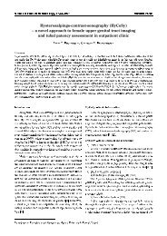
Archives of Perinatal Medicine 18(4), 229-232, 2012CASE REPORTTorsion of adnexal cyst in utero – case reportANNA DERA-SZYMANOWSKA 1 , MARIOLA ROPACKA-LESIAK 1 , MICHAŁ BŁASZCZYŃSKI 2 ,MARTA SZYMANKIEWICZ 3 , GRZEGORZ H. BRĘBOROWICZ 1AbstractBackground: <strong>Fetal</strong> tumors represent rare group of abnormalities however ovarian cysts are the most frequenttype of abdominal tumors in female newborns. The diagnosis has become more frequent due to the prenatalultrasonography monitoring. Due to the possible complications of such abnormalities it is important to be awareof the management and treatment options. The literature presents different management options from conservativeapproach such as wait and see policy to more aggressive as intrauterine cyst aspiration or surgery. <strong>Case</strong>report: We have presented the management and treatment of large complex fetal ovarian cyst diagnosed prenatally.Towards the end of pregnancy the cyst changed its consistency; it contained fluid/debris levels which didshow some calcifications and possible hemorrhagic fluid suggesting torsion. The patient was delivered by a cesareansection and the neonate was referred for selective abdominal surgery. Conclusion: <strong>Fetal</strong> ovarian cysts arerare however they may cause serious complications. The decision regarding the management and treatment offetal ovarian cyst should depend on the size, appearance of the cyst and visible complications. Ultrasonographicmonitoring allows differentiation of the ovarian mass (simple/complex) and application of appropriate management.We think that simple ovarian cyst should be managed by wait and see policy and close postnatal follow-upand complex ovarian cysts should be managed by early surgery especially if symptomatic.Key words: fetal ovaria cystIntroduction<strong>Fetal</strong> tumors represent rare group of abnormalitieshowever ovarian cysts are the most frequent type of abdominaltumors in female newborns [1]. The rates ofdetection of fetal ovarian cysts have increased since theadvent of routine antenatal sonography. The first case ofan ovarian cyst was reported in 1889 in a stillborn prematurewhereas the first successfully treated ovariancyst in newborn period was done by Bulfamonte in1942 [2]. The etiology of fetal ovarian cysts has not beenentirely clarified. Most likely, it results from fetal exposureto maternal and fetal gonadotrophins, found oftenin the mothers having increased levels of beta-HCG(diabetes, Rh isoimmunization and toxemia). Other suspectedhypotheses include fetal immaturity, fetal hypothyroidismas well as placental insufficiency in additionto incomplete maturation of the gonadostat [3]. Variouscomplications are described in association with ovariancysts: compression of other viscera, rupture of the cyst,hemorrhage but the most common is ovarian torsionwith possible consequent loss of the ovary. According toNussbaum’s classification, ovarian cysts can be dividedinto simple (completely anechoic) and complex (characterizedby fluid-debris level, clot, septa, and echogenicwall) cysts suggesting torsion [4]. There is still considerablecontroversy regarding treatment of ovarian cysts.The literature presents different management optionssuch as wait and see policy, aspiration or surgery. In thiscase we present the management as well as successfultreatment of large complex neonatal ovarian cyst diagnosedantenatally.<strong>Case</strong> presentationA 34 year-old mother at 33 week of gestation, gravida2, with a history of spontaneous abortion was admittedto the Department of Perinatology and Gynecologybecause of lower abdominal pain and suspicion offetal ovarian cyst. <strong>Fetal</strong> ultrasound had shown a 67 × 43mm simple anechoic cyst at the right side of fetal pelvis(Fig. 1, 2). The otherwise normal anatomy of the gastrointestinaland urinary tracts in a female fetus washighly suggestive of an ovarian cyst. Otherwise the fetalanatomy was within the norms. All the routine hematologicaland biochemical investigations were within normallimits. After diagnosis the mother was followed byserial ultrasound scans every 2 weeks to check for thesonographic disappearance of the cyst or for possibleshift to the pattern of torsion. The scan at 36 weeks re-1 Perinatology and Gynecology Department, Poznan University of Medical Sciences, Poland2 Chilgren Surgery, Traumatology and Urology Department, Poznan University of Medical Sciences, Poland3 Neonatology Department, Poznan University of Medical Sciences, Poland
230A. Dera-Szymanowska, M. Ropacka-Lesiak, M. Błaszczyński, M. Szymankiewicz, G.H. Bręborowiczvealed different echogenicity of the ovarian cyst and possiblechange to complex type of the cyst (Fig. 3).Fig. 4 . The ultrasound scan of the ovary shows a shift to echogeniccomplex ovarian cyst (fluid/debris level) at 39 weeksFig. 1 . <strong>Fetal</strong> ultrasound shows a 67 × 43 mm simpleanechoic cyst at the right side of fetal pelvis at 33 weeksThe cyst did not change in size however there was changeof the consistency and echogenicity. It containedfluid/debris levels which did show some calcificationsand possible hemorrhagic fluid. The patient underwentcesarean section at 40 week of gestation and deliveredfemale newborn weighing 3770 g and Apgar score 10, 10.Fig. 2. The fetal abdominal cyst in 3D scanFig. 5. The ovarian cyst in 3D scan at 39 weeks.The complex cyst with intracystic septa, fluid/debris levelFig. 3. The hyperechogenic complex ovarian cystat 36 weeks reflecting the change of cyst characterThe diameter was 65 mm. Following, at 39 weeks thepatient was admitted to the hospital for elective cesareansection, however at that time the ultrasound scanof the ovary showed shift to echogenic complex ovariancyst (fluid/debris level) (Fig. 4, 5).The fetus was evaluated by neonatologist who confirmedgood neonatal state and the presence of ovarian cystwithout any other abnormalities of anatomical structures.The only disturbing abnormality detected was increasedlevel of AFP (26138 ng/ml) and CEA (30.5 ng/ml). The patient was transported to the Department ofPediatric Surgery 6 days after delivery for selective abdominalsurgery. Laparoscopic exploration revealeda large dark brown mass localized in right abdominalcompartment extending from subhepatic region to thepelvis. After evacuation of a dense hemorrhage fluidfrom the cyst the torsion of right ovary secondary to thelarge ovarian cyst was found as a cause of pathology.Right salpingo-oophorectomy was performed because
Torsion of adnexal cyst in utero – case report 231necrosis made the distinction of normal ovarian tissueimpossible. After excision of the adnexa there were twosolid adhesions between right ovary and the wall of jejunum,which were excised and the wall of the jejunumwas checked. The histopathology examination indicatedthe presence of hemorrhagic necrosis and foci of dystrophiccalcifications. With these features, a diagnosis ofa congenital ovarian cyst with torsion was given. Thepostoperative course of the patient was uneventful andneonate was discharged home on the 3 rd postoperativeday. The ambulatory follow-up indicated normal physicaldevelopment without additional problems.DiscussionThe incidence of ovarian cysts has been estimatedat more than 30% (this estimate is based on an investigationof stillborns or infants who died within 28 daysafter birth) [5]. <strong>Ovarian</strong> cysts arise from mature follicleswhich are usually < 2 cm in diameter, cysts larger thanthat size are considered pathological which can be diagnosedusually beyond 28 weeks. The diagnosis has becomemore frequent due to the prenatal ultrasonographymonitoring. Torsion is the most common complication(50-70%) and it has been observed to occur more frequentlyduring fetal life than postnatal. Some studieshave reported that the outcome of such complication isrelated to the length of the cyst pedicle which is morepredictive than cyst dimension [6], however unfortunatelyit may only be evaluated during surgery. Complicationsuch as torsion may cause inflammatory adhesionswhich were observed in case of our patient and probablyinduced the increase in the AFP and CEA level. Othercomplications may include intracystic hemorrhage, rupture,less frequently urinary tract or bowel obstructionand rarely autoamputation of the cyst. However themanagement of ovarian cyst should be based on the sizeand echopattern of the cyst. Few authors have shownthat unilateral simple cyst is suggestive of benign process.It is known that cyst < 4 cm have a tendency forspontaneous resolution both prenatally and after deliverywhich means that they can be managed conservatively byserial scans. Bagolan et al. managed 34 simple ovariancyst < 5 cm with wait and see policy and they have observedspontaneous regression in 26 cases, 1 persistenceat birth and 7 cases of torsion [10]. Others have reportedthat simple cysts even up to 8 cm can safely bemonitored by ultrasound, as spontaneous resolution canoccur within 4-5 months [4, 15]. That is why we thinkthat the most appropriate clinical approach in case ofsimple ovarian cysts is to adopt a wait and see policywhich requires periodic ultrasound monitoring to assessthe course of the condition. In our patient the diagnosiswas made at 33 weeks of pregnancy, before that the patientwas monitored regularly by ultrasonography. Basedon the regular US scans for few weeks we were observinga large simple ovarian cyst that is why we decidedto proceed with the wait and see policy.Another management option presented by few studiesis intrauterine ovarian-cyst aspiration [9, 11]. Bagolanet al. in their study performed in utero aspiration of14 simple cysts measuring $ 5 cm observing resolutionin 12 cases and torsion in 2 concluding that “in utero”aspiration of ovarian cyst is a safe procedure. They havealso recommended surgical management for cysts withultrasound pattern of torsion persisting postnataly [10].However others think that continued hormonal stimulationleads to regrowth of the cyst [12], can lead to peritonitisdue to rupture or spillage of cyst fluid but mostimportantly the increased possibility of wrong diagnosis.US scan cannot distinguish benign and malignant lesionespecially in the presence of complex mass. There havebeen reports of cystoadenoma and bilateral granulosacellcarcinomas of the ovary have been reported ina 30-week-old fetus [7, 8] Because we weren’t certainabout the type of the ovarian mass we did not want toproceed with intrauterine aspiration.Another question which has to be asked is what isthe best time for delivery in case of cyst with the ultrasoundpattern of torsion. In decision making we have toconsider maturity of the fetus, possible attempts to savethe ovary and possible complications. We think that dueto the possibility of ultrasound monitoring we can observethe ovary on day time bases and be able to makethe decision according to the changes that we observe.Since there was no change in the size of the ovary andwe were not sure if the ultrasonographic evidence ofcomplex cyst is actually a torsion we decided not toproceed with early delivery. The patient was delivered assoon as we were sure that the fetus obtained pulmonarymaturity. According to the literature early delivery is notcurrently recommended.What to do after delivery? Due to the fact that ultrasoundis nonspecific when it comes to interpreting thenature of fetal ovarian cyst and does not give us a sureanswer about the possibility of torsion we should takea moment to think about treatment option. The possibilitiesinclude wait and see policy, laparoscopy explorationsand laparotomy. According to the study by Kwak et al.,postnatal symptomatic cysts or cysts with diametergreater than 5 cm that do not regress or enlarge should
232A. Dera-Szymanowska, M. Ropacka-Lesiak, M. Błaszczyński, M. Szymankiewicz, G.H. Bręborowiczbe treated, but uncomplicated asymptomatic cysts lessthan 5 cm in diameter should only be observed and reassessedby serial ultrasonography [13]. Whereas, Kessleret al. recommended US-guided aspiration of asymptomaticlarge ovarian cysts for salvage or for decompressionif torsion occurs. Surgery was to be reservedfor patients with acute torsion, intestinal obstruction andintestinal volvulus [14]. Sometimes such a choice oftreatment could be dangerous even with lethal demise[15]. Monney-Noche et al. found that most of complexappearing ovarian cysts were torsed. Because ultrasonographywas not able to distinquish torsed from hemorragiccyst the early surgery was recommended to definediagnosis and assessment of ovarian viability [16]. In ourcase we have decided to proceed with surgery becausewe were unsure about the character of ovarian cyst, wewanted to find out if there is any functional ovarian tissueleft as well as visualize other structures which couldbe involved in the process.To conclude the decision regarding the managementand treatment of fetal ovarian cyst should depend on thesize, appearance of the cyst and visible complications.Ultrasonographic monitoring allows differentiation of theovarian mass (simple/complex) and application of appropriatemanagement. We think that simple ovarian cystshould be managed by wait and see policy and close postnatalfollow-up and complex ovarian cysts should be managedby early surgery especially if symptomatic.References[1] Chiaramonte C., Piscopo A., Cataliotti F. (2001) <strong>Ovarian</strong>cysts in newborns. Pediatr. Surg. Int. 17: 171-174.[2] Tehrani F., Kavehmanesh Z., Kaveh M., Tanha F. (2007)Neonatal <strong>Ovarian</strong> <strong>Cyst</strong>: A case report. Iran J. Pediatr.17(3): 379-382.[3] Mudholkar V., Acharya A., Kulkarini A., Hirgude S. (2011)Antenatally diagnosed neonatal ovarian cyst with torsion.Indian Journal of Pathology and Microbiology 54(1): 228-229.[4] Nussbaum A.R., Sanders R.C., Hartman J.S. et al. (1988)Neonatal ovarian cysts. Sonographic pathologic correlation.Radiology 168: 817-21.[5] Meizner I., Levy A., Katz M., Maresh A.J. et al. (1991) <strong>Fetal</strong>ovarian cysts: Prenatal ultrasonographic detection andpostnatal evaluation and treatment. Am. J. Obstet. Gynecol.164(3): 874-8.[6] Sakala E.P., Leon Z.A., Rouse G.A. (1991) Managementof antenatally diagnosed fetal ovarian cysts. Obstet. Gynecol.Surv. 46: 407-414.[7] Brown M.F., Hebra A., McGeehin K., Ross A.J. (1993)<strong>Ovarian</strong> masses in children. A review of 91 cases of malignantand benign masses. J. Pediatr. Surg. 28: 930-932.[8] Hengster P., Menardi G. (1992) <strong>Ovarian</strong> cysts in the newborn.Pediatr. Surg. Int. 7: 372-375.[9] Bagolan P., Rivosecchi M., Giorlandino C., Bilancioni E.et al. (1992) Prenatal diagnosis and clinical outcome ofovarian cysts. J. Pediatr. Surg. 27: 879-881.[10] Bagolan P., Giorlandino C., Nahom A., Bilancioni E. et al.(2002) The management of fetal ovarian cysts. J. Pediatr.Surg. 37: 25-30.[11] Sapin E., Bargy F., Lewin F., Baron J.M. et al. (1994) Managementof ovarian cysts detected by prenatal ultrasound.Eur. J. Pediatr. Surg. 4: 137-140.[12] Brandt M.L., Lucks F.I., Filiatraul D., Garel L. et al. (1991)Surgical indications in antenatally diagnosed ovariancysts. J. Pediatr. Surg. 26: 276-282.[13] Kwak D.K., Seok Sohn Y., Kim S.K. et al. (2006) Clinicalexperiences of fetal ovarian cyst: Diagnosis and consequence.J. Korean Med. Sci. 21: 690-4.[14] Kessler A., Nagar H., Graif M., Ben-Sira L. et al. (2006)Percutaneus drainage as the treatment of choice for neonatalovarian cysts. Pediatr. Radiol. 36(9): 954-8.[15] Puligandla P.S., Laberge J.M. (2009) Lethal outcome afterpercutaneous aspiration of a presumed ovarian cyst in aneonate. Semin. Pediatr. Surg. 18: 119-121.[16] Monnery-Noche M.-E., Auber F., Jouannic J.-M. et al.(2008) <strong>Fetal</strong> and neonatal ovarian cysts: is surgery indicated?Prenat. Diagn. 28: 15-20.J Anna Dera-SzymanowskaPerinatology and Gynecology DepartmentPoznan University of Medical Sciences60-535 Poznań, ul. Polna 33, Polande-mail: annaszerszen@wp.pl