Lab Topic Three
Lab Topic Three
Lab Topic Three

You also want an ePaper? Increase the reach of your titles
YUMPU automatically turns print PDFs into web optimized ePapers that Google loves.
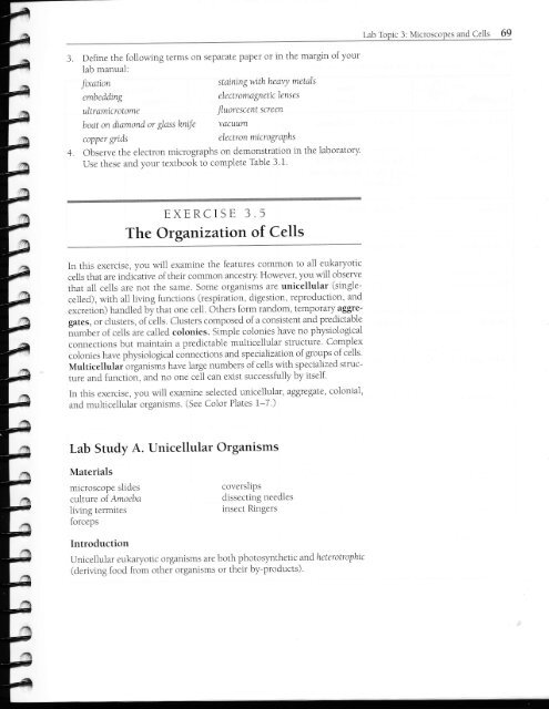
and Cells3. Define the following terms on separate paper or in the margin of yourlab manual:stainingwithheavy metalsJtxationembeddingultramicrotomeboat on diamond or glasshniJecopper gridselectromagnetic lensesJluorescent screenvactLumelectron micrograPhs4. Observe the electron micrographs on demonstlation in the laboratoryUse these and your textbook to complete Table 3.1.EXERCISE 3.5The Organizatuort of Cellsln this exercise, you will examine the features common to all eukaryoticcells that are indicative of their common ancestDl However, you will observethat all cells are not the same. Some organisms are unicellular (singlece11ed),wlth al1living functions (respiration, digestion, reproduction, andexcre[ion) handled by that one cell. Others form random, temporary aggregates,or clusters, of iells. Clusters composed of a consis[ent and predictablei.,mb"r of cells are ca11ed colonies. Simple colonies have no physiologicalconnections but maintain a predictable mu1ticel1u1al sllucture Complexcolonies have physiological conneclions and speciallzation of groups of celis.Multicellulaiorganisms have large numbers of ce11s with specialized structureand function, and no one ce1l can exisl successfully by itself.ln this exercise, you wili examine selected unicellular, aggregate, colonial,and multicellular organisms. (See Color Plates 1-7')<strong>Lab</strong> Study A. Unicellular OrganismsMaterialsmicroscope slidesculture of Amoebalivi.ng termitesforcepscoverslipsdissectrng needlesinsect RingersIntroductionUnicellular eukaryotic organisms are both photosynthetic and heterotrophic(deriving food from other organismstheir by-products)'
Procedurel. Examine a livingAnrorbc (Figure 3.5) under the compound microscope.Amoebas are aquatic organisms commonly found in ponds. To transfera specimen to your slide, fo11ow these procedures:a. Place the culture dish containing the amoeba under thc dissectingmicroscope, and focus on the bottom of the dish. The amoeba will appearas a whitish, irregularly shaped organi.sm attached to the bottom.b. Using a clean pipette (it is importanL not to interchange pipettesbe[ween culture dishes), transler a drop with several amoebas to yourmicroscope slide. To do this, squeeze the pipette bulb before youplace the tip under the surface of the water. Disturbing the cuhureas litLle as oossrhle 'r'r-"- ninette a dron of uater liith debris from the bt,ttomol the culture dish. You may use your stereoscopic microscopeto scan the slide to locate amoebas before continuing.c. Cover your preparation with a clean coverslip.d. L nder l.rw power on the compound scope. scan the slidc to locatean amoeba. Center the specimen in your iield of riew: then switchto higher powers.e. Identify the following structures in the amoeba:CeIl membrane is the boundary that separaies the organism fromitc crrrrnrrndino- '^..-..._s.Ectoplasm is the thin, transparent layer of cytopiasm drrectly beneaththe cell membrane.Endoplasm is the granular cytoplasm containing the ce11 organelles.The nucleus is the grayish. Iootball-shaped body that is somewhatgranular in appearance This.rrganelle. which directs the cellularactivities, will often be seen movrng within the endoplasm.Contractile vacuoles are clear. spherical vesicles of r rrying sizesthat gradually enlarge as they fi ll with excess \\aler. Once y.ru'relocated a vacuole, watch it fill and then empty its contents into thesurroundinp'-ent ironment. These vacutrles servc an excrelorv funcb'I -" 'tion for the amoeba.Food vacuoles are smal1, dark. irregularly shaped vesicles withinrhe cndnnlacm Thev cnnr:rin rrndioe..ted {ood n2rt icle.,'"'b' -.' r*" '' '' "'Pseudopodia t false feet ) are fingerlike pro.iectitrns of the eytoplasm.The; are used for locomotion as well as lor trapping and engulfi nglood in a process ca11ed phagocytosis.). Examine Trirhottymphrr under a compound microscope. You will li rsthave to scparatc the Triclronympha (Frgure l.ol f rom the termite withwhich rt lives rn a q.mbrotic relationship. Tnchonymphaand other organicmqneerrnr the orrl nf the tcrmitec urhr're thev dioect rlrnnd nart iclec-*r]....5*.'../-.b.".'..\?-r*'...'."enten hr the insect. Termiles lack the ....^../../.."'t) enzvmes necessarv to drpest uoodand are dependent on kichonympha to make the nutrients in the woodavailable to them. Tichonyntpha has become so well adapted to the environmentof the termite's gut that it cannot survlve outside of it.To obtaln a specimen.a. Place a couple of drops of insect Ringers (a saline solution that isisotonic to the internal environment of insects) on a clean mlcroscopes1ide.b. Using forceps or your fingers. transler a termite into the drop ol Ringers.<strong>Lab</strong> <strong>Topic</strong> 3: Micro>copes and Cells 7Ia^t"f^"nh"^^^Endop asmEctop asmContractrle vacuoleFood vacuolePseudopod aFigure 3.5.Anroeba. An Arrrucb.r movcs u)ingpscudopodia Obsen e the lir ingnrornicrnrrrcino *- the arrmnnrndb'microscope. (See Color Plate l.)NucleusE ^^^Woodt^q,/aA%)A)(iS+rODKry.,Figure 3.6.Trichonympha. A community ofmicroorganisms, rncluding Trtchonympha,inhabits the intestine of the termite .Following the procedure in Exercise 3.5,lqh Strr.lr ..._r A .. diqnerce .... th| -..- ...emtcroorganisms and locate the cellular structuresrn TrLchonyntpha. (See Color Piate 2.)
a. To obtain a specimen, use a dissecting needle to brush off a smallamount of the green growth on the piece of tree bark provided intoa drop of water on a clean microscope slide. Avoid scraping barkonto the sllde. Cover the preparation with a clean coverslip.b. Observe at highest power that these ce11s are aggregates: The size ofthe ce11 groupings is random, and there are no permanen[ connectionsbetween cells.c. Observe several smali cell groupings and avoid iarge clumps of cel1s.Cellular detail may be obscure.Examine liing Scenedc-smus under the compound microscope . Scenedesmts(Figure 3.8) is an aquatic green alga that is common in aquaria and po1-luted water.a. To obtain a specimen, place a drop from the culture dish (using aclean pipette) onto a clean microscope slide, and cover it with a cleancoverslip.b. Observe that the cells of this organism form a simple colony: Thecells always occur in groups of from four to eighr cells, and rhey arepermanently united.c. Identify the following structures:The nucleus is the spherical organelle in the approxima[e middle ofeach cell.Vacuoles are the transparent spheres that tend [o occur at either endof the cells.Spines are the transparent prqections tha[ occur on the two end ceils.Examine liingVoltoxunder the compound microscope. \.6lvox(Figure 3.9)is an aquatic green alga that also i.s common in aquaria, ponds, and lakes.<strong>Lab</strong> <strong>Topic</strong> 3: Mlcroscopes and Cells 73a. To obtain a speci.men, prepare a wet mount as you did for Scenedesmuswith the following addrtion: Before placing a drop of the culture onyour slide, place several glass chips on the slide. This will keep thecoverslip from crushing these spherical organisms.Cytoplasmic strandb. Observe that the cells of this organism form a large complex colony.ChloroplaslApproximately 500 to 50,000 cells (depending on the species) arepermanently united; [here are cytoplasmi connections between ce11s;and some cells are specialized for reproducrion.c. identify the following structures:Individual ce1ls each possess the following structures: cell wall,nucleus, vacuole, chloroplasts, flagella (two per cell).Cytoplasmic strands form connec[ions between adjacent cel1s.Daughter colonies are smaller spheres within the larger colony.These are produced asexually, and when they are large enough. thcywill be discharged from the parent colony into the surrounding enr,rronment.ChloroplastFigure 3.8.Scenedesmus. Scenedesmus s anaquatic alga that usually occursin simple colonies of four cellsconnected by an outer cell wa1l.(See Color Plate 4.)DaughtercolonresFigure 3.9.Volvox. In this complex colony. rheindividual cells are interconnected bycytoplasmic ttrands lo form a sphere.Small clusters of cells, called daughtercolonies. are specialized for reproduction.(See Color Plate 5.)
NucleusFigure 3.10.CytoplasmicstranosChloroplastElodea. Elodeais an aquatrc Plantcommonly grown in freshwateraquaria. The cell strLtctures may bedifficult to see because of the threedimensionalcel1 shape and thepresence of a large central vacuole.(See Color Plate 6.)<strong>Lab</strong> Study C. Multicellular OrganismsMaterialsmicroscope slidesdropper bottles of watertoothpickscoverslipsEIodeamethylene bluelntroductionMultlcellular organisms are composed of groups of specialized ce1ls, calledtissues, that together perform particular funclions for the organism. Tissues,in turn, may be grouped to form organs' and organs may be grouped intoorgan systems. In this lab study, you will examine some of the ceiis that compoiethe basic tissue types of plants and animals.ProcedurePlant Cells1. The major characteristics of a typical plant ce11 are readily seen in theleal celis ol E;odea. a common aquaric plant lFigure ].l0) Prepare awet mount and exami.ne one of the youngest (smallest) leaves from a sprigof Elodeaunder the compound microscope.2. Identify the following structures:The cell wall is the rigrd outel framework sunounding the cell. This strucruregives rhe cell a definite shape and suppoft. It is not found in animalcells.Protoplasm is the organized conlents of the cell, exclusive of thece11wall.cytoplasm rs the protoplasm of the cel1, exclusive of the nucleus.Chloroplasts are the green, spherical organelles often seen movingwithin tie cyroplasm. These organelles carry the pigment chlorophyllthat is involved in photoslnthesis. As the microscope light heats up thecells, the chloroplasts may begin to move quite rapidly rn a plocesscalled cytopl asmic streaming. or cyclosis.The nucleus is the usually spherical, transparent organelle within thecytoplasm. This structure controls cell metabolism and division.The vacuole is a membrane-bound sac within the cytoplasm that isfilled with water and dissolved substances. This structure serves to storemetabolic wastes and gives the ce1l suppoft by means of turgor pressure.Animal ce1ls also have vacuoles, but they are not as large and conspicuousas those found in Plants.What three structures observed inElodea are unlque to plants?etfr\FEf,€n-lttncnIf,t==37FFFfeF
_T-4. Compare your observations ol Elodeausing the compound scope withthose made in Exercise 3.3 using the stereoscopic sc.pe . List the structuresseen wlth each:Stereoscopic:Compound:Animal Cellsl. Animals are multicelluiar heterotrophic organisms that ingest organrcmaner. They are composed of ce11s that can be categorized into fourmajor tlssue groups: epithelial, connective, muscle, and nervous trs_sue. In this lab study, you r,vi11 examine epithelial cel1s. similar to theepidermal cells of plants, epithelial cells occur on the outside of animalsand serve to prorect the animal from water loss, mechanical ir-r.]ury,and loreign invaders. in addltlon, epithelial cells line interior cavrtiesand ducts in animals. Examine the epirheliar cells (Figure 3.11) thar /^\form tl-re linlng of yourCyinneroolasrrcheek. To obtain a speciJen, follow thlsprocedure:a. with a clean toothpick, gentl)/ scrape the rnside of your cheek severaltlmes.b R:rll the scraping into a drop of warer on a clean microscope slide,add a srnall drop of methylene blue, and cover wirh a coveislip.c. Using the compound microscope, r,-iew the cells under higher powers.2. observe that these ce1ls are extremeiy flat and so may be folded over onthemselves. Arrempt to locate several ce11s that u." ,-rot badly iolded,and study their detail.] ldcnrify rhc following srrucrures;The cell membrane is the boundary that separates the cell from itssurroundings.The nucleus is the 1arge, circular organelre near the micldle of the cell.cytoplasm is the granular contents of the ce1l, exclusive of the nucleus.uelimembraneNrc.ersFigure 3.11.Human epithelial cells. The epirhellalcel1s that line your cheek are thtn, flatcells that you can remove easily fromyour cheek by scraping ir with a toorhpick.(See Color Plate 7.)<strong>Lab</strong> Study D. UnknownsMaterialsmicroscope slidescoverslipspond water or culture of unknownsIntroductionuse this lab study to see if you ha'e met the objectives of this lab toprc. asyou carry our this lab study, (1) think carefully about using correcr microscopictechniqr-res; (2) dlstinguish organisms wirh differenicellular organrz-ationor configuratlon; (3) note how the clifferenr organisms are similai yetdifferent; and (,1) nore cell differences.
elProcedure1. Examine several drops of the culture of pond watel that you collected,or examine the unknown culture provided by the instructor.2. Record in Table 3.2 the characteristics of at least four differentorganisms.!IIeiand CellsTable 3.2Characteristics of Organisms Found in Pond WaterTirbeUnknownIMeans ofLocomotionCelI Wall(+/-)Chloroplasts(+/-) Organization€rgr*2345Questions for Review1. List several organelles tha[ are visible with the electron microscope butthat were not visible with your microscope'2. Descnbe at least two types of materials or observations that would necessitatethe use of the stereoscopic microscope'3. What characteristics do all eukaryotic cells have in common?+.a.Whatcellularfeaturesdifferentiateplantsfromanimals?b. How are [he struc[ures ihat are unique to plants important to theirsuccess?*A^6.heeeeeI*e#*f,fre
l. In your own words, describe the evolutionary trend for increasing organismalcomplexity, using examples from this 1ab to illustrate your answer.2. We often imply that multicellular organisms are more advanced (and thereforemore successful) than unicellular or colonral organisms. Explainwhy thls rs not true, using examples from this lab or elsewhere.Following is a hst of tissues that have specialized functions and demonstratecorresponding specialization of subcellular structure. Match thetissue wlth the letter of the cell structures and organelles iisted to theright that would be abundant in these cel1s. (Refer ro Table 3.1.)Tissues', "-'.]nf theI Fnzvme (nrot pin)-ccr-rpt ino npllcncnrtor.2 Insect fliphr mrrscles3 Cells linino ....Dthe ...- rc , - sptrat orypassages4. White blood cells thar engulfand dest ror, inv:adinollncrericCell Structures sndOrganellesa. plasma membraneb. mitochondria^/-^1-:L. uurBr dPPdrdLu>d. chloroplaste. endoplasmicreticulumfgcilia znA f qooll^vacuoleh. ribosomei. lysosomeinernxicnmpc5. Leaf ceils of cacti