- Page 1 and 2: World Health Organization Classific
- Page 3 and 4: This volume was produced in collabo
- Page 5 and 6: Contents1 Tumours of the kidney 9WH
- Page 7 and 8: CHAPTER 1Tumours of the KidneyCance
- Page 9 and 10: TNM classification of renal cell ca
- Page 11 and 12: one quarter of kidney cancers in bo
- Page 13 and 14: Familial renal cell carcinomaM.J. M
- Page 15 and 16: Table 1.02Genotype - phenotype corr
- Page 17 and 18: ABFig. 1.11 A Multiple cutaneous le
- Page 21 and 22: Clear cell renal cell carcinomaD.J.
- Page 23 and 24: Fig. 1.20 Clear cell renal cell car
- Page 25 and 26: Papillary renal cell carcinomaB. De
- Page 27 and 28: ABFig. 1.29 Papillary renal cell ca
- Page 29 and 30: Fig. 1.33 Chromophobe RCC with sarc
- Page 31 and 32: Carcinoma of the collectingducts of
- Page 33 and 34: Renal medullary carcinomaC.J. Davis
- Page 35 and 36: Renal carcinomas associated withXp1
- Page 37 and 38: Renal cell carcinoma associated wit
- Page 39 and 40: Papillary adenoma of the kidneyJ.N.
- Page 41 and 42: of this tumour. Microscopic extensi
- Page 43 and 44: ABFig. 1.59 Metanephric adenoma. A
- Page 45 and 46: esults in intratumoral aneurysms. O
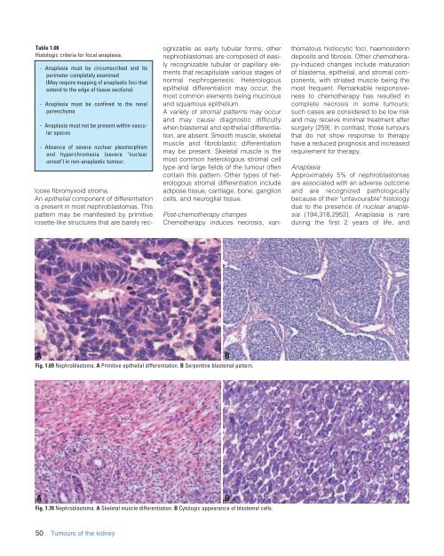
- Page 47: peritumoural fibrous pseudocapsule.
- Page 51 and 52: Nephrogenic rests andnephroblastoma
- Page 53 and 54: Cystic partially differentiatedneph
- Page 55 and 56: ABFig. 1.80 Clear cell sarcoma of t
- Page 57 and 58: ABFig. 1.85 Rhabdoid tumour of the
- Page 59 and 60: transcription factor is fused to th
- Page 61 and 62: LeiomyosarcomaS.M. BonsibDefinition
- Page 63 and 64: AngiomyolipomaG. MartignoniM.B. Ami
- Page 65 and 66: melanocytic and smooth muscle marke
- Page 67 and 68: Multinucleated and enlarged ganglio
- Page 69 and 70: HaemangiomaP. TamboliDefinitionHaem
- Page 71 and 72: Fig. 1.107 Juxtaglomerular cell tum
- Page 73 and 74: Intrarenal schwannomaI. Alvarado-Ca
- Page 75 and 76: Mixed epithelial and stromal tumour
- Page 77 and 78: Synovial sarcoma of the kidneyJ.Y.
- Page 79 and 80: Renal carcinoid tumourL.R. BéginDe
- Page 81 and 82: Primitive neuroectodermal tumour(Ew
- Page 83 and 84: Paraganglioma / PhaeochromocytomaPh
- Page 85 and 86: LeukaemiaA. OraziInterstitial infil
- Page 87 and 88: WHO histological classification of
- Page 89 and 90: TNM classification of carcinomas of
- Page 91 and 92: The risk of bladder cancer goes dow
- Page 93 and 94: Tumour spread and stagingUrinary bl
- Page 95 and 96: feature in such patients undergoing
- Page 97 and 98: AFig. 2.12 Infiltrative urothelial
- Page 99 and 100:
AFig. 2.15 A Infiltrating urothelia
- Page 102 and 103:
it difficult to identify genes lead
- Page 104 and 105:
Alterations of 9p21 and p15/p16 bel
- Page 106 and 107:
nostic role in invasively growing b
- Page 108 and 109:
Urothelial hyperplasiaJ.I. EpsteinU
- Page 110 and 111:
Urothelial papillomaC. BuschS.L. Jo
- Page 112 and 113:
Somatic geneticsUltrastructure, ant
- Page 114 and 115:
In spite of the overall orderly app
- Page 116 and 117:
Urothelial carcinoma in situI.A. Se
- Page 118 and 119:
aberrations, often including high l
- Page 120 and 121:
grade carcinomas recur frequently (
- Page 122 and 123:
AFig. 2.47 Squamous cell carcinoma.
- Page 124 and 125:
grading to be a significant morphol
- Page 126 and 127:
AFig. 2.57 Adenocarcinoma. A High p
- Page 128 and 129:
Urachal carcinomaA.G. AyalaP. Tambo
- Page 130 and 131:
Clear cell adenocarcinomaE. OlivaDe
- Page 132 and 133:
Small cell carcinomaF. AlgabaG. Sau
- Page 134 and 135:
mon in females by 1.4:1 {1845}. The
- Page 136 and 137:
RhabdomyosarcomaI. LeuschnerDefinit
- Page 138 and 139:
AngiosarcomaJ. ChevilleDefinitionAn
- Page 140 and 141:
Malignant fibrous histiocytomaJ. Ch
- Page 142 and 143:
Granular cell tumourI.A. Sesterhenn
- Page 144 and 145:
LymphomasA. MarxDefinitionMalignant
- Page 146 and 147:
ABCFig. 2.87 Metastatic tumours to
- Page 148 and 149:
makes 4.6% of lower urinary tracttu
- Page 150 and 151:
and patients with pT 2 tumours have
- Page 152 and 153:
Table 2.07Anatomic classification o
- Page 154 and 155:
prominent basal epithelial cell lay
- Page 156 and 157:
WHO histological classification of
- Page 158 and 159:
Acinar adenocarcinomaJ.I. EpsteinF.
- Page 160 and 161:
have been seen in high-risk countri
- Page 162 and 163:
ABCFig. 3.07 A Pelvic metastases of
- Page 164 and 165:
expression of PSMA in serum of both
- Page 166 and 167:
AFig. 3.12 A Organ-confined adenoca
- Page 168 and 169:
glands. These are perineural invasi
- Page 170 and 171:
have occasional p63 immunoreactive
- Page 172 and 173:
ABFig. 3.25 A Pseudohyperplastic ad
- Page 174 and 175:
PSA positive. Associated acinar ade
- Page 176 and 177:
AAFig. 3.36 Schematic diagram of th
- Page 178 and 179:
AFig. 3.39 A Gleason score 3+3=6. B
- Page 180 and 181:
and prostatectomy Gleason score{375
- Page 182 and 183:
Table 3.01Prostate cancer susceptib
- Page 184 and 185:
AFig. 3.52 A Immunohistochemistry f
- Page 186 and 187:
ABFig. 3.57 A Pathological stage an
- Page 188 and 189:
Fig. 3.59 Preoperative PSA levels (
- Page 190 and 191:
ABFig. 3.61 A Flat and tufting patt
- Page 192 and 193:
ABFig. 3.65 High grade PIN with foa
- Page 194 and 195:
Table 3.04Risk of subsequent carcin
- Page 196 and 197:
sue. Transrectal needle core biopsi
- Page 198 and 199:
Urothelial carcinomaD.J. GrignonDef
- Page 200 and 201:
CK20 in the majority of cases and h
- Page 202 and 203:
Basal cell carcinomaP.H. TanA. Bill
- Page 204 and 205:
ABFig. 3.84 Small cell carcinoma. A
- Page 206 and 207:
ABFig. 3.87 A Malignant phyllodes t
- Page 208 and 209:
Haematolymphoid tumoursK.A. Iczkows
- Page 210 and 211:
gliomas should not be designated as
- Page 212 and 213:
CHAPTER 4XTumours of the of the Tes
- Page 214 and 215:
TNM classification of germ cell tum
- Page 216 and 217:
Germ cell tumoursP.J. WoodwardA. He
- Page 218 and 219:
senting symptom: back or abdominalp
- Page 220 and 221:
ABFig. 4.03 Germ cell tumours genet
- Page 222 and 223:
process at all {1524}. In addition,
- Page 224 and 225:
ABFig. 4.12 Comparison of morpholog
- Page 226 and 227:
small tumour insufficient to produc
- Page 228 and 229:
ABCFig. 4.22 Seminoma. A Pseudoglan
- Page 230 and 231:
ABFig. 4.27 Spermatocytic seminoma.
- Page 232 and 233:
Differential diagnosesDifferential
- Page 234 and 235:
Histologic patternsMicrocystic or r
- Page 236 and 237:
AFig. 4.40 Yolk sac tumour. A Pleom
- Page 238 and 239:
TeratomaTeratomas are generally wel
- Page 240 and 241:
ABCFig. 4.49 Teratoma. A Teratoma w
- Page 242 and 243:
ABFig. 4.52 Teratoma with somatic t
- Page 244 and 245:
ABFig. 4.59 Mixed germ cell tumours
- Page 246 and 247:
ABCFig. 4.64 Leydig cell tumour. A
- Page 248 and 249:
AFig. 4.69 Androgen insensitivity s
- Page 250 and 251:
tinguished from Sertoli cell nodule
- Page 252 and 253:
ound and have spherical, regularly
- Page 254 and 255:
Tumours containing both germ cell a
- Page 256 and 257:
Miscellaneous tumours of the testis
- Page 258 and 259:
Lymphoma and plasmacytoma of thetes
- Page 260 and 261:
Tumours of collecting ducts and ret
- Page 262 and 263:
Tumours of paratesticular structure
- Page 264 and 265:
EpidemiologyNodular mesothelial hyp
- Page 266 and 267:
ABFig. 4.104 Papillary cystadenoma
- Page 268 and 269:
Fig. 4.109 Desmoplastic small round
- Page 270 and 271:
Fig. 4.115 Leiomyosarcoma. Coronal,
- Page 272 and 273:
Secondary tumoursC.J. DavisDefiniti
- Page 274 and 275:
CHAPTER 5Tumours of the PenisThe in
- Page 276 and 277:
Malignant epithelial tumoursA.L. Cu
- Page 278 and 279:
ABFig. 5.05 A Well differentiated s
- Page 280 and 281:
ABFig. 5.10 Squamous cell carcinoma
- Page 282 and 283:
Fig. 5.17 Low grade papillary carci
- Page 284 and 285:
5 cm in diameter. The term has been
- Page 286 and 287:
Melanocytic lesionsA.G. AyalaP. Tam
- Page 288 and 289:
Fig. 5.29 Lobular capillary haemang
- Page 290 and 291:
ABFig. 5.34 A Kaposi sarcoma of the
- Page 292 and 293:
LymphomasA. MarxDefinitionPrimary p
- Page 294 and 295:
ContributorsDr Lauri A. AALTONENRes
- Page 296 and 297:
Dr Kyu Rae KIMDepartment of Patholo
- Page 298 and 299:
Dr Thomas M. ULBRIGHT*Department of
- Page 300 and 301:
3.03.01 Dr D.M. Parkin03.02 WHO/NCH
- Page 303 and 304:
115. Ariel I, Sughayer M, Fellig Y,
- Page 305 and 306:
246. Birt AR, Hogg GR, Dube WJ (197
- Page 307 and 308:
374. Cardone G, Malventi M, Roffi M
- Page 309 and 310:
502. Corti B, Carella R, Gabusi E,
- Page 311 and 312:
633. Donhuijsen K, Schmidt U, Richt
- Page 313 and 314:
768. Fischer J, Palmedo G, von Knob
- Page 315 and 316:
900. Goedert JJ, Cote TR, Virgo P,
- Page 317 and 318:
1028. Hartmann A, Dietmaier W, Hofs
- Page 319 and 320:
1162. Iezzoni JC, Fechner RE, Wong
- Page 321 and 322:
1293. Keetch DW, Catalona WJ (1995)
- Page 323 and 324:
1424. Ladanyi M, Lui MY, Antonescu
- Page 325 and 326:
1548. Lopez-Beltran A, Croghan GA,C
- Page 327 and 328:
1681. McLaughlin JK, Silverman DT,
- Page 329 and 330:
1816. Moudouni SM, En-Nia I, Rioux-
- Page 331 and 332:
1945. Ohori M, Wheeler TM, Kattan M
- Page 333 and 334:
2070. Phillips G, Kumari-Subaiya S,
- Page 335 and 336:
2199. Riou G, Barrois M, Prost S, T
- Page 337 and 338:
2327. Schmidt L, Junker K, Weirich
- Page 339 and 340:
2450. Smith G, Elton RA, Beynon LL,
- Page 341 and 342:
2572. Tamboli P, Mohsin SK, Hailema
- Page 343 and 344:
2693. van Echten J, Timmer A, van d
- Page 345 and 346:
2816. Wick MR, Berg LC, Hertz MI (1
- Page 347 and 348:
2937. Zhuang Z, Park WS, Pack S, Sc
- Page 349 and 350:
CCNB, 226CCND1, 105, 108CCND2, 226,
- Page 351 and 352:
JJuvenile type granulosa cell tumou
- Page 353 and 354:
Promoter methylation, 21Prostate sp