Isotopic signatures, foraging habitats and trophic ... - Cebc - CNRS
Isotopic signatures, foraging habitats and trophic ... - Cebc - CNRS
Isotopic signatures, foraging habitats and trophic ... - Cebc - CNRS

You also want an ePaper? Increase the reach of your titles
YUMPU automatically turns print PDFs into web optimized ePapers that Google loves.
158 Coral Reefs (2011) 30:155–165<br />
<strong>and</strong> a few months in large-bodied animals; whereas plasma<br />
typically provides information over shorter time periods<br />
(Hobson <strong>and</strong> Clark 1992, 1993; Hildebr<strong>and</strong> et al. 1996;<br />
Haramis et al. 2001; Bearhop et al. 2002). The situation is<br />
different for ectothermic vertebrates, especially snakes<br />
which exhibit very slow metabolic turnover (Pough 1980).<br />
Most snakes feed infrequently, with the cycle from preycapture<br />
to the completion of digestion often requiring at<br />
least 2 weeks. Because it is physiologically impossible for<br />
a snake to renew its entire blood composition after a single<br />
meal, each additional prey item can affect the predator’s<br />
overall isotopic signature only slightly. Between infrequent<br />
successful <strong>foraging</strong> trips, the snakes must rely on their<br />
body reserves (e.g., long-term fat <strong>and</strong> muscle stores) to<br />
buffer long starvation periods <strong>and</strong> to sustain their general<br />
metabolism (including anabolism <strong>and</strong> hence blood maintenance).<br />
Most blood components last more than a few<br />
weeks <strong>and</strong> are progressively synthesized using reserves <strong>and</strong><br />
income. Overall, because integration times are likely to be<br />
months rather than weeks in these reptiles, whole blood<br />
isotopic <strong>signatures</strong> are likely to provide a longer-term<br />
picture of a species’ <strong>foraging</strong> ecology than can direct<br />
feeding observations or stomach content analyses.<br />
We sampled muscle (*0.5 g) from 10 individuals of<br />
each main prey species ([10% by number of the diet,<br />
Table 1) of both sea krait species. For one prey type consumed<br />
by both species of sea kraits (Conger sp.), the state<br />
of digestion precluded species-specific identification. Thus,<br />
we sampled five specimens of Conger sp. from each of the<br />
snake species. Whole blood <strong>and</strong> muscle samples were<br />
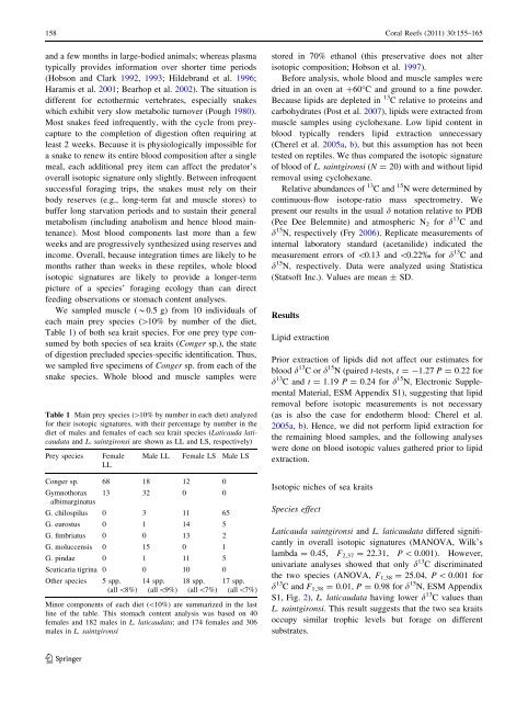
Table 1 Main prey species ([10% by number in each diet) analyzed<br />
for their isotopic <strong>signatures</strong>, with their percentage by number in the<br />
diet of males <strong>and</strong> females of each sea krait species (Laticauda laticaudata<br />
<strong>and</strong> L. saintgironsi are shown as LL <strong>and</strong> LS, respectively)<br />
Prey species Female<br />
LL<br />
Male LL Female LS Male LS<br />
Conger sp. 68 18 12 0<br />
Gymnothorax<br />
albimarginatus<br />
13 32 0 0<br />
G. chilospilus 0 3 11 65<br />
G. eurostus 0 1 14 5<br />
G. fimbriatus 0 0 13 2<br />
G. moluccensis 0 15 0 1<br />
G. pindae 0 1 11 5<br />
Scuticaria tigrina 0 0 10 0<br />
Other species 5 spp. 14 spp. 18 spp. 17 spp.<br />
(all\8%) (all\9%) (all\7%) (all\7%)<br />
Minor components of each diet (\10%) are summarized in the last<br />
line of the table. This stomach content analysis was based on 40<br />
females <strong>and</strong> 182 males in L. laticaudata; <strong>and</strong> 174 females <strong>and</strong> 306<br />
males in L. saintgironsi<br />
123<br />
stored in 70% ethanol (this preservative does not alter<br />
isotopic composition; Hobson et al. 1997).<br />
Before analysis, whole blood <strong>and</strong> muscle samples were<br />
dried in an oven at ?60°C <strong>and</strong> ground to a fine powder.<br />
Because lipids are depleted in 13 C relative to proteins <strong>and</strong><br />
carbohydrates (Post et al. 2007), lipids were extracted from<br />
muscle samples using cyclohexane. Low lipid content in<br />
blood typically renders lipid extraction unnecessary<br />
(Cherel et al. 2005a, b), but this assumption has not been<br />
tested on reptiles. We thus compared the isotopic signature<br />
of blood of L. saintgironsi (N = 20) with <strong>and</strong> without lipid<br />
removal using cyclohexane.<br />
Relative abundances of 13 C <strong>and</strong> 15 N were determined by<br />
continuous-flow isotope-ratio mass spectrometry. We<br />
present our results in the usual d notation relative to PDB<br />
(Pee Dee Belemnite) <strong>and</strong> atmospheric N2 for d 13 C <strong>and</strong><br />
d 15 N, respectively (Fry 2006). Replicate measurements of<br />
internal laboratory st<strong>and</strong>ard (acetanilide) indicated the<br />
measurement errors of \0.13 <strong>and</strong> \0.22% for d 13 C <strong>and</strong><br />
d 15 N, respectively. Data were analyzed using Statistica<br />
(Statsoft Inc.). Values are mean ± SD.<br />
Results<br />
Lipid extraction<br />
Prior extraction of lipids did not affect our estimates for<br />
blood d 13 Cord 15 N (paired t-tests, t =-1.27 P = 0.22 for<br />
d 13 C <strong>and</strong> t = 1.19 P = 0.24 for d 15 N, Electronic Supplemental<br />
Material, ESM Appendix S1), suggesting that lipid<br />
removal before isotopic measurements is not necessary<br />
(as is also the case for endotherm blood: Cherel et al.<br />
2005a, b). Hence, we did not perform lipid extraction for<br />
the remaining blood samples, <strong>and</strong> the following analyses<br />
were done on blood isotopic values gathered prior to lipid<br />
extraction.<br />
<strong>Isotopic</strong> niches of sea kraits<br />
Species effect<br />
Laticauda saintgironsi <strong>and</strong> L. laticaudata differed significantly<br />
in overall isotopic <strong>signatures</strong> (MANOVA, Wilk’s<br />
lambda = 0.45, F2,37 = 22.31, P \ 0.001). However,<br />
univariate analyses showed that only d 13 C discriminated<br />
the two species (ANOVA, F 1,38 = 25.04, P \ 0.001 for<br />
d 13 C <strong>and</strong> F1,38 = 0.01, P = 0.98 for d 15 N, ESM Appendix<br />
S1, Fig. 2), L. laticaudata having lower d 13 C values than<br />
L. saintgironsi. This result suggests that the two sea kraits<br />
occupy similar <strong>trophic</strong> levels but forage on different<br />
substrates.