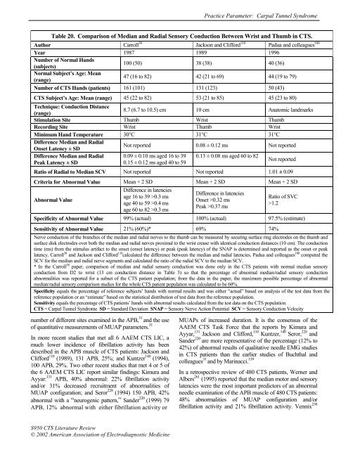
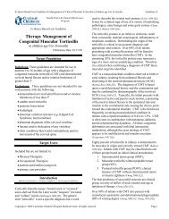
<strong>Practice</strong> <strong>Parameter</strong>: Carpal Tunnel SyndromeTable 20. Comparison <strong>of</strong> Median <strong>and</strong> Radial Sensory Conduction Between Wrist <strong>and</strong> Thumb in CTS.Author Carroll 38 Jackson <strong>and</strong> Clifford 110 Padua <strong>and</strong> colleagues 188Year 1987 1989 1996Number <strong>of</strong> Normal H<strong>and</strong>s(subjects)100 (50) 38 (38) 40 (36)Normal Subject’s Age: Mean(range)47 (16 to 82) 42 (21 to 69) 44 (19 to 79)Number <strong>of</strong> CTS H<strong>and</strong>s (patients) 161 (101) 131 (123) 50 (43)CTS Subject’s Age: Mean (range) 45 (22 to 82) 53 (21 to 85) 45 (23 to 80)Technique: Conduction Distance(range)8.7 (6.7 to 10.5) cm 10 cm Anatomic l<strong>and</strong>marksStimulation Site Thumb Wrist ThumbRecording Site Wrist Thumb WristMinimum H<strong>and</strong> Temperature 30°C 31°C 31°CDifference Median <strong>and</strong> RadialOnset Latency ± SDNot reported 0.08 ± 0.12 ms Not reportedDifference Median <strong>and</strong> Radial 0.09 ± 0.10 ms aged 16 to 39 0.13 ± 0.08 ms aged 60 to 82Peak Latency ± SD0.15 ± 0.12 ms aged 40 to 59Not reportedRatio <strong>of</strong> Radial to Median SCV Not reported Not reported 1.01 ± 0.09Criteria for Abnormal Value Mean + 2 SD Mean + 2 SD Mean + 2 SDAbnormal ValueDifference in latenciesage 16 to 39 >0.3 msage 40 to 59 >0.4 msage 60 to 82 >0.3 msDifference in latenciesOnset >0.32 msPeak >0.37 msRatio <strong>of</strong> SVC>1.2Specificity <strong>of</strong> Abnormal Value 99% (actual) 100% (actual) 97.5% (estimate)Sensitivity <strong>of</strong> Abnormal Value 21% (60%)* 69% 74%Nerve conduction <strong>of</strong> <strong>the</strong> branches <strong>of</strong> <strong>the</strong> median <strong>and</strong> radial nerves to <strong>the</strong> thumb can be measured by securing surface ring electrodes on <strong>the</strong> thumb <strong>and</strong>surface disk electrodes over both <strong>the</strong> median <strong>and</strong> radial nerves proximal to <strong>the</strong> wrist crease with identical conduction distances (10 cm). The conductiontime (ms) from <strong>the</strong> stimulus artifact to <strong>the</strong> onset (onset latency) or peak (peak latency) <strong>of</strong> <strong>the</strong> SNAP is determined <strong>and</strong> reported as <strong>the</strong> onset or peaklatency. Carroll 38 <strong>and</strong> Jackson <strong>and</strong> Clifford 110 calculated <strong>the</strong> difference between <strong>the</strong> median <strong>and</strong> radial latencies. Padua <strong>and</strong> colleagues 188 computed <strong>the</strong>SCV for <strong>the</strong> median <strong>and</strong> radial nerve segments <strong>and</strong> calculated <strong>the</strong> ratio <strong>of</strong> <strong>the</strong> radial SCV to <strong>the</strong> median SCV.* In <strong>the</strong> Carroll 38 paper, comparison <strong>of</strong> median <strong>and</strong> radial sensory conduction was done only in <strong>the</strong> CTS patients with normal median sensoryconduction from D2 to wrist (13 cm conduction distance in Table 3) so that <strong>the</strong> percentage <strong>of</strong> abnormal median/radial sensory conductionabnormalities was reported for a subset <strong>of</strong> <strong>the</strong> CTS patient population; from <strong>the</strong> data in <strong>the</strong> paper, <strong>the</strong> maximum possible percentage <strong>of</strong> abnormalmedian/radial sensory comparison studies for <strong>the</strong> whole CTS patient population was calculated to be 60%.Specificity equals <strong>the</strong> percentage <strong>of</strong> reference subjects’ h<strong>and</strong>s with normal results <strong>and</strong> was ei<strong>the</strong>r “actual” based on analysis <strong>of</strong> <strong>the</strong> test data from <strong>the</strong>reference population or an “estimate” based on <strong>the</strong> statistical distribution <strong>of</strong> test data from <strong>the</strong> reference population.Sensitivity equals <strong>the</strong> percentage <strong>of</strong> CTS patients’ h<strong>and</strong>s with abnormal results calculated from <strong>the</strong> test data on <strong>the</strong> CTS population.CTS = Carpal Tunnel Syndrome SD = St<strong>and</strong>ard Deviation SNAP = Sensory Nerve Action Potential SCV = Sensory Conduction Velocitynumber <strong>of</strong> different sites examined in <strong>the</strong> APB, 31 <strong>and</strong> <strong>the</strong> use<strong>of</strong> quantitative measurements <strong>of</strong> MUAP parameters. 31In more recent studies that met all 6 AAEM CTS LIC, amuch lower incidence <strong>of</strong> fibrillation activity has beendescribed in <strong>the</strong> APB muscle <strong>of</strong> CTS patients: Jackson <strong>and</strong>Clifford 110 (1989), 131 APB, 25%; <strong>and</strong> Kuntzer 140 (1994),100 APB, 29%. Two o<strong>the</strong>r recent studies that met 4 or 5 <strong>of</strong><strong>the</strong> 6 AAEM CTS LIC report similar findings: Kimura <strong>and</strong>Ayyar: 131 APB, 40% abnormal: 22% fibrillation activity<strong>and</strong>/or 31% decreased recruitment <strong>of</strong> abnormalities <strong>of</strong>MUAP configuration; <strong>and</strong> Seror 228 (1994) 150 APB, 42%abnormal with a “neurogenic pattern,” S<strong>and</strong>er 220 (1999) 79APB, 12% abnormal with ei<strong>the</strong>r fibrillation activity orMUAPs <strong>of</strong> increased duration. It is <strong>the</strong> consensus <strong>of</strong> <strong>the</strong>AAEM CTS Task Force that <strong>the</strong> reports by Kimura <strong>and</strong>Ayyar, 131 Jackson <strong>and</strong> Clifford, 110 Kuntzer, 140 Seror, 228 <strong>and</strong>S<strong>and</strong>er 220 are more representative <strong>of</strong> <strong>the</strong> percentage (12% to42%) <strong>of</strong> abnormal results <strong>of</strong> qualitative needle EMG studiesin CTS patients than <strong>the</strong> earlier studies <strong>of</strong> Buchthal <strong>and</strong>colleagues 31 <strong>and</strong> by Marinacci. 159In a retrospective review <strong>of</strong> 480 CTS patients, Werner <strong>and</strong>Albers 261 (1995) reported that <strong>the</strong> median motor <strong>and</strong> sensorylatencies were <strong>the</strong> most important predictors <strong>of</strong> an abnormalneedle examination <strong>of</strong> <strong>the</strong> APB muscle <strong>of</strong> 480 CTS patients:48% abnormalities <strong>of</strong> MUAP configuration <strong>and</strong>/orfibrillation activity <strong>and</strong> 21% fibrillation activity. Vennix 258S950 CTS <strong>Literature</strong> <strong>Review</strong>© 2002 American Association <strong>of</strong> Electrodiagnostic Medicine
<strong>Practice</strong> <strong>Parameter</strong>: Carpal Tunnel Syndromenoted that 95% <strong>of</strong> CTS with evidence <strong>of</strong> denervation onneedle EMG had APB CMAP amplitudes less than 7 mVbut agreed with Werner <strong>and</strong> Albers 261 that <strong>the</strong>se models arenot applicable in <strong>the</strong> clinical setting to predict denervation inindividual CTS patients.In 1999, Gnatz <strong>and</strong> Conway 87 debated <strong>the</strong> issue <strong>of</strong> <strong>the</strong>performing needle EMG <strong>of</strong> <strong>the</strong> APB muscle <strong>and</strong> o<strong>the</strong>r h<strong>and</strong><strong>and</strong> limb muscles in CTS patients. Gnatz 87 concluded that<strong>the</strong> needle EMG is important in all CTS suspects because<strong>the</strong> discomfort <strong>and</strong> expense <strong>of</strong> <strong>the</strong> exam was outweighed by<strong>the</strong> diagnostic information obtained. Conway 87 recommendedthat needle EMG be performed only when pathologyproximal to <strong>the</strong> carpal tunnel was suspected. 13,184 Gnatz <strong>and</strong>Conway 87 agreed that more studies are needed to evaluate<strong>the</strong> question <strong>of</strong> performing needle EMG in every patientsuspected <strong>of</strong> CTS. There are a few o<strong>the</strong>r studies whichaddress this issue. 176Sympa<strong>the</strong>tic Skin Response in CTSKuntzer, 140 in a study that met all 6 AAEM CTS LIC,reported a low (10%) incidence <strong>of</strong> abnormalities with anEDX test <strong>of</strong> <strong>the</strong> sympa<strong>the</strong>tic skin response (SSR) in <strong>the</strong> CTSpatients (Table 21). Verghese, 259 in a study that met 5 <strong>of</strong> 6AAEM CTS LIC, reported that 24% (33/139) <strong>of</strong>symptomatic h<strong>and</strong>s <strong>of</strong> CTS patients had a prolonged SSRlatency (>1.72 s). However, Sener, 223 in a study that met all6 AAEM CTS LIC, reported that none <strong>of</strong> <strong>the</strong> 44symptomatic limbs in CTS patients showed a SSR latencygreater than limbs <strong>of</strong> 20 normal subjects (Table 21). Sener 223used sternal stimulation to avoid <strong>the</strong> potential effect <strong>of</strong>afferent dysfunction in a limb on <strong>the</strong> SSR results. Theseresults indicate that CTS is an unlikely cause <strong>of</strong> mediannerve SSR abnormalities. Fur<strong>the</strong>rmore, <strong>the</strong> SSR study, like<strong>the</strong> F-wave study, does not localize <strong>the</strong> abnormality to <strong>the</strong>CTS segment <strong>of</strong> <strong>the</strong> median nerve. Interestingly bothVerghese 259 <strong>and</strong> Sener 223 noted over half <strong>of</strong> <strong>the</strong> CTS patientscomplained <strong>of</strong> at least 1 symptoms in <strong>the</strong> affected h<strong>and</strong> thatmay indicate autonomic dysfunction: swelling <strong>of</strong> <strong>the</strong> h<strong>and</strong>or fingers, dryness, excessive perspiration, pallor, red orpurple discoloration, <strong>and</strong> coolness. However, for <strong>the</strong> reasonsnoted above, SSR studies are not recommended as an EDXstudy to diagnose CTS patients.The Effect <strong>of</strong> Limb Ischemia, Dynamic H<strong>and</strong> Exercises,<strong>and</strong> Brief <strong>and</strong> Sustained Wrist Positioning on MedianNCSs in CTSEffect <strong>of</strong> Limb Ischemia on Median Nerve Conduction inCarpal Tunnel Syndrome. In 1953, Gilliatt <strong>and</strong> Wilson 86described <strong>the</strong> production <strong>of</strong> pares<strong>the</strong>sia in limbs <strong>of</strong> CTSpatients with a pneumatic tourniquet. As noted above, in1963, Fullerton 76 evaluated <strong>the</strong> effect <strong>of</strong> upper extremityischemia on median motor conduction in <strong>the</strong> forearm <strong>and</strong>h<strong>and</strong> <strong>and</strong> suggested that transient nocturnal symptoms weredue to median nerve ischemia. Limb ischemia caused <strong>the</strong>median <strong>the</strong>nar CMAP amplitude to fall to less than 40% <strong>of</strong><strong>the</strong> initial value after 25 minutes in 7 out <strong>of</strong> 15 CTS patientswhereas <strong>the</strong> median <strong>the</strong>nar CMAP amplitude in normalsubjects remained above 50% <strong>of</strong> <strong>the</strong> initial value after 30minutes <strong>of</strong> ischemia.Effect <strong>of</strong> Dynamic H<strong>and</strong> Exercise on Median NerveConduction in CTS. In 1994, Clifford, 48 in a study thatmet all 6 AAEM CTS LIC, evaluated <strong>the</strong> effect <strong>of</strong> 4minutes <strong>of</strong> repetitive wrist <strong>and</strong> finger movements on mediansensory nerve conduction to D4 <strong>and</strong> found a significantdifference between a group <strong>of</strong> CTS patients <strong>and</strong> normalsubjects. However, <strong>the</strong> difference was <strong>of</strong> insufficientmagnitude to discriminate individual CTS patients fromcontrol subjects. Therefore, <strong>the</strong> effects <strong>of</strong> repetitive wrist<strong>and</strong> finger movements on median NCS in CTS patients areclassified as investigational at this time.Effect <strong>of</strong> Sustained Wrist Positioning on Median NerveConduction in CTS. The effect <strong>of</strong> sustained (1 minute orgreater) active or passive wrist <strong>and</strong> finger positioning(maximal flexion or extension) on median sensory <strong>and</strong>motor nerve conduction in normal subjects <strong>and</strong> CTSpatients has been evaluated by several investigators:Schwartz, 222 Marin, 158 Dunnan, 62 Werner, 262 Hansson <strong>and</strong>Nilsson, 97 Rosecrance, 213 <strong>and</strong> Kiernan. 126Initial studies which focused on <strong>the</strong> effect <strong>of</strong> sustainedwrist positioning on <strong>the</strong> median distal sensory latency<strong>and</strong> <strong>the</strong> median motor distal latency produced conflictingresults: Schwartz, 222 Marin, 158 <strong>and</strong> Dunnan. 62 More recentstudies have focused on <strong>the</strong> effect <strong>of</strong> prolongedpositioning on <strong>the</strong> amplitude <strong>of</strong> <strong>the</strong> median SNAP <strong>and</strong>reported more promising results: Hansson <strong>and</strong> Nilsson, 97Rosecrance, 213 <strong>and</strong> Kiernan. 126 Hansson <strong>and</strong> Nilsson, 97 ina study that met 5 <strong>of</strong> <strong>the</strong> 6 AAEM CTS LIC, evaluated<strong>the</strong> effect <strong>of</strong> prolonged (up to 45 minutes) passive wristflexion on <strong>the</strong> median SNAP amplitude <strong>and</strong> determined<strong>the</strong> time (T50) it takes for <strong>the</strong> SNAP amplitude to fall toone-half <strong>the</strong> baseline value. Rosecrance <strong>and</strong>colleagues, 213 in a study that met all 6 AAEM CTS LIC,evaluated <strong>the</strong> recovery time for <strong>the</strong> SNAP amplitude toreturn to baseline after 5 minutes <strong>of</strong> active extreme wrist<strong>and</strong> fingers flexion. In both reports, <strong>the</strong> changes in <strong>the</strong>SNAP amplitude during (Hansson <strong>and</strong> Nilsson 97 ) or after(Rosecrance <strong>and</strong> colleagues 213 ) sustained wristpositioning distinguished CTS patients from normalsubjects. Because o<strong>the</strong>rs have not yet confirmed <strong>the</strong>seresults, <strong>the</strong> effects <strong>of</strong> sustained wrist positioning onmedian nerve conduction in CTS patients are classified asinvestigational at this time.Hansson <strong>and</strong> Nilsson 96,97 also provided evidence that <strong>the</strong>effect <strong>of</strong> prolonged extreme wrist flexion was due toischemia <strong>and</strong> not to compression <strong>of</strong> <strong>the</strong> nerve. The results <strong>of</strong><strong>the</strong> recent studies by Hansson <strong>and</strong> Nilsson 96,97 <strong>and</strong>Rosecrance <strong>and</strong> colleagues 213 are consistent with <strong>the</strong> effectMuscle & Nerve Supplement X 2002 S951