Nuclear Technology Review 2011 - IAEA
Nuclear Technology Review 2011 - IAEA
Nuclear Technology Review 2011 - IAEA

You also want an ePaper? Increase the reach of your titles
YUMPU automatically turns print PDFs into web optimized ePapers that Google loves.
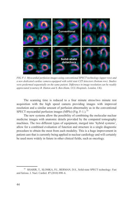
The scanning time is reduced to a four minute stress/two minute rest<br />
acquisition with the high speed camera providing images with improved<br />
resolution and a similar amount of perfusion abnormality as in the conventional<br />
SPECT myocardial perfusion images (MPIs) (Fig. F-1.). 19<br />
The new systems allow the possibility of combining the molecular nuclear<br />
medicine images with anatomic details provided by the computed tomography<br />
machines. The two different types of equipment, merged into ‘hybrid systems’,<br />
allow for a combined evaluation of function and structure in a single diagnostic<br />
procedure to obtain the most from each modality. This is a huge improvement in<br />
patient care that is currently being applied in nuclear cardiology and will certainly<br />
be used more widely in future in other clinical fields, such as oncology.<br />
19 SHARIR, T., SLOMKA, P.J., BERMAN, D.S., Solid-state SPECT technology: Fast<br />
and furious. J. Nucl. Cardiol. 17 (2010) 890–6.<br />
44<br />
Conventional<br />
Solid-state<br />
detectors<br />
FIG. F-1. Myocardial perfusion images using conventional SPECT technology (upper row) and<br />
a new dedicated cardiac camera equipped with solid state CZT detectors (bottom row). Studies<br />
were performed sequentially on the same patient. Difference in image resolution can be readily<br />
appreciated (courtesy B. Hutton and S. Ben-Haim, UCL Hospitals, London, UK).