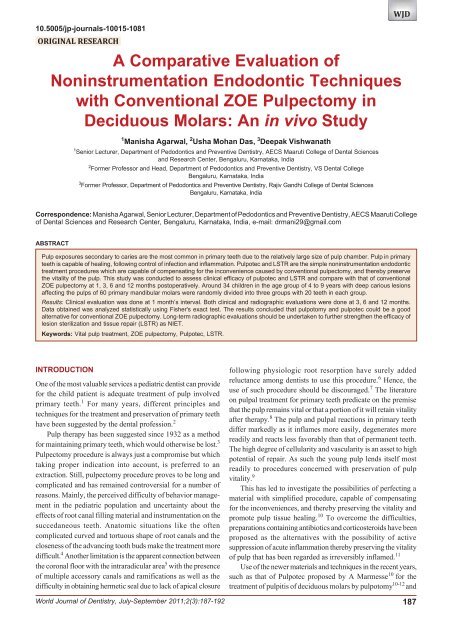
to View In Vivo Study - universaldental.com.pk
to View In Vivo Study - universaldental.com.pk
to View In Vivo Study - universaldental.com.pk
- No tags were found...
You also want an ePaper? Increase the reach of your titles
YUMPU automatically turns print PDFs into web optimized ePapers that Google loves.
Manisha Agarwal et al<br />
another alternative, the concept of lesion sterilization and tissue<br />
repair (LSTR) therapy proposed by Hoshino 13 and Iwaku<br />
et al 14 can prove <strong>to</strong> be beneficial alternatives <strong>to</strong> conventional<br />
ZOE pulpec<strong>to</strong>my. They employ the use of minute amounts of<br />
corticosteroids and mixture of antibacterial drugs respectively,<br />
<strong>to</strong> sterilize the root canal system but not mechanical procedure.<br />
These clinical procedures are simple and do not require multiple<br />
visits and have been designated as noninstrumentation<br />
endodontic treatment techniques (NIET). 15<br />
Both the techniques present numerous advantages when<br />
<strong>com</strong>pared <strong>to</strong> conventional pulpec<strong>to</strong>my. One can mention<br />
significant time saving, easy access <strong>to</strong> the coronal part of the<br />
pulp which is treated with simplified procedure. 12<br />
However, there are few reports in the literature regarding<br />
the use and clinical efficacy of these procedures. There are also<br />
no previous studies concerning which of these simplified<br />
techniques give the higher percentage of success. Moreover,<br />
data is also insufficient regarding the clinical efficacy of these<br />
noninstrumentation techniques when <strong>com</strong>pared with<br />
conventional procedures in primary teeth.<br />
So, this study was conducted <strong>to</strong> determine the clinical<br />
efficacy of Pulpo<strong>to</strong>my, Pulpotec and LSTR therapy as<br />
noninstrumentation endodontic procedures and simultaneously<br />
<strong>com</strong>pare their clinical and radiographic results with those of<br />
conventional ZOE pulpec<strong>to</strong>my as control on primary mandibular<br />
molars.<br />
METHODOLOGY<br />
A short-term clinical study was conducted on 60 primary<br />
mandibular molars, showing signs of pulpal involvement in 4<br />
<strong>to</strong> 9 years old children free from systemic disease, who reported<br />
<strong>to</strong> the Department of Pedodontics and Preventive Dentistry,<br />
VS Dental College and Hospital, Bengaluru, <strong>In</strong>dia.<br />
CRITERIA FOR TOOTH SELECTION USING<br />
CLINICAL AND RADIOGRAPHIC EXAMINATION<br />
1. Primary molars with vital carious pulp exposure that show<br />
evidence of hyperemia with or without partial necrosis or<br />
abscess formation<br />
2. No clinical symp<strong>to</strong>m or evidence of radicular pulp<br />
degeneration<br />
3. Radiographic features:<br />
• No radiographic signs of internal/external root<br />
resorption<br />
• No furcation radiolucency<br />
4. No pathologic mobility<br />
5. No sinus or fistula formation<br />
6. Teeth should be res<strong>to</strong>rable.<br />
The selected teeth were divided randomly in<strong>to</strong>:<br />
Group 1 (Fig. 1)<br />
Control<br />
Twenty primary mandibular molars were treated with<br />
conventional ZOE pulpec<strong>to</strong>my procedure according <strong>to</strong> Payne<br />
et al, 2004. 16<br />
188<br />
Local anesthesia was given followed by Rubber dam<br />
application. Access cavity was prepared using no. 56 fissure<br />
bur in NSK high speed airo<strong>to</strong>r handpiece. Complete amputation<br />
of coronal pulp using spoon excava<strong>to</strong>r was done <strong>to</strong> gain entrance<br />
in<strong>to</strong> the root canal identified at the floor of pulp chamber. Pulp<br />
tissue extirpation was done using no. 15, 20 H files, one at a<br />
time. Working length established 1 mm short of apex by<br />
inserting fine files by taking IOPAR. Following which<br />
biomechanical preparation was done using H files, rotating them<br />
<strong>to</strong> engage the pulp tissue and removed in a pull back motion<br />
with frequent irrigation with normal saline. The canals dried<br />
using sterile absorbent paper points for obturation with a paste<br />
of ZOE mixed <strong>to</strong> medium consistency, delivered using<br />
lentulospirals and the material was finally condensed using root<br />
canal pluggers. Pos<strong>to</strong>perative IOPAR was taken after<br />
<strong>com</strong>pletion of procedure. The <strong>to</strong>oth was then res<strong>to</strong>red after 24<br />
hours with stainless steel crown.<br />
Group 2 (Fig. 2)<br />
Experimental<br />
Fig. 1: Conventional ZOE pulpec<strong>to</strong>my<br />
Twenty primary mandibular molars were treated with pulpo<strong>to</strong>my<br />
and pulpotec (Pulpotec kit contains powder and liquid, carbide<br />
Fig. 2: Pulpo<strong>to</strong>my and Pulpotec<br />
JAYPEE
WJD<br />
A Comparative Evaluation of Noninstrumentation Endodontic Techniques with Conventional ZOE Pulpec<strong>to</strong>my in Deciduous Molars<br />
surgical bur, endo bur, diamond pear shaped bur and paste filler)<br />
procedure. 10<br />
After preoperative assessment, local anesthesia was given<br />
along with rubber dam placement. Roof of pulp chamber was<br />
removed using surgical bur after which vital pulp was excised<br />
from the chamber by means of endo bur. Shape the pulp chamber<br />
using diamond pear shaped bur. Pulpotec liquid and powder<br />
were blended <strong>to</strong> obtain thick, creamy consistency of the paste<br />
which was inserted in<strong>to</strong> the pulp chamber using paste filler.<br />
The cavity was then sealed with temporary ZOE cement. The<br />
patient was then asked <strong>to</strong> bite progressively but firmly on the<br />
cot<strong>to</strong>n placed between the two dental arches, so that the paste<br />
clings <strong>to</strong> the walls of the pulp cavity as well as <strong>to</strong> the root canal<br />
orifices. Excess cement was eliminated and pos<strong>to</strong>perative<br />
IOPAR taken after <strong>com</strong>pletion of procedure. Once the initial<br />
set has occurred (after 7 hours), second session was undertaken<br />
<strong>to</strong> <strong>com</strong>plete the treatment by seating stainless steel crown.<br />
Group 3 (Fig. 3)<br />
Experimental<br />
Twenty primary mandibular molars were treated with ‘lesion<br />
sterilization and tissue repair therapy’ (LSTR-3 Mix-MP, mixture<br />
of drug <strong>com</strong>bination of ciprofloxacin 500, metronidazole 400<br />
and minocycline 100 in a ratio of 1:3:3 prepared with macrogol<br />
and propylene glycogol in ointment form) procedure. 15<br />
Once the preoperative assessment was made, local<br />
anesthesia administration and rubber dam application were<br />
done. Access cavity was then prepared using no. 56 fissure bur<br />
in high speed NSK airo<strong>to</strong>r handpiece. 5% NaOCl immersed in<br />
cot<strong>to</strong>n was applied <strong>to</strong> control the hemorrhage, followed by<br />
application of the antibiotic paste <strong>to</strong> the pulpal floor. The <strong>to</strong>oth<br />
was then sealed with GIC and pos<strong>to</strong>perative IOPAR was taken<br />
after <strong>com</strong>pletion of procedure. Permanent res<strong>to</strong>ration was done<br />
by cementing stainless steel crown in second appointment after<br />
24 hours.<br />
The children were recalled for clinical evaluation at the<br />
interval of 1 month; clinical and radiographic 3, 6 and 12<br />
months.<br />
Scoring Criteria 17<br />
Scoring for clinical success of teeth:<br />
0. Failure<br />
1. No pain symp<strong>to</strong>ms<br />
2. No tenderness <strong>to</strong> percussion<br />
3. No swelling<br />
4. No fistula<br />
5. No pathologic mobility.<br />
Scoring for radiographic success of teeth:<br />
0. Failure<br />
1. No radicular radiolucency<br />
2. No internal/external root resorption<br />
3. No periodontal ligament space widening.<br />
[Failure (0): Defined as presence of any one or more of the<br />
above clinical and/or radiographic signs and symp<strong>to</strong>ms].<br />
RESULTS<br />
A <strong>to</strong>tal of 60 mandibular primary molars, 26 first molars and<br />
34 second molars in 34 children (18 males, 16 females) were<br />
endodontically treated in group I (ZOE) and in group 2<br />
(Pulpotec) and 3 (lesion sterilization and tissue repair) where<br />
noninstrumentation directed endodontic treatment was carried<br />
out. The symp<strong>to</strong>ms of the patients were recorded regarding the<br />
status of the pulp and single sitting roof canal procedure was<br />
carried out (Table 1).<br />
Statistical Analysis was done using Fisher’s exact test <strong>to</strong><br />
<strong>com</strong>pare the proportion of failure/success (Tables 2 <strong>to</strong> 5) in the<br />
three groups at different time intervals (Graphs 1 <strong>to</strong> 4). Null<br />
hypothesis H0 taken was that there is no significant difference<br />
in the proportion of failures among the three groups, i.e.<br />
p1 = p2 = p3 and the alternate hypothesis H1 was that at least<br />
one pair of the groups differ with level of significance being<br />
Table 1: Distribution of teeth according <strong>to</strong> presenting symp<strong>to</strong>ms<br />
Group Teeth with Acute symp<strong>to</strong>ms Abscess Asymp<strong>to</strong>matic<br />
deep carious<br />
lesions and<br />
exposed pulp<br />
1 20 13 — 7<br />
2 20 12 1 7<br />
3 20 13 — 7<br />
Table 2: Cumulative distribution of failure and success in each<br />
group at 1 month<br />
Group Failures Success p-value (Fisher’s test)<br />
Fig. 3: LSTR (3 Mix-MP placed over the pulp stump)<br />
1 (n = 16) 0 16 (100%) 1.000<br />
2 (n = 20) 0 20 (100%)<br />
1 (n = 16) 0 16 (100%) 0.024<br />
3 (n = 20) 6 (30%) 14 (70%)<br />
2 (n = 20) 0 20 (100%) 0.020<br />
3 (n = 20) 6 (30%) 14 (70%)<br />
World Journal of Dentistry, July-September 2011;2(3):187-192 189
Manisha Agarwal et al<br />
Table 3: Cumulative distribution of failure and success in each<br />
group at 3 months<br />
Group Failures Success p-value<br />
(Fisher’s test)<br />
1 (n = 16) 0 16 (100%) 1.000<br />
2 (n = 20) 1(15%) 19 (95%)<br />
1 (n = 16) 0 16 (100%) < 0.001<br />
3 (n = 18) 10 (55.56%) 8 (44.44%)<br />
2 (n = 20) 0 20 (100%) 0.001<br />
3 (n = 18) 10 (55.56%) 8 (44.44%)<br />
Table 4: Cumulative distribution of failure and success in each<br />
group at 6 months<br />
Group Failures Success p-value<br />
(Fisher’s test)<br />
1 (n = 14) 1 (7.14%) 13 (92.86%) 1.000<br />
2 (n = 19) 1 (5.26%) 18 (94.74%)<br />
1 (n = 14) 1 (7.14%) 13 (92.86%) 0.003<br />
3 (n = 18) 11 (61.11%) 7 (38.89%)<br />
2 (n = 19) 1 (5.26%) 18 (97.74%) < 0.001<br />
3 (n = 18) 11 (61.11%) 7 (38.89%)<br />
Graph 2: Graphic representation of success and failure in each<br />
group at 3 months<br />
Table 5: Cumulative distribution of failure and success in each<br />
group at 12 months<br />
Group Failures Success p-value<br />
(Fisher’s test)<br />
1 (n = 14) 3 (21.43%) 11 (78.57%) 0.288<br />
2 (n = 19) 1 (5.26%) 18 (94.73%)<br />
1 (n = 14) 3 (21.43%) 11 (78.57%) 0.016<br />
3 (n = 18) 12 (66.67%) 6 (33.33%)<br />
2 (n = 19) 1 (5.26%) 18 (94.73%) < 0.001<br />
3 (n = 18) 12 (66.67%) 6 (33.33%)<br />
Graph 3: Graphic representation of success and failure in each<br />
group at 6 months<br />
Graph 1: Graphic representation of success and failure in each<br />
group at 1 month<br />
Graph 4: Graphic representation of success and failure in each<br />
group at 12 months<br />
α = 0.05. The decision criterion was <strong>to</strong> reject the null hypothesis<br />
if p < 0.05 and accept the alternate hypothesis. Otherwise we<br />
accept H0.<br />
<strong>In</strong>ference: The following Tables 2 <strong>to</strong> 5 give us the significance<br />
value, i.e. the p-value for various <strong>com</strong>putations. We notice that<br />
p < 0.05 between group 1, group 3 and group 2, group 3 at all<br />
time intervals. Thus, we reject H0 and conclude that there is a<br />
significant difference in the proportion of failures/success<br />
190<br />
between group 1, group 3 and group 2, group 3. But for group<br />
1 and group 2, p > 0.05, at all time intervals. Therefore, we<br />
accept the null hypothesis H0 and conclude that there is no<br />
significant difference in the proportion of failures/success<br />
between group 1 and group 2.<br />
DISCUSSION<br />
One of the most valuable services a pediatric dentist can provide<br />
for the child patient is adequate treatment of pulp involved<br />
JAYPEE
WJD<br />
A Comparative Evaluation of Noninstrumentation Endodontic Techniques with Conventional ZOE Pulpec<strong>to</strong>my in Deciduous Molars<br />
primary teeth. 1 Maintaining the integrity and health of oral<br />
tissues is the primary objective of pulp treatment.Conservative<br />
treatments are re<strong>com</strong>mended for primary teeth whose pulps have<br />
the potential <strong>to</strong> recover once the irritation is removed.<br />
Various techniques have been described for conserving<br />
primary molars whose pulps have be<strong>com</strong>e nonvital or have<br />
degenerated <strong>to</strong> the extent that they are not good candidates for<br />
vital pulp therapy. Most of these techniques can be assigned <strong>to</strong><br />
one of two major classifications. Representatives of one group<br />
advocate removing the contents of pulp chamber and placing<br />
some sort of medication over the radicular pulp stump for a<br />
varying period of time. Representatives of the second group<br />
advocate removing all accessible pulp tissue, controlling the<br />
organisms and their <strong>to</strong>xins with a rotation of drugs, and res<strong>to</strong>ring<br />
the canals with an absorbable filling material. 18<br />
McDonalds R advocates partial pulpec<strong>to</strong>my technique for<br />
deciduous teeth that have hyperemic pulp without painful<br />
pulpitis. 19 The usual treatment applied <strong>to</strong> pulpitis on a vital<br />
<strong>to</strong>oth is pulpec<strong>to</strong>my. 12 ZOE is the most widely used preparation<br />
for primary <strong>to</strong>oth pulpec<strong>to</strong>mies. 20 Erausqin and Muruzabal used<br />
ZOE as a root canal filling material in 141 rats followed from 1<br />
<strong>to</strong> 90 days. They noted that ZOE irritated the periapical tissues<br />
and caused necrosis of bone and cementum. 21<br />
<strong>In</strong> 1982, Jerrell and Ronk presented a case report of<br />
overfilled ZOE pulpec<strong>to</strong>my in which succedaneous premolar<br />
was malformed. 22 Coll et al in 1985 reported a more than 80%<br />
success rate of one-visit ZOE pulpec<strong>to</strong>my followed a mean time<br />
of 70 months. They found ZOE retained in the tissue after eight<br />
of 17 molars exfoliated. 23<br />
Ana<strong>to</strong>mic situations like the often-<strong>com</strong>plicated shape of root<br />
canals and the closeness of the advancing <strong>to</strong>oth bud make root<br />
canal treatment difficult. 5 Groter (1967) 24 advocated <strong>to</strong> avoid<br />
the use of instruments <strong>com</strong>pletely while Spedding (1973) 25<br />
advocated <strong>to</strong> use instruments in the root canals <strong>to</strong> the point of<br />
resistance. Yacobi et al (1991) 26 were in favor of minimal<br />
biomechanical preparation. Garcia Godoy (1987) 27 preferred<br />
<strong>to</strong> enlarge the root canals only <strong>to</strong> the level of occlusal plane of<br />
the permanent <strong>to</strong>oth germ.<br />
Hobson 1970 28 found that tubules in the dentinal walls of<br />
the root canals, in 70% of the samples of extracted teeth with<br />
necrotic in both radicular and coronal pulp, were penetrated by<br />
microorganisms. He concluded that it would be desirable,<br />
therefore, when treating nonvital infected primary teeth <strong>to</strong> use<br />
an antibacterial drug capable of penetrating the tissues and<br />
controlling infection in the dentinal walls.<br />
Several investiga<strong>to</strong>rs agree that <strong>to</strong>tal removal of the pulp<br />
tissue from the root canals of primary teeth cannot be achieved<br />
because of their <strong>com</strong>plex and variable morphology. It is also<br />
difficult <strong>to</strong> eliminate the wide range of organisms in infected<br />
root canals. Thus, the particular quality of the medicament/paste<br />
determines the prognosis in the endodontic treatment of infected<br />
primary teeth. Identifying the best formulation of ingredients<br />
and techniques <strong>to</strong> predictably produce pulpal healing remains<br />
elusive. It is generally agreed that prognosis after any type of<br />
the pulp therapy improves in the absence of contamination by<br />
pathogenic organisms. Thus, bio<strong>com</strong>patible neutralization of<br />
any existing pulpal contamination and prevention are worthy<br />
goals in vital pulp therapy. If the treatment material in direct<br />
contact with pulp also has some inherent quality that promotes,<br />
stimulates or accelerates a true tissue healing response, so much<br />
better, however, it is recognized that vital pulp tissue can recover<br />
from a variety of insults spontaneously in a favorable<br />
environment. 29<br />
Noninstrumentation endodontic treatment procedures<br />
applied <strong>to</strong> the use of Pulpotec and lesion sterilization and tissue<br />
repair are based on the above concepts.<br />
The overall success rate of ZOE (78.5%) was consistent<br />
with the results of Mortazavi M and Mesbahi M (2004) 39 who<br />
also reported the overall success rate (clinical and radiographic)<br />
of 78.5% for ZOE at the end of 10 <strong>to</strong> 16 months follow-up<br />
period. The results were also in <strong>com</strong>parison with the study done<br />
by Mani, Chawla and Tewari et al (2000) 34 who reported 83.3%<br />
clinical and radiographic success rate in the ZOE group followed<br />
up <strong>to</strong> 6 months.<br />
The overall success of 94% in “Pulpotec” at the end of 12<br />
months was also in agreement with the results of Maramesse A<br />
(1989) 13 who conducted clinical trials of Pulpotec for long-term<br />
and found a success rate of 80% in deciduous teeth. Our results<br />
were also consistent with those of Dodeyan SA, Donkaya IP<br />
(2003) 36 who reported 100% success rate of Pulpotec during 6<br />
months evaluation.<br />
<strong>In</strong> this in vivo study, the overall (clinical and radiographic)<br />
success rates in LSTR at the end of 12 months were not on<br />
agreement with those of Hoshino E (2004) 16 who reported 80%<br />
clinical success of the procedure. Overall three patients were<br />
seen with intraoral swelling at the end of 6 months follow-up<br />
and the main <strong>com</strong>plications encountered were radicular<br />
radiolucency and internal resorption on radiographic followup<br />
at 6 and 12 months intervals. The poor success rate can be<br />
attributed <strong>to</strong> the fact that radiographic criteria were also used<br />
<strong>to</strong> assess the success, which was not done in the earlier studies.<br />
There are limited and conflicting reports in the literature <strong>to</strong><br />
substantiate the clinical and radiographic success rate of LSTR.<br />
From the above study, it was concluded that “Pulpotec”<br />
can be used as a proven, safe and effective alternative <strong>to</strong><br />
conventional ZOE pulpec<strong>to</strong>my in teeth with pulpal exposure<br />
secondary <strong>to</strong> caries with or without partial necrosis, which<br />
eliminates the need <strong>to</strong> instrument in<strong>to</strong> the canals and simplifies<br />
the procedure.<br />
We re<strong>com</strong>mend further long-term clinical and radiographic<br />
evaluations for LSTR before it can be re<strong>com</strong>mended as NIET.<br />
CONCLUSIONS<br />
1. Our study concluded that pulpo<strong>to</strong>my and pulpotec could be<br />
a good alternative for conventional ZOE pulpec<strong>to</strong>my<br />
2. Long-term clinical and radiographic evaluations should be<br />
undertaken <strong>to</strong> further strengthen the efficacy of LSTR as<br />
NIET.<br />
World Journal of Dentistry, July-September 2011;2(3):187-192 191
Manisha Agarwal et al<br />
REFERENCES<br />
1. Rifkin A. A simple, effective, safe technique for the root canal<br />
treatment of abscessed primary teeth. J Dent Child Nov-Dec<br />
1980;435-41.<br />
2. Boller RJ. Reactions of pulpo<strong>to</strong>mized teeth <strong>to</strong> zinc oxide and<br />
formocresol type drugs. J Dent Child July-Aug 1972:52-61.<br />
3. Kubota K, Golden BE, Penugonda B. Root canal filling materials<br />
for primary teeth: A review of the literature. J Dent Child May-<br />
June 1992;225-27.<br />
4. Holan G, Fucks AB. A <strong>com</strong>parison of pulpec<strong>to</strong>mies using ZOE<br />
and KRI paste in primary molars: A retrospective study. Pediatric<br />
Dentistry 1993;15:403-07.<br />
5. Moss SJ, Addels<strong>to</strong>n R, Goldsmith ED. His<strong>to</strong>logic study of pulpal<br />
floors of deciduous molars. JADA 1965;70:372.<br />
6. Mathewson RJ, Primoch RE, Morrison JT. Fundamentals of<br />
pediatric dentistry (3rd ed). Quintessence Publishing Co <strong>In</strong>c<br />
1995:257-84.<br />
7. Massler N. Preventive endodontics. DCNA Nov 1967;670.<br />
8. Gould JM. Root canal therapy for infected primary molar teeth:<br />
Preliminary repot. J Dent Child July-Aug 1972;23-27.<br />
9. Stewart RE, Barber TK, Trouman KC, Wei SHY. Pediatric<br />
dentistry scientific foundations and clinical practice. The CV<br />
Mosby Company.<br />
10. http://www.pulpotec.<strong>com</strong>.<br />
11. Schroeder U, Graneth L. On internal dentine resorption of<br />
deciduous molars treated by pulpo<strong>to</strong>my and capped with calcium<br />
hydroxide. Odon<strong>to</strong>l Review 1971;22:179.<br />
12. Marmesse A. Final reports of clinical trials of pulpotec<br />
(Translation of the original text). Unpublished article (http://<br />
www.pulpotec.<strong>com</strong>).<br />
13. Hoshino E. Sterilization of carious lesions by drugs. J of the<br />
Japanese Association for Dental science 1990;9:32-37.<br />
14. Iwaku M, Hoshino E, Kota K. Lesion sterilization and tissue<br />
repair therapy: New pulpal treatment, how <strong>to</strong> conserve infected<br />
pulps. Tokyo, Japan: Nihon-Shika-Horon 1996.<br />
15. Takushige T, Cruz EV, Moral AA, Hoshino E. Endodontic<br />
treatment of primary teeth using a <strong>com</strong>bination of antibacterial<br />
drugs. <strong>In</strong>ternational endodontic Journal 2004;37:132-38.<br />
16. Payne RG, Kenny DJ, Johns<strong>to</strong>n DH, Judd PL. Two-year out<strong>com</strong>e<br />
study of zinc oxide-eugenol root canal treatment for vital primary<br />
teeth. J Can Dent Assoc 1993;59:528-36.<br />
17. Agamy HA, Bakry NS, Mounir MMF, Avery DR. Comparison<br />
of mineral trioxide aggregate and formocresol as pulp capping<br />
agents in pulpo<strong>to</strong>mized primary teeth. Ped Dent 2004;26:<br />
302-09.<br />
18. Starkey PE. Pulpec<strong>to</strong>my and root canal filling in primary molars:<br />
Report of a case. J Dent Child May-June 1973;49-53.<br />
19. Donald RE, Avery DR, Dean JA. Dentistry for the child and<br />
adolescent. St Louis: CV Mosby Company 1978:146-68.<br />
20. Sardian R, Coll JA. A long-term follow-up on the retention rate<br />
of zinc oxide eugenol filler after primary <strong>to</strong>oth pulpec<strong>to</strong>my.<br />
Pediatric Dentistry 1993;15:249-53.<br />
21. Erausquin J, Muruzabal M. Root canal fillings with zinc oxideeugenol<br />
cement in the rat molar. Oral Surg Oral Med Oral Pathol<br />
1967;24:547-58.<br />
22. Jerrell RG, Ronk SL. Developmental arrest of a succedaneous<br />
<strong>to</strong>oth following pulpec<strong>to</strong>my in a primary <strong>to</strong>oth. J Pedod<br />
1983;6:337-42.<br />
23. Coll JA, Josell S, Casper JS. Evaluation of a one-appointment<br />
formocresol pulpec<strong>to</strong>my technique for primary molars. Ped Dent<br />
1985;7:123-29.<br />
24. Groter JA. Pulp therapy in primary teeth. J Dent Child<br />
1967;34:508-10.<br />
25. Spedding RH. Root canal treatment for primary teeth. DCNA<br />
1973;17:105-24.<br />
26. Yacobi R, Kenny DJ, Judd PL, Johns<strong>to</strong>n DH. Evolving primary<br />
pulp therapy techniques. JADA 1991;122:83-85.<br />
27. Garcia Godoy C. Evaluation of iodoform paste in root canal<br />
therapy for infected primary teeth. J Dent Child 1987;54:30-34.<br />
28. Hobson P. Pulp treatment of deciduous teeth. BDJ 1970;128:<br />
232-8, 275-82.<br />
29. Mortazavi M, Mesbahi M. Comparison of zinc oxide and<br />
eugenol, and Vitapex for root canal treatment of necrotic primary<br />
teeth. <strong>In</strong>t J of Ped Dent 2004;14:417-24.<br />
192<br />
JAYPEE


