to View In Vivo Study - universaldental.com.pk
to View In Vivo Study - universaldental.com.pk
to View In Vivo Study - universaldental.com.pk
- No tags were found...

You also want an ePaper? Increase the reach of your titles
YUMPU automatically turns print PDFs into web optimized ePapers that Google loves.
Manisha Agarwal et al<br />
another alternative, the concept of lesion sterilization and tissue<br />
repair (LSTR) therapy proposed by Hoshino 13 and Iwaku<br />
et al 14 can prove <strong>to</strong> be beneficial alternatives <strong>to</strong> conventional<br />
ZOE pulpec<strong>to</strong>my. They employ the use of minute amounts of<br />
corticosteroids and mixture of antibacterial drugs respectively,<br />
<strong>to</strong> sterilize the root canal system but not mechanical procedure.<br />
These clinical procedures are simple and do not require multiple<br />
visits and have been designated as noninstrumentation<br />
endodontic treatment techniques (NIET). 15<br />
Both the techniques present numerous advantages when<br />
<strong>com</strong>pared <strong>to</strong> conventional pulpec<strong>to</strong>my. One can mention<br />
significant time saving, easy access <strong>to</strong> the coronal part of the<br />
pulp which is treated with simplified procedure. 12<br />
However, there are few reports in the literature regarding<br />
the use and clinical efficacy of these procedures. There are also<br />
no previous studies concerning which of these simplified<br />
techniques give the higher percentage of success. Moreover,<br />
data is also insufficient regarding the clinical efficacy of these<br />
noninstrumentation techniques when <strong>com</strong>pared with<br />
conventional procedures in primary teeth.<br />
So, this study was conducted <strong>to</strong> determine the clinical<br />
efficacy of Pulpo<strong>to</strong>my, Pulpotec and LSTR therapy as<br />
noninstrumentation endodontic procedures and simultaneously<br />
<strong>com</strong>pare their clinical and radiographic results with those of<br />
conventional ZOE pulpec<strong>to</strong>my as control on primary mandibular<br />
molars.<br />
METHODOLOGY<br />
A short-term clinical study was conducted on 60 primary<br />
mandibular molars, showing signs of pulpal involvement in 4<br />
<strong>to</strong> 9 years old children free from systemic disease, who reported<br />
<strong>to</strong> the Department of Pedodontics and Preventive Dentistry,<br />
VS Dental College and Hospital, Bengaluru, <strong>In</strong>dia.<br />
CRITERIA FOR TOOTH SELECTION USING<br />
CLINICAL AND RADIOGRAPHIC EXAMINATION<br />
1. Primary molars with vital carious pulp exposure that show<br />
evidence of hyperemia with or without partial necrosis or<br />
abscess formation<br />
2. No clinical symp<strong>to</strong>m or evidence of radicular pulp<br />
degeneration<br />
3. Radiographic features:<br />
• No radiographic signs of internal/external root<br />
resorption<br />
• No furcation radiolucency<br />
4. No pathologic mobility<br />
5. No sinus or fistula formation<br />
6. Teeth should be res<strong>to</strong>rable.<br />
The selected teeth were divided randomly in<strong>to</strong>:<br />
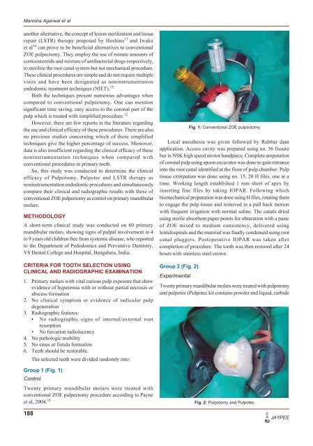
Group 1 (Fig. 1)<br />
Control<br />
Twenty primary mandibular molars were treated with<br />
conventional ZOE pulpec<strong>to</strong>my procedure according <strong>to</strong> Payne<br />
et al, 2004. 16<br />
188<br />
Local anesthesia was given followed by Rubber dam<br />
application. Access cavity was prepared using no. 56 fissure<br />
bur in NSK high speed airo<strong>to</strong>r handpiece. Complete amputation<br />
of coronal pulp using spoon excava<strong>to</strong>r was done <strong>to</strong> gain entrance<br />
in<strong>to</strong> the root canal identified at the floor of pulp chamber. Pulp<br />
tissue extirpation was done using no. 15, 20 H files, one at a<br />
time. Working length established 1 mm short of apex by<br />
inserting fine files by taking IOPAR. Following which<br />
biomechanical preparation was done using H files, rotating them<br />
<strong>to</strong> engage the pulp tissue and removed in a pull back motion<br />
with frequent irrigation with normal saline. The canals dried<br />
using sterile absorbent paper points for obturation with a paste<br />
of ZOE mixed <strong>to</strong> medium consistency, delivered using<br />
lentulospirals and the material was finally condensed using root<br />
canal pluggers. Pos<strong>to</strong>perative IOPAR was taken after<br />
<strong>com</strong>pletion of procedure. The <strong>to</strong>oth was then res<strong>to</strong>red after 24<br />
hours with stainless steel crown.<br />
Group 2 (Fig. 2)<br />
Experimental<br />
Fig. 1: Conventional ZOE pulpec<strong>to</strong>my<br />
Twenty primary mandibular molars were treated with pulpo<strong>to</strong>my<br />
and pulpotec (Pulpotec kit contains powder and liquid, carbide<br />
Fig. 2: Pulpo<strong>to</strong>my and Pulpotec<br />
JAYPEE