Answers - Virtual Pathology at the University of Leeds
Answers - Virtual Pathology at the University of Leeds
Answers - Virtual Pathology at the University of Leeds

Create successful ePaper yourself
Turn your PDF publications into a flip-book with our unique Google optimized e-Paper software.
FRCP<strong>at</strong>h Part 2 in Histop<strong>at</strong>hology Spring 2012<br />
Surgical <strong>P<strong>at</strong>hology</strong><br />
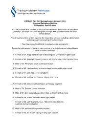
<strong>Answers</strong><br />
One route to examin<strong>at</strong>ion success is to read <strong>the</strong> question and <strong>the</strong>n <strong>at</strong>tempt to answer it.<br />
The question is:<br />
‘You should provide a written report to <strong>the</strong> requesting clinician including a<br />
description <strong>of</strong> <strong>the</strong> lesion, a clear final diagnosis, and a clinical comment putting<br />
your diagnosis into its clinical context.<br />
You may suggest additional investig<strong>at</strong>ions as appropri<strong>at</strong>e.’<br />
The short answers to <strong>the</strong> cases are given in <strong>the</strong> list below. On <strong>the</strong> following pages are<br />
summaries <strong>of</strong> <strong>the</strong> expected full answers and <strong>the</strong> points th<strong>at</strong> examiners were looking for<br />
when marking <strong>the</strong> answers. These are all based on <strong>the</strong> original diagnostic reports.<br />
To achieve a pass on this part <strong>of</strong> <strong>the</strong> examin<strong>at</strong>ion an average mark <strong>of</strong> 2.5 per case is<br />
needed – a total <strong>of</strong> <strong>at</strong> least 50.<br />
To do th<strong>at</strong> not only do you need make a clear diagnosis but you also need to put your<br />
diagnosis into its clinical context.<br />
1 - M 58. Orchidectomy for infective orchitis.<br />
Classical seminoma<br />
2 - F 18. Cyst surrounding lower left 8 tooth (also removed). Present many years,<br />
slowly enlarging, ? dentigerous.<br />
Odontogenic ker<strong>at</strong>ocyst<br />
3 - F 48. Breast screening p<strong>at</strong>ient. Mammotome / Vacuum-Assisted Core Biopsy <strong>of</strong> 5<br />
mm focus <strong>of</strong> microcalcific<strong>at</strong>ion in left upper outer quadrant. Clinical assessment<br />
= normal (P1), Radiological assessment = probably benign (R3), Ultrasound<br />
assessment = Normal (U1)<br />
Benign microcalcific<strong>at</strong>ion and fibrocystic disease/ columnar cell change<br />
4 - F 56. 3 cm mass in <strong>the</strong> lower lobe <strong>of</strong> <strong>the</strong> right lung. PET-CT shows moder<strong>at</strong>e<br />
uptake in RLL mass only. Section from right lower lobectomy.<br />
Atypical carcinoid tumour
5 – M 50. ‘Painful left epididymal swelling. Failed to respond to medical <strong>the</strong>rapy.<br />
Anxieties about fertility’. Received, a 25 mm length <strong>of</strong> ? vas <strong>at</strong>tached to a pearshaped<br />
firm mass 42 x 30 x 17 mm, with a homogeneous creamy-white cut<br />
surface.<br />
Necrotising granulom<strong>at</strong>ous inflamm<strong>at</strong>ion<br />
6 - M 73. Papillom<strong>at</strong>ous expansile nasal polyp filling left nasal cavity. ? Inverted<br />
papilloma.<br />
Primary sino-nasal intestinal type adenocarcinoma<br />
7 – F 63. Oesophageal tumour diagnosed as poorly differenti<strong>at</strong>ed carcinoma with some<br />
neuroendocrine differenti<strong>at</strong>ion. Has had several rounds <strong>of</strong> chemo<strong>the</strong>rapy<br />
followed by gastro-oesophagectomy. No tumour visible macroscopically.<br />
Section <strong>of</strong> gastro-oesophageal junction.<br />
High grade dysplasia suspicious for lamina propria invasion (intramucosal<br />
carcinoma) in Barrett’s oesophagus<br />
8 – F 24. Products <strong>of</strong> conception. Missed miscarriage <strong>at</strong> 5/40.<br />
Complete hyd<strong>at</strong>idiform mole in twin pregnancy<br />
9 – F 88. Lobul<strong>at</strong>ed. Well-defined part cystic part solid lesion in <strong>the</strong> right breast.<br />
Central wide excision, including nipple.<br />
8 mm high cytonuclear grade encysted papillary carcinoma, completely excised.<br />
10 – F 46. Cervical lymphadenop<strong>at</strong>hy. ? NHL on FNA. Left cervical lymph node<br />
biopsy.<br />
Follicular lymphoma, grade 3A<br />
11 – F 37. Sc<strong>at</strong>tered lumpy lesion on forehead, neck and left hand. Hint <strong>of</strong> annular<br />
configur<strong>at</strong>ion. Incisional biopsy from dorsum <strong>of</strong> left hand.<br />
Granuloma annulare<br />
12 – F 32. Referral cytology <strong>of</strong> severe dyskaryosis ?invasive (code 5). At colposcopy,<br />
<strong>the</strong>re was dense aceto-white seen over a large area. Loop excision <strong>of</strong> <strong>the</strong><br />
transform<strong>at</strong>ion zone was done and this is one section from it.<br />
CIN3 and CGIN<br />
13 – F 31. 10 cm mass in right lobe <strong>of</strong> liver – resected.<br />
Inflamm<strong>at</strong>ory / telangiect<strong>at</strong>ic hep<strong>at</strong>ocellular adenoma
14 - M 63. Large retroperitoneal mass extending from around <strong>the</strong> right kidney down into<br />
<strong>the</strong> inguinal canal. On sectioning <strong>the</strong> tumour <strong>the</strong>re was a tennis ball sized solid<br />
area lying adjacent to <strong>the</strong> kidney. Section from <strong>the</strong> edge <strong>of</strong> <strong>the</strong> solid area.<br />
Well differenti<strong>at</strong>ed liposarcoma with de-differenti<strong>at</strong>ed component<br />
15 – M 24. Pigmented lesion excised from <strong>the</strong> left mid back.<br />
Pigmented spindle cell naevus <strong>of</strong> Reed<br />
16 – F 48. Ulcer<strong>at</strong>ed area in anal canal.<br />
Basaloid squamous cell carcinoma<br />
17 - F 17. Biopsy <strong>of</strong> skin lesion from right hip. ? epidermal cyst.<br />
Aneurysmal derm<strong>at</strong><strong>of</strong>ibroma<br />
18 – F 75. Altered bowel habit. Normal colon on sigmoidoscopy. Endoscopic biopsy.<br />
Collagenous colitis<br />
19 – M 28. Past medical history <strong>of</strong> eczema. Widespread superficial blistering with<br />
appearance suggesting an impetiginised eruption.<br />
Impetiginised pemphigus foliaceus<br />
20 – F 30. Enlarged firm mobile right axillary lymph node.<br />
Foreign body giant cell reaction to silicone
Slide 2<br />
F 18. Cyst surrounding lower left 8, also removed. Present many years, slowly<br />
enlarging, ? dentigerous.<br />
Oral biopsy, left mandible: A cystic structure measuring 20 x 13 x 5mm. The surface<br />
is intact and smooth. The specimen is bisected longitudinally to reveal s<strong>of</strong>t creamy<br />
contents. Both sections embedded in 1A. Also received is a tooth measuring 13 x 12 x<br />
10mm. This was returned to <strong>the</strong> pot.<br />
Sections show an odontogenic cyst. The cyst is lined by a thin squamous epi<strong>the</strong>lium<br />
th<strong>at</strong> shows palisading <strong>of</strong> <strong>the</strong> basal cells and surface paraker<strong>at</strong>osis. The cyst wall is<br />
composed <strong>of</strong> fibrous tissue. There are sc<strong>at</strong>tered odontogenic epi<strong>the</strong>lial rests and a few<br />
s<strong>at</strong>ellite (daughter) cysts. The fe<strong>at</strong>ures are those <strong>of</strong> an odontogenic ker<strong>at</strong>ocyst.<br />
Left mandible; odontogenic ker<strong>at</strong>ocyst.<br />
1.5 Malignant diagnosis<br />
2.0 Benign diagnosis but not correctly classified as odontogenic ker<strong>at</strong>ocyst<br />
2.5 Diagnosis <strong>of</strong> odontogenic ker<strong>at</strong>ocyst<br />
3.0 Correct diagnosis, with correct identific<strong>at</strong>ion <strong>of</strong> s<strong>at</strong>ellite (daughter) cysts and<br />
comment on <strong>the</strong> increased risk <strong>of</strong> recurrence
Slide 3<br />
Case 12H2811<br />
F 48. Breast screening p<strong>at</strong>ient. Mammotome / Vacuum-Assisted Core Biopsy <strong>of</strong> 5 mm<br />
focus <strong>of</strong> microcalcific<strong>at</strong>ion in left upper outer quadrant. Clinical assessment = normal<br />
(P1), Radiological assessment = probably benign (R3), Ultrasound assessment =<br />
Normal (U1)<br />
1.5 Any malignant diagnosis (DCIS or invasive malignancy)<br />
2.0 Atypical ductal hyperplasia (ADH)<br />
2.5 Benign fibrocystic change and columnar cell change.<br />
3.0 Benign calcific<strong>at</strong>ion. B2. Discuss <strong>at</strong> MDT. Correl<strong>at</strong>e with imaging findings and<br />
clinical history. If <strong>the</strong>se correl<strong>at</strong>e <strong>the</strong>n no fur<strong>the</strong>r action with p<strong>at</strong>ient back to<br />
normal recall.
Slide 4
Slide 5
Slide 6<br />
M 73. Papillom<strong>at</strong>ous expansile nasal polyp filling left nasal cavity. ?Inverted papilloma.<br />
ENT biopsy, nasal polyp, left: Multiple tan haemorrhagic and cream coloured<br />
fragments <strong>of</strong> tissue toge<strong>the</strong>r measuring 40 x 30 x up to 8mm. All tissue is embedded in<br />
three cassettes, 1A to 1C.<br />
Sections show a papillary tumour with occasional tubular form<strong>at</strong>ions supported by<br />
haemorrhagic fibrous stroma. The tumour cells resemble those seen in colonic<br />
neoplasms and occasional goblet cells are also present. Mitotic figures are readily found<br />
and <strong>at</strong>ypical mitosis is seen. In some blocks, <strong>the</strong> tumour is seen adjacent to nasal<br />
mucosa and underlying sino-nasal bone. The fe<strong>at</strong>ures are those <strong>of</strong> a papillary<br />
adenocarcinoma.<br />
Immunohistochemistry shows th<strong>at</strong> most <strong>of</strong> <strong>the</strong> tumour cells are positive for CK20 and a<br />
small proportion are also positive for CK7. Staining for CEA is also positive. Sc<strong>at</strong>tered<br />
cells are strongly positive for granular cytoplasmic chromogranin and synaptophysin;<br />
staining for CD56 is neg<strong>at</strong>ive.<br />
The fe<strong>at</strong>ures are in keeping with a primary sino-nasal intestinal type adenocarcinoma. If<br />
this proves to be <strong>the</strong> case, <strong>the</strong> subtype would be papillary/ colonic type. The differential<br />
diagnosis is a metast<strong>at</strong>ic deposit and this should be excluded by clinical staging. Lower<br />
GI tract would be <strong>the</strong> most likely primary site. Clinical correl<strong>at</strong>ion and discussion <strong>at</strong> <strong>the</strong><br />
MDT is advised.<br />
Left nasal cavity; intestinal-type adenocarcinoma, most likely primary but metastasis<br />
should be excluded.<br />
1.5 Benign diagnosis<br />
2.0 Unequivocal diagnosis <strong>of</strong> metast<strong>at</strong>ic adenocarcinoma<br />
2.5 Diagnosis <strong>of</strong> primary sino-nasal intestinal type adenocarcinoma<br />
3.0 Diagnosis <strong>of</strong> primary sino-nasal intestinal type adenocarcinoma, with appropri<strong>at</strong>e<br />
differential diagnosis, suggestions for immuno and MDT referral
Slide 7
Slide 8
Slide 9
Slide 10<br />
Slide 11
Slide 12<br />
32 year old with a referral cytology <strong>of</strong> severe dyskaryosis ?invasive (code 5). At<br />
colposcopy, <strong>the</strong>re was dense acetowhite seen over a large area. Loop excision <strong>of</strong> <strong>the</strong><br />
transform<strong>at</strong>ion zone was done and this is one section from it.<br />
1 if invasive neoplasia is diagnosed.<br />
1.5 or 2 if one or <strong>the</strong> o<strong>the</strong>r dysplasia is overlooked and o<strong>the</strong>r comments are not given,<br />
particularly <strong>the</strong> need for levels and correl<strong>at</strong>ion.<br />
2.5 is given if CIN 3 and CGIN are mentioned but o<strong>the</strong>r comments such as excision<br />
margins, correl<strong>at</strong>ion, etc are not included. Provided <strong>the</strong>y do not call it invasive<br />
neoplasia.<br />
3 if some <strong>of</strong> <strong>the</strong> fe<strong>at</strong>ures are not mentioned.<br />
3.5 should describe this as <strong>the</strong> transform<strong>at</strong>ion zone with CIN 3 on <strong>the</strong> surface and also<br />
colonising occasional glands and extensive high grade CGIN. In this section <strong>the</strong> CIN is<br />
completely excised but <strong>the</strong> CGIN is present <strong>at</strong> <strong>the</strong> endocervical margin. There is no<br />
invasive neoplasia. The good candid<strong>at</strong>es will ask for levels to exclude invasion and to<br />
ensure th<strong>at</strong> <strong>the</strong> CGIN is definitely incompletely excised. They will also suggest fur<strong>the</strong>r<br />
coploscopy excision and will indic<strong>at</strong>e <strong>the</strong> depth <strong>of</strong> <strong>the</strong> deepest gland involved by <strong>the</strong><br />
CGIN. The most important fe<strong>at</strong>ure to be mentioned is <strong>the</strong> apparent non-correl<strong>at</strong>ion with<br />
<strong>the</strong> referral cytology. They should suggest a review <strong>of</strong> <strong>the</strong> cytology and, if required, a<br />
discussion <strong>at</strong> a colposcopy MDT meeting.
Slide 13
Slide 14<br />
History: 63 year old male with large retroperitoneal mass extending from around <strong>the</strong><br />
right kidney down into <strong>the</strong> inguinal canal. On sectioning <strong>the</strong> tumour <strong>the</strong>re is a tennis ball<br />
sized solid area lying adjacent to <strong>the</strong> kidney. Section from <strong>the</strong> edge <strong>of</strong> <strong>the</strong> solid area.<br />
Answer: Should describe tumour with two components – Well diff liposarcoma with<br />
collections <strong>of</strong> variably sized adipocytes lying in loose fibrous stroma with numerous<br />
tumour giant cells. Occasional lipoblasts but not prominent so may be missing in some<br />
sections.<br />
De-differenti<strong>at</strong>ed component with sheets <strong>of</strong> spindle cells in loose stroma, mitotic activity<br />
+, extensive central necrosis and area <strong>of</strong> more pleomorphic spindle cells.<br />
Good/excellent candid<strong>at</strong>e will make diagnosis <strong>of</strong> de-differenti<strong>at</strong>ed liposarcoma on<br />
history (classical site) and morphology + will suggest confirm<strong>at</strong>ion with immuno +/- ISH<br />
for MDM2 amplific<strong>at</strong>ion (3.5) and correl<strong>at</strong>ion with radiology.<br />
An adequ<strong>at</strong>e answer will describe <strong>the</strong> tumour – label it as a sarcoma and provide a<br />
suggested immunostaining panel to subtype – e.g. S100 for MPNST, Desmin amd<br />
SMA for leiomyosarcoma, pancytoker<strong>at</strong>in to exclude sarcom<strong>at</strong>oid carcinoma.
Slide 15<br />
This pigmented lesion was excised from <strong>the</strong> left mid back <strong>of</strong> this 24-year-old man.<br />
The lesion shows good radial symmetry and is predominantly intraepidermal, being<br />
composed <strong>of</strong> expansile junctional nests <strong>of</strong> spindle and epi<strong>the</strong>lioid melanocytes showing<br />
quite heavy pigment<strong>at</strong>ion. There is some limited spread <strong>of</strong> melanocytes into <strong>the</strong> upper<br />
epidermis but <strong>the</strong> striking fe<strong>at</strong>ure is <strong>the</strong> heavy pigment<strong>at</strong>ion th<strong>at</strong> is present, with melanin<br />
pigment present in all layers <strong>of</strong> <strong>the</strong> epidermis and in melanophages in <strong>the</strong> underlying<br />
papillary dermis.<br />
There is no substantial dermal component and <strong>the</strong>re is no epidermal ulcer<strong>at</strong>ion or<br />
dermal mitoses.<br />
This lesion is a benign pigmented spindle cell naevus <strong>of</strong> Reed. Diagnostic fe<strong>at</strong>ures <strong>of</strong><br />
malignant melanoma are not present.<br />
The lesion has been completely excised with 2mm l<strong>at</strong>eral margins.<br />
In view <strong>of</strong> <strong>the</strong> benign diagnosis <strong>the</strong>re is no indic<strong>at</strong>ion for any fur<strong>the</strong>r local surgery <strong>at</strong> this<br />
time.<br />
1.5 Malignant diagnosis<br />
2.0 No conclusion or suggestion <strong>of</strong> malignancy<br />
2.5 Correct diagnosis <strong>of</strong> pigmented spindle cell naevus <strong>of</strong> Reed<br />
3.0 Correct diagnosis with full description<br />
3.5 Correct diagnosis with full description and advice on no fur<strong>the</strong>r surgery
Slide 16<br />
48 female. Ulcer<strong>at</strong>ed area in anal canal.<br />
(Basaloid) squamous carcinoma <strong>of</strong> rectum<br />
Traditionally basaloid SCC is said to have a poorer prognosis than usual type SCC.<br />
One <strong>of</strong> <strong>the</strong> main problems is <strong>the</strong> variability within tumours and now reliability in<br />
diagnosing this sub-type. It probably does confer a slightly poorer prognosis as <strong>the</strong><br />
fe<strong>at</strong>ures <strong>of</strong> basaloid carcinoma are also those <strong>of</strong> poorer differenti<strong>at</strong>ion. In general<br />
histological fe<strong>at</strong>ures are much less useful in predicting prognosis in anal cancer than<br />
size and nodal st<strong>at</strong>us.<br />
1.5 Fails to recognise a malignant tumour or clearly wrong malignant diagnosis - e.g.<br />
lymphoma<br />
2 Indecisive answer as to n<strong>at</strong>ure (benign / malignant) or says malignant but without<br />
favouring carcinoma<br />
2.5 "Carcinoma"<br />
3 Squamous carcinoma<br />
3.5 Recognises basaloid fe<strong>at</strong>ures
Slide 17<br />
F 17. Biopsy <strong>of</strong> skin lesion from right hip. ? epidermal cyst.<br />
1.5 Any malignant diagnosis<br />
2.0 Benign report without clear correct diagnosis<br />
2.5 Correct diagnosis <strong>of</strong> benign aneurysmal derm<strong>at</strong><strong>of</strong>ibroma, completely excised<br />
3.0 Correct diagnosis plus comment th<strong>at</strong> <strong>the</strong> aneurysmal variant <strong>of</strong> derm<strong>at</strong><strong>of</strong>ibroma<br />
has no<br />
malignant potential and should be cure by complete excision
Slide 18
Slide 19
Slide 20