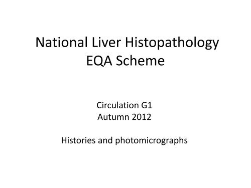
National Liver Histopathology EQA Scheme
here - Virtual Pathology at the University of Leeds
here - Virtual Pathology at the University of Leeds
- No tags were found...

You also want an ePaper? Increase the reach of your titles
YUMPU automatically turns print PDFs into web optimized ePapers that Google loves.
<strong>National</strong> <strong>Liver</strong> <strong>Histopathology</strong><br />
<strong>EQA</strong> <strong>Scheme</strong><br />
Circulation G1<br />
Autumn 2012<br />
Histories and photomicrographs
Case G1/398<br />
42 M<br />
Hepatitis C PCR +ve. Ex IVDU. For HCV<br />
treatment.<br />
1 core 17mm long (please also see VG and retic<br />
on website)
398
398
398
398
398
398
398
398
Case G1/399<br />
47 M<br />
Haemochromatosis ? Cirrhotic<br />
3 cores, 7, 3 and 2mm (please also see Perl’s,<br />
VG and retic on website)
399
399
399
399
399
Case G1/400<br />
32 F<br />
HBV Diagnosed 4 years ago, ? when acquired (African<br />
name). High viral PCR, Ag +ve, raised ALT. For<br />
staging.<br />
1 core 14mm long (photomics HbsAg and HbcAg;<br />
please also see retic and VG on website)
400
400
400
400
400
400
400<br />
HBsAg
400<br />
HBcsAg
Case G1/401<br />
37 M<br />
Deranged LFTs. High BMI. ?fatty liver disease<br />
One tan core of tissue 14mm in length. All<br />
embedded. (anticipated connective tissue<br />
stains on website)
401
401
401
401
401
401
401
Case G1/402<br />
68 F<br />
CT/US - heterogeneous liver echotexture ?HCC<br />
lesional biopsy<br />
Tan Core 20mm (anticipated connective tissue<br />
stain on website)
402
402
402
402
402
402
402
Case G1/403<br />
78 M<br />
Admitted with liver dysfunction, worsening<br />
bilirubin/AST/ALT. Aetiology unclear. US<br />
guided biopsy.<br />
1 core 17mm (no connective tissue stain<br />
available)
403
403
403
403
403
403
Case G1/404<br />
43 F<br />
HCV PCR +ve Alcohol excess. deranged LFTs<br />
1 core 15mm long (please also see retic and VG<br />
on website)
404
404
404
404
404
404
404
Case G1/405<br />
46 F<br />
Abnormal LFTs, positive ANF, 1:1000, raised IgG<br />
16mm core biopsy (photomic Shikata x10;<br />
negative for copper –associated protein; no<br />
other connective tissue stains available)
405
405
405
405
405
405
405
405
405<br />
Post treatment
Case G1/406<br />
72 F<br />
<strong>Liver</strong> Metastasis and colorectal primary segment VI.<br />
Stuck to diaphragm Gerota's fascia.<br />
Irregular liver wedge 140x60x45mm with portion of<br />
diaphragm. Necrotic pale nodule 45 x 32 x 40mm<br />
close to surgical margin and diaphragm, adjacent<br />
smaller nodule, block 5 from smaller nodule
406
406
406
406
406
Case G1/407<br />
62 M<br />
Cirrhotic <strong>Liver</strong>. HCC and 2-3 indeterminate lesions on<br />
CT. Ascites, banded varices, multiple lesions on<br />
MRI/CT felt consistent with HCC at HPB cancer MDM<br />
Vaguely nodular but not cirrhotic liver with numerous<br />
areas of vascular ectasia, 14 & 44mm lesions looking<br />
like sclerosed haemangiomas. No HCC like lesion
407
407
407
407
407
407
407
Case G1/408<br />
28 M<br />
Left hemihepatectomy for tumour<br />
Left lobe of liver measuring 150x140x80mm,<br />
with a circumscribed pale yellow tumour<br />
abutting the capsule. The background liver<br />
does not appear to be cirrhotic
408
408
408
408
408
Case G1/409<br />
30 F<br />
Non specific abdominal pain. Pedunculated<br />
tumour on imaging<br />
<strong>Liver</strong> tissue imaging 60x50x35mm composed of<br />
the entire lesion, uniform brown with nodular<br />
appearance containing a central white scar
409
409
409
409
409