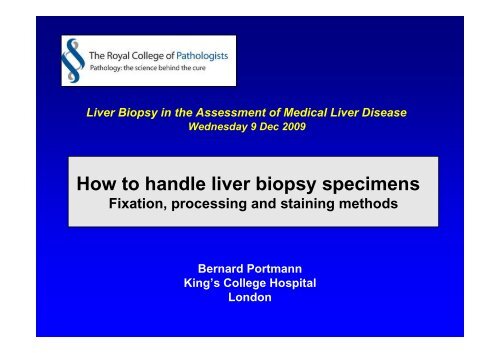
How to handle liver biopsy specimens
Prof B Portmann 11.20 - Virtual Pathology at the University of Leeds
Prof B Portmann 11.20 - Virtual Pathology at the University of Leeds
- No tags were found...
You also want an ePaper? Increase the reach of your titles
YUMPU automatically turns print PDFs into web optimized ePapers that Google loves.
Liver Biopsy in the Assessment of Medical Liver Disease<br />
Wednesday 9 Dec 2009<br />
<strong>How</strong> <strong>to</strong> <strong>handle</strong> <strong>liver</strong> <strong>biopsy</strong> <strong>specimens</strong><br />
Fixation, processing and staining methods<br />
Bernard Portmann<br />
King’s College Hospital<br />
London
Percutaneous <strong>liver</strong> <strong>biopsy</strong><br />
• Invasive technique<br />
• Risk of morbidity<br />
• Risk of mortality<br />
exceedingly low, but<br />
existent<br />
Essential not <strong>to</strong> loose<br />
information during<br />
handling / processing<br />
of these small <strong>specimens</strong>
Liver Biopsy – Processing / Interpretation<br />
Potential problems<br />
• Small specimen (1:50’000th of<br />
the <strong>liver</strong>)<br />
⇒ US guided specimen taken<br />
by radiologists = thinner / smaller<br />
• Fragmentation<br />
? artefact or fibrotic <strong>liver</strong><br />
• Sampling variation<br />
• Limited numbers of tissue responses<br />
<strong>to</strong> a broad range of injuries of different aetiologies
Liver Biopsy – Processing / Interpretation<br />
Importance of<br />
• High standard processing<br />
• Sensible use of available techniques<br />
• His<strong>to</strong>pathologist’s ‘expertise’<br />
• Clinical information<br />
• Interactive discussion between pathologist<br />
and clinician
Liver <strong>biopsy</strong><br />
Specimen collection / fixation<br />
• For conventional his<strong>to</strong>logy<br />
Prompt immersion in buffered formalin or<br />
formol saline<br />
• Special fixative for EM (Limited diagnostic<br />
value for <strong>liver</strong> disease)<br />
• Culture (PUO)<br />
• Fresh tissue on wet blotting paper mainly Cu<br />
[Fe] (use water for injection)<br />
• Snap frozen tissue : enzymology - metabolic<br />
disorder (specialized centres)
Handling the <strong>biopsy</strong> specimen<br />
Liver <strong>biopsy</strong> <strong>specimens</strong> are best<br />
left floating in the fixative solution<br />
No blotting paper<br />
Avoid rough packing<br />
between foam sponge<br />
Use fine mesh cassettes or<br />
lens-paper wrapping
Handling the <strong>biopsy</strong> specimen<br />
Essential <strong>to</strong> embed specimen flat, especially<br />
when dis<strong>to</strong>rted, in order <strong>to</strong> get most of the<br />
core(s) cut by each micro<strong>to</strong>me stroke<br />
Paraffin wax blocks<br />
Glass slides
Routine staining in our Labora<strong>to</strong>ry<br />
• 16 sequential sections are pair-mounted on 8<br />
slides numbered 1 <strong>to</strong> 8<br />
1, 8 = H & E (x 2)<br />
<br />
<br />
<br />
<br />
3 = Silver for reticulin<br />
5 = Perls’ for iron<br />
6 = Periodic acid Schiff after diastase<br />
7 = Orcein (modified Shikata’s)<br />
2, 4 = Held unstained<br />
Xxxxxxxxxxx<br />
Xxxxxxx<br />
xxxxxxxx
Routine slides x 8 (2 held unstained)<br />
3285/09<br />
H&E-1<br />
3285/09<br />
Perls’<br />
3285/09<br />
Reticulin<br />
3285/09<br />
Orcein<br />
3285/09<br />
dPAS<br />
3285/09<br />
H&E-2
Gordon-Sweets’ silver method for reticulin<br />
Normal<br />
Collagenisation<br />
Un<strong>to</strong>ned<br />
Un<strong>to</strong>ned<br />
Collapsed<br />
HV<br />
Un<strong>to</strong>ned<br />
Toned
NRH
Mild portal fibrosis<br />
Early septa<br />
Bridging septa<br />
Cirrhosis
Active<br />
Inactive
Periodic acid Schiff (PAS)<br />
• Cell loss / entrapped hepa<strong>to</strong>cytes<br />
in fibrous tissue<br />
• Negatively stained s<strong>to</strong>rage cells<br />
dPAS<br />
• Negative in glycogen s<strong>to</strong>rage disorders<br />
(highly soluble glycogen)<br />
No significant additional information<br />
Not performed routinely
PAS after diastase<br />
Performed routinely<br />
Bile duct basement membranes<br />
α1-antitrypsin globules<br />
Scavenger macrophages
Orcein staining: requirements<br />
• Optimal fixation and processing<br />
(Folds, scratches dark brown)<br />
• Adequate pre-staining oxidation<br />
(renew KMNO4 weekly)<br />
= same as that used for reticulin)<br />
• pH of orcein solution
Orcein<br />
• Hepatitis B surface antigen<br />
Homogeneous cy<strong>to</strong>plasmic inclusions<br />
• Low sensititvity ( ↓ in buffer formalin)<br />
• Does not detect membranous deposition<br />
• Elastic tissue<br />
• Age of areas of collapse → not very useful)<br />
• Vascular thrombi
Orcein<br />
Copper associated protein Dark brown granules<br />
• Chronic cholestasis (periportal / periseptal)<br />
• Wilson’s disease<br />
random ± Kupffer cells<br />
• Indian childhood cirrhosis / Cu associated<br />
neonatal disorders / Cu <strong>to</strong>xicosis<br />
• Physiological (infant <strong>liver</strong> up <strong>to</strong> 2 mos of age)<br />
• Cirrhosis of any aetiology (patchy)
Copper handling by hepa<strong>to</strong>cytes<br />
Caeruloplasmin<br />
A<strong>to</strong>x 1<br />
Trans-Golgi<br />
Membranes<br />
(WDP – ATP7B)<br />
CCS<br />
SOD (cy<strong>to</strong>sol)<br />
Cu(I)<br />
Ctr1<br />
Cox 17<br />
Cyt C oxydase<br />
Bile<br />
canaliculus<br />
Mi<strong>to</strong>chondrion<br />
Cu(II)-albumin<br />
in portal blood<br />
Cu-GSH<br />
Cu-Metallothionein<br />
Lysosome<br />
(Excess Cu /<br />
Orcein++)
Copper and copper associated protein<br />
Thionein<br />
s<br />
s<br />
s<br />
s<br />
Cu ++<br />
Cu ++ Rhodanine +<br />
Oxidation (KMnO 4 )<br />
Thionein<br />
SO 2<br />
H<br />
SO 2<br />
H<br />
SO 2<br />
H<br />
SO 2<br />
H<br />
Orcein +
Copper / Copper-associated protein<br />
Orcein<br />
Rhodanine<br />
Immediate processing<br />
After 3 wks in formol saline
Copper and copper associated protein<br />
• Copper easily removed by formal saline<br />
(especially if acidic)<br />
•Copper-binding protein unaffected by fixation<br />
• Early Wilson :<br />
- Cy<strong>to</strong>solic Cu highly soluble + <strong>to</strong>o small<br />
<strong>to</strong> be seen his<strong>to</strong>logically<br />
- Lysosomal Cu binding protein may not<br />
be significant yet<br />
⇒ negative staining does not exclude WD
Perls’ for iron<br />
Ferritin + haemosiderin<br />
are ferric compounds stained<br />
by the Perls’ technique<br />
• Ferritin: diffuse blue blush<br />
(DD artefact)<br />
• Haemosiderin: intense blue<br />
granules<br />
Parenchymal iron graded on a<br />
0 - 4+ scale<br />
1 = mild periportal/periseptal deposition<br />
4 = massive deposits (no acinar gradient)<br />
2 and 3 = intermediate<br />
Grade 1-2<br />
Grade 4
Iron overload<br />
(Liver siderosis / haemosiderosis)<br />
• Genetic haemochroma<strong>to</strong>sis<br />
HFE / non-HFE associated (juvenile, adult)<br />
Mutational analysis (C282Y +/+)<br />
Iron tissue estimation now rarely requested<br />
• Chronic anaemia (genetic, acquired)<br />
• Blood transfusions<br />
(Kupffer cells / macrophagic siderosis ++)
Immunohis<strong>to</strong>chemistry<br />
• Numerous commercial antibodies <strong>to</strong><br />
choose from<br />
against structural components,<br />
viral or secre<strong>to</strong>ry material<br />
• Variably useful for diagnosis or research<br />
• Balance between diagnostic need,<br />
interest and cost effectiveness<br />
• To be used sensibly given the small<br />
amount of tissue
Viral proteins<br />
• Hepatitis B (HBs – HBc)<br />
HDV (delta)<br />
• [Hepatitis C]<br />
• Cy<strong>to</strong>megalovirus (CMV)<br />
• Adenovirus<br />
• Herpes virus<br />
• EBV (ISH / EBER)<br />
• Cryp<strong>to</strong>sporidium
HBV<br />
Vertical transmission<br />
HBc tissue expression ++<br />
Minimal inflammation<br />
HBc<br />
HBs carrier<br />
HBV DNA -ve, ↑↑ HBs in serum and <strong>liver</strong><br />
HBc<br />
Immunocompromized host<br />
Active viral replication<br />
HBs
HCV anti-core region antibody<br />
Not useful for diagnosis<br />
or screening<br />
• Adequate results only in<br />
some immunocompromized<br />
hosts with high virus load<br />
in tissue<br />
HCV recurrence in <strong>liver</strong><br />
allograft with rapid<br />
progression <strong>to</strong> cirrhosis
Liver allograft<br />
CMV (microabscess)<br />
CMV early phase Ab<br />
Necrosis due <strong>to</strong><br />
adenovirus<br />
Adenovirus group Ab
Structural antigens<br />
• Cy<strong>to</strong>keratins 7,19 [AE1/AE3] = biliary epithelium<br />
• Cy<strong>to</strong>keratins 8, 18 / ‘Hepa<strong>to</strong>cyte-specific<br />
antigen’(HePar1) = hepa<strong>to</strong>cytes<br />
• CD68, HLA-DR = Kupffer cells<br />
• FVIII-associated Ag, CD34 = negative on normal<br />
sinusoidal endothelium<br />
• CD10, CD13, pCEA = canalicular regions<br />
Enzymes<br />
• Gamma-glutamyl-transpeptidase (γ GT)<br />
• Bile salt export protein (BSEP)<br />
• Multidrug resistance 3 (MDR3)
CK7<br />
Chronic cholestasis / CK7<br />
Chronic rejection<br />
Duc<strong>to</strong>paenia
Progressive familial intrahepatic cholestasis (PFIC2 – BSEP deficiency)<br />
γ-GT<br />
BSEP<br />
PFIC2<br />
Normal
Progressive familial intrahepatic cholestasis<br />
(PFIC3 – MDR3 deficiency)<br />
Morphology<br />
- Cholestasis<br />
- Progressive periportal fibrosis<br />
- Ductular reaction - Duc<strong>to</strong>paenia<br />
⇒ Biliary cirrhosis<br />
(adolescents / young adults)<br />
Imunostaining for MDR3<br />
• Infants / young children<br />
No staining in MDR3 deficiency<br />
• Adolescents/adults (late onset)<br />
Normal staining<br />
(?functional deficiency)<br />
MDR3 immuno (4 yr)<br />
MDR3 control
Biopsy taken <strong>to</strong> assess focal lesion<br />
• Best <strong>to</strong> evaluate firstly H&E sections<br />
• Spare sections on PLL coated slides<br />
held for immunohis<strong>to</strong>chemistry<br />
• Panel of antibodies <strong>to</strong> assess neoplasms does<br />
not differ from that used in any other organ<br />
• Due <strong>to</strong> scarcity of tissue more judicious<br />
selection recommended based on his<strong>to</strong>logical<br />
appearances + clinical guidance<br />
• Remember neuroendocrine neoplasms
Electron microscopy<br />
• Requires 1mm 3 pieces in glutaraldehyde<br />
• May contribute <strong>to</strong> diagnosis in<br />
some metabolic disorders<br />
- FIC1 disease (Byler)<br />
- Mi<strong>to</strong>chondriopathy, Wilson<br />
- S<strong>to</strong>rage disorders<br />
• Technique generally <strong>to</strong>o demanding for<br />
the diagnostic yield<br />
• Risk of removing information for his<strong>to</strong>logy<br />
assessment
Minute <strong>biopsy</strong> with diagnostic features<br />
Importance of careful processing + clinical information<br />
• Clinical information:<br />
? Chronic <strong>liver</strong> disease<br />
- Small nodular fragment<br />
lined by fibrosis<br />
- Dilated cholangioles<br />
with bile plugs<br />
• Differential diagnosis<br />
Cirrhotic nodule ? Biliary<br />
? Sepsis<br />
von Meyenburg complex /<br />
Congenital hepatic fibrosis
Additional clinical information<br />
– Portal hypertension / Haematemesis<br />
– No jaundice - Normal <strong>liver</strong> enzymes<br />
Diagnosis : Congenital hepatic fibrosis<br />
Subsequent confirma<strong>to</strong>ry<br />
<strong>biopsy</strong> specimen
<strong>How</strong> <strong>to</strong> <strong>handle</strong> needle <strong>liver</strong> <strong>biopsy</strong><br />
Invading technique - small specimen<br />
High standard processing <strong>to</strong> avoid lost of<br />
information<br />
Sensible use of available techniques<br />
especially immunohis<strong>to</strong>chemistry<br />
Seek second opinion for challenging cases<br />
(but limited assistance if poorly processed<br />
+ paraffin block empty)<br />
Importance of<br />
• Clinical awareness on the part of the pathologist<br />
• Clinical information given by clinicians<br />
• Interactive discussion between pathologist<br />
and clinician
King’s College Hospital<br />
Institute of<br />
Liver Studies


