th - 1988 - 51st ENC Conference
th - 1988 - 51st ENC Conference
th - 1988 - 51st ENC Conference

You also want an ePaper? Increase the reach of your titles
YUMPU automatically turns print PDFs into web optimized ePapers that Google loves.
29 <strong>th</strong> ���� - <strong>1988</strong> Rochester<br />
Chair: Stanley Opella<br />
Local Arrangements: Nick Zumbulyadis<br />
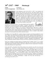
There was some concern about <strong>th</strong>e nor<strong>th</strong>ern location selected for<br />
<strong>th</strong>e 29<strong>th</strong> <strong>ENC</strong>. Fortunately, Rochester New York lived up to its<br />
reputation as <strong>th</strong>e Flower City by having unseasonably warm<br />
wea<strong>th</strong>er in April of <strong>1988</strong>. Notably, <strong>th</strong>is was <strong>th</strong>e first <strong>ENC</strong><br />
organized by Judi<strong>th</strong> Sjoberg and her Science Managers company,<br />
and <strong>th</strong>is relationship has had a profound effect on all subsequent<br />
<strong>ENC</strong>s by ensuring <strong>th</strong>at <strong>th</strong>e meeting arrangements are of <strong>th</strong>e same<br />
high quality as <strong>th</strong>e scientific presentations. Also, <strong>th</strong>is meeting led<br />
to <strong>th</strong>e <strong>ENC</strong> being included on <strong>th</strong>e New York Times list of key<br />
meetings on <strong>th</strong>e scientific speaker's circuit.<br />
Twenty years have passed since <strong>th</strong>e 29<strong>th</strong> <strong>ENC</strong>, and <strong>th</strong>is is <strong>th</strong>e first<br />
time <strong>th</strong>at I have looked at <strong>th</strong>e scientific program since <strong>th</strong>e meeting<br />
was held. I am struck by <strong>th</strong>e prescience of so many of <strong>th</strong>e presentations. There were entire sessions<br />
devoted to magic angle sample spinning, ordered biological systems, and dynamic nuclear<br />
polarization, in addition to <strong>th</strong>ose in <strong>th</strong>e more general areas of pulse sequence development,<br />
materials and biological imaging, and <strong>th</strong>ings <strong>th</strong>at now would be referred to as exotica.<br />
The magic angle sample spinning session introduced a number of advances <strong>th</strong>at have transformed<br />
<strong>th</strong>is field of research. It started wi<strong>th</strong> a talk on NMR strategies and high-speed MAS by Gary Maciel,<br />
which could be given today until you notice <strong>th</strong>at <strong>th</strong>e abstract mentions speeds “inching toward 30<br />
kHz.” The experimental NMR me<strong>th</strong>ods discussed in <strong>th</strong>e talks Measurements of two-dimensional<br />
NMR powder patterns in rotating solids (Takehiko Terao), 13 C- 15 N Rotational Echo Double<br />
Resonance (Jake Schaefer), and Rotational Resonance in solid state NMR (Bob Griffin) are still<br />
being refined and combined to provide <strong>th</strong>e pulse sequences applied in contemporary studies of<br />
polycrystalline proteins.<br />
In <strong>th</strong>e session on ordered biological systems, two of <strong>th</strong>e talks were particularly notable for where<br />
<strong>th</strong>ey have led. In <strong>th</strong>e talk Multinuclear experiments for <strong>th</strong>e determination of oligosaccharide<br />
structure in liquid crystal phases, Jim Prestegard described how orientational information could be<br />
obtained from weakly aligned biomolecules <strong>th</strong>rough measurements of what would become residual<br />
dipolar couplings, now an essential element of nearly all protein NMR studies in solution. And Tim<br />
Cross used his talk Dynamics of Gramicidin A transmembrane channel by solid state 15 N NMR to<br />
introduce <strong>th</strong>e interplay of structure and dynamics <strong>th</strong>at dominate current solid-state NMR studies of<br />
aligned membrane proteins.<br />
The Program for <strong>th</strong>e 29<strong>th</strong> <strong>ENC</strong> reflected <strong>th</strong>e input of <strong>th</strong>e NMR community and discussions and<br />
compromises among <strong>th</strong>e members of <strong>th</strong>e Executive Committee. At <strong>th</strong>e time of <strong>th</strong>e meeting, I<br />
<strong>th</strong>ought it went well, and <strong>th</strong>e participants I heard from were complimentary. I didn't reflect on <strong>th</strong>e<br />
quality of <strong>th</strong>e meeting during <strong>th</strong>e intervening twenty years. Now, my reaction is one of<br />
astonishment. The scientific presentations were so far ahead of <strong>th</strong>eir time, <strong>th</strong>at it took a while for<br />
<strong>th</strong>em to have <strong>th</strong>eir impact. The credit for <strong>th</strong>e success of <strong>th</strong>e 29<strong>th</strong> <strong>ENC</strong> belongs solely wi<strong>th</strong> <strong>th</strong>e<br />
practitioners of experimental NMR spectroscopy who showcased <strong>th</strong>eir ideas and results in<br />
Rochester.
. r<br />
<strong>ENC</strong>, I .<br />
29<strong>th</strong> Experimental Nuclear Magnetic Resonance Spectroscopy <strong>Conference</strong><br />
Rochester, New York, April 17-21, <strong>1988</strong><br />
<strong>Conference</strong> Office<br />
750 Audubon<br />
East Lansing, MI 48823<br />
(517) 332-3667<br />
Executive Committee<br />
Stanley J. Opella, Chair<br />
University of Pennsylvania<br />
Department of Chemistry<br />
Philadelphia, PA 19104<br />
(215) 898-6459<br />
A.N, Garroway, Chair-Elect<br />
Naval Research Laboratory<br />
Code 6120<br />
Washington, DC 30375<br />
(202) 757-2323<br />
N. Zumbulyadis, Local Arrangements<br />
Eastman Kodak Company<br />
Corporate Research Laboratories<br />
Rochester, NY 14650<br />
(716) 722-1409<br />
Edward O. Stejskal, Secretary<br />
Nor<strong>th</strong> Carolina State University<br />
Department of Chemistry<br />
Raleigh, NC 27695<br />
(919) 737-2998<br />
Mary W. Baum, Treasurer<br />
Princeton University<br />
Department of Chemistry<br />
Princeton, NJ 08544<br />
(609) 452-3892<br />
Lynne Batchelder<br />
Ad Bax<br />
Bernhard Bluemich<br />
Jo-Anne K. Bonesteel<br />
R. Andrew Byrd<br />
Paul W. Cope<br />
Colin Fyfe<br />
Myra Gordon<br />
Lynn Jelinski<br />
Gary E. Maciel<br />
Charles G. Wade<br />
Welcome to <strong>th</strong>e 29<strong>th</strong> <strong>ENC</strong>!<br />
It is a pleasure to welcome all participants to Rochester for <strong>th</strong>e 29<strong>th</strong><br />
<strong>ENC</strong>. Just as <strong>th</strong>e mon<strong>th</strong> of April invariably presages <strong>th</strong>e May bloom in<br />
Rochester, <strong>th</strong>e Flower City, <strong>th</strong>e fecund discussions at <strong>th</strong>e <strong>ENC</strong> always lead<br />
to new research in experimental NMR spectroscopy.<br />
The high quality and originality of <strong>th</strong>e abstracts for <strong>th</strong>e oral and poster<br />
presentations demonstrate <strong>th</strong>at <strong>th</strong>e <strong>ENC</strong> continues as <strong>th</strong>e premier forum for<br />
<strong>th</strong>e field of experimental NMR spectroscopy. These abstracts provide an<br />
instant snapshot of <strong>th</strong>is vigorous and dynamic field. The program is<br />
described in <strong>th</strong>is notebook wi<strong>th</strong> <strong>th</strong>e abstracts for <strong>th</strong>e oral presentations near<br />
<strong>th</strong>e schedule and <strong>th</strong>e poster abstracts in a separate section. All of <strong>th</strong>e talks will<br />
be given in <strong>th</strong>e Nor<strong>th</strong> Exhibition Hall. All posters should be set-up on<br />
Sunday in <strong>th</strong>e Lilac Ballroom of <strong>th</strong>e Convention Center and remain up<br />
<strong>th</strong>roughout <strong>th</strong>e meeting; <strong>th</strong>ey should be taken down after <strong>th</strong>e 5:30 pm closing<br />
of <strong>th</strong>e Wednesday afternoon poster session. The presenters of posters wi<strong>th</strong><br />
even numbered abstracts should be at <strong>th</strong>eir posters between 2:30 pm and<br />
5:30 pm on Monday and <strong>th</strong>e presenters wi<strong>th</strong> odd numbered abstracts between<br />
2:30 pm and 5:30 pm on Wednesday.<br />
The <strong>ENC</strong> also provides many opportunities to renew old friendships and<br />
to establish new ones. Please be sure to wear your registration badge during<br />
all scientific and social activities. There will be a Welcome Reception<br />
Sunday evening beginning at 7:00 pm in <strong>th</strong>e Convention Center Galleria.<br />
There are coffee breaks between morning sessions. Lunch will be served in<br />
<strong>th</strong>e Sou<strong>th</strong> Exhibition Hall. Consult <strong>th</strong>e enclosed restaurant guide for insight<br />
into <strong>th</strong>e local gourmet scene. All hospitality suites are located in <strong>th</strong>e<br />
Convention Center and Holiday Inn. They will close at 2:00 am in accord<br />
wi<strong>th</strong> local ordinances. The <strong>Conference</strong> Cocktail Party will be held between<br />
6:00 pm and 7:00 pm on Wednesday in <strong>th</strong>e Ballroom Foyer on <strong>th</strong>e second<br />
level of <strong>th</strong>e Rochester Plaza Hotel and is open to all participants.<br />
All comments and suggestions are welcome and will help A1 Garroway,<br />
Chair of <strong>th</strong>e 30<strong>th</strong> <strong>ENC</strong>, plan <strong>th</strong>e meeting for April 2-6, 1989 at <strong>th</strong>e Asilomar<br />
<strong>Conference</strong> Center in Pacific Grove, California.<br />
Please join me in participating in a successful 29<strong>th</strong> <strong>ENC</strong>,<br />
Chair, 29<strong>th</strong> <strong>ENC</strong>
PROGRAM: This conference program has divided<br />
lectures and posters into two sections. The abstracts<br />
of presentations appear in <strong>th</strong>e appropriate section. The<br />
au<strong>th</strong>or index is located in an additional section. The<br />
index references <strong>th</strong>e page number where <strong>th</strong>e abstract<br />
appears.<br />
Presentors of oral papers should arrive about 15<br />
minutes before <strong>th</strong>e session is scheduled to begin. If you<br />
are using slides, please give <strong>th</strong>em to <strong>th</strong>e projectionist<br />
before <strong>th</strong>e start of <strong>th</strong>e session.<br />
Posters have been numbered. If your poster is an even<br />
number, you must be present in <strong>th</strong>e poster session on<br />
Monday afternoon. If your poster is an odd number, you<br />
should be present at <strong>th</strong>e Wednesday session.<br />
Posters should be mounted on Sunday evening. All<br />
poster spaces have been numbered. Please be sure<br />
to mount your poster in <strong>th</strong>e space <strong>th</strong>at corresponds to<br />
your poster number in <strong>th</strong>e program.<br />
LOCATION OF SESSIONS: Oral sessions are in <strong>th</strong>e<br />
Nor<strong>th</strong> Exhibit Hall. Poster sessions are in <strong>th</strong>e Lilac<br />
Ballroom.<br />
EMPLOYMENT CENTER: The employment center will<br />
maintain a file of resumes. If you wish to register, please<br />
come to <strong>th</strong>e center on Monday morning. Notices of<br />
employment positions may be placed on <strong>th</strong>e bulletin<br />
board designated for <strong>th</strong>at purpose.<br />
REGULATIONS: The following regulations are in <strong>th</strong>e<br />
best interests of <strong>th</strong>e conference:<br />
. No smoking in any session, including <strong>th</strong>e poster<br />
sessions.<br />
. Registration badges must be worn to all con-<br />
ference activities, including hospitality suites, <strong>th</strong>e<br />
welcome reception, and <strong>th</strong>e Wednesday cocktail<br />
party.<br />
The 29<strong>th</strong> <strong>ENC</strong><br />
Rochester, New York<br />
April 17-21, <strong>1988</strong><br />
. Your cooperation is requested in closing hospitali-<br />
ty suites at 2:00 a.m. in compliance wi<strong>th</strong><br />
Rochester liquor laws.<br />
. The opening of hospitality suites should not<br />
conflict wi<strong>th</strong> conference sessions.<br />
HOSPITALITY SUITES: Hospitality suites are located<br />
in <strong>th</strong>e Holiday Inn, as well as <strong>th</strong>e Convention Center.<br />
The suites in <strong>th</strong>e Holiday Inn are on <strong>th</strong>e mezzanine level<br />
(same level as <strong>th</strong>e skywalk) and in parlors on <strong>th</strong>e upper<br />
floors.<br />
CONFER<strong>ENC</strong>E REFRESHMENTS: Coffee Breaks will<br />
be available on Monday, Tuesday, Wednesday and<br />
Thursday mornings in <strong>th</strong>e GaUeria of <strong>th</strong>e Convention<br />
Center at <strong>th</strong>e times indicated in <strong>th</strong>e program.<br />
There will also be refreshments served during <strong>th</strong>e poster<br />
sessions on Monday and Wednesday afternoons.<br />
LUNCH: A cafeteria-style lunch may be purchased in<br />
<strong>th</strong>e Sou<strong>th</strong> Exhibit Hall. Wi<strong>th</strong> <strong>th</strong>e number of people<br />
expected to use <strong>th</strong>is service, long lines at <strong>th</strong>e beginning<br />
are inevitable. However, <strong>th</strong>e crowd will be served as<br />
efficiently as possible. Please be patient.<br />
If you prefer to go out for lunch, a restaurant guide is<br />
located in <strong>th</strong>is program.<br />
NIAGARA FALLS EXCURSION: The Niagara Falls<br />
excursion buses will load at <strong>th</strong>e (~onvention center main<br />
entrance between 12:15 and 12:30 p.m. Lunch will be<br />
served on board <strong>th</strong>e buses. Non-U.S. citizens must bring<br />
a passport or appropriate papers for Canadian customs.<br />
The buses will return at approximately 6:30 p.m.<br />
COCKTAIL PARTY: The cocktail party hosted by<br />
Varian Associates will be in <strong>th</strong>e Rochester Plaza Hotel<br />
(across <strong>th</strong>e river). It will be in <strong>th</strong>e ballroom foyer, second<br />
level, between 6 and 7:00 p.m.<br />
2
The Executive Committee of <strong>th</strong>e 29<strong>th</strong> <strong>ENC</strong><br />
gratefully acknowledges <strong>th</strong>e financial support for<br />
<strong>th</strong>e conference from <strong>th</strong>e following organizations:<br />
Academic Press<br />
Aldrich Chemical<br />
Amplifier Research<br />
Bruker<br />
Cambridge Isotope Laboratories<br />
Chemagnetics<br />
Chemical Dynamics<br />
Creative Electronics<br />
Dory Scientific<br />
Drusch<br />
Eastern Analytical Symposium<br />
Electronic Navigation Industries<br />
GE NMR instruments<br />
IBM - Almaden Research Center<br />
ICN<br />
ICON Services<br />
Isotec<br />
JEOL<br />
Eastman Kodak<br />
Merck Sharp and Dohme Isotopes<br />
M-R Resources<br />
Nalorac Cryogenics<br />
New Era Enterprises<br />
New Me<strong>th</strong>ods Research<br />
Norell<br />
Oxford Instruments<br />
Pergamon Journals<br />
Phospho-Energetics<br />
Sciteq<br />
STN International<br />
Siemens Medical Systems<br />
Spectral Data Services<br />
Spectroscopy Imaging Systems<br />
Stevens Creek Software<br />
Tecmag<br />
Varlan Associates<br />
Wilmad Glass<br />
John Wiley & Sons<br />
Xerox
The Executive Committee of <strong>th</strong>e 29<strong>th</strong> <strong>ENC</strong><br />
gratefully acknowledges financial support for<br />
underwriting <strong>th</strong>e following:<br />
Isotec<br />
Program binders<br />
Programmed Test Sources<br />
Tote bags<br />
Amplifier Research and Programmed Test Sources<br />
Welcome reception<br />
Stanley J. Opella, Chair<br />
University of Pennsylvania<br />
Department of Chemistry<br />
Philadelphia, PA 19104<br />
(215) 898-6459<br />
A.N. Garroway, Chair-Elect<br />
Naval Research Laboratory<br />
Code 6120<br />
Washington, DC 20375<br />
(202) 747-2323<br />
N. Zumbulyadis, Local Arrangements<br />
Eastman Kodak Company<br />
Corporate Research Laboratories<br />
Rochester, NY 14650<br />
(716) 722-1409<br />
Edward O. Stejskal, Secretary<br />
Nor<strong>th</strong> Carolina State University<br />
Department of Chemistry<br />
Raleigh, NC 27695<br />
(919) 737-2998<br />
Bruker Instruments<br />
Coffee breaks<br />
GE NMR Instruments<br />
Monday Poster refreshments<br />
JEOL<br />
Wednesday Poster refreshments<br />
Varian Associates<br />
Wednesday cocktail party<br />
EXECUTIVE COMMITTEE<br />
4<br />
Mary Wo Baum, Treasurer<br />
Princeton University<br />
Department of Chemistry<br />
Princeton, NJ 08544<br />
(609) 452-3892<br />
Lynne Batchelder<br />
Ad Bax<br />
Bernhard Bluemich<br />
Jo-Anne K. Bonesteel<br />
R. Andrew Byrd<br />
Paul W. Cope<br />
Colin Fyfe<br />
Myra Gordon<br />
Lynn Jelinski<br />
Gary E. Maciel<br />
Charles G. Wade
101<br />
101<br />
101<br />
101<br />
101<br />
101<br />
101<br />
VENDOR SUITE LOCATIONS<br />
101 J<br />
101 K<br />
102 A<br />
102<br />
102<br />
102<br />
103<br />
103<br />
103<br />
103<br />
A<br />
B<br />
C<br />
D<br />
E<br />
F<br />
G<br />
C<br />
D<br />
E &F<br />
A<br />
B<br />
C<br />
D<br />
&H<br />
&B<br />
Exchange<br />
Fairfax<br />
Genessee<br />
Huron<br />
Tiffany<br />
1425<br />
1500<br />
1200<br />
925<br />
CONVENTION CENTER<br />
Spectroscopy Imaging Systems<br />
Chemagnetics<br />
GE NMR Instruments<br />
Doty Scientific<br />
Bruker Instruments<br />
Electronic Navigation Industries<br />
New Me<strong>th</strong>ods Research<br />
M-R Resources<br />
Varian Associates<br />
JEOL, USAJLtd<br />
Intermagnetics General<br />
Tecmag<br />
Bruker Instruments<br />
ICN Stable Isotopes<br />
Chemical Dynamics<br />
Sciteq<br />
Phospho-Energetics<br />
HOLIDAY INN MEZZANINE<br />
ICON Services<br />
Cambridge Isotope Laboratories<br />
Programmed Test Sources<br />
Amplifier Research<br />
Merck Sharp & Dohme<br />
Isotec<br />
HOLIDAY INN SUITES<br />
Nalorac<br />
Oxford Instruments<br />
Wilmad Glass<br />
STN International
A<br />
A<br />
0<br />
0<br />
,-..1<br />
I<br />
Z<br />
Z<br />
0<br />
"c<br />
N<br />
"1"<br />
i<br />
0<br />
,.I<br />
Q.<br />
Q.<br />
....... :" " " J U J '.a~<br />
Ji~ E!-~ .....<br />
~--J: - El-i .<br />
J ~ F1<br />
t<br />
. . . .<br />
....... .1~- ]:-' ~ °'<br />
,<br />
L~<br />
>-,<br />
rd~<br />
X<br />
I~o .,.-i<br />
c~ r.z.,<br />
u<br />
X<br />
LT4
i<br />
L<br />
3~,.ols I 0',<br />
I<br />
===~ ~ F',~ R ', : tl - I!<br />
U ' II<br />
I 4 U I<br />
n , H ~, ~/.., < ++ R+,<br />
",l u", n LlOS ~-olbm~ ~i~-niP',<br />
°~ L<br />
0<br />
0<br />
Z<br />
rr u<br />
c/]<br />
0<br />
0<br />
~9<br />
0<br />
E<br />
I- ,,,=I l.,,m- ,<br />
°mlll<br />
= Ii<br />
=0 0 0<br />
30vN3nOl~<br />
o o<br />
Z<br />
u~<br />
0,-~<br />
I---I<br />
~0<br />
0<br />
u<br />
0<br />
0<br />
0 0<br />
---r ~-+-T '+++ "<br />
£33~£$ tlv~<br />
Z<br />
]oo<br />
u-I<br />
0~0 u~<br />
u-I ~x4<br />
o i c'm.,~F<br />
w<br />
0<br />
r._)<br />
----I<br />
I
Restaurants in Rochester<br />
Restaurants close to <strong>th</strong>e Convention Center are located in some unlikely<br />
places. This selection is intended to help you find <strong>th</strong>em. No claims are made as<br />
to completeness, and you may very well find a restaurant you like <strong>th</strong>at is not on<br />
<strong>th</strong>e list. Wi<strong>th</strong>in about a mile of <strong>th</strong>e Convention Center <strong>th</strong>ere are many more<br />
eating places <strong>th</strong>an <strong>th</strong>ere are in <strong>th</strong>e immediate downtown area. Wi<strong>th</strong> a car or<br />
some extra time you may want to try some of <strong>th</strong>e outlying places mentioned at <strong>th</strong>e<br />
end of <strong>th</strong>e list.<br />
First for <strong>th</strong>e more exoensive olaces. In Rochester <strong>th</strong>is means about $25 for<br />
a full meal. exceot where noted.<br />
The first revolving restaurant in New York, The Changing Scene, is<br />
located on <strong>th</strong>e top floor of First Federal Plaza, <strong>th</strong>e tall building next to <strong>th</strong>e<br />
Rochester Plaza.<br />
Chapel's is located in <strong>th</strong>e former City Hall, a renovated historic setting.<br />
is very good, but in <strong>th</strong>e evening you will spend at least $40 per person, and <strong>th</strong>e<br />
meal usually spans several hours.<br />
Edwards at 13 S. Fitzhugh is just off Main street, several blocks west of<br />
<strong>th</strong>e river. It is one of <strong>th</strong>e well-established, quality restaurants in Rochester, but is<br />
also likely to be more expensive <strong>th</strong>an most.<br />
Joseph's is a good Italian restaurant located at 169 N. Chestnut, about a<br />
block from <strong>th</strong>e Eastman Theater. The prices are well wi<strong>th</strong>in reason.<br />
The Riverview Cafe in <strong>th</strong>e Rochester Plaza is also a good choice.<br />
Meals in <strong>th</strong>e moderate ranae should run about $15.<br />
An excellent oriental restaurant is <strong>th</strong>e Bangkok, 155 State Street, across<br />
from <strong>th</strong>e Rochester Plaza. It has bo<strong>th</strong> a Chinese and a Thai menu, and will give<br />
you a 5% discount on evening meals if you show your registration card. Lunch is<br />
at <strong>th</strong>e menu price.<br />
Beams Restaurant, 106 Andrews Street at <strong>th</strong>e corner of St. Paul one<br />
block nor<strong>th</strong> of <strong>th</strong>e Holiday Inn is pleasant. Some of <strong>th</strong>eir menu items have a<br />
"heal<strong>th</strong>-food" note.<br />
A new restaurant <strong>th</strong>at comes wi<strong>th</strong> good recommendations is The Filling<br />
Station. The building formerly was just what it says. It is located at 30 Mount<br />
Hope Ave., just across <strong>th</strong>e Inner Loop.<br />
Trebor's, on State Street across from <strong>th</strong>e Rochester Plaza, is popular<br />
among <strong>th</strong>e business crowd at lunch.<br />
It
Gellert's, next door to <strong>th</strong>e Eastman Theater on Gibb's Street has a<br />
simple, but pleasant atmosphere. It caters in large part to <strong>th</strong>e <strong>th</strong>eater crowd.<br />
Sibley's, one of <strong>th</strong>e downtown department stores, has a surprisingly<br />
good restaurant on its top floor, as well as snack bars and a pastry shop on <strong>th</strong>e<br />
ground floor. In McCurdy's are <strong>th</strong>e Garden Room and Oak Room.<br />
If you have a reallv tiaht budoet, <strong>th</strong>ere are also some choices.<br />
There are a couple of soup and sandwich places in Midtown Plaza,<br />
which used to be several downtown city blocks and now is a completely<br />
enclosed shopping mall located at Main and Clinton, two blocks east of <strong>th</strong>e<br />
Convention Center. Try <strong>th</strong>e Great Canadian Soup Company. There is also<br />
a Burger King in <strong>th</strong>is complex. The Top of <strong>th</strong>e Plaza (take <strong>th</strong>e elevator)<br />
does not fall in <strong>th</strong>e inexpensive class, but is reputed to have good hamburgers for<br />
lunch and would be good for a more expensive evening meal..<br />
There are McDonalds on Main Street, about one block east of <strong>th</strong>e<br />
Convention Center, and on State Street, across <strong>th</strong>e street from <strong>th</strong>e Rochester<br />
Plaza.<br />
Sweet Dawn's, at <strong>th</strong>e corner of Main Street and Stone, is good for soup<br />
and sandwiches.<br />
Across State Street from <strong>th</strong>e Kodak Office building is Rubino's, selling<br />
submarine sandwiches and <strong>th</strong>e associated items. Rubino's also runs <strong>th</strong>e<br />
Salumeria Care in Midtown Plaza.<br />
If you want to aet a little fur<strong>th</strong>er from <strong>th</strong>e Convention Center, look on<br />
Alexander Street, whi-ch crosses East Avenue on <strong>th</strong>e o<strong>th</strong>er Side of <strong>th</strong>e Inner Loop,<br />
on Park Avenue, which crosses Alexander about a block sou<strong>th</strong> of East Avenue,<br />
or on Monroe Avenue, which crosses Alexander still fur<strong>th</strong>er Sou<strong>th</strong>.. Some of <strong>th</strong>e<br />
restaurants on Alexander are ra<strong>th</strong>er expensive. The Park Avenue area is a bit<br />
"yuppie" and spills over onto Monroe. The best restaurant bargains are probably<br />
on Monroe. Several different e<strong>th</strong>nic groups are represented.<br />
If YOU have a car. <strong>th</strong>ere is a wide choice of restaurants. You might want to<br />
try <strong>th</strong>e Spring House, 3001 Monroe Avenue (take <strong>th</strong>e freeway), an inn <strong>th</strong>at<br />
originally was located next to <strong>th</strong>e Erie Canal. (The canal moved; <strong>th</strong>e restaurant is<br />
where it always was, but if you look closely off to <strong>th</strong>e side behind <strong>th</strong>e building you<br />
can see a trace of <strong>th</strong>e old ditch.) On <strong>th</strong>e present-day canal, but much fur<strong>th</strong>er out,<br />
is Richardson's Canal House, 1474 March Road, <strong>th</strong>e only restaurant in<br />
Rochester listed in <strong>th</strong>e Mobil guide.<br />
9
8:30 a.m.<br />
8:35- 10:05<br />
10:05 - 10:25<br />
10:25- 12:15<br />
12:15 p.m.<br />
2:30 - 5:30<br />
7:30 - 9:30<br />
8:30 - 10:20 a.m.<br />
10:20 - 10:40<br />
10:40- 12:10<br />
12:10 p.m.<br />
12:15- 12:30<br />
7:30 - 9:30<br />
MONDAY, APRIL 18, <strong>1988</strong><br />
PROGRAM<br />
Opening remarks, S. J. Opella, Chair.<br />
Low Temperatures end Fields.<br />
G. Maciel, Session Chair.<br />
Nor<strong>th</strong> Exhibit Hall.<br />
Coffee Break.<br />
Convention Center Gaileria.<br />
Compliments of Bruker Instruments.<br />
Magic Angle Sample Spinning.<br />
E. Stejskal, Session Chair.<br />
Nor<strong>th</strong> Exhibit Hall.<br />
Lunch. A cafeteria style lunch may be purchased in <strong>th</strong>e Sou<strong>th</strong> Exhibit Hall.<br />
Poster Session.<br />
Lilac Ballroom.<br />
Au<strong>th</strong>ors of even numbered posters present.<br />
Refreshments compliments of GE NMR Instruments.<br />
Detection and Analysis.<br />
M. Baum, Session Chair.<br />
Nor<strong>th</strong> Exhibit Hall.<br />
TUESDAY, APRIL 19, <strong>1988</strong><br />
PROGRAM<br />
Two-Dimensional Spectroscopy.<br />
C. Wade, Session Chair.<br />
Nor<strong>th</strong> Exhibit Hall.<br />
Coffee Break.<br />
Convention Center Galleria.<br />
Selective Pulse Sequences.<br />
A. Bax, Session Chair.<br />
Lunch. A cafeteria-style lunch may be purchased in <strong>th</strong>e Sou<strong>th</strong> Exhibit Hall.<br />
Niagara Falls Excursion buses depart.<br />
Lunch will be served on <strong>th</strong>e buses.<br />
Afternoon free! Don't miss <strong>th</strong>e George Eastman House Containing <strong>th</strong>e International<br />
Museum of Photography, or walk along <strong>th</strong>e river to view <strong>th</strong>e Falls.<br />
High Tc Superconductors.<br />
L. Jelinski, Session Chair.<br />
Nor<strong>th</strong> Exhibit Hall.<br />
11
8:30 - 10:10 a.m.<br />
10:10- 10:30<br />
10:30- 12:10<br />
12:10 p.m.<br />
2:30 - 5:30<br />
6:00 - 7:00<br />
7:00 p.m.<br />
8:30 - 10:05 a.m.<br />
10:05- 10:25<br />
10:25 o 12:05<br />
WEDNESDAY, APRIL 20, <strong>1988</strong><br />
PROGRAM<br />
Materials Imaging.<br />
A. Garroway, Session Chair.<br />
Nor<strong>th</strong> Exhibit Hall.<br />
Coffee Break.<br />
Convention Center Galleria.<br />
Biological Imaging.<br />
R. A. Byrd, Session Chair.<br />
Nor<strong>th</strong> Exhibit Hall.<br />
Lunch. A cafeteria-style lunch may be purchased in <strong>th</strong>e Sou<strong>th</strong> Exhibit Hall.<br />
Poster Session.<br />
Au<strong>th</strong>ors of odd numbered posters present.<br />
Refreshments compliments of JEOL.<br />
Cocktail party.<br />
Rochester Plaza Hotel, Ballroom Foyer.<br />
Compliments of Varian Associates.<br />
Open to all conference registrants.<br />
<strong>Conference</strong> Banquet.<br />
The Future of Conventional and Electronic Imaging;<br />
Robert Hunt, University of London.<br />
Rochester Plaza Ballroom.<br />
Tickets required.<br />
THURSDAY, APRIL 21, <strong>1988</strong><br />
PROGRAM<br />
Ordered Biological Systems.<br />
L. Batchelder, Session Chair.<br />
Nor<strong>th</strong> Exhibit Hall.<br />
Coffee Break.<br />
Convention Center Galleria.<br />
Dynamic Nuclear Polarization.<br />
N. Zumbulyadis, Session Chair.<br />
Nor<strong>th</strong> Exhibit Hall.<br />
<strong>Conference</strong> Adjourned.<br />
See you next year at Asilomar, April 2-6, 1989.<br />
12
8:30 a.m.<br />
8:35 a.m.<br />
9:05 a.m.<br />
9:15 a.m.<br />
9:25 a.m.<br />
9:35 a.m.<br />
10:05 a.m.<br />
MONDAY MORNING<br />
LOW TEMPERATURES AND FIELDS<br />
G. Maciel, Session Chair<br />
Opening remarks, S. J. Opella, Chair.<br />
T 1 Mechanisms of Solids Immersed in 3He.<br />
O. Gonen, P. L. Kuhns, *J. S. Waugh.<br />
Untruncation of Dipole-Dipole Couplings in Solids,<br />
or Zero Field NMR Entirely in High Field.<br />
R. Tycko.<br />
Interpretation of <strong>th</strong>e NMR Nutation Spectra.<br />
*A. Samoson, E. Lippmaa.<br />
High Resolution Electrophoretic NMR (ENMR) of a Mixture.<br />
*T. R. Saarinen, C. S. Johnson.<br />
Collaborative Projects in NMR.<br />
A. Pines.<br />
Break.<br />
13
cON 8:35 ]<br />
It.<br />
T 1 MECHANISMS OF SOLIDS IMMERSED IN 3He<br />
O. Gonen, P.L. Kuhns, and J.S. Waugh*, MIT, Cambridge, MA. 02139<br />
Spins (I) at <strong>th</strong>e surface of a solid can be relaxed by neighboring 3He<br />
spins. Thereafter <strong>th</strong>e interior of <strong>th</strong>e solid is relaxed <strong>th</strong>rough spin diffusion.<br />
ForQ|gn (S) spins in <strong>th</strong>e surface layer inhibit <strong>th</strong>e escape of I-magnetization<br />
to <strong>th</strong>e interior, and S spins distributed <strong>th</strong>roughout <strong>th</strong>e bulk may inhibit spin<br />
diffusion altoge<strong>th</strong>er. A variety of 1H, 2H, and 29Si measurements in Si02<br />
i11ustrate and quantify <strong>th</strong>ese effects. Examples will be presented of selective<br />
spectroscopy of adsorbed species in sub-monolayer coverage.<br />
14
f~<br />
I UNTRUNCATIO~I OF DIPOLE-DIPOLE COUPLINGS IN SOLIDS, OR ZERO FIELD<br />
MON 9:0S I NMR ENTIRELY IN HIGH FIELD. Robert Tycko, AT&T Bell Laboratories,<br />
Murray Hill, Nj, 07974.<br />
N::R spectra of powdered or noncrystalline solids in high field commonly exhibit broad<br />
lines <strong>th</strong>at arise from <strong>th</strong>e dependence of <strong>th</strong>e nuclear magnetic dipole-dipole couplings<br />
on molecular orientation. New experiments will be described in which <strong>th</strong>at orientation<br />
dependence is removed by <strong>th</strong>e combination of rapid sample rotation wi<strong>th</strong> a synchronized<br />
rf pulse sequence.• The sample rotation and pulse sequence have <strong>th</strong>e effect of con-<br />
verting <strong>th</strong>e usual truncated dipole-dipole couplings into an untruncated form. The<br />
result is N,~IR spectra wi<strong>th</strong> sharp lines and splittings <strong>th</strong>at depend only on inter-<br />
nuclear distances, i.e. spectra wi<strong>th</strong> a "zero field" appearance <strong>th</strong>at are obtained<br />
entirely in high field. Such spectra provide a means of studying molecular conforma-<br />
tions in solids, wi<strong>th</strong>out requiring single crystals. The <strong>th</strong>eory behind untruncation<br />
experiments will be presented along wi<strong>th</strong> experimental spectra of simple organic<br />
solids.<br />
15
[--~N 9:15<br />
INTERPRETATION OF THE NMR NUTATION SPECTRA. A. Samoson * and E.<br />
I Lippmaa, Institute of Chemical Physics and Biophysics, Estonian<br />
Academy of Sciences, 200001Tallinn, USSR.<br />
The quadrupole interaction parameters of half integer spin nuclei are<br />
accessible from <strong>th</strong>e dependence of NMR central transition signal on <strong>th</strong>e rf excitation<br />
pulse leng<strong>th</strong>. The Fourier analysis yields (nutatlon) spectra, consisting at most<br />
of 21 major lines. The lines can be associated wi<strong>th</strong> single quantum coherences in a<br />
rotating magnetic field created by <strong>th</strong>e rf pulse. The magnetization vectors<br />
describing spin evolution in <strong>th</strong>e rotating magnetic field nutate in different senses,<br />
depending on <strong>th</strong>e quantum numbers of respective energy levels. This provides for<br />
fur<strong>th</strong>er unravelling of 2D spectra via hypercomplex Fourier transform. The ratio of<br />
a first moment to integral intensity of <strong>th</strong>e nutation spectra gives a good estimate<br />
for <strong>th</strong>e quadrupole interaction constant. The nutation spectroscopy applied to <strong>th</strong>e<br />
study of zeolites, glasses and organic conductors provided for identification of<br />
various nuclear sites and interpretation of complicated ID spectra.<br />
Current address: Department of Chemistry, University of California, Berkeley,<br />
CA 94720.<br />
16
MON 9:25<br />
HIGH RESOLUTION ELECTROPHORETIC ~ (ENMR) OF A MIXTURE:<br />
Timo<strong>th</strong>y R. Saarlnen' and~les S. Johnson, Jr., University of Nor<strong>th</strong><br />
Carolina, Dept. of Chem., Chapel Hill, NC 27599-3290<br />
Electrophoretic mobilities have been measured in situ using<br />
pulsed field gradient NMR (PFGNMR). Several components in a mixture<br />
can be studied simultaneously by Fourier transformation of <strong>th</strong>e second<br />
half of <strong>th</strong>e spin echo. For a U-tube configuration application of an<br />
electric field across <strong>th</strong>e sample resu/ts ~n a cosinusoidal modulation<br />
of spectral peak amplitudes, cos(Kv= t) where K equals <strong>th</strong>e area of <strong>th</strong>e<br />
gradient pulse times <strong>th</strong>e gyrcmagnetic ratio, v: is <strong>th</strong>e drift velocity<br />
of <strong>th</strong>e i'<strong>th</strong> species, and t is <strong>th</strong>e duration of <strong>th</strong>e electric field<br />
pttlse. By working at low ionic streng<strong>th</strong>s electric fields of up to 50<br />
V/cm could be applied for i sec before convection ~as detected by a<br />
change in <strong>th</strong>e amplitude of <strong>th</strong>e HOD peak. The cationic mobilities in<br />
a mixture of tetra-me<strong>th</strong>yl and tetra-e<strong>th</strong>yl anmonJum chloride in D20<br />
were determined. Application of <strong>th</strong>e technique for studying emulsions<br />
looks prc~lising.<br />
17
IION 9 : 35 [<br />
COLLABORATIVE PROJECTS IN NMR: A. Pines, University of Califomia,<br />
Berkeley, CA 94720<br />
Topics for discussion will be selected from among <strong>th</strong>e following:<br />
1. Quantum phase of <strong>th</strong>e photon and its relevance to magnetic resonance.<br />
2. Multiple-pulse NMR in zero field.<br />
3. Zero field NMR studies of small local asymmetries.<br />
4. 2-Dimensional studies of molecular conformations in liquid crystals.<br />
5. Clustering of molecules in zeolites studied by Xenon and multiple-quantum NMR.<br />
6. Detection of quadrupole resonance by a SQUID detector at low temperature.<br />
7. Iterative spin decoupling schemes for solids.<br />
8. High-temperature NMR of silicate glasses and liquids.<br />
Some of <strong>th</strong>ese projects are carried out in collaboration wi<strong>th</strong> M.V. Berry<br />
(Physics, Bristol); J. Clarke (Physics, Berkeley); J. Fraissard (Chemistry, Paris); J.<br />
Guckenheimer (Ma<strong>th</strong>ematics, Cornell); C.J. Radke (Chemical Engineering, Berkeley)<br />
and J. Stebbins (Geology, Stanford).<br />
18
• . r,<br />
10:25 a.m.<br />
10:55 a.m.<br />
11:05 a.m.<br />
11:35 a.m.<br />
11:45 a.m.<br />
12:15 p.m.<br />
MONDAY MORNING<br />
MAGIC ANGLE SAMPLE SPINNING<br />
E. Stejskal, Session Chair<br />
NMR Strategies and High-Speed MAS.<br />
*G. E. Maciel, C. E. Bronnimann,<br />
S. F. Dec, B. L. Hawkins, R. A. Wind.<br />
Measurements of Two-Dimensional NMR Powder Patterns in<br />
Rotating Solids.<br />
T. Nakai, J. Ashida, *T. Terao.<br />
13C-lSN Rotational Echo Double Resonance.<br />
T. Gullion, *J. Schaefer.<br />
2D Chemical Shift Anisotropy Correlation Spectroscopy.<br />
A new Sample Positioning Mechanism Which Simplifies<br />
Measurement of Chemical Shift Anisotropies in Complex<br />
Single Crystals.<br />
M. H. Sherwood, *D. W. Alderman,<br />
D. M. Grant.<br />
Rotational Resonance in Solid State NMR.<br />
D. P. Raleigh, M. H. Levitt,<br />
F. Creuzet, *R. G. Griffin.<br />
Lunch.<br />
19
MON i0:25<br />
NMR STRATEGI'ES AND HIGH-SPEED MAS, G.E. Maciel * C.E. Bronnimann<br />
, 9<br />
S.F.Dec, B.L. Hawkins and R.A. Wind, Department of Chemistry,<br />
Colorado State Univesity, Ft. Collins, CO 80523<br />
Wi<strong>th</strong> MAS speeds inching toward 30 KHz, a variety of important possibilities<br />
and issues arise in solid-state NMR. One of <strong>th</strong>e most direct benefits of high-speed<br />
MAS is <strong>th</strong>e ability to reduce spinning sidebands and r~e~move <strong>th</strong>em from spectral<br />
regions of interest. This is especially beneficial in ~'Al NMR, allowing <strong>th</strong>e use<br />
of high fields wi<strong>th</strong> <strong>th</strong>e corresponding reduction in <strong>th</strong>e second-order quadrupole<br />
effect. Examples will be shown.<br />
The temptation to employ high-speed MAS as a high-resolution ~oli~-state IH<br />
NMR technique must be considered in relation to <strong>th</strong>e nature of <strong>th</strong>e~H-~H dipolar<br />
interactions in each sla~_P1~e ~ Direct comparisons of CRAMPS and MAS only r,~ults<br />
show <strong>th</strong>at when strong ipolar interactions are present, as expected, <strong>th</strong>e<br />
CRAMPS approach provides far superior results.<br />
The anticipated interference of high-speed MAS wi<strong>th</strong> CP efficiency i ~H r ~ dily<br />
demonstrated, even in systems wi<strong>th</strong> strong dipolar interactions. In - C CP<br />
experiments carried out as a function of MAS speed, <strong>th</strong>e Hartmann-Hahn match curves<br />
differ dramatically for carbons wi<strong>th</strong> directly attached hydrogens relative to carbons<br />
wi<strong>th</strong>out. Hence, <strong>th</strong>e use of high-speed MAS to overcome spinning sideband<br />
problems in high-field CP-MAS experiments seems problematical. One potential<br />
approach to <strong>th</strong>is kind Of problem may be stop-and-go (STAG) spinning, in which CP<br />
occurs during a static-sample period in <strong>th</strong>e STAG-sequence. Some STAG results will<br />
be shown.<br />
20
~'-O-i 0 N i0:55<br />
MEASUREMENTS OF TWO-DIMENSIONAL NMR POWDER PATTERNS IN ROTATING<br />
ISOLIDS.T. Nakal, J. Ashida and T. Terao , Department of Chemistry,<br />
Faculty of Science, Kyoto University, Kyoto 606, Japan.<br />
Switching-angle sample-splnnlng techniques for measuring <strong>th</strong>e heteronuclear<br />
dipolar/chemlcal shift 2D powder patterns are reported. The techniques have <strong>th</strong>e<br />
advantages of <strong>th</strong>e high slgnal-to-nolse ratio and <strong>th</strong>e low distortion of <strong>th</strong>e spectrum<br />
compared wi<strong>th</strong> <strong>th</strong>ose in stationary powder samples. Fur<strong>th</strong>ermore, for compounds wi<strong>th</strong><br />
more <strong>th</strong>an one chemically distinct nucleus, <strong>th</strong>e individual 2D powder patterns can be<br />
separately obtained by 3D NMR. Practical applications of <strong>th</strong>ese techniques are<br />
demonstrated wi<strong>th</strong> <strong>th</strong>e 13C 2D powder patterns of calcium formate, polye<strong>th</strong>ylene, and<br />
polyacetylene. The chemical shift tensors and proton positions in calcium formate<br />
were obtained for <strong>th</strong>e two crystallographlcally inequivalent formate ions, which<br />
agree wi<strong>th</strong> <strong>th</strong>e results already reported by single crystal studles of 13C NMR and<br />
neutron diffraction. The chemical shift principal axes in polye<strong>th</strong>ylene were found<br />
to be only approximately along <strong>th</strong>e symmetry directions of <strong>th</strong>e CH 2 group, indicating<br />
a strong perturbation of <strong>th</strong>e electric environment by <strong>th</strong>e crystal field.<br />
Current address: Department of Chemistry, University of California, Berkeley,<br />
"CA 94720.<br />
21
I 13C-15N ROTATIONAL ECHO DOUBLE RESONANCE<br />
~0N 11:05 i<br />
Terry Gullion and Jacob Schaefer*<br />
Dept. of Chemistry, Washington Univ., St. Louis, MO 63130<br />
Dephasing of 13C rotational echos in solids containing pairs of<br />
dipolar coupled 13C and 15N spins occurs when <strong>th</strong>e sign of <strong>th</strong>e C-N<br />
interaction is reversed by some trains of 15N ~ pulses wi<strong>th</strong><br />
periods less <strong>th</strong>an <strong>th</strong>at of <strong>th</strong>e rotor. Fourier transforms of <strong>th</strong>e<br />
echos wi<strong>th</strong> and wi<strong>th</strong>out <strong>th</strong>e ~ pulses lead to a 13C NMR difference<br />
spectrum which arises only from <strong>th</strong>ose 13C's wi<strong>th</strong> 15N neighbors.<br />
This rotational-echo double-resonance (REDOR) experiment combines<br />
elements of <strong>th</strong>e spin-echo double-resonance (SEDOR) technique used<br />
by Slichter to observe 13C-170 couplings in static solids, wi<strong>th</strong><br />
<strong>th</strong>e dephasing properties of ~ pulse trains used by Lippmaa and by<br />
Waugh to characterize 13C chemical<br />
shift tensors in rotating solids.<br />
REDOR is easier to perform<br />
<strong>th</strong>an 13C-15N double-cross<br />
REDOR<br />
polarization because <strong>th</strong>e<br />
technically difficult<br />
H H CP<br />
DECOUPLE<br />
Hartmann-Hahn match between weakly ~<br />
coupled carbons and nitrogens is c J ~ i<br />
avoided. In addition, REDOR<br />
differences can be as much as an N --~ll<br />
order of magnitude greater <strong>th</strong>an <strong>th</strong>e<br />
corresponding double-cross rotor i i<br />
differences for <strong>th</strong>e same<br />
13C-15N containing sample.<br />
22<br />
II II<br />
N<br />
W<br />
I I I<br />
! ACOUmE<br />
i<br />
|
MON 11:3S 12D CHEMICAL SHIFT ANISOTROPY CORRELATION SPECTROSCOPY. A NEW<br />
SAMPLE POSITIORING MECHANISM WHICH SIMPLIFIES MEASUREMENT OF CHEMICAL SHIFT<br />
ANISOTROPIES IN COMPLEX SINGLE CRYSTALS. Mark H. Sherwood , D.W. Alderman~ &<br />
D.M. Grant, Dept. of Chemistry, University of Utah, Salt Lake City, UT 84112<br />
2D chemical shift anisotropy (CSA) correlation spectroscopy permits <strong>th</strong>e<br />
measurement of CSA tensors in complex single crystals wi<strong>th</strong> far more peaks <strong>th</strong>an<br />
have been tractable wi<strong>th</strong> 1D techniques (1). Such measurements open <strong>th</strong>e<br />
possibility of using CSA tensors as structural and conformational probes in<br />
large molecules. The basis of <strong>th</strong>e technique is to obtain 2D spectra in which<br />
<strong>th</strong>e peaks are located by <strong>th</strong>e chemical shift at two different single crystal<br />
orientations. The spectra are obtained by moving <strong>th</strong>e crystal between <strong>th</strong>e two<br />
orientations during <strong>th</strong>e mixing time of a chemical exchange 2D pulse sequence.<br />
It will be shown how <strong>th</strong>e complete CSA tensors for all <strong>th</strong>e nuclei in a<br />
complex single crystal can be determined by measuring peak frequencies at only<br />
six well chosen orientations of <strong>th</strong>e crystal and correlating <strong>th</strong>ese measurements<br />
wi<strong>th</strong> <strong>th</strong>e 2D technique. The special geometry of a mechanism to accomplish <strong>th</strong>e<br />
necessary orientation and reorientation will be explained and <strong>th</strong>e device itself<br />
installed in a 200 MHz probe exhibited. In order to measure all <strong>th</strong>e tensors in<br />
a single crystal <strong>th</strong>e sample need be mounted only once in <strong>th</strong>e mechanism and six<br />
2D spectra obtained.<br />
Six 2D spectra which determine <strong>th</strong>e carbon-13 CSA tensors in a single<br />
crystal of sucrose will be shown. Sucrose has 12 carbons per molecule and two<br />
molecules per unit cell so <strong>th</strong>at 24 peaks are observed.<br />
The possibilities of <strong>th</strong>e technique for measurement of tensors in<br />
much more complicated molecules will be discussed.<br />
(1) C.M. Carter, D.W. Alderman, and D.M. Grant, J. Magn. Reson.<br />
65, 183 (1985) and 73, 114 (1987).<br />
23
HON ii :45<br />
02139 U.S.A.<br />
ROTATIONAL RESONANCE IN SOLID STATE NMR<br />
D.P. Raleigh , M.H. Levitt, F. Creuzet and R.G. Griffin<br />
I Massachusetts Institute of Technology, Cambridge, MA<br />
In magic angle spinning experiments on samples containing dilute<br />
homonuclear dipolar coupled spin pairs, rotational resonance (R 2)<br />
occurs when <strong>th</strong>e spinning speed is adjusted so <strong>th</strong>at <strong>th</strong>e condition<br />
~iso- = n~ is satisfied. Here ~iso is <strong>th</strong>e difference in isotropic<br />
r<br />
chemical shifts, ~ is <strong>th</strong>e spinning speed, and n is an integer. Under<br />
<strong>th</strong>ese conditions t~e fllp-flop term of <strong>th</strong>e dipolar coupling is<br />
reintroduced into <strong>th</strong>e effective Hamiltonian, and <strong>th</strong>e normally sharp<br />
resonance lines broaden and split. In addition, a rapid oscillatory<br />
exchange of Zeeman-order between <strong>th</strong>e dipolar coupled spins is observed.<br />
The time dependence of <strong>th</strong>e exchange and <strong>th</strong>e spectral lineshapes agree<br />
wi<strong>th</strong> numerical simulations which include <strong>th</strong>e dipolar coupling and <strong>th</strong>e<br />
relative orientation of shielding tensors. The me<strong>th</strong>od is potentially<br />
useful for estimating <strong>th</strong>rough-space dipolar couplings, and <strong>th</strong>erefore<br />
internuclear distances in polycrystalline solids.<br />
24
7:30 p.m.<br />
8:00 p.m.<br />
8:10 p.m.<br />
8:40 p.m.<br />
8:50 p.m.<br />
MONDAY EVENING<br />
DETECTION AND ANALYSIS<br />
M. Baum, Session Chair<br />
Pressure -- An Essential Experimental Variable in NMR<br />
Studies of <strong>th</strong>e Dynamic Behavior of Chemical Systems.<br />
J. Jonas.<br />
The 13C Relaxation Behavior of E<strong>th</strong>ane Through Its Critical<br />
Point.<br />
R. F. Evilia, S. L. Whittenburg,<br />
*J. M. Robert.<br />
Flow NMR and DNP Studies of Dense Fluids.<br />
*H. C. Dorn, T. E. Glass, L. Allen,<br />
R. Gitti, C. Tsaio, C. Wild,<br />
C. S. Yannoni.<br />
The World and Wonders of 3H NMR Spectroscopy.<br />
P. G. Williams.<br />
NMR Approaches to Understanding Natural and Syn<strong>th</strong>etic<br />
Enzymes.<br />
J. K. M. Sanders.<br />
25
MON Eve 7:301 PRESSURE - AN ESSENTIAL EXPERIMENT~ VARIABLE IN NMR STUDIES OF<br />
THE DYNAMIC BEHAVIOR OF CHEMICAL SYSTEMS: Jiri Jonas , University of Illinois,<br />
Department of Chemistry, Urbana, Illinois. 61801.<br />
An overview of several high pressure NMR studies performed in our laboratory<br />
illustrates <strong>th</strong>e essential role of pressure (density) in <strong>th</strong>e investigation of <strong>th</strong>e<br />
dynamic behavior of chemical systems. After a brief introduction devoted to <strong>th</strong>e<br />
novel experimental high pressure NMR techniques, two projects are discussed.<br />
The two examples deal wi<strong>th</strong> <strong>th</strong>e application of multinuclear high resolution N-MR<br />
spectroscopy at high pressure. First, <strong>th</strong>e results of <strong>th</strong>e study (C.-L. Xie, D.<br />
Campbell, J. Jonas, J. Chem. Phys., in press, <strong>1988</strong>) of <strong>th</strong>e dynamical solvent effects<br />
on <strong>th</strong>e rotation of coordinated e<strong>th</strong>ylene in ~-CsHsRh(CpHA) p demonstrate <strong>th</strong>e unique<br />
information about <strong>th</strong>e reaction rates in soluti6n-obtaln~d-from <strong>th</strong>e high pressure NMR<br />
experiments. Second, <strong>th</strong>e pressure effects on <strong>th</strong>e main phase transition, ln L-a-<br />
dipalmitoyl phosphatidyl vesicles are investigated by proton decoupled lJc natural<br />
abundance NMR spectroscopy (C.-L. Xie, P. J. Grandinetti, D. Driscoll, A. Jonas,<br />
J. Jonas, PNAS, in press).<br />
The concluding remarks emphasize <strong>th</strong>e wide range of problems <strong>th</strong>at can be studied<br />
by <strong>th</strong>e high pressure NMR techniques.<br />
26
HON Eve 8:00<br />
The 13C Relaxation Behavior of E<strong>th</strong>ane Through Its Critical Point<br />
Ronald F. Evilia and Scott L. Whittenburg:" Dept. of Chemistry,<br />
Univ. of New Orleans, New Orleans, La. 70148<br />
, Jan M. Robert: Dept. of Chemistry, S.G. Mudd Bldg. #6, Lehigh<br />
Univ., Be<strong>th</strong>lehem, Pa. 18015<br />
The longitudinal relaxation time of 13C in <strong>th</strong>e e<strong>th</strong>ane molecule has been<br />
measured over a temperature range of -i01 to +50°C, for a sample at <strong>th</strong>e<br />
critical density. T l appears to vary wi<strong>th</strong> temperature, as anticipated;<br />
however, a discontinuity in <strong>th</strong>e relaxation behavior Is apparent at <strong>th</strong>e<br />
critical point. From <strong>th</strong>e experimental data, <strong>th</strong>e critical constant may<br />
be obtained.<br />
27
[---~N Eve 8:10]<br />
FLOW NMR AND DNP STUDIES OF DENSE FLUIDSz H. C. Dorn , T. E.<br />
Glass, L° Allen, R° Gittl, C° Tsaio, C° Wild, Department of Chemistry,<br />
Virginia Polytechnic Institute and State University, Blacksburg, VA 24061<br />
and C. S. Yannoni, IBM Almaden Research Center, 650 Harry Road, San Jose, CA<br />
95120.<br />
Recent experiments in our laboratory have demonstrated <strong>th</strong>at a flowing liquid<br />
bolus provides a convenient way to independently optimize <strong>th</strong>e "EPR" and "NMR"<br />
portions of <strong>th</strong>e dynamic nuclear polarization DNP experiment. Specifically,<br />
flow -H DNP experiments at I0 GHz were found to require only 0.5-4 watts of<br />
microwave power in order to achieve saturation (s:l) of a given electron spin<br />
transition for a number of stable spin labels [e.g., tri-t-butylphenoxides,<br />
nitroxides, etc.). Flow H DNP results and applications will be presented for<br />
various flowing fluids (e.g., supercritlcal fluids and liquids). In addition,<br />
a new technique potentially appl~cable for monitoring surfaces, "s011d-liquld<br />
Intermolecular transfer [SLIT), H DNP" will also he discussed.<br />
28
THE WORLD AND WONDERS OF 3H NMR SPECTROSCOPY: Philip G.<br />
MON illiam ," National Tritium Labeling Facility, Lawrence Berkeley Laboratory 75-123,<br />
Eve 8 4 0<br />
Omverslty of California, Berkeley, California 94720.<br />
The NTLF is a national User Facility, funded by <strong>th</strong>e National Institutes of Heal<strong>th</strong>. The Facility combines<br />
he availability of high levels of carrier free tritium gas, extensive radiochemical purification resources, and an<br />
n-house NMR instrument dedicated to tritium NMR spectroscopy. The NTLF combines its User service<br />
"unction wi<strong>th</strong> core and collaborative research based on <strong>th</strong>e use of hydrogen isotopes.<br />
Tritium is an excellent nucleus for NMR observation, but NMR applications in <strong>th</strong>e chemical and biological<br />
;ciences have been very limited in number. "Onepulse" tritium measurements can quickly and cleanly give <strong>th</strong>e<br />
:hemical shift and relative abundance of tritons in a sample, and in combination wi<strong>th</strong> o<strong>th</strong>er physical me<strong>th</strong>ods<br />
:an rapidly assure quality control in labelling experiments. In catalysis hydrogen isotope exchange is readily<br />
nonitored, wi<strong>th</strong> <strong>th</strong>e relative incorporation at each position of a substrate yielding specificity rules for <strong>th</strong>e<br />
:atalyst as well as mechanistic detail.<br />
Hydrogenation and halogen replacement reactions are <strong>th</strong>e cornerstone of high level tritium labelling<br />
~rocedures. Little is known about concomitant side-reactions, but <strong>th</strong>ese are extremely important when specific<br />
abelling is required. Observation of tritium NMR peaks from supposedly "unlabelled" positions obviates<br />
hese extra mechanisms, and allows <strong>th</strong>e choice of appropriate precursors and reaction conditions for <strong>th</strong>e<br />
iesirccl tritiation. ' ~<br />
As one example, allylic me<strong>th</strong>yl exchange in <strong>th</strong>e hydrogenation of [3-me<strong>th</strong>yl styrene to yield n-<br />
~ropylbenzene is readily detected, and <strong>th</strong>e full range of isotopomers can be distinguished by J-resolved<br />
;pectroscopy. Secondly, tritio-dehalogenation of 2-chloro-2'-d.eoxyadenosine wi<strong>th</strong> pure "1"2 does not give<br />
~roduct wi<strong>th</strong> <strong>th</strong>e <strong>th</strong>eoretical specific activity, and factors influencing <strong>th</strong>is "dilution" may be followed.<br />
Important and developing uses of tritium NMR spectroscopy include monitoring of <strong>th</strong>e conversion of<br />
ntermediates in biological systems, studies of substrate binding, and as an aid in spectral elucidation of proton<br />
qMR spectra. The use of modern multipulse techniques in concert wi<strong>th</strong> simple and elegant older sequences<br />
ms <strong>th</strong>e potential for giving a great deal of conformational and coupling information, <strong>th</strong>rough <strong>th</strong>e interaction of<br />
~-H and 1-H atoms. NMR work at <strong>th</strong>e Tritium Facility is intent on establishing <strong>th</strong>e benefits and problems<br />
~ssociated wi<strong>th</strong> tritium NMR spectroscopy of many diverse substrates - from simple organics to solids and<br />
nacromolecules.<br />
29
Eve 8"50] NMR APPROACHES TO UNDERSTANDING<br />
NATURAL AND SYNTHETIC ENZYMES<br />
Jeremy K. M. Sanders*<br />
Department of Chemistry<br />
University Chemical Laboratory<br />
Lensfield Road<br />
Cambridge CB2 1EW<br />
Recent advances in NMR spectroscopy have given us powerful new tools for<br />
observing and understanding <strong>th</strong>e details of chemical and biochemical transformations. The<br />
problem faced by <strong>th</strong>e chemist or biochemist is how to choose <strong>th</strong>e best spectroscopic<br />
s~ategy for solving a particular problem. We must look at each step in <strong>th</strong>e reaction or<br />
metabolic process from <strong>th</strong>e point of view of individual nuclei: <strong>th</strong>ese are our potential<br />
reporters. Once we know how <strong>th</strong>e shift or coupling environment for each potential reporter<br />
is likely to change, we can design NMR experiments <strong>th</strong>at select only <strong>th</strong>e spins of interest,<br />
even ff <strong>th</strong>e sample is a living organism. These ideas will be illustrated by a wide range of<br />
examples including <strong>th</strong>e following:<br />
1. An in vivo deuterium NMR study of formaldehyde dismutases in bacterial<br />
cultures.<br />
2. Selective 1H and 13C N]V[R measurements of gluta<strong>th</strong>ione biochemistry in<br />
bacterial and mammalian cells.<br />
3. One- and two-dimensional IH and 13C studies of iigand binding to 'syn<strong>th</strong>etic<br />
enzymes' based on porphyrins.<br />
30
8:30 a.m.<br />
9:00 a.m.<br />
9:10 a.m.<br />
9:20 a.m.<br />
9:50 a.m.<br />
10:20 a.m.<br />
TUESDAY MORNING<br />
TWO-DIMENSIONAL SPECTROSCOPY<br />
C. Wade, Session Chair<br />
Patterns and Relaxations.<br />
P. Pfandler, U. Eggenberger, D. Limat,<br />
S. Wimperis, J. -M. Bohlen,<br />
*G. Bodenhausen.<br />
Two Dimensional Linear Prediction NMR Spectroscopy.<br />
*H. Gesmar, J. J. Led.<br />
Multivariate Techniques for Enhancement of Two<br />
Dimensional NMR Spectra.<br />
H. Grahn, *F. Delaglio,<br />
M. W. Roggenbuck, G. C. Levy.<br />
2D Rot=ing Frame Spectroscopy: New Approaches and<br />
Prospects.<br />
*C. Griesinge~ C. Schonenberge~<br />
R. R. Ernst, R. Bruschweile~<br />
New Twists to Some Old Experiments.<br />
*A. Bax, D. Marion, L. Lerner,<br />
R. Tschudin.<br />
Break.<br />
31
.TUE 8"30<br />
J PATTERNS AND RELAXATION: P. pfvandler, U. Eggenberger, D. Limat, S.<br />
Wimperis, J.-M. BOhlen and G. Bodenhausen, Institut de Chimie Organique, Universit6<br />
de Lausanne, Switzerland<br />
Pattern recognition in 2D NMR spectroscopy is rapidly coming of age, to <strong>th</strong>e<br />
point where <strong>th</strong>e automated analysis of spectra of weakly coupled spin systems wi<strong>th</strong><br />
well-resolved multiplets no longer presents a genuine challenge. We are now<br />
turning our attention to strongly coupled spectra, such as arise from dipolar-coupled<br />
spin systems in liquid crystalline solutions, to double-quantum spectra which suffer<br />
from inhomogeneous excitation because of unpredictable coupling streng<strong>th</strong>s, and to<br />
spectra which suffer from extensive overlap due to aliasing as a result of deliberate<br />
undersampling.<br />
Dipole-dipole relaxation not only gives rise to <strong>th</strong>e familiar Overhauser effect<br />
(migration of Zeeman order ), but, in <strong>th</strong>e presence of cross-correlation of pairs<br />
of dipolar interactions, may give rise to longitudinal <strong>th</strong>ree-spin order .<br />
Such terms, which may reveal information about angles subtended by internuclear<br />
vectors, can be observed selectively by means of triple-quantum filtration<br />
techniques. Cross-correlation can also lead to unexpected coherence transfer<br />
pa<strong>th</strong>ways, particularly when bo<strong>th</strong> chemical shift anisotropy and dipolar interactions<br />
are involved.<br />
32
TWO DIMENSIONAL LINEAR PREDICTION NMR SPECTROSCOPY<br />
TUE 9:00 [ ,<br />
Henrik Gesmar and Jens J. Led<br />
University of Copenhagen, Dept. of Chemical Physics<br />
The H.C. Orsted Institute, 5, Universitetsparken<br />
DK-2100 Copenhagen, Denmark.<br />
Linear prediction has been introduced into <strong>th</strong>e field of NMR spectroscopy as a valu-<br />
able me<strong>th</strong>od of quantitative spectral estimation (1,2). Its applicability has been de-<br />
monstrated even in case of broad band spectra wi<strong>th</strong> many narrowly spaced resonances (3<br />
l_'.e, cases where LSQ curve fitting procedures (4) would seem to be unfeasible.<br />
In <strong>th</strong>e present study it is demonstrated <strong>th</strong>at <strong>th</strong>e application of <strong>th</strong>e linear predic-<br />
tion principle can be extended to include two dimensional NIIR spectroscopy, wi<strong>th</strong>out<br />
increasing <strong>th</strong>e computation time drastically.<br />
Examples are presented and <strong>th</strong>e advantages as well as <strong>th</strong>e pitfalls of <strong>th</strong>e procedure<br />
are discussed.<br />
(I) H. Barkhuijsen, R. de Beer, W.M.M.j. Bov6e, and D. van Ormondt,<br />
J. Magn. R eson. 6_~I, 465 (1985).<br />
(2) J. Tang, C.P. Lin, I.I.K. Bov~nan, and J.R. Norris, J. Hagn. Reson. 6_22, 167 (1985).<br />
(3) H. Gesmar and J.J. Led, J. Magn. Reson. (<strong>1988</strong>). In press.<br />
(4) F. Abildgaard, H. Gesmar, and J.J. Led, J. Magn. Reson. (<strong>1988</strong>). In press.
TUE 9:10<br />
MULTIVARIATE TECHNIQUES FOR ENHANCElVlENT<br />
OF TWO DIMENSIONAL NMR SPECTRA<br />
Hans Grahn, Frank Delaglio °, Mark W. Roggenbuck and George C. Levy<br />
NMR and Data Processing Laboratory, NIH Resource and CASE Center,<br />
Syracuse University, Syracuse 13244-1200.<br />
By using multivariate representations of 2D NMR spectra, we show <strong>th</strong>at systematic noise<br />
such as tl and t2 ridges can be modeled by a Principal Component Analysis (PCA) me<strong>th</strong>od.<br />
Later <strong>th</strong>ese noise models can be subtracted from <strong>th</strong>e original data wi<strong>th</strong>out distorting <strong>th</strong>e<br />
spectral features.<br />
In addition, PCA can generate reconstructions of 2D spectra, which are solely based on <strong>th</strong>e<br />
systematic information from <strong>th</strong>e data, and <strong>th</strong>us exclude random noise. Special data<br />
transformations can be applied in conjunction wi<strong>th</strong> PCA in order to emphasize or reduce<br />
specific features; <strong>th</strong>is approach is employed in a diagonal suppression scheme for 2D NOE<br />
spectra. All of <strong>th</strong>ese me<strong>th</strong>ods can be combined to optimize data in preparation for<br />
automated, multivariate-based spectral analysis procedures, which benefit greatly from such<br />
improvements.<br />
34
TUE 9"20 ]<br />
2D ROTATING FRAME SPECTROSCOPY NEW APPROACHES AND PROSPECTS: C.<br />
Griesinger, C. SchOnenberger, R. Briischweiler, W.O. Scrensen, and R.R. Ernst,<br />
Laboratorium fiir Physikalische Chemie, EidgenOssische Technische Hochschule,<br />
8092 Ziidch, Switzerland<br />
The general potential of rotating frame spectroscopy is explored in view of <strong>th</strong>e<br />
elucidation of structure and <strong>th</strong>e study of molecular dynamics. Cross relaxation,<br />
coherence transfer, as well as spectral features in <strong>th</strong>e rotating frame show<br />
properties distinct from <strong>th</strong>ose in <strong>th</strong>e laboratory frame <strong>th</strong>at can be exploited for<br />
molecular studies. Several techniques have been described so far <strong>th</strong>at involve<br />
rotting frame concepts. In <strong>th</strong>is lecture, a unified treatment of rotating frame<br />
experiments is applied and new sequences wi<strong>th</strong> improved performance are<br />
presented.<br />
It is known <strong>th</strong>at <strong>th</strong>e standard techniques of rotating frame spectroscopy suffer<br />
from interference of coherent and incoherent transfer processes. New types of<br />
mixing sequences allow <strong>th</strong>e exclusive selection of a single mechanism. This can lead<br />
to a more accurate quantification of transfer rates. The new approaches are<br />
illustrated by application to biomolecules.<br />
The potential of more exotic rotating frame techniques is discussed.<br />
Experiments involving transfer of higher spin order in <strong>th</strong>e rotating frame or<br />
frequency-selective spin locking are conceivable. The incorporation of rotating<br />
frame and laboratory frame sequences into 3D expcriments will be illustrated by<br />
protein spectra.<br />
35
I TUE 9:50 I NEW TWISTS TO SOME OLD EXPERIMENTS<br />
Ad u=u~, . . uumln~H~e . . . . Marlon, Laura lamer and Rolf Tschudin<br />
Laboratory of Chemical Physics, NIDDK, National Institutes of Heal<strong>th</strong>,<br />
Be<strong>th</strong>esda, M_D 20892.<br />
A number of modifications to existing 2D experiments are pr?po~d.<br />
Improvements in sensitivity and resolution of <strong>th</strong>e ~H-detected ~H-~JC<br />
long range correlation (HMBC) experiment can be obtained by recording <strong>th</strong>e<br />
spectrum in <strong>th</strong>e absorption mode in <strong>th</strong>e F, dimension and absolute value<br />
mode in F2. A recipe for non-interactive phasing of <strong>th</strong>is and all o<strong>th</strong>er<br />
types of 2D spectra will be presented.<br />
A slightly different approach for suppressing zero quantum artefacts<br />
from NOESY spectra will be described and several mixing schemes for <strong>th</strong>e<br />
HOHAHA/TOCSY experiment will be discussed and compared <strong>th</strong>eoretically and<br />
experimentally. It is found <strong>th</strong>at <strong>th</strong>e optimal scheme depends on <strong>th</strong>e<br />
electronics used for phase shifting, i.e., on <strong>th</strong>e type of spectrometer<br />
used.<br />
36
10:40 a.m.<br />
11:00 a.m.<br />
11:20 a.m.<br />
11:30 a.m.<br />
11:40 a.m.<br />
12:10 p.m.<br />
TUESDAY MORNING<br />
SELECTIVE PULSE SEQU<strong>ENC</strong>ES<br />
A. Bax, Session Chair<br />
Composite Pulses: New Applications and Me<strong>th</strong>ods.<br />
T. Bielecki, *M. H. Levitt,<br />
J. L. Sudmeier, W. H. Bachovchin.<br />
Water Suppression Techniques for <strong>th</strong>e Generation of Pure<br />
Phase NMR Spectra.<br />
V. Sklenar.<br />
Elimination of Phase Roll, Solvent Suppression, and Uniform<br />
Spin-1 Excitation wi<strong>th</strong> Shaped Pulses.<br />
W. S. Warren, M. McCoy, *A. Hasenfeld.<br />
Frequency Switched Inversion Pulses and Their Application<br />
to Broadband Decoupling.<br />
T. Fujiwara, *K. Nagayama.<br />
Fun wi<strong>th</strong> Genes.<br />
R. Freeman.<br />
Lunch.<br />
37
TUE 10"40 I<br />
COMPOSITE PULSES: NEW APPLICATIONS AND METHODS<br />
Tony Bielecki and Malcolm H. Levitt ,<br />
Massachusetts Institute of Technology, Cambridge, MA 02139;<br />
James L. Sudmeier and William H. Bachovchin,<br />
Tufts University School of Medicine, Boston, MA 02111<br />
The application of composite pulses as solvent peak suppression<br />
sequences will be discussed. A combination of coherent averaging<br />
<strong>th</strong>eory wi<strong>th</strong> numerical optimization allows one to find simple<br />
six-pulse sequences which hold <strong>th</strong>e phase of excited signals almost<br />
constant, while cutting out a narrow, flat notch at <strong>th</strong>e solvent<br />
resonance. This helps greatly to reduce distortions of <strong>th</strong>e baseline<br />
and of broad or overlapping signals. Suppression ratios are not<br />
dramatic, but seem to be sufficient, especially in conjunction wi<strong>th</strong><br />
an improved receiver design.<br />
It is also hoped to show results for composite pulses where <strong>th</strong>e<br />
component pulses have different frequencies. New advances in direct<br />
digital frequency syn<strong>th</strong>esis have made it feasible to achieve very<br />
fast jumps in carrier frequency, while maintaining phase coherence.<br />
This is expected to allow short but very broadband composite pulses,<br />
which will have implications in low-power population inversion and<br />
decouplinq experiment~.<br />
38
TUE ii'00 J<br />
WATER SUPPRESSION TECHNIQUES FOR THE GENERATION<br />
OF PURE PHASE NMR SPECTRA<br />
Vladimir Sklen~<br />
Institute of Scientific Instruments, Czechoslovak<br />
Academy of Sciences, CS-612 64 BRNO, Czechoslovakia<br />
A large variety of selective excitation techniques are available<br />
for suppression of <strong>th</strong>e intense H20 resonance in <strong>th</strong>e NMR spectra<br />
of water soluble compounds. However, only a few me<strong>th</strong>ods yield spec-<br />
tra <strong>th</strong>at are free of phase (and cosequently baseline) distorsions<br />
and can be applied in <strong>th</strong>e pure absorption 2D NMR experiments. New<br />
class of two stage selective excitation techniques <strong>th</strong>at offer very<br />
good water suppression, ideal phase profile and different amplitude<br />
profiles will be discussed. These me<strong>th</strong>ods use time shared hard pulse<br />
sequences or combinations of soft and hard pulses and take advan-<br />
tage of <strong>th</strong>e phase cycling to achieve desired properties. Application<br />
to <strong>th</strong>e measurement of pure phase 2D NMR spectra of small proteins<br />
and DNA fragments will be presented.<br />
39
TUE 11:20<br />
ELIMINATION OF PHASE ROLL, SOLVENT SUPPRESSION, AND UNIFORM SPIN-I<br />
EXCITATION WITH SHAPED PULSES: Warren S. Warren, Mark McCoy and<br />
Andy Hasenfeld*, Department of Chemistry, Princeton University,<br />
Princeton, NJ 08544<br />
We have recently shown <strong>th</strong>at purely amplitude modulated or phase/amplitude modu-<br />
lated pulses can eliminate phase roll while exciting regions as narrow as 15 Hz; can<br />
produce undistorted two-dimensional spectra off resonance while completely eliminating<br />
<strong>th</strong>e solvent peak; and can excite a broader quadrupolar powder pattern for <strong>th</strong>e same<br />
amplifier peak power. All of <strong>th</strong>ese experiments were done wi<strong>th</strong> a slightly modified<br />
commercial spectrometer. Theoeretical work has uncovered a new infinite family of<br />
pulses wi<strong>th</strong> a rectangular excitation profile and complete insensitivity to r.f. field<br />
streng<strong>th</strong> (similar to <strong>th</strong>e (sech(aT)) l+Di pulses demonstrated by Silver), but <strong>th</strong>e<br />
additional degrees of freedom permit improved phase characteristics and give new<br />
insight into <strong>th</strong>e effects of pulse shaping.<br />
References:<br />
M. McCoy and W.S. Warren, Chem. Phys. Lett. 133, 165 (1987).<br />
F. Loaiza, M. McCoy, S. Hammes and W.S. Warren, J. Mag. Res. (in press).<br />
A. Hasenfeld, Phys. Rev. Left. (submitted).<br />
40
TUE 11:30 J<br />
FREQU<strong>ENC</strong>Y SWITCHED INVERSION PULSES AND THEIR APPLICATION TO<br />
BROADBAND DECOUPLING; Toshimichi Fujiwara and Kuniaki Nagayama<br />
Biometrology Lab, JEOL Ltd. Nakagami, Akishima, Tokyo 196, Japan<br />
First, <strong>th</strong>e broadband inversion pulses wi<strong>th</strong> coherent<br />
frequency switching were designed. They are made of a few<br />
180°-like pulses which are different in frequency of about<br />
1.5 x B , where B indicates streng<strong>th</strong> of r.f. field. The<br />
refined frequency differences and pulse wid<strong>th</strong>s were numerically<br />
searched under <strong>th</strong>e constraint of symmetry about offset frequency.<br />
The operative frequency range of <strong>th</strong>ese pulses is about<br />
1.2 x B x n, where n is <strong>th</strong>e number of frequencies used, or <strong>th</strong>e<br />
number of 180 ° pulses in <strong>th</strong>e sequence. Second, its performance<br />
and <strong>th</strong>e tolerance to inhomogeneity of B field were improved by<br />
<strong>th</strong>e phase cycling of 0 ° , 150 ° , 60 ° , 150 ° , 0°. * Finally,<br />
decoupling pulse sequences were constructed from <strong>th</strong>ese improved<br />
inversion pulses using <strong>th</strong>e phase cycle employed in MLEV-4. The<br />
performance of <strong>th</strong>ese pulse sequences was experimentally tested,<br />
and <strong>th</strong>eoretically evaluated wi<strong>th</strong> two scaling factors; J-scaling<br />
factor which characterizes <strong>th</strong>e decoupling on a long time scale<br />
(long period scaling) and a scaling factor which characterizes<br />
<strong>th</strong>e decoupling on a short time (short period scaling).<br />
*R.Tycko, A. Pines, Chem. Phys. Letters iii, 462 (1984).<br />
41
TUE 11:40<br />
FUN WITH GENES: Ray Freeman, Cambridge University,<br />
] Cambridge CB2 IEP, England.<br />
If you are bored by endlessly searching For minlma in multidimensional<br />
hyperspace, why not try a new approach to <strong>th</strong>e design of NMR pulse sequences?<br />
Now it is <strong>th</strong>e scientist who makes all <strong>th</strong>e important decisions while <strong>th</strong>e<br />
computer merely calculates some figure of merit - for example a frequency-domain<br />
response function. The idea is not to optimlse an experiment but to discover<br />
new ones. Suppose we are interested in shaped selective radiofrequency pulses~<br />
we might multiply a Gaussian by a sinc function or a polynomial and truncate<br />
<strong>th</strong>e tails. Each contribution to <strong>th</strong>e overall shape is assigned a "gene", a<br />
numerical value which can be incremented or decremented to represent a<br />
"mutation". fhe computer evaluates <strong>th</strong>e frequency-domain excitation spectrum<br />
and displays it on an oscilloscope screen, toge<strong>th</strong>er wi<strong>th</strong> its eight "offspring"<br />
obtained by changing one or two genes. If some "desirable" trend can be<br />
recognized, <strong>th</strong>e operator selects <strong>th</strong>at pattern as <strong>th</strong>e parent for <strong>th</strong>e next<br />
generation; if not he chooses arbitrarily or even recklessly, it doesn't<br />
matter. Eventually after several generations some<strong>th</strong>ing new emerges. After<br />
all, <strong>th</strong>at is how we all originated, <strong>th</strong>rough Darwinian natural selection.<br />
Ihis "genetic evolution" technique (I-3) is very general; it has been used in<br />
aeronautical engineering and in <strong>th</strong>e automotive industry. It might just put <strong>th</strong>e<br />
fun back into our own specialized 11ttle NMR games.<br />
(I) R. Dawkins, "The Blind Watchmaker", Longman 1986.<br />
(2) I. Rechenberg, "Evolutionsstra(e~ie'~ Frommann-Holtzboog, 1973<br />
(3) R. Freeman and X.L. Wu, J. Magn. Reson., 75, 18a (1987).<br />
42
7:30 p.m.<br />
8:00 p.m.<br />
8:30 p.m.<br />
8:50 p.m.<br />
TUESDAY EVENING<br />
HIGH Tc SUPERCONDUCTORS<br />
L. Jelinski, Session Chair<br />
Magnetic Resonance Studies of High Temperature<br />
Superconductors.<br />
C. P. Slichter.<br />
s3,6sCu NQR Studies of High Tc Oxide Superconductors.<br />
*W. W. Warren, R. E. Walstedt,<br />
G. F. Brennert, R. F. Bell, R. J. Cava,<br />
G. P. Espinosa, J. P. Remeika.<br />
Influence of High Temperature Superconductors on <strong>th</strong>e<br />
Design of NMR Spectrometers.<br />
*H. Hill, G. Kneip.<br />
Cu NQR YBa2CuzOx wi<strong>th</strong> Varying Oxygen Content.<br />
*A. J. Vega, W. E. Farne<strong>th</strong>,<br />
R. K. Bordia, E. M. McCarron.<br />
43
I TUE Eve 7"301MAGNETIC RESONANCE STUDIES OF HIGH TEMPERATURE SUPERCONDUCTORS:<br />
Charles P. Slichter, University of Illinois at Champaign-Urbana, Urbana, IL 61801<br />
Nuclear spin-lattice relaxation times give important information about<br />
superconductors. The talk will briefly review <strong>th</strong>e reasons. Then, it will turn to<br />
Cu NMR and NQR studies of powders of <strong>th</strong>e 40 K superconductor Lal.85Ba.15CuO 4 and NMR<br />
studies of single crystals of <strong>th</strong>e 90 K superconductor YBa2Cu307_ 6.<br />
Supported <strong>th</strong>rough <strong>th</strong>e University of Illinois Materials Research Laboratory by <strong>th</strong>e<br />
Department of Energy Division of Materials Research under Contract DE-AC01-<br />
76ERO1198.<br />
44
• . r<br />
T[-~-UE ~ t~ve 8:00! 63,65Cu NQR STUDIES OF HIGH T OXIDE SUPERCONDUCTORS:<br />
W. W. Warren, Jr.*, R. E. Walstedt, G. F. Brennert, R. F c. Bell, R. J. Cava, G. P. Espinosa,<br />
and J. P. Remeika, AT&T Bell Laboratories, Murray Hill, NJ 07974<br />
Nuclear quadrupole resonance provides a local, site selective probe which has proven highly<br />
informative for studies of oxide superconductors. In <strong>th</strong>is talk I will review our investigations<br />
using <strong>th</strong>e 6~,6SCu resonances in <strong>th</strong>e 90 K superconductor YBa2Cu3OT_8" The crystal structure<br />
of YBa2Cu307 contains two inequivalent Cu sites (Cu-O ".chains" and "planes") whose NQR<br />
lines fall at roughly 22 MI-Iz and 31.5 MHz. Al<strong>th</strong>ough electron band <strong>th</strong>eory calculations imply<br />
similar electronic structure at <strong>th</strong>e two sites, spin-lattice relaxation time measurements reveal<br />
strikingly different behavior in bo<strong>th</strong> <strong>th</strong>e normal and superconducting states. In <strong>th</strong>e normal<br />
states, <strong>th</strong>e 22 MHz sites relax by an enhanced Korringa process <strong>th</strong>at exhibits <strong>th</strong>e usual roughly<br />
linear temperature dependence for <strong>th</strong>e relaxation rate. At 31 MHz, in contrast, <strong>th</strong>e rate<br />
increases only slightly wi<strong>th</strong> increasing temperature. Below To, spin-lattice relaxation reflects<br />
<strong>th</strong>e excitation spectrum of unpaired electrons ("quasiparticles"). Again, <strong>th</strong>e relaxation<br />
behavior is dramatically different for <strong>th</strong>e two sites. An exponential representation of <strong>th</strong>e<br />
relaxation rate yields a value 2A / kT c -- 2.4 for <strong>th</strong>e 22 MI-Iz sites (A is <strong>th</strong>e superconducting<br />
energy gap) while for 31.5 MHz, <strong>th</strong>e corresponding value is 2A / kT c -- 8.3. Reduced oxygen<br />
content (8 = 0.3) leads to relaxation at <strong>th</strong>e 31.5 MHz sites reduced by a factor - 1/1500 but<br />
only a modest 30 ~o effect at 22 MHz. This indicates <strong>th</strong>at <strong>th</strong>e former sites carry almost no<br />
conduction electron density and, by implication, do not participate in <strong>th</strong>e 60 K<br />
superconductivity observed in <strong>th</strong>is phase. The effects of extrinsic paramagnetic moments, <strong>th</strong>e<br />
assignment of <strong>th</strong>e NQR lines to <strong>th</strong>e respective Cu sites, and <strong>th</strong>e implications of <strong>th</strong>ese results of<br />
models of superconductivity will be discussed.<br />
45
TUE Eve 8" 30 I<br />
INFLU<strong>ENC</strong>E OF HIGH TEMPERATURE SUPERCONDUCTORS ON THE DESIGN OF NMR<br />
SPECTROMETERS: Howard Hill* and George Kneip, Varian Associates, Palo<br />
Alto, CA 94303<br />
Recent discoveries of materials which are superconducting at liquid<br />
nitrogen temperatures raise <strong>th</strong>e possibility of significant changes in <strong>th</strong>e<br />
design and operation of NMR spectrometers. While <strong>th</strong>e most obvious impact<br />
may be on <strong>th</strong>e superconducting magnet itself and <strong>th</strong>e associated cryogenics,<br />
<strong>th</strong>ere could also be major changes in <strong>th</strong>e way in which <strong>th</strong>e magnet and<br />
spectrometer are operated. In addition, <strong>th</strong>e probe and rf electronics may<br />
also be enhanced by <strong>th</strong>e use of superconducting components.<br />
A number of aspects of NMR spectrometer design will be discussed to<br />
indicate how performance and operation may be affected by fur<strong>th</strong>er<br />
advances in superconducting technology.<br />
46
TUE Eve 8-50 } Cu NQR OF YBa2Cu30 x WITH VARYING OXYGEN CONTENT:<br />
A. J. Vega*, W. E. Farne<strong>th</strong>, R. K. Bordia, and E. M. McCarron, Central<br />
Research and Development Department, E. I. du Pont de Nemours and<br />
Company, Experimental Station, Wilmington, Delaware 19898~<br />
The 63Cu and 65Cu NQR spectra of YBa2Cu30 x show a strong dependence<br />
on <strong>th</strong>e oxygen content when x is varied from 6 to 7. For x=7 two<br />
signals are observed at room temperature. The room-temperature<br />
signals generally consist of a short-T 1 (< 1 ms) and a long-T 1<br />
component (~ 100 ms). The relative intensity of <strong>th</strong>e short-T 1<br />
component gradually decreases from 100% to 0% when x is decreased<br />
from 7.0 to 6.0. In addition, <strong>th</strong>e line shapes of <strong>th</strong>e two T 1<br />
components are strongly dependent on <strong>th</strong>e oxygen content. While <strong>th</strong>e<br />
short Tl's are attributed to a Korringa-type relaxation mechanism<br />
involving <strong>th</strong>e conduction electrons, it may be assumed <strong>th</strong>at <strong>th</strong>e Cu<br />
sites wi<strong>th</strong> <strong>th</strong>e longer T 1 values are not directly associated wi<strong>th</strong> <strong>th</strong>e<br />
conduction process. The NQR data can <strong>th</strong>us be used to help interpret<br />
<strong>th</strong>e strong dependence of T c on <strong>th</strong>e oxygen content of <strong>th</strong>ese<br />
superconducting materials.<br />
47
8:30 a.m.<br />
9:00 a.m.<br />
9:25 a.m.<br />
9:35 a.m.<br />
9:45 a.m.<br />
10:10 a.m.<br />
WEDNESDAY MORNING<br />
MATERIALS IMAGING<br />
A. Garroway, Session Chair<br />
Removal of Extraneous Line Broadening for Solid State<br />
Imaging: How Much Is Enough?<br />
J. B. Miller, *A. N. Garroway.<br />
New Imaging of Rigid Solids.<br />
D. G. Cory, A. Reichwein, J. Van Os,<br />
*W. So Veeman.<br />
A Static NMR Image of a Rotating Object.<br />
*S. Matsui, K. Sekihara, H. Shiono,<br />
H. Kohno.<br />
Solid State Back Projection Imaging.<br />
J. Listerud, *G. Drobny.<br />
Fluid and Solid State NMR Imaging Techniques for Studying<br />
<strong>th</strong>e Processing of Advanced Ceramic Components.<br />
*J. L. Ackerman, L. Garrido,<br />
W. A. Ellingson, J. D. Weyand.<br />
Break.<br />
48
~X<br />
[I'IED 8:30 I<br />
REMOVAL OF EXTRANEOUS LINE BROADENING FOR SOLID STATE<br />
IMAGING: HOW MUCH IS ENOUGH, J. B. Miller and A. N. Garroway*,<br />
Naval Research Laboratory, Code 6122, Washington D. C. 20375-5000<br />
There are questions in materials science which can be<br />
addressed by NMR imaging. Under some circumstances, <strong>th</strong>ere is<br />
sufficient molecular motion wi<strong>th</strong>in <strong>th</strong>e specimen so <strong>th</strong>at <strong>th</strong>e now<br />
familiar techniques of medical NMR imaging (MRI) can be applied<br />
almost directly. However, <strong>th</strong>e more general case requires me<strong>th</strong>ods<br />
specifically for imaging rigid solids.<br />
In <strong>th</strong>is talk we present a number of solid state imaging<br />
approaches. Homonuclear line narrowing sequences are appended to<br />
<strong>th</strong>e usual Fourier transform imaging procedure to produce two-<br />
dimensional images of solids. Even rare sDin imaging is tenable,<br />
as o<strong>th</strong>er workers have shown. Though <strong>th</strong>e 13C signal is weaker<br />
<strong>th</strong>an <strong>th</strong>at of IH, heteronuclear decoupling is generally more<br />
efficient <strong>th</strong>an homonuclear decoupling in organic polymers. But<br />
reducing only <strong>th</strong>e dipolar contribution to <strong>th</strong>e linewid<strong>th</strong> may not<br />
be sufficient for best spatial resolution. We demonstrate <strong>th</strong>e<br />
removal of chemical shift-like broadening (isotropic and<br />
anisotropic chemical shifts, susceptibility, static field<br />
inhomogeneity) for bo<strong>th</strong> abundant (~H) and rare spin (13C) solid<br />
state imaging. We also present line narrowing me<strong>th</strong>ods and <strong>th</strong>e<br />
first results for solid state imaging wi<strong>th</strong> a surface coil.<br />
49
h<br />
[WED 9-00 I<br />
NMR IMAGING OF RIGID SOLIDS<br />
l<br />
D.G. CORY, A. REICHWEIN, J.W.M. VAN OS, W.S. VEEMAN<br />
LABORATORY OF PHYSICAL CHEMISTRY<br />
UNIVERSITY OF NIJMEGEN<br />
6525 ED NIJMEGEN, NETHERLANDS<br />
IH NMR images of solids have been obtained by performing proton line narrowing<br />
(MREV-8 and ~S) concurrently wi<strong>th</strong> imaging. This combination of techniques allows<br />
a wide variety of imaging experiments. We employ a rotating magnetic field gradient<br />
synchronized to <strong>th</strong>e spinning of <strong>th</strong>e sample (1). Image reconstruction techniques are<br />
used by varying <strong>th</strong>e phase difference between <strong>th</strong>e gradient and <strong>th</strong>e spinner.<br />
Distortion of images due to <strong>th</strong>e distribution of isotropic chemical shift are<br />
eliminated by a numerical procedure in <strong>th</strong>e time domain (2). The present resolution<br />
is ~ 30 ~lm.<br />
(I) D.G. Cory, J.W.M. van Os, W.S. Veeman; J.M.R. in press.<br />
D.G. Cory, A.M. Reichwein, J.W.M. van Os, W.S. Veeman; Chem. Phys Letters,<br />
in press.<br />
(2) D.G. Cory, A. Reichwein, W.S. Veeman; J.M.R., submitted.<br />
50<br />
(~)
. r<br />
WED 9:25 ]<br />
A STATIC NMR IMAGE OF A ROTATING OBJECT<br />
S. Matsui,* K. Sekihara, H. Shiono, and H. Kohno<br />
Central Research Laboratory, Hitachi, Ltd.<br />
P.O. Box 2, Kokubunji, Tokyo 185, Japan.<br />
An approach to imaging of a rotating object is described and demonstrated experimentally.<br />
The principle is to apply field gradients such <strong>th</strong>at <strong>th</strong>e N~ signal from <strong>th</strong>e<br />
rotating object observed under <strong>th</strong>e applied gradients results in appropriate scanning<br />
in <strong>th</strong>e spatial frequency domain, or <strong>th</strong>e k space. The scanning pattern must cover <strong>th</strong>e<br />
k space as uniformly as possible. A static image of <strong>th</strong>e rotating object can be obtained<br />
from such a scanning pattern by suitable data processing.<br />
When <strong>th</strong>e whole object is moving, one must consider <strong>th</strong>e field gradients in <strong>th</strong>e<br />
moving object frame, G (t), (not in <strong>th</strong>e laboratory frame, ~R(t)). Then, <strong>th</strong>e signal<br />
scanning pattern in <strong>th</strong>e object-frame k<br />
r<br />
space is<br />
(t) = W gr (t')dt' = Y 0<br />
r<br />
I<br />
Here, D_ is a transformation depending on <strong>th</strong>e object motion. In <strong>th</strong>e case of rotation<br />
about t~e Y axis at an angular frequency~o s, D G is given by<br />
(o t 0<br />
DG= 0 s 1 0 s<br />
t 0 cos ~ t .<br />
-sin ~s s<br />
In our preliminary two-dimensional (x,z) experiment, a gradient sequence in <strong>th</strong>e<br />
laboratory frame, B~(t) = (Go~O t, 0, Go), was applied to obtain a spiral scanning<br />
K<br />
pattern in <strong>th</strong>e object frame, k ~t) = (~G~tsin~ t, 0, Y G^tcos~ t). A phantom,<br />
r u V S<br />
consisting of two water-filled capillaries (~l.5Sand 2 nnn 1.d.), was rotated at 180<br />
Hz. The obtained proton image was consistent wi<strong>th</strong> <strong>th</strong>e dimensions of <strong>th</strong>e phantom.<br />
51
IWED 9:35 J<br />
SOLID STATE BACK PROJECTION IMAGING<br />
JOHN LISTERUD AND GARY DROBNY<br />
DEPARTMENTS OF ELECTRICAL ENGINEERING AND CHEMISTRY<br />
UNIVERSITY OF WASHINGTON, SEATTLE, WA 98195<br />
Abstract<br />
The requirements of an NMR imaging system dedicated to materials science will be<br />
quite distinct from <strong>th</strong>ose of medical imaging. Not <strong>th</strong>e least of <strong>th</strong>ese differences will be <strong>th</strong>e<br />
degree of flexibility demanded of a research laboratory system as compared to <strong>th</strong>e turn-<br />
key philosophy of <strong>th</strong>e clinical imager. In particular, <strong>th</strong>e materials sciences challenge <strong>th</strong>e<br />
spectroscopist to combine <strong>th</strong>e classic NMR spectroscopies wi<strong>th</strong> <strong>th</strong>e imaging experiment.<br />
To <strong>th</strong>ese ends we describe <strong>th</strong>e construction of a multi-purpose microscopic NMR imaging<br />
probe for use on a standard spectrometer, and <strong>th</strong>e efficient adaptation of standard two<br />
dimensional NMR data processing utility to image processing. The probe is capable of<br />
a variety of experiments, including <strong>th</strong>e Kumar-Welti- Ernst experiment, backprojection<br />
by mechanical rotation of <strong>th</strong>e sample, and backprojection by electronic rotation of gradi-<br />
ents. Because of its simplicity, backprojection promises to be especially straightforward<br />
to combine wi<strong>th</strong> spectroscopic techniques such as chemical shift and multiple quantum<br />
spectroscopy. Fur<strong>th</strong>ermore, "macro" feature of <strong>th</strong>e standard two dimensional NMR data<br />
processing utility has a natural extension to tailored image processing, as demonstrated<br />
here by Tl and diffusion weighting of image grey scales.<br />
52
WED 9" 45 IFLUID AND SOLID STATE NMR IMAGING TECHNIQUES FOR STUDY-<br />
ING THE PROCESSING OF ADVANCED CERAMIC COMPONENTS: Jerome L. Ackerman, *~<br />
Leoncio Garrido, ~ William A. Ellingson, b and John D. Weyand; ~ ~Department of Radiology, NMR<br />
Facility, Massachusetts General Hospital, Boston, MA 02114; bMaterials and Components Technol-<br />
ogy Division, Argonne National Laboratory, Argonne, IL 60439; CResearch Laboratory, ALCOA<br />
Technical Center, Alcoa Center, PA 15069<br />
Nuclear magnetic resonance imaging has had major impact on medical diagnosis, but has only<br />
recently been investigated for its potential as an analytical tool in <strong>th</strong>e study of nonbiological ma-<br />
terials. Applying imaging techniques to, for example, a polymeric material offers <strong>th</strong>e possibility of<br />
studying many of <strong>th</strong>e physical and chemical properties normally analyzed wi<strong>th</strong> NMR, but now also<br />
in a manner which allows <strong>th</strong>e determination of how <strong>th</strong>ese properties vary wi<strong>th</strong> position wi<strong>th</strong>in <strong>th</strong>e<br />
specimen.<br />
We have used bo<strong>th</strong> fluid-state and solid-state NMR imaging to study <strong>th</strong>e processing of advanced<br />
ceramic materials. Elevated porosity or nonuniform binder distribution in green-state (unfired)<br />
specimens can lead to flaws in <strong>th</strong>e final densified part, and subsequent failure in service. To image<br />
<strong>th</strong>e porosity of a part, we introduce a carefully chosen tracer fluid into <strong>th</strong>e specimen by vacuum<br />
impregnation. We define <strong>th</strong>e NMR-derived porosity in a region as <strong>th</strong>e fraction of maximum signal<br />
intensity normalized to <strong>th</strong>at of pure tracer fluid. We image <strong>th</strong>e polymeric binders (burned out during<br />
final sintering) of green-state specimens wi<strong>th</strong> adaptations of standard 2DFT spin-echo techniques,<br />
wi<strong>th</strong> <strong>th</strong>e echo times TE reduced from <strong>th</strong>e typical clinical range of 15 to 100 msec to <strong>th</strong>e range of<br />
600 itsec to 4 msec, in correspondence to <strong>th</strong>e T2's of <strong>th</strong>ese materials. We employ image processing<br />
'and display techniques to enhance <strong>th</strong>e understandability of <strong>th</strong>e image data, and to derive measures<br />
of t, niformity such as porosity distribution functions.<br />
53
10:30 a.m.<br />
11:00 a.m.<br />
11:10 a.m.<br />
11:35 a.m.<br />
11:45 a.m.<br />
12:10 p.m.<br />
WEDNESDAY MORNING<br />
BIOLOGICAL IMAGING<br />
R. A. Byrd, Session Chair<br />
Multivolume Selective Spectroscopy, Deuterium Imaging and<br />
lzC-NMR as Tools for <strong>th</strong>e Study of<br />
in vivo Metabolism.<br />
*J. Seelig, S. Muller, J. Link,<br />
S. Cerdan.<br />
Human in vivo Spectroscopy at 4.0T.<br />
D. Hentschel, J. Vetter, R. Ladebeck,<br />
*M. J. Albright.<br />
Self Shielded Gradient Coils and Their Applications to<br />
Imaging.<br />
*P. B. Roemer, W. A. Edelstein,<br />
G. H. Glover.<br />
Quantification of Blood Flow and Tissue Perfusion via<br />
Deuterium NMR -- The Novel Use of D20 as a Freely<br />
Diffusible Tracer.<br />
*J. J. H. Ackerman, S. G. Kim, C. S. Ewy,<br />
N. N. Becker, Y. C. Hwang, R. A. Shalwitz.<br />
Angiography Using Magnetic Resonance.<br />
*A. Macovski, D. G. Nishimura.<br />
Lunch.<br />
54
i<br />
MULTIVOLUME SELECTIVE SPECTROSCOPY, DEUTERIUM IMAGING AND. 13C-NMR<br />
~ED 10:30 I AS TOOLS FOR THE STUDY OF IN VIVO METABOLISM<br />
Joachim Seelig , S. MUller, J. Link, and S. Cerdan, Biocenter, University of Basel,<br />
Basel, Switzerland<br />
The main problem of in vivo spectroscopy is <strong>th</strong>e precise selection of <strong>th</strong>e region of<br />
interest wi<strong>th</strong> sufficient sensitivity. Image-guided, simultaneous sgectroscopy of<br />
multiple volumes is possible via <strong>th</strong>e application of frequency selective pulses<br />
31<br />
composed of several excitation frequencies. The feasibility of multivolume P-NMR<br />
spectroscopy will be demonstrated for phantoms and for human heart. The availability<br />
of a rampable magnet allows routine MR-Imaging at 1.5 T and MR-Spectroscopy at 2 T<br />
wi<strong>th</strong> only 20 min intervals. Deuterium MR-Imaging will be introduced as a new me<strong>th</strong>od<br />
for contrast enhancement and flow. The application of 13C-NMR to <strong>th</strong>e in vivo study<br />
of liver metabolism of rat will be discussed.<br />
55
WED ii:00 J<br />
HUMAN IN VIVO SPECTROSCOPY AT 4.0T<br />
Dietmar Hentschel, Jurgen Vetter, Ralf Ladebeck, and Michael J. Albright*<br />
Siemens AG, D-8520 Erlangen, FDG<br />
A 4 T six-coil SCM wi<strong>th</strong> a warm bore of 1.25 m diameter was designed<br />
wi<strong>th</strong> high homogeneity for use wi<strong>th</strong> in vivo spectroscopy. Computer optimized<br />
design was used to correct terms up to 10<strong>th</strong> order. The magnet can be ramped<br />
to 4 T in 1 hour. The rated current is 376 A, and <strong>th</strong>e stored field energy is 39<br />
MJ. The field drift is less <strong>th</strong>an 3.6 x 10-e/h, and bare homogeneity of 100 ppm<br />
can be corrected to less <strong>th</strong>an +2.5 ppm for a 50 cm dsv. The total magnet<br />
weight is 10.6 tons.<br />
Increased spectral dispersion will be shown by comparison wi<strong>th</strong> 2 T<br />
spectra. Human in vivo 31p spectra at 4 T show resolution of <strong>th</strong>e different<br />
PDE resonances, and, on some spectra, separation of <strong>th</strong>e dinucleotides and<br />
nucleoside diphosphosugars upfield of <strong>th</strong>e (z-ATP peak.<br />
High field RF penetration will be demonstrated wi<strong>th</strong> a 1H image at 4 T.<br />
56
[WED 11-10 I SELF SHIELDED GRADIENT COILS AND THEIR APPLICATIONS<br />
TO IMAGING: P. B. Roemer *1, W. A. Edelstein 1, G. H. Glover 2. (1) GE Corporate Research and<br />
Development Center, Schenectady, NY 12345. (2) GE Medical Systems, Milwaukee, WI 53188.<br />
Gradient induced eddy currents adversely affect many imaging and volume spectroscopy techniques. In <strong>th</strong>is<br />
talk we cover <strong>th</strong>ree aspects of gradient coil design and <strong>th</strong>eir applications: i) <strong>th</strong>e nature of gradient induced<br />
eddy currents and <strong>th</strong>eir correctable and uncorretable components; ii) <strong>th</strong>e design of self-shielded gradient coils<br />
and limits to which <strong>th</strong>ey can reduce eddy currents; iii) <strong>th</strong>e impact of eddy currents on imaging and selective<br />
volume spectroscopy experiments.<br />
We show <strong>th</strong>at unshielded gradient coils of conventional design produce eddy currents on <strong>th</strong>e order of 20%<br />
of <strong>th</strong>e gradient field. Shaping of <strong>th</strong>e gradient waveform is typically used to compensate for eddy currents and<br />
<strong>th</strong>is technique in general can only be used to correct a single point in space. O<strong>th</strong>er points in <strong>th</strong>e imaging<br />
volume will have time dependent field errors on <strong>th</strong>e order of a few ten<strong>th</strong>s of a percent. We show <strong>th</strong>at <strong>th</strong>is<br />
level of eddy currents can adversely affect many imaging experiments such as multi-echo multi-slice, cardiac<br />
cine, phase contrast angiogrophy and volume selective spectroscopy. By using self-shielded gradient coils it<br />
is readily possible to reduce eddy currents by at least ano<strong>th</strong>er factor of 30, making <strong>th</strong>em almost undetectable.<br />
Results are presented for coils we have designed and operated <strong>th</strong>at range from small fast gradients (20 G/cm<br />
100 usec risetime, 15 cm bore) operated in small bore imaging systems to whole body imaging gradients<br />
(1G/cm, 500 usec, 66 cm bore).<br />
57
Wed 11:55<br />
QUANTIFICATION OF BLOOD FLOW AND TISSUE PERFUSION VIA DEUTERIUM<br />
I NMR-THE NOVEL USE OF D=O AS A FREELY DIFFUSIBLE TRACER:<br />
Joseph J.H. Ackerman i W , Seong-Ci Kim*, Coleen S. Ewy*, Nancy N.<br />
56cker*, Yuying C. Hwang*, and Robert A. Shalwitz2; Departments of Chemistry* and<br />
Pediatrics 2, Washington University, St. Louis, MO 631301 and 631102 .<br />
NMR has proven to be a valuable technique wi<strong>th</strong> which to monitor metabolic events<br />
nondestructively in intact biological systems. The past decade has witnessed dramatic<br />
advances in <strong>th</strong>e development of such spectroscopic analyses employing alp, 13C, and *H<br />
nuclides. Our laboratory has recently introduced a new approach, employing deuterium<br />
NMR in concert wi<strong>th</strong> D20 as a freely diffusible aqueous tracer, for <strong>th</strong>e measurement of<br />
blood flow and tissue perfusion I'2 This me<strong>th</strong>od borrows heavily from multicompart-<br />
ment kinetic modeling used wi<strong>th</strong> diffusible radiotracers such as H2*SO and 133Xe but,of<br />
course, does not require <strong>th</strong>e special handling procedures associated wi<strong>th</strong> radioactive<br />
labels. In addition, <strong>th</strong>e deuterium NMR blood flow determination can be carried out<br />
concomitant wi<strong>th</strong> NMR metabolic analysis, <strong>th</strong>us, correlating in one measurement impaired<br />
substrate delivery and its physiologic consequences. In brief, <strong>th</strong>e tissue or organ in<br />
which blood flow is to be determined is labeled wi<strong>th</strong> D20 via ei<strong>th</strong>er intravenous, intra<br />
arterial or intratissue bolus injection. Ongoing capillary blood flow, diffusion and<br />
proton-deuteron exchange serve to distribute HOD <strong>th</strong>roughout <strong>th</strong>e tissue's aqueous space<br />
Fur<strong>th</strong>er blood flow (unlabeled) <strong>th</strong>en washes out <strong>th</strong>e deuterium residue. The residue<br />
decay (washout) curve is accurately defined via external monitoring, i.e., 2H NMR.<br />
Single*, ~ and multicompartment modeling 3'4 and knowledge of <strong>th</strong>e blood:tissue<br />
partition coefficient (readily determined independently of <strong>th</strong>e NMR residue decay curve<br />
allows derivation of blood flow and perfusion in units of ml-blood/(lO0 g-tissue,min).<br />
The extension of <strong>th</strong>is me<strong>th</strong>od to NMR flow-imaging appears feasible s . [References: (i)<br />
J.J.H. Ackerman et al., Proc. Natl. Acad. Sci. USA, 84, 4099 (1987); (2) J.J.H.<br />
Ackerman et al., N.Y. Acad. Sci., 508, 89 (1987); (3) S.-G. Kim et al., Cancer<br />
Research, accepted (1987); (4) S.-G. Kim, et al., Magn. Reson..Med., submitted<br />
(1987); (5) C.S. Ewy eC al., Magn. Reson. Med., submitted (1987).]<br />
58
[WED 11:45 ] ANGIOGRAPHY USING MAGNETIC RESONANCE, Albert<br />
Nishimura, Dept. of EE, Stanford University, Stanford CA 94305<br />
Macovski~ and Dwight G<br />
Vessel disease is <strong>th</strong>e number one killer of western man, wi<strong>th</strong> coronary artery lesions<br />
<strong>th</strong>e principal source of heart disease, and carotid artery lesions <strong>th</strong>e principal source<br />
of strokes. We will present an array of techniques for studying <strong>th</strong>ese vessels wi<strong>th</strong><br />
magnetic resonance imaging, a completely non-invasive procedure. Each of <strong>th</strong>e approaches<br />
involves one or more combinations of <strong>th</strong>ree basic techniques: Phase shift in <strong>th</strong>e pres-<br />
ence of a gradient, wash-in wash-out effects, or cancellation excitation. The first is<br />
a classical phenomenon whereby moving material acquires a phase shift proportional to<br />
<strong>th</strong>e velocity component in <strong>th</strong>e gradient direction. Systems have been developed which<br />
subtract <strong>th</strong>e signals from two sequences having differing first gradient moments, <strong>th</strong>us<br />
displaying solely <strong>th</strong>e moving material. The second approach involves an upstream excita-<br />
tion followed by a downstream readout, or a variety of similar approaches. We have been<br />
~articularly successful wi<strong>th</strong> an approach where <strong>th</strong>e source of blood is subjected to an<br />
Lnversion excitation on one sequence and left unexcited on <strong>th</strong>e next. When <strong>th</strong>ese are<br />
subtracted, using identical readout sequences, all static tissue cancels while <strong>th</strong>e<br />
vessels are clearly visualized due to <strong>th</strong>e large difference between <strong>th</strong>e inverted and<br />
fresh spins. The <strong>th</strong>ird approach involves excitations which provide a net excitation for<br />
moving material only, while statlc material is left unexcited using a form of driven<br />
equilibrium. These vessel images are structured as projections <strong>th</strong>rough <strong>th</strong>e volume of<br />
interest so <strong>th</strong>at <strong>th</strong>e entire vessel is visualized despite its tortuous pa<strong>th</strong>. One of<br />
<strong>th</strong>e major problems is <strong>th</strong>e phase shift produced by higher order moments, such as accel-<br />
eration due to turbulence. This can cause loss of signal in regions of narrowing, often<br />
I crucial portion of <strong>th</strong>e image. Imaging sequences can be designed to make <strong>th</strong>e readout<br />
ery close to <strong>th</strong>e excitation, minimizing <strong>th</strong>is problem.<br />
I<br />
59
8:30 a.m.<br />
8:55 a.m.<br />
9:20 a.m.<br />
9:30 a.m.<br />
9:40 a.m.<br />
10:05 a.m.<br />
THURSDAY MORNING<br />
ORDERED BIOLOGICAL SYSTEMS<br />
L. Batchelder, Session Chair<br />
"Te<strong>th</strong>ered" Biological Systems: Results from NMR<br />
Spectroscopy.<br />
*L. Jelinski, R. W. Behling, D. Live,<br />
T. Yamane.<br />
Multinuclear Experiments for <strong>th</strong>e Determination of Oligo-<br />
saccharide Structure in Liquid Crystal Phases.<br />
*J. H. Prestegard, P. Ram,<br />
L. T. Mazzola.<br />
Dynamics of <strong>th</strong>e Gramicidin A Transmembrane Channel by<br />
Solid State lSN NMR.<br />
L. K. Nicholson, M. T. Brenneman,<br />
P. V. Lograsso, *T. A. Cross.<br />
Natural Abundance 13C and 14N NMR of Bacterial Osmolytes<br />
in vivo.<br />
*B. A. Lewis, S. C. Cayley,<br />
S. Padmanabhan, M. T. Record.<br />
NMR Studies of Anti-Spin Label Monoclonal Antibodies.<br />
*G. S. Rule, H. M. McConnell.<br />
Break.<br />
60
THU 8:30 "TETHERED" BIOLOGICAL SYSTEMS: RESULTS FROM NMR SPECTROSCOPY<br />
Lynn W. Jelinski," Ronald W. Behling, David Live, + and Tetsuo Yamane, AT&T Bell Laboratories, Murray Hill, NJ 07974.<br />
By "te<strong>th</strong>ered" biological systems, we mean assemblies in which two or more molecules arc ei<strong>th</strong>er covalently or transiently<br />
joined toge<strong>th</strong>er for <strong>th</strong>e purpose of biological action. Te<strong>th</strong>ered systems are a recurring <strong>th</strong>eme in biophysics; examples include<br />
enzyme - subswate complexes, DNA - protein interactions, receptor - ligand binding, and antigen - antibody recognition.<br />
NMR spectroscopy is exceptionally well-suited to address key questions regarding bo<strong>th</strong> site - site recognition and chain<br />
folding in te<strong>th</strong>ere.d biological assemblies.<br />
We will briefly describe <strong>th</strong>ree examples of te<strong>th</strong>ered assemblies. In <strong>th</strong>e first, polymer - peptide hybrids were constructed of<br />
polystyrene onto which was grown oligoglycines wi<strong>th</strong> varying but monodisperse chain leng<strong>th</strong>s. Using solid state deuterium<br />
NMR spectroscopy, we showed <strong>th</strong>at peptidc - peptide association occurred when <strong>th</strong>e chain leng<strong>th</strong>s were sufficient to form one<br />
overlap of <strong>th</strong>e polyglycine II triple helical repeat.<br />
In ano<strong>th</strong>er example, high resolution proton NMR was used to study <strong>th</strong>e binding of small ligands to <strong>th</strong>e acetylcholine receptor.<br />
Once <strong>th</strong>e strategy was in place for observation of binding by NMR, 2D-NOE experiments were performed to determine <strong>th</strong>e<br />
conformation of acetylcholine in its receptor-bound state. The results show <strong>th</strong>at <strong>th</strong>e conformation of <strong>th</strong>e receptor in <strong>th</strong>e<br />
bound state is significantly different from <strong>th</strong>at when free in solution, suggesting <strong>th</strong>at structure-activity relationships based<br />
solely on X-ray or solution conformations must be approached wi<strong>th</strong> caution.<br />
We are presently attempting to answer <strong>th</strong>e question of whe<strong>th</strong>er nucleation sites for protein folding are formed during protein<br />
syn<strong>th</strong>esis. To answer <strong>th</strong>is question, we are preparing "fake" ribosomes (polyacrylamide) onto which <strong>th</strong>e initial peptide<br />
segments of <strong>th</strong>e S-peptide of ribonuclease are attached. These "fake" ribosomes are designed to mimic <strong>th</strong>e way in which a<br />
protein would be syn<strong>th</strong>esized in <strong>th</strong>e cell. These te<strong>th</strong>ered structures will be compared to <strong>th</strong>e NMR spectra of <strong>th</strong>e S-peptide,<br />
whose solution state conformation is being determined.<br />
Taken toge<strong>th</strong>er, our results suggest <strong>th</strong>at a combination of modem NMR techniques and cleverly chosen systems will have a<br />
substantial impact on our understanding of <strong>th</strong>e structure and function of complex hybrid biological systems.<br />
+ Department of Chemistry, Emery University, Atlanta, GA 30322<br />
61
THU 8:55 ]<br />
MULTINUCLEAR EXPERIMENTS FOR THE DETERMINATION OF<br />
OLIGOSACCHARIDE STRUCTURE IN LIQUID CRYSTAL PHASES: J. H. Prestegard~,<br />
Pree<strong>th</strong>a Ram, and L. T. Mazzola, Department of Chemistry, Yale University,<br />
New Haven, CT 06511.<br />
The dependence of quadrupole and magnetic dipole splittings on orientation<br />
of internuclear vectors relative to applied magnetic fields can, in<br />
principle, provide a great deal of information on <strong>th</strong>e conformation and<br />
orientation of molecules in llquld crystal phases. In practice, however,<br />
it is difficult to obtain a sufficient number of independent spllttlngs to<br />
reduce <strong>th</strong>e ambiguity inherent in <strong>th</strong>e multlvalued solutions for spllttlngs<br />
in terms of angles, and to eliminate order parameters which scale<br />
spllttlngs in <strong>th</strong>ese environments.<br />
We will show <strong>th</strong>at in some cases, it is possible to obtain sufficient<br />
information by combining data from 2H quadrupole spllttlngs, 13C-IH dipolar<br />
spllttlngs, and IH-IH dipolar spllttlngs. Observation of <strong>th</strong>ese spllttlngs<br />
is facilitated by use of liquid crystal phases which are relatively free of<br />
background signals, and use of experiments which exploit unique<br />
spectroscopic properties of quadrupole and dipole couplings. In o<strong>th</strong>er<br />
cases, spectroscopic information can be supplemented wi<strong>th</strong> energetic<br />
descriptions of molecular conformations contained in molecular mechanics<br />
and molecular dynamics programs. Examples of structure determination on<br />
surface associated dlsaccharides and membrane anchored glycoltpids will be<br />
given.<br />
82
• / I<br />
DYNAMICS OF THE GRAMICIDIN A TRANSMEMBRANE CHANNEL BY<br />
THU 9:20 I SOLID STATE 15N NMR: L.I~ Nicholson, M. T. Brenneman, P.V. LoGrasso and<br />
T.A. Cross, Florida State University, Institute of Molecular Biophysics and<br />
Department of Chemistry, Tallahassee, Florida 32306.<br />
The dynamics of specific sites in <strong>th</strong>e peptide backbone of <strong>th</strong>e gramicidin A cation selective<br />
transmembrane channel have been studied using solid state 15 N NMR. Gramicidin A is a polypeptide<br />
consisting of fifteen amio acids which dimerizes to form a single stranded helical pore in a lipid bilayer.<br />
Its generally accepted structure is <strong>th</strong>e ~6.3 helix which, due to <strong>th</strong>e uniquely alternating L/D amino acid<br />
sequence places <strong>th</strong>e bydrophobic side chains on <strong>th</strong>e outside of <strong>th</strong>e channel where <strong>th</strong>ey interact wi<strong>th</strong> <strong>th</strong>e<br />
hydrocarbon core of <strong>th</strong>e bilayer, and <strong>th</strong>e polar peptide linkages along <strong>th</strong>e interior of <strong>th</strong>e channel which<br />
enhances solvation of <strong>th</strong>e channel ion. Al<strong>th</strong>ough gramicidin is <strong>th</strong>e most extensively studied channel, an<br />
atomic resolution mechanism Of ion transport is not known. Characterization of motions of various groups<br />
wi<strong>th</strong>in <strong>th</strong>e channel backbone will help to elucidate <strong>th</strong>e specific interactions <strong>th</strong>at result in transport of <strong>th</strong>e ion<br />
across <strong>th</strong>e membrane. Motions of specific sites along <strong>th</strong>e channel backbone have been detected by observing<br />
<strong>th</strong>e averaging of <strong>th</strong>e 15N chemical shift anisotropy (CSA) tensor as a function of temperature in bo<strong>th</strong> oriented<br />
and unoriented samples. It has previously been shown <strong>th</strong>at fast overall channel rotation occurs in and<br />
above <strong>th</strong>e lipid phase transition region, and <strong>th</strong>at <strong>th</strong>e axis of rotation coincides wi<strong>th</strong> <strong>th</strong>e channel axis which is<br />
parallel to <strong>th</strong>e bilayer normal. This global rotation becomes slow on <strong>th</strong>e 3kHz timeframe of <strong>th</strong>e NMR<br />
experiment when <strong>th</strong>e temperature is below <strong>th</strong>e onset of <strong>th</strong>e phase transition. Recent studies of <strong>th</strong>e temperature<br />
dependence of <strong>th</strong>e 15N spectra of bo<strong>th</strong> oriented and unoriented samples show evidence for local motions of <strong>th</strong>e<br />
peptide linkages existing above <strong>th</strong>e onset of <strong>th</strong>e gel to liquid crystalline phase transition, and <strong>th</strong>at <strong>th</strong>e<br />
amplitude of <strong>th</strong>ese motions varies along <strong>th</strong>e channel backbone. These local motions have a large amplitude<br />
at <strong>th</strong>e monomoer - monomer juction where <strong>th</strong>e peptide linkage planes contribute a proton to <strong>th</strong>e hydrogen<br />
bonds linking <strong>th</strong>e two monomers. The temperature dependence of oriented samples where yield a very<br />
sharp resonance above <strong>th</strong>e phase transition region has proved to be a very sensitive indicator of dynamics<br />
when <strong>th</strong>e temperature is lowered. The resonance linewid<strong>th</strong> below <strong>th</strong>e phase transition reflects directly on <strong>th</strong>e<br />
range of orientations swept out by <strong>th</strong>e dynamic process at higher temperatures. This new tool for assessing<br />
dynamics should have broad application in systems <strong>th</strong>at can be oriented.<br />
63
I NATURAL ABUNDANCE 13 C and 14 N NMR (IF BACTERIAL OSIIOLYIES IN VlYO. B.A.<br />
THU 9 • 3 0 .I Lewis,eS.C.Cayley, S. Padmanabhan, and II.T. Record, dr. Dept. of Chemistry,<br />
University of Wisconsin, Madison Wl 53706.<br />
Bacteria such as E. Coil and_5, lyphimurium are capable ol growir 0 under conditions of moderately<br />
high osmotic stress, up to about 0.7 molar salt. To adapt to such hi~h-osmolarity enviror~ments, <strong>th</strong>e b~~l.erial<br />
cell accumulates potassium ions and also syn<strong>th</strong>esizes or accurnulal.es one or more small orgonic molecules.<br />
These include <strong>th</strong>e anion glutamate and <strong>th</strong>e neutral or zwitterionic molec..ules prol ine, glycine betaine (I,I,I,I,H-<br />
trime<strong>th</strong>yl glycine) and/or trehalase, a glucose dimer. Because <strong>th</strong>ese small molecules are accumulated to<br />
intracellular concentrations on <strong>th</strong>e order of 0.5 molal, <strong>th</strong>ey are re~Jily observed in dense cell slurries by<br />
natural abundance 13 C NMR on our Bruker AM360 wi<strong>th</strong> a I 0 turn br-c,~lband probe. I '-1N NI'IR is al~ u.~ful to<br />
observe glycine betaine, which h~ a relatively narrow 14 N spectrum due to <strong>th</strong>e symmetric environment of<br />
<strong>th</strong>e nitrogen and its lack of exchangeable protons.<br />
We are able to measure <strong>th</strong>e relative and absolute amounts of <strong>th</strong>e various or~nic osmolytes ~..~:umulaled<br />
by <strong>th</strong>e bacteria in vivo under a variety of environmental conditions. In minimal medium wi<strong>th</strong> 0.5 M l'laCI,<br />
trehalose and glutamate are <strong>th</strong>e on!y small organic molecules pre~nt in high amounts. If I mM proline is added<br />
to <strong>th</strong>e medium, it is accumulated to nearly 0.4 M intracellular ly, wi<strong>th</strong> some diminution of <strong>th</strong>e trehsl~-e., and<br />
glutamate levels. Glycine betaine, however, also supplied at I raM, is accumulated to about 0.5 M, and<br />
trehalose is completely eliminated.<br />
Under <strong>th</strong>ese, high salt conditions, significant amounts of rf power are absorbed by <strong>th</strong>e sample,<br />
particularly at <strong>th</strong>e high frequencies of 90 IiHz for 13 C and 360 for i H. Thus for- <strong>th</strong>e 13 C e×periments we<br />
employ gated proton decoupling to minimize sample heating. In ~.klition, <strong>th</strong>e pulse leng<strong>th</strong>s must be calibrated<br />
for each sample, and internal standards must be used for quantitative measurement.<br />
64
I THU 9:40 I<br />
NMR STUDIES OF ANTI-SPIN LABEL MONOCLONAL ANTIBODIES: D. J. Leahy, G. S. Rule',<br />
and H. M. McConnell, Dept. of Chemistry, Stanford University, Stanford, CA, 94305.<br />
Current progress towards <strong>th</strong>e application of NMR to <strong>th</strong>e study of proteins has been largely<br />
restricted to protein molecules of low (10-15 KDa) molecular weight. We are currently utilizing<br />
a number of approaches to obtain interpretable proton NMR spectra from a large protein, <strong>th</strong>e Fab<br />
fragment of an immunoglobulin.<br />
The NMR spectra are simplified by me<strong>th</strong>ods of deuteration. We deuterate <strong>th</strong>e Fab fragment<br />
Wi<strong>th</strong> bo<strong>th</strong> perdeuterated and partially deuterated amino acids. The partial deuteration results in <strong>th</strong>e<br />
removal of J coupling between protons. Thus we can observe and study individual resonance lines<br />
from aromatic protons of Tyr and Trp residues, and <strong>th</strong>e me<strong>th</strong>yl groups of Val, Leu, lie, Thr, or Ala<br />
residues.<br />
The proton NMR spectra are fur<strong>th</strong>er simplified by utilizing a spin-labelled hapten to<br />
selectively broaden NMR signals from protons near <strong>th</strong>e combining site. Chemical exchange of <strong>th</strong>e<br />
hapten modulates <strong>th</strong>is broadening effect and spectra obtained at different occupancy levels can yield<br />
measurements of <strong>th</strong>e distance between <strong>th</strong>e hapten and <strong>th</strong>e amino acid residues in <strong>th</strong>e range of 8-17<br />
A.<br />
By combining distance measurements, resonance assignments, and molecular modelling we<br />
intend to develop a working model of <strong>th</strong>e antibody combining site.<br />
65
10:25 a.m.<br />
10:55 a.m.<br />
11:05 a.m.<br />
11:35 a.m.<br />
THURSDAY MORNING<br />
DYNAMIC NUCLEAR POLARIZATION<br />
N. Zumbulyadis, Session Chair<br />
New Techniques for Dynamic Nuclear Polarization.<br />
W. Th. Wenckebach.<br />
Time Domain ENDOR Studies of Disordered Solids.<br />
P. J. Tindall, M. Bernardo,<br />
*H. Thomann.<br />
Dynamic Nuclear Polarization: A Me<strong>th</strong>od for Surface-<br />
Selective NMR.<br />
*G. G. Maresch, R. D. Kendrick,<br />
C. S. Yannoni, M. E. Galvin.<br />
Dynamic Nuclear Polarization in <strong>th</strong>e Nuclear Rotating Frame.<br />
*R. A. Wind, H. Lock, L. Li,<br />
G. E. Maciel.<br />
66
THU 10"2S I<br />
NEW TECHNIQUES POR<br />
DYNAMIC NUCLEAR POLARIZATION<br />
W.Th. W<strong>ENC</strong>KEBACH<br />
KAMERLINGH ONNES LABORATORY, P.O.BOX 9506,<br />
2300 RA LEIDEN, THE NETHERLANDS<br />
Two new techniques for dynamic nuclear polarization have recently been<br />
developed: Microwave induced optical nuclear polarization (MIONP) and<br />
nuclear orientation via electron spin lock (NOVEL). In <strong>th</strong>e first technique<br />
photoexcited states wi<strong>th</strong> an electron spin are used to polarize nuclear spins.<br />
As in classical dynamic nuclear polarization <strong>th</strong>e electron spin polarization<br />
k transferred to <strong>th</strong>e nuclear spins using cw microwave irradiation. In <strong>th</strong>e<br />
second technique <strong>th</strong>e electron spin polarization is transferred by means of<br />
pulsed microwave techniquea First <strong>th</strong>e electron spin is locked by a ~-pulse<br />
followed by a ~--phase shift. Then <strong>th</strong>e nuclear spins and <strong>th</strong>e electron spins<br />
are brought to mutual resonance by choosing <strong>th</strong>e Rabi frequency of <strong>th</strong>e<br />
latter equal to Larmor frequency of <strong>th</strong>e former. As a result polarization<br />
tr-~uffer ~ obee~ed.<br />
67
ITHU 10"55 I TIME DOMAIN ENDOR STUDIES OF DISORDERED SOLIDS: P. J. Tindall, M.<br />
Bernardo, and H. Thomann, EXXON Corporate Research Laboratory, Route 22 East,<br />
Annandale, N. J. 08801<br />
Spectral simplification, resolution enhancement, and sensitivity enhancement are well<br />
known advantages of multiple frequency techniques used in NMR. The ability to<br />
coherently excite and coherently transfer longitudinal or transverse magnetization<br />
among sub-levels of <strong>th</strong>e spin system elgenstates is fundamental for <strong>th</strong>e success of<br />
most of <strong>th</strong>ese experiments and is only possible wi<strong>th</strong> time domain pulsed excitation. Ir<br />
contrast to NMR, <strong>th</strong>e most widely applied multiple resonance technique in ESR, <strong>th</strong>e<br />
ENDOR experiment, has traditionally been performed in <strong>th</strong>e frequency domain. However,<br />
recent advances in instrumentation have now made time domain ENDOR more feasible.<br />
The time domain analog of <strong>th</strong>e CW-ENDOR exper-lment is magnetization transfer (MT)<br />
ENDOR using <strong>th</strong>e Davies pulse sequence. MT-ENDOR has <strong>th</strong>e advantage <strong>th</strong>at <strong>th</strong>e ENDOR<br />
enhancement does not depend on <strong>th</strong>e ratio of <strong>th</strong>e electron and nuclear T 1 rates as it<br />
does in CW-ENDOR. Fur<strong>th</strong>ermore, time domain excitation also makes possible more<br />
complex double resonance experiments which depend on coherence transfer, such as<br />
CT-ENDOR and splnor ENDOR recently demonstrated by Mehring et al. The general<br />
applicability of <strong>th</strong>ese techniques to disordered solids will be governed by electron<br />
T I and T m (phase memory) times which are typically shortened by disorder effects.<br />
Fortunately, in many cases of interest, relaxation times for hydrocarbon radicals in<br />
condensed hydrocarbons are sufficiently long for successful magnetization and<br />
coherence transfer experiments even at room temperature. Experiments on transition<br />
metal ion complexes and metal clusters are possible at liquid He temperatures. Some<br />
recent time domain ENDOR results on isolated coal macerals, polyacetylene, and frozer<br />
solutions of transition metal ion complexes will be presented.<br />
68
I THU 11 : 05 I<br />
DYNAMIC NUCLEAR POLARIZATION: A METHOD FOR SURFACE-SELECTIVE NMR<br />
G. G. Maresch, R. D. Kendrick, and C. S. Yannoni*<br />
IBM Research Division, Almaden Research Center, San Jose, California<br />
and<br />
M. E. Galvin<br />
AT&T Bell Laboratories, Murray Hill, New Jersey<br />
Dynamic Nuclear Polarization (DNP) holds promise as a me<strong>th</strong>od for <strong>th</strong>e study of molecules<br />
at <strong>th</strong>e surface of materials which contain unpaired electrons. The idea in <strong>th</strong>e kind of<br />
experiments <strong>th</strong>at will be described here is to use bulk samples, but to selectively polarize<br />
nuclei in molecules <strong>th</strong>at sit on <strong>th</strong>e Internal surfaces of "islands" of electron-rich material<br />
<strong>th</strong>at has been dispersed in <strong>th</strong>e bulk. Selectivity is achieved via <strong>th</strong>e short range of <strong>th</strong>e<br />
electron-nuclear coupling. The bulk material may be an organic or inorganic solid or<br />
polymer (e.g. polye<strong>th</strong>ylene), while <strong>th</strong>e electrons may be localized in metal clusters or<br />
small islands of a semiconductor like polyacetylene. The requirement of confining <strong>th</strong>e<br />
dynamic polarization of even rare nuclei like 13C to <strong>th</strong>e surface of <strong>th</strong>e electron-rich islands<br />
poses a challenge and our efforts to do <strong>th</strong>is have resulted in <strong>th</strong>e confirmation of a<br />
"<strong>th</strong>ree-spin" effect. Al<strong>th</strong>ough related observations have been made in DNP studies of<br />
dilute solutions of free radicals 1, <strong>th</strong>e <strong>th</strong>ree-spin effect discussed here arises from a<br />
number of dynamical processes peculiar to <strong>th</strong>e static configuration of <strong>th</strong>e <strong>th</strong>ree spins<br />
(electron, proton and carbon in <strong>th</strong>is case) in solids.<br />
. R. E. Richards and J. W. White, Disc. Faraday Soc. 3._44, 96 (1962);<br />
K. H. Hausser and F. Reinbold, Phys. Lett. 2 , 53 (1962).<br />
69
THU 11:35 I<br />
DYNAMIC NUCLEAR POLARIZATION IN THE NUCLEAR ROTATING FRAME, R~A.<br />
Wind, H. Lock, L. Li and G.E. Maciel, Department of Chemistry,<br />
Colorado State University, Ft. Collins, CO 80523.<br />
Traditionally Dynamic Nuclear Polarization (DNP) has been used to enhance <strong>th</strong>e<br />
nuclear polarization directed along <strong>th</strong>e external magnetic field. Several DNP mecha-<br />
nisms can be responsible for <strong>th</strong>is polarization enhancement, depending on <strong>th</strong>e time-<br />
dependence and character of <strong>th</strong>e electron-nuclear interactions: <strong>th</strong>e Overhauser<br />
effect, <strong>th</strong>e Solid-state effect and <strong>th</strong>e direct and indirect <strong>th</strong>ermal mixing effects.<br />
In solids containing organic radicals all mechanisms often determine <strong>th</strong>e observed<br />
enhancement. It will be shown <strong>th</strong>at it is also possible to enhance <strong>th</strong>e nuclear<br />
polarization in its rotating frame. This is obtained by irradiating wi<strong>th</strong> a strong<br />
r.f. field at <strong>th</strong>e nuclear Larmor frequency during <strong>th</strong>e microwave irradiation. Com-<br />
pared wi<strong>th</strong> <strong>th</strong>e conventional me<strong>th</strong>od of DNP <strong>th</strong>is technique has several advantages:<br />
(i) <strong>th</strong>e build-up time of <strong>th</strong>e nuclear polarization is determined by <strong>th</strong>e rotating-<br />
frame relaxation time, which is often much shorter <strong>th</strong>an <strong>th</strong>e Zeeman relaxation time;<br />
(ii) The 'forbidden' transition rate, which determines <strong>th</strong>e enhancement due to <strong>th</strong>e<br />
indirect <strong>th</strong>ermal mixing effect, is several orders ofmagnitude larger in <strong>th</strong>e rotating<br />
frame <strong>th</strong>an in <strong>th</strong>e laboratory frame. As a result <strong>th</strong>e enhancement due to <strong>th</strong>is effect<br />
is one to two orders of magnitude larger in <strong>th</strong>e rotating frame <strong>th</strong>an in <strong>th</strong>e labora-<br />
tory frame; (iii) If <strong>th</strong>e Zeeman relaxation time is short, <strong>th</strong>e DNP enhancement in <strong>th</strong>e<br />
laboratory frame can be reduced, whereas <strong>th</strong>e rotating-frame enhancement remains<br />
large; (iv) rotating-frame DNP of abundant spins opens <strong>th</strong>e possibility for multiple-<br />
contact cross-polarization (CP) experiments and for CP experiments wi<strong>th</strong> long contact<br />
times, independent of <strong>th</strong>e value of <strong>th</strong>e rotating-frame relaxation time.<br />
Applications will be shown of rotating-frame 1H DNP and 13C-1H CP experiments<br />
in doped styrene and coal.<br />
70
THIS SECTION CONTAINS A LISTING OF POSTERS<br />
FOLLOWED BY THE ABSTRACTS IN THE SAME SEQU<strong>ENC</strong>E<br />
AS THE LISTING.<br />
PLEASE NOTE THE NUMBER PRECEDING EACH TITLE<br />
IN THE LISTING. EACH POSTER SHOULD BE MOUNTED<br />
ON THE BOARD WITH THE CORRESPONDING NUMBER.<br />
AUTHORS OF POSTERS WITH EVEN NUMBERS SHOULD BE<br />
PRESENT DURING THE POSTER SESSION ON MONDAY AFTERNOON.<br />
AUTHORS OF POSTERS WITH ODD NUMBERS SHOULD BE<br />
PRESENT DURING THE WEDNESDAY AFTERNOON SESSION.<br />
71
1 SOLID PHASE CARBON-13 NMR STUDIES OF CROWN ETHERS AND THEIR<br />
COMPLEXES; *BUCHANAN, G W, MORAT, C, KIRBY, R A, RATCLIFFE, C I<br />
AND RIPMEESTER, J A; CARLETON UNIV, OTTAWA, CANADA.<br />
2 A NEW APPROACH FOR QUANTITATIVE 13C-NMR SPECTROSCOPY OF COAL;<br />
*BOTTO, R E, MUNTEAN, J V AND STOCK, L M; ARGONNE NAT'L<br />
LABORATORY, ARGONNE, IL.<br />
3 31P SOLID STATE NMR STUDIES OF ZrP, Mg3P2, MgP4, AND CdPS3;<br />
*NIISAN, R A AND VANDERAH, T A; NAVAL WEAPONS CTR, CHINA LAKE,<br />
CA.<br />
4 HIGH RESOLUTION SPECTRA OF LIQUIDS IN INHOMOGENEOUS<br />
ENVIRONMENTS AS ONTAINED BY MASS; *RUTAR, V; IOWA STATE UNIV,<br />
AMES, IA.<br />
5 CONSTRAINED DECONVOLUTION OF 2D NMR SPECTRA AND IMAGES;<br />
*SOLE, P, DELAGLIO, F AND LEVY, G C; NEW METHODS RESEARCH,<br />
SYRACUSE, NY.<br />
6 AUTOMATED HI SPECTRA MADE ON AN XL-300;<br />
UPjOHN CO, KALAMAZOO, MI.<br />
*SLOMP, G; THE<br />
7 CALCULATION OF 29SI MAS NMR CHEMICAL SHIFT FROM SILICATE<br />
MINERAL STRUCTURE; *SHERRIFF, B L AND GRUNDY, H D; MCMASTER<br />
UNIV, HAMILTON, CANADA.<br />
8 NONLINEAR INCOHERENT SPECTROSCOPY; PAFF, J AND *BLUMICH, B;<br />
MAX-PLANCK-INST F POLYMERFORSCHUNG, MAINZ, FRG.<br />
72
9 ELIMINATION OF PHASE ROLL, SOLVENT SUPPRESSION, AND<br />
UNIFORM SPIN-1 EXCITATION WITH SHAPED PULSES; WARREN, W S,<br />
MCCOY, M AND *HASENFELD, A; PRINCETON UNIV, PRINCETON, NJ.<br />
10 127I NMR STUDY OF QUADRUPOLAR ECHOES IN KI;<br />
C; MCGILL UNIV, MONTREAL, CANADA.<br />
*SANCTUARY, B<br />
11 AN NMR STUDY OF MISCIBLE BLENDS IN CONCENTRATED SOLUTION;<br />
*CROWTHER, M W, CABASSO, I AND LEVY, G C; NEW METHODS RESEARCH,<br />
SYRACUSE, NY.<br />
12 POT<strong>ENC</strong>Y OF FLUORINATED ETHER ANESTHETICS CORRELATES WITH<br />
SPIN-SPIN RELAXATION TIME IN BRAIN; *D'AVIGNON, D A, HAYCOCK, J<br />
C AND EVERS, A S; WASHINGTON, UNIV, ST LOUIS, MO.<br />
13 QUANTIFICATION OF BLOOD FLOW AND TISSUE PERFUSION VIA<br />
DEUTERIUM NMR - THE NOVEL USE OF D20 AS A FREELY DIFFUSIBLE<br />
TRACER; *ACKERMAN, J J H, KIM, S-G, EWY, C S, BECKER, N N,<br />
HWANG, Y C AND SHALWITZ, R A; WASHINGTON UNIV, ST LOUIS, MO.<br />
14 SILICON-29 MASNMR ANALYSIS OF SINTERED Si3N4 CERAMICS;<br />
*CARDUNER, K R; FORD MOTOR CO, DEARBORN, MI.<br />
15 19F CRAMPS OF INORGANIC FLUORIDE COMPOUNDS; *SMITH, K A<br />
AND BURUM, D P; COLGATE-PALMOLIVE, PISCATAWAY, NJ.<br />
16 13C NMR RELAXATION STUDIES OF GLUCONATE AND MANCANESE-<br />
GLUCONATE INTERACTIONS~ *CARPER, W R AND COFFIN, D B; WICHITA<br />
STATE UNIV, WICHITA, KS.<br />
73
17 QUANTITATIVE 2D NMR STUDIES OF PROTON EXCHANGE IN AMMONIUM<br />
ION; *PERRIN, C L AND DWYER, T J; UNIV OF CALIFORNIA, SAN<br />
DIEGO, CA.<br />
18 TWO-DIMENSIONAL NMR STUDIES OF THE CONFORMATIONS OF<br />
BRADYKININ IN AQUEOUS SOLUTION AND IN THE PRES<strong>ENC</strong>E OF MICELLES;<br />
*LEE, S C AND RUSSELL, A F; PROCTER & GAMBLE, CINCINNATI, OH.<br />
19 DIPOLAR AND SPIN-ROTATION POLARIZATION OF METHYL GROUP<br />
SPINS; *MURPHY, M AND WHITE, D; UNIV OF PENNSYLVANIA,<br />
PHILADELPHIA, PA.<br />
20 NMR SIGNAL PROCESSING USING PADE APPROXIMANT AND LINEAR<br />
PREDICTION Z-TRANSFORM METHOD; *TANG, J, ZENG, Y AND NORRIS, J<br />
R; ARGONNE NAT'L LABORATORY, ARGONNE, IL.<br />
21 24 DETECTION OF LONG-RANGE 1H-19F COUPLINGS USING A<br />
HETERONUCLEAR EQUIVALENT OF THE COSY PULSE SEQU<strong>ENC</strong>E; *HUGHES D<br />
W AND BAIN, A D; MCMASTER UNIV, HAMILTON, CANADA.<br />
22 STUDIES OF PHOSPHORYLATED SITES IN PROTEINS USING 1H-31p 2-<br />
DIMENSIONAL NMR; *LIVE, D H AND EDMONDSON, D E; EMORY UNIV,<br />
ATLANTA, GA.<br />
23 A SOLID-STATE 2H AND 13C NMR STUDY OF THE STRUCTURE OF<br />
POLYANILINES; *KAPLAN, S, CONWELL, E M, RICHTER, A F AND<br />
MACDIARMID, A G; UNIV OF PENNSYLVANIA, PHILADELPHIA, PA.<br />
24 TWO-DIMENSIONAL FLUORINE NMR;<br />
UNIV OF FLORIDA, GAINESVILLE, FL.<br />
74<br />
*PLANT, H D AND BREY, W S;
25 NUCLEAR MAGNETIC RESONANCE STUDIES OF GROUP VI METAL<br />
CARBONYLS ON OXIDE SUPPORTS; *SHIRLEY, W M; WICHITA STATE UNIV,<br />
WICHITA, KS.<br />
26 A NOVEL METHOD FOR DETERMINING ACTIVATION ENERGIES AND<br />
CORRELATION TIMES FROM NMR SPIN-LATTICE RELAXATION DATA;<br />
*FINEMAN, M A; SAN DIEGO STATE UNIV, SAN DIEGO, CA.<br />
27 COLLECTION OF PHOSPHORUS-31NMR SPECTRA FROM RAT PUPS WITH<br />
INDUCED HYPERTHERMIA; *FORD, J J, TABER, K H AND BRYAN, R N;<br />
BAYLOR MAGNETIC RESONANCE CTR, HOUSTON, TX.<br />
28 HIGH PRESSURE DEUTERIUM SOLID STATE NMR OF POLYCRYSTALLINE<br />
CdPS3 INTERCALATED WITH PYRIDINE; *MCDANIEL, P L, LIU, G AND<br />
JONAS, J; UNIV OF ILLINOIS, URBANA, IL.<br />
29 SOLID-STATE NMR STUDY OF THE STRUCTURE AND DYNAMICS OF<br />
PLANT POLYESTERS AND INTACT PLANT CUTICLE; *GARBOW, J R<br />
ZLOTNIK-MAZORI, T, FERRANTELLO, L M AND STARK, R E; MONSANTO CO,<br />
ST LOUIS, MO.<br />
30 A STATIC NMR IMAGE OF A ROTATING OBJECT; *MATSUI, S,<br />
SEKIHARA, K, SHIONO, H AND KOHNO, H; HITACHI LTD, TOKYO, JAPAN.<br />
31 DELAYED REFOCUSSING TWO-DIMENSIONAL NMR IN ROTATING SOLIDS;<br />
*KOLBERT, A C, RALEIGH, D P, LEVITT, M H AND GRIFFIN, R G; MIT,<br />
CAMBRIDGE, MA.<br />
32 MEASUREMENTS OF TWO-DIMENSIONAL NMR POWDER PATTERNS IN<br />
ROTATING SOLIDS; NAKAI, T, ASHIDA, J AND *TERAO, T; KYOTO UNIV,<br />
KYOTO, JAPAN.<br />
75
33 INTERPRETATION OF THE NMR NUTATION SPECTRA; *SAMOSON,<br />
A AND LIPPMAA, E; ESTONIAN ACADEMY OF SCI<strong>ENC</strong>ES, TALLINN, USSR.<br />
34 RF PUMPING EFFECTS IN HEXAMETHYLENETETRAMINE; *SANDERS, J<br />
P, FINEMAN, M A AND BURNETT, L J; SAN DIEGO STATE UNIV, SAN<br />
DIEGO, CA.<br />
35 DYNAMIC NUCLEAR POLARIZATION STUDIES OF A MOLECULARLY DOPED<br />
POLYMER; WIND, R A, LI, L, MACIEL, G E, *ZUMBULYADIS, N AND<br />
YOUNG, R H; EASTMAN KODAK CO, ROCHESTER, NY.<br />
36 FREQU<strong>ENC</strong>Y SWITCHED INVERSION PULSES AND THEIR<br />
APPLICATION TO BROADBAND DECOUPLING; FUJIWARA, T AND *NAGAYAMA,<br />
K; JEOL LTD, TOKYO, JAPAN.<br />
37 MOLECULAR MOTIONS IN SOLIDS MEASURED FROM 13C LINEWIDTHS;<br />
NICELY, V A AND *HENRICHS, P M; EASTMAN KODAK CO, ROCHESTER, NY.<br />
38 HIGH RESOLUTION ELECTROPHORETIC NMR (ENMR) OF A MIXTURE;<br />
*SAARINEN, T R AND JOHNSON, C S; UNIV OF NORTH CAROLINA, CHAPEL<br />
HILL, NC.<br />
39 2D NMR STUDIES AT 600 MHZ OF A PROTEIN-DNA COMPLEX USING<br />
IMPROVED TECHNIQUES FOR WATER SUPPRESSION AND HETERONUCLEAR<br />
CORRELATION SPECTROSCOPY; *OTTING, G, LEUPIN, W, EUGSTER, A AND<br />
WUTHRICH, K; INST F MOLEKULARBIOLOGIE UND BIOPHYSIK, ZURICH,<br />
SWITZERLAND.<br />
40 ALTERNATE METHODS FOR COLLECTION OF 2D-NMR SPECTRA;<br />
*RINALDI, P AND IVERSON, D; UNIVERSITY OF AKRON, AKRON, OH.<br />
76
~k 41 A HYPO-RELAXATION AGENT; SIMULTANEOUS USE WITH HYPER-<br />
RELAXATION AGENTS TO IMPROVE LOCALIZED CONTRAST IN NMR IMAGING;<br />
*LEE, J P; NEW ENGLAND DEACONESS HOSP, BOSTON, MA.<br />
42 SEQU<strong>ENC</strong>E-SPECIFIC 1H NMR ASSIGNMENTS FOR COBROTOXIN;<br />
C AND WANG, C; NATIONAL TSING HUA UNIV, HSINCHU, TAIWAN. *YU,<br />
43 COHERENT AVERAGING THEORY UNDER THE CONDITION OF STRONG<br />
PULSES OF FINITE WIDTH AND ITS APPLICATION; *XIAOLING, W,<br />
SHANMIN, Z AND XUEWEN, W; EAST CHINA NORMAL UNIVERSITY,<br />
SHANGHAI, P R CHINA.<br />
44 TWO DIMENSIONAL LINEAR PREDICTION NMR SPECTROSCOPY;<br />
*GESMAR, H AND LED, J J; UNIVERSITY OF COPENHAGEN, COPENHAGEN,<br />
DENMARK.<br />
45 INTERGLYCOSIDIC 13C-1H COUPLING CONSTANTS AN APPROACH TO<br />
DISACCHARIDE AND POLYSACCHARIDE CONFORMATIONS; *MORAT, C AND<br />
TARAVEL, R F; CARLETON UNIVERSITY, OTTAWA, CANADA.<br />
46 CARBON-13 SPECTRAL ASSIGNMENTS OF DNA OLIGOMERS APPLICATION<br />
OF PROTON-DETECTED HETERONUCLEAR 2D-NMR; *ASHCROFT, J AND<br />
COWBURN, D; THE ROCKEFELLER UNIVERSITY, NEW YORK, NY.<br />
47 A NEW MODEL FOR HARTMANN-HAHN CROSS RELAXATION IN NMR; *WU,<br />
X L, *ZHANG, S M AND *WU, X W; EAST CHINA NORMAL UNIV, SHANGHAI,<br />
P R CHINA.<br />
48 SEMUT SPECTRAL EDITING, CALIBRATION OF RF FIELD STRENGTHS,<br />
AND TOSS AT HIGH SPINNING SPEEDS IN 13 C CP/MAS NMR OF SOLIDS;<br />
*NIELSEN, N C , BILDSOE, H, JAKOBSEN, H J AND SORENSEN, 0 W;<br />
UNIVERSITY OF AARHUS, ARHUS C, DENMARK.<br />
7"7
49 CHEMICAL SHIFT IMAGING OF HUMAN INTERNAL ORGANS AT 1.5T;<br />
*THOMA, W J, TAYLOR, J S, NELSON, S J AND BROWN, T R; FOX CHASE<br />
CANCER CENTER, PHILADELPHIA, PA.<br />
50 PIQABLE AUTOMATIC AND RELIABLE QUANTIFICATION OF LOW<br />
SIGNAL TO NOISE SPECTRA; *NELSON, S J AND *BROWN, T R; FOX<br />
CHASE CANCER CENTER, PHILADELPHIA, PA.<br />
51 SOLID STATE NMR INVESTIGATIONS OF CERAMICS AND GLASSES WITH<br />
EXTREMELY LONG SPIN-LATTICE RELAXATION TIMES; *HAMMOND, T E ,<br />
BOYER, R D AND MOONEY, J R; BP AMERICA RESEARCH & DEVELOPMENT,<br />
CLEVELAND, OH.<br />
52 INTERCONVERSION OF VAL<strong>ENC</strong>E TAUTOMERS IN CYCLOBUTADIENE-<br />
13C2 IN AN ARGON MATRIX; *ORENDT, A M, *ARNOLD, B R,<br />
*RADZISZEWSKI, J G, *FACELLI, J C, *MALSCH, K D, *STRUB, H,<br />
*GRANT, D M AND *MICHL, J; UNIVERSITY OF UTAH, SALT LAKE CITY,<br />
UT.<br />
53 NMR CHEMICAL SHIFT ASSIGNMENTS BY ISOLATION OF MOLECULAR<br />
CONFORMATIONS IN SOLUTION AT LOW TEMPERATURES PLATINUM-<br />
PHOSPHINE COMPLEXES; *LUCK, L A, BUSHWELLER, C H AND RHEINGOLD,<br />
A L; UNIVERSITY OF VERMONT, BURLINGTON, VT.<br />
54 A 13C CP/MAS AND 2H WIDELINE VARIABLE TEMPERATURE STUDY OF<br />
BECLOMETHASONE DIPROPIONATE--HEXANE INCLUSION COMPLEX; *EARLY,<br />
T A AND PUAR, M S; GE NMR INSTRUMENTS, FREMONT, CA.<br />
55 HUMAN IN VIVO SPECTROSCOPY AT 4.0T; HENTSCHEL, D,<br />
VETTER, J, LADEBECK, R AND *ALBRIGHT, M J; SIEMENS MEDICAL<br />
SYSTEMS INC, ISELIN, NJ.<br />
56 THE SOURCE OF AN ARTIFACT IN THE 1H - 1H DECOUPLED<br />
HETERONUCLEAR CHEMICAL SHIFT CORRELATION EXPERIMENT; *BAIN, A D,<br />
HUGHES, D W AND HUNTER, H N; MCMASTER UNIVERSITY, HAMILTON,<br />
ONTARIO, CANADA.<br />
78
57 STRUCTURAL STUDIES OF LIPIDS IN FIELD ORDERED MODEL<br />
MEMBRANES; *RAM, P, *O'BRIEN, P AND PRESTEGARD, J H; YALE<br />
UNIVERSITY, NEW HAVEN, CT.<br />
58 MEASUREMENT OF T1 RELAXATION RATES OF COUPLED SPINS VIA 2D<br />
ACCORDION SPECTROSCOPY WITH APPLICATION TO ACYL CARRIER PROTEIN;<br />
*KAY, L E, *FREDERICK, A F AND PRESTEGARD, J Hi YALE UNIVERSITY,<br />
NEW HAVEN, CT.<br />
59 UNTRUNCATION OF DIPOLE-DIPOLE COUPLINGS IN SOLIDS, OR<br />
ZERO FIELD NMR ENTIRELY IN HIGH FIELD; *TYCKO, R~ AT&T BELL<br />
LABORATORIES, MURRAY HILL, NJ.<br />
60 GLUCOSE METABOLISM IN PERFUSED HEARTS MONITORED BY 13C NMR<br />
SPECTROSCOPY A MORE SENSITIVE INDICATOR OF ALTERED FLOW THAN<br />
HIGH ENERGY PHOSPHATE LEVELS~ *CHACKO, V P, WEISS, R G,<br />
GLICKSON, J D AND GERSTENBLITH, G; JOHN HOPKINS MEDICAL<br />
INSTITUTIONS, BALTIMORE, MD.<br />
61 CU NQR OF YBA2CU30X WITH VARYING OXYGEN CONTENT; *VEGA,<br />
A J, FARNETH, W E, BORDIA, R K AND McCARRON, E M; E I DU PONT<br />
DE NEMOURS AND COMPANY, WILMINGTON, DE.<br />
62 MEASUREMENT OF 13C-15N DIPOLAR COUPLINGS IN SOLIDS; *BORK,<br />
V, GULLION, T, HING, A AND SCHAEFER, J; WASHINGTON UNIVERSITY,<br />
ST LOUIS, MO.<br />
63 EFFECT OF 15N PULSE SPACINGS ON 13C-15N REDOR; *GULLION, T<br />
AND SCHAEFER, J; WASHINGTON UNIVERSITY, ST LOUIS, MO.<br />
64 MICROSCOPIC IMAGING OF LIVE MOUSE AT 400 MHZ; *SARKAR, S K,<br />
GREIG, R AND MATTINGLY, M; SMITH KLINE & FR<strong>ENC</strong>H LABORATORIES<br />
KING OF PRUSSIA, PA.<br />
79
65 APPLICATION OF A ONE DIMENSIONAL IMAGING EXPERIMENT;<br />
*BORAH, B AND *SZEVERENYI, N M; NORWICH EATON PHARMACEUTICALS<br />
INC, NORWICH, NY.<br />
66 NMR IMAGING TECHNIQUES IN MATERIALS SCI<strong>ENC</strong>E; *CHU, S AND<br />
FOXALL, D; SPECTROSCOPY IMAGING SYSTEMS, FREMONT, CA.<br />
67 DEVELOPMENTS IN NITROGEN-14 NMR SPECTROSCOPY; *MCNAMARA, R,<br />
*RAMANATHAN, K V AND *OPELLA, S J; UNIVERSITY OF PENNSYLVANIA,<br />
PHILADELPHIA, PA.<br />
68 CONFORMATIONAL ANALYSIS VIA VICINAL CARBON-HYDROGEN<br />
COUPLING~ *WATERHOUSE, A; TULANE UNIVERSITY, NEW ORLEANS, LA.<br />
69 VECTOR GRAPHICS TO DEPICT MULTIPULSE NMR; *WATERHOUSE, A L<br />
AND *GARBETT, S P; TULANE UNIVERSITY, NEW ORLEANS, LA.<br />
70 2H NMR STUDIES OF MOTIONS IN SOLIDS D2S AND D2SE; COLLINS,<br />
M J, *RATCLIFFE, C I AND RIPMEESTER, J A; NATIONAL RESEARCH<br />
COUNCIL OF CANADA, OTTAWA, ONTARIO, CANADA.<br />
71 STUDIES OF FLAVODOXIN BY HOMONUCLEAR AND HETERONUCLEAR<br />
NMR TECHNIQUES; *THANABAL, V AND WAGNER, G; UNIVERSITY OF<br />
MICHIGAN, ANN ARBOR, MI.<br />
72 SCUBA, A WAY TOWARDS COMPLETE 1H SPECTRA IN PROTEINS, AND<br />
EFFICIENT USE OF 15N LABELS IN PROTEINS; *MUELLER, L, WEBER, P<br />
L AND BROWN, S C; SMITH KLINE & FR<strong>ENC</strong>H LABORATORIES, KING OF<br />
PRUSSIA, PA.<br />
80<br />
2D
73 THE AUTOMATED NMR LABORATORY; SPANTON, S G, FRUEHAN, P AND<br />
*STEPHENS, R L; ABBOTT LABORATORIES, NORTH CHICAGO, IL.<br />
74 PH EFFECTS ON THE SOLUTION CONFORMATION OF SHIKIMATE-3-<br />
PHOSPHATE DETERMINATION BY NMR AND DISTANCE GEOMETRY<br />
CALCULATIONS; *CASTELLINO, S, LEO, G C AND SAMMONS, R D;<br />
MONSANTO AGRICULTURAL COMPANY, ST LOUIS, MO.<br />
75 ASSIGNMENTS OF 31P AND 1H RESONANCES IN OLIGONUCLEOTIDES BY<br />
TWO DIMENSIONAL HETERONUCLEAR HARTMANN-HAHN SPECTROSCOPY;<br />
*ZAGORSKI, M G, KALNIK, M W, GAO, X, NORMAN, D AND KOUCHAKDJIAN,<br />
M; COLUMBIA UNIVERSITY, NEW YORK, NY.<br />
76 NMR CHARACTERIZATION OF THE GLYPHOSATE-SHIKIMATE-3-<br />
PHOSPHATE ENZYME DEAD-END COMPLEX; CASTELLINO, S, *LEO, G C,<br />
SAMMONS, R D AND SIKORSKI, J A; MONSANTO AGRICULTURE COMPANY,<br />
ST LOUIS, MO.<br />
77 PULSE SHAPING AND SELECTIVE EXCITATION THE EFFECT OF<br />
SCALAR COUPLING; *BAZZO, R, BOYD, J AND SOFFE, N; UNIVERSITY<br />
OF OXFORD, OXFORD, UK.<br />
78 PHOSPHATE PLASTICIZER DYNAMICS IN GLASSY POLYMER BLENDS BY<br />
31P CSA LINESHAPES; *INGLEFIELD, P T, JONES, A A, ROY, A K,<br />
CAULEY, B J AND KAMBOUR, R P; CLARK UNIVERSITY, WORCESTER, MA.<br />
79 NON UNIFORM SAMPLING IN NMR EXPERIMENTS; MANASSEN, Y AND<br />
*NAVON, G; TEL AVIV UNIVERSITY, TEL AVIV, ISRAEL.<br />
80 HIGH RESOLUTION MR IMAGING AT 4.7T OF THE CENTRAL NERVOUS<br />
SYSTEM IN RATS; *WANG, P C, *MURAKI, A, ARAJAN, S, *WAMBABE, C,<br />
*GUIDOTTI, A AND *CARVLIN, M~ GEORGETOWN UNIVERSITY, WASHINGTON,<br />
DC.<br />
81
81 SODIUM IMAGING OF OCULAR TUMORS; *KOHLER, S J, KOLODNY, N<br />
H AND BALASUBRAMANIAM, S; HARVARD MEDICAL SCHOOL, BOSTON, MA.<br />
82 BROADBAND PULSES FOR EXCITATION AND INVERSION IN I=1<br />
SYSTEMS; *RALEIGH, D P, OLEJNICZAK, E T AND GRIFFIN, R G;<br />
CAMBRIDGE, MA.<br />
MIT,<br />
83 DEUTERIUM NATURAL ABUNDANCE NMR SPECTROSCOPY MONOTERPENE<br />
BIOSYNTHESIS, THE LINALOOL-LIMONENE CONNECTION; *LEOPOLD, M F,<br />
EPSTEIN, W W AND GRANT, D M; UNIVERSITY OF UTAH, SALT LAKE CITY,<br />
UT.<br />
84 A PROBE WITH HIGHER DECOUPLING EFFICI<strong>ENC</strong>Y AND SENSITIVITY<br />
FOR SOLID STATE NMR EXPERIMENTS; *JIANG, Y J, WOOLFENDEN, W R,<br />
SHERWOOD, M H, ALDERMAN, D W, PUGMIRE, R J AND GRANT, D M;<br />
UNIVERSITY OF UTAH, SALT LAKE CITY, UT.<br />
85 COMPUTER PATTERN MATCHING IN 2D INADEQUATE SPECTRA;<br />
*CURTIS, J , MAYNE, C L, ALDERMAN, D W, PUGMIRE, R J AND GRANT,<br />
D M; UNIVERSITY OF UTAH, SALT LAKE CITY, UT.<br />
86 THE USE OF J-SPECTRUM TYPE PULSE SEQU<strong>ENC</strong>ES IN COUPLED<br />
RELAXATION STUDIES; *FANG, L, MAYNE, C L AND GRANT, D M;<br />
UNIVERSITY OF UTAH, SALT LAKE CITY, UT.<br />
87 2D CHEMICAL SHIFT ANISOTROPY CORRELATION SPECTROSCOPY A<br />
NEW SAMPLE POSITIONING MECHANISM WHICH SIMPLIFIES MEASUREMENT OF<br />
CHEMICAL SHIFT ANISOTROPIES IN COMPLEX SINGLE CRYSTALS;<br />
*SHERWOOD, M H, ALDERMAN, D W AND GRANT, D M; UNIVERSITY OF<br />
UTAH, SALT LAKE CITY, UT.<br />
88 SOLID STATE 113CD NUCLEAR MAGNETIC RESONANCE STUDY OF<br />
EXCHANGED MONTMORILLONITES; *BANK, S, BANK, J F AND ELLIS, P D;<br />
SUNY AT ALBANY, ALBANY, NY.<br />
82
89 13C NMR ASSIGNMENTS OF DNA OLIGONUCLEOTIDES AND THE DRUG<br />
NETROPSIN; *BOUDREAU, E, *HYMAN, T, *LAPLANTE, S *MARTIN, G,<br />
*JACKSON, G AND *BORER, P; SYRACUSE UNIVERSITY, SYRACUSE, NY.<br />
90 THREE-DIMENSIONAL STRUCTURE DETERMINATION OF DNA<br />
~D(TAGCGCTA)]2; *WANG, S, DELSUC, M, LEVY, G, BORER, P AND<br />
LAPLANTE, S; SYRACUSE UNIVERSITY, SYRACUSE, NY.<br />
91 OPTIMIZATION OF NMR DATAPROCESSING WITH PARALLEL COMPUTERS;<br />
*HOFFMAN, R E AND LEVY, G C; SYRACUSE UNIVERSITY, SYRACUSE, NY.<br />
92 CHARACTERIZATION OF NORMAL BRAIN TISSUE USING MRI<br />
PARAMETERS AND A STATISTICAL ANALYSIS SYSTEM; *HYMAN, T J,<br />
LEVY, G C, KURLAND, R J AND SHOOP, J D; SYRACUSE UNIVERSITY<br />
SYRACUSE, NY.<br />
93 TOWARD A COMPUTER ASSISTED ANALYSIS OF NOESY SPECTRA A<br />
MULTIVARIATE PATTERN RECOGNITION ANALYSIS OF DNA AND RNA NOESY<br />
SPECTRA; GRAHN, H, DELAGLIO, F, EDLUND, U, ROGGENBUCK, M W AND<br />
*BORER, P; SYRACUSE UNIVERSITY, SYRACUSE, NY.<br />
94 MULTIVARIATE TECHNIQUES FOR ENHANCEMENT OF TWO<br />
DIMENSIONAL NMR SPECTRA; GRAHN, H, *DELAGLIO, F, ROGGENBUCK, M<br />
W AND LEVY, G C; SYRACUSE UNIVERSITY, SYRACUSE, NY.<br />
95 NIH RESOURCE FOR MULTI-NUCLEI NMR AND DATA PROCESSING AT<br />
SYRACUSE UNIVERSITY; *HEFFRON, G J, LIPTON, A S, BISHOP, K D,<br />
LAPLANTE, S R, BORER, P N AND LEVY, G C; SYRACUSE UNIVERSITY,<br />
SYRACUSE, NY.<br />
96 AN EVALUATION OF NEW PROCESSING PROTOCOLS FOR IN VIVO NMR;<br />
*MAZZEO, A R AND LEVY, G C; SYRACUSE UNIVERSITY, SYRACUSE, NY.<br />
83
97 2DNMR DETERMINATION OF 13C SPIN-LATTICE RELAXATION TIMES IN<br />
BPTI BY INDIRECT DETECTION; *NIRMALA, N R AND WAGNER, G;<br />
UNIVERSITY OF MICHIGAN, ANN ARBOR, MI.<br />
98 CHEMICAL EXCHANGE OF HETERONUCLEAR LONGITUDINAL TWO-SPIN<br />
ORDER (IZSZ) A DYNAMIC PROBE OF CONFORMATIONAL ISOMERIZATION IN<br />
PROTEINS; *MONTELIONE, G T AND WAGNER, G~ UNIVERSITY OF<br />
MICHIGAN, ANN ARBOR, MI.<br />
99 TEACHING MRI USING COMPUTER ANIMATION; *HORNAK, J P;<br />
ROCHESTER INSTITUTE OF TECHNOLOGY, ROCHESTER, NY.<br />
100 NOVEL RESONATOR DESIGNS;<br />
*BRYANT, R G AND *HORNAK, J P;<br />
TECHNOLOGY, ROCHESTER, NY.<br />
*MARSHALL, E, *LISTINSKY, J J,<br />
ROCHESTER INSTITUTE OF<br />
101 THE USE OF VARIABLE ANGLE SAMPLE SPINNING TO ASSESS<br />
AROMATIC CLUSTER SIZE IN COALS, COAL CHARS AND CARBONACEOUS<br />
MATERIALS; *SOLUM, M S, SETHI, N K, FACELLI, J C, WOOLFENDEN, W<br />
R, PUGMIRE, R J AND GRANT, D M; UNIVERSITY OF UTAH, SALT LAKE<br />
CITY, UT.<br />
102 STRONG 181TA QUADRUPOLE INTERACTIONS DETECTED VIA CROSS-<br />
RELAXATION TO HYDROGEN BY PROTON SPIN-LATTICE RELAXATION RATE<br />
STUDY IN TAH.322; *TORGESON, D R, HAN, J-W AND BARNES, R G;<br />
IOWA STATE UNIVERSITY, AMES, IA.<br />
103 PRE-PULSE SEQU<strong>ENC</strong>E - AN INVERSION PULSE T (Y) AND A DELAY<br />
TIME ( T3); *LIN, F T AND LIN, F M; UNIVERSI OF PITTSBURGH,<br />
PITTSBURGH, PA.<br />
104 TAYLOR TRANSFORMATION OF 2D NMR M SERIES FROM TIME<br />
DIMENSION TO POLYNOMIAL DIMENSION FOR CONVENIENT DETERMINATION<br />
OF CROSS RELAXATION RATES IN NOESY SPECTRA; *HYBERTS, S G AND<br />
WAGNER, G; UNIVERSITY OF MICHIGAN, ANN ARBOR, MI.<br />
84
105 EVALUATION OF DOUBLE TUNED CIRCUITS USED IN NMR;<br />
VARIAN ASSOCIATES, PALO ALTO, CA.<br />
*ZENS, T;<br />
106 ARTIFACTS IN ECHO-PLANAR IMAGING; *AVRAM, H E, CROOKS, L<br />
E AND KRAMER, D M; DIASONICS MRI, SOUTH SAN FRANCISCO, CA.<br />
107 TWO DIMENSIONAL NMR SOFTWARE IN THE WORKSTATION<br />
ENVIRONMENT; *DELAGLIO, F, SOLE, P, GRAHN, H, MACUR, A,<br />
BEGEMANN, J, CROWTHER, M, HOFFMAN, R AND LEVY, G C; NEW METHODS<br />
RESEARCH INC, SYRACUSE, NY.<br />
108 PERFORMANCE COMPARISON OF DOUBLE-TUNED SURFACE COILS;<br />
*FITZSIMMONS, J R, BROOKER, H R, KUAN, W AND BECK, B; UNIV OF<br />
FLORIDA, GAINESVILLE, FL.<br />
109 NMR STUDY OF ALKALINE HYDROLYSIS OF POLY-(ACRYLONITRILE)<br />
*LOVY, J AND STOY, V; KINGSTON TECHNOLOGIES INC, DAYTON,<br />
110 DISCRETE ANALYSIS OF STOCHASTIC NMR USING WIENER SERIES;<br />
*WONG, S T S, NEWMARK, R D AND ROOS, M S; UNIV OF CALIFORNIA,<br />
BERKELEY, CA.<br />
111 STOCHASTIC NMR IMAGING WITH OSCILLATING GRADIENTS;<br />
S T S, *ROOS, M S AND NEWMARK, R D; UNIV OF CALIFORNIA,<br />
BERKELEY, CA.<br />
WONG,<br />
112 RECENT EXTENSIONS OF NOESYSIM, A PROGRAM FOR RAPID<br />
COMPUTATION OF NOESY INTENSITY MATRICES FROM ATOMIC COORDINATES<br />
AND EXPERIMENTAL CONDITIONS; *EATON, H L, ANDERSEN, N H AND LAI,<br />
X; UNIV OF WASHINGTON, SEATTLE, WA.<br />
B5
113 31P MAGENTIC RESONANCE IMAGING OF SOLID CALCIUM PHOSPHATES:<br />
POTENTIAL FOR CHEMICAL IMAGING OF BONE; ACKERMAN, J L, *RALEIGH,<br />
D P AND GLIMCHER, M J; MIT, CAMBRIDGE, MA.<br />
114 VOLUME LOCALIZED SPECTRAL EDITING USING ZERO QUANTUM<br />
COHER<strong>ENC</strong>E CREATED IN A STIMULATED ECHO PULSE SEQU<strong>ENC</strong>E; *SOTAK,<br />
C H AND FREEMAN, D M; GE NMR INSTRUMENTS, FREMONT, CA.<br />
115 USE OF PURE ABSORPTION PHASE 31P/1H 2D COLOC NMR SPECTRA<br />
FOR ASSIGNMENT OF 31P SIGNALS OF OLIGONUCLEOTIDES; FU, J M,<br />
SCHROEDER, S A, JONES, C R, SANTINI, R AND *GORENSTEIN, D G;<br />
PURDUE UNIV, W LAFAYETTE, IN.<br />
116 MODIFICATION TO A JEOL GX270 WIDEBORE SPECTROMETER FOR<br />
MAGNETIC RESONANCE IMAGING: PETROGRAPHIC APPLICATIONS; *DOUGHTY,<br />
D A AND MAEREFAT, N L; NIPER, BARTLESVILLE, OK.<br />
117 RESONANT EFFECTS IN CP-MAS SPECTRA OF HOMONUCLEAR DIPOLAR-<br />
COUPLED SPIN SYSTEMS; *BARBARA, T M AND HARBISON, G S; SUNY,<br />
STONY BROOK, NY.<br />
118 APPLICATION OF N-H HETERONUCLEAR CORRELATION SPECTROSCOPY<br />
TO SEVERAL 15N ENRICHED PROTEINS; *MOOBERRY, E S, STOCKMAN, B J,<br />
YUAN, B, OH, B H AND MARKLEY, J L; UNIV OF WISCONSIN, MADISON,<br />
WI.<br />
119 13C LABELING AND HIGH RESOLUTION 1H 2-D NMR: MAKING<br />
UNNATURAL ESTERS STAND UP AND BE COUNTED; *HELMS, G L,<br />
NIEMCZURA, W P AND MOORE, R E; UNIV OF HAWAII, HONOLULU, HI.<br />
120 SPECIATION OF WATER IN GLASSES BY HIGH-SPEED 1H MAS-NMR;<br />
*ECKERT, H, YESINOWSKI, J P, SILVER, L A AND STOLPER, E M; UNIV<br />
OF CALIFORNIA, SANTA BARBARA, CA.<br />
8B
121 BROADBAND SPIN DECOUPLING IN THE PRES<strong>ENC</strong>E OF SCALAR<br />
INTERACTIONS; *SHAKA, A J, LEE, C J AND PINES, A; UNIV OF<br />
CALIFORNIA, BERKELEY, CA.<br />
122 MULTINUCLEAR TWO-DIMENSIONAL APPROACHES TO SEQU<strong>ENC</strong>E-<br />
SPECIFIC RESONANCE ASSIGNMENTS IN A PROTEIN" 13C-13C, 13C-15N,<br />
1H-13C, 1H-15N, AND 1H-1H CORRELATIONS IN ANABAENA 7120<br />
FLAVODOXIN; *STOCKMAN, B J, WESTLER, W M, MOOBERRY, E S AND<br />
MARKLEY, J L; UNIV OF WISCONSIN, MADISON, WI.<br />
123 SOLID STATE NUCLEAR MAGNETIC RESONANCE INVESTIGATIONS OF<br />
ORGANOPHOSPHONIC ACID ADSORPTION ON ALUMINA; *DANDO, N R,<br />
WEISERMAN, L F AND MARTIN, E S; ALCOA TECH CENTER, ALCOA CENTER,<br />
PA.<br />
124 A COMPLETELY INTEGRATED NETWORK OF HOME-BUILT AND<br />
COMMERCIAL NMR SPECTROMETERS~ *BOUCHARD, D A AND OPELLA, S J;<br />
UNIV OF PENNSYLVANIA, PHILADELPHIA, PA.<br />
125 NMR VS CIRCULAR DICHROISM: WHAT CAN WE SAY ABOUT HELICITY?;<br />
*BEHLING, R W, MIRAU, P A AND JELINSKI, L W; AT&T BELL LABS,<br />
MURRAY HILL, NJ.<br />
126 EXPERIMENTAL EVALUATION OF NMR IMAGING PROBES; *TALAGALA,<br />
S L AND HALL, L D; UNIV OF BRITISH COLUMBIA, VANCOUVER, CANADA.<br />
127 HETERONUCLEAR TWO-DIMENSIONAL NMR METHODS FOR THE<br />
DETERMINATION OF THE PRIMARY STRUCTURE OF PEPTIDES; BORNEMANN,<br />
V, CHESNICK, A S, HELMS, G, MOORE, R E AND *NIEMCZURA, W P;<br />
UNIV OF HAWAII, HONOLULU, HI.<br />
128 VOLUME-SELECTIVE SIGNAL SUPRESSION IN SURFACE-COIL NMR<br />
SPECTROSCOPY" COMPARISON OF THREE MEHTODS; SMITH, C D, THOMAS,<br />
G S AND *SMITH, S L; UNIV OF KENTUCKY, LEXINGTON, KY.<br />
87
129 IN VIVO VOLUME LOCALIZED SURFACE COIL SPECTROSCOPY WITH<br />
ISIS AND DRESS: THE CHEMICAL SHIFT DISPLACEMENT; SMITH, C D,<br />
THOMAS, G S AND *SMITH, S L; UNIV OF KENTUCKY, LEXINGTON, KY.<br />
130 SEQUENTIAL ASSIGNMENT OF AMIDE PROTONS IN -HELICES IN<br />
LARGE PROTEINS; *SPARKS, S W, BAX, A AND TORCHIA, D A~ NIH,<br />
BETHESDA, MD.<br />
131 MASS TRANSFER PROCESSES STUDIED BY NMR IMAGING; HALL, L D<br />
AND *WEBB, A G; ADDENBROOKES HOSP, CAMBRIDGE, ENGLAND.<br />
132 NATURAL ABUNDANCE 13C AND 14N NMR OF BACTERIAL<br />
OSMOLYTES IN VIVO; *LEWIS, B A, CAYLEY, S C, PADMANABHAN, S AND<br />
RECORD, M T; UNIV OF WISCONSIN, MADISON, WI.<br />
133 CHARACTERIZATION OF HUMAN BLOOD PLASMA USING VERY HIGH<br />
FIELD RESOLUTION ENHANCED PRM DIFFER<strong>ENC</strong>E SPECTROSCOPY; *DADOK,<br />
~ BOTHNER-BY, A A, MISHRA, P, WILKINSON, D A, GILES, R H,<br />
EVEDO, H F, SHRIVASTAVA, P N AND JARAMILLO, B; CARNEGIE<br />
MELLON UNIV, PITTSBURGH, PA.<br />
134 PERFUSION PROBE FOR A BRUKER AM-400 WIDE-BORE SPECTROMETER;<br />
*ANDERSON, M E, CHOBANIAN, M, MOOBERRY, E S, MARKLEY, J L AND<br />
ARUS, C; UNIV OF WISCONSIN, MADISON, WI.<br />
135 HOMONUCLEAR TWO DIMENSIOANL 13C DOUBLE QUANTUM CORRELATION<br />
SPECTROSCOPY (2D 13C{13C}DQC AND IH-{13C}HETCOR AS PRIMARY TOOLS<br />
FOR SPIN SYSTEM AND HEME ASSIGNMENTS IN CYTOCHROME C553; *REILY,<br />
M D, ULRICH, E L, WESTLER, W M AND MARKLEY, J L" UNIV OF<br />
WISCONSIN, MADISON, WI.<br />
136 THREE-DIMENSIONAL STRUCTURE OF TURKEY OVOMUCOID THIRD<br />
DOMAIN BY 2D-NMR SPECTROSCOPY AND DISTANCE GEOMETRY CALCULATIONS;<br />
*DARBA, P, KREZEL, A FEJZO J, MACURA S, ROBERTSON, A D AND<br />
MARKLEY, J L; UNIV 6F WISC6NSIN, MADISON, WI.<br />
88
137 TWO-DIMENSIONAL 13C{15N}, 13C{13C} AND 1H{13C} CHEMICAL<br />
SHIFT CORRELATION IN PROTEINS: SEQU<strong>ENC</strong>E-SPECIFIC ASSIGNMENT OF<br />
RESONANCES IN 13C AND 15N-LABELED STREPTOMYCES SUBTILISIN<br />
INHIBITOR; *WESTLER, W M, KAINOSHO, M, NAGAO, H, TOMONAGA, N<br />
AND MARKLEY, J L; UNIV OF WISCONSIN, MADISON, WI.<br />
138 FERREDOXIN FROM ANABAENA 7120" UNIFORM CARBON-13 AND/OR<br />
NITROGEN-15 ENRICHMENT AND NUCLEAR MAGNETIC RESONANCE<br />
INVESTIGATIONS; *OH, B H, WESTLER, W M, DARBA, P AND MARKLEY, J<br />
L; UNIV OF WISCONSIN, MADISON, WI.<br />
139 TWO-DIMENSIONAL HYDROGEN-I NUCLEAR MAGNETIC RESONANCE<br />
STUDIES OF STAPHYLOCOCCAL NUCLEASE- SPIN SYSTEM ASSIGNMENTS IN<br />
THE (NUCLEASE H124L).DEOXYTHYMIDINE-3',5'-BISPHOSPHATE CA2+<br />
TERNARY COMPLEX; *WANG, J AND MARKLEY, J L; UNIV OF WISCONSIN,<br />
MADISON, WI.<br />
140 DIRECT OBSERVATION OF LONG RANGE HETERONUCLEAR SPLITTINGS<br />
IN PROTON 2DJ SPECTRA; PRATUM, T K, *HAMMEN, P K AND ANDERSEN,<br />
N H; UNIV OF WASHINGTON, SEATTLE, WA.<br />
141 BRANCH LOCATION STUDIED BY SOLVENT SWELLING AND SOLID<br />
STATE NMR IN ISOTOPICALLY ENRICHED ETHYLENE-I-BUTENE COPOLYMERS;<br />
*MCFADDIN, D; QUEEN'S UNIV, ONTARIO, CANADA.<br />
142 INDIRECT DETECTION OF 14N<br />
*GARROWAY, A N AND MILLER, J B;<br />
DC.<br />
M=2 (OVERTONE) NMR TRANSITIONS;<br />
NAVAL RESEARCH LAB, WASHINGTON,<br />
143 DIFFERENTIAL DEVELOPMENT OF MULTIPLE-QUANTUM COHER<strong>ENC</strong>E IN<br />
A L!QUID CRYSTAL; *GERASIMOWICZ, W V, GARROWAY, A N AND MILLER<br />
J B, NAVAL RESEARCH LAB, WASHINGTON, DC.<br />
144 1H AND 13C REFOCUSED GRADIENT IMAGING OF SOLIDS; *MILLER,<br />
J B AND GARROWAY, A N; NAVAL RESEARCH LAB, WASHINGTON, DC.<br />
89
145 DERIVATION OF POLYMER RHEOLOGICAL CONSTANTS FROM THE<br />
VISCOSITY AND TEMPERATURE DEPEND<strong>ENC</strong>E OF 13C NMR RELAXATION<br />
PARAMETERS; *BRANDOLINI, A J; MOBIL CHEMICAL CO, EDISON, NJ.<br />
146 SOME APPLICATIONS OF THE WATR EXPERIMENT; *RABENSTEIN, D<br />
L, GUO, W AND SMITH, E; UNIV OF CALIFORNIA, RIVERSIDE, CA.<br />
147 THERMALLY INDUCED VOLUME CHANGES IN A BLOCK COPOLYMER;<br />
*CAU, F AND LACELLE, S; UNIV DE SHERBROOKE, QUEBEC, CANADA.<br />
148 CHALLENGES TO THE CLASSICAL MODELS OF REACTIVITY;<br />
B; CORNELL UNIV, ITHACA, NY.<br />
149 19F NMR STUDIES OF FLUORINE SUBSTITUTED Ba2YCu307-x;<br />
C E, WHITE, D, DAVIES, P K AND STUART, J A; UNIV OF<br />
PENNSYLVANIA, PHILADELPHIA, PA.<br />
*LYONS,<br />
150 ISOTOPE DETECTED NOE EXPERIMENTS ON 13C LABELED tRNA Phe;<br />
*GMEINER, W H AND POULTER, C D; UNIV OF UTAH, SALT LAKE CITY,<br />
UT.<br />
151 INVERSION RECOVERY CROSS POLARIZATION NMR STUDY OF<br />
MORPHOLOGY IN POLYETHYLENES; *YANG, T S AND RITCHEY, W M;<br />
WESTERN RESERVE UNIV, CLEVELAND, OH.<br />
*LEE,<br />
CASE<br />
152 NMR STUDY OF NAPHTHALENE TRANSPORT AND RELAXATION IN THE<br />
NAPHTHALENE-SUPERCRITICAL ETHYLENE SYSTEM~ *WOO, K W, ADAMY, S<br />
AND JONAS, J; UNIV OF ILLINOIS, URBANA, IL.<br />
90
153 NMR CHARACTERIZATION OF THE SOLUTION, GEL AND SOLID<br />
STRUCTURES OF [(1-3)-B-D-GLUCAN (CURDLAN)]; BOLTON, P H,<br />
*GIAMMATTEO, P J AND STIPANOVIC, A J; TEXACO INC, BEACON, NY.<br />
154 DISCRIMINATION BETWEEN SYMMETRIC AND ASYMMETRIC HYDROGEN<br />
BONDS BY ISOTOPIC PERTURBATION OF EQUILIBRIUM; PERRIN, C L AND<br />
*THOBURN, J D; UCSD, LA JOLLA, CA.<br />
155 NMR STUDIES OF PHOSPHATIDYLCHOLINES AND<br />
THIOPHOSPHATIDYLCHOLINES; *BASTI, M M AND LAPLANCHE, L A;<br />
NORTHERN ILLINOIS UNIV, DEKALB, IL.<br />
156 ROTATING FRAME COHER<strong>ENC</strong>E TRANSFER DUE TO TUNNELLING;<br />
*JOHNSTON, E R; HAVERFORD COLLEGE, HAVERFORD, PA.<br />
157 INTACT STRUCTURE OF ACLACINOMYCIN-A;<br />
AND CHARI, M; RICE UNIV, HOUSTON, TX.<br />
*KOOK, A M, ARORA, S<br />
158 SOME TRICKS OF THE TRADE FOR BETTER 2D NMR SPECTRA (OU<br />
COMMENT MONTER UNE MAYONNAISE A LA MAIN..); *MARION, D AND BAX,<br />
A; NIH, BETHESDA, MD.<br />
159 DEVELOPMENT OF FLUORINATED, NMR ACTIVE SPIN TRAPS FOR<br />
STUDIES OF FREE RADICAL CHEMISTRY; *SELINSKY, B S, LEVY, L A,<br />
MOTTEN, A G AND LONDON, R E; NIEHS, RESEARCH TRIANGLE PARK, NC.<br />
160 A 27AL MAS STUDY OF AMORPHOUS ANODIC ALUMINA: STRUCTURAL<br />
INFORMATION COMBINED WITH QUANTITATIVE UNCERTAINTY; *FARNAN, I,<br />
DUPREE~ R, SMITH, M E, JEONG, Y S AND THOMPSON, G; STANFORD<br />
UNIV, STANFORD, CA.<br />
91
161 LOCALIZED PROTON SPECTROSCOPY AND SPECTROSCOPIC IMAGING OF<br />
THE HUMAN BRAIN; *LUYTEN, P AND HOLLANDER, J D; PHILIPS<br />
MEDICAL SYSTEMS, BEST, THE NETHERLANDS.<br />
162 HIGH RESOLUTION 13C-1H SHIFT CORRELATION WITH FULL 1H-1H<br />
DECOUPLING; *PERPICK-DUMONT, M, REYNOLDS, W F AND ENRIQUEZ, R G;<br />
UNIV OF TORONTO, TORONTO, CANADA.<br />
163 13C AND 15N MASS SPECTRA OF LABELED STAPHYLOCCOCAL<br />
NUCLEASE CRYSTALS; *COLE, H B R AND TORCHIA, D A; NIH,<br />
BETHESDA, MD.<br />
164 MODIFICATION OF A BRUKER WH-300 SPECTROMETER FOR BROADBAND/<br />
HIGH POWER SOLID STATE NMR EXPERIMENTS; *SIMPLACEANU, V AND HO,<br />
C; CARNEIGE MELLON UNIV, PITTSBURGH, PA.<br />
165 IN VIVO PHOSPHOROUS-31NMR STUDIES OF HUMAN BRAIN AT 1.5T;<br />
*KAPLAN, D, PANCHALINGAM, K, MCEVOY, J, SPIKER, D, KESHAVAN, M S,<br />
WOLF, G E AND PETTEGREW, J; PITTSBURGH NMR INST, PITTSBURGH, PA.<br />
166 SOLID STATE NMR STUDY ON THE STRUCTURE OF GRAMICIDIN A;<br />
*TENG Q NORTH, C L, BRENNEMAN, M T, LOGRASSO, P V AND CROSS, T<br />
A; FLORIDA STATE UNIV, TALLAHASSEE, FL.<br />
167 DYNAMICS OF THE GRAMICIDIN A TRANSMEMBRANE CHANNEL BY<br />
SOLID STATE 15N NMR; NICHOLSON, L K, BRENNEMAN, M T, LOGRASSO,<br />
P V AND *CROSS, T A; FLORIDA STATE UNIV, TALLAHASSEE, FL.<br />
168 IN VIVO 31P AND 1H NMR SPECTROSOCPY AND IMAGING OF RAT<br />
LIVER EXPOSED TO HALOCARBONS; TOWNER, R A, *BRAUER, M, FOXALL,<br />
D AND JANZEN, E G; UNIV OF GUELPH, GUELPH, CANADA.<br />
92
169 SPECTROSCOPY WITH EXACT APODIZATION TRANSFORMATION (SWEAT);<br />
*LISICKI, M, BOTHNER-BY, A A, SHUKLA, R, DADOK, J AND VAN ZIJL,<br />
P C M; CARNEIGE MELLON INST, PITTSBURGH, PA.<br />
170 FLOW-COMPENSATED NMR IMAGING TECHNIQUES FOR RHEOLOGY OF<br />
SUSPENSIONS; *MAJORS, P D, ALTOBELLI, S A, FUKUSHIMA, E AND<br />
GIVLER, R C; LOVELACE MEDICAL FOUNDATION, ALBUQUERQUE, NM.<br />
171 RAPID ROTATING FRAME IMAGING WITH RETENTION OF CHEMICAL<br />
SHIFT INFORMATION; MACDONALD, P M, *METZ, K R AND BOEHMER, J P;<br />
HARVARD MEDICAL SCHOOL, BOSTON, MA.<br />
172 MAGIC ANGLE SPINNING SEPARATED LOCAL FIELD SPECTROSCOPY:<br />
SOME EXPERIMENTAL OBSERVATIONS RELEVANT TO THE DETERMINATION OF<br />
C-H DISTANCES BY NMR; *WEBB, G G AND ZILM, K W; YALE UNIV, HEW<br />
HAVEN, CT.<br />
173 DETERMINATION OF H-H BOND DISTANCES IN TRANSITION METAL<br />
DIHYDROGEN COMPLEXES BY SOLID STATE NMR; CHINN, M, COZINE, M,<br />
HEINEKEY, M, KUBAS, G, *MILLAR, J AND ZILM, K; YALE UNIV, NEW<br />
HAVEN, CT.<br />
174 DESIGN OF A HIGH RESOLUTION HIGH PRESSURE DOUBLE RESONANCE<br />
NMR PROBE; *GRANDINETTI, P J, VANDERVELDE, D, XIE, C-L, WALKER,<br />
N A AND JONAS, J; UNIV OF ILLINOIS, URBANA, IL.<br />
175 CARBON-13 CP/MAS NMR STUDY OF THE NYLON-6 POLYMOPHS AND<br />
DYNAMICS; *WANG, D, HU, J, YAN, X, WANG, G AND QIAN, B; WUHAN<br />
INST OF PHYSICS, HUBEI, PR CHINA.<br />
176 SINGLE CRYSTAL NMR STUDIES OF 113CD COMPLEXES AND 113CD<br />
NMR OF CADMIUM PROTOPORPHYRIN IX AND CADMIUM MYOGLOBIN;<br />
*KENNEDY, M A AND ELLIS, P D; UNIV OF SOUTH CAROLINA, COLUMBIA,<br />
SC.<br />
93
177 THE ADSORPTION OF Rb+ AND Cs+ TO TRANSITION ALUMINAS BY<br />
87Rb AND 113Cs SOLID STATE NMR SPECTROSCOPY; *CROCKETT, B AND<br />
ELLIS, P D; UNIV OF SOUTH CAROLINA, COLUMBIA, SC.<br />
178 CROSS-POLARIZATION MAS NMR OF 27AI IN - AND -ALUMINA;<br />
*MORRIS, H D AND ELLIS, P D; UNIV OF SOUTH CAROLINA, COLUMBIA,<br />
SC.<br />
179 DYNAMICS OF CHAIN SEGMENTS IN THERMOSET RESINS; *FRY, C G<br />
AND LIND, A C; MCDONNELL DOUGLAS RES LABS, ST LOUIS, MO.<br />
180 170/1H NMR MICROSCOPY AT CWRU; *MATEESCU, G, YVARS, G,<br />
PAZARA, D AND ALLDRIDGE, N A; CASE WESTERN RESERVE, CLEVELAND,<br />
OH.<br />
181 APPLICATION OF I-D AND 2-D SODIUM-23 MAGNETIZATION<br />
TRANSFER NMR TO STUDY TRANSMEMBRANE CATION EXCHANGE; *SHUNGU, D<br />
C AND BRIGGS, R W; UNIV OF FLORIDA, GAINESVILLE, FL.<br />
182 SUPPRESSION OF ARTIFACTS IN MULTIPLE ECHO NUCLEAR MAGNETIC<br />
RESONANCE; *BARKER, G J AND MARECI, T H; UNIV OF FLORIDA,<br />
GAINESVILLE, FL.<br />
183 QUANTITATION OF EXCHANGE RATES USING THE RED NOESY<br />
SEQU<strong>ENC</strong>E; *COCKMAN, M D AND MARECI, T H; UNIV OF FLORIDA,<br />
GAINESVILLE, FL.<br />
184 MULTINUCLEAR NMR METHODOLOGY FOR DECONVOLUTING NATURAL<br />
MIXTURES AND CATALYTICALLY ACTIVE LAYER SILICATES; *THOMPSON, A<br />
R, CARRADO, K A AND BOTTO, R E; ARGONNE NATL LAB, ARGONNE, IL.<br />
94
185 PARSING THE EDITED 1H NMR SIGNALS INTO 12C-IH AND 13C-IH<br />
SUBSPECTRA" A STRATEGY TO STUDY SPECIFIC ACTIVITY IN VIVO;<br />
*JUE, T; YALE UNIV, NEW HAVEN, CT.<br />
186 THE 13C RELAXATION BEHAVIOR OF ETHANE THROUGH ITS<br />
CRITICAL POINT; EVILIA, R F, WHITTENBURG, S L AND *ROBERT, J M;<br />
LEHIGH UNIV, BETHLEHEM, PA.<br />
187 NMR INVESTIGATION OF THE CYCLOPHILIN-CYCLOSPORIN COMPLEX~<br />
*HEALD, S L, GOOLEY, P R, ARMITAGE, I M, JOHNSON, W C, HARDING,<br />
M W AND HANDSCHMACHER, R E; YALE UNIV, HEW HAVEN, CT.<br />
188 INHIBITION OF ALANINE RACEMASE BY THE PHOSPHATE ANALOG OF<br />
ALANINE, 1-(AMINOETHYL)PHOSPHATE (ALA-P): IDENTIFICATION OF A<br />
SCHIFF BASE LINKAGE IN THE ENZYME-INHIBITOR COMPLEX BY SOLID<br />
STATE 15N-NMR; *COPIE, V, FARACI, W S, WALSH, C T AND GRIFFIN,<br />
R G; MIT, CAMBRIDGE, MA.<br />
189 DYNAMIC AND CONFROMATIONAL STRUCTURE OF CORD FACTOR<br />
GLYCOLIPIDS IN MODEL MEMBRANES AS DETERMINED BY SOLID-STATE 2H<br />
NMR; *BYRD, R A AND LIM, T K; US FDA, BETHESDA, MD.<br />
190 THE VISUALIZATION OF PROBE ELECTRIC FIELDS;<br />
AND CHEN, C-N; NIH, BETHESDA, MD.<br />
*HOULT, D I<br />
191 NMR ANALYSIS AND IMAGING OF OIL CORES; *EDELSTEIN, W A,<br />
VINEGAR, H J, ROEMER, P B, TUTUNJIAN, P N AND MUELLER, 0 M; GE<br />
CORPORATE RES & DEV CENTER, SCHENECTADY, NY.<br />
192 RESOLUTION ENHANCEMENT OF PHOSPHORUS-31 SPECTRA: THE USE<br />
OF CDTA IN PERCHLORIC ACID EXTRACTS OF DICTYOSTELIUM DISCOIDEUM;<br />
*WILLIAMSON, K L AND FROMM, E F; MOUNT HOLYOKE COLLEGE, SOUTH<br />
HADLEY, MA.<br />
95
193 ADDITIVITY OF CARBON-13 SPIN-LATTICE RELAXATION TIMES IN<br />
OCTENES; *WILLIAMSON, K L, SIMONDS, M A AND STENGLE, T R;<br />
MOUNT HOLYOKE COLLEGE, SOUTH HADLEY, MA.<br />
194 CPMAS ANALYSIS OF A POLYIMIDE/GLASS CIRCUIT BOARD; *MYERS-<br />
ACOSTA, B L AND SELOVER, S J; LOCKHEED SPACE CO, SUNNYVALE, CA.<br />
195 A STUDY ON 3',5'-AMP BY TWO-DIMENSIONAL DOUBLE QUANTUM<br />
SPECTROSCOPY IN 'H NMR; *WU, G, GUO, W, HUANG, Y, JIANG, S AND<br />
LIAN, S; YORK UNIV, NORTH YORK, CANADA.<br />
196 TRIFLUOROETHOXY DERIVATIVES: SELECTIVE DEACTIVATION OF<br />
OXYGEN CONTAINING FUNCTIONAL GROUPS IN LANTHANIDE INDUCED SHIFTS<br />
AND/OR RELAXATION NMR STUDIES; *WILD, C, TSIAO, C, GLASS, T E,<br />
ROY, J AND DORN, H C; VPI&SU, BLACKSBURG, VA.<br />
197 TIME DOMAIN ENDOR STUDIES OF DISORDERED SOLIDS;<br />
TINDALL, P J, BERNARDO, M AND *THOMANN, H; EXXON CORP RES LAB,<br />
ANNANDALE, NJ.<br />
198 NUMERICAL STUDIES OF STIMULATED ESEEM WAVEFORMS;<br />
AND *THOMANN, H; EXXON CORP RES LAB, ANNANDALE, NJ.<br />
JIN, H<br />
199 HIGH PRESSURE 13C CROSS-POLARIZATION AND SPIN RELAXATION<br />
STUDY OF ADAMANTANE; *PRINS, K 0 AND VAN DER PUTTEN, D; UNIV<br />
OF AMSTERDAM, AMSTERDAM, THE NETHERLANDS.<br />
200 INTERPRETATION OF 13C NMR MIXTURE SPECTRA BY MULTIVARIATE<br />
ANALYSIS; *BREKKE, T, KVALHEIM, 0 M AND SLETTEN, E; UNIV OF<br />
BERGEN, BERGEN, NORWAY.<br />
9B
. z<br />
201 THE CORRELATION OF 1H-19F COUPLINGS BY HETERONUCLEAR MODE<br />
PULSED DECOUPLING (HUMPD); *GRODE, S H AND GILLIS, R W; THE<br />
UPJOHN CO, KALAMAZOO, MI.<br />
202 SOLID STATE BACK PROJECTION IMAGING; LISTERUD, J AND<br />
*DROBNY, G; UNIV OF WASHINGTON, SEATTLE, WA.<br />
203 LONG-RANGE SHIELDING AND CHEMICAL SHIFT IN SILICON CARBIDE<br />
POLYTYPES; RICHARDSON, M F, *HARTMAN, J S AND GUO, D; BROCK<br />
UNIV, ONTARIO, CANADA.<br />
204 RECENT PROGRESS IN HIGH RESOLUTION NMR OF SOLIDS;<br />
*BRONNIMANN, C E, DEC, S L, FRYE, J S, HAWKINS, B L AND MACIEL,<br />
G E; COLORADO STATE UNIV, FT. COLLINS, CO.<br />
205 HIGH-FIELD PULSED GRADIENT DIFFUSION MEASUREMENTS;<br />
R L AND SCHLEICH, T; UNIV OF CALIFORNIA, SANTA CRUZ, CA. *HANER,<br />
206 THE WORLD AND WONDERS OF 3H NMR SPECTROSCOPY;<br />
*WILLIAMS, P G; UNIV OF CALIFORNIA, BERKELEY, CA.<br />
97
1 I~ I SOLID PHASE CARBON-13 NMR STUDIES OF CROWN ETHERS AND THEIR<br />
COMPLEXES. G.W. Buchanan*, C. Morat and R.A. Kirby, Ottawa-Carleton Chemistry<br />
Institute, Dept. of Chemistry, Carleton University, Ottawa Canada KIS 586.<br />
C.I. Ratcliffe and J.A. Ripmeester, Chemistry Division, N.R.C. Ottawa Canada KIA ORg.<br />
For a series of 12-crown-4, 14-crown-4 and 18-crown-6 e<strong>th</strong>ers, i~C CPMAS spectra<br />
have been used to determine <strong>th</strong>e asymmetric units present in <strong>th</strong>e crystals. Results<br />
are compared wf<strong>th</strong> <strong>th</strong>ose available from x-ray crystallographic data. In several<br />
18-crown-6 complexes, low temoerature spectra have been recorded which reflect<br />
retardation, on <strong>th</strong>e NMR timescale, of rotational motion oresent in <strong>th</strong>e solids at<br />
298K. Torsional angle contributions to <strong>th</strong>e observed 13C chemical shifts at low<br />
temperature will be delineated.<br />
I<br />
-- 2 I A NEW APPROACH FOR QUANTITATIVE 13C-NMR SPECTROSCOPY OF COALt:<br />
Robert E. Botto*, John V. Muntean and Leon M. Stock, Chemistry Division, Argonne<br />
National Laboratory, 9700 Sou<strong>th</strong> Cass Avenue, Argonne, Illinois 60439.<br />
Several researchers recently have discussed <strong>th</strong>e scope and limitations of solid<br />
13C-NMR spectroscopy for <strong>th</strong>e analysis of fossil fuels. Several, including our-<br />
selves, have established <strong>th</strong>at only 50-70% of <strong>th</strong>e 13C nuclei in coals are observed<br />
in Bloch-decay or CP/MAS solid NMR experiments. This poster concerns recent work<br />
from our laboratories directed toward <strong>th</strong>e achievement of more quantitative<br />
spectroscopic results. One phase of our work centers on <strong>th</strong>e use of tetrakis(tri-<br />
me<strong>th</strong>ylsilyl)silane as a qualitative chemical shift standard and as a quantitative<br />
internal standard for <strong>th</strong>e measurement of <strong>th</strong>e observable quantity of 13C nuclei.<br />
The o<strong>th</strong>er phase of <strong>th</strong>is study focuses on <strong>th</strong>e application of samarium(ll) iodide<br />
for <strong>th</strong>e selective removal of organic free radicals from coal. Reduction of <strong>th</strong>e<br />
radical content is essential for <strong>th</strong>e realization of quantitative spectroscopic<br />
results.<br />
tWork performed under <strong>th</strong>e auspices of <strong>th</strong>e Office<br />
Sciences, Division of Chemical Sciences, U.S. Department<br />
contract number W-31-I09- ENG-38.<br />
98<br />
of Basic Energy<br />
of Energy, under
i m 3 I<br />
31p SOLID STATE NMR STUDIES OF ZrP, Mg3P2, MgP4, AND CdPS3,<br />
R. A. Nissan* and'T. A. Vanderah, Chemistry Division, Research Department,<br />
Naval Weapons Center, China Lake, CA 93555.<br />
The 31p solid state NMR spectra of ZrP, Mg3P2, MgP4, and CdPS3 are reported. Static and marc<br />
angle spinning (MAS) spectra were obtained for each compound. In all cases, chemical shift anisotropy and<br />
<strong>th</strong>e effects of dipolar broadening were sufficiently reduced by <strong>th</strong>e MAS me<strong>th</strong>od to reveal <strong>th</strong>e isotropic<br />
hemical shifts for each crystallographically distinct phosphorus. The observed resonances were assigned to<br />
he different types of phosphorus by considering <strong>th</strong>e structural details of each compound. In ZrP <strong>th</strong>e two<br />
.'hemical shifts of +128.4 and +187.5 ppm (relative to 85% H3PO4) were assigned to P occupying <strong>th</strong>e<br />
¢¢yckoff 2d and 2a sites, respectively. Assignments were confirmed by quenching of ZrP0.92 <strong>th</strong>us<br />
mriching <strong>th</strong>e 2a site. In Mg3P2, two resonances from <strong>th</strong>e 24d and 8a site P atoms were observed at -262.3<br />
md -239.6 ppm, respectively. In MgP4 two types of phosphorus, one type coordinated to two Mg and two<br />
P and <strong>th</strong>e o<strong>th</strong>er to <strong>th</strong>ree P and one Mg, gave chemical shifts of-109.2 and -6.1 ppm, respectively. From<br />
,?.dPS3 only one resonance at +104.9 ppm is observed as all P atoms are crystallographically:equivalent.<br />
I HIGH RESOLUTION SPECTRA OF LIQUIDS IN INHOMOGENEOUS ENVIRONMENTS<br />
AS OBTAINED BY MASS<br />
V. Rutar, Department of Chemistry, Iowa State University, Ames, Iowa 50011<br />
Magic angle sample spinning successfully reduces line broadening arising from<br />
differences in magnetic susceptibility. Al<strong>th</strong>ough <strong>th</strong>is advantage seems<br />
superficial in NMR spectroscopy of solids, it offers interesting new<br />
possibilities in studies of some "liquid-like" samples. Many biological systems<br />
represnet significant examples where MASS improves resolution and sensitivity.<br />
IH and 13C spectra of plant seeds facilitate nondestructive determination of oil<br />
composition <strong>th</strong>us allowing rapid development of better varieties. Detection of<br />
dissolved sugars monitors germination processes wi<strong>th</strong>out destroying <strong>th</strong>e sample.<br />
MASS of liquids can be applied also to o<strong>th</strong>er objects which are small enough to<br />
fit into <strong>th</strong>e spinner cavity (typical volume is about 1 cm~), because spinning<br />
frequencies are moderate (100-500 Hz). Precise balancing of rotors is not<br />
essential and mechanical stress does not appear dangerous.<br />
99
S<br />
Pascale Sole*, a b<br />
Frank Delaglio, a'<br />
George C. Levy, b<br />
I<br />
CONSTRAINED DECONVOLUTION OF 2D NMR SPECTRA AND IMAGES<br />
a<br />
b New Me<strong>th</strong>ods Research, Inc., 719 E. Genesee Street, Syracuse, NY 13210<br />
Syracuse University, Department of Chemistry, Syracuse, NY 13210<br />
We have been examining a fast constrained deconvolution me<strong>th</strong>od used to<br />
achieve better resolution in 2D spectra and images. The me<strong>th</strong>od uses<br />
estimates of <strong>th</strong>e noise mean and variance in conjunction wi<strong>th</strong> a<br />
criterion of spectral quality to reconstruct <strong>th</strong>e 'best' result. The<br />
quality criterion is a smoo<strong>th</strong>ness constraint, such as <strong>th</strong>e minimization<br />
of <strong>th</strong>e second derivative amplitude or a maximization of <strong>th</strong>e entropy of<br />
<strong>th</strong>e spectrum. Bo<strong>th</strong> approaches are illustrated and compared.<br />
We show <strong>th</strong>at <strong>th</strong>e use of a Taylor approximation in <strong>th</strong>e derivation of<br />
<strong>th</strong>e maximum entropy deconvolution filter allows an implementation much<br />
faster <strong>th</strong>an conventional iterative techniques. The measured spectrum<br />
is modeled as <strong>th</strong>e sum of a 2D random noise process and an ideal<br />
spectrum convolved wi<strong>th</strong> a generalized point spread function. Spectral<br />
lineshape parameters can be used to choose an appropriate point spread<br />
function. Once <strong>th</strong>is convolution function is chosen, <strong>th</strong>e<br />
resolutlon-enhanced spectrum can be rapidly reconstructed using <strong>th</strong>e<br />
approximate form of <strong>th</strong>e entropy in an iterative Newton-Raphson scheme.--<br />
..<br />
6<br />
AUTOMATED HI ~E~I'kA MADE ON AN XL-300: George S!omp. The Upjohn<br />
. . .<br />
Company, Kalamazoo, MI 49001<br />
An automated program for preparing routine IH spectra is described. The progra~<br />
can run unattended saving much operator time, makes uniform spectra, and ~jnimizes<br />
human error. The program employs <strong>th</strong>ree levels of easily-modified MAGICAL "m macros and<br />
features"<br />
I. A wide variety of solvents are known and <strong>th</strong>e program corrects for <strong>th</strong>e<br />
ambiguous d7-DMF lock.<br />
2. Prompts ask for a title and o<strong>th</strong>er identification if desired.<br />
3. Escape is offered at potential trouble spots and resume is provided if manual<br />
interaction was taken.<br />
4. Current activity is always displayed.<br />
5. Output is identified wi<strong>th</strong> title, date, event time, me<strong>th</strong>od of referencing,<br />
ignored intense peaks and broad downfield lines.<br />
6. The f.i.d, is saved on a floppy.<br />
Spectra are referenced to ~.IS, solvent, or by default and <strong>th</strong>en <strong>th</strong>ey are scaled<br />
vertically ignoring solvent, TMS, and unimportant intense lines. Threshold for <strong>th</strong>e<br />
line list is offered at 3X noise level or if ignored it will iterate to a maximum of<br />
88 lines, or less for small molecules. The spectrum, integral, parameters, title, and<br />
o<strong>th</strong>er information are plotted on II x 17 blank paper using an HP7550 at a standard<br />
wid<strong>th</strong> of IOPPM. Broad downfield peaks are inset at left of plot.<br />
Potential troubles and <strong>th</strong>eir remedy will be illustrated.<br />
100
CALCULATION OF 2951MAS NMR CHEMICAL SHIFT FROM SILICATE<br />
MINERAL STRUCTURE: Sherriff, Barbara L. " and G:rundy H Douglas<br />
Department of Geology, McMasterUniversity, Hamilton, Ontario, L8S4MI...<br />
There have been many attempts to correlate 295i MAS nmr chemical shift<br />
wi<strong>th</strong> various parameters of silicate mineral structure. In our studies<br />
of mineral systems such as scapolites and Feldspars we Found <strong>th</strong>ese<br />
correlations to be inadequate For <strong>th</strong>e interpretation of <strong>th</strong>e complex nmr<br />
spectra.<br />
Silicate crystal structures were retrieved from a database and<br />
manipulated wi<strong>th</strong> a computer graphics modelling program• Fur<strong>th</strong>er<br />
calculations revealed a simple correlation between 29Si MAS nmr<br />
chemical shift and molecular geometry <strong>th</strong>at Is applicable to all<br />
silicate minerals. It is based on <strong>th</strong>e magnetic anisotropy and valence<br />
of <strong>th</strong>e bond between <strong>th</strong>e terminal oxygen atoms of <strong>th</strong>e silicate<br />
tetrahedron and <strong>th</strong>e second nearest neighbour cation to <strong>th</strong>e silicon.<br />
The correlation, which is based on 76 data points and has a<br />
correlationn coefficient of 0.911 wi<strong>th</strong> a standard deviation of 0.7ppm,<br />
can be used to calculate <strong>th</strong>e chemical shift and hence to assess <strong>th</strong>e<br />
validity of different structural models.<br />
X-ray diffraction me<strong>th</strong>ods can only determine <strong>th</strong>e average of <strong>th</strong>e<br />
AI-O and Si-O leng<strong>th</strong>s for each tetrahedral (T) site in <strong>th</strong>e case of<br />
minerals wi<strong>th</strong> AI-Si disorder. Comparison of measured chemical shifts<br />
wi<strong>th</strong> <strong>th</strong>ose calculated For structures wi<strong>th</strong> different T-O distances can<br />
give an estimate of AI content.<br />
8 NONLINEAR INCOHERENT SPECTROSCOPY<br />
J. Paff and B. BiOmich*<br />
Max-Planck-Institut fur Polymerforschung, 6500 Mainz, F.R. Germany<br />
In incoherent spectroscopy <strong>th</strong>e Fourier transforms of <strong>th</strong>e nonlinear<br />
cross-correlation functions of excitation and response are multidimen-<br />
sional spectra which correspond to <strong>th</strong>e nonlinear susceptibilities. In<br />
stochastic NMR spectroscopy <strong>th</strong>e Fourier transform of <strong>th</strong>e cross-correla-<br />
tion algori<strong>th</strong>m has been applied in <strong>th</strong>e past for <strong>th</strong>e computation of 2D<br />
spectra in terms of 2D cross-sections <strong>th</strong>rough <strong>th</strong>e 3D spectra of <strong>th</strong>e<br />
<strong>th</strong>ird order nuclear magnetic susceptibility. 1<br />
We have tested <strong>th</strong>e explicit time domain <strong>th</strong>ird order cross-correla-<br />
tion for <strong>th</strong>e derivation of 2D cross-sections <strong>th</strong>rough <strong>th</strong>e 3D time corre-<br />
lation function. After 2D FT one obtains z-COSY or exchange and MQ type<br />
2D spectra. This approach is of interest, since <strong>th</strong>e evaluation can be<br />
executed in an analog fashion in parallel for each data point of <strong>th</strong>e 2D<br />
time domain matrix. In <strong>th</strong>is way <strong>th</strong>e multiplex advantage may be introdu-<br />
ced to <strong>th</strong>e additional dimension in 2D spectroscopy wi<strong>th</strong> <strong>th</strong>e ultimate<br />
goal to measure a complete 2D spectrum wi<strong>th</strong>in a few TI. The procedure is<br />
presently being implemented to obtain dead time free 2D ESR spectra,<br />
taking advantage of <strong>th</strong>e low power of continuous stochastic excitation.<br />
The state of <strong>th</strong>e art is described, and examples from NMR spectroscopy<br />
are given.<br />
i) B. BiOmich. Progr. NMR Spectrosc. 19, 331 (1987).<br />
101
ELIMINATION OF PHASE ROLL, SOLVENT SUPPRESSION, AND UNIFORM SPIN-1<br />
EXCITATION WITH SHAPED PULSES: Warren S. Warren, Mark McCoy and<br />
Andy Hasenfeld*, Department of Chemistry, Princeton University,<br />
Princeton, NJ 08544<br />
We have recently shown <strong>th</strong>at purely amplitude modulated or phase/amplitude modu-<br />
lated pulses can eliminate phase roll while exciting regions as narrow as 15 Hz; can<br />
produce undistorted two-dimensional spectra off resonance while completely eliminating<br />
<strong>th</strong>e solvent peak; and can excite a broader quadrupolar powder pattern for <strong>th</strong>e same<br />
amplifier peak power. All of <strong>th</strong>ese experiments were done wi<strong>th</strong> a slightly modified<br />
commercial spectrometer. Theoeretical work has uncovered a new infinite family of<br />
pulses wi<strong>th</strong> a rectangular excitation profile and complete insensitivity to r.f. field<br />
streng<strong>th</strong> (similar to <strong>th</strong>e (sech(~T)) I+5i pulses demonstrated by Silver), but <strong>th</strong>e<br />
additional degrees of freedom permit improved phase characteristics and give new<br />
insight into <strong>th</strong>e effects of pulse shaping.<br />
References:<br />
M. McCoy and W.S. Warren, Chem. Phys. Left. 133, 165 (1987).<br />
F. Loaiza, M. McCoy, S. Hammes and W.S. Warren, J. Mag. Res. (in press).<br />
A. Hasenfeld, Phys. Rev. Lett. (submitted).<br />
--<br />
i0<br />
I 127I NMR STUDY OF QUADRUPOLAR ECHOES IN KI: B. C. Sanctuary,<br />
McGill University, Montreal, Quebec H3A 2K6.<br />
5<br />
The NMR observables of a system of single spin ~ nuclei can be described in terms<br />
k<br />
of <strong>th</strong>e 2k-pole alignments population ~0 (i ~ k ¢ 5) and <strong>th</strong>e q-<strong>th</strong> quantum coher-<br />
k k<br />
ences ~±q (k ~ q ~ 5, q ~ O), where <strong>th</strong>e polarizations, ~q, are <strong>th</strong>e expectation<br />
values of a set of k-<strong>th</strong> rank spherical tensor operators, or multlpoles. Theor-<br />
etical calculations are first given on <strong>th</strong>e NMR spln-echo responses of <strong>th</strong>is system,<br />
perturbed by a distribution of static electric quadrupole interactions, to various<br />
sequences of up to <strong>th</strong>ree intense r.f. pulse. Following <strong>th</strong>is, we describe <strong>th</strong>e<br />
results of some experiments on 127I in KI aimed at verifying <strong>th</strong>ese calculations<br />
and determining <strong>th</strong>e relaxation behavior of <strong>th</strong>e polarizations.<br />
102
AN NMR STUDY OF MISCIBLE BLENDS<br />
IN CONCENTRATED SOLUTION<br />
ii I *a b<br />
olly W. Crow<strong>th</strong>er ,Israel Cabasso ,and George C. Levy Department<br />
of Chemistry, Syracuse University, Syracuse, New York, 13210<br />
acurrent Address- New Me<strong>th</strong>ods Research, Inc., 719 E. Genesee Street<br />
b Syracuse, New York, 13210<br />
Department of Chemistry, State University of New York-ESF<br />
Syracuse, New York, 13210<br />
High-resolution proton spectra of <strong>th</strong>e miscible polymer blend<br />
polystyrene/poly(vinyl me<strong>th</strong>yl e<strong>th</strong>er) (PS/PVME) in concentrated<br />
solution have been used to examine intermolecular interactions. The<br />
spectral resolution achieved in solution allows <strong>th</strong>e polymer components<br />
and <strong>th</strong>e chemically different types of protons wi<strong>th</strong>in each component to<br />
be well resolved. A one-dimensional cross-relaxation experiment shows<br />
<strong>th</strong>at <strong>th</strong>e polymers are intimately mixed in toluene solution but not in<br />
chloroform. The concentration <strong>th</strong>reshold for observable magnetization<br />
exchange between <strong>th</strong>e polymer pair (in toluene) lies between 30 and 40<br />
wt% total polymer, of which 50 wt% is polystyrene. The chemical shift<br />
difference between <strong>th</strong>e me<strong>th</strong>ine and me<strong>th</strong>oxy resonances of PVME is found<br />
to vary wi<strong>th</strong> <strong>th</strong>e mole ratio of PVME to <strong>th</strong>e total aromatic<br />
functionality, from ei<strong>th</strong>er PS or toluene. Linewid<strong>th</strong> vs. temperature<br />
measurements seem to indicate hindrance of motion for <strong>th</strong>e blend in<br />
toluene at elevated temperatures, as <strong>th</strong>e gross phase separation is<br />
approached, <strong>th</strong>at is not observed for <strong>th</strong>e pure homopolymer. A<br />
two-dimensional exchange experiment was performed at a series of<br />
mixing times to measure <strong>th</strong>e intra- and intermolecular spin-diffusion<br />
rates. Specific intermolecular rates could not be differentiated in<br />
<strong>th</strong>e presence of <strong>th</strong>e very fast intramolecular distribution of <strong>th</strong>e<br />
_magnetization via_spin diffusion.<br />
[<br />
~<br />
POT<strong>ENC</strong>Y OF FLUORINATED ETHER ANESTHETICS CORRELATES WITH SPIN-SPIN<br />
RELAXATION TIME IN BRAIN:<br />
12 J D. Andre' d'Avfgnon 1. Joanna C. Haycock 2 and Alex S. Evers 2"<br />
Departments of Chemistry I and Anes<strong>th</strong>esiology 2, Washington University, St. Louis, MO<br />
631301 and 631102 .<br />
19F NMR signals arising from fluorinated anes<strong>th</strong>etics can readily be observed in<br />
brain tissue excised from anes<strong>th</strong>etized rats. When brain is suspended in D20-saline<br />
two anes<strong>th</strong>etic environments as characterized by different spin-spin (Tp) relaxation<br />
times can be observed I. The major anes<strong>th</strong>etic compartment (bound anes<strong>th</strong>etic) is highly<br />
immobilized (T 2 < 5 msec) while <strong>th</strong>e minor component is believed in exchange wi<strong>th</strong> <strong>th</strong>e<br />
aqueous phase (T 2 > 9 msec). The following observations are made:<br />
* T 2 times of <strong>th</strong>e bound component for a variety of anes<strong>th</strong>etic e<strong>th</strong>ers in brain correlate<br />
well wi<strong>th</strong> anes<strong>th</strong>etic potency as measured by ED50. Respective T2/EDso values<br />
are: Me<strong>th</strong>oxyflurane, T 2 = 0.62 msec, ED50 = 0.0046 arm; isoflurane, T2-& 2.39,<br />
ED.^ = 0 016 arm; Enflurane, To = 2.90msec, ED.^ = 0.022 arm; fluroxene, T 2 = 4.30<br />
~U " ~ ~U<br />
ED50 = 0.035 arm.<br />
* Hexafluore<strong>th</strong>ane, a non-anes<strong>th</strong>etic, shows a far greater T 2 time (18 msec) in brain<br />
<strong>th</strong>an <strong>th</strong>e anes<strong>th</strong>etics studied. Hexafluore<strong>th</strong>ane partitions well into perinephric<br />
*<br />
adipose tissue and poorly into brain, suggesting low affinity for a binding site<br />
in brain.<br />
Anes<strong>th</strong>etics incorporated in adipose tissue show much longer T2's <strong>th</strong>an in brain<br />
(200 - 400 msec) while hexafluore<strong>th</strong>ane is 230 msec.<br />
Our conclusions from <strong>th</strong>is work are:<br />
* The correlation of anes<strong>th</strong>etic potency wi<strong>th</strong> spin-spin relaxation time suggests<br />
anes<strong>th</strong>etics wi<strong>th</strong> highest binding affinity (greatest immobilization) are <strong>th</strong>e most<br />
potent anes<strong>th</strong>etics.<br />
* Brain tissue contains b~nding sites for anes<strong>th</strong>etics which provide a markedly dif-<br />
ferent motional environment <strong>th</strong>an sites found in adipose tissue.<br />
References: 1A~S. Evers et al. Nature 328, 157 (1987).<br />
103
- - QUANTIFICATION OF BLOOD FLoW AND TISSUE PERFUSION VIA DEUTERIUM<br />
13 I NMR-THE NOVEL USE OF D20 AS A FREELY DIFFUSIBLE TRACER:<br />
Joseph J.H. Ackerman 1" , Seong-Gi Kim I , Coleen S. Ewy I , Nancy N.<br />
5ecker I , Yuying C. Hwang I , and Robert A. Shalwitz2; Departments of Chemistry I and<br />
Pediatrics 2, Washington University, St. Louis, MO 631301 and 631102 .<br />
NMR has proven to be a valuable technique wi<strong>th</strong> which to monitor metabolic events<br />
nondestructively in intact biological systems. The past decade has witnessed dramatic<br />
advances in <strong>th</strong>e development of such spectroscopic analyses employing 31p, 13C, and *H<br />
nuclides. Our laboratory has recently introduced a new approach, employing deuterium<br />
NMR in concert wi<strong>th</strong> D20 as a freely diffusible aqueous tracer, for <strong>th</strong>e measurement of<br />
blood flow and tissue perfusion 1'2 This me<strong>th</strong>od borrows heavily from multicompart-<br />
ment kinetic modeling used wi<strong>th</strong> diffusible radiotracers such as H2150 and 133Xe but,of<br />
course, does not require <strong>th</strong>e special handling procedures associated wi<strong>th</strong> radioactive<br />
labels. In addition, <strong>th</strong>e deuterium NMR blood flow determination can be carried out<br />
concomitant wi<strong>th</strong> NMR metabolic analysis, <strong>th</strong>us, correlating in one measurement impaired<br />
substrate delivery and its physiologic consequences. In brief, <strong>th</strong>e tissue or organ in<br />
which blood flow is to be determined is labeled wi<strong>th</strong> D20 via ei<strong>th</strong>er intravenous, intra-<br />
arterial or intratissue bolus injection. Ongoing capillary blood flow, diffusion and<br />
proton-deuteron exchange serve to distribute HOD <strong>th</strong>roughout <strong>th</strong>e tissue's aqueous space.<br />
Fur<strong>th</strong>er blood flow (unlabeled) <strong>th</strong>en washes out <strong>th</strong>e deuterium residue. The residue<br />
decay (washout) curve is accurately defined via external monitoring, i.e., 2H NMR.<br />
Single*, 2 and multicompartment modeling 3'4 and knowledge of <strong>th</strong>e blood:tissue<br />
partition coefffcient (readily determined independently of <strong>th</strong>e NMR residue decay curve~<br />
allows derivation of blood flow and perfusion in units of ml-blood/(100 g-tissue,min).<br />
The extension of <strong>th</strong>is me<strong>th</strong>od to NMR flow-imaging appears feasible s . [References: (i)<br />
J.J.H. Ackerman et al., Proc. Natl. Acad. Sci. USA, 84, 4099 (1987); (2) J.J.H.<br />
Ackerman e~ al., N.Y. Acad. Sci., 508, 89 (1987); (3) S.-G. Kim et al., Cancer<br />
Research, accepted (1987); (4) S.-G. Kim, et al., Magn. Reson. Med., submitted<br />
(1987); (5) C.S. Ewy eC al., Magn. Reson. Med., submitted (1987).]<br />
14 ] SILICON-29 MASNMR ANALYSIS OF SINTERED Si3N 4 CERAMICS:<br />
K. R. Carduner, Ford Motor Company, Dearborn, Michigan, 48121<br />
Silicon nitride is finding application in <strong>th</strong>e transportation industry in<br />
various engine components such as valves, valve seats, or wrist pins <strong>th</strong>at<br />
will operate wi<strong>th</strong> less wear and improved <strong>th</strong>ermal characteristics compared<br />
to metal components. Gas turbine rotors constructed from Si3N 4 ceramic<br />
can operate at higher speeds and temperatures wi<strong>th</strong>out <strong>th</strong>e deformation or<br />
fatigue observed in turbines manufactured from high temperature alloys.<br />
Si3N 4 ceramics are made by conversion at high temperature of ~-Si3N 4<br />
precursor powder packed into a mold to ~-Si3N 4 into which is mixed<br />
approximately 5% of a "sintering aid" such as Y203 . The precursor powder<br />
must be mostly a-Si3N 4 and/or amorphous Si3N 4 phases and low in SiO 2 or<br />
Si2N20 for effective microstructure development during sintering. An NMR<br />
technique to analyze for phase purity in precursor powders has recently<br />
appeared (K. R. Carduner, e t al, Anal. Chem. 59, 2794, 1987). Presently,<br />
it is shown <strong>th</strong>at NMR can provide insight into sintered ceramic as well.<br />
Test ceramics, which are not readily pulverizable, were machined into<br />
cylinders and inserted into alumina MAS rotors. 29Si MASNMR spectra can<br />
distinguish between <strong>th</strong>e ~ and any residual ~-Si3N 4 phase, and it has also<br />
been possible to detect, assign resonances of, and quantify <strong>th</strong>e grain<br />
boundary phases <strong>th</strong>at result from reaction of yttria and Si3N 4. The grain<br />
boundary phases, which are at <strong>th</strong>e level of i to 2 wt%, are often respon-<br />
sible for limiting <strong>th</strong>e high temperature streng<strong>th</strong> of Si3N 4 ceramics.<br />
Quantification of <strong>th</strong>e grain boundary phases by o<strong>th</strong>er techniques are<br />
compared wi<strong>th</strong> <strong>th</strong>e NMR results. NMR shows strong promise as a me<strong>th</strong>od to<br />
study crystalline versus amorphous morphology of <strong>th</strong>e grain boundary<br />
pha~=s.<br />
104
15 II9F CRAMPS OF INORGANIC FLUORIDE COMPOUNDS:<br />
*Karen Ann Smi<strong>th</strong> and Douglas P. Burum , Colgate-Palmolive, 909 River Road,<br />
Piscataway, NJ 08854, and Bruker Instruments, Inc., Manning Park, Billerica,<br />
MAOI821.<br />
The major mineral component of human dental enamel is hydroxyapatite.<br />
Fluoride treatment of apatite can result in formation of calcium fluoride<br />
and/or fluoroapatlte, depending on treatment conditions. In addition,<br />
dentifrices may contain various sodium or potassium salts which could result<br />
in a variety of fluoride-contalnlng compounds precipitating or forming. Many<br />
of <strong>th</strong>ese compounds have large fluorlne-fluorlne dipolar couplings, which<br />
broaden <strong>th</strong>e spectra and ma~ resolution of individual resonances difficult<br />
wi<strong>th</strong> MASS alone. However F CRAMPS allows identification and resolution of<br />
calcium fluoride, fluoroapatite, sodium and potassium fluoride, and sodium<br />
and potassium monofluorophosphate, even when all are present simultaneously.<br />
Fluorine-19 has a large chemical shift range, which can be a problem in using<br />
multl-pulse techniques. Here, quad detection (achieved by data sampling in<br />
all 4 2~wlndows in <strong>th</strong>e MRev-8 cycle and appropriate data manipulation) was<br />
used to double <strong>th</strong>e effective sweep wid<strong>th</strong> of <strong>th</strong>e multiple pulse sequence, and<br />
cover <strong>th</strong>e range of chemical shifts needed.<br />
Spectra taken wi<strong>th</strong> 19F CRAMPS, as well as details of <strong>th</strong>e pulse sequence and<br />
data handling used will be presented.<br />
16 I 13C NMR RELAXATION STUDIES OF GLUCONATE AND MANGANESE-GLUCONATE<br />
INTERACTIONS, W. Robert Carper* and David B. Coffin, Department of Chemistry, Wichita<br />
State University, Wichita, KS 67208.<br />
13<br />
The effect of temperature on <strong>th</strong>e spin-lattice (R I) and spin-spin (Rp) C relaxa-<br />
tion rates of gluconate and manganese(II)-gluconate solutions is determined in D~O.<br />
We observe a R~ vs. temperature minimum for gluconate solutions similar to <strong>th</strong>at ~b-<br />
served in solia-liquid phase transitions. Nuclear Overhauser enhancement factors<br />
indicate predominately dipolar relaxation mechanisms for all except <strong>th</strong>e carbonyl<br />
carbon. Activation energies and chemical shifts indicate a molecular reorientation<br />
involving <strong>th</strong>e carbonyl carbon which results in changes in solvation (hydrogen bond-<br />
ing) effects. Addition of manganese(II) to gluconate in D~O results in an observed<br />
temperature minimum in R 1 vs. reciprocal temperature plots for all except <strong>th</strong>e carbonyl<br />
carbon atom. Activation energies fur<strong>th</strong>er support <strong>th</strong>e concept of changes in solvent-<br />
manganese-gluconate interactions affected by a change in intra-molecular structure.<br />
This work has been supported by a grant from NIDDKD (DK 38853).<br />
105
I~ 17 J QUANTITATIVE 2D NMR STUDIES OF PROTON EXCHANGE IN AMMONIUM ION.<br />
Charles L. Perrin and Tammy J. Dwyer,* Department of Chemistry, University-of<br />
California, San Diego, La Jolla, California 92093.<br />
To investigate kinetic isotope effects on proton exchange, we have studied<br />
ammonium ion by a combination of isotopic substitution and quantitative twodimensional<br />
NMR. Solutions of ISNHANO~ in a 3:2 mixture of D20:H20 result in five<br />
isotopomers of <strong>th</strong>e ammonium ion, four of which are distinguishable in <strong>th</strong>e IH NMR<br />
spectrum. At pH < 1 <strong>th</strong>e protons exchange only wi<strong>th</strong> water. This is detected as<br />
crosspeaks connecting each isotopomer wi<strong>th</strong> its nearest neighbor(s). Base-catalyzed<br />
exchange involves transfer of a proton from an ammonium ion to an ammonia. In <strong>th</strong>is<br />
case, crosspeaks are observed which connect <strong>th</strong>e different spin states of <strong>th</strong>e nitrogen<br />
nucleus as well as <strong>th</strong>e individual isotopomers of <strong>th</strong>e ammonium ion. It has been<br />
shown (Perrin and Gipe, J. Am. Chem. Soc. 1984, 106, 4036) <strong>th</strong>at each crosspeak can<br />
be integrated to determine <strong>th</strong>e site-to-site rate constants for <strong>th</strong>e individual<br />
exchange processes. The rate constant for base-catalyzed proton exchange obtained<br />
by <strong>th</strong>is me<strong>th</strong>od is 2.7 x 10⁸ M -1 sec -1. This agrees nicely wi<strong>th</strong> a value obtained<br />
previously. More importantly, an isotope effect of kD/k H = 0.56 was observed for<br />
<strong>th</strong>is process.<br />
18 J TWO-DIMENSIONAL NMR STUDIES OF THE CONFORMATIONS OF BRADYKININ IN<br />
AQUEOUS SOLUTION AND IN THE PRES<strong>ENC</strong>E OF MICELLES: Susannie C. Lee and Anne F.<br />
Russell, Procter and Gamble Co., Miami Valley Laboratories, P.O. Box 398707,<br />
Cincinnati, Ohio 45239<br />
The conformational properties of a nonapeptide hormone, bradykinin, have been<br />
determined by two-dimensional NMR techniques at 500 MHz. In particular, homonuclear<br />
Hart_mann-Hahn (HOHAHA) and rotating frame cross-relaxation (ROESY) experiments were<br />
essential in <strong>th</strong>e assignment of resonances and <strong>th</strong>e elucidation of <strong>th</strong>e structure of<br />
<strong>th</strong>is 1280 Da polypeptide. Our studies indicate <strong>th</strong>at bradykinin exists, in aqueous<br />
solution, ei<strong>th</strong>er as a completely disordered structure or as an average of several<br />
conformations in fast exchange. To gain a better understanding of <strong>th</strong>e structural<br />
properties of bradykinin in a cell membrane receptor environment, various micellar<br />
systems were examined for <strong>th</strong>eir ability to stabilize a preferred conformation.<br />
Three membrane mimetic systems were studied: sodium dodecyl sulfate (SDS),<br />
myristoyl-lysophosphatidyl choline, and dodecyl phosphocholine. The optimal system<br />
for <strong>th</strong>is investigation was a mixture of bradykinin and sodium dodecyl sulfate<br />
(perdeuterated) at a 1:5 molar ratio, as confirmed by <strong>th</strong>e temperature-dependent<br />
behavior of <strong>th</strong>e amide protons. Under <strong>th</strong>ese conditions, we were able to detect <strong>th</strong>e<br />
presence of a gamma turn at residues 7-9 of bradykinin. Detailed structural<br />
information, in <strong>th</strong>e presence of SDS, was obtained from quantitative 2-D NOE analyses<br />
and distance geometry calculations.<br />
106
19 I DIPOLARAND SPIN-ROTATION POLARIZATION OF METHYL GROUP SPINS<br />
Michael Murphy and David White, Dept. of Chemistry<br />
University of Pennsylvania, Philadelphia, PA 19104<br />
We have investigated <strong>th</strong>e proton NMR of molecules such as CH3CN , CH3CmCH ,<br />
and CH3CI trapped at low concentrations in solid Kr. The me<strong>th</strong>yl groups in<br />
<strong>th</strong>ese matrices undergo nearly free quantum rotation, allowing for polarization<br />
of me<strong>th</strong>yl spins to occur via coupled spin and rotational relaxation following<br />
a temperature jump. Besides proton-proton dipolar polarization (<strong>th</strong>e 'Haupt<br />
I<br />
effect' ), we have identified additional spin polarizations, namely, <strong>th</strong>ose<br />
associated wi<strong>th</strong> secular spin-rotation and heteronuclear dipolar interactions.<br />
Due to <strong>th</strong>e magnetic isolation of <strong>th</strong>e molecules, well-resolved powder line-<br />
shapes are obtained from which <strong>th</strong>e contributions of different spin observables<br />
may be distinguished. We provide evidence <strong>th</strong>at a finite spin-rotation cou-<br />
pling exists and determine <strong>th</strong>e coupling constant for CH3CN/Kr by examining <strong>th</strong>e<br />
signal component due to polarized 'spin-rotation magnetization'. Heteronuclear<br />
2 Is<br />
dipolar polarization is demonstrated in CH3CN/Kr. Lineshape analyses are<br />
shown to provide new information regarding <strong>th</strong>e me<strong>th</strong>yl group rotational levels.<br />
I. J. Haupt, Phys. Lett. 38A, 389 (1972); Z. Naturforsch 28a, 98 (1973)<br />
2. M. Murphy and D. White, J. Chem. Phys. 86, 1640 (1987)<br />
I -- 2 0 I NMR SIGNAL PROCESSING USING PADE APPROXIMANT AND LINEAR<br />
PREDICTION Z-TRANSFORM METHOD: J. Tang*, Y. Zeng and J. R. Norris, Chemistry Division,<br />
Argonne National Laboratory, Argonne, IL 60439<br />
Linear prediction (LP) <strong>th</strong>eory has been widely applied to digital signal processing to overcome<br />
truncation and noise problems often encountered by <strong>th</strong>e fast Fourier transform me<strong>th</strong>od. Here, a new<br />
approach 1 is proposed for NMR spectral analysis wi<strong>th</strong> enhanced resolution and sensitivity using Pad6<br />
rational approximation 2-s and linear prediction z-transform. 4 In <strong>th</strong>e conventional LP me<strong>th</strong>ods 4 such as<br />
LPQRD or LPSVD, <strong>th</strong>e whole spectrum is analyzed. In order to resolve all <strong>th</strong>e spectral lines a very large<br />
LP filter leng<strong>th</strong>, usually several times <strong>th</strong>at of <strong>th</strong>e total number of spectral components, has to be used.<br />
In contrast, <strong>th</strong>is me<strong>th</strong>od can be used to zoom into a small section of <strong>th</strong>e whole spectrum for analysis if<br />
<strong>th</strong>e spectral contents in some zones are of particular interest. Thus, <strong>th</strong>is me<strong>th</strong>od uses a much shorter<br />
LP filter leng<strong>th</strong> and requires a smaller computer memory and shorter computational time. As LPQRD or<br />
LPSVD, <strong>th</strong>is me<strong>th</strong>od also yields a table of spectral parameters wi<strong>th</strong>out additional efforts required by FFT.<br />
Applications of <strong>th</strong>is me<strong>th</strong>od and <strong>th</strong>e comparisons wi<strong>th</strong> LPQRD or LPSVD will be presented. O<strong>th</strong>er LP<br />
me<strong>th</strong>ods using computationally efficient autoregression (AR) or Burg algori<strong>th</strong>m s are particularly useful for<br />
2-D NMR signal processing. By LP extrapolation of <strong>th</strong>e unobserved FID and application of line-<br />
narrowing apodization functions one can significantly improve spectral resolution while avoiding sinc-<br />
wiggling artifacts due to data truncation.<br />
.<br />
2.<br />
3.<br />
4.<br />
.<br />
J. Tang and J. R. Norris, Nature, (in press).<br />
E. Yeramian and P. Claverie, Nature 326, 169 (1987).<br />
J. Tang and J. R. Norris, J. Magn. Reson. (in press).<br />
J. Tang and J. R. Norris, in "Electronic Magnetic Resonance of <strong>th</strong>e Solid State', Vol. 1, p. I11<br />
(1987) (Ed., J. Weil), The Canadian Society for Chemistry, Ottawa, Canada.<br />
J. Tang and J. R. Norris, Chem. Phys. Lett. 131, 252 (1986).<br />
This work was supported by <strong>th</strong>e U.S. Department of Energy, Office of Basic Energy Sciences,<br />
Division of Chemical Sciences under contract W-31-109-Eng-38.<br />
107
21 DETECTION OF LONG-RANGE IH-19F COUPLINGS USING A HETERONUCLEAR<br />
[ EQUIVALENT OF THE COSY PULSE SEQU<strong>ENC</strong>E.<br />
Donald W.Hughes and Alex D.Bain, Department of Chemistry, McMaster University,<br />
Hamilton, Ontario. Canada. LSS 4MI.<br />
Heteronuclear chemical shift correlation has become an indispensable technique for<br />
assigning <strong>th</strong>e spectra of natural products. The original pulse sequence (I)<br />
IH : D1 - 90 ° - t I - 90 °<br />
X : 90 ° - ACQ<br />
has been largely neglected by spectroscopists because of <strong>th</strong>e complications arising from<br />
heteronuclear coupling in bo<strong>th</strong> dimensions. Recently <strong>th</strong>is pulse sequence was reexamined<br />
wi<strong>th</strong> a phase-sensitive modification (2). The principle advantage <strong>th</strong>is heteronuclear<br />
correlation (HETCOSY) me<strong>th</strong>od is <strong>th</strong>at like <strong>th</strong>e homonuclear COSY experiment it is a<br />
robust technique i.e.resistant to errors. HETCOSY can produce correlations between<br />
protons and X nuclei wi<strong>th</strong>out prior knowledge of X-IH coupling constants. This feature<br />
makes HETCOSY useful for establishing correlations between protons and X nuclei such<br />
as 19F and 31p, especially in cases where <strong>th</strong>e X-IH couplings are not well resolved. The<br />
present study deals wi<strong>th</strong> <strong>th</strong>e application of HETCOSY in identifying which fluorine<br />
nuclei are responsible for long-range IH-19F couplings in corticosteriods related<br />
to fluocinonide.<br />
I. A.A.Maudsley and R.R. Ernst. Chem. Phys. Lett.50, 368 (1977)<br />
2. A.D. Bain. J. Magn. Reson. In press (<strong>1988</strong>)<br />
I -- 22 I<br />
STUDIES OF PHOSPHORYLATED SITES IN PROTEINS USING IH -<br />
David H. Live and Dale E. Edmondson #<br />
31p<br />
2-DIMENSIONAL NMR<br />
*Department of Chemistry and #Department of Biochemistry, Emory University,<br />
Atlanta, GA 30322<br />
The application of proton detected IH - 31p multiquantum 2-dimensional<br />
NMR to directly studying <strong>th</strong>e phophorylated sites of proteins is demonstrated<br />
here. This approach works well for proteins up to molecular weight of about 40<br />
kD in spite of <strong>th</strong>e fact ~at <strong>th</strong>e 2D spectrum is mediated by small 3 or more<br />
bond couplings between ~P and protons on phosphorylated amino acid residues.<br />
Results are presented for <strong>th</strong>e protein Azotobacter flavodoxin, an electron<br />
carrier in nitrogen fixation. The results provide <strong>th</strong>e first direct evidence<br />
for <strong>th</strong>e existence of a phosphate diester linkage between a seryl and a<br />
<strong>th</strong>reonyl residue in <strong>th</strong>e protein. Data from <strong>th</strong>is protein are compared to <strong>th</strong>at<br />
from phosphoserine, phospho<strong>th</strong>reonine and ovalbumin.<br />
108
2 3- I A SOLID-STATE 2H and 13C NMR STUDY OF THE STRUCTURE OF POLYANILINES:<br />
Samuel Kaplan* and Es<strong>th</strong>er M. Conwell, Xerox Webster Research Center, 800 Phillips Rd. 011ll-39D,<br />
Webster, NY lt1580; Alan F. Richter and Alan G. MacDiarmid" Department of Chemistry, University of<br />
Permsylania" Philadelphia, PA 1910tl.<br />
Polyaniline, syn<strong>th</strong>esized by <strong>th</strong>e electrochemical or chemical oxidative polymerization of aniline, can<br />
exist as a number of unique structures of <strong>th</strong>e form<br />
. . . . . ly~ . . . . . . x<br />
1A 2A<br />
where 0-
25<br />
-- I NUCLEAR MAGNETIC RESONANCE STUDIES OF GROUP VI METAL CARBONYLS ON<br />
OXIDE SUPPORTS: William M. Shirley, Department of Chemistry, Wichita State<br />
University, Wichita, Kansas 67208.<br />
High-resolution solid-state NMR has become an important technique for<br />
characterizing heterogeneous catalysts. In <strong>th</strong>is study, 13C NMR was used to<br />
characterize surface species prepared by allowing Cr(CO)6 , Mo(CO)6 , and W(CO)6 to<br />
react wi<strong>th</strong> alumina and zeolite supports. Surface trlcarbonyl species, stable up to<br />
200°C for all <strong>th</strong>ree metals, have been observed by NMR on ei<strong>th</strong>er y-alumlna or a NaX<br />
zeolite. The large downfleld shift of 247 ppm observed for Cr(CO)3/NaX indicates an<br />
anionic species. The molybdenum and tungsten carbonyls on alumina show a strong<br />
signal from an intermediate species wi<strong>th</strong> 4 or 5 CO llgands. Al<strong>th</strong>ough diffuse<br />
reflectance visible spectroscopy indicates an intermediate species for <strong>th</strong>e chromium<br />
carbonyl on <strong>th</strong>e NaX zeolite, <strong>th</strong>e NMR signal for <strong>th</strong>is species is barely observable<br />
using maglc-angle spinning (MAS). Interpretation of MAS spectra in <strong>th</strong>e presence or<br />
absence of cross polarization (CP) provides information on <strong>th</strong>e mobility of surface<br />
species on alumina. While <strong>th</strong>e spinning sldebands of <strong>th</strong>e 13C resonance from<br />
Mo(CO)3/alumlna using CP/MAS indicate a very broad resonance (400 ppm), <strong>th</strong>e<br />
intermediate carbonyl has a smaller chemical shift anlsotropy and is not enhanced by<br />
CP. The Mo(CO)3 species is apparently static while <strong>th</strong>e intermediate species is<br />
ra<strong>th</strong>er mobile. Mobility is also indicated for Cr(CO)3/NaX since <strong>th</strong>e resonance is<br />
only about 150 ppm wide. This resonance is narrow enough to be followed from a room<br />
temperature powder pattern to a relatively narrow llne above 150°C using a<br />
conventional liquids probe at 7 T.<br />
_ _<br />
26 I A NOVEL METHOD FOR DETERMING ACTIVATION ENERGIES AND<br />
CORRELATION TIMES FROM NMR SPIN-LATTICE RELAXATION DATA<br />
Morton A. Fineman *<br />
Department of Physics<br />
San Diego State University<br />
San Diego,CA 92182<br />
The task of deducing <strong>th</strong>e activation energies and correlation times from spin-<br />
lattice relaxation data consisting of relaxation time measurements at various<br />
temperatures at a fixed Larmor frequency has, in <strong>th</strong>e past, been accomplished<br />
by employing complicated and tedious iterative programs. In <strong>th</strong>is paper a novel<br />
technique is described which permits one to find accurate values of <strong>th</strong>e activation<br />
energy and correlation times easily and quickly wi<strong>th</strong>out <strong>th</strong>e use of a computer.<br />
The only requirement is <strong>th</strong>at a minimum value for <strong>th</strong>e spin-lattice relaxation<br />
time is observed. A typical set of data from an NMR experiment on penta-<br />
deuterated e<strong>th</strong>ane will be treated to demonstrate <strong>th</strong>is me<strong>th</strong>od. Results<br />
obtained by <strong>th</strong>is technique for several o<strong>th</strong>er compounds reported in <strong>th</strong>e literature<br />
will be compared to <strong>th</strong>e recorded literature values.<br />
The support of EE&G Incorporated , <strong>th</strong>e US Federal Aviation Agency and<br />
<strong>th</strong>e US Navy is gratefully acknowledged.<br />
110
27 1 COLLECTION OF PHOSPHORUS-31 NMR SPECTRA FROM RAT<br />
PUPS WITH INDUCED HYPERTHERMIA: Joseph J. Ford*, Ka<strong>th</strong>erine H. Taber<br />
and R. Nick Bryan, Baylor Magnetic Resonance Center, Houston, Texas 77030<br />
It is well known <strong>th</strong>at hyper<strong>th</strong>ermia in young animals will induce<br />
seizures, which involve <strong>th</strong>e expenditure of a large amount of energy, and<br />
eventually dea<strong>th</strong>. Phosphorous-31 NMR can be used to monitor how seizures<br />
affect <strong>th</strong>e levels of <strong>th</strong>e high energy phosphorous metabolites. Measuring <strong>th</strong>e<br />
phosphorous-31 NMR spectrum on a young, 5-20 day old, rat pup while<br />
inducing hyper<strong>th</strong>ermia and monitoring <strong>th</strong>e EEG presents a technical challenge.<br />
To collect <strong>th</strong>e data, it was necessary to physically restrain <strong>th</strong>e animal to<br />
insure <strong>th</strong>at anes<strong>th</strong>etics not affect <strong>th</strong>e results. There is very little free space<br />
inside <strong>th</strong>e probe <strong>th</strong>at is placed in <strong>th</strong>e NMR magnet and <strong>th</strong>e probe is at least 1<br />
foot inside <strong>th</strong>e narrow (70 mm) cylinder of <strong>th</strong>e NMR magnet. It was<br />
necessary to gently restrain <strong>th</strong>e animal in <strong>th</strong>is isolated, high magnetic field<br />
enviroment, while monitoring <strong>th</strong>e EEG and internal body temperature, and<br />
adjusting <strong>th</strong>e body temperature. A specially designed water blanket and a<br />
commercial <strong>th</strong>ermocouple probe were used to monitor and maintain body<br />
temperture and <strong>th</strong>e use of long lead electrodes enabled <strong>th</strong>e collection of some<br />
EEG data while <strong>th</strong>e animal was in <strong>th</strong>e magnet. Evaluation of <strong>th</strong>e equipment<br />
and techniques used will also be presented.<br />
-- 28 I HIGH PRESSURE DEUTERIUM SOLID STATE NMR OF POLYCRYSTALLINE CdPS 3<br />
I INTERCALATED WITH PYRIDINE: P. L. McDaniel, G. Liu and J. Jonas, University of<br />
Illinois, Urbana, IL 61801<br />
O O<br />
The use of <strong>th</strong>e quadrupole echo sequence (90 -T-90 ) for <strong>th</strong>e collection of<br />
• X. .<br />
deuterium powder pattern lineshapes which provzde inform~tzon about <strong>th</strong>e dynamcs of<br />
<strong>th</strong>e deuterated molecule is well known. We have applied <strong>th</strong>is technique to <strong>th</strong>e study<br />
of ds-pyridine intercalated into <strong>th</strong>e VDW gap of <strong>th</strong>e lamellar CdPS 3 host. Fur<strong>th</strong>ermore,<br />
we d~veloped a probe which allows us to study <strong>th</strong>is system at pressures up to 4.5 kbar.<br />
Pressure experiments were performed for four iso<strong>th</strong>erms, (270K, 300K, 330K and<br />
360K). Due to <strong>th</strong>e absence of any large amplitude reorientational motion at 270K,<br />
pressure had a minimal effect on <strong>th</strong>e lineshape. At 300K, 330K and 360K, however,<br />
increasing pressure results in a decrease in <strong>th</strong>e VDW gap size which in turn has a<br />
marked effect on <strong>th</strong>e reorienting pyridine molecule. Increasing pressure results in<br />
a reduction of <strong>th</strong>e proportion of motionally reduced pyridine (a result of rapid<br />
reorientational motion of 3-fold or higher symmetry about an in-plane axis perpendi-<br />
cular to <strong>th</strong>e molecular C 2 symmetry axis) to <strong>th</strong>e rigid pyridine component.<br />
These high pressure deuteDium solid state NMR experiments show <strong>th</strong>at an elevation<br />
in pressure produces lineshapes similar to <strong>th</strong>ose obtained wi<strong>th</strong> a decrease in<br />
temperature. These two me<strong>th</strong>ods of affecting intercalate dynamics are basically very<br />
different. Thermal expansion of <strong>th</strong>e host material resulting in a larger VDW gap would<br />
be small. The application of pressure, however, results in an actual alteration of<br />
<strong>th</strong>e VDW gap dimension. The use of high pressure to generate changes in <strong>th</strong>e VDW gap<br />
could be useful in <strong>th</strong>e simulation of <strong>th</strong>e effect on intercalated molecules of similar<br />
host compounds whose differences lie only in <strong>th</strong>eir VDW gap size.<br />
111
2 ~ I SOLID-STATE NMR STUDY OF THE STRUCTURE AND<br />
DYNAMICS OF PLANT POLYESTERS AND INTACT PLANT CUTICLE.<br />
Joel R. Garbow', Tatyana Zlotnik-Mazori ~, Lisa M. Ferrantello ~ and Ru<strong>th</strong> E. Stark ~. .<br />
Life Sciences NMR Center, Monsanto Company, St. Louis, MO 63198 and #Department of<br />
Chemistry, College of Staten Island, City University of New York, Staten Island, NY 10301.<br />
Cutin and suberin are <strong>th</strong>e structural polymers of plant cuticle, functioning in conjunction<br />
wi<strong>th</strong> lipid waxes and carbohydrate cell walls as effective barriers to <strong>th</strong>e environment. In <strong>th</strong>is<br />
poster, we report on a high-resolution solid-state 13C NMR study of <strong>th</strong>ese plant polyesters,<br />
designed to determine how <strong>th</strong>eir substituted fatty-acid constituents are linked toge<strong>th</strong>er in<br />
a functionally useful way. Additional molecular information is derived from NMR analysis<br />
of cutin-wax and suberin-cell wall assemblies, as well as from solid residues remaining after<br />
partial depolymerization.<br />
Cross-polarization magic-angle spinning 13C NMR spectra wi<strong>th</strong> dipolar decoupling have<br />
been used to identify and quantitate <strong>th</strong>e magnetically distinct carbons of <strong>th</strong>ese solid biopoly-<br />
mers. In samples for which a subset of <strong>th</strong>e aliphatic carbons is sufficiently flexible to yield<br />
direct-polarization spectra wi<strong>th</strong> scalar decoupling, a quantitative comparison of immobile<br />
and mobile groups has been made. 13C and 1H spin-relaxation experiments (Tip(C), Tip(H)<br />
and TI(C)) have been used to probe polyester motions in <strong>th</strong>e kHz and MHz frequency<br />
regimes, to examine crosslink structures <strong>th</strong>at maintain cuticle integrity and to explore <strong>th</strong>e<br />
nature of cutin-wax and suberin-cell wall interactions.<br />
-- 3o j<br />
A STATIC NMR IMAGE OF A ROTATING OBJECT<br />
S. Matsui,* K. Sekihara, H. Shiono, and H. Kohno<br />
Central Research Laboratory, Hitachi, Ltd.<br />
P.O. Box 2, Kokubunji, Tokyo 185, Japan<br />
An approach to imaging of a rotating object is described and demonstrated experimentally.<br />
The principle is to apply field gradients such <strong>th</strong>at <strong>th</strong>e NMR signal from <strong>th</strong>e<br />
rotating object observed under <strong>th</strong>e applied gradients results in appropriate scanning<br />
in <strong>th</strong>e spatial frequency domain, or <strong>th</strong>e k space. The scanning pattern must cover <strong>th</strong>e<br />
k space as uniformly as possible. A static image of <strong>th</strong>e rotating object can be obtained<br />
from such a scanning pattern by suitable data processing.<br />
When <strong>th</strong>e whole object is moving, one must consider <strong>th</strong>e field gradients in <strong>th</strong>e<br />
moving object frame, 6 (t), (not in <strong>th</strong>e laboratory frame, CR(t)). Then, <strong>th</strong>e signal<br />
r<br />
scanning pattern in <strong>th</strong>e object-frame k space is<br />
r<br />
r<br />
Here, D_ is a transformation depending on <strong>th</strong>e object motion. In <strong>th</strong>e case of rotation<br />
about t~e Y axis at an angular frequency(u s , D G is given by<br />
0 no/<br />
D G = 0 s 1 st<br />
t 0 cos ~ t<br />
-sin ~s s<br />
In our preliminary two-dimensional (x,z) experiment, a gradient sequence in <strong>th</strong>e<br />
laboratory frame, ~R(t) = (GoW t, 0, Go), was applied to obtain a spiral scanning<br />
pattern in <strong>th</strong>e object frame, k ~t) = (~G~tsinw t, 0, ~G^tcos~ t). A phantom,<br />
r u U s<br />
consisting of two water-filled capillaries ~l.SSand 2 mm i.d.), was rotated at 180<br />
Hz. The obtained proton image was consistent wi<strong>th</strong> <strong>th</strong>e dimensions of <strong>th</strong>e phantom.<br />
112
31 DELAYED REFOCUSSING TWO-DIMENSIONAL NMR IN ROTATING SOLIDS<br />
A.C. Kolbert *'1'2, D.P. Raleigh 1'2, H.B. Levitt 2, R.G. Grlffin 2<br />
1<br />
Department of Chemistry<br />
and<br />
2<br />
Francis Bitter National Magnet Laboratory<br />
Massachusetts Institute of Technology<br />
Cambridge, HA 02139<br />
We describe a new class of two-dimenslonal MASS NHR experiments designed to<br />
measure small coupling tensors. The experiment, in its simplest form, involves<br />
<strong>th</strong>e placement of a K-pulse at tl/2 after cross-polarizatlon, <strong>th</strong>at is, in <strong>th</strong>e<br />
middle of <strong>th</strong>e evolution period, followed by unrestricted sampling during t~.<br />
The effect of <strong>th</strong>e n-pulse is to delay rotational echo formation in t I resulting<br />
in <strong>th</strong>e FID in t. having rotor echoes spaced at 2T . The 2-D spectrum resulting<br />
i<br />
from <strong>th</strong>is experiment will have rotational sideban~s spaced at ~ /2 in <strong>th</strong>e ~.<br />
dimension, while maintaining <strong>th</strong>e effective spinning speed in ~2 ~ A fur<strong>th</strong>er i<br />
example of experiments in <strong>th</strong>is class is provided by an experiment which yields<br />
rotational sldebands at ~r/3 in ~I' and involves <strong>th</strong>e placement of E-pulses at t 1<br />
and 2ti/3.<br />
32<br />
MEASUREMENTS OF TWO-DIMENSIONAL NMR POWDER PATTERNS IN ROTATING<br />
ISOLIDS.T. Nakal, J. Ashida and T • Terao* • Department ~ of Chemistr- y•<br />
Faculty of Science• Kyoto University, Kyoto 606• Japan.<br />
Switching-angle sample-spinnlng techniques for measuring <strong>th</strong>e heteronuclear<br />
dipolar/chemical shift 2D powder patterns are reported. The techniques have <strong>th</strong>e<br />
advantages of <strong>th</strong>e high signal-to-noise ratio and <strong>th</strong>e low distortion of <strong>th</strong>e spectrum<br />
compared wi<strong>th</strong> <strong>th</strong>ose in stationary powder samples• Fur<strong>th</strong>ermore, for compounds wi<strong>th</strong><br />
more <strong>th</strong>an one chemically distinct nucleus• <strong>th</strong>e individual 2D powder patterns can be<br />
separately obtained by 3D NMR. Practical applications of <strong>th</strong>ese techniques are<br />
demonstrated wi<strong>th</strong> <strong>th</strong>e 13C 2D powder patterns of calcium formate, polye<strong>th</strong>ylene, and<br />
polyacetylene. The chemical shift tensors and proton positions in calcium formate<br />
were obtained for <strong>th</strong>e two crystallographically inequivalent formate ions, which<br />
agree wi<strong>th</strong> <strong>th</strong>e results already reported by single crystal studies of 13C NMR and<br />
neutron diffraction. The chemical shift principal axes in polye<strong>th</strong>ylene were found<br />
to be only approximately along <strong>th</strong>e symmetry directions of <strong>th</strong>e CH 2 group• indicating<br />
a strong perturbation of <strong>th</strong>e electric environment by <strong>th</strong>e crystal field•<br />
Current address: Department of Chemistry• University of California, Berkeley•<br />
CA 94720.<br />
113
33<br />
INTERPRETATION OF THE NMR NUTATION SPECTRA. A. Samoson* and E.<br />
I Lippmaa, Institute of Chemical Physics and Biophysics, Estonian<br />
Academy of Sciences, 200001Tallinn, USSR.<br />
The quadrupole interaction parameters of half integer spin nuclei are<br />
accessible from <strong>th</strong>e dependence of NMR central transition signal on <strong>th</strong>e rf excitation<br />
pulse leng<strong>th</strong>. The Fourier analysis yields (nutation) spectra, consisting at most<br />
of 21 major lines. The lines can be associated wi<strong>th</strong> single quantum coherences in a<br />
rotating magnetic field created by <strong>th</strong>e rf pulse. The magnetization vectors<br />
describing spin evolution in <strong>th</strong>e rotating magnetic field nutate in different senses,<br />
depending on <strong>th</strong>e quantum numbers of respective energy levels. This provides for<br />
fur<strong>th</strong>er unravelling of 2D spectra via hypercomplex Fourier transform. The ratio of<br />
a first moment to integral intensity of <strong>th</strong>e nutation spectra gives a good estimate<br />
for <strong>th</strong>e quadrupole interaction constant. The nutation spectroscopy applied to <strong>th</strong>e<br />
study of zeolites, glasses and organic conductors provided for identification of<br />
various nuclear sites and interpretation of complicated ID spectra.<br />
Current address: Department of Chemistry, University of California, Berkeley,<br />
CA 94720.<br />
34 I<br />
RF PUMPING EFFECTS IN HEXAMETHYLENETETRAMINE<br />
John P. Sanders *, Morton A. Fineman and Lowell J. Burnett<br />
Department of Physics, San Diego State University<br />
San Diego, CA 92182<br />
Couplings between <strong>th</strong>e hydrogen and nitrogen nuclei in hexa-<br />
me<strong>th</strong>ylenetetramine (HMTA) produce weakly-allowed transitions at<br />
frequencies distinct from ei<strong>th</strong>er <strong>th</strong>e proton or <strong>th</strong>e nitrogen Lar-<br />
mor frequencies. Pumping <strong>th</strong>ese weakly-allowed transitions wi<strong>th</strong> a<br />
CW rf source produces changes in <strong>th</strong>e proton magnetization or,<br />
equivalently, in <strong>th</strong>e apparent proton relaxation time, which are<br />
detectable by conventional pulse NMR techniques.<br />
In HMTA, significant effects were observed at rf pumping frequen-<br />
cies as far as 4.7 MHz away from <strong>th</strong>e proton Larmor frequency of<br />
19.14 MHz. Evidence for a frequency-dependent structure in <strong>th</strong>e<br />
response to rf pumping was also observed. Comparable effects<br />
were not observed for mannitol, a compound wi<strong>th</strong> similar proton<br />
relaxation properties <strong>th</strong>at does not contain nitrogen.<br />
The support of EG&G Incorporated, <strong>th</strong>e US Federal Aviation Agency<br />
and <strong>th</strong>e US Navy is gratefully acknowledged.<br />
114
35 DYNAMIC NUCLEAR POLARIZATION STUDIES OF A MOLECULARLY<br />
DOPED POLYMER by Robert A. Wind, Liyun Li, and Gary E. Maciel,<br />
Department of Chemistry, Colorado State University, Fort Collins, CO 80523,<br />
Nicholas Zumbulyadis," Corporate Research Labora~ri~, Eastman Kodak<br />
Company, Rochester, NY 14650, and Ralph I-L Young, Copy Products Research and Development,<br />
Eastman Kodak Company, Roches~r, NY 14650.<br />
Small organic, charge-transporting molecules doped into inert polymer mal~ices offer many advantages<br />
as model systems for <strong>th</strong>e study of elect~nic processes in amorphous materia~ We have studied<br />
samples of bisphenol-A-polycarbonate doped wi<strong>th</strong> various amounts of trianisylaml-lum percMorate<br />
and U~misylamine using pm~m DNP and C-13 DNP/CPMAS and DNP/HDMAS experiment~ The<br />
H DNP experiments indicam <strong>th</strong>at <strong>th</strong>e electron-proton interactious have bo<strong>th</strong> a time-independent and<br />
a ~ d e n t component. The former lead to enhancements due to <strong>th</strong>e solid-stats and <strong>th</strong>ermal<br />
mixing effects, <strong>th</strong>e latter to an Overhauser enhancement. The Overhauser enhancement is positive,<br />
indicating <strong>th</strong>at scalar electron-proton interactions dominat~ The addition of free amine reduces <strong>th</strong>e<br />
proportion of Overhauser enhancement.<br />
The C-13 DNP/FIDMAS experimenm indicate differcntiat proton nuclear Overhauser enhancement as<br />
well as a mixture of solid-state, <strong>th</strong>ermal mixing and Overhauser enhancement due to <strong>th</strong>e unpaired<br />
• ele~mnL The implications of <strong>th</strong>ese obeervations for charge mobility and small molecu/e clustering<br />
in <strong>th</strong>e polymeric matrix will be diwume&<br />
36 I<br />
FREQU<strong>ENC</strong>Y SWITCHED INVERSION PULSES AND THEIR APPLICATION TO<br />
BROADBAND DECOUPLING; Toshimichi Fujiwara and Kuniaki Nagayama<br />
Biometrology Lab, JEOL Ltd. Nakagami, Akishima, Tokyo 196, Japan<br />
First, <strong>th</strong>e broadband inversion pulses wi<strong>th</strong> coherent<br />
frequency switching were designed. They are made of a few<br />
180°-like pulses which are different in frequency of about<br />
1.5 x B , where B indicates streng<strong>th</strong> of r.f. field. The<br />
refined frequency differences and pulse wid<strong>th</strong>s were numerically<br />
searched under <strong>th</strong>e constraint of symmetry about offset frequency.<br />
The operative frequency range of <strong>th</strong>ese pulses is about<br />
1.2 x B x n, where n is <strong>th</strong>e number of frequencies used, or <strong>th</strong>e<br />
number of 180 ° pulses in <strong>th</strong>e sequence. Second, its performance<br />
and <strong>th</strong>e tolerance to inhomogeneity of B field were improved by<br />
<strong>th</strong>e phase cycling of 0 °, 150 ° , 60 ° , 150 ° , 0°. * Finally,<br />
decoupling pulse sequences were constructed from <strong>th</strong>ese improved<br />
inversion pulses using <strong>th</strong>e phase cycle employed in MLEV-°4. The<br />
performance of <strong>th</strong>ese pulse sequences was experimentally tested,<br />
and <strong>th</strong>eoretically evaluated wi<strong>th</strong> two scaling factors; J-scaling<br />
factor which characterizes <strong>th</strong>e decoupling on a long time scale<br />
(long period scaling) and a scaling factor which characterizes<br />
<strong>th</strong>e decoupling on a short time (short period scaling).<br />
*R.Tycko, A. Pines, Chem. Phys. Letters ll__!l, 462 (1984).<br />
115
37 IMOLECULAR MOTIONS IN SOLIDS MEASURED FROM 13C<br />
LINEWIDTHS: V. A. Nicely and P. M. Henrichs,* Eastman Kodak<br />
Company, Rochester, NY, 14650.<br />
We have found linewid<strong>th</strong>s of <strong>th</strong>e 13C resonances from solids<br />
to be a sensitive indicator of molecular motions. Often non-motional<br />
contributions to <strong>th</strong>e linewid<strong>th</strong>s obscure <strong>th</strong>e motional linewide<strong>th</strong> due<br />
to motion in such cases. Fourier transformation of an entire echo<br />
train to give a spectrum broken into spikes (as demonstrated by<br />
Garroway and later by Zilm) is an efficient way to treat spin-echo<br />
results. The shapes of <strong>th</strong>e individual spikes contain <strong>th</strong>e information<br />
about motion. For examples spinning at <strong>th</strong>e magic angle, <strong>th</strong>e spacing<br />
of <strong>th</strong>e echo pulses must be an integral multiple of <strong>th</strong>e rotation<br />
period. Dime<strong>th</strong>yl sulfone, for which motions are well known, is a<br />
convenient test material. The linewid<strong>th</strong>s are sensitive to bo<strong>th</strong><br />
me<strong>th</strong>yl rotations and reorientation of <strong>th</strong>e_whole molecule. In polymer<br />
below <strong>th</strong>e glass-transition temperature, -~C linewid<strong>th</strong>s reflect <strong>th</strong>e<br />
distribution of correlation times associated wi<strong>th</strong> local chain<br />
motions such as ring flips, me<strong>th</strong>yl reorientations, and me<strong>th</strong>ylene<br />
oscillations. Results have been obtained from bisphenol-A<br />
polycarbonate, poly(e<strong>th</strong>ylene tereph<strong>th</strong>alate), and poly(e<strong>th</strong>ylene<br />
isoph<strong>th</strong>alate). This me<strong>th</strong>od is a useful way to probe motions in<br />
polymers and o<strong>th</strong>er materials for which isotopic labelling is not<br />
practical.<br />
38 HIGH RESOLUTION ELECTROPHORETIC NMR (ENMR) OF A MDflWJRE:<br />
Timo<strong>th</strong>y R. Saarinen and Charles S. Johnson, Jr., University of Nor<strong>th</strong><br />
Carolina, Dept. of Chem., Chapel Hill, NC 27599-3290<br />
Electrophoretic mobilities have been measured in situ using<br />
pulsed field gradient NMR (PFGNMR). Several components in a mixture<br />
can be studied simultaneously by Fourier transformation of <strong>th</strong>e second<br />
b~if of <strong>th</strong>e spin echo. For a U-tube conficjuration application of an<br />
e!ectl'ic field across <strong>th</strong>e sample results in a cosinusoidal modulation<br />
of spectral peak amplitudes, cos(Kv:t) where K equals <strong>th</strong>e area of <strong>th</strong>e<br />
gradient pulse times <strong>th</strong>e gyrcmagnetic ratio, v is <strong>th</strong>e drift velocity<br />
of <strong>th</strong>e i'<strong>th</strong> species, and t is <strong>th</strong>e duration of <strong>th</strong>e electric field<br />
pulse. By working at low ionic streng<strong>th</strong>s electric fields of up to 50<br />
V/cm could be applied for i sec before convection was detected by a<br />
change in <strong>th</strong>e amplitude of <strong>th</strong>e HOD peak. The cationic mobilities in<br />
a mixture of tetra-me<strong>th</strong>yl and tetra-e<strong>th</strong>yl ammonium chloride in D.O<br />
were determined. Application of <strong>th</strong>e technique for studying emu/sions<br />
looks prcnlising.<br />
116
I<br />
39<br />
2D NMR STUDIES AT 600 MHZ OF A PROTEIN-DNA COMPLEX USING IMPROVED TECHNIQUES<br />
FOR WATER SUPPRESSION AND HETERONUCLEAR CORRELATION SPECTROSCOPY<br />
C. OTTINC*, W. LEUPIN, A. EUCSTER, AND K. WOTHRICH, INSTITUT FOR<br />
MOLEKULARBIOLOCIE UND BIOPHYSIK, ETH, CH-8093 ZORICH<br />
The N-terminal DNA-binding domain 1-76 of <strong>th</strong>e P22 c2 repressor was investigated<br />
in a I:I complex wi<strong>th</strong> a 16-base pair DNA duplex related to <strong>th</strong>e ORI binding<br />
site. Starting from <strong>th</strong>e sequence-specific resonance assignments previously<br />
established for <strong>th</strong>e isolated protein and <strong>th</strong>e DNA duplex, resonance assignments<br />
could be made for <strong>th</strong>e complex using conventional 2D NMR techniques. For NOESY<br />
in H20 an improved scheme was developed which suppresses <strong>th</strong>e water resonance<br />
wi<strong>th</strong>out presaturation at <strong>th</strong>e end of <strong>th</strong>e mixing time, and provides uniform<br />
excitation in bo<strong>th</strong> dimensions, except for a region of approximately +/-1.5 ppm<br />
in ~2 centered about <strong>th</strong>e water line. To obtain simplified IH-NMR spectra of <strong>th</strong>e<br />
complex, a protein preparation wi<strong>th</strong> 15N enriched lysine and arginine residues<br />
was prepared. The resonances of <strong>th</strong>e protons directly bound to 15N were <strong>th</strong>en<br />
selectively observed using 15N(~ 2) half-filtered NOESY. IH- detected 15 N<br />
correlation spectroscopy was performed using <strong>th</strong>e pulse sequence by Bodenhausen<br />
and Ruben (Chem. Phys. Lett. 69, 185 (1980)). To record [IH,15N]-COSY spectra<br />
<strong>th</strong>e performance of <strong>th</strong>e experiment was improved by insertion of short spin lock<br />
pulses which purge all undesired signals originating from protons not directly<br />
bound to 15N. The purge pulses enabled us to use <strong>th</strong>is experiment also for<br />
[IH,13C]-COSY at natural abundance wi<strong>th</strong> a 5mM solution of repressor 1-76, and<br />
to extend <strong>th</strong>e pulse sequence wi<strong>th</strong> a TOCSY-type mixing period to obtain relayed<br />
correlations.<br />
~ ALTERNATIVE METHODS FOR COLLECTION OF 2D-NMR SPECTRA: Peter<br />
40 I Rinaldi* and Dan Iverson+, Department of Chemistry, The<br />
University of Akron, Akron, OH 44325* and Varian Instruments, 611 Hansen Way,<br />
Palo Alto, CA 94303+.<br />
The standard me<strong>th</strong>od for obtaining 2D-NMR spectra involves collection of<br />
all NT transients at a single evolution time before incrementing tl, where NT<br />
is an integral multiple of cycles such as CYCLOPS to systematically reduce ar-<br />
tifacts, and n is <strong>th</strong>e number of t I increments.<br />
(90x-O-90x-AT)NT, (90x-0-90y-AT)NT, (90x-T-9Ox-AT)NT,<br />
(90x-T-90y-AT)NT, (90x-2T-9Ox-AT)NT, (90x-2T-90y-AT)NT,<br />
.... (90x-nT-90x-AT)NT, (90x-nT-9Oy-AT)N T<br />
When magnetization is not permitted to fully decay (as is typically <strong>th</strong>e<br />
case) <strong>th</strong>is me<strong>th</strong>od produces systematic t I artifacts such as <strong>th</strong>e characteristic<br />
"false" COSY crosspeaks <strong>th</strong>at appear from sharp singlets. Alternative orders<br />
for collection of 2D-NMR data have been examined using <strong>th</strong>e COSY sequence.<br />
Collection of a single transient for all values of t I and repeating <strong>th</strong>e se-<br />
quence NT times provides considerably better artifact suppression and at <strong>th</strong>e<br />
same time requires fewer steady state cycles if NT is small.<br />
[(90x-0-90x-AT), (90x-T-9Ox-AT), (90x-2T-90x-AT),<br />
.... (90x-nT-9Ox-AT), (90x-0-90y-AT),<br />
(90x-T-9Oy-AT), (90x-2T-90y-AT) .... , (90x-nT-90y-AT)]NT<br />
Sample data obtained from trans-stilbene and me<strong>th</strong>y me<strong>th</strong>acrylate are<br />
shown to illustrate <strong>th</strong>e level of artifact reduction.<br />
117
41<br />
A HYPO-RELAXATION AGENT; SIMULTANEOUS USE WITH HYPER-RELAXATION<br />
AGENTS TO IMPROVE LOCALIZED CONTRAST IN NMR IMAGING.<br />
Jona<strong>th</strong>an P. Lee *<br />
Department of Diagnostic Radiology<br />
The New England Deaconess Hospital and Harvard Medical School<br />
There are at least six types of i~rac~ions which can contribute to spin-<br />
lattice (Tl) relaxation in NMR of solutionsl The relative difference in <strong>th</strong>e<br />
amount <strong>th</strong>af any specific interaction contributes to <strong>th</strong>e total relaxation rate<br />
at discrete locations is believed to be a major contributing factor in image<br />
contrast. So called "contrast agents" (CAs) act in part by increasing <strong>th</strong>e<br />
contribution of paramagnetic interaction to <strong>th</strong>e rate of T 1 relaxation. While<br />
<strong>th</strong>e relative difference between T 1 relaxation at discrete locations is increased,<br />
it can be argued <strong>th</strong>at <strong>th</strong>ere is an overall decrease in <strong>th</strong>e potential dynamic<br />
range for image contrast.<br />
If one were to decrease <strong>th</strong>e contribution of ano<strong>th</strong>er type of interaction<br />
which contributes to T I relaxation, and fur<strong>th</strong>ermore be able to simultaneously<br />
effect spatial localization between <strong>th</strong>is decrease and <strong>th</strong>e increase observed<br />
from CAs, <strong>th</strong>en it follows <strong>th</strong>at <strong>th</strong>e potential dynamic range of image contrast<br />
would be extended. In practice <strong>th</strong>is would "push" one location's T] up, and<br />
ano<strong>th</strong>er's down, <strong>th</strong>us extending in bo<strong>th</strong> directions <strong>th</strong>e relative differences in<br />
T I relaxation and <strong>th</strong>ereby <strong>th</strong>e relative image contrast.<br />
I. Becker, Edwin D.; in High Resolution NMR, Academic Press, 1980.<br />
4 9<br />
]<br />
l Sequence-specific H NMR Assignments for Cobrotoxin<br />
Chin Yu*, Chi-Yina ~Jano<br />
Chemistry Department , National Tsing Hua University, Hsinchu, Taiwan<br />
Cobrotoxin is a neurotoxic protein isolated from <strong>th</strong>e venom of<br />
Taiwan cobra (Naja naja atra). This protein, which blocks <strong>th</strong>e<br />
neuromuscular transmission at <strong>th</strong>e post-synaptic membrane by <strong>th</strong>e<br />
specific binding to <strong>th</strong>e acetylcholine receptors, contains 62 amino<br />
acid residues (Mr 6949) wi<strong>th</strong> four disulfide brid~es.<br />
The assignment of <strong>th</strong>e IH NIIR spectr~n at 30°C of cobrotoxin is<br />
described and ducumented. The assignments are based entirely on <strong>th</strong>e<br />
amino acid sequence, phase-sensitive homonuclear 2D NMR experiments,<br />
idenfication of complete spin systems, NOEs, and studies of pH<br />
dependence of NMR spectrum on 400 MHz.<br />
118
43<br />
Coherent Averaging Theory Under <strong>th</strong>e Condition of Strong<br />
P, dses o4 Finite Wid<strong>th</strong> and Its Application<br />
Wu Xiaoling, Zhang Shanmin , and Wu Xuewen<br />
Department of Physics, East China Normal University,<br />
Shanghai 200062, P.R.China<br />
Abstract<br />
A <strong>th</strong>eory, Coherent Averaging Theory Under <strong>th</strong>e Condition of<br />
Strong Pulses of Finite Wid<strong>th</strong>, is presented by using perturbation<br />
me<strong>th</strong>od of quantum mechanics combining wi<strong>th</strong> Coherent Averaging<br />
Theory. It is proved <strong>th</strong>at under <strong>th</strong>e condition of strong pulse,<br />
i.e. when <strong>th</strong>e interaction coupled wi<strong>th</strong> RF field , ~I , is<br />
stronger <strong>th</strong>an <strong>th</strong>e internal interaction among spins <strong>th</strong>emselves,<br />
~;nt , by a factor of more <strong>th</strong>an six , instead of <strong>th</strong>e whole ~<br />
only <strong>th</strong>e secular part in ~i~t wi<strong>th</strong> respect to ~1 , which<br />
commutes wi<strong>th</strong> ~1 , needs to be remained during <strong>th</strong>e pulse°<br />
Compared wi<strong>th</strong> <strong>th</strong>e me<strong>th</strong>od commonly used to deal wi<strong>th</strong> pulses<br />
wi<strong>th</strong> finite wid<strong>th</strong>, <strong>th</strong>is technique is of much more convenient<br />
and of little lower degree of approximation in <strong>th</strong>e applications.<br />
Some applications of <strong>th</strong>is me<strong>th</strong>od in various experiments are<br />
discussed. Especially, equipped wi<strong>th</strong> <strong>th</strong>is technique , we have<br />
designed a windowless solid echo pulse sequence and a solid<br />
state broadband composite 180 ° pulse, which are superior to<br />
<strong>th</strong>ose scheme~generally utilized.<br />
119
--<br />
I 44<br />
TWO DIMENSIONAL LINEAR PREDICTION NMR SPECTROSCOPY<br />
] •<br />
Henrik Gesmar and Jens J. Led<br />
University of Copenhagen, Dept. of Chemical Physics<br />
The H.C. 8rsted Institute, 5, Universitetsparken<br />
DK-2100 Copenhagen, Denmark.<br />
Linear prediction has been introduced into <strong>th</strong>e field of NMR spectroscopy as a valu-<br />
able me<strong>th</strong>od of quantitative spectral estimation (1,2). Its applicability has been de-<br />
monstrated even in case of broad band spectra wi<strong>th</strong> many narrowly spaced resonances (3),<br />
i.e. cases where LSQ curve fitting procedures (4) would seem to be unfeasible.<br />
In <strong>th</strong>e present study it is demonstrated <strong>th</strong>at <strong>th</strong>e application of <strong>th</strong>e linear predic-<br />
tion principle can be extended to include two dimensional N!IR spectroscopy, wi<strong>th</strong>out<br />
increasing <strong>th</strong>e computation time drastically.<br />
Examples are presented and <strong>th</strong>e advantages as well as <strong>th</strong>e pitfalls of <strong>th</strong>e procedure<br />
are discussed.<br />
(I) H. Barkhuijsen, R. de Beer, W.M.M.Jo Bov6e, and D. van Ormondt,<br />
J. !.lag n. Reson. 6_~I, 465 (1985).<br />
(2) J. Tang, C.P. Lin, M.K. Bov~nan, and J.R. Norris, J_. ~agn. Reson. 6_22, 167 (1985).<br />
(3) H. Gesmar and J.J. Led, J. !4agn. Reson. (<strong>1988</strong>). In press.<br />
(4) F. Abildgaard, H. Gesmar, and J.J. Led, J. Magn. Reson. (<strong>1988</strong>). In press.<br />
45 I INTERGLYCOSIDIC 13C_I H COUPLING CONSTANTS. AN APPROACH TO<br />
D ACCHARIDE AND POLYSACCHARIDE CONFORMATIONS. C. Morat, LEDSS, University of Grenoble<br />
R.F. Taravel, CERMAV-CNRS: F38402 Saint Martin d'Heres FRANCE.<br />
Interglycosidic C-H coupling constants have been measured for different<br />
disaccharides (me<strong>th</strong>yl B-cellobioside, me<strong>th</strong>yl B-maltoside, octa-O acetyl B-gentiobiose<br />
me<strong>th</strong>yl B-isomaltoside, B-D-mannobiose) and one oolysaccharide (5-3-6 triacetyl<br />
cellulose) in natural abundance by <strong>th</strong>e selective 2D-J heteronuclear exoeriment.<br />
Their values give access to <strong>th</strong>e torsion angle of <strong>th</strong>e glycosidic link when used in<br />
conjunction wi<strong>th</strong> a KARPLUS-tyDe relationship.<br />
120
46<br />
CARBON-13 SPECTRAL ASSIGNMENTS OF DNA OLIGOMERS: APPLICATIONS OF PROTON-DETECTED<br />
HETERONUCLEAR 2D-NMR: J. Ashcroft*, and D. Cowbum, The Rockefeller Univ., New York, New York, 10021-6399<br />
Heteronuclear multi.spin coherence proton detected chemical shift correlated NMR (HMP-COSY), may be<br />
used to obtain 2D IH-{13C} correlated spectra. Carbon resonances can <strong>th</strong>en be assigned via NOESY and COSY derived<br />
proton assignnmnts. Proton-carbon correlated spectra can also provide useful information wi<strong>th</strong>out supplementation. In<br />
<strong>th</strong>e 1D proton spectxum of DNA oligomers, <strong>th</strong>e resonances arising from <strong>th</strong>e 1', 3', 4', 5' and <strong>th</strong>e cytidine H5 protons all<br />
occur wi<strong>th</strong>in an approximam.ly 2 ppm wide region, while in <strong>th</strong>e 1D carbon spectra <strong>th</strong>ese groups, except <strong>th</strong>e 1' and 4' are<br />
well separated. In <strong>th</strong>e 1H-{tJC} correlated spectrum all groups are distinct, and.group assignments are greatly facilitated.<br />
Proton and .cybon chemical shifts, along wi<strong>th</strong> single-bond coupling constants (zJcH obtaided in <strong>th</strong>e HMP-COSY experi-<br />
ment when laC decoupfing is not appfied during acquisition), can be used to assign resonances to a specific type of<br />
nucleotide residue. For example, <strong>th</strong>e adenine C2 and <strong>th</strong>e cytidine C5 chemical shifts are unique, enabling identification of<br />
<strong>th</strong>ese resonances. Also, <strong>th</strong>e purine base CH pairs exhibit IJcrl'S at least 30 Hz. greater <strong>th</strong>an pyrimidine base CH pairs.<br />
The I-IMP-COSY experiment uses a mixing period of duration 1/2./, to ensure maximum coherence transfer<br />
between proton and carbon. For single-bond coupling, <strong>th</strong>is value ranges from 2.5 msec. to 4.0 msec., (J = 125 to 200<br />
Hz.). If <strong>th</strong>e mixing period is between 40 and 100 msec., <strong>th</strong>e HMP-COSY experiment is optimized for multiple bond cou-<br />
piing, (J = 12.5 to 5 Hz.). In such an experiment one can obtain a proton-carbon correlated spectra, which contains<br />
proton-non-protonated carbon cross peaks. Thus, assignment of carbonyl and quaternary carbons is possible.<br />
In principle <strong>th</strong>e above two me<strong>th</strong>ods can be used to assign all carbon resonances in a DNA duplex. For practi-<br />
cal reasons,-- extreme spectral crowding, cancellation of anti-phase peaks, and complications of spectral interpretation due<br />
to strong-coupling,-- <strong>th</strong>e 2', 2" and 5' carbon resonances can not be fully assigned using <strong>th</strong>ese techniques. The use of<br />
proton-detected IH-{IH-13C}-RELAY experiments to obtain <strong>th</strong>e 2', 2" and 5' carbon assignments is examined.<br />
The effectiveness of different pulse sequences used to obtain IH-{13C} I-IMP-COSY spectra are compared.<br />
Examples obtained from <strong>th</strong>e study of <strong>th</strong>e duplexes d(TAGCGCTA)2, d(GGTATACC)2 , d(GGAATTCC)2 , are shown.<br />
Supported by grants from NSF, NIH, and <strong>th</strong>e Keck Foundation<br />
121
A New Model for Hartmann-Hahn<br />
Cross Relaxation in NI,iR<br />
Wu Xiaoling , Zhang Shanmin and Wu Xuewen<br />
Department of Physics, East China Normal University,<br />
Shanghai 200062, P.R.China<br />
Ab s tract<br />
It was found out for <strong>th</strong>e first time <strong>th</strong>at Hartmann-I{ahn<br />
cross relaxation between rare and abundant spins in IZ~.~ proceeds<br />
in two stages: first, a fast energy exchange between each rare<br />
spin S and its directly bonded I spins, and <strong>th</strong>en, a much slower<br />
one between <strong>th</strong>ese SI subsystems and remaining I spins. During<br />
n<br />
<strong>th</strong>e cross relaxation, especially in <strong>th</strong>e first stage, <strong>th</strong>e I spin<br />
system is not always in a quasiequilibrium state and so is not<br />
always describable by a single temperature.<br />
122
a<br />
b<br />
48<br />
SEMUT SPECTRAL EDITING, CALIBRATION OF RF FIELD STRENGTHS, AND<br />
TOSS AT HIGH SPINNING SPEEDS IN 13C CPIMAS NMR OF SOLIDS<br />
N.C.Nielsen *a, H.Bildsee a, H.J.Jakobsen a, and O.W. Ssrensen b<br />
Department of Chemistry, University of Aarhus, DK-8000 Aarhus C, Denmark<br />
Laboratorium fur Physikalische Chemie, ETH, CH-8092 Zurich, Switzerland<br />
Pulse techniques for spectral editing have become popular tools for assign-<br />
ment of liquid state 13C NMR spectra. This work describes ~xtension of <strong>th</strong>e con-<br />
cept of spectral editing to include 1D and 2D SEMUT editing of 13C CP/MAS NMR<br />
spectra for solids. Fur<strong>th</strong>ermore, as solid state NMR multipulse experiments are<br />
extremely sensitive to missetting of pulse timings we also report a 2D CP/MAS<br />
pulse sequence for fast and accurate calibration of rf field streng<strong>th</strong>s. The se-<br />
quences are based on principles known from ID and 2D NMR experiments of liquids<br />
combined wi<strong>th</strong> techniques for obtaining high-resolution NMR spectra of solids.<br />
Finally, we present new and improved four and six ~-pulse TOSS sequences for ef-<br />
ficient suppression of spinning sidebands under various experimental condi-<br />
tions. Compared to earlier sequences <strong>th</strong>e new TOSS schemes are advantageous for<br />
high-speed MAS experiments, for samples wi<strong>th</strong> short T2's, or for efficient dipo-<br />
lar dephasing of protonated carbons in 13C CP/MAS NMR at high speeds. Experimen-<br />
tal results obtained using our new sequences will be presented.<br />
49 * I CHEMICAL SHIFT IMAGING OF HUMAN INTERNAL ORGANS AT 1.5T<br />
William J. Thoma , June S. Taylor, Sarah J. Nelson and Truman R. Brown.<br />
Fox Chase Cancer Center, Philadelphia, PA 19111<br />
For NMR spectroscopy to be clinically useful, <strong>th</strong>e sensitivity, quantification and<br />
volume localization must be optimized. Sensitivity is a function of field streng<strong>th</strong>,<br />
homogeneity and rf coll design, quantification can be a~hieved by post-acquisitlon<br />
processing. We have implemented chemical shift imaging (CSI) on a 1.5T Siemens<br />
Magnetom (clinical imager) to localize volumes of interest. The simplest me<strong>th</strong>od of<br />
localization is to combine a I-D version of CSI wi<strong>th</strong> a surface coll to achieve 3-D<br />
localization. Localized signal is obtained by spin excitation by a non-selectlve rf<br />
pulse from <strong>th</strong>e surface coil followed by an incremented phase-encodlng gradient pulse of<br />
3.1 msec duration. The ADC is turned on immediately after <strong>th</strong>e rf pulse; data obtained<br />
during <strong>th</strong>e gradient-on time is zeroed before fourier transformation. The technique has<br />
been used to obtain heart and liver spectra (Figure la, b, respectively) in 8 min. The<br />
spectra were obtained wi<strong>th</strong> a i0 cm, 2 turn surface coll and were from 1.5 and 1.0 cm<br />
<strong>th</strong>ick slices, respectively. The repetition time was i sec wi<strong>th</strong> <strong>th</strong>e pulse amplitude<br />
adjusted to produce a nominal 90 ° pulse in <strong>th</strong>e region of interest. 3-D CSI sequences<br />
~ve also been implemented.<br />
T.R. Brown, B.M. Kincaid and K. Ugurbil. PNAS 79, 3523 (1982).<br />
Figure i.<br />
123
-- 50 I PIQABLE: AUTOMATIC AND RELIABLE QUANTIFICATION OF LOW SIGNAL TO<br />
NOISE SPECTRA. Sarah J. Nelson and Truman R. Brown, Fox Chase Cancer Center,<br />
Philadelphia, PA<br />
Interpretation of <strong>th</strong>e results of in vivo spectroscopy requires a rapid, unbiased and<br />
reproducible me<strong>th</strong>od for quantifying low signal to noise spectra. Typically, <strong>th</strong>ese<br />
spectra have variable baseline, a variety of peak shapes and some partially overlapping<br />
peaks. We have recently developed a technique which performs peak identification,<br />
quantification and automatic baseline estimation (hence PIQABLE) for such spectra. The<br />
original version of PIQABLE was calibrated on simulated spectra (Nelson and Brown,<br />
1987). We have now refined <strong>th</strong>e algori<strong>th</strong>ms to include <strong>th</strong>e options of automatically<br />
estimating phase correction parameters and of detecting and estimating areas of<br />
partially overlapping peaks. The accuracy of <strong>th</strong>ese refinements has been examined on<br />
simulated data which have peak signal to noise ratios in <strong>th</strong>e range I00:I to 3:1. The<br />
parameter values and error estimates which PIQABLE provides are accurate to wi<strong>th</strong>in <strong>th</strong>e<br />
limitations imposed by <strong>th</strong>e random noise. The new version of PIQABLE has been applied<br />
to a wide range of experimental data, including human calf, liver, heart and brain.<br />
Because <strong>th</strong>e analysis is automated, it has proved particularly useful for application to<br />
situations where multiple spectra are collected such as kinetic data or chemical shift<br />
imaging data.<br />
-- 51 I SOLID STATE NMR INVESTIGATIONS OF CERAMICS AND GLASSES<br />
WITH EXTREMELY LONG SP•IN-LATTICE RELAY~TION TIMES: T. E. Hammond*,<br />
R. D. Boyer, J. R. Mooney, BP America Research & Development, Cleveland,<br />
Ohio 44128.<br />
Several inorganic ceramics and glasses have been studied by solid state<br />
N~[R which have been found to have extremely long spim-lattice relaxation<br />
times. These materials are typically void of hydrogens. Therefore,<br />
single pulse, Bloch decay-experiments are usually <strong>th</strong>e only me<strong>th</strong>od which<br />
can be used to acquire an NMR spectrum. Included in <strong>th</strong>ese studie~ have<br />
been <strong>th</strong>e C-13 and Si-29 spectra of alpha silicon carbide, Si-29 spectrum<br />
of silicon sulfide glasses, and <strong>th</strong>e Y-89 spectrum of yttrium oxides.<br />
The worst case appears to be for certain compositions of <strong>th</strong>e silicon<br />
sulfide glass, where <strong>th</strong>e Si-29 T I can be on <strong>th</strong>e order of 15-25 hours.<br />
The silicon carbide appears to have a di,tribution of T.'s, ranging from<br />
several seconds to over a <strong>th</strong>ouoand seconds. In <strong>th</strong>e SiC case, it is beiieved<br />
<strong>th</strong>at metal impurities provide paramagnetic sites <strong>th</strong>at induce relaxation<br />
of neighboring silicon atoms. Since <strong>th</strong>ere are no abundant spin active<br />
nuclei present, spin diffusion is not a possible mechanism to induce<br />
relaxation of <strong>th</strong>ose silicon atoms removed from <strong>th</strong>e paramagnetic centers.<br />
Yttrium Tl'S appear to be on <strong>th</strong>e order of 200-1500 seconds for several<br />
yttrium compounds studied.<br />
124
'--- V 5 2 j INTERCONWERSION OF VAL<strong>ENC</strong>E TAUTO~RS IN<br />
CYCLOBUTADIENE-LIC.~ [NAN &RGON MATRIX<br />
~ Onmd~ B. P,. Arnold, J. G. Radziszewski, J. C. FaceUi, IC D. Malsch, H. Sumb, D. lvL Crank and J. Michl<br />
Department of C3~¢mistry, Univer;ity of Utah, Sail Lake City, Utah 84112<br />
The static 13C ]qMR dipolar spectrum of cyclobutadiene in an argon matrix is ~ . Vicinal]y 13C labeled<br />
cyclobutadiene was generated photochemically fzom 1,2-13C2-cyclobutene-3,4.dJcart~xylic anhydride which wa<br />
diluted in argon at a matrix ratio of about I:I00.<br />
0<br />
13 248 nm 13<br />
"--"--- .,. • CO . C02<br />
Ar 13<br />
13 25K A 13<br />
o B<br />
Dipolar spectra of <strong>th</strong>e p~'ursor, cyclobutadiene, and <strong>th</strong>e dimer of cyclobutadiem were at] obtained.<br />
Previous matrix isolation polarized IR specm)scopy results indicate <strong>th</strong>at cyclobutadiene is ei<strong>th</strong>er rotating in <strong>th</strong>(<br />
matrix and/or interconverting between its two tautomers, A and B. Spectral mmuladons of <strong>th</strong>e expected pauem for<br />
<strong>th</strong>e above cas~ as we].l as for <strong>th</strong>e case of a nonmta~g noninterconverting molecule were completed. "Fnese simu.la-<br />
fioas show <strong>th</strong>at <strong>th</strong>ere is interconversion between <strong>th</strong>e two valence mummers, A and B,which is at least comparable to<br />
53 I<br />
NMR CHEMICAL SHIFT ASSIGNMENTS BY ISOLATION OF<br />
MOLECULAR CONFORMATIONS IN SOLUTION AT LOW TEMPERATURES.<br />
PLATINUM- PHOSPHINE COMPLEXES: L. A. Luck*, C. H.<br />
Bushweller, A. L. Rheingold, Department of Chemistry,<br />
University of Vermont, Burlington, Vermont 05404<br />
31p{IH} DNMR has been used to probe <strong>th</strong>e stereodynamics of a series<br />
of [(t-C~Hg:~2PR]2PtCl complexes. (R= CH3, C6H5, C6HsCH2, C2H5) In all<br />
cases, below 150K, <strong>th</strong>e spectra indicate as many as four sub-spectra (4<br />
conformations). Assignment of <strong>th</strong>e NMR peaks to specific conformations<br />
has in <strong>th</strong>e past been speculative. This poster will show how we solved<br />
<strong>th</strong>e dilemma. We will show how X-ray crystallographic data in<br />
conjunction wi<strong>th</strong> isolation of specific conformations in solution at low<br />
temperatures allowed unequivocal assignment of 31p{IH} NMR signals to<br />
specific conformations.<br />
125
s4 IA 13C CP/MAS AND 2H WIDELINE VARIABLE TEMPERA-<br />
TURE STUDY OF BECLOMETHASONE DIPROPIONATE--HEXANE IN-<br />
CLUSION COMPLEX: Thomas A. Early1-* and Mohindar S. Puar§, ~GE NMR<br />
Instruments, Fremont, Califorina 94539 §Schering-Plough Corp., Bloomfield,<br />
New Jersey 07003<br />
A solid inclusion complex, beclome<strong>th</strong>asone dipropionate--hexane, has<br />
been previously studied by magic angle spinning NMR me<strong>th</strong>ods 1. The complex<br />
presents an interesting system for CP/MAS study because of <strong>th</strong>e wide range of<br />
motions present. For example, even <strong>th</strong>ough <strong>th</strong>e hexane me<strong>th</strong>yl reorients at a rate<br />
consistent wi<strong>th</strong> liquid-like tumbling, <strong>th</strong>e hexane carbons easily cross polarize.<br />
When <strong>th</strong>e hexane guest molecule is deuterated, and wideline dueterium<br />
spectra are obtained at a variety of temperatures ranging from 100 ° C to below<br />
-160 ° C, lowering <strong>th</strong>e sample temperature results in an unexpected narrowing of<br />
2H Pake powder pattern resonances of <strong>th</strong>e hexane-d]4 guest molecule. This in-<br />
teresting temperature behavior leads to models <strong>th</strong>at 13C CP/MAS can distinguish.<br />
1 "Carbon-13 Nuclear Magnetic Resonance Studies of a Solid Inclusion Com-<br />
plex", T.A. Early, J.F. Haw, A. Bax, G.E. Maciel, and M.S. Puar, The Journal of<br />
Physical Chemistry, 88, 324(1984).<br />
SS<br />
I<br />
HUMAN IN VIVO SPECTROSCOPY AT 4.0T<br />
Dietmar Hentschel, Jurgen Vetter, Ralf Ladebeck, and Michael J. AIbright*<br />
Siemens AG, D-8520 Erlangen, FDG<br />
A 4 T six-coil SCM wi<strong>th</strong> a warm bore of 1.25 m diameter was designed<br />
wi<strong>th</strong> high homogeneity for use wi<strong>th</strong> in vivo spectroscopy. Computer optimized<br />
design was used to correct terms up to 10<strong>th</strong> order. The magnet can be ramped<br />
to 4 T in 1 hour. The rated current is 376 A, and <strong>th</strong>e stored field energy is 39<br />
MJ. The field drift is less <strong>th</strong>an 3.6 x 10⁻⁸/h, and bare homogeneity of 100 ppm<br />
can be corrected to less <strong>th</strong>an +2.5 ppm for a 50 cm dsv. The total magnet<br />
weight is 10.6 tons.<br />
Increased spectral dispersion will be shown by comparison wi<strong>th</strong> 2 T<br />
spectra. Human in vivo 31p spectra at 4 T show resolution of <strong>th</strong>e different<br />
PDE resonances, and, on some spectra, separation of <strong>th</strong>e dinucleotides and<br />
nucleoside diphosphosugars upfield of <strong>th</strong>e (z-ATP peak.<br />
High field RF penetration will be demonstrated wi<strong>th</strong> a 1H image at 4 T.<br />
126
[<br />
~ THE SOURCE OF AN ARTIFACT IN THE IH - IH DECOUPLED<br />
$6 I HETERONUCLEAR CHEMICAL SHIFT CORRELATION EXPERIMENT<br />
Alex D • Bain* , Donald W. Hughes, Dept. of Chemistry, McMaster University,<br />
Hamilton, Ontario, Canada LBS 4MI; and Howard N. Hunter, National<br />
Research Council of Canada, Biotechnology Research Institute, Montreal,<br />
Quebec, Canada H4P 2R2.<br />
When <strong>th</strong>e IH - IH decoupled variation of <strong>th</strong>e heteronuclear<br />
two-dimensional chemical shift correlation experiment (shown below) is<br />
applied to me<strong>th</strong>ylene groups wi<strong>th</strong> inequivalent protons, a strong artifact<br />
may show up at <strong>th</strong>e average chemical shift of <strong>th</strong>e two protons. The<br />
artifact does not appear to be a strong coupling effect, since it appears<br />
in me<strong>th</strong>ylene groups wi<strong>th</strong> large proton chemical shift differences•<br />
However, it behaves apparently erratically wi<strong>th</strong> respect to <strong>th</strong>e shift<br />
difference. Simulations wi<strong>th</strong> <strong>th</strong>e SIMPLTN program indicated a strong<br />
dependence on <strong>th</strong>e delay D2 in <strong>th</strong>e sequence, and <strong>th</strong>is led to a full<br />
explanation. The mechanism for <strong>th</strong>is artifact is presented, along wi<strong>th</strong><br />
experimental confirmation.<br />
90 90 180 90<br />
,H ~-] t,/2 ~ t,/2<br />
I 3 C<br />
180<br />
V-<br />
90<br />
io2<br />
90<br />
F7<br />
: ACQUIRE<br />
57 I STRUCTURAL STUDIES OF LIPIDS IN FIELD ORDERED MODEL<br />
MEMBRANES: Pree<strong>th</strong>a Ram*, Maureen P. O'Brien* and J. H.<br />
Prestegard, Department of Chemistry, Yale Un.iversity, New Haven,<br />
CT 06511<br />
. . . . . . . . . . . °<br />
Oriented, as opposed to randomly dispersed samples of<br />
biological membranes . o~ model membranes offer unusual<br />
opportunities for structural characteriztion. Quadrupole<br />
splittings in deuterium NMR spectra can, for example, be resolved<br />
in multiply labelled compounds and interpreted in terms of<br />
preferred orientation relative to membrane surfaces. In <strong>th</strong>e past<br />
most me<strong>th</strong>ods for obtaining oriented samples have relied on<br />
mechanical orientation. More recently magnetic field ordered<br />
systems have been developed. We compare data on two types of<br />
magnetic field ordered micelles: potassium laurate and bile<br />
salt/dimyristoylphosphatidylcholine wi<strong>th</strong> phospholipid bilayers<br />
mechanically oriented on glass plates. The physical<br />
characteristics of <strong>th</strong>ese systems are investigated using<br />
quadrupole splittings from deuterium NMR and Carbon-13<br />
dipolar couplings obtained from Magic Angle Spinning experiments.<br />
Me<strong>th</strong>ods are presented for obtaining structural data on<br />
conformational aspects of specifically labelled lipids and lipid<br />
like molecules such as phosphatidylcholine, myristic acid and a<br />
galactose terminal lipid. Resulting quadrupole coupling data are<br />
merged wi<strong>th</strong> molecular energetics information derived from a<br />
molecular mechanics program to determine probable geometry at <strong>th</strong>e<br />
bilayer interface.<br />
127
$8 I<br />
MEASUREMENT OF T I RELAXATION RATES OF COUPLED SPINS VIA 2D ACCORDION<br />
SPECTROSCOPY WITH APPLICATION TO ACYL CARRIER PROTEIN<br />
Lewis E. Kay*, Anne F. Frederick*, and James H. Prestegard<br />
Department of Chemistry, Yale University, New Haven, CT 06511<br />
Nuclear magnetic resonance spectroscopy (NMR) is widely recognized as<br />
a useful technique for probing dynamic and structural properties of<br />
macromolecules. Analysis is most commonly approached <strong>th</strong>rough measurements<br />
of NOE spectra. However, <strong>th</strong>e same I/r 6 distance dependence <strong>th</strong>at makes NOE<br />
spectra useful in assessing structure enters into spin-lattice or T 1<br />
relaxation times. In principle, T 1 recoveries can be used for structural<br />
analyses. Application of conventional ID NMR techniques or use of<br />
information on <strong>th</strong>e diagonal in 2D experiments is often impossible due to<br />
lack of spectral resolution, and recently proposed combinations of<br />
inversion recovery experiments wi<strong>th</strong> two dimensional COSY detection (IR-<br />
COSY) are very time consuming.<br />
We have developed a pulse sequence based on <strong>th</strong>e accordion experiment,<br />
which provides a framework for <strong>th</strong>e reduction of <strong>th</strong>e time consuming <strong>th</strong>ree<br />
dimensional IR-COSY experiment to a more practical 2D experiment. We will<br />
present applications of <strong>th</strong>is sequence to <strong>th</strong>e measurement of IH TlS in Acyl<br />
Carrier Protein (ACP), a small protein of molecular weight 8800 D. IH TlS<br />
obtained from a sample of ACP in <strong>th</strong>e absence of metal and in <strong>th</strong>e presence<br />
of Mn 2+ have been used as constraints in molecular mechanics calculations<br />
in order to locate <strong>th</strong>e metal binding sites in <strong>th</strong>is protein.<br />
59 UNTRUNCATIO.~I OF DIPOLE-DIPOLE COUPLINGS IN SOLIDS, OR ZEP, O FIELD<br />
I NMR ENTIRELY IN HIGH FIELD. Robert Tycko, AT&T Bell Laboratories,<br />
Murray Hill, NJ, 07974.<br />
N;:R spectra of powdered or noncrystalline solids in high field commonly exhibit broad<br />
lines <strong>th</strong>at arise from <strong>th</strong>e dependence of <strong>th</strong>e nuclear magnetic dipole-dipole couplings<br />
on molecular orientation. New experiments will be described in which <strong>th</strong>at orientation<br />
dependence is removed by <strong>th</strong>e combination of rapid sample rotation wi<strong>th</strong> a synchronized<br />
rf pulse sequence. The sample rotation and pulse sequence have <strong>th</strong>e effect of con-<br />
verting <strong>th</strong>e usual truncated dipole-dipole couplings into an untruncated form. The<br />
result is N~IR spectra wi<strong>th</strong> sharp lines and splittings <strong>th</strong>at depend only on inter-<br />
nuclear distances, i.e. spectra wi<strong>th</strong> a "zero field" appearance <strong>th</strong>at are obtained<br />
entirely in high field. Such spectra provide a means of studying molecular conforma-<br />
tions in solids wi<strong>th</strong>out requiring single crystals. The <strong>th</strong>eory behind untruncation<br />
experiments will be presented along wi<strong>th</strong> experimental spectra of simple organic<br />
solids.<br />
128
60<br />
GLUCOSE METABOLISM IN PERFUSED HEARTS MONITORED BY<br />
I13C NMR SPECTROSCOPY: A MORE SENSITIVE INDICATOR OF<br />
ALTERED FLOW THAN HIGH ENERGY PHOSPHATE LEVELS.<br />
V. P. Chacko*, R. G. Weiss, J. D. Glickson and G. Gerstenbli<strong>th</strong><br />
The Johns Hopkins Medical Institutions, 600 N. Wolfe St., Baltimore, MD 21205<br />
Cardiac metabolism was studied in <strong>th</strong>e intact, beating, perfused rat heart using<br />
13C NMR Spectroscopy and correlated wi<strong>th</strong> function and 31p NMR assessment of high<br />
energy phosphates, inorganic phosphate, and cellular pH during normal (15 ml/min)<br />
and graded reductions (5 ml/min & 2 ml/min) in coronary flow. Despite a 50% mean<br />
reduction in developed pressure at 5 ml/min flow, <strong>th</strong>ere was no change in levels of<br />
PCr, ATP, Pi or pH <strong>th</strong>roughout <strong>th</strong>e 60 minute period. There were, however, marked<br />
metabolic changes detected by 13C NMR monitoring levels of glutamate and lactate.<br />
These changes were not due to decreased delivery of glucose during low flow, as<br />
matched reductions in delivery achieved by lowering perfusate glucose during normal<br />
flow did not reproduce <strong>th</strong>e low flow profile. The results are consistent wi<strong>th</strong> an<br />
increased dependence on anaerobic metabolism, and a reduction in tricarboxylic acid<br />
cycle flux during low flow. A delayed time to half maximum enrichment of <strong>th</strong>e C2<br />
glutamate peak, but not <strong>th</strong>e increased lactate levels, could be produced by<br />
decreasing workload to match <strong>th</strong>at present during low flow by decreasing perfusate<br />
calcium concentration, indicating a close relationship between workload and <strong>th</strong>is 13C<br />
NMR index of TCA flux. Fur<strong>th</strong>er reduction in flow to 2 ml/minute resulted in fur<strong>th</strong>er<br />
delay in <strong>th</strong>e time to half maximum enrichment of C2 glutamate isotopomer, higher<br />
livels of lactate, as well as <strong>th</strong>e classical "ischemic" changes in <strong>th</strong>e 31p NMR<br />
spectra. Thus, 13C NMR spectroscopy can be used to characterize metabolic changes<br />
during reduced flow and altered workloads and is more sensitive <strong>th</strong>an 31p NMR<br />
spectroscopy in identifying hypofunctional myocardium in which modest flow (supply)<br />
reductions are accompanied by a balanced down-regulated workload (demand).<br />
6~ ] Cu NQR OF YBa2CusO x WITH VARYING OXYGEN CONTENT:<br />
A. J. Vega*, W. E. Farne<strong>th</strong>, R. K. Bordia, and E. M. McCarron, Central<br />
Research and Development Department, E. I. du Pont de Nemours and<br />
Company, Experimental Station, Wilmington, Delaware 19898.<br />
The 63Cu and 6SCu NQR spectra of YBasCusO x show a strong dependence<br />
on <strong>th</strong>e oxygen content when x is varied from 6 to 7. For x=7 two<br />
signals are observed at room temperature. The room-temperature<br />
signals generally consist of a short-T 1 (< 1 ms) and a long-T 1<br />
component (~ 100 ms). The relative intensity of <strong>th</strong>e short-T I<br />
component gradually decreases from 100% to 0% when x is decreased<br />
from 7.0 to 6.0. In addition, <strong>th</strong>e line shapes of <strong>th</strong>e two T 1<br />
components are strongly dependent on <strong>th</strong>e oxygen content. While <strong>th</strong>e<br />
short Tl'S are attributed to a Korringa-type relaxation mechanism<br />
involving <strong>th</strong>e conduction electrons, it may be assumed <strong>th</strong>at <strong>th</strong>e Cu<br />
sites wi<strong>th</strong> <strong>th</strong>e longer T 1 values are not directly associated wi<strong>th</strong> <strong>th</strong>e<br />
conduction process. The NQR data can <strong>th</strong>us be used to help interpret<br />
<strong>th</strong>e strong dependence of T c on <strong>th</strong>e oxygen content of <strong>th</strong>ese<br />
superconducting materials.<br />
129
MEASUREMENT OF 13C-15N DIPOLAR COUPLINGS IN SOLIDS<br />
62 J<br />
Vincent Bork*, Terry Gullion, Andy Hing, and Jacob Schaefer<br />
Washington University, St. Louis, Missouri<br />
The 13C-15N dipolar coupling in solids is measured directly<br />
in 2D double-window Dipolar Rotational Spin Echo (DRSE)<br />
experiments utilizing ei<strong>th</strong>er an odd or even number of = pulses<br />
after <strong>th</strong>e first rotor period. Inserting a ~ pulse into bo<strong>th</strong><br />
carbon and nitrogen channels is equivalent to leaving bo<strong>th</strong> out in<br />
<strong>th</strong>e even double-window C-N DRSE experiment. An odd double-window<br />
DRSE experiment involves using just one • pulse, which can be<br />
placed in ei<strong>th</strong>er <strong>th</strong>e carbon or nitrogen channel. The net C-N<br />
dephasing in <strong>th</strong>e even double-window experiment after one complete<br />
rotor period of dipolar modulation is zero, and its resulting<br />
dipolar powder pattern is indistinguishable from <strong>th</strong>at observed in<br />
a single window DRSE experiment. In contrast, net dephasing<br />
occurs <strong>th</strong>roughout <strong>th</strong>e dipolar evolution period of <strong>th</strong>e odd<br />
double-window experiment because<br />
<strong>th</strong>e single ~ pulse reverses <strong>th</strong>e<br />
sign of <strong>th</strong>e C-N dipolar j<br />
interaction. The dipolar H ~ DEOOUPLE<br />
modulation period is <strong>th</strong>us twice<br />
O-N DIPOLAR MODULATION<br />
<strong>th</strong>e rotor period, and <strong>th</strong>e c ~ ~ i ~=~"~<br />
resulting Fourier transform<br />
reveals a new powder pattern wi<strong>th</strong> N ~ r ~ ~ = _ _ _ ~ _ _ _ _<br />
dipolar sidebands at intervals of<br />
half <strong>th</strong>e rotor frequency, rotor I I I<br />
63 I EFFECT OF 15N PULSE SPACINGS ON 13C-15N REDOR<br />
TERRY GULLION* and JACOB SCHAEFER<br />
Dept. of Chemistry, Washington Univ., St. Louis, MO 63130<br />
Manipulation of <strong>th</strong>e 13C-15N dipolar interaction in a rotational<br />
echo double resonance (REDOR) experiment can lead to a major<br />
reduction in amplitude of 13C rotational echos. (The basic REDOR<br />
experiment will be presented in <strong>th</strong>e Monday Morning Session on<br />
Magic Angle Sample Spinning.) The REDOR experiment produces a<br />
13 C- 15 N dipolar interacti o n <strong>th</strong>at has a non-zero average over each<br />
rotor period. The location of <strong>th</strong>e ~ pulses during each rotor<br />
cycle, and <strong>th</strong>e number of rotor cycles during which <strong>th</strong>e ~ pulses<br />
are applied, bo<strong>th</strong> affect <strong>th</strong>is average. For example, a string of<br />
pulses spaced by one-<strong>th</strong>ird of a rotor period (TR/3) produces<br />
full rotational echos, whereas a string of ~ pulses placed at<br />
Tr/3,Tr,4Tr/3,2Tr, 7TR/3,3Tr, .... produces almost complete<br />
destruction of <strong>th</strong>e 13C rotational echos. In addition, while <strong>th</strong>e<br />
amplitudes of <strong>th</strong>e rotational<br />
echos initially decrease<br />
wi<strong>th</strong> <strong>th</strong>e first few cycles,<br />
amplitudes of <strong>th</strong>e residual<br />
echos oscillate wi<strong>th</strong><br />
increasing number of rotor<br />
cycles.<br />
__J<br />
H ~ CP<br />
REDOR<br />
DECOUPLE<br />
7~<br />
N<br />
,30 root I I I I
. °- F<br />
64 I<br />
MICROSCOPIC IMAGING OF LIVE MOUSE AT 400 MHz<br />
Susanta K. Sarkar*, Russell Greig and Mark Mattingly'<br />
Smi<strong>th</strong> K1ine & French Laboratories, King of Prussia, PA 19406-0939, and<br />
'Bruker Instruments, Billerica, MA, 01821<br />
The development of NMR microscopy is potentially useful in determining<br />
<strong>th</strong>e fine structure of pa<strong>th</strong>ological lesions, and in particular in monitoring<br />
<strong>th</strong>e grow<strong>th</strong> and spread of malignant tumors in small animals. However, since<br />
<strong>th</strong>e signal to noise ratio is <strong>th</strong>e key limitation for imaging experiments wi<strong>th</strong><br />
microscopic resolution, it is necessary to do <strong>th</strong>ese experiments at higher<br />
field streng<strong>th</strong>.<br />
He demonstrate here <strong>th</strong>e feasibility of obtaining live mouse images wi<strong>th</strong> a<br />
resolution of lOOxlOOx650 I~m at 400 MHz. Examples wtll tnclude images of<br />
human tumor xenografts tn nude mtce and mouse kidney. A wide bore Bruker 400<br />
MHz NMR spectrometer, modified for imaging experiments, was used for <strong>th</strong>ese<br />
experiments.<br />
65 I<br />
APPLICATION OF A ONE DIMENSIONAL IMAGING EXPERIMENT=<br />
Babul Borah, Norwich Eaton Pharmaceuticals, Inc., Norwich, NY 13815 and<br />
Nikolaus M. Szeverenyi, SUNY Heal<strong>th</strong> Science Center, Syracuse, NY 13210<br />
Al<strong>th</strong>ough <strong>th</strong>e trend has been towards increasing complexity in imaging experiments,<br />
we have found a useful application for a one dimensional imaging experiment in quan-<br />
tifying and characterizing <strong>th</strong>e fluid changes in <strong>th</strong>e rat leg as a result of ar<strong>th</strong>ritis.<br />
A large rf probe is used to insure <strong>th</strong>at BI is uniform in <strong>th</strong>e region of <strong>th</strong>e rat<br />
leg and a single linear magnetic field gradient is applied continuously in <strong>th</strong>e di-<br />
rection of <strong>th</strong>e leg. A spin echo pulse sequence provides a signal which maps <strong>th</strong>e<br />
spatial distribution of water and fat along <strong>th</strong>e leg. In order to make quantitative<br />
measurements on <strong>th</strong>e leg, a reference capsule containing water is placed Just beyond<br />
<strong>th</strong>e paw. TI and T2 measurements can be obtained using <strong>th</strong>e same techniques as in<br />
spectroscopy and suggest <strong>th</strong>at <strong>th</strong>ere are two fluid components which are sensitive to<br />
infl-,,,atory soft tissue changes. One component has a T2 of 34 ms and <strong>th</strong>e o<strong>th</strong>er 120<br />
ms. These components increase in concentration by a factor of 3 and 7 respectively<br />
as <strong>th</strong>e lesion of <strong>th</strong>e Joint progresses and appear to peak in 15-20 days after <strong>th</strong>e<br />
induction of <strong>th</strong>e ar<strong>th</strong>ritis in <strong>th</strong>e rat.<br />
131
- -<br />
i<br />
66<br />
NMR IMAGING TECHNIQUES IN MATERIALS SCI<strong>ENC</strong>E<br />
Simon Chu* and David Foxall.<br />
Spectroscopy Imaging Systems, Fremont, CA 94538.<br />
We have used NMR imaging as a non-destructive tool to monitor transition and diffusion<br />
between liquid and solid phases in a number of different practical examples. A major problem<br />
associated wi<strong>th</strong> imaging techniqes to date is a lack of ability to quantify fundamental physical<br />
parameters. We have paid strict attention to <strong>th</strong>e problem of obtaining quantitative results from<br />
NMR images.<br />
Adhesives, resins and piasters provide practical examples of systems undergoing<br />
solidification by different mechanisms, which include condensation reaction (silicone glue),<br />
evaporation of solvent (plastic wood filler) and hydration of water (plaster). The<br />
me<strong>th</strong>anol/polyme<strong>th</strong>yl me<strong>th</strong>acrylate system is an example of liquid permeation into solids, of<br />
practical significance in aircraft windshields. Water/agarose provides a controlled model system<br />
for diffusion studies.<br />
We will present results of <strong>th</strong>ese systems in <strong>th</strong>e form of imaging time course studies and<br />
analysis to obtain kinetic data and diffusion coefficients. A disscusion of <strong>th</strong>e problems<br />
encountered in making our measurements and how we have attempted to overcome <strong>th</strong>em should<br />
provide some insight into how NMR imaging can be used in materials science studies.<br />
67<br />
Ramana<strong>th</strong>an,<br />
Philadelphia,<br />
I<br />
DEVELOPMENTS IN NITROGEN-14 NMR SPECTROSCOPY: R. McNamara, K.V.<br />
and S.J. Opella, Department of Chemsitry, University of Pennsylvania,<br />
Pennsylvania 19104<br />
Recent results which extend <strong>th</strong>e utility of nitrogen-14 NMR spectroscopy will<br />
be presented. The experiments involve measurement of relaxation parameters of<br />
model peptides over a range of temperatures down to liquid helium temperaturs and<br />
of nitrogen-carbon dipolar couplings. For example, t4N overtone decoupling results<br />
in simplification of 13C spectra and helps as an additional assignment tool leading to<br />
structure determination. Two-dimensional NMR experiments <strong>th</strong>at enable<br />
measurement of carbon-nitrogen, dipolar couplings will be presented. The 14N<br />
relaxation data will be discussed in terms of possible sensitivity enhancement at low<br />
temperatures and o<strong>th</strong>er applications such as 14N spin exchange experiments.<br />
132
68<br />
69<br />
CONFORMATIONAL ANALYSIS via<br />
VICINAL CARBON-HYDROGEN COUPLING<br />
Andrew L. Waterhouse<br />
Chemistry Department, Tulane University<br />
New Orleans, LA 70118<br />
The Fully Coupled (FUP) Correlation Experiment has been used to analyze<br />
conformation and stereochemistry on two dissimilar compounds, strychnine<br />
and camphor. The FUP experiment detects <strong>th</strong>e vicinal coupling between<br />
hydrogeus and carbons, and <strong>th</strong>e magnitude of <strong>th</strong>is coupling is appraised by<br />
<strong>th</strong>e presence or absence of cross-peaks in <strong>th</strong>e spectrum plot. This corre-<br />
lates wi<strong>th</strong> a first-order approximation Of <strong>th</strong>e Karplus torsional angle-<br />
relationship. The FUP experiment works better <strong>th</strong>an o<strong>th</strong>er similar me<strong>th</strong>ods<br />
<strong>th</strong>at detect vicinal C-H coupling because it has no J-filters to alter <strong>th</strong>e<br />
signal intensity of <strong>th</strong>e coupling.<br />
VECTOR GRAPHICS TO DEPICT MULTIPULSE NMR<br />
Andrew L. Waterhouse<br />
Shawn P. Garbett<br />
Department of Chemistry, Tulane University<br />
New Orleans, Louisiana 70118<br />
An educational program which graphically presents <strong>th</strong>e vector diagrams<br />
often used in explaining multipulse NMR is written for <strong>th</strong>e Commodore<br />
Amiga. It greatly enhances <strong>th</strong>e clarity of <strong>th</strong>e vector explanations as one<br />
can actually see vectors refocus. "Experiments can be entered wi<strong>th</strong> up to<br />
<strong>th</strong>ree independent signals, each of which can be coupled to 0-3<br />
independent nuclei. While <strong>th</strong>e experiment is being run, <strong>th</strong>e vectors are<br />
displayed in <strong>th</strong>ree dimensions, and ~e pulse sequence is shown wi<strong>th</strong> <strong>th</strong>e<br />
current time point indicated. The presentation can be interrupted at any<br />
point. Presentation of a lesson can be done interactively or selected<br />
experiments can be easily recorded on standard videotape for later<br />
playback. Copies of <strong>th</strong>e program are available.<br />
133
70 I 2H NMR STUDIES OF MOTIONS IN SOLID D2S AND D2Se:<br />
M.J. Collins, C.I. Ratcliffe,* and J.A. Ripmeester, Chemistry Division,<br />
Research Council of Canada, Ottawa, Ontario, Canada KIA OR9<br />
National<br />
There have been a number of tH and 2H NMR studies of motions in <strong>th</strong>e <strong>th</strong>ree solid phases<br />
of H2S. There are, however, a number of ambiguities concerning <strong>th</strong>ese results, and <strong>th</strong>e<br />
also yield little insight into <strong>th</strong>e nature of <strong>th</strong>e motion in phase II. The current work<br />
mainly concerns 2H powder lineshapes of D2S as a function of temperature. New work on<br />
phase II of D2Se is also included, since <strong>th</strong>e behaviour appears similar to <strong>th</strong>at of D2S<br />
phase II. In phase III of D2S close to <strong>th</strong>e phase transition <strong>th</strong>e lineshapes indicate<br />
<strong>th</strong>e onset of a motion which is best explained as 180" flips, <strong>th</strong>ough o<strong>th</strong>er alternatives<br />
suggested by earlier IH T I_ results will be discussed. To be compatible wi<strong>th</strong><br />
dielectric results <strong>th</strong>is motion must be about <strong>th</strong>e dipole axis of <strong>th</strong>e molecule. Phase I<br />
of D 25 and D2Se proved to be much more interesting. The narrowed lineshapes indicate<br />
an axially symmetric motion in <strong>th</strong>e fast motion limit, over <strong>th</strong>e whole temperature range<br />
of phase II, but <strong>th</strong>e averaged quadrupole coupling constant decreases substantially as<br />
<strong>th</strong>e temperature increases. This suggests a fast motion for which angular parameters<br />
are varying as a function of T. Al<strong>th</strong>ough a unique motion cannot be assigned possible<br />
models will be presented.<br />
71 ] STUDIES OF FLAVODOXIN BY HOMONUCLEAR AND<br />
HETERONUCLEAR 2D NMR TECHNIQUES. V. Thanabal* & Gerhard Wagner,<br />
Biophysics Research Division, Institute of Science and Technology, University<br />
of Michigan, Ann Arbor, MI 48109<br />
Most studies of protein conformations by NMR have concentrated so far on molecules of<br />
molecular masses below 10 kDalton. We have approached assignments of flavodoxin from<br />
Megasphaera elsdenii which has a molecular weight of 15 kDahon (137 residues). Al<strong>th</strong>ough <strong>th</strong>e<br />
classical 2D NMR pulse sequences (COSY, NOESY, RELAY) yielded satisfactory spectra we<br />
have heavily used TOCSY (HOHAHA) spectra and heteronuclear techniques for characterization of<br />
<strong>th</strong>e amino acid spin systems. Compared to COSY and RELAY experiments <strong>th</strong>ese techniques suffer<br />
much less from cancellation of antiphase cross peaks due to a large linewid<strong>th</strong>. TOCSY spectra<br />
were recorded wi<strong>th</strong> an MLEV17 spin lock as described by Bax and Davis. Experiments wi<strong>th</strong><br />
different mixing times were used to obtain a complete set of connectivities. Heteronuclear 1H-13C<br />
COSY experiments were recorded ei<strong>th</strong>er wi<strong>th</strong> a heteronuclear multiple quantum evolution period or<br />
wi<strong>th</strong> a double DEPT editing sequence to separate CH, CH2 and CH3 carbons. The purpose of <strong>th</strong>e<br />
heteronuclear experiments was mainly to elucidate <strong>th</strong>e spin systems of <strong>th</strong>e coupled protons. First<br />
sequential assignments obtained wi<strong>th</strong> <strong>th</strong>ese techniques will be presented.<br />
A.Bax and D.G.Davis J. Magn. Reson. 65, 355-360 (1985).<br />
134
72 I<br />
SCUBA, A MAY TOHARDS COMPLETE IH SPECTRA IN PROTEINS, AND EFFICIENT USE OF<br />
15N LABELS IN PROTEINS.<br />
Luciano Mueller*, Paul L. Heber and Stephen C. Brown<br />
Smi<strong>th</strong> Kline & French Laboratories<br />
Research & Development Division<br />
P.O. Box 1539<br />
King of Prussia, Pennsylvania 19406-0939<br />
A common problem in proton NMR experiments performed in H20 solution is<br />
<strong>th</strong>e loss of resonances associated wi<strong>th</strong> CH saturation of <strong>th</strong>e solvent peak. Two<br />
recently developed tricks help to alleviate <strong>th</strong>e problem. I. Hu<strong>th</strong>rich and<br />
coworkers proposed to precede 2D sequences wi<strong>th</strong> a homonuclear mixing block<br />
(TOCSY, HOHAHA) to remagnetize <strong>th</strong>e saturated HC=-peaks in proteins. 2. He<br />
proposed a me<strong>th</strong>od based on N0E type cross-relation (SCUBA=SCheme w£<strong>th</strong><br />
Unprecedented Bad Acronym) which is claimed to be more efficient <strong>th</strong>an TOCSY.<br />
Details of SCUBA, which allows protons to brea<strong>th</strong> under water, will be<br />
presented.<br />
The combination of proton-nigrogen-15 chemical shift correlation wi<strong>th</strong><br />
TOCSY, COSY and NOESY appears to be a powerful tool to sort out ambiguities<br />
caused by severe resonance overlaps in proteins. Experimental realizations of<br />
<strong>th</strong>ese me<strong>th</strong>ods will be presented toge<strong>th</strong>er wi<strong>th</strong> software which aids and<br />
automates analysis of heteronuclear spectra.<br />
73 1<br />
THE AUTOMATED NMR LABORATORY<br />
by<br />
Stephen G. Spanton, Peter Fruehan and Richard L. Stephens*<br />
Department of Analytical Research, Abbott Laboratories<br />
Nor<strong>th</strong> Chicago, IL 60064<br />
The integration of an NMR spectrometer wi<strong>th</strong> a robotic workstation and<br />
an external computer network provides a~l efficient means of preparing and<br />
acquiring spectra of l~rge numbers of NMR samples. In <strong>th</strong>e system being<br />
developed at Abbott, a chemist identifies his sample to a computer network.<br />
The computer returns a barcode label which is put on <strong>th</strong>e sample vial.<br />
The robot subsequently reads <strong>th</strong>e barcode, identifying <strong>th</strong>e sample, adds <strong>th</strong>e<br />
previously specified solvent, filters and transfers <strong>th</strong>e solution to an NMR<br />
tube, and inserts <strong>th</strong>e sample into <strong>th</strong>e spectrometer magnet. The computer<br />
system <strong>th</strong>en downloads appropriate commands to <strong>th</strong>e spectrometer which<br />
executes <strong>th</strong>e desired experiment. The resulting data file is transfered to <strong>th</strong>e<br />
computer network where it is numbered, entered into a database, archived,<br />
Fourier transformed and sent to <strong>th</strong>e plotter nearest <strong>th</strong>e submitting chemist.<br />
135
74<br />
PH EFFECTS ON THE SOLUTION CONFORMATION OF<br />
SHIKIMATE-3-PHOSPHATE: DETERMINATION BY NMR AND<br />
DISTANCE GEOMETRY CALCULATIONS.<br />
Stephen Castellino*, Gregory C. Leo and R. Doug Sammons<br />
Monsanto Agricultural Company, A Unit of Monsanto Company,<br />
800 N. Lindbergh Boulevard, St. Louis, Missouri 63167<br />
The solution structure was determined for shikimate-3-phosphate (S3P), a cyclohexenyl<br />
sugar derivative which is a metabolite in aromatic biosyn<strong>th</strong>esis. Nuclear Overhauser effects (NOE)<br />
and coupling constants were measured as a function of solution pH. The preferred conformation<br />
found experimentally is <strong>th</strong>e pseudo-axial chair wi<strong>th</strong> <strong>th</strong>e 3-phosphate occupying an axial position.<br />
This agrees wi<strong>th</strong> <strong>th</strong>e conformation determined by force field calculations (MM-2). Comparisons of<br />
experimental and <strong>th</strong>eoretical <strong>th</strong>ree dimensional structures will be shown as a function of pH.<br />
7s I<br />
ASSIGNMENTS OF 31p AND IH RESONANCES IN OLIGONUCLEOTIDES BY TWO DIMENSIONAL<br />
HETERONUCLEAR HARTMANN-HAHN SPECTROSCOPY. M.G. Zagorski*, M.W. Kalnik, X.Gao,<br />
D. Norman, and M. Kouchakdjian, Department of Biochemistry and Molecular<br />
Biophysics, College of Physicians and Surgeons, Columbia University, New York,<br />
New York 10032, USA.<br />
A modified he~eronuc~ar 2D shift correlation experiment is demonstrated<br />
for assignment of H and P resonances in a variety of DNA olig~ucleotides<br />
ranging in size from a self-complementar~1octamer (seven unique P atoms) up to<br />
a complementary nonamer (sixteen unique J~P atoms). The modified pulse scheme<br />
incorporates a MLEV-17 composite pulse scheme for achieving net magnetization<br />
transfer among protons via a homonuclear Hartmann-Hahn (HOHAHA) type cross<br />
polarization. This net homonuclear magnetization transfer is <strong>th</strong>en relayed to<br />
<strong>th</strong>e --P atoms via an INEPT sequence and consequently recorded and processed to<br />
provide high quality pure phase absorption spectra via <strong>th</strong>e hypercomplex me<strong>th</strong>od.<br />
Despite <strong>th</strong>e relatively broad lin~ of <strong>th</strong>ese large DNA sequences, we were able to<br />
unambiguously assign all of <strong>th</strong>e -~P resonances and many of <strong>th</strong>e C-I', C-2', C-4'<br />
and C-5' sugar protons. These assignments were greatly facilitated by <strong>th</strong>e<br />
presence of rel~ cross peaks spanning 4 and 5 bonds from <strong>th</strong>e C-I' and C-2'<br />
protons to <strong>th</strong>e --P nuclei. This relayed system removed many ambiguities present<br />
from overlap of <strong>th</strong>e C-3', C-4' and C-5' resonances. Al<strong>th</strong>ough <strong>th</strong>e experiment<br />
described here does not provide <strong>th</strong>e gains in sensitivity of <strong>th</strong>e heteronuclear<br />
multiple quantum coherence (indirect) experiments, it clearly provides an order<br />
of magnitude improvement in sensitivity and resolution over ~her currently<br />
available me<strong>th</strong>ods for (direct) detecting <strong>th</strong>e less sensitive --P nuclei.<br />
136
76<br />
NMR CHARACTERIZATION OF THE<br />
GLYPHOSATE-SHIKIMATE-3-PHOSPHATE- ENZYME<br />
DEAD-END COMPLEX.<br />
Stephen Castellino, Gregory C. Leo', R. Douglas Sammons and James A.<br />
Sikorski<br />
Monsanto Agriculture Company, a Unit of Monsanto Company, .800 N.<br />
Lindbergh Boulevard, St. Louis, MO 63167<br />
The herbicidal dead-end ternary complex of glyphosate wi<strong>th</strong> EPSP syn-<br />
<strong>th</strong>ase (EC 2.5.1.19 from E. toll) and <strong>th</strong>e substrate shikimate-3-phosphate<br />
(S3P) has been characterized by 13C,1S N and 31p .NMR. The enzyme bound<br />
glyphosate and S3P have unique chemical shifts. The phosphorus chemical<br />
shifts observed for glyphosate are shown to be dependent upon <strong>th</strong>e position<br />
of <strong>th</strong>e phosphorus atom relative to <strong>th</strong>e free electrons of <strong>th</strong>e nitrogen atom.<br />
77 I<br />
PULSE SHAPING AND SELECTIVE<br />
EXCITATION : THE EFFECT OF SCALAR COUPLING<br />
R. Bazzo0, J. Boyd and N. Soffe, Dept. of Biochemistry,<br />
University of Oxford, Oxford, UK.<br />
Recently, a good deal of interest has centred around <strong>th</strong>e<br />
use of shaped pulses for a variety of ID and 2D experiments.<br />
Long low power rectansular pulses or waWeforms shaped like<br />
Hyperbolic Secants, Sine: func.tions, Oaussian or half-<br />
Gaussian functions have been used in <strong>th</strong>ese experiments.<br />
For very seleotlve pulses <strong>th</strong>e averase r.f. power is<br />
adjusted to be <strong>th</strong>e same order of masnltude as <strong>th</strong>e J<br />
couplin S. A <strong>th</strong>eoretical description suitable for sele,=:tive<br />
shaped pulses is siven, which includes <strong>th</strong>e effect of <strong>th</strong>e<br />
scalar couplin 6. The excitation profile for <strong>th</strong>e different<br />
coherence components of <strong>th</strong>e irradiated spin will be shown<br />
for <strong>th</strong>e commonly used shaped waveforms.<br />
137
PHOSPHATE PLASTICIZER DYNAMICS IN GLASSY POLYMER BLENDS BY 3~F~ CSA<br />
-- LINESHAPES: Paul T. Inglefield*, Alan A. Jones, Ajoy K. Roy and<br />
I 78 IBonnie J. Cauley (Department of Chemistry, Clark University,<br />
Worcester, MA 01610) and Roger P. Kambour (Polymer Physics and Engineering, General<br />
Electric Company, Research and Development Center, Schenectady, NY 12301)<br />
The ability of 3tp CSA lineshape collapse to critically determine subtle details of<br />
<strong>th</strong>e microscopic dynamics in <strong>th</strong>e solid state is presented. The particular example of<br />
<strong>th</strong>e motion of phosphorus containing diluents in solid polymer blends is considered.<br />
No sub-glass transition mechanical loss peaks are observed in 50:50 blends of poly<br />
(2,6-dime<strong>th</strong>yl-1,4-phenylene oxide) and polystyrene. However if organic phosphate,<br />
)lasticizing diluents (e.g. trioctyl phosphate) are added, <strong>th</strong>e modulus is lowered and<br />
a broad low temperature loss peak appears. Non-spinning 3Xp lineshapes were observed<br />
and clearly indicate <strong>th</strong>e onset of plasticizer motion in <strong>th</strong>e glass. An axially symme-<br />
tric lineshape at low temperature evolves to a narrow line below <strong>th</strong>e glass transition.<br />
At intermediate temperatures a superficially bimodal lineshape is observed indicative<br />
of a broad distribution of correlation times. The plasticizer motion appears to be<br />
isotropic in nature and detailed quantification of <strong>th</strong>e dynamics is possible. The dy-<br />
namics of different phosphate diluents correlates wi<strong>th</strong> <strong>th</strong>e glass transition of <strong>th</strong>e<br />
pure diluent and it is postulated <strong>th</strong>at <strong>th</strong>e plasticizer motion is responsible for <strong>th</strong>e<br />
low temperature mechanical relaxation.<br />
7 9 I NON UNIFORM SAMPLING IN NMR EXPERIMENTS<br />
Y. Manassen and G. Navon*, School of Chemistry, Tel Aviv University,<br />
Ramat Aviv, Tel Aviv 69978, Israel.<br />
In many NMR experiments <strong>th</strong>e resonance frequencies and <strong>th</strong>e<br />
linewid<strong>th</strong>s are known a-priory and only <strong>th</strong>e peak intensities are sought.<br />
This includes spectroscopic imaging experiments and 2-D NMR experiments<br />
(such as COSY and NOESY), where <strong>th</strong>e possible frequencies of <strong>th</strong>e peaks<br />
and <strong>th</strong>eir approximate linewid<strong>th</strong>s can be obtained from <strong>th</strong>e 1-D spectrum.<br />
It is shown here <strong>th</strong>at a significant improvement in sensitivity can be<br />
obtained in such cases by collecting <strong>th</strong>e data in a non-uniform manner<br />
instead of constant dwell times. It is fur<strong>th</strong>er shown how <strong>th</strong>e error in <strong>th</strong>e<br />
intensity calculation can be estimated, and how <strong>th</strong>is can be used to select<br />
<strong>th</strong>e sampling times so <strong>th</strong>at <strong>th</strong>e <strong>th</strong>e sensitivity is maximized. The increase<br />
in sensitivity is particularly pronounced in cases wi<strong>th</strong> small number of<br />
peaks and in cases where <strong>th</strong>e distribution of <strong>th</strong>e peaks in <strong>th</strong>e spectrum is<br />
non-uniform. Our simulation experiments indicate <strong>th</strong>at <strong>th</strong>e calculated<br />
amplitudes are not very sensitive to errors in <strong>th</strong>e linewid<strong>th</strong>s.<br />
138
8o I<br />
HIGH RESOLUTION MR IMAGING AT 4.7T OF THE CENTRAL NERVOUS SYSTEM<br />
IN RATS: Paul C. Wang, Alan Muraki, Sunder Rajan, Charles<br />
Wambabe, Alessandro Guidotti, Mark Carvlin, Georgetown<br />
University, Washington, DC 20007.<br />
The rat is used commonly as an animal model in neurological<br />
research. One of <strong>th</strong>e inherent problems has been to discern<br />
extremely small structures and detect pa<strong>th</strong>ological changes in <strong>th</strong>e<br />
rat's brain. High resolution in vivo nuclear magnetic resonance<br />
images of <strong>th</strong>e brain and spinal cord in rats were obtained using a<br />
4.7 Tesla magnet wi<strong>th</strong> a 33 cm bore imaging system (Spectroscopy<br />
Imaging System, Fremont, CA). Detailed anatomic structures are<br />
revealed due to <strong>th</strong>e high resolving capability (resolution: ]20 um<br />
x 120 um, 4 averages). The slice <strong>th</strong>ickness was 1.5 mm. A 2-<br />
Gauss/cm gradient coil wi<strong>th</strong> a 2-inch diameter saddle shaped RF<br />
coil was used. The RF coil was connected to <strong>th</strong>e matching and<br />
running circuit by means of a 50~ transmission cable. Standard<br />
spin warp techniques wi<strong>th</strong> TR range from 0.3 sec to 3.0 sec and TE<br />
range from 22 msec to 50 msec were utilized to obtain <strong>th</strong>e TI and<br />
T2 weighted images. Detailed microscopic structures such as <strong>th</strong>e<br />
basal ganglia, ventricular system, and olfactory bulb as well as<br />
defining <strong>th</strong>e gray matter from <strong>th</strong>e white matter are clearly shown.<br />
In vivo relaxation times of <strong>th</strong>e brain tissue at 4.7 Tesla were<br />
also measured.<br />
81 J SODIUM IMAGING OF OCULAR TUMORS: Susan J. Kohler *,a, Nancy H. Kolodnyb, c,<br />
and Swarna Balasubramaniam b, aBrigham and Womens Hospital, Boston MA 02115; bWellesley<br />
College, Wellesley MA 02181; CMassachusetts Eye and Ear Infirmary, Boston MA 02114<br />
Operating at field streng<strong>th</strong>s of 1.5 and 1.9 T, we have developed 23Na magnetic resonance<br />
imaging protocols which provide high resolution multi-echo sodium images in short times. Using<br />
rapidly switchable gradients wi<strong>th</strong> minimum eddy currents we have implemented a multi-echo gradient<br />
echo technique (J. Granot, J. Magn. Reson. 68, 575 (1986)) which produces acomplete <strong>th</strong>ree-<br />
dimensional set of useable ocular images wi<strong>th</strong> 2x2x2mm voxels in four minutes by co-adding eight<br />
echoes (TE=3.5ms). Alternatively, longer acquisition times may be used, and <strong>th</strong>e echoes processed<br />
individually to yield eight T2-weighted image sets. Since <strong>th</strong>e echo time is only 3.5 ms, T2*'s may be<br />
readily calculated.<br />
Enucleated human and bovine eyes have been studied by <strong>th</strong>ese techniques. Conditions including<br />
intraocular tumor, retinal detachment, and vitreous hemorrhage have been clearly visualized in <strong>th</strong>ese<br />
systems. We have developed procedures to allow voxel by voxel calculations of T2 ° values from <strong>th</strong>e<br />
eight echoes generated from <strong>th</strong>e gradient echo technique we employ. At 1.9 T <strong>th</strong>e T2 ° value measured<br />
from saline solutions is approximately 55 ms. The addition of increasing concentrations of<br />
Tris3DyTTHA shift reagent shortens <strong>th</strong>e T2* values as expected, reaching a value of 8.5 ms for a<br />
solution containing 0.75 M NaCI and 0.05 M shift reagent. The measurement of T2* in <strong>th</strong>e vitreous of<br />
enucleated bovine eyes is more difficult due to increased noise, but preliminary results indicate T2*<br />
values on <strong>th</strong>e order of 50 ms.<br />
Related studies of <strong>th</strong>e "visibility" of sodium in <strong>th</strong>e bovine vitreous have provided <strong>th</strong>e<br />
interesting and provocative result <strong>th</strong>at <strong>th</strong>e sodium in <strong>th</strong>is environment has a visibility of<br />
approximately 80%. This value is intermediate between <strong>th</strong>e 100% visibility predicted for sodium in<br />
an isotropic environment and <strong>th</strong>e 40% visibility predicted for sodium in an anisotropic environment,<br />
and is suggestive of two sodium pools wi<strong>th</strong>in <strong>th</strong>e vitreous.<br />
139
82<br />
BROADBAND PULSES FOR EXCITATION AND INVERSION IN I=i<br />
SYSTEMS<br />
D.P. Raleigh*, E.T. Olejniczak 2 and R.G. Griffin<br />
Massachusetts Institute of Technology, Cambridge, MA 02139 U.S.A.;<br />
2Abbott Park Laboratories, Nor<strong>th</strong> Chicago, IL 60046.<br />
New composite pulses for exciting and inverting <strong>th</strong>ree-level systems<br />
are presented. The n/2-pulse is designed for use in quadrupole echo<br />
spectroscopy and has a bandwid<strong>th</strong> comparable to existing sequences and<br />
is slightly shorter. Two new broadband n-pulses are presented which<br />
have bandwid<strong>th</strong>s larger <strong>th</strong>an o<strong>th</strong>er existing I=I inverting pulses wi<strong>th</strong>out<br />
being significantly longer. The composite n-pulses have bandwid<strong>th</strong>s<br />
exceeding + 2m .. Experimental examples and numerical calculations are<br />
presented ~hic~ldemonstrate <strong>th</strong>e usefulness of <strong>th</strong>ese sequences.<br />
83<br />
I<br />
DEUTERIUM NATURAL ABUNDANCE NMR SPECTROSCOPY:<br />
MONOTERPENE BIOSYNTHESIS, THE LINALOOL-LIMONENE CONNECTION<br />
M.F. Leopold , William W. Epstein, and David M. Grant<br />
Department of Chemistry, University of Utah<br />
Salt Lake City, Utah 84112<br />
The advent of high-field NMR has allowed <strong>th</strong>e accurate and reproducible<br />
measurement of relative deuterium intensities at natural abundance. Most recently<br />
<strong>th</strong>is technique has been applied to <strong>th</strong>e biosyn<strong>th</strong>esis of limonene in citrus and<br />
<strong>th</strong>e results supportl<strong>th</strong>e regiospeci~ic conversion of <strong>th</strong>e proposed ~__-terpinyl<br />
cation to limonene. Study of <strong>th</strong>e -H NMR of limonene suggested <strong>th</strong>e analysis of<br />
an acyclic precursor such as linalool pyrophosphate. Bo<strong>th</strong> linalool acetate<br />
and limonene have been isolated from oil of bergamot and studied to assess<br />
continuity of relative deuterium integration and fractionation due to secondary<br />
deuterium isotope effects.<br />
.OPP<br />
I. Leopold, M.F.; Epstein, W.W.; Grant, D.M.J. Am. Chem. Soc. <strong>1988</strong>, ii0, 616.<br />
140
A PROBE WITH HIGHER DECOUPLING EFFICI<strong>ENC</strong>Y AND<br />
F SENSITIVITY FOR SOLID STATE NMR EXPERIMENTS<br />
84 I<br />
Yi Jin Jiang * Warner R. Woolfenden Mark H. Sherwood Don W. Alderman<br />
Ronald d. Pugmire, and David M. Grant, Departments of Chemistry and Fuels<br />
Engineering, University of Utah, Salt Lake City, Utah 84112<br />
A more efficient double-tuned 13C/1H probe circuit has been developed espe-<br />
cially for higher decoupllng efficiency and improved sensitivity of <strong>th</strong>e observa-<br />
tion channel (13C).<br />
Comparing <strong>th</strong>is wi<strong>th</strong> <strong>th</strong>e circuit in our previous paper (1), <strong>th</strong>is modification<br />
of <strong>th</strong>e circuit of <strong>th</strong>e circuit not only eliminates an expensive capacitor, but<br />
also extends <strong>th</strong>e space inside <strong>th</strong>e probe, which lessens <strong>th</strong>e arcing problem.<br />
Because <strong>th</strong>e capacitor C4 has been replaced by a relatively small stray<br />
capacitance, <strong>th</strong>e inductance L1 can be increased, resulting in a more efficient<br />
decoupling power in <strong>th</strong>e sample coil.<br />
In a solid state NMR probe, for use at high frequencies (1H at 200 MHz or<br />
above) a small coil is often required, and hence <strong>th</strong>e inductance of <strong>th</strong>e coil is<br />
small. This decreases <strong>th</strong>e sensitivity of <strong>th</strong>e 13C channel. This problem can be<br />
alleviated by replacing <strong>th</strong>e single X/4 coaxial cable in <strong>th</strong>e original circuit (1)<br />
wi<strong>th</strong> <strong>th</strong>ree M4 cables in parallel, decreasing <strong>th</strong>e inductance from ooint B to<br />
ground, and increasing <strong>th</strong>e sensitivity of <strong>th</strong>e sample coil of <strong>th</strong>e 13C channel.<br />
(1) Yi Jin diang, Ronald d. Pugmire and David M. Grant, d. Magn. Reson. 71,<br />
485 (1987).<br />
COMPUTER PATTERN MATCHING IN 2D INADEQUATE SPECTRA<br />
85 I<br />
I<br />
Janet Curtis", Charles L. Mayne, Don W. Alderman, Ronald J. Pugmire ÷ and David M. Grant<br />
Department of Chemistry, University of Utah, Salt Lake City, Utah 84112<br />
+ Deparmaent of Fuels Engineering, University of Utah<br />
An automated system for extraction of 13C-13C connectivites as deteci~ by <strong>th</strong>e phase-sensitive, 2D<br />
INADEQUATE NMR experiment is being developed. The cormectivites are contained in characteristic AX or AB<br />
patterns consisting of pairs of plus-minus doublets in <strong>th</strong>e 2D data set which have equal double quantum<br />
frequencies, vl:X~. The software accepts a line list from a quantitative 1D 13C spectrum, sons <strong>th</strong>e lines by intensity,<br />
and selects pairs wi<strong>th</strong> commensurate intensities to establish subsets of <strong>th</strong>e full data set to be searched for a possible<br />
carbon-carbon bond. The program uses a simplex algori<strong>th</strong>m to perform non-linear surface-fitting for each coupled<br />
pair of spins. The adjustable parameters are intensities of <strong>th</strong>e doublets, 1Jcc, <strong>th</strong>e chemical shifts, and <strong>th</strong>e T 2<br />
relaxation parameters. The double quantum frequency is simply <strong>th</strong>e algebraic sum of <strong>th</strong>e chemical shifts. For each<br />
statistically significant pattern obtained, a bond is assigned between <strong>th</strong>e two coupled carbon nuclei. The antiphase<br />
doublets are a pattern more easily recognized in spectra wi<strong>th</strong> low S/N and <strong>th</strong>e computer algori<strong>th</strong>m takes advantage<br />
of <strong>th</strong>is additional information.<br />
The simulation routines will be described. The pattern matching software has been tested for spectra wi<strong>th</strong> up<br />
to 11 shifts and 10 couplings . . . .<br />
The objective of <strong>th</strong>e research is <strong>th</strong>e extraction of maximum information from <strong>th</strong>e 2D INADEQUATE spectra<br />
in complex mixtures such as fossil fuels and in large natural product molecules, etc.<br />
141
86 THE USE OF J-SPECTRUM TYPE PULSE SEQU<strong>ENC</strong>ES<br />
IN COUPLED RELAXATION STUDIES<br />
Liu Fang*, Charles L. Mayne, David M. Grant<br />
Department Of Chemistry, University of Utah, Salt Lake City, Utah 84112<br />
The accuracy wi<strong>th</strong> which one can determine spectral densities in a coupled spin relaxation experiment is<br />
strongly dependent on <strong>th</strong>e nature of <strong>th</strong>e non-equilibrium states from which <strong>th</strong>e spin system is allowed to relax.<br />
Pulse sequences similar to <strong>th</strong>ose used to obtain 2D J-spectra have been used to prepare initial non-equilibrium<br />
states of an AX2 spin system. Figure 1 shows partially relaxed 13C spectra from such a preparation. The sample<br />
studied was 1-decanol-l-13C. Table I compares <strong>th</strong>e values of <strong>th</strong>e relaxation parameters (spectral densities) and<br />
marginal standard deviations obtained from a dataset including <strong>th</strong>e J-spectral preparation to <strong>th</strong>ose obtained from a<br />
dataset including only <strong>th</strong>e preparations previously reported. Inclusion of data obtained using <strong>th</strong>e new preparation<br />
significantly reduces <strong>th</strong>e marginal standard deviations of several of <strong>th</strong>e spectral densities and reduces correlations<br />
among <strong>th</strong>e spectral densities. Thus, a significant improvement in <strong>th</strong>e structural and dynamical parameters extracted<br />
from <strong>th</strong>e spectral densities is achieved.<br />
V ----V---'<br />
Figure 1<br />
Table I: Spectral Densities for 1-Decanol-1-13C in Diglyme at 0 C.<br />
Spectral<br />
Density<br />
JAX<br />
JXX<br />
JXAX<br />
JAXX<br />
JA<br />
Jx<br />
i)o(<br />
Wi<strong>th</strong>out J-Spectral<br />
Preparation<br />
0.09751 (0.00093)<br />
0.06625 (0.0068)<br />
0.00606 (0.0027)<br />
0.05794 (0.0013)<br />
0.0 (locked)<br />
0.05553 (0.0074)<br />
0.065 (0.0323)<br />
Wi<strong>th</strong> J-Spectral<br />
Preparation<br />
0.09645 (0.00076)<br />
0.06219 (0.0033)<br />
0.00399 (0.0025)<br />
0.05635 (0.00091)<br />
0.0 (locked)<br />
0.06456 (0.0038)<br />
0.033 (0.0047)<br />
87 12D CHEMICAL SHIFT ANISOTROPY CORRELATION SPECTROSCOPY. A NEW<br />
SAMPLE POSITIORING MECHANISM WHICH SIMPLIFIES MEASUREMENT OF CHEMICAL SHIFT<br />
ANISOTROPIES IN COMPLEX SINGLE CRYSTALS. Mark H. Sherwood*, D.W. Alderman, &<br />
D.M. Grant, Dept. of Chemistry, University of Utah, Salt Lake City, UT 84112<br />
2D chemical shift anisotropy (CSA) correlation spectroscopy permits <strong>th</strong>e<br />
measurement of CSA tensors in complex single crystals wi<strong>th</strong> far more peaks <strong>th</strong>an<br />
have been tractable wi<strong>th</strong> 1D techniques (1). Such measurements open <strong>th</strong>e<br />
possibility of using CSA tensors as structural and conformational probes in<br />
large molecules. The basis of <strong>th</strong>e technique is to obtain 2D spectra in which<br />
<strong>th</strong>e peaks are located by <strong>th</strong>e chemical shift at two different single crystal<br />
orientations. The spectra are obtained by moving <strong>th</strong>e crystal between <strong>th</strong>e two<br />
orientations during <strong>th</strong>e mixing time of a chemical exchange 2D pulse sequence.<br />
It will be shown how <strong>th</strong>e complete CSA tensors for all <strong>th</strong>e nuclei in a<br />
complex single crystal can be determined by measuring peak frequencies at only<br />
six well chosen orientations of <strong>th</strong>e crystal and correlating <strong>th</strong>ese measurements<br />
wi<strong>th</strong> <strong>th</strong>e 2D technique. The special geometry of a mechanism to accomplish <strong>th</strong>e<br />
necessary orientation and reorientation will be explained and <strong>th</strong>e device itself<br />
installed in a 200 MHz probe exhibited. In order to measure all <strong>th</strong>e tensors in<br />
a single crystal <strong>th</strong>e sample need be mounted only once in <strong>th</strong>e mechanism and six<br />
2D spectra obtained.<br />
Six 2D spectra which determine <strong>th</strong>e carbon-13 CSA tensors in a single<br />
crystal of sucrose will be shown. Sucrose has 12 carbons per molecule and two<br />
molecules per unit cell so <strong>th</strong>at 24 peaks are observed.<br />
The possibilities of <strong>th</strong>e technique for measurement of tensors in<br />
much more complicated molecules will be discussed.<br />
(1) C.M. Carter, D.W. Alderman, and D.M. Grant, J. Magn. Reson.<br />
6_55, 183 (1985) and 73, 114 (1987).<br />
142
88 I SOLID STATE ll3cd I~CLEAR }~GENETIC RESONANCE STUDY OF<br />
EXCHAI~GED ~.IOI~-~.IORII/X)NITES: Shelton Bank *a , Janet F. Bank a and<br />
Paul D. Ellis b Departments of Chemistry, aSUNY at Albany, Albany,<br />
New York 12222 and b<strong>th</strong>e University of Sou<strong>th</strong> Carolina, Columbia,<br />
Sou<strong>th</strong> Carolina 2920g<br />
The solid state ll3cd nuclear magnetic resonance spectra of<br />
cadmium adsorbed on montmorillonite clays were investigated. ~o<br />
cadmium oxoanion compents of different linewid<strong>th</strong>s wi<strong>th</strong> different<br />
sensitivities to preparation conditions are identified. Echo train<br />
experiments at various temperatures indicate <strong>th</strong>at <strong>th</strong>e linewid<strong>th</strong>s<br />
are governed by homogenous contributions ra<strong>th</strong>er <strong>th</strong>an chemical shift<br />
dispersion. These results on cadmium environments are considered<br />
in relationship to models derived from adsorption studies of cadmium<br />
and o<strong>th</strong>er metals on clays and related materials.<br />
89<br />
13C NMR ASSIGNMENTS OF DNA OLIGONUCLEOTIDES AND THE DRUG NETROPSIN<br />
Eilis Boudreau, Tim Hyman, Steven LaPlante, Gilles Martin, Graham<br />
Jackson and Philip Borer. NIH Resource for NMR and Data Anaysis,<br />
Syracuse University, Syracuse, NY 13244-1200.<br />
13C nmr resonance assignments of DNA oligonucleotides have<br />
proceeded in two directions. The two-dimensional me<strong>th</strong>od is<br />
presented in a seperate poster by D. Cowburn and J. Ashcroft. The<br />
one-dimensional me<strong>th</strong>od uses spectral comparisons of chemical shift<br />
and chemical shift versus temperature:trends. We have applied a<br />
statistical analysis me<strong>th</strong>od, Bayes Maximum Likelihood, to <strong>th</strong>e<br />
assignment of C-nmr spectra of <strong>th</strong>e bases of oligonucleotides.<br />
We achieved 100% discrimination wi<strong>th</strong> <strong>th</strong>e parameters chemical<br />
shift, temperature, a sequence factor representing <strong>th</strong>e neighboring<br />
bases and <strong>th</strong>e difference from <strong>th</strong>e C5 carbon of <strong>th</strong>e base. The<br />
technique produced 97% accuracy when used to characterize a<br />
spectrum previously assigned by <strong>th</strong>e visual me<strong>th</strong>od.<br />
Netropsin is an anti-tumor,l~nti-cancer drug in which <strong>th</strong>ere is<br />
considerable interest. The C nmr resonances of <strong>th</strong>e I d~g<br />
Netropsin have been assigned using <strong>th</strong>e two-dimensional "H---C<br />
chemical shift correlated and <strong>th</strong>e Attached Proton Test<br />
experiments.<br />
143
9o j<br />
THREE-DIMENS~ONAL STUCTURE DETERMINATION OF DNA: [d(TAGCGCTA) ]~.<br />
Sophia ~ang , Marc Delsuc +, George Levy, Philip Borer and Stev~n<br />
LaPlante , NMR and Data Processing Laboratory, NIH Resource and<br />
Biophysics Program, Syracuse University, Syracuse, NY13244-1200.<br />
+ICSN-CNRS 911 90, Gif-Sur-Yvette, France.<br />
The solution structure of [d(TAGCGCTA)] has been solved by<br />
NOESY-distance-restrained simulations. S~ructures determined by<br />
different me<strong>th</strong>ods were systematically compared (e.g. restrained<br />
dynamics and distance geometry analysis). Assignments have been<br />
completed for all <strong>th</strong>e non-exchangeable proton resonances using NOESY<br />
and double quantum filtered COSY. NOESY spectra were acquired at 50,<br />
i00, 200, 400, 800 and 1200 msec. Volumes were obtained by several<br />
integration algori<strong>th</strong>ms and <strong>th</strong>ose me<strong>th</strong>ods were compared. Completely<br />
isolated peaks and well resolved crosspeaks were used in <strong>th</strong>e<br />
analysis. Distances were calculated from comparison of <strong>th</strong>e initial<br />
NOE build-up rate wi<strong>th</strong> <strong>th</strong>e reference distances (i.e. H2'-H2", H5-H6).<br />
I 91 OPTIMIZATION OF NMR DATAPROCESSING WITH PARALLEL<br />
COMPUTERS: Roy E. Hoffman* and George C. Levy, Nor<strong>th</strong>east Parallel<br />
Architectures Center and NIH Resource, Bowne Hall, Syracuse<br />
University, Syracuse, NY 13244-1200, USA.<br />
For 40 years, most computers have been based on a single<br />
processor in what is known as <strong>th</strong>e Von Neumann architecture. In<br />
<strong>th</strong>e early 1970's, vector and array processors were introduced for<br />
scientific data processing and subsequently for NMR data<br />
reduction. It is only recently <strong>th</strong>at computers wi<strong>th</strong> parallel<br />
processors have become widely available to NMR researchers and<br />
<strong>th</strong>e advent of compilers wi<strong>th</strong> parallel and vector optimization<br />
has enabled <strong>th</strong>e modification of algori<strong>th</strong>ms in order to achieve<br />
speeds previously only possible wi<strong>th</strong> supercomputers.<br />
In <strong>th</strong>is work an Alliant FX/80 system was used. This contains<br />
8 parallel processors, each wi<strong>th</strong> a vector facility, and has a<br />
maximum performance speed of 189 Mflops. Wi<strong>th</strong> a physical memory<br />
of 128 Mbytes and a virtual memory of 2 Gbytes it is ideal for 2D<br />
and 3D-NMR processing.<br />
iD-Fourier transforms are 20 times faster on <strong>th</strong>e Alliant<br />
<strong>th</strong>an on a VAX 8800 and 120 times faster <strong>th</strong>an on a VAX 11/750.<br />
However, <strong>th</strong>e speed o~ a single processor on <strong>th</strong>e Alliant when used<br />
wi<strong>th</strong>out vector optimization is comparable to a VAX 8800. We<br />
report increases of computational speed for o<strong>th</strong>er algori<strong>th</strong>ms such<br />
as 2D lineshape analysis, plotting, and peak-picking. These<br />
increases in speed are achieved in part by <strong>th</strong>e optimization<br />
facilities in <strong>th</strong>e compiler but much of <strong>th</strong>e enhancement arises<br />
from changes in <strong>th</strong>e source code.<br />
144
- - 92 l<br />
CHARACTERIZATION OF NORMAL BRAIN TISSUE USING MRI<br />
PARAMETERS AND A STATISTICAL ANALYSIS SYSTEM. Timo<strong>th</strong>y J. Hyman*,<br />
George C. Levy, Department of Chemistry, Syracuse University,<br />
Syracuse, NY 13244, Robert J. Kurland, Jon D. Shoop, MRI Facility,<br />
Geisinger Medical Center, Danville PA, 17822.<br />
Doctors and scientis~ have been working toward techniques <strong>th</strong>at<br />
utilize Magnetic Resonance Imaging for characterizing tissue types<br />
in <strong>th</strong>e human body. Our study introduces a technique for <strong>th</strong>e<br />
characterization of normal brain tissue. We will present a<br />
statistical analysissystem for discriminating 13 regions in <strong>th</strong>e<br />
brain for 49 volunteers. The statistical me<strong>th</strong>od implemented in our<br />
technique is Bayes Maximum Likelihood which produced a<br />
discrimination accuracy of 90.8% for <strong>th</strong>ree sets of age groups. The<br />
me<strong>th</strong>od for calculating TI, T2 and proton density values implemented<br />
single-echo sequences, along wi<strong>th</strong> multiple-echo and inversion<br />
recovery sequences, in an effort to eliminate <strong>th</strong>e affects of<br />
diffusion and of <strong>th</strong>e inaccurate 90" pulse. T2 values calculated from<br />
single-echo images were found to have a higher accuracy of<br />
reproducibility and discrimination <strong>th</strong>an T2 values calculated from<br />
multiple-echo images. The nine parameters showed excellent<br />
reproducibility wi<strong>th</strong> percent standard deviations between 7% and 18%.<br />
Finally, our study shows <strong>th</strong>at a better discrimination is obtained<br />
when using all nine parameters ra<strong>th</strong>er <strong>th</strong>an using just <strong>th</strong>ree<br />
traditional parameters or <strong>th</strong>ree averaged parameters.<br />
93 TOWARD A COMPUTER ASSISTED ANALYSIS OF NOESY SPECTRA:<br />
A MULTIVARIATE PATYERN RECOGNITION ANALYSIS OF<br />
DNA AND RNA NOESY SPECTRA<br />
Hans Grahn, Frank Delaglio, Ulf Edlundt, Mark W. Roggenbuck and Phil Borer*<br />
NMR and Data Processing Laboratory, NIH Resource and CASE Center,<br />
Syracuse University, NY 13244-1200.<br />
Two dimensional NMR spectra of biomolecules present us wi<strong>th</strong> a weal<strong>th</strong> of data. However, if we wish to<br />
access <strong>th</strong>is information on a routine basis, automated me<strong>th</strong>ods for spectral assignment are essential, since<br />
<strong>th</strong>e spectra are so complex. A 2D NOESY spectrum of a relatively small DNA or RNA fragment can contain<br />
several hundreds of cross-peaks. The situation is especially critical for RNA spectra, which typically include<br />
several regions of severe overlap or minimal resolution. Therefore, even <strong>th</strong>e most basic task of selecting <strong>th</strong>e<br />
peaks to be included in an initial analysis is difficult and time-consuming. For <strong>th</strong>e same reasons, <strong>th</strong>e actual<br />
procedures of manual assignment are also difficult. In <strong>th</strong>is study we show <strong>th</strong>at several of <strong>th</strong>e above issues<br />
can be addressed by appealing to multivariate representations of <strong>th</strong>e NOESY spectra. The analysis generates<br />
projections of <strong>th</strong>e multivariate space by calculating principal components, wi<strong>th</strong> ra<strong>th</strong>er remarkable<br />
consequences. These projections directly identify relevant spectral bands. Subsequent multivariate analysis<br />
can provide peak assignment information according to type of base or conformation, and can even supply<br />
reliability estimates for proposed assignments, based on previously assigned spectra. The techniques are<br />
illustrated for separation of different structural segments of an RNA duplex NOESY spectra of<br />
(CACAUGUG) 2.<br />
NMR Research Group, Department of Chemistry, Ume~ University, S-901 87 Ume~, Sweden.<br />
14,5
94<br />
MULTIVARIATE TECHNIQUES FOR ENHANCEMENT<br />
OF TWO DIMENSIONAL NMR SPECTRA<br />
Hans Grahn, Frank Delaglio °, Mark W. Roggenbuck and George C. Levy<br />
NMR and Data Processing Laboratory, NIH Resource and CASE Center,<br />
Syracuse University, Syracuse 13244-1200.<br />
By using multivariate representations of 2D NMR spectra, we show <strong>th</strong>at systematic noise<br />
such as tl and t2 ridges can be modeled by a Principal Component Analysis (PCA) me<strong>th</strong>od.<br />
Later <strong>th</strong>ese noise models can be subtracted from <strong>th</strong>e original data wi<strong>th</strong>out distorting <strong>th</strong>e<br />
spectral features.<br />
In addition, PCA can generate reconstructions of 2D spectra, which are solely based on <strong>th</strong>e<br />
systematic information from <strong>th</strong>e data, and <strong>th</strong>us exclude random noise. Special data<br />
transformations can be applied in conjunction wi<strong>th</strong> PCA in order to emphasize or reduce<br />
specific features; <strong>th</strong>is approach is employed in a diagonal suppression scheme for 2D NOE<br />
spectra. All of <strong>th</strong>ese me<strong>th</strong>ods can be combined to optimize data in preparation for<br />
automated, multivariate-based spectral analysis procedures, which benefit greatly from such<br />
improvements.<br />
- - NIH RESOURCE FOR MULTI-NUCLEI NMR AND DATA PROCESSING<br />
I AT SYRACUSE UNIVERSITY: Gregory J. Heffron*, Andrew<br />
95 ] S. Lipton, Karl D. Bishop, Steven R. Laplante, Philip<br />
N. Borer and George C. Levy, syracuse university,<br />
Bowne Hall, Syracuse, New York 13244-1200<br />
The NIH Resource at Syracuse University combines research and<br />
services in high sensitivity multi-nuclear nmr spectroscopy wi<strong>th</strong><br />
advanced spectroscopic and o<strong>th</strong>er data processing capabilities.<br />
Emphasis is placed upon biological nmr and innovative processing<br />
me<strong>th</strong>ods. Instrumentation includes a General Electric GNS00 11.7<br />
Tesla multi-nuclear nmr spectrometer, a Bruker WM360 8.5 Tesla<br />
multi-nuclear nmr spectrometer, and a Cryomagnet Systems 5.8 Tesla<br />
multi-nuclear nmr spectrometer. Data processing facilities include<br />
five Sun-based SpecStations and a Stellar Graphics Supercomputer<br />
(May) networked wi<strong>th</strong> all of <strong>th</strong>e spectrometers. Additionally, <strong>th</strong>e<br />
Resource network is connected via Telnet and ftp to campus and<br />
worldwide networks, including several very powerful computers in<br />
S.U.'s Nor<strong>th</strong>east Parallel Architectures Center.<br />
The "Syracuse concept" of nmr and computer networking will be<br />
presented wi<strong>th</strong> recent examples and tests of 2-dimensional maximum<br />
entropy Fourier spectral deconvolution processing on RNA and DNA<br />
data sets. These me<strong>th</strong>ods introduce few distortions and greatly<br />
clarify presentation of data. Users are welcome and inquiries may<br />
be directed to Gregory Heffron.<br />
146
96<br />
AN EVALUATION OF NEW PROCESSING PROTOCOLS FOR IN-VIVO NMR<br />
A.R. Mazzeo and G.C. Levy<br />
NIH Research Resource for Multi-Nuclei NMR and Data<br />
Processing, Syracuse University, Syracuse, NY 13244-1200.<br />
In-vivo NMR spectroscopy is often characterized by relatively<br />
broad resonances of low signal-to-noise superimposed on a<br />
curved baseline formed by broad but, in some cases, real<br />
resonances. A variety of processing techniques have been<br />
used in <strong>th</strong>e past to obtain "quantitative" information from<br />
<strong>th</strong>ese difficult spectra, wi<strong>th</strong> varied success. Here, several<br />
different processing protocols, using software tools<br />
developed in <strong>th</strong>is laboratory for spectral optimization, are<br />
used and evaluated for quantification of test spectra. Bo<strong>th</strong><br />
syn<strong>th</strong>etic and experimental data sets were processed using<br />
conventional techniques (convolution difference) and wi<strong>th</strong> new<br />
protocols involving Maximum Entropy Fourier Spectral<br />
Deconvolution (MEFSD) and Linear Prediction (LP). In some<br />
cases, baselines were corrected using an "intelligent"<br />
baseline conditioning routine. Results show <strong>th</strong>e advantages<br />
and limitations of <strong>th</strong>e various techniques.<br />
We would like to acknowledge NIH Grant RR-01317 for support.<br />
97 [2DNMR DETERMINATION OF '3C SPIN-LATTICE<br />
RELAXATION TIMES IN BPTI BY INDIRECT DETECTION: N.R.Nirmala~and<br />
Gerhard Wagner, Biophysics Research Division, University of Michigan, Ann<br />
Arbor, MI 48109.<br />
13C spin-lattice relaxation times provide an important clue for determining <strong>th</strong>e<br />
mobility of a protein in solution. This is of interest in itself, but it is also essential for<br />
resolving ambiguities concerning variations in structures obtained from calculations of<br />
<strong>th</strong>ree-dimensional structures using NMR data. If <strong>th</strong>ere exists a high degree of mobility in<br />
only selected parts of <strong>th</strong>e molecule, <strong>th</strong>is will be reflected in <strong>th</strong>e variation of spin-lattice<br />
relaxation times of <strong>th</strong>e corresponding 13C nuclei. Therefore, knowledge of <strong>th</strong>e 13C Tl's will<br />
aid in <strong>th</strong>e study of <strong>th</strong>ree-dimensional structures of proteins in solution. Since typical<br />
spectra of proteins are heavily overlapped, individual 13C Tl's can be determined only by<br />
two-dimensional NMR. Fur<strong>th</strong>ermore, direct measurement of 13C spin-lattice relaxation<br />
times is hampered by <strong>th</strong>e low sensitivity of <strong>th</strong>e 13C nucleus, requiring long measuring<br />
times and high sample concentrations. Proton detection increases <strong>th</strong>e sensitivity of <strong>th</strong>e<br />
experiment by a factor of ( 7H/7c)2 and is <strong>th</strong>erefore preferred. In <strong>th</strong>e experiment<br />
described, a double transfer was used [1,2]. Proton magnetization was converted to 13C z-<br />
magnetization using a DEPT-type sequence. The 13C z-magnetization was <strong>th</strong>en allowed to<br />
relax and reconverted to proton magnetization using a reverse DEPT sequence, wi<strong>th</strong><br />
decoupling of <strong>th</strong>e 13C nucleus during detection. The experiment was performed wi<strong>th</strong><br />
natural abundance l aC on basic pancreatic trypsin inhibitor (BPTI). Individual Tl's of <strong>th</strong>e<br />
u-carbons were determined.<br />
,<br />
1. L. E. Kay, T. L. Jue, B. Bangerter and P. C. Demou, J. Mag. Res., 73, 558 (1987).<br />
2. V. Sklenar, D. Torchia and A. Bax, J. Mag. Res., 73, 375 (1987).<br />
147
F 98<br />
I CHEMICAL EXCHANGE OF HETERONUCLEAR LONGITUDINAL<br />
TWO-SPIN ORDER (IzSz): A DYNAMIC PROBE OF CONFORMATIONAL<br />
ISOMERIZATION IN PROTEINS Gaetano T. Montelione* and Gerhard<br />
Wagner, Biophysics Research Division, University of Michigan, Ann<br />
Arbor, MI 48109.<br />
Chemical exchange spectroscopy can provide information about rates of<br />
conformational isomerization for systems which are in slow dynamic<br />
equilibrium. In such measurements it is often necessary to distinguish<br />
magnetization transfer <strong>th</strong>rough chemical exchange from cross-relaxation due<br />
to dipolar coupling. This distinction can be made by developing longitudinal<br />
two-spin order (i.e. zz-order) via scaler coupling wi<strong>th</strong>in one conformer and<br />
transferring it by chemical exchange to <strong>th</strong>e o<strong>th</strong>er conformer(s) which are in<br />
slow-exchange on <strong>th</strong>e chemical shift timescale 1,2. We have employed <strong>th</strong>is<br />
concept in developing 2D-NMR pulse sequences which characterize <strong>th</strong>e chemical<br />
exchange of natural abundance heteronuclear two-spin order (IzSz). These<br />
"heteronuclear zz-exchange" experiments provide information about bo<strong>th</strong> rate<br />
and equilibrium constants for slow dynamic processes in polypeptides and<br />
proteins. The me<strong>th</strong>ods are applicable to studies of slow peptide-bond<br />
isomerization, aromatic ring rotations, and <strong>th</strong>e folding / unfolding dynamic<br />
equilibria of small proteins.<br />
1. Bodenhausen et al. (1984) J. Mag. Reson. 59: 542.<br />
2. Wagner et al. (1985) J. Am Chem. Soc. 107: 6440.<br />
I -- 99 I TEACHING MRI USING COMPUTER ANIMATION, Joseph P. Hornak,<br />
Rochester Institute of Technology, l~ochester, NY 14623<br />
Involving undergraduate students in magnetic resonance research requires a carefully<br />
planned education program in <strong>th</strong>e principles of magnetic resonance. Such a program often<br />
requires <strong>th</strong>e student to learn <strong>th</strong>e principles independently as <strong>th</strong>ere are usually no appropriate<br />
courses at <strong>th</strong>e sophomore and jumor level. Several dynamic aspects of NMR spectroscopy and<br />
imaging are difficult for <strong>th</strong>e student to understand when textbooks wi<strong>th</strong> static diagrams are used,<br />
and consequently, significant amounts of time are spent by <strong>th</strong>e research advisor explaining <strong>th</strong>ese<br />
concepts which could better be taught by o<strong>th</strong>er means. One solution to <strong>th</strong>is problem is to utilize<br />
computer animation for teaching magnetic resonance. A computer based teaching package of <strong>th</strong>e<br />
basics of NMB. imaging is described which presents several of <strong>th</strong>e dynamic processes of magnetic<br />
resonance wi<strong>th</strong> computer animation and text which simultaneously appear on a computer screen.<br />
Some of <strong>th</strong>e topics taught by <strong>th</strong>is package are <strong>th</strong>e rotating frame, pulse sequences, <strong>th</strong>e behavior of<br />
magr/etization during a two dimensional imaging sequence, and two dimensional Fourier<br />
transforms.<br />
148
. .<br />
I00 I NOVEL RESONATOR DESIGNS, E. Marshall, J.J. Listinsky, R.G. Bryant,<br />
J.P. Hornak, University of Rochester, Rochester, NY 14642 and Rochester Institute of<br />
Technology, Rochester, NY 14623<br />
Certain flat sample geometries and anatomies are not conveniently studied by NMR in a<br />
coil of cylindrical symmetry due to poor filling factor or on a surface coil due to signal roll-off. A<br />
single turn solenoid is a high efficiency transmit and receive coil wi<strong>th</strong> a nearly homogeneous RF<br />
magnetic field and sensitivity which may be used for NMR imaging and spectroscopy. We have<br />
designed a single turn solenoid wi<strong>th</strong> rectangular symmetry called a ribbonator. The LC circuit of<br />
<strong>th</strong>e ribbonator is formed from a sheet of copper wrapped around a rectangular form. A gap<br />
between <strong>th</strong>e two edges of <strong>th</strong>e sheet is bridged by <strong>th</strong>e capacitive elements of <strong>th</strong>e resultant LC<br />
circuit. Ribbonators retain <strong>th</strong>e favorable properties of a conventional cylindrical STS such as a<br />
high Q and nearly uniform excitation and receive fields. As a result <strong>th</strong>e ribbonator efficiently<br />
produces MR signals from fiat objects placed wi<strong>th</strong>in its volume. Holes may be cut in <strong>th</strong>e side of<br />
<strong>th</strong>e inductor to allow easier insertion of samples and do not significantly perturb its properties.<br />
The resonance equation for <strong>th</strong>is rectangular parallelopiped shaped resonator will be discussed.<br />
Contour maps of <strong>th</strong>e RF magnetic field along wi<strong>th</strong> images of <strong>th</strong>e hand and wrist obtained from a<br />
ribbonator will be presented.<br />
101<br />
THE USE OF VARIABLE ANGLE SAMPLE SPINNING TO ASSESS AROMATIC CLUSTER<br />
SIZE IN COALS, COAL CHARS AND CARBONACEOUS MATERIALS<br />
Mark S. Solum °, Naresh K. Se<strong>th</strong>i, Julio C. FaceUi, Warner R. Woolfenden,<br />
Ronald J. Pugmire and David M. Grant<br />
Deparunents of Fuels Engineering and Chemistry, University of Utah,<br />
Salt Lake City, Utah 84112<br />
Variable angle sample spinning and powder pattern lineshape analysis techniques have been<br />
employed to study <strong>th</strong>e 13C shielding tensov~ in bituminous coals, an<strong>th</strong>racites, inertinite macerals, and coal<br />
cha~. The shielding tensors have been analyzed as a superposition of different bands due to benzene-<br />
like, condensed (bridgehead and inner) and substituted carbons. A comparison of experimental data and<br />
<strong>th</strong>eoretical calculations on model compounds containing 1-4 aromatic rings plus circumcoronene (C54HIs)<br />
support <strong>th</strong>e interpretation of <strong>th</strong>e shielding tensor data observed in coals and coal derived materials.<br />
Determination of <strong>th</strong>e ratio of non-protonated to protonated aromatic carbons obtained on <strong>th</strong>e coals,<br />
macerals, and chats by spectroscopic analysis are in good agreement wi<strong>th</strong> elemental analysis and previous<br />
dipoar dephasing NMR experiments. The me<strong>th</strong>od <strong>th</strong>erefore constitutes a valuable way to analyze <strong>th</strong>e<br />
strucnue of high rank coals and coal derived carbonaceous residues. The mole fraction of condensed<br />
inner ring carbons obtained by <strong>th</strong>is technique is used to estimate <strong>th</strong>e average cluster size in <strong>th</strong>ese<br />
polycondensed axomalic hydrocarbon materials. These data along wi<strong>th</strong> results from dipolar dephasing<br />
techniques on coals ate <strong>th</strong>en used as input parameters in coal devolatization modeling.<br />
149
102<br />
] STRONG 181Ta OUADRUPOLE INTERACTIONS DETECTED VIA CROSS-RELAXATION<br />
TO HYDROGEN BY PROTON SPIN-LATTICE RELAXATION RATE STUDY IN TAB.322 : D.R.<br />
Torgeson*, J-W. Han and R.G. Barnes, Ames Laboratory + and Department of<br />
Physics, Iowa State University, Ames, Iowa 50011<br />
Proton spin-lattice relaxation rate R 1 measurements of hydrogen in <strong>th</strong>e<br />
Ta2H metallic hydride phase of TaH 0 322 at 130 K have been made as a function<br />
of proton magnetic resonance frequencies from 24 to 105 MHz. Contributions to<br />
<strong>th</strong>e proton Rl.arise from conduction elegl[ons, long-range diffusion (at higher<br />
temperatures) i an~ cross-relaxation by ISITa spins.<br />
The measured H relaxation rates R 1 show a strong, extremely broad (35 MHz<br />
wide) peak or collection R~ peaks centered at 70 MHz <strong>th</strong>at we attribute to<br />
cross-relaxation by <strong>th</strong>e 1°iTa spins which are <strong>th</strong>emselves strongly relaxed by<br />
conduction electrons and quadrupole interactions.<br />
Interpretation of <strong>th</strong>ese resplls is complicated by <strong>th</strong>e increasing streng<strong>th</strong><br />
of <strong>th</strong>e Zeeman splitting of <strong>th</strong>e ~SITa nuclear electric quadrupole energy levels<br />
<strong>th</strong>at results from <strong>th</strong>e stepped increase in <strong>th</strong>e external magnetic field<br />
necessary for <strong>th</strong>e proton R 1 measurements from 24 to 105 MHz. From <strong>th</strong>ese<br />
"spectra", we estimate <strong>th</strong>e 181Ta pure quadrupole frequency ~0 to be - 40 MHz<br />
and <strong>th</strong>e electric field gradient (EFG) asymmetry parameter-~-- 0.5.<br />
The or<strong>th</strong>orhombic crystal structure of Ta2H and <strong>th</strong>e hydrogen occupation of<br />
alternate planes of tetrahedral interstitial sites wi<strong>th</strong>in <strong>th</strong>e structure<br />
indicate <strong>th</strong>e crystalline EFG to have an asymmetry parameter ~ ~ 0.6. A more<br />
detailed interpretation of <strong>th</strong>e results will be given, as well as a description<br />
of <strong>th</strong>e experimental procedures employed to complete <strong>th</strong>ese measurements.<br />
+Operated for <strong>th</strong>e USDOE by Iowa State Univ. under contract No. W-7405-Eng-82.<br />
1p.A. Hornung, A.D. Khan, D.R. Torgeson and R.G. Barnes, Z. Phys. Chemie Neue<br />
Folge 116, 577-86 (1979).<br />
i03 I PRE-PULSE SEQU<strong>ENC</strong>E -- AN INVERSION PULSE (~) AND A<br />
DELAY TIME (aT3): . =Fu-Tyan Lin, Department of Chemistry, University of<br />
Pittsburgh, Pittsburgh, PA 15260 and Fu-Mel Lin, Calgon Corporation,<br />
Pittsburgh, PA 152~-<br />
The use of pre-pulse sequence which includes an inversion pulse<br />
(~) and a delay time (D 2) to null <strong>th</strong>e intensity of a selected peak in<br />
NMR experiments was developed in <strong>th</strong>is work. This delay time D 2<br />
defined as aT 3 is applied right after W pulse and before <strong>th</strong>e pulse<br />
sequence for data acquisition. Here T 3 is a delay time to obtain<br />
zero magnetization for a sufficient long dealy time D 1 before ~ pulse.<br />
1<br />
T 3 is equal to (2n2)T 1 derived from Bloch equations, and =(I - ~)T 1<br />
measured from <strong>th</strong>e experiments. For m~l~iple scans (n), <strong>th</strong>e experi-<br />
(n-l)T3 z<br />
mental coefficient a = exp[- 4n(DI+A+I) ], where A is <strong>th</strong>e free<br />
induction decay (FID) acquisition time. For a single scan wi<strong>th</strong><br />
D 1 > 5T I, <strong>th</strong>e a value becomes I. The presequence of inversion-delay<br />
has <strong>th</strong>e advantages of selective suppression, more easier and accurate<br />
T 1 determination, and <strong>th</strong>e separation of longer T 1 and shorter T 1 peaks<br />
of a molecule.<br />
150
104<br />
TAYLOR TRANSFORMATION OF 2D NMR "Um SERIES<br />
FROM TIME DIMENSION TO POLYNOMIAL DIMENSION<br />
FOR CONVENIENT DETERMINATION OF CROSS<br />
RELAXATION RATES IN NOESY SPECTRA<br />
Sven G. Hyberts* & Gerhard Wagner<br />
Biophysics Research Division, IST; University of Michigan;<br />
2200 Bonisteel Blvd; Ann Arbor, MI 48109<br />
A series of 14 NOESY specra wi<strong>th</strong> 1;m values ranging from 10 ms to 75 ms, was<br />
subject to a point-by-point Taylor transformation around l:m=0. This yields a<br />
zero-order spectrum containing <strong>th</strong>e diagonal and base-line offset, a 1st-order<br />
spectrum containing only <strong>th</strong>e cross relaxation rates and a 2nd-order spectrum<br />
containing <strong>th</strong>e spin-diffusion and Tl-relaxation effects. To improve <strong>th</strong>e signal-<br />
to-noise ratio we have truncated <strong>th</strong>e Taylor expansion after <strong>th</strong>e second order<br />
term. The cross relaxation rate can now be determined directly by integration of<br />
<strong>th</strong>e desired cross-peak in <strong>th</strong>e 1st-order spectrum. In <strong>th</strong>e usual approach, <strong>th</strong>e<br />
selected cross-peak has to be integrated in each 2D spectrum and <strong>th</strong>en, <strong>th</strong>e<br />
build-up curve has to be fitted to determine <strong>th</strong>e cross relaxation rate. This is<br />
quite labour intensive for a macromolecule wi<strong>th</strong> many cross-peaks preventing<br />
<strong>th</strong>e quantitative analysis of NOESY spectra in most structural work sofar. The<br />
me<strong>th</strong>od proposed provides a more convenient measurement of <strong>th</strong>e cross<br />
relaxation Pates and may <strong>th</strong>us encourage quantitative analysis of NOESY<br />
spectra.<br />
- - lOS<br />
I<br />
EVALUATION OF DOUBLE TUNED CIRCUITS USED IN NMR: Toby Zens*<br />
Varian Associates, Palo Alto, CA 94303<br />
Double tuned circuits are frequently used in NMR probes as a<br />
simple means of exciting and detecting nuclei in a single coil<br />
device. General examples of different double tuned<br />
will be examined in terms of efficiency and<br />
application to NMR probes. Examples will include<br />
used in CPMAS and surface coil probes.<br />
151<br />
circuits<br />
practical<br />
circuits
1 0 6 I AR'nFACT'3 IN ECHO-PLANAR IMAGING<br />
Hector E. Avram * 1), Lawrence E. Crooks 2) and David M. Kramer 1)<br />
1) Diasonics MRI, 533 Cabot Rd., Sou<strong>th</strong> San Francisco, CA 94080<br />
2) University of California, San Francisco, 400 Grandview Dr., Sou<strong>th</strong> San Francisco, CA 94080<br />
Since its introduction in 1978 (1), Echo-Planar imaging has developed into a real clinical posibility for<br />
imaging of <strong>th</strong>e human body when very high speed is required as in <strong>th</strong>e case of uncooperative patients and<br />
children (2). Echo-Planar allows acquisition of an entire image in a time under 0.1 sec.. This technique is<br />
based on <strong>th</strong>e use of succesive gradient-recalled echoes, individually phase encoded, to generate a 2DFT<br />
image. By switching readout gradient polarity, phase distortions occur which generate image artifacts<br />
mainly a ghost image L/2 away from <strong>th</strong>e primary image (where L is <strong>th</strong>e y-image dimension). It is found <strong>th</strong>at<br />
<strong>th</strong>ese phase distortions which arise mainly from magnetic field inhomogeneities, gradient instabilities and<br />
eddy current distributions, are to a certain extent predictable and <strong>th</strong>at wi<strong>th</strong> proper zero and first order<br />
phasing of <strong>th</strong>e echoes such artifacts are minimized if not eliminated.<br />
A scheme for an efficient way to phase correct <strong>th</strong>e phase encoded projections will be presented.<br />
(1) Mansfield P, Pykett IL, J Magn Reson 1978; 29:355-373<br />
(2) Crooks LE, et al, Radiology <strong>1988</strong>;166:157-163<br />
... 107<br />
'I~VO DIMENSIONAL NMR SOFTWARE IN THE<br />
WORKSTATION ENVIRONMENT<br />
Frank Delaglio °, Pascale Sole1", Hans Grahnl", Alex Macur,<br />
John Begemann, Molly Crow<strong>th</strong>er, Roy Hoffmanl", and George C. Levy.<br />
New Me<strong>th</strong>ods Research, Inc., 719 East Genesee Street, Syracuse, NY 13210.<br />
We present several techniques for optimal analysis of 2D NMR spectra, which rely bo<strong>th</strong> on <strong>th</strong>e<br />
computational power and advanced graphics capabilities of modern scientific workstations. Examples<br />
include me<strong>th</strong>ods from <strong>th</strong>e field of image processing, such as morphological filters, histogram<br />
equalizations, and various segmentation procedures. Such techniques, which improve data visibility,<br />
are most valuable when results can be obtained and examined quickly in an interactive scheme.<br />
O<strong>th</strong>er examples involve surface fitting of 2D spectra, a task which is of course computationally<br />
strenuous, but also benefits from flexible graphics for presentation and evaluation of results. We use<br />
surface fitting to compensate for baseplane distortions, measure 2D NOE peak volumes, and to<br />
simulate DQF-COSY crosspeak multiplets.<br />
An outline of o<strong>th</strong>er me<strong>th</strong>ods newly implemented in <strong>th</strong>e NMR2 two dimensional NMR software<br />
system is presented, including interactive bicomplex 2D phasing, 2D solvent signal subtraction, and<br />
connectivity analysis. We also illustrate our first-generation implementations for 3D NMR processing<br />
and presentation.<br />
"~ NMR and Data Processing Laboratory, NIH Resource and CASE Center, Syracuse University,<br />
Syracuse, NY 13244-1200.<br />
152
108<br />
PERFORMANCE COMPARISON OF DOUBLE-TUNED SURFACE COILS<br />
J.R. Fitzsimmons*, H.R. Brooker, W. Kuan, and B. Beck<br />
Departments of Radiology and Physics<br />
University of Florida, Sainesville, FL 32611<br />
Recently, several groups have tried to extend <strong>th</strong>e utility of radio frequency (rf)<br />
coils used in NMR spectroscopy and imaging by designing rf coils which resonate at<br />
two or more frequencies (I-3). This study evaluates <strong>th</strong>e advantages and disadvan-<br />
tages of several of <strong>th</strong>e more recent designs suggested by <strong>th</strong>e literature including<br />
one of our own making. In particular, we compare I) <strong>th</strong>e design suggested by<br />
Schnall, et al (I) which makes use of rf "trap" circuits, 2) <strong>th</strong>e loop gap resonator<br />
approach suggested by Grist, et al (2), 3) <strong>th</strong>e transformer coupled planar design by<br />
Fitzsimmons, et al (3). In addition, a set of single tuned coils were constructed<br />
(85 MHz and 34 MHz) to serve as <strong>th</strong>e standard for <strong>th</strong>e above designs. All of <strong>th</strong>ese<br />
designs were constructed using <strong>th</strong>e same conductor type (#14 solid copper), capacitor<br />
type (Sprague IOTCC series), diameter coil (4.8cm) and balanced matching scheme.<br />
Two phantoms were used for <strong>th</strong>ese experiments, one containing DeO and saline to<br />
approximate <strong>th</strong>e loading effects of <strong>th</strong>e human arm muscle and <strong>th</strong>e o<strong>th</strong>er using dilute<br />
phosphoric acid wi<strong>th</strong> saline. The unloaded and loaded Q of each coil was measured<br />
using a sweep generator and 50 ohm bridge and each coil was evaluated on a 2.0T SIS<br />
imager/spectrometer using a one pulse experiment. Signal to noise (S/N) measure-<br />
ments were made for each coil at each frequency. Significant differences were found<br />
between coils and wi<strong>th</strong>in coils on different frequencies.<br />
This research is supported by grants from <strong>th</strong>e National Institute of Heal<strong>th</strong><br />
(P41-RR-02278) and <strong>th</strong>e Veterans Administration Medical Service<br />
I. Schnall, MD, Subramanian, VH, Leigh, JS, and Chance, B. J. Magn. Resort. 65,<br />
122-129 (1985).<br />
2. Grist, T.M., J.B. Kneeland, A. Jesmanowicz, W. Froncisz, and J.S. Hyde.<br />
Fif<strong>th</strong> Annual Meeting of <strong>th</strong>e Society of Mag. Res. in Med. August, 1986.<br />
~. J.R. Fitzsimmons, H.R. Brooker, B. Beck, Mag. Res. In Med. 5,471-477,1987.<br />
I-- 10g I NMR STUDY OF ALKALINE HYDROLYSIS OF POLY-(ACRYLONITRILE)<br />
(PAN): J. Lovy*, V. Stoy, Kingston Technologies, Inc., Dayton, New<br />
Jersey, 08810.<br />
Al<strong>th</strong>ough alkaline hydrolysis of PAN has been utilized commercially<br />
as well as studied for many decades, its mechanism was not sufficiently<br />
known.<br />
According to <strong>th</strong>e former studies, <strong>th</strong>e mechanism of hydrolysis of<br />
PAN was essentially identical wi<strong>th</strong> hydrolysis of an isolated nitrile<br />
group. The formation of cyclic groups of tetrahydronaph<strong>th</strong>yridine type<br />
was suggested as a mere minor by-product.<br />
By detailed NMR study we have found <strong>th</strong>at products of alkaline<br />
hydrolysis contain acrylamidine (which was never reported before) in<br />
substantial concentrations. Its formation requires a cyclic mechanism<br />
involving at least two CN groups in 1,3 position.<br />
The fur<strong>th</strong>er studies of akaline hydrolysis of PAN and a model<br />
compound (glutaronitrile) have shown <strong>th</strong>at <strong>th</strong>e cyclic mechanism is <strong>th</strong>e<br />
dominant one. The influence of reaction conditions and media was also<br />
studied.<br />
153
1 1 0<br />
-- [ DISCRETE ANALYSIS OF STOCHASTIC NMR USING WIENER SERIES:<br />
S.T.S. Wong*, R.D. Newmark & M.S. Roos, Donner Laboratory, Lawrence Berkeley Laboratory, University<br />
of California, Berkeley, California 94720.<br />
Stochastic NMR is an efficient alternative to conventional NMR techniques for spectroscopy and imaging<br />
<strong>th</strong>at can reduce <strong>th</strong>e peak RF power requirement by several orders of magnitude. The stochastic experiment<br />
is analysed by a Wiener series expansion of <strong>th</strong>e non-linear NMR system wi<strong>th</strong> a discrete Gaussian white noise<br />
process for <strong>th</strong>e input. This diifers from Kaiser's analysis for continuous excitation (JMR 48, 293, 1982). A<br />
me<strong>th</strong>od for correcting <strong>th</strong>e distortions in spectra (images) reconstructed by simple 1D cross-correlation due to<br />
<strong>th</strong>e NMR system non-linearity will be presented.<br />
The experiment consists of a series of RF pulses wi<strong>th</strong> Rip angles being a sample of a discrete Gaussian<br />
white noise process. One data point is sampled after every RF pulse. Spectral (image) information is obtained<br />
by Fourier transforming <strong>th</strong>e 1D cross-correlation of <strong>th</strong>e sampled data points wi<strong>th</strong> <strong>th</strong>e white noise sequence.<br />
By modeling <strong>th</strong>e system wi<strong>th</strong> a set of difference equations, analytic expressions for <strong>th</strong>e signal power and<br />
<strong>th</strong>e reconstructed spectra (projections) were obtained. These expressions allow us to choose <strong>th</strong>e Rip angle<br />
variance which maximizes signal-to-noise ratio. Unfortunately, <strong>th</strong>e Rip angle variance <strong>th</strong>at gives <strong>th</strong>e maximum<br />
signal-to-noise ratio is large enough to cause <strong>th</strong>e magnetization response to saturate, giving rise to distortions<br />
in <strong>th</strong>e reconstructed spectra (projections).<br />
When <strong>th</strong>e non-linear magnetization response is expanded into a Volterra series, <strong>th</strong>e desired non-distorted<br />
spectrum corresponds to <strong>th</strong>e linear kernel, hl, of <strong>th</strong>e series. However, <strong>th</strong>ere is no easy way to obtain hl<br />
<strong>th</strong>eoretically or experimentally. The non-linear response cam be expanded into a series of Wiener functionals<br />
which can be obtained by multi-dimensional cross-correlations of <strong>th</strong>e sampled data wi<strong>th</strong> <strong>th</strong>e Gaussian white<br />
noise excitation. The 1D cross-correlation gives <strong>th</strong>e first order Wiener kernel, kl, which approaches hl as<br />
<strong>th</strong>e Rip angle variance approaches zero. As <strong>th</strong>e system becomes more non-linear, hl becomes dispersed into<br />
Wiener functionals of order higher <strong>th</strong>an one: hl = kl + ki(3) + k1(s) + ...... , where k](3), k1(s) etc., are <strong>th</strong>e linear<br />
components of <strong>th</strong>e higher order Wiener functionals. Expressions for kl, ki(3) and k1(s) have been derived.<br />
The sum/=i + ki(3) reduces <strong>th</strong>e distortions significantly for <strong>th</strong>e Rip angle variance which gives <strong>th</strong>e maximum<br />
signal-to-noise ratio.<br />
11 1 J STOCHASTIC NMR IMAGING WITH OSCILLATING GRADIENTS:<br />
S.T.S. Wong, M.S. Roos*, R.D. Newmark, Donner Laboratory, Lawrence Berkeley Laboratory, University of<br />
California, Berkeley, California 94720.<br />
The analysis of NMR imaging and volume selective spectroscopy wi<strong>th</strong> oscillating gradients (A. Macovski,<br />
J. Mag. Res. Med., vol. 2, p. 29-39, 1985) has been extended to include stochastic excitation. Advantages of<br />
<strong>th</strong>e me<strong>th</strong>od described relative to conventional techniques are a large reduction in <strong>th</strong>e peak RF power required<br />
relative to deterministic pulse excitation and <strong>th</strong>e elimination of switching transients using oscillating gradients.<br />
The stochastic experiment consists of a sequence of RF pulses where <strong>th</strong>e Rip angles are a sample of a<br />
discrete Gaussian white noise process. One data point is sampled after every RF pulse in <strong>th</strong>e presence of a B0<br />
gradient which varies sinusoidally <strong>th</strong>roughout <strong>th</strong>e experiment. The magnetization response to RF excitations<br />
is assumed to be linear, which is valid in <strong>th</strong>e limit of small Rip angles. The sampled signal is <strong>th</strong>en cross-<br />
correlated wi<strong>th</strong> <strong>th</strong>e product of <strong>th</strong>e Gaussian white noise sequence and a phase demodulation kernel derived<br />
from <strong>th</strong>e time-varying gradient waveform in order to reconstruct an image.<br />
The expected value of <strong>th</strong>e reconstructed image has <strong>th</strong>e form of a localization function convolved wi<strong>th</strong> <strong>th</strong>e<br />
spin distribution. This function is<br />
e -°/T' 3o (2~Gsin(WmO'/2) z~,<br />
\ Wm /<br />
where 3o is <strong>th</strong>e zero-order Bessel function, G and co. are, respectively, <strong>th</strong>e amplitude and frequency of <strong>th</strong>e<br />
siausoidal gradient. The time lag of <strong>th</strong>e cross-correlation, o, is a free parameter <strong>th</strong>at may be used to manipulate<br />
T2 contrast. Integrating over o also improves localization, resulting in Jo(TGz/a~,n). The sidelobes of <strong>th</strong>e<br />
localization function can be reduced by including harmonics of <strong>th</strong>e gradient frequency in <strong>th</strong>e kernel, allowing<br />
syn<strong>th</strong>esis of a localization function from a series of Bessel functions of increasing order.<br />
The variance of a given pixel of <strong>th</strong>e image obtained by cross-correlation does not approach zero in <strong>th</strong>e<br />
limit of infLnite observation time. It becomes part of <strong>th</strong>e overall image noise in additon to observation noise.<br />
Bo<strong>th</strong> types of noise can be reduced by averaging images reconstructed using separate samples of <strong>th</strong>e excitation<br />
process.<br />
154<br />
. - -
RECENT EXTENSIONS OF NOESYSIM, A PROGRAM FOR RAPID COMPUTATION<br />
112 [OF NOESY INTENSITY MATRICES FROM ATOMIC COORDINATES AND EXPERI-<br />
MENTAL CONDITIONS. Hugh L. Eaton*, Niels H. Andersen, and Xiaonian Lai, University of Wash-<br />
ington, Seattle, WA 98195.<br />
The NOESYSIM program has been extended to allow calculation of NOESY matrices at any of<br />
four levels of <strong>th</strong>eory or approximation: 1) linear-limit isolated spin pairs; 2) isolated spin pairs wi<strong>th</strong><br />
leakage correction; 3) summation over all possible <strong>th</strong>ree spin systems; and 4) complete experiment<br />
simulation by numeric integration (J. Magn. Reson. 74, 212). The latter implicitly includes<br />
all spins and is equivalent to CORMA (Borgias and James, 28<strong>th</strong> <strong>ENC</strong>) for idealized experiments<br />
wi<strong>th</strong> an effectively infinite repetition interval. Me<strong>th</strong>ods 3) and 4), which yield matrices <strong>th</strong>at lack<br />
diagonal symmetry for experiments wi<strong>th</strong> short repetition times, give superior agreement wi<strong>th</strong><br />
experimental data. We find <strong>th</strong>at <strong>th</strong>e <strong>th</strong>ree spin approximation, which requires considerably less<br />
computation time <strong>th</strong>an me<strong>th</strong>od 4, holds to 4- 10% for all systems examined <strong>th</strong>roughout <strong>th</strong>e range<br />
wr c = 0.18 - 12. The <strong>th</strong>ree spin approximation <strong>th</strong>us appears to be suitable for conformational<br />
refinement procedures based on experimental NOESY data. In contrast, we find <strong>th</strong>at isolated spin<br />
pair approximation frequently yields NOESY cross-peak intensities <strong>th</strong>at differ from exact <strong>th</strong>eory by<br />
as much as a factor of 3, which corresponds.to as much as a 40% error in distance estimates.<br />
For <strong>th</strong>e purposes of NOESY analysis carried-out wi<strong>th</strong>out computer assistance, <strong>th</strong>e <strong>th</strong>ree spin<br />
approximation is equivalent to<br />
1 {S~j Sj~ rm<br />
27" m k Sii "}" Sj3 } = Oi~ "}- -2- Z tTjk Oki<br />
k<br />
an equation <strong>th</strong>at holds to better <strong>th</strong>an 10% <strong>th</strong>roughout <strong>th</strong>e correlation time range <strong>th</strong>at is found for<br />
molecules in solution states amenable to high resolution NMR.<br />
[--<br />
{ 115 [alp MAGNETIC RESONANCE IMAGING OF SOLID CALCIUM PHOSPHATES:<br />
POTENTIAL FOR CHEMICAL IMAGING OF BONE: Jerome L. Ackerman, a Daniel P. Raleigh, *b,c and<br />
Melvin J. Glimcher; a aDepartment of Radiology, NMR Facility, Massachusetts General Hospital Boston, MA<br />
02114; bDepartment of Chemistry, Massachusetts Institute of Technology Cambridge, MA 02139; CFrancis Bit-<br />
ter National Magnet Laboratory, Massachusetts Institute of Technology Cambridge, MA 02139; aLaboratory<br />
for <strong>th</strong>e Study of Skeletal Disorders and Rehabilitation, Department of Or<strong>th</strong>opedic Surgery, Harvard Medical<br />
School, Children's Hospital Medical Center, Boston, MA 02115<br />
The use of NMR imaging of biological systems has been almost exclusively restricted to <strong>th</strong>e fluid com-<br />
ponents of tissues. An exciting possible application of NMR imaging is <strong>th</strong>at of imaging of <strong>th</strong>e phosphate<br />
resonance in mineralized tissue. Previous spectroscopic experiments have demonstrated <strong>th</strong>e potential of 31p<br />
NMR in elucidating <strong>th</strong>e chemical composition of <strong>th</strong>e mineral phase of bone. Wi<strong>th</strong> <strong>th</strong>e eventual goal of ex-<br />
tending <strong>th</strong>ese measurements to <strong>th</strong>e imaging domain, we have been developing me<strong>th</strong>ods for <strong>th</strong>e production of<br />
phosphorus images in calcium hydroxyapatite (an accepted model for <strong>th</strong>e major mineral phase of bone).<br />
We have obtained one-dimensional projections of <strong>th</strong>e alp resonance in syn<strong>th</strong>etic hydroxyapatite for speci-<br />
mens oil <strong>th</strong>e order of 0.5 to 1.0 cm in linear extent at 7.4 T field streng<strong>th</strong>. Because of <strong>th</strong>e solid state nature of<br />
<strong>th</strong>ese samples, short alp spin-spin relaxation times under 1 msec occur, necessitating echo times on <strong>th</strong>e order:<br />
of 1 msec and phase-encoding magnetic field gradient pulses under 500 /zsec. Al<strong>th</strong>ough such T3 values are<br />
easily managed by spectrometers, <strong>th</strong>ey are well below <strong>th</strong>e range of minimum echo times (typically 15 msec or<br />
greater) achievable by clinical imagers. Optimal projection quality and shortest total image acquisition times<br />
result from pulsed gradient phase-encoding of <strong>th</strong>e spatial dimension, using a compensating gradient pulse to<br />
cancel <strong>th</strong>e distorting effects of gradient waveform transients. The exceedingly long alp spin-lattice relaxation<br />
times could lead to potentially intolerable image acquisition times; we have reduced <strong>th</strong>ese wi<strong>th</strong> a flipback<br />
pulse teclmique. These me<strong>th</strong>ods should be of general utility in <strong>th</strong>e multinuclear imaging of a wide variety of<br />
solids of interest in biophysics .and <strong>th</strong>e materials sciences.<br />
155
-- VOLUME LOCALIZED SPECTRAL EDITING USING ZERO<br />
QUANTUM COHER<strong>ENC</strong>E CREATED IN A STIMULATED ECHO<br />
1 1 4 [ PUI~E SEQU<strong>ENC</strong>E: Christopher H. Sotak and Dominique M. Free-<br />
man, General Electric NMR Instruments, Fremont, CA, 94539.<br />
Wi<strong>th</strong> <strong>th</strong>e advent of myriad pulsed field gradient me<strong>th</strong>ods for volume localization, it is now<br />
possible to obtain reasonable in vivo specua from a region of interest based upon an image. For<br />
protons, we have found <strong>th</strong>e STimulated Echo (STE) technique (1-4) to give good results. Unfor-<br />
tunately, <strong>th</strong>e direct observation protonated metabolites /n vivo is frequendy precluded by <strong>th</strong>e pres-<br />
ence of interfering resonances. Consequently, some me<strong>th</strong>od of spectral editing is usually required,<br />
in conjunction wi<strong>th</strong> <strong>th</strong>e localization technique, to extract <strong>th</strong>e metabolite information.<br />
We have developed a volume localized spectral editing technique using zero quantum<br />
coherences (ZQC's) created in a STE pulse sequence (5). In addition to localization, <strong>th</strong>e STE<br />
sequence (Figu£roe 1) generates ZQ (and higher order) coherences in coupled spin systems following<br />
<strong>th</strong>e first two 90 pulses. The ZQC's evolve during <strong>th</strong>e interval t 1 and manifest <strong>th</strong>emselves as an<br />
amplitude modulation of <strong>th</strong>e corresponding single quantum sign~tl generated following <strong>th</strong>e <strong>th</strong>ird<br />
90 pulse. The ZQ modulation frequency equals <strong>th</strong>e chemical shift difference (in Hz) between <strong>th</strong>e<br />
coupled spins. Noncoupled spins, on <strong>th</strong>e o<strong>th</strong>er hand, experience no modulation during <strong>th</strong>e t 1<br />
period since isolated spin-lf2 nuclei only undergo single quantum transitions. Subtracting two<br />
volume localized spectra wi<strong>th</strong> <strong>th</strong>e appropriate ZQ evolution periods constructively adds signal<br />
from metabolites wi<strong>th</strong> coupled spins and cancels signal from interfering noncoupled resonances.<br />
In addition to discriminating against noncoupled spins, it is also possible to distinguish<br />
among various types of coupled spin systems based upon differences in <strong>th</strong>eir ZQ frequencies.<br />
These frequencies are elucidated in a two-dimensional experiment where <strong>th</strong>e ZQ evolution period<br />
is incremented. Subsequent two-dimensional Fourier transformation yields a plot of chemical shift<br />
vs. ZQ modulation frequency. Peaks due to coupled metabolites are separated in <strong>th</strong>e ZQ fre-<br />
quency domain based upon chemical shift differences between <strong>th</strong>e respective coupled spins. Peaks<br />
due to noncoupled spins appear at zero frequency.<br />
We have applied <strong>th</strong>ese techniques /n vivo to measure 5 to 10 mM lactate concentrations in<br />
implanted mouse tumors in <strong>th</strong>e presence of interfering lipid resonances.<br />
1. J. Frahm, K. D. Merboldt, and W. Hanicke, J. Magn. Reson. 72, 502 (1987).<br />
2. G. McKinnon, Works in Progress, 5<strong>th</strong> Annual Meeting of <strong>th</strong>e Society of Magnetic Resonance<br />
in Medicine, Montreal, August 19-22, 1986, 168.<br />
3. J. Granot, J. Magn. Reson. 70, 488 (1986).<br />
4. R. Kimmieh and D. Hoepfel, J. Magn. Reson. 72, 379 (1987).<br />
5. C.H. Sotak and D. M. Freeman, J. Magn. Reson., in press, (<strong>1988</strong>).<br />
C<br />
O<br />
I C<br />
115<br />
I [ USE OF PURE ABSORPTION PHASE 31p/IH 2D COLOC<br />
NMR SPECTRA FOR ASSIGNMENT OF zip SIGNALS OF OLIGONUCLEOTIDES:<br />
Josepha M. Fu, Stephen A. Schroeder, Claude R. Jones*, Robert Santini* and David G. Goren-<br />
stein*, Department of Chemistry, Purdue University, W. Lafayette, IN 47907<br />
Chemical shift data in 31p NMR spectroscopy serves as an important probe of <strong>th</strong>e conformation<br />
and dynamics of nucleic acids. A major limitation in <strong>th</strong>e use of zip NMR has been <strong>th</strong>e difficuly<br />
in assigning <strong>th</strong>e signals. An 1tO labeling me<strong>th</strong>odology has previously been used for assigning<br />
<strong>th</strong>e zip signals, however, <strong>th</strong>is me<strong>th</strong>odology is ra<strong>th</strong>er costly and time consuming. A 2D 31p _<br />
1H COLOC NMR approach is shown to provide a staightforward, convenient alternative for<br />
assigning <strong>th</strong>e zip of even moderately sized oligonucleotide duplexes. The COLOC pulse sequence<br />
was modified to emphasize 31p _ 1H scalar couplings and to produce pure absorption ph~qe<br />
spectra. The 31p _ l H COLOC spectra showed enhanced sensitivity and resolution relative to 31p<br />
- 1H heteronuclear correlation spectra. The 31p chemical shifts of <strong>th</strong>e self-complementary 14 base<br />
pair oligonucleotide, d(TGTGAGCGCTCACA)2, were determined from <strong>th</strong>e COLOC spectrum,<br />
based upon IH assignments determined by 2D 1H - tH COSY and NOESY spectra. The zip<br />
assignments were verified by 1~O labeling. The zip chemical shifts of ano<strong>th</strong>er oligonucleotide,<br />
d(TATGAGCGCTCATA)2, were also determined by 31p _ l H COLOC and 1H - IH COSY and<br />
NOESY.<br />
156<br />
C<br />
N<br />
k
116 ]<br />
NODIFICATIONS TO A JEOL GX270 WIDEBORE SPECTROMEI'ER<br />
FOR NAGNETIC RESONANCE IMAGING: PETROGRAPHIC APPLICATIONS<br />
By<br />
National<br />
Daryl A. Doughty and Nicida L. Maerefat<br />
Institute for Petroleum and Energy Production<br />
Bartlesvllle, OK 74005<br />
ABSTRACT<br />
Modification of an existing jEOL GX270/89 NMR spectrometer for imaging<br />
studies is described. Inhouse modifications were made because of <strong>th</strong>e<br />
specialized nature of <strong>th</strong>e petrographic samples and <strong>th</strong>eir restrictions on probe<br />
geometry. Because of <strong>th</strong>e wide proton linewid<strong>th</strong>s measured at 270 MHz for<br />
fluids contained in porous rock <strong>th</strong>e magnetic field of <strong>th</strong>e spectrometer was<br />
reduced to 1.41T (60 MHz proton frequency) for <strong>th</strong>e imaging studies. Details<br />
concerning <strong>th</strong>e probe/gradlent coil assembly and <strong>th</strong>e spectrometer interface<br />
board containing <strong>th</strong>e gradient control and slice-selection circuits are<br />
presented. Results showing <strong>th</strong>e operation of <strong>th</strong>e spectrometer in imaging<br />
phantoms, brine in sandpacks, and fluids in Berea, Cottage Grove, and<br />
Cleveland sandstone cores are also presented.<br />
11 7 J RESONANT EFFECTS IN CP-MAS SPECTRA OF HOMONUCLEAR<br />
DIPOLAR-COUPLED SPIN SYSTEMS ~ Thomas M. Barbara and Gerard S. Harbison:<br />
Department of Chemistry, SUNY at Stony Brook, Stony Brook, NY 11794-5400<br />
The CP-MAS spectra of systems which contain homonuclear dipolar couplings between inequivalent spin-I/2<br />
sped,,, exhibit sisnlflcant broadening of <strong>th</strong>e coupled resonances, which is particularly pronounced when <strong>th</strong>e rotational<br />
frequency vr is • areal/integer multiple of <strong>th</strong>e isotropic chemical 8hilt difference between <strong>th</strong>e nuclei ~6. This resonant<br />
effect has been known for over 20 years but has recently attracted considerable interest. We have developed two<br />
formalissm for underetendlng and calculating <strong>th</strong>e lineshspes of <strong>th</strong>ese systems. The first involves a basis transformation<br />
to • rotating frame which reduces <strong>th</strong>e problem to <strong>th</strong>e fsmJ/Jar double-resonance ¢~e for • two-level system. In <strong>th</strong>e<br />
simplest pmudble model system which exhibits <strong>th</strong>ese phenomena (two coupled spin= wi<strong>th</strong>out • ahlelding anlsotropy<br />
but wi<strong>th</strong> distinct isotropic chemical shifts) <strong>th</strong>e solution of <strong>th</strong>e equations of motion for <strong>th</strong>e on-resonance case, where<br />
A6 -- J0r or 2~'r, gives • splitting which is linearly proportional to <strong>th</strong>e dipolar coupling and dependent on <strong>th</strong>e angle<br />
p between <strong>th</strong>e dipolar and <strong>th</strong>e rotor axis. This anKsdar-dependent splitting leads in unoriented samples to scaled<br />
powder patterns, whose quite distinct shapes for A6 ----- JPr and A6 = 2vr reflect <strong>th</strong>e different dependence on/~ of<br />
<strong>th</strong>ese two rotor resonances. The wid<strong>th</strong> of <strong>th</strong>ese patterns is simply (~/'2D/'.~) 6n~l DI~ for Che ~, ~.d ;;r r=o-=ce~<br />
respectively, (D being <strong>th</strong>e dipolar coupling). These values, while exact only for vonishin~y small D, are correct<br />
to wi<strong>th</strong>in S% for D < =*r- Dipolar effects when A6 ~ =*r or 2=*r can similarly be viewed as Bloch-Siegert shifts<br />
Jn <strong>th</strong>e double rwommce formalism. These shifts are asaln an83dar-dependent and <strong>th</strong>erefore also lead to apparent<br />
line-broadening; <strong>th</strong>ey also causo <strong>th</strong>e center of <strong>th</strong>e sis~al to be ahifted away from its uncoupled frequency.<br />
Chemical shielding anleotropy destroys <strong>th</strong>ese sim~le relationships between <strong>th</strong>e linewid<strong>th</strong> and dipolar coupling;<br />
it also causes resonances to appear at nvr for n y~ I or 2. The double-resonance picture is however still intuitively<br />
useful, and gives magnitudes for <strong>th</strong>e dipolar linewid<strong>th</strong>, particularly off-resonance. To calculate exact lineshapes in<br />
<strong>th</strong>ese cases, we have used • numerical solution to <strong>th</strong>e Liouville equation mdng a Rnnge-Kutta algori<strong>th</strong>nL This is<br />
surprisingly modest in its use of computer time, and can be used to calculate spectra to • high degree of accuracy.<br />
Spectra c~Iculsted ~ <strong>th</strong>is me<strong>th</strong>od are in excellent alpreement wi<strong>th</strong> experimental results ond wi<strong>th</strong> <strong>th</strong>ose of <strong>th</strong>e double<br />
resommce fonnalim~ The advantages of <strong>th</strong>e me<strong>th</strong>od are <strong>th</strong>at it does not require assumptions about <strong>th</strong>e ¢yc]Jcity of<br />
<strong>th</strong>e interactien= (as average Hami]tonlen <strong>th</strong>eory does), <strong>th</strong>eir relative aises or <strong>th</strong>e sdiabaticity of <strong>th</strong>e perturbations,<br />
and <strong>th</strong>at it can be extended wi<strong>th</strong> little modification to almost any <strong>th</strong>eoretical problem in NM~<br />
157
118 l APPLICATION OF N-H HETERONUCLEAR CORRELATION<br />
SPECTROSCOPY TO SEVERAL 15N ENRICHED PROTEINS<br />
Ed S. Mooberry* , Brian J. Stockman, Bin Yuan, Byung Ha Oh, and John L. Markley<br />
National Magnetic Resonance Facility at Madison and Department of Biochemistry,<br />
College of Agricultural and Life Sciences, University of Wisconsln-Madlson, Madison,<br />
WI 53706<br />
Several applications of N-H heteronuclear correlation spectroscopy (HETCOR) are<br />
described for 15N enriched proteins. An experimental arrangement is shown for<br />
obtaining 15N decoupled spectra. The proton chemical shift is removed from <strong>th</strong>e<br />
nitrogen dimension by using time proportional phase incrementation. Examples are<br />
shown for [95% ul 15N]Anabaena 7120 flavodoxin (I), [95X ul 15N]Anabaena 7120<br />
ferredoxln and [95% 15N-Leu]staphylococcal nuclease . The HETCOR spectra were<br />
obtained by using Bruker reverse electronics wi<strong>th</strong> <strong>th</strong>e new 451MHz IF frequency and a<br />
5--- reverse probe. Water elimination was accomplished by solvent presaturatlon for<br />
<strong>th</strong>e flavodoxin and ferredoxin spectra. For nuclease, <strong>th</strong>e spln-echo sequence (wi<strong>th</strong>out<br />
decoupling) recently published by Sklenar and Bax was used (2).<br />
I. B.J. Stockman, W.M. Westler, E.S. Mooberry, and J.L. Markley, Biochemlstry'27, 136-<br />
142 (<strong>1988</strong>). 1<br />
2. V. Sklenar and A. Bax, J. Magn. Res. 74, 469-479 (1987).<br />
[Supported by NIH Grants RR02301, RR02781, and GM35976, NSF Grant PCM-845048, and<br />
USDA Competitive Research Grant 85-CRCR-I-1598.]<br />
119 I 13C LABELING AND HIGH RESOLUTION 1H 2-D NMR: MAKING<br />
UNNATURAL ESTERS STAND UP AND BE COUNTED<br />
G.L. Helms~ W.P. Niemczura and R.E. Moore; Dept. of Chemistry,<br />
University of Hawaii, Honolulu, HI. 96822<br />
Polyhydroxylated natural products which also contain ester func-<br />
tionalities can be formidable structure problems. It is often<br />
necessary to peracetylate <strong>th</strong>ese compounds to alleviate solubility<br />
problems or to increase spectral dispersion. Unfortunately <strong>th</strong>e<br />
sites of natural esterification now become indistinguishable from<br />
<strong>th</strong>ose of <strong>th</strong>e introduced esters. Ace~ylg~io ~ wi<strong>th</strong> 1,1'-'~C acetic<br />
anhydride yields products in which -J --C--H couplings label <strong>th</strong>e<br />
sites of <strong>th</strong>e introduced esters. These small couplings (I-4 Hz)<br />
can be visualized even in crowded spectral regions by using<br />
homonuclear H 2-D J Resolved or high resolution phase sensitive<br />
COSY spectra. In bo<strong>th</strong> cases <strong>th</strong>e heteronuclear coupling leads to a<br />
splitting of <strong>th</strong>e cross peak multiplet patterns in <strong>th</strong>e F2 dim-<br />
ension. This splitting not only indicates <strong>th</strong>e location of t~e<br />
introduced esters but also allows <strong>th</strong>e determination of <strong>th</strong>e-J<br />
heteronuclear coupling constant. Examples taken from our work on<br />
novel cyclic peptides and cyclodextrins will be presented.<br />
158
I<br />
12 o I SPECIATION OF WATER IN GLASSES BY HIGH-SPEED 1H MAS-NMR<br />
Hellmut Eckert *+, James P. Yesinowski*§, Lynn A. Silver Y , and Edward M.<br />
Stolper ¥, + Department of Chemistry, UC Santa Barbara, Goleta CA 93106,<br />
§Division of Chemistry and Chemical Engineering, California Institute of<br />
Technology, Pasadena, CA 91125, YDivision of Geological and Planetary<br />
Sciences, California Institute of Technology, Pasadena, CA 91125.<br />
The state of water in silicate glasses has received considerable attention bo<strong>th</strong><br />
in geology and materials science and much effort has been devoted to<br />
identifying and quantitating <strong>th</strong>e H20 and OH species present. This work<br />
describes <strong>th</strong>e first application of high resolution 1H nuclear magnetic resonance<br />
me<strong>th</strong>ods to <strong>th</strong>is problem. 1H MAS-NMR results are reported on a series of<br />
syn<strong>th</strong>etic and naturally-occurring silicate glasses containing 0.04 to 9.4 wt.%<br />
H20. Spinning at 8 kHz results in substantial line-narrowing; in addition,<br />
extensive spinning sideband patterns are observed <strong>th</strong>at reflect <strong>th</strong>e<br />
inhomogeneous character of <strong>th</strong>e 1H-1H dipolar couplings wi<strong>th</strong>in anisotropically<br />
constrained water molecules. The 1H MAS-NMR spectra can be simulated as <strong>th</strong>e<br />
sum of <strong>th</strong>e individual spectra of <strong>th</strong>e model compounds tremolite (OH) and<br />
analcite (H20), and <strong>th</strong>e species concentrations <strong>th</strong>us obtained are in good<br />
agreement wi<strong>th</strong> IR results. The agreement of <strong>th</strong>e experimental 1H MAS-NMR<br />
spectra wi<strong>th</strong> simulations based on compounds, in which <strong>th</strong>e hydrogen-bearing<br />
species are structurally isolated, indicates <strong>th</strong>at no preferential clustering<br />
occurs.<br />
121 J BROADBAND SPIN DECOUPLING IN THE PRES<strong>ENC</strong>E OF SCALAR INTERACTIONS<br />
A. J. Shaka ~, C. J. Lee and A. Pines, University of California, Berkeley,<br />
Berkeley, California 94720<br />
Successful broadband spin decoupling sequences like WALTZ-16 are based on<br />
an underlying model of isolated IS spin pairs. Almost all organic molecules<br />
of interest, however, contain networks of scalar-coupled protons. We have<br />
analyzed <strong>th</strong>e effect of proton scalar coupling on broadband decoupling and<br />
devised a new series of broadband decoupling sequences which we call <strong>th</strong>e<br />
DIPSI sequences. The DIPSI sequences of'fer <strong>th</strong>e same high standard of decoupling<br />
as WALTZ-16 in <strong>th</strong>e isolated IS case, but over somewhat smaller bandwid<strong>th</strong>s.<br />
in <strong>th</strong>e presence of proton-proton coupling <strong>th</strong>e DIPSI sequences are superior<br />
to WALTZ-16, offering an improvement in resolution and sensitivity for<br />
carbon-13 spectroscopy.<br />
The principles used to construct <strong>th</strong>e DIPSI ~eq,,~nrP~ can be applied to<br />
a number of related experiments in two-dimensional spectroscopy.<br />
159
1 22 I IMULTINUCLEAR TWO-DIMENSIONAL APPROACHES TO SEQU<strong>ENC</strong>E-SPECIFIC<br />
RESONANCE ASSIGNMENTS IN A PROTEIN: 13C-13C, 13C-15N, IH-13C, IH-15N, AND IH-IH<br />
CORRELATIONS IN ANABAENA 7120 FLAVODOXIN: Brian J. Stockman*, William M. Westler, Ed<br />
S. Mooberry, and John L. Markley. Department of Biochemistry, College of Agricultural<br />
and Life Sciences, 420 Henry Mall, University of Wisconsln-Madlson, Madison, WI 53706.<br />
A sequential assignment procedure based on heteronuclear correlations is presented. A<br />
two-dlmensional (2D) 13C[13C] Double Quantum Correlation (DQC) NMR experiment (125.76<br />
MHz) has been applied to [26% ul 13C]flavodoxin (MW 21,000). The uniqueness of <strong>th</strong>e<br />
carbon spin systems for 18 of <strong>th</strong>e 20 amino acid types (Asx and Glx degeneracies can be<br />
distinguished via 13C-15N correlations) allowed many aliphatic and aromatic side<br />
chains to be completely outlined, ending wi<strong>th</strong> <strong>th</strong>e carbonyl carbon. Carbon spin<br />
systems were <strong>th</strong>en sequentially assigned in <strong>th</strong>e following way. Carbonyl assignments<br />
were extended across <strong>th</strong>e peptide bond to <strong>th</strong>e alpha nitrogen of <strong>th</strong>e following residue<br />
using 2D 13C-15N correlations of [26% ul 13C, 95% ul 15N]flavodoxin. Amide protons<br />
were assigned using 2D IH-15N correlations (H20 solvent), and were correlated to <strong>th</strong>e<br />
alpha carbon protons of <strong>th</strong>e same residue by a double-quantum-filtered COSY experiment.<br />
2D IH-13C correlations were <strong>th</strong>en used to cross assign alpha protons to alpha carbons,<br />
<strong>th</strong>us allowing identification of <strong>th</strong>e following residue via its carbon spin system.<br />
Alternatively, 13C-15N correlations could be used to assign <strong>th</strong>e alpha carbons of <strong>th</strong>e<br />
next residue (bo<strong>th</strong> procedures could be used for redundancy or to overcome unfavorable<br />
resolution). The advantages of using <strong>th</strong>is strategy for sequential assignments<br />
compared to a homonuclear iH-IH strategy are <strong>th</strong>e relative ease wi<strong>th</strong> which carbon spin<br />
systems can be assigned in comparison to proton spin systems, and <strong>th</strong>e reliability of<br />
correlations based on scalar coupling as opposed to dipolar coupling. Assignments can<br />
be extended to side-chain proton spin systems via IH-Ioc correlations to carbon spin<br />
systems. [Supported by USDA Competitive Research Grant 85-CRCR-I-1589, NSF Grant<br />
RR023021, and NIH Grants RR023021, RR02781, and GM07215.]<br />
SOLID STATE NUCLEAR MAGNETIC RESONANCE INVESTIGATIONS OF<br />
123 I ORGANOPHOSPHONIC ACID ADSORPTION ON ALUMINA<br />
Neal R. Dando; Larry F. Weiserman and Edward S. Martin<br />
Aluminum Company of America, Alcoa Technical Center, Alcoa Center, PA 15069<br />
Me<strong>th</strong>yl and phenyl phosphonic acid adsorption on gan~na alumina was investigated by<br />
phosphorus-31, carbon-13, and aluminum-27 solid state NMR spectrometry. The<br />
population of chemisorbed and physisorbed species was investigated over a 5-20%<br />
loading range. Physisorption was observed at <strong>th</strong>e lowest loading studied (5%) and<br />
increased monotonically as a function of loading. The population of chemisorbed<br />
species remained constant <strong>th</strong>roughout <strong>th</strong>e loading range studied. Motional dynamics<br />
of <strong>th</strong>e adsorbed species were evaluated by a series of static, magic angle spinning,<br />
high power decoupling and relaxation experiments. Carbon-13 and aluminum-27 data,<br />
while less sensitive to surface phenomena, allowed for more complete characterization<br />
of <strong>th</strong>e substrate and adsorbates.<br />
1 6 0 - - -
124<br />
I A COMPLETELY INTEGRATED NETWORK OF HOME--BUILT<br />
AND COMMERCIAL NMR SPECTROMETERS. Donald A. Bouchard*<br />
and Stanley J. Opella, Department of Chemistry, Univ. of Pennsylvania,<br />
Philadelphia, PA 19104-6323<br />
A description of a local-area laboratory network consisting of <strong>th</strong>ree home-<br />
built DEC microVAX-II controlled spectrometer nodes, two DEC PDP-<br />
11/23 commercial spectrometer nodes, and a central data-processing node<br />
will be presented. The design of <strong>th</strong>e microVAX-II home--built spectrometers<br />
will also be presented. The spectrometer software provides a system to de-<br />
sign and execute NMR experiments of any complexity, integrating <strong>th</strong>e large<br />
base of existing applications wi<strong>th</strong> NMR spectroscopy. The local-area NMR<br />
network is bridged to a departmental computer facility consisting of a VAX<br />
11/785 and 1i/750 each utilizing DECnet and TCP/IP networking protocols,<br />
providing transparent links to most national and international networks.<br />
125 NMR vs. CIRCULAR DICHROISM: WrHAT CAN WE SAY ABOUT<br />
HELICITY? Ronald W. Behling,* Peter A. Mirau, and Lynn W. Jelinski, AT&T Bell Laboratories,<br />
Murray Hill, NJ 07974.<br />
The S-peptide is formed by enzymatic cleavage of ribonuclease A, and is composed of amino acids 1 -<br />
20. The S-peptide can be recombined wi<strong>th</strong> residues 21 - 124 (<strong>th</strong>e S-protein), and <strong>th</strong>is non-covalent<br />
complex is enzymatically active and its crystal su'ucture is known. The crystal structure shows <strong>th</strong>at<br />
residues 3 - 13 form an a-helix <strong>th</strong>at is virtually identical to <strong>th</strong>e (x-helix formed by residues 3 - 13 in<br />
intact ribonuclease A. Circular dichroism studies suggest <strong>th</strong>at <strong>th</strong>e S-peptide is approximately 50%<br />
helical under certain salt, pH, and temperature conditions.<br />
We present results <strong>th</strong>at address <strong>th</strong>e question: what does 50% helical mean? High resolution proton<br />
NMR spectra were obtained for <strong>th</strong>e S-peptide in 1.0 M NaC1, pD 5.3, and 0 *C. These spectra include<br />
2D-double quantum filtered COSY, 2D-NOE, and selective and non-selective T: experiments. The<br />
relaxation experiments suggest <strong>th</strong>at <strong>th</strong>e S-peptide in solution has dimensions <strong>th</strong>at are substantially<br />
different from <strong>th</strong>ose predicted from <strong>th</strong>e crystal structure, and <strong>th</strong>at considerable internal motions are<br />
present. The measured 2D-NOE was compared wi<strong>th</strong> <strong>th</strong>e 2D-NOE calculated from <strong>th</strong>e crystal structure.<br />
The experimental NOE's are substantially different from <strong>th</strong>e predicted NOE's, illustrating <strong>th</strong>at all of <strong>th</strong>e<br />
details of <strong>th</strong>e peptide conformation are not preserved in solution.<br />
161
126 I<br />
EXPERIMENTAL EVALUATION OF NMR IMAGING PROBES<br />
S. L. Talagala* and L. D. Hall, Department of Chemistry,<br />
University of British Columbia, Vancouver, B.C., Canada<br />
Several probe designs suitable for high field NMR Imaging have been<br />
suggested in <strong>th</strong>e recent years to overcome <strong>th</strong>e deficiencies of traditional<br />
solenoid and saddle shaped coils. The variety of designs proposed demonstrate<br />
<strong>th</strong>e difficulty in optimizing all necessary criteria wi<strong>th</strong> a single design.<br />
This study presents an experimental evaluation of <strong>th</strong>e performance of four<br />
commonly used probe designs.<br />
The four designs chosen for evaluation, <strong>th</strong>e birdcage design, Alderman &<br />
Grant design, split-ring resonator, and <strong>th</strong>e saddle coil were all constructed<br />
wi<strong>th</strong> <strong>th</strong>e same diameter (7.5cm) and leng<strong>th</strong> for direct comparison. The criteria<br />
chosen to evaluate <strong>th</strong>e performance of each design included <strong>th</strong>e probe 0-factor<br />
(loaded and unloaded), <strong>th</strong>e effect of sample on <strong>th</strong>e probe resonance frequency<br />
and <strong>th</strong>e homogeneity of <strong>th</strong>e rf magnetic field inside <strong>th</strong>e probe. In addition,<br />
<strong>th</strong>e signal-to-nolse ratio and <strong>th</strong>e 90 ° pulse wid<strong>th</strong> for a "point" phantom were<br />
also determined.<br />
Experimental results obtained indicate <strong>th</strong>e relative merits and demerits of<br />
different probe designs. In summary, it is seen <strong>th</strong>at higher 0 values are<br />
afforded by <strong>th</strong>e resonator probe designs (birdcage, Alderman-Grant and<br />
split-ring) and <strong>th</strong>at <strong>th</strong>e split-ring resonator gives <strong>th</strong>e best results. In<br />
general, <strong>th</strong>e dielectric losses associated wi<strong>th</strong> <strong>th</strong>e resonator probes are lower<br />
<strong>th</strong>an <strong>th</strong>at of <strong>th</strong>e saddle coil, and <strong>th</strong>e Alderman-Grant design provides <strong>th</strong>e best<br />
performance wi<strong>th</strong> respect to <strong>th</strong>is parameter. Qualitative estimates show <strong>th</strong>at<br />
<strong>th</strong>e resonators generate higher inductive losses compared to <strong>th</strong>e saddle coil.<br />
The best rf homogeneity in all <strong>th</strong>ree axes is provided by <strong>th</strong>e split-ring<br />
resonator. The rf homogeneity of <strong>th</strong>e saddle coil is comparable to <strong>th</strong>at of <strong>th</strong>e<br />
Alderman-Grant design in <strong>th</strong>e transverse plane and <strong>th</strong>e former offers better<br />
performance along <strong>th</strong>e longitudinal axis.<br />
127 I HETERONUCLEAR TWO-DIMENSIONAL NNR METHODS FOR THE DETERM]NATION<br />
OF THE PRIMARY STRUCTURE OF PEPTIDES<br />
VoLker Bornemann, A. Scott Chesnick, Gregory Helms, Richard E.<br />
Moore and Walter P. Miemczura*<br />
Department of Chemistry, University of Hawaii, Honolulu, HI 96822.<br />
The common me<strong>th</strong>od of determining <strong>th</strong>e primary structure of peptides and<br />
small proteins using RMR spectroscopy involves a combination of homonuctear<br />
experiments in bo<strong>th</strong> protonated and deuterated solvents. These experiments are<br />
generally <strong>th</strong>e most straight-forward me<strong>th</strong>od for solving <strong>th</strong>is problem. There are<br />
several drawbacks to <strong>th</strong>is approach. Data must be obtained in <strong>th</strong>e presence of<br />
protonated solvents. Also, key information is obtained from proton-proton nOe<br />
experiments which can be weak or sometimes absent due to chemical exchange. Finally,<br />
<strong>th</strong>e me<strong>th</strong>od breaks down for non-standard amino acids such as beta amino acids or <strong>th</strong>ose<br />
which are chemically modified. Recently <strong>th</strong>e improved sensitivity of modern<br />
spectrometers and <strong>th</strong>e introduction of proton detected direct and long-range<br />
heteronuctear chemical shift correlation experiments has opened a whole new avenue<br />
towards solving <strong>th</strong>e problem of peptide sequencing wi<strong>th</strong> NNR spectroscopy. Now<br />
hi<strong>th</strong>erto unavailable information concerning <strong>th</strong>e direct and remote connectivities<br />
between carbon or nitrogen and hydrogens can now be obtained on reasonable quantities<br />
of material. Wi<strong>th</strong> <strong>th</strong>e aide of tow level isotopic enrichment (ca 1OX uniform N-15<br />
<strong>th</strong>roughout <strong>th</strong>e molecule) even carbon-nitrogen correlations can be determined. Our<br />
work involves <strong>th</strong>e isolation and identification of biologically active natural<br />
products from blue-green algae. In many instances <strong>th</strong>ese compounds are peptidat in<br />
nature. Because of <strong>th</strong>e modified nature of <strong>th</strong>ese molecules <strong>th</strong>e standard NNR<br />
sequencing techniques are often inadequate. We have found <strong>th</strong>at a broad range of not<br />
only homonuclear but also heteronuctear experiments are often needed in order to<br />
completely characterize <strong>th</strong>ese new compounds. By combining direct and tong-range<br />
correlation of carbon and nitrogen to protons wi<strong>th</strong> carbon-nitrogen correlation<br />
experiments it is possible to determine <strong>th</strong>e primary sequence of peptides using one<br />
end two bond correlation experiments. We will present two recent peptidat compounds<br />
which could not be sequenced wi<strong>th</strong> homonuctear two-dimensional experiments due to<br />
structural modifications or ambiguous homonuctear data. For bo<strong>th</strong> of <strong>th</strong>ese compounds<br />
<strong>th</strong>e availability of data from <strong>th</strong>e heteronuclei was essential in solving <strong>th</strong>e unknown<br />
structures. ALl information wilt be provided for <strong>th</strong>e experiments presented. ]n<br />
addition, hardware and probe modifications necessary for performing <strong>th</strong>e carbon-<br />
nitrogen correlation experiments wilt be discussed.<br />
162
I-- 1 2 8 IVOLUME-SELECTIVE SIGNAL SUPPRESSION IN SURFACE-COIL NrMR<br />
SPECTROSCOPY: COMPARISON OF THREE METHODS. C.D. Smi<strong>th</strong>, G.S. Thomas, S.L. Smi<strong>th</strong>*,<br />
Magnetic Resonance Center, University of Kentucky, Lexington, KY 40506<br />
We compared <strong>th</strong>e radio-frequency (RF) field profile of a 3.0cm 31p-tuned surface<br />
coil, using <strong>th</strong>ree me<strong>th</strong>ods to suppress signal close to <strong>th</strong>e coil while collecting<br />
signal from deeper regions. These me<strong>th</strong>ods are: (I) Bendal's dep<strong>th</strong> pulse using bo<strong>th</strong><br />
first order and second order elimination, (2) spatially-selective prpsaturation wi<strong>th</strong><br />
low-power pulses, and (3) Erst-angle optimization (in a surface coil, <strong>th</strong>is amounts to<br />
a second form of spatially-selective presaturation). The latter two me<strong>th</strong>ods may be a<br />
useful "poor man's" alternative to gradient or multiple coil technique for collecting<br />
in vivo 31p spectra of brain, wi<strong>th</strong> reduced contamination from overlying muscle, for<br />
example. The basis of comparison is intensity ratio of <strong>th</strong>e signal profile at<br />
critical distances from <strong>th</strong>e coil.<br />
Experiments were performed on a Spectroscopy Imaging Systems VIS 4.7 tesla<br />
system, oeprating at 81MHz for phosphorus. The surface coil was placed perpendicular<br />
to a 0.5cm rectangular slab phantom containing 3M sodium dihydrogen phosphate<br />
(T 1 = .Is); <strong>th</strong>is form for <strong>th</strong>e sample makes slice selection gradients unnecessary for<br />
imaging. Images of <strong>th</strong>e RF profiles and associated intensity traces, plotted to <strong>th</strong>e<br />
same scale and window settings to aid visual comparison, are highly instructive and<br />
will be presented. Advantages and disadvantages of each me<strong>th</strong>od for in vivo<br />
applications are discussed.<br />
- - 12 9 I!N VIVO VOLUME LOCALIZED SURFACE COIL SPECTROSCOPY WITH ISIS AND<br />
DRESS: THE CHEMICAL SHIFT DISPLACEmeNT. C.D. Smi<strong>th</strong>, G.S. Thomas, and S.L. Smi<strong>th</strong>*<br />
Magnetic Resonance Center, University of Kentucky, Lexington, KY 40506<br />
A problem in gradient volume selection spectroscopy, dependent directly on main<br />
field streng<strong>th</strong>, is <strong>th</strong>e chemical shift effect, which can be summarized as follows:<br />
<strong>th</strong>e volumes from which signal is collected for individual lines in a spectrum are<br />
displaced in space, <strong>th</strong>e displacement proportional to <strong>th</strong>eir chemical shift. This<br />
means, for e::ample, two peaks in a single rat brain 31p spectrum may originate from<br />
opposite hemispheres. Some spectral lines may be attenuated or absent because <strong>th</strong>e<br />
corresponding volumes lie outside <strong>th</strong>e brain entirely.<br />
In DRESS <strong>th</strong>e selected volumes consist of displaced parallel planes wi<strong>th</strong> centers<br />
.,eparated by distance S, in centimeters, S = o • Bo/g " 102 , where O is <strong>th</strong>e<br />
separation in parts per million between spectral lines of interest, Bo is <strong>th</strong>e main<br />
field in Tesla and g <strong>th</strong>e gradient streng<strong>th</strong> in gauss per centimeter. The gradient<br />
effect on chemical shift is negligible.<br />
In <strong>th</strong>e ISIS experiment, selected voiumes consist of a diagonally displaced stack<br />
of cubes corresponding to each line; <strong>th</strong>e overlap in volume between <strong>th</strong>e cubes depends<br />
bo<strong>th</strong> on gradient streng<strong>th</strong> and pulse bandwid<strong>th</strong>. Wi<strong>th</strong> some combinations of <strong>th</strong>ese, <strong>th</strong>e<br />
volumes may not overlap at all. For example, in a 4.7 tesla system at phorphorus<br />
frequency wi<strong>th</strong> a nominal ma::imum gradient streng<strong>th</strong> of 2.0 gauss/cm, <strong>th</strong>e distance<br />
between centers of cubes corresponding to opposite ends of a typical spectrum is<br />
0.94cm. The effect is significant when considering localization to volumes of <strong>th</strong>is<br />
order= e.g., rat brain. Nuclei wi<strong>th</strong> a large chemical shift range, e.g., carbon,<br />
su~er greatest wi<strong>th</strong> <strong>th</strong>e phenomenon. Spatial variation of metabolic parameters, e.g.,<br />
phosphorylation potential, over a range of centimeters must also be considered in<br />
interpretation of spectra obtained using <strong>th</strong>ese me<strong>th</strong>ods; <strong>th</strong>e assumption of tissue<br />
homogeneity over <strong>th</strong>is range should be included in such interpretations. We wi!7. sho~<br />
in vivo spectra obtained using ISIS and DP~SS demonstrating <strong>th</strong>e above considerations.<br />
163
130 I<br />
SEQUENTIAL ASSIGNMENT OF AMIDE PROTONSIN o~-HELICES IN LARGE PROTEINS<br />
Steven W. Sparks +*, Ad Bax ++, and Dennis A. Torchla +<br />
NIDR +, NIDDK++,National Institutes of Heal<strong>th</strong>, Be<strong>th</strong>esda, MD 20892<br />
We describe an approach <strong>th</strong>at yields sequential assignments of proton signals in u-helices in<br />
proteins <strong>th</strong>at are too large to apply <strong>th</strong>e standard assignment strategy. Deuteration of<br />
non-exchangeable protons is used to enhance dNN connectivities in <strong>th</strong>e protein NOESY spectrum,<br />
<strong>th</strong>ereby revealing long sequences of dNN connectivities <strong>th</strong>at are characteristic of (x-helices. Double<br />
labeling wi<strong>th</strong> 13C/15N is used to edit and assign signals in proton detected heteronuclear shift<br />
correlation (HMQC) spectra of <strong>th</strong>e protein. The sequential assignments are obtained by comparing<br />
i<br />
<strong>th</strong>e amide proton chemical shifts in <strong>th</strong>e NOESY and HMQC spectra. We show <strong>th</strong>at <strong>th</strong>is meihod<br />
provides assignments for all amide protons in <strong>th</strong>e <strong>th</strong>ree (x-helical domains of staphylococcal<br />
nuclease complexed wi<strong>th</strong> pdTp and Ca 2+, MW = 18 kDa. The fact <strong>th</strong>at <strong>th</strong>e assignments were obtained<br />
at a low protein concentration (1.5 raM), and at physiological temperature (36.5 °) and pH (7.7),<br />
indicates <strong>th</strong>at <strong>th</strong>is approach can be applied to a wide range of proteins. The HMC)C spectra also<br />
provide assignments of protons outside of <strong>th</strong>e (x-helices. We show <strong>th</strong>at <strong>th</strong>ese assignments can be<br />
used as starting points for sequential assignments of o<strong>th</strong>er structural domains in <strong>th</strong>e protein.<br />
131<br />
f<br />
MASS TRANSFER PROCESSES STUDIED BY NMR IMAGING.<br />
L.D.HALL AND A.G.WEBB*<br />
Laboratory for Medicinal Chemistry,<br />
Level 4, Radio<strong>th</strong>erapeutic Centre<br />
Addenbrookes Hospital, Hills Road,<br />
Cambridge. CB2 2QQ. England.<br />
Diffusional processes play an important role in many chemical and<br />
biological systems. Examples include <strong>th</strong>e rate determination of chemical<br />
reactions, and <strong>th</strong>e mass transport properties of biological membranes. NMR<br />
imaging can be used to follow <strong>th</strong>ese processes by exploiting <strong>th</strong>e difference in<br />
relaxation properties between <strong>th</strong>e species of interest and <strong>th</strong>e medium <strong>th</strong>rough<br />
which it is diffusing. Quantitative information concerning diffusion<br />
coefficients can <strong>th</strong>en be calculated.<br />
We will present examples of <strong>th</strong>e diffusion of common solvents <strong>th</strong>rough<br />
industrially important polymers such as polyme<strong>th</strong>ylme<strong>th</strong>acrylate. In addition we<br />
have studied <strong>th</strong>e temporal and spatial localisation of <strong>th</strong>e aerially catalysed<br />
reduction of hydroquinone to produce a semiquinone anionic free radical. This<br />
series of radical anions play an important role in various biological pa<strong>th</strong>ways.<br />
All experiments were carried out on an Oxford 2T wide-bore ( 31 cms. )<br />
magnet. The maximum gradient streng<strong>th</strong> of lOmT/m produced a slice <strong>th</strong>ickness of<br />
3.5mm. An inversion recovery spin echo refocussed imaging sequence was used to<br />
produce T 1 weighted images.<br />
164
--<br />
is 2 I<br />
NATURAL ABUNDANCE 13 C and 14 N NMR OF BACTERIAL OSMOLYTES IN VlVO. B.A.<br />
Lewis,~'S.C.Cayley, S Pedmanabhan, and M.T. Record, Jr. Dept. of Chemistry,<br />
University of Wisconsin, Madison Wl 53706.<br />
Bacteria such as E. Coli and _5. Typhimurium are capable of growing under conditions of moderately<br />
high osmotic stress, up to about 0.7 molar salt. To adapt to such high-osmolarity environments, <strong>th</strong>e bacterial<br />
cell accumulates potassium ions and also syn<strong>th</strong>esizes or accumulates one or more small organic molecules.<br />
These include <strong>th</strong>e anion glutamate and <strong>th</strong>e neutral or zwitterionic molecules praline, glycine betaine ( N,N,N-<br />
trime<strong>th</strong>yl glycine) and/or trehalose, a glucose dimer. Because <strong>th</strong>ese small molecules are~ccumulated to<br />
intracollular concentrations on <strong>th</strong>e order of 0.5 molal, <strong>th</strong>ey are readily observed in dense cell slurries by<br />
natural abundance 13 c NMR on our Bruker AM360 wi<strong>th</strong> a I 0 mm broadband probe. 14 N NMR is also useful to<br />
observe glycine betaine, which has a relatively narrow 14 N spectrum due to <strong>th</strong>e symmetric environment of<br />
<strong>th</strong>e nitrogen and its lack of exchangeable protons.<br />
We are able to measure <strong>th</strong>e relative and absolute amounts of <strong>th</strong>e various organic osmolytes accumulated<br />
by <strong>th</strong>e bacteria in viva under a variety of environmental conditions. In minimal medium wi<strong>th</strong> 0.5 M NaCl,<br />
trehalose and glutamate are <strong>th</strong>e only small organic molecules present in high amounts. If I mM praline is added<br />
to <strong>th</strong>e medium, it is accumulated to nearly 0.4 M intracellularly, wi<strong>th</strong> some diminution of <strong>th</strong>e trehalose and<br />
glutamate levels. 61ycine heroine, however, also supplied at I mM, is accumulated to about 0.5 M, and<br />
trehalose is completely eliminated.<br />
Under <strong>th</strong>ese high salt conditions, significant amounts of rf power are absorbed by <strong>th</strong>e sample,<br />
particularly at <strong>th</strong>e high frequencies of 90 MHz for 13 c and 360 for I H. Thus for <strong>th</strong>e i 3 C experiments we<br />
employ gated proton dacoupling to minimize sample heating. In addition, <strong>th</strong>e pulse leng<strong>th</strong>s must be calibrated<br />
for each sample, and internal standards must be used for quantitative measurement.<br />
-- 133 !<br />
CHARACTERIZATION OF HUMAN BLOOD PLASMA USING VERY HIGH FIELD<br />
DIFFEER<strong>ENC</strong>E SPECTROSCOPY.<br />
Dadok, J.*, Bo<strong>th</strong>ner-By, A. A., Mishra, P.K., Carnegie Mellon<br />
15213<br />
Wilkinson, D. A., Giles, R. H., Acevedo, H. F., Shrivastava,<br />
Allegheny-Singer Research Institute, Pittsburgh, PA 15213<br />
RESOLUTION ENHANCED PMR<br />
Univ., Pittsburgh, PA<br />
P.N., Jarmillo, B.,<br />
Blood plasma from cancerous patients was compared wi<strong>th</strong> plasma from heal<strong>th</strong>y males and<br />
females using 620 MHz PMR spectra wi<strong>th</strong> various degrees of resolution enhancement. The<br />
variations in <strong>th</strong>e content of VLDL, LDL and HDL as well as of o<strong>th</strong>er plasma components<br />
was evaluated wi<strong>th</strong> <strong>th</strong>e use of difference spectroscopy. Preliminary results indicate<br />
<strong>th</strong>at <strong>th</strong>is technique may provide useful information on <strong>th</strong>e physiological state of <strong>th</strong>e<br />
blood plasma. We could see also systematic and substantial differences in plasma of<br />
heal<strong>th</strong>y males and females and we feel <strong>th</strong>at comparison should be made wi<strong>th</strong>in well de-<br />
fined groups of <strong>th</strong>e same sex.<br />
References:<br />
I. Fossel,<br />
2. Bell, D.<br />
Letters,<br />
E.T., Carr, J.M. and McDonagh, J., N. Engl. J. Med.,315 1369-1376 (1986).<br />
J., Sandler, P. J., Macleod, A. F., Turner, P. R., LaVille, A., FEB<br />
219, 239-273 (1987).<br />
165
V FERFUSION PROBE FOR A BRUKER AM-400 WIDE-BORE SPECTROMETER.<br />
134 J Mark E. Anderson e# , Michael #Chob a ni an A, Ed S. Mooberry ~. John L.<br />
Markley # and Carlos Ar~s ~. National Magnetic Resonance Facility<br />
at Madison and Department of Biochemistry, College Of AAgriculture and Life Sciences,<br />
University of Wisconsin-Madison, Madison, Wl 53706. University of Wisconsin,<br />
School of Medicine, Madison, WI 53792, ~b Department of Biochemistry, Autonomous<br />
University of Barcelona, Barcelona, Spain.<br />
We constructed a probe for <strong>th</strong>e Bruker AM-400 wide-bore spectrometer <strong>th</strong>at<br />
permits <strong>th</strong>e perfusion of kidney proximal tubules. The design was inspired by <strong>th</strong>e<br />
work of Y. Boulanger, et al., but we chose a solenoidal coil geometry and doubly-<br />
tuned <strong>th</strong>e probe to P-31 and H-2 [1]. The deuterium channel facilitates <strong>th</strong>e shimming<br />
of <strong>th</strong>e uncommon geometry. Since <strong>th</strong>e proximal tubules of <strong>th</strong>e kidney are very oxygen-<br />
dependent, flow rates on <strong>th</strong>e order of 200 ml/min are necessary in a perfused system.<br />
The probe's design permits flow rates up to 1,000 ml/min. The construction of <strong>th</strong>e<br />
perfusion chamber permits easy sample access and minimizes <strong>th</strong>e chances for leaks and<br />
disruptions of coil geanetry. The temperature is monitored by a <strong>th</strong>ermocouple<br />
located in <strong>th</strong>e effluent side of <strong>th</strong>e perfusion chamber. We have tested <strong>th</strong>e system<br />
and have found <strong>th</strong>at it is possible to maintain cell viability for over 12 hours.<br />
The proximal tubules are isolated and injected into hollow dialysis fibers <strong>th</strong>at are<br />
<strong>th</strong>en inserted into <strong>th</strong>e perfusion chamber. The ATP levels of <strong>th</strong>e cells rose to a<br />
steady state value after two hours of perfusion and remained at <strong>th</strong>ose levels for <strong>th</strong>e<br />
duration of <strong>th</strong>e experiment. Wi<strong>th</strong> <strong>th</strong>is system and modifications to <strong>th</strong>e electronics,<br />
a wide range of metabolic experiments of sensitive cells are possible.<br />
[1] Y. Boulanger, P. Vinay, M.T. Phan Viet, R. Guardo and M. Desroches, Magn. Reson.<br />
Ned. 2, 495-500 (1985).<br />
[Supported by: U.S.-Spain Joint Grant CCA-8510/098, NIH grants RR02301 and RR027gl,<br />
NSF Grant PCM-84504g, NKF 133M007, <strong>th</strong>e U.S. Department of Agriculture, and <strong>th</strong>e<br />
University of Wisconsin.]<br />
BO,~IONUCLEAR TWO DIMENSIONAL saC DOUBLE GUANTUM CORRELATION<br />
V SPECTROSCOPY (2D xJC[x~C]DOC) AND IH-{13C]HETCOR AS PRIMARY TOOLS<br />
135 J FOR SPIN SYSTEM AND HE~ ASSIGNmeNTS IN CYTOCHROME Csss: Michael<br />
D. Reilye, Eldon L. Ulrich, William M. Westler and John L. Markley, Department of<br />
Biochemistry, College of Agricultural and Life Sciences, 420 Henry Mall, University of<br />
Wisconsin-Madison, Madison, WI 53706.<br />
The first step in sequence-speciflc resonance assignments, identification of<br />
individual spin systems, has traditionally involved time-consuming acquisition and<br />
analysis of several 2D experiments <strong>th</strong>at correlate scalar-coupled proton networks. We<br />
present an alternative me<strong>th</strong>od for spin system identification <strong>th</strong>at relies mainly on<br />
scalar a3C-IsC and I~C-IH coupling. The IJC[ISC}DOC experiment is first used to<br />
assign carbon spin systems. Next, <strong>th</strong>e IH[13C]HETCOR experiment is used to extend<br />
<strong>th</strong>ese assignments to carbon-bound proton resonances. Amide and amine NH resonances<br />
are <strong>th</strong>en identified and sequential assignments made by XH(I:C)MR-HECTOR(HMBC) or a<br />
combination of NOESY and COSY. This approach has several advantages over proton-<br />
proton me<strong>th</strong>ods. First, <strong>th</strong>ere are eighteen unique amino acid xsC-IsC coupling patterns<br />
and only eight unique IH-IH spin systems. Second, primary assignments are based on<br />
conformation-independent one-bond 13C-I~C and x:C-ZH connectivities. Proton me<strong>th</strong>ods<br />
rely on dihedral-angle-dependent <strong>th</strong>ree bond coupling, and so expected cross peaks may<br />
be weak or nonexistent. Third, proteins have fewer carbons <strong>th</strong>an protons, and <strong>th</strong>ese<br />
have a larger chemical shift range~ <strong>th</strong>is simplifies analysis for larger proteins or<br />
for proteins <strong>th</strong>at have a significant amount of random coil structure. Four<strong>th</strong>, xJC-IsC<br />
connectivities currently provide <strong>th</strong>e only general means of making unambiguous aromatic<br />
side chain assignments. Finally, <strong>th</strong>e new me<strong>th</strong>od reduces <strong>th</strong>e time needed for resonance<br />
assignments. Isotope-enriched proteins can be obtained inexpensively by <strong>th</strong>e use of<br />
modern blotechnology me<strong>th</strong>ods. We demonstrate <strong>th</strong>e technique for cytochrome csss from<br />
Anabaena 7120 uniformly labeled to 26% in I~C. Computer programs are being developed<br />
to automate first and second order assignments based on <strong>th</strong>ese data. [Supported by<br />
USDA 85-CRCR-1-1589, NSF PCM-g4504g and NIH RR02301, RR02781.]<br />
166
. °<br />
THREE-DIMENSIONAL STRUCTURE OF TURKEY OVOMUCOID THIRD DOMAIN BY 2D-<br />
136 J NMR SPECTROSCOPY, AND DISTANCE GEOMETRY CALCULATIONS. Prashan<strong>th</strong><br />
Darba , Andrzej grezel, ~asna Fejzo, S. Macura, Andrew D. Robertson<br />
and 3ohn L. Markley, National Magnetic Resonance Facility at Madison and Department of<br />
Biochemistry, College of Agricultural and Life Sciences, University of Wisconsin-<br />
Madison, Madison, WI 53706.<br />
Turkey ovomucoid <strong>th</strong>ird domain (OMTKY3) is a small 56-residue protein <strong>th</strong>at inhibits<br />
serine proteinases. Previous studies [1,2] based on chemical shift differences have<br />
revealed <strong>th</strong>at subtle changes in <strong>th</strong>e tertiary structure occur near and remote from <strong>th</strong>e<br />
cleavage site upon hydrolyzing <strong>th</strong>e reactive-site peptide bond. Recently 247 inter-<br />
proton distances were determined from a series of 2D-NMR experiments. Some of <strong>th</strong>e<br />
distances were estimated crudely from <strong>th</strong>e number of contour levels of well resolved<br />
peaks in a single NOESY spectrum; o<strong>th</strong>ers were determined accurately from a set of<br />
NOESY/ROESY experiments obtained wi<strong>th</strong> 7 different mixing times. [3]<br />
These NMR distance constr'aints were used in two different distance geometry<br />
programs, DG900 [4] based on a metric matrix approach and DISMAN [5] which uses a<br />
variable target function, to determine <strong>th</strong>e solution structure of OMTKY3. A comparison<br />
of <strong>th</strong>e structures calculated from <strong>th</strong>ese me<strong>th</strong>ods wi<strong>th</strong> <strong>th</strong>e X-ray structure of OMTKY3 [6]<br />
will be presented. Implications of crude versus accurate distances on <strong>th</strong>e structure<br />
determination and convergence properties of <strong>th</strong>e two distance geometry programs will be<br />
discussed.<br />
[1] A.D. Robertson, W.M. Westler, and 3.L. Markley, Biochemistry, in press. [2] G.I.<br />
Rhyu and J.L. Markley, Biochemistry, in press. [3] 3. Fejzo, S. Macnra, and 3.L.<br />
Markley, unpublished data. [4] 3. Thomason, M. Day, and I.D. Kuntz, nA Vectorized<br />
Distance Geometry Program, n in prep. [5] W. Braun and N. Go, 3. Mol. Biol. 186, 611-<br />
626 (1985). [6] R.$. Read, M. Fujinaga, A.R. Sielecki, and M.N.G. James, Biochemistry<br />
22, 4420-4433 (1983).<br />
[Supported by: NTH Grants RR 02301 and GM 35976 and <strong>th</strong>e University of Wisconsin-<br />
Madison.]<br />
I<br />
-- TWO-DIb~ENSIONAL xIC(XSN}, x3C{x3C} AND XH{x3C} CHEMICAL SHIFT<br />
137 I CORRELATION IN PROTEINS: SI~U<strong>ENC</strong>E-SPECIFIC ASSIGN~[ENT OF<br />
RESONANCES IN xsC AND XSN LABELED S.TREPTOMYCES SUBTILISIN INHIBITOR. William M.<br />
Westler, *~ M. Kainosho, # H. Nagao, # N. Tomonaga,# and 3ohn L. Markley ~. ~Department<br />
of Biochemistry, College of A~ricnltural and Life Sciences, University of Wisconsin-<br />
Madison, Madison, WI 53706. ~Department of Chemistry, Faculty of Science, Tokyo<br />
Metropolitan University, Fnkazawa, Setagaya-ku, Tokyo, 158 Japan.<br />
Applications of heteronuclear two-dimensional NMR to proteins and o<strong>th</strong>er<br />
macromolecules are proving to be extremely useful for resonance assignments in large<br />
proteins. Here we demonstrate <strong>th</strong>e use of two-dimensional shift correlation me<strong>th</strong>ods<br />
between ssC and ISN, ssC and IsC, and aH and ssC for determinin 8 sequence-specific<br />
assignments in Streptomyces snbtilisin inhibitor (SSI). Two-dimensional ssC{SSN}<br />
correlation spectroscopy is used to detect coupling between selectively ssC labeled<br />
me<strong>th</strong>ionine ssC o resonances at position i in <strong>th</strong>e protein sequence and <strong>th</strong>e SSN labeled<br />
amide peak of <strong>th</strong>e next sequential residue (i+1) in [ul 60% SSN, 99% a3C o me<strong>th</strong>ionine]<br />
SSI. The known assignments of <strong>th</strong>e me<strong>th</strong>ionine xsC o peaks allow sequence-specific<br />
assignment of <strong>th</strong>e XSN a peaks of <strong>th</strong>e i+1 amino acid residue We also report here a<br />
novel me<strong>th</strong>od for sequence-specific assignments of carbon and proton resonances in xsC<br />
labeled proteins by <strong>th</strong>e use of 3 two-dimensional me<strong>th</strong>ods: xsC[s3C} double-quantum<br />
correlation, SB(ssC} one-bond chemical shift correlation, and SH{ssC] multiple-bond<br />
correlation. By <strong>th</strong>ese me<strong>th</strong>ods, <strong>th</strong>e NMR spin system assignments of <strong>th</strong>e nine lencine<br />
residues in [85% ul s3C]leucine SSl are extended from <strong>th</strong>e previously assigned<br />
carbonyl carbons to <strong>th</strong>e intraresidne alpha carbons, alpha protons, and to <strong>th</strong>e alpha<br />
protons of <strong>th</strong>e next sequential amino acid. [Supported by Grants in Aid from <strong>th</strong>e<br />
Ministry of Education of Japan (60430033, 60880022, 62220026), NIH grants RR02301 and<br />
RR02781, NSF Grant PLM-845048, <strong>th</strong>e U. S. Department of Agriculture, and <strong>th</strong>e<br />
University of Wisconsin.]<br />
167
[ 1 3 8 J FERREDOXIN FROM ANABAENA 7120 : UNIFORM CARBON-13 AND/OR<br />
NITROGEN-15 ENRICHMENT AND NUCLEAR MAGNETIC RESONANCE INVESTIGATIONS<br />
Byung Ha Oh% _ William M. Westler, Prashan<strong>th</strong> Darba and John L. Markley0<br />
Department of Biochemistry, 420 Henry Mall, University of Wisconsin-Madison,<br />
Madison, WI53706.<br />
Uniformly carbon-13 (28%) and/or nitrogen-15 (~9b~) enriched ferredoxin (plant<br />
type, 2Fe-2S*) were obtained by growing Anabaena 7120 (a cyanobactertum) wi<strong>th</strong><br />
26% x3C CO 2 and/or )95% XSN KNO~ as <strong>th</strong>e sole carbon and/or nitrogen source. By<br />
applying two-dimensional (2D) ssc[x3C} double-quantum correlated spectroscopy<br />
(xsC{xSC}DOC) to <strong>th</strong>e uniformly xsC enriched ferredoxin, <strong>th</strong>e carbon spin systems<br />
of 75 of <strong>th</strong>e 98 amino acid residues in <strong>th</strong>e protein were identified and<br />
classified by <strong>th</strong>e amino acid type. Most of <strong>th</strong>e carbon spin systems <strong>th</strong>at were<br />
not observed probably correspond to amino acid residues near to <strong>th</strong>e paramagnetic<br />
center. The striking feature of <strong>th</strong>is experiment is <strong>th</strong>e ease of arC spin system<br />
analysis: <strong>th</strong>e data set was analyzed in about a week. Because xH-xsC groups can<br />
be assigned via <strong>th</strong>e xR detected xsC experiment, <strong>th</strong>is approach provides an<br />
efficient way for analyzing xH spin systems. "sC{XSN] heteronuclear correlated<br />
spectroscopy (xsC[XSN}RETCGR) applied to [ul 26% xsC, ul )95% XSN]ferredoxin<br />
revealed Co/N cross peaks from 83 of <strong>th</strong>e peptide bonds and 6 side chains (Gin,<br />
Ash). By applying <strong>th</strong>e x3C{xSC}DOC and ssC{XSN]RETCOR experiments, it should be<br />
possible to obtain sequential assignments along <strong>th</strong>e pepttde backbone wi<strong>th</strong> a<br />
single dual xsC/XSN labeled protein. These two novel 2D experiments which<br />
exploit direct one-bond coupling, represent a new me<strong>th</strong>odology for assigning<br />
protein NblR spectra. [Supported by USDA Competitive Research Grant 85-CRCR-1-<br />
1598, NIB Grant RR02301, NIH Grant RR02781 and NSF Grant PCg-845048.]<br />
TWO-DIMENSIONAL HYDROGEN-1 NUCLEAR MAGNETIC RESONANCE STUDIES OF<br />
159 1 STAPHYLOCOCCAL N~CLEASE: SPIN SYSTEM ASSIGNMENTS IN THE (NUCLEASE<br />
B124L) "DEOXYTHYMIDINE-3 ' ,5 '-BISPHOSPHATE" CA2 + TERNARY COMPLEX<br />
Jinfeng Wang and John L. Markley, National Magnetic Resonance Facility at Madison<br />
and Department of Biochemistry, College of Agriculture and Life Sciences, University h<br />
of Wisconsin, Madison, WI 53706<br />
Two-dimensional NMR me<strong>th</strong>ods have been used to assign resonances from <strong>th</strong>e<br />
aromatic and a substantial number of non-aromatic residues in <strong>th</strong>e XH NMR spectra of<br />
<strong>th</strong>e staphylococcus aureus V8 variant nuclease ternary complex: (nuclesse H124L)"<br />
deoxy<strong>th</strong>ymidine-3',5'-bisphosphate'Ca z+. Specific assignments are presented for all<br />
14 of <strong>th</strong>e aromatic spin systems. The assignment me<strong>th</strong>ods used relied heavily on <strong>th</strong>e<br />
two-dimensional NMR experiments. The aromatic ring resonances were identified by<br />
combining ROHAHA, COSY, and NOESY experiments. Ambiguities in distinguishing<br />
between phenylalanine and tyrosine spin systems were resolved by making use of<br />
sequential backbone assignment of two unique dipeptide segments in <strong>th</strong>e primary<br />
structure of staphylococcal nuclease, and by comparison of NOE data wi<strong>th</strong> <strong>th</strong>e<br />
structure derived from single crystal X-ray diffraction.<br />
Heteronuclear two-dimensional NMR studies have assisted <strong>th</strong>e assignments and<br />
provide more information for detailed analysis of <strong>th</strong>e conformation of nuclease<br />
H124L in <strong>th</strong>e presence and absence of pdTp and Ca 2+.<br />
[Supported by Nil] Grant GM35976 and NSF Grant DMB 84-10222; NMR studies were<br />
supported by NSF Grant PCM-845048, NIH Grants RR02301 and RR02781, <strong>th</strong>e USDA and <strong>th</strong>e<br />
University of Wisconsin.]<br />
168
~-'-- 140<br />
DIRECT OBSERVATION OF LONG RANGE HETERONUCLE~ SPLIETINGS IN<br />
• PROTON 2DJ SPECTRA: T. K. Pratum, P. K. Hammen* and N. H.<br />
] Andersen, University of Washington, Seattle WA 98195<br />
Pr0ton-detected heteronuclear 2DJ-resolved experiments have been designed<br />
a/_lo~ring <strong>th</strong>e observation of long range heteronuclear couplings (nJcH). Placement<br />
of a 180 ~ carbon pulse at <strong>th</strong>e midpoint of <strong>th</strong>e hon~nuclear 2DJ evolution period pr~ah~ces<br />
a signal n~dulated by bo<strong>th</strong> heteronuclear and hamonuclear coupling. Selective<br />
detection of protons ~ri<strong>th</strong> long range coupling to 13C nuclei surmounts <strong>th</strong>e inherent<br />
d C~p'ul c r~l~Be difficulty encount~d wi<strong>th</strong> proton dete.ct.ion. For <strong>th</strong>is selection <strong>th</strong>e<br />
se at <strong>th</strong>e nlidpoint of t I is n~Ddulated or an additlona_l n~ulated 180 n C_I<br />
pulse is placed ei<strong>th</strong>er at <strong>th</strong>e end of t I or at <strong>th</strong>e beginning but a fixed, (2 JCH ) ,<br />
delay a~ter <strong>th</strong>e initial proton 90 ° pulse. %~nese lead to differs_noes in <strong>th</strong>e signal<br />
detected, and corLsequently in data presentation. Of <strong>th</strong>em <strong>th</strong>e most useful sequence<br />
, ,2 "* I I "'*<br />
IH I I Acq(e.)<br />
In <strong>th</strong>is case, a 45 ° projection of <strong>th</strong>e 2D data matrix elindnates proton J couplings<br />
preser~ring <strong>th</strong>e long range heteronuclear couplings. A~litude reduction of <strong>th</strong>e mod-<br />
ulated 13C pulse, nmk/_ng it sendselective for a desired sl~ect-ral region, pr~)vides<br />
ano<strong>th</strong>er advantage. It elind_nates all but <strong>th</strong>e couplings to <strong>th</strong>e selected carbons<br />
which is beneficial when several couplings exist wi<strong>th</strong> nearly identical n~gnitude.<br />
%~nese me<strong>th</strong>eds have been developed ~sing ar~m~itic an~o acids as test systems<br />
• ~ri<strong>th</strong> selective 13 C edifying in <strong>th</strong>e carbonyl region. %~ne experiment has been extend-<br />
ed to l~=-ptides where it provides a means for assigning diastereotopic me<strong>th</strong>ylene<br />
protons and deter~dzling <strong>th</strong>e side cha/in dihedral angles.<br />
141 I BRANCH LOCATION STUDIED BY SOLVENT SWELLING AND SOLID<br />
STATE NMR IN ISOTOPICALLY ENRICHED ETHYLENE-I-BUTENE COPOLYHERS:<br />
D. HcFaddin, Queen's University, Kingston, Ontario, Canada. K7L 3N6<br />
Homogenous e<strong>th</strong>ylene-l-butene copolymers (isotoplcally enriched<br />
at <strong>th</strong>e me<strong>th</strong>yl group) have been studied by solid state NHR, as a<br />
function of crystallization conditions and comonomer content. Two<br />
enviror~nents are seen for <strong>th</strong>e branches Ln all cases.<br />
Ln <strong>th</strong>e solution crystallized samples <strong>th</strong>e me<strong>th</strong>yl region of <strong>th</strong>e<br />
spectrum is characterized by two broad overlapping peaks. However,<br />
<strong>th</strong>e addition of excess solvent (carbon tetrachloride) causes an<br />
increase in <strong>th</strong>e mobility of <strong>th</strong>e amorphous chains and improved<br />
resolution in <strong>th</strong>e me<strong>th</strong>yl region of <strong>th</strong>e spectrum. The solvent does<br />
not penetrate <strong>th</strong>e crystalline regions of <strong>th</strong>e polymer and <strong>th</strong>erefore<br />
only effects <strong>th</strong>e amorphous branches of <strong>th</strong>e sample.<br />
In a 0.2 tool% sample (crystallized from <strong>th</strong>e melt)<br />
approximately 18% of <strong>th</strong>e branches reside in a restricted region,<br />
while 37% of <strong>th</strong>e branches exist in a crystalline-like (non-swollen)<br />
region in <strong>th</strong>e solution crystallized polymer. In a 2.5 mol% sample<br />
approx~ately 12% (melt crystallized) and 22% (solution<br />
crystallized) of <strong>th</strong>e total branches exist in <strong>th</strong>is crystalline-like<br />
(non-swollen) region of <strong>th</strong>e polymer. These sidechains are probably<br />
located in a defective crystalline overlayer.<br />
169
142 ]<br />
TRANSITIONS:<br />
Laboratory,<br />
INDIRECT DETECTION OF 14N ~M=2 (OVERTONE) NMR<br />
A. N. Garroway* and J. B. Miller, Naval Research<br />
Code 6122, Washington D. C. 20375-5000<br />
For quadrupolar spin systems in high magnetic field,<br />
<strong>th</strong>e ~M=2 NMR transition is weakly allowed, due to <strong>th</strong>e slight<br />
distortion of <strong>th</strong>e pure Zeeman states by <strong>th</strong>e quadrupolar<br />
perturbation. For such overtone transitions, <strong>th</strong>e <strong>th</strong>eory and<br />
direct observation of <strong>th</strong>e 14N resonance, occurring at about twice<br />
<strong>th</strong>e 14N Larmor frequency, have been already presented by o<strong>th</strong>er<br />
workers. Here we extend <strong>th</strong>e me<strong>th</strong>od by using <strong>th</strong>e IH spin system<br />
to detect indirectly <strong>th</strong>e nitrogen overtone transition. First IH<br />
dipolar order is created and <strong>th</strong>en progressively destroyed by<br />
repetitive contacts wi<strong>th</strong> <strong>th</strong>e nitrogen rotating frame Zeeman<br />
reservoir; we monitor <strong>th</strong>e loss of <strong>th</strong>e IH signal. The 14N<br />
irradiation is stepped in frequency during <strong>th</strong>e cross-relaxation<br />
to compensate partly for <strong>th</strong>e reduced effective 14N rf field<br />
streng<strong>th</strong>. The merit of <strong>th</strong>e indirect over <strong>th</strong>e direct detection<br />
scheme is <strong>th</strong>e corresponding increase in signal intensity. We<br />
demonstrate 14N indirect detection me<strong>th</strong>ods on some crystalline<br />
solids including hexame<strong>th</strong>ylene tetramine.<br />
14 3 I DIFFERENTIAL DEVELOPMENT OF MULTIPLE-QUANTUM COHER<strong>ENC</strong>E IN A<br />
LIQUID CRYSTAL: W. V. Gerasimowicz*l", A. N.Garroway, and J. B. Miller, Chemistry Division, Code<br />
6122, Naval Research Laboratory, Washington, D. C. 20375-5000.<br />
A 2:1 molar mixture of 4'-cyanophenyl-4-n-heptylbenzoate and 4'-cyano-phenyl-4-n-<br />
butylbenzoate exhibits nematic liquid crystal behavior in <strong>th</strong>e temperature range from 25 o<br />
to 50o C. The proton NMR spectra suggest <strong>th</strong>at two regimes of dipolar interaction are<br />
present L e. <strong>th</strong>ose characteristic of partially-isolated proton spin pairs, presumably on <strong>th</strong>e<br />
phenyl rings, and weakly-coupled spins originating from <strong>th</strong>e alkyl chain moieties<br />
comprising <strong>th</strong>e remainder of <strong>th</strong>e system. A solid-echo pulse sequence permits <strong>th</strong>e<br />
observation and separation of <strong>th</strong>ese unique regions wi<strong>th</strong>in <strong>th</strong>e molecules on <strong>th</strong>e basis of<br />
<strong>th</strong>eir differing relaxation properties. We have combined <strong>th</strong>is technique wi<strong>th</strong> multiple-<br />
quantum NMR, so <strong>th</strong>at <strong>th</strong>e spins are first prepared by means of a variable delay solid-echo<br />
sequence followed by MQ NMR experiments. Differential development of proton spin<br />
coherence can <strong>th</strong>en be distinguished (in <strong>th</strong>is case) for different segments of <strong>th</strong>e same<br />
molecule. We find <strong>th</strong>at over <strong>th</strong>e course of <strong>th</strong>e multiple-quantum preparation times, <strong>th</strong>e<br />
phenyl proton pairs do not interact appreciably wi<strong>th</strong> <strong>th</strong>e remaining protons of <strong>th</strong>e molecule.<br />
1"Permanent Address: U. S. D. A., Eastern Regional Research Center, 600 E. Mermaid Lane,<br />
Philadelphia, PA 19118<br />
170
144 I<br />
I IH AND 13C REFOCUSED GRADIENT IMAGING OF SOLIDS<br />
J. B. Miller* and A. N. Garroway<br />
Naval Research Laboratory, Code 6120<br />
Washington, DC 20375-5000<br />
We have previously demonstrated a technique for removing<br />
distortions from NMR images due to chemical shift and susceptibility<br />
effects which we call refocused gradient imaging (RGI). The technique<br />
relies on <strong>th</strong>e Carr-Purcell pulse sequence to refocus <strong>th</strong>e chemical-<br />
shift-like evolution of <strong>th</strong>e spins. The sign of <strong>th</strong>e gradient is<br />
switched synchronously wi<strong>th</strong> <strong>th</strong>e rf pulses so <strong>th</strong>at gradient evolution<br />
is not refocused.<br />
Here we extend refocused gradient imaging to <strong>th</strong>e observation of<br />
solids. For high natural abundance spins where <strong>th</strong>e homonuclear<br />
dipole-dipole interaction dominates, <strong>th</strong>e Carr-Purcell sequence is<br />
replaced by a pulse sequence which simultaneously refocuses <strong>th</strong>e<br />
dipolar and chemical-shift-like interactions. For low natural<br />
abundance spins <strong>th</strong>e Carr-Purcell sequence is used. Where necessary,<br />
high power decoupling of heteronuclei may be added.<br />
We describe <strong>th</strong>e pulse sequences used for RGI of solids and show<br />
examples of bo<strong>th</strong> IH and 13C images. The relative merits of IH and<br />
13C RGI are discussed. We find <strong>th</strong>at because of differences in<br />
experimental parameters and more efficient line-narrowing for 13C RGI,<br />
<strong>th</strong>e low natural abundance of 13C is not a severe limitation for carbon<br />
imaging of solids.<br />
145 I<br />
DERIVATION OF POLYMER RI~IEOLOGICAL CONSTANTS<br />
FROM THE VISCOSITY AND TEMPERATURE DEPEND<strong>ENC</strong>E OF xsC NMP.<br />
RELAXATION PARAMETERS: Anita J. Brandolini, Mobil Chemical<br />
Company, Edison Laboratory, P.O. Box 240, Edison, New<br />
Jersey 08818<br />
NMR relaxation parameters characterize polymer chain<br />
motions on a local scale; <strong>th</strong>eological constants describe<br />
<strong>th</strong>e viscoelastic properties of a bulk material.<br />
CorrelatinE bulk property measurements wi<strong>th</strong> spectroscopic<br />
data enables one to determine <strong>th</strong>e contribution of local<br />
seEmental reorientations to overall chain motions. The<br />
IsC NMR llnewid<strong>th</strong>s of low molecular-weight<br />
polyisobutylenes exhibit power-law dependences on sample<br />
viscosity. The exponent, which is different for each<br />
carbon type (quaternary, me<strong>th</strong>ylene, and me<strong>th</strong>yl), specifies<br />
<strong>th</strong>e fractional contribution of each carbon type to <strong>th</strong>e<br />
polymer's free volume and monomeric friction coefficient.<br />
Fur<strong>th</strong>ermore, <strong>th</strong>e IsC NMR linewid<strong>th</strong>s have a<br />
Williams-Landel-Ferry (WLF) dependence on temperature,<br />
which is <strong>th</strong>e same form observed for <strong>th</strong>e temperature<br />
variation of many bulk viscoelastic properties. The<br />
derived values for free volume and <strong>th</strong>ermal expansion<br />
coefficient aEree wi<strong>th</strong> published rheological parameters to<br />
wi<strong>th</strong>in ±10%.<br />
171
146 I SO~ APPLICATIONS OF THE ~TR EXPERI~IENT: Dallas L. Rabenstein,<br />
Uei Guo and Erin Smi<strong>th</strong>, Department of Chemistry, University of<br />
California, Riverside, California, 92521.<br />
In <strong>th</strong>e WATR (w_ater ~ttenuation by T__ 2 r_elaxation) experiment, <strong>th</strong>e<br />
water resonance is eliminated by selectively decreasing <strong>th</strong>e spin-spin<br />
relaxation time of <strong>th</strong>e water protons by chemical exchange and <strong>th</strong>en<br />
measuring <strong>th</strong>e spectrum by <strong>th</strong>e Carr-Purcell-Meiboom-Gill (CPMG) pulse<br />
sequence.l, 2 Wi<strong>th</strong> <strong>th</strong>is me<strong>th</strong>od, <strong>th</strong>e water resonance can be selectively<br />
eliminated and resonances at <strong>th</strong>e chemical shift of <strong>th</strong>e water resonance<br />
can be observed. In <strong>th</strong>is poster, we describe several applications of<br />
<strong>th</strong>e WATR me<strong>th</strong>od, including measurement of IH-NMR spectra of peptides in<br />
99% H20/1% D20 , observation of resonances for protons bonded to<br />
natural-abundance15N in peptide bonds, and measurement of IH-NMR<br />
spectra for aqueous samples, including biological fluids. Resonances<br />
for protons on <strong>th</strong>e a-carbons of peptides are normally obscurred by <strong>th</strong>e<br />
water resonance, however <strong>th</strong>ey are readily observed by <strong>th</strong>e WATR me<strong>th</strong>od<br />
and can provide useful information in studies of <strong>th</strong>e chemistry of peptides,<br />
as will be illustrated by results from studies of <strong>th</strong>e<br />
<strong>th</strong>iol/disulfide chemistry of peptides.<br />
I D.L. Rabenstein, S. Fan and T.T. Nakashima<br />
541 (1985).<br />
J. Magn. Reson., 64,<br />
2 D.L. Rabenstein and S. Fan, Anal. Chem. 58, 3178 (1986).<br />
147<br />
-- I THERMALLY INDUCED VOLUtlE CI~NGES IN A BLOCK COPOL~IER: Franco Cau*<br />
Serge Lacelle, D~partement de chimie, Universit6 de Sherbrooke, Sherbrooke, Quebec<br />
CANADA JIK 2R1<br />
Recently some temperature induced molar volume changes have been reported for<br />
solutions of block copolymers of propylene oxide (P) and e<strong>th</strong>ylene oxide (E) wi<strong>th</strong><br />
structures of <strong>th</strong>e type (E) _ (D) - E (i). We have studied <strong>th</strong>e expansibility of a<br />
m n o<br />
- P - wi<strong>th</strong> IH T I and lineshape as a function of <strong>th</strong>e<br />
polymer solution of El00 44 El00<br />
temperature and concentration. The resonances of <strong>th</strong>e E and "D monomers are well<br />
resolved <strong>th</strong>ereby permitting to monitor <strong>th</strong>e microscopic environments in <strong>th</strong>e different<br />
blocks of <strong>th</strong>e Dolymer. Our findings include I) <strong>th</strong>e volume changes can be associated<br />
wi<strong>th</strong> <strong>th</strong>e P block, 2) determinatio~3of <strong>th</strong>e overlap <strong>th</strong>reshold concentration, 3) <strong>th</strong>e<br />
radius of Ryration scales wi<strong>th</strong> N ~ , where N is <strong>th</strong>e degree of polymerization. These<br />
results will be discussed in <strong>th</strong>e light of various scalinE behavior (2).<br />
i. R.K. ~Jilliams, H.A. Simard, C. Jolicoeur, J. Phys. Chem. 89, 179, (1985).<br />
2. P.C. de Gennes, Scalin~ Concepts in Polymer Physics, Cornell U. Press 1979.
~<br />
(POSTER ABSTRACT) BARBARA LYONS/CORNELL UNIVERSITY<br />
CHALLENGES TO THE CLASSICAL MODELS OF REACTIVITY<br />
148 I The possibility of heavy atom tunneling, in <strong>th</strong>is case <strong>th</strong>at of<br />
carbon atoms, has been investigated <strong>th</strong>rough calculational me<strong>th</strong>ods by several different<br />
researchers in recent years. However, <strong>th</strong>e molecules of interest used in <strong>th</strong>e calculations<br />
of <strong>th</strong>ese researchers have not been readily feasible as models for study in <strong>th</strong>e<br />
laboratory. To test <strong>th</strong>e <strong>th</strong>eory of heavy atom tunneling a suitable molecule needed to bE<br />
found: one which would lend itself to available laboratory techniques. We turned to <strong>th</strong>e<br />
molecule semibullvalene as a possibility, due partly to its startlingly low energy of<br />
activation(5.5+0.1kcal/mol at -140°C).<br />
Semibul~alene undergoes a degenerate cope rearrangement at room temperature. The<br />
classical'~C isotope effect for <strong>th</strong>e rearrangement at -170°C is k../k.~=l.04. However<br />
~ 1~<br />
by performing a simplistic quantum mechanical calculation we preozcted <strong>th</strong>at, including<br />
tunneling contributions, <strong>th</strong>e isotope effect for semibullvalen~3at -170°C should be<br />
about kl2/k =2 8 The k value is determined from <strong>th</strong>e fully C subst, molecule.<br />
3 " " 13<br />
To test t~zs hypo<strong>th</strong>esis we turned to variable temperature n.m.r.line-shape analysis<br />
using a 400 MHz spectrometer. Spectra were taken of <strong>th</strong>e natural abundance molecule fro~<br />
-160°C to -35°C in 3°C increments. Likewise, after <strong>th</strong>e all C molecule had been syn<strong>th</strong>esized,<br />
spectra were taken in <strong>th</strong>e same temperature range. A two-site exchange model<br />
was used on a mainframe computer to calculate <strong>th</strong>e relevant n.m.r, line-shapes for each<br />
temperature. This gave us <strong>th</strong>e rat~_constants for <strong>th</strong>e natural abundance molecule.<br />
However, upon comparing <strong>th</strong>e all~3C spectra to <strong>th</strong>e ~tural abundance spectra by overlap<br />
plotting an unpleasant fact was noticed. The all--C line-shape wid<strong>th</strong> for each temperature<br />
was <strong>th</strong>e same as <strong>th</strong>e natural abundance line-shape wid<strong>th</strong> for <strong>th</strong>at same temperature.<br />
Unfortunately only one conclusion was possible: <strong>th</strong>at <strong>th</strong>e fullyl3c molecule had<br />
virtually <strong>th</strong>e same rate constants at any given temperature. Therefore, in at least <strong>th</strong>e<br />
temperature range we were looking at, tunneling was not a major contributing factor<br />
to <strong>th</strong>e reaction process.<br />
149<br />
I 19F NMR STUDIES OF FLUORINE SUBSTITUTED Ba2YCu307_ x<br />
C. E. Lee*, D. White, P.K. Davies, J. A. Stuart<br />
University of Pennsylvania, Philadelphia, PA 19104<br />
Gaseous phase exchange has been used to introduce substantial<br />
concentrations of fluorine into or<strong>th</strong>orhombic perovskite powder<br />
samples. The temperature dependence of <strong>th</strong>e NMR lineshape and<br />
signal amplitude in <strong>th</strong>e normal and superconducting states are<br />
presented for several fluorine concentrations. In general, it is found<br />
<strong>th</strong>at <strong>th</strong>e presence of <strong>th</strong>e oxy-fluoride phase does not significantly<br />
affect <strong>th</strong>e superconducting transition temperature of <strong>th</strong>e bulk<br />
sample, but does decrease <strong>th</strong>e extent of field exclusion, i.e., <strong>th</strong>e<br />
Meissner effect of <strong>th</strong>e bulk powder. Some preliminary spin lattice<br />
relaxation data are presented in which <strong>th</strong>e 19F NMR is used as a<br />
probe of <strong>th</strong>e electronic environment of <strong>th</strong>e oxide in <strong>th</strong>e normal and<br />
superconcucting states.<br />
173
-- 150 I ISOTOPE DETECTED NOE EXPERIMENTS ON 13C LABELED tRNAPhe: William<br />
H. Gmeiner*and C. Dale Poulter, Department of Chemistry, University<br />
of Utah, Salt Lake City, UT 84112<br />
Isotope directed and isotope detected nuclear Overhauser effect experiments are<br />
performed on tRNA^ ne from E. coli in which all <strong>th</strong>e adenine residues were labeled at<br />
position 8 wi<strong>th</strong> ±jC. The experiments are used to establish <strong>th</strong>e presence of <strong>th</strong>e<br />
unusual Hoogsteen type base-pair in <strong>th</strong>e solution structure of <strong>th</strong>e molecule. A<br />
relaxation p~file of <strong>th</strong>e lab~edlsite is presented in which <strong>th</strong>e effect of two<br />
quadrupolar --N spins on <strong>th</strong>e -C- H spin system is analyzed. The rel~ation effect<br />
on <strong>th</strong>e imino proton of uridine in adenosine-uridine base-pairs by <strong>th</strong>e--C label is<br />
used to measure <strong>th</strong>e relative populations of Watson-Crick and Hoogsteen base-pairs in<br />
a chloroform soluble model system.<br />
- - isi<br />
I<br />
Inversion Recovery Cross Polarization (IRCP) techniques are used to probe <strong>th</strong>e<br />
structures of morphologlcally different polye<strong>th</strong>ylenes. Crystalline and amorphous<br />
components were resolved by <strong>th</strong>eir different cross polarization (CP) rates which were<br />
discriminated by an IRCP pulse sequence. At least two major magnetically distinct<br />
environments were detected in each sample. The arystalllne-amorphous interfaclal<br />
domains were also detected in some samples. The cross polarization time, TCH, of<br />
each phase was compared among <strong>th</strong>ese morphologically different samples and can be<br />
interpreted in terms of structural details.<br />
The effect on TCH by varying magic angle spinning speed and spln-lock power<br />
were also studied. The chemical shift anlsotropy (CSA) of each component and <strong>th</strong>e<br />
degree of crystalllnlty of <strong>th</strong>ese semicrystalllne polymers were also discussed in <strong>th</strong>e<br />
study.<br />
174
1 52 I ~IR STUDY OF NAPHTHALENE TRANSPORT AND P~ELA)LATIOtq IN THE NAPHTHA-<br />
LENE-SUPERCRITICAL ETIIYLENE SYSTEM: K. W. Woo , S. Adamy and J. Jonas, University<br />
of Illinois, Urbana, IL 61801<br />
The purpose of <strong>th</strong>is study is to investigate <strong>th</strong>e motional dynamics of naph<strong>th</strong>alene<br />
in <strong>th</strong>e naph<strong>th</strong>alene-e<strong>th</strong>ylene supercritical mixture using T 1 and diffusion measurements.<br />
The deuterium TI for d,-naDh<strong>th</strong>alene were measure'd along <strong>th</strong>ree iso<strong>th</strong>erms<br />
_ _o . i . o -. . , . f<br />
(lu, 30, 4J C) in <strong>th</strong>e solld-supercrltlcal e<strong>th</strong>ylene ohase at pressures rom i00 to<br />
i000 bar, and along two iso<strong>th</strong>erms (70, 78°C) . from 20-200 bar for <strong>th</strong>e liquid naph<strong>th</strong>a-<br />
lene-suoercritica] e<strong>th</strong>ylene phase. The pressure dependences of <strong>th</strong>e relaxation rate<br />
of d_-naoh<strong>th</strong>alene in <strong>th</strong>e two phases show quite a different behavior. In particu]ar,<br />
tile effect of dissolved e<strong>th</strong>ylene dominates tile motional characteristic of naph<strong>th</strong>alene<br />
in <strong>th</strong>e liquid naph<strong>th</strong>alene phase. In order to complement <strong>th</strong>e data, <strong>th</strong>e diffusion<br />
coefficients of naph<strong>th</strong>alene were also measured using tile fixed-field gradient Bessel<br />
• function analysis technique. The experimental data are interpreted in terms of<br />
current <strong>th</strong>eoretical models. A brief discussion of tlfe potential of i,~ techniques<br />
to orovide unique molecular level information on suDercritical fluid systems is<br />
also included.<br />
NMR CHARACTERIZATION OF THE SOLUTION, GEL AND<br />
153 SOLIDS STRUCTURES OF [(I-3)-~-D-GLUCAN (CURDLAN)]<br />
P. H. Bolton and P. J. Giammatteo*, Wesleyan<br />
University, Middletown, CT and A. J. Stipanovic, Texaco Research<br />
Center, Beacon, NY, 12508<br />
Naturally occuring microbial polysaccharides represent an<br />
important class of compounds whose characteristic properties are<br />
used in applications ranging from foods to enhanced oil recovery.<br />
While typically composed of one to five simple sugars per repeat<br />
unit, polysaccharides can exhibit complex tertiary structure such<br />
as single, double and/or triple helices. Through hydrogen<br />
bonding and/or cross-linking mechanisms, <strong>th</strong>ese systems can form<br />
interchain networks. Fur<strong>th</strong>er, varying degrees of crystallinity<br />
can result depending on bo<strong>th</strong> <strong>th</strong>e monomeric makeup and <strong>th</strong>e<br />
linkages betweeen monomers on <strong>th</strong>e polysaccharide. Depending on<br />
precipitation, hydration and/or solution conditions, Curdlan,<br />
[(l-3)-~-D-Glucan], a linear homopolymer of glucose, can exist in<br />
one of <strong>th</strong>ree distinct solid structures, can form <strong>th</strong>ermally<br />
setting gels, or can exist in ei<strong>th</strong>er a triple or single helix in<br />
solution. CP/MAS 13C NMR was used to study <strong>th</strong>e solid and gel<br />
forms and a variety of 2D solution NMR experiments were employed<br />
to elucidate <strong>th</strong>e molecular conformation,<br />
| • •<br />
self-assoclatlon<br />
mechanism and gel domain structure of Curdlan. All experimental<br />
procedures and applications will be presented and discussed.<br />
175
-- 154 I<br />
DISCRIMINATION BETWEEN SYMMETRIC AND ASY~9~ETRIC HYDROGEN BONDS BY<br />
ISOTOPIC PERTURBATION OF EQUILIBRIUM. Charles L. Perrin and John<br />
D. Thoburn*, Department of Chemistry, University of California,<br />
San Diego, La Jolla, California 92093.<br />
The me<strong>th</strong>od of isotopic perturbation of equilibrium was used to<br />
distinguish symmetric and asymmetric hydrogen bonds in monoanions<br />
of two carboxylic acids. Oxygen-18 substituted maleic and succinic<br />
acids were prepared by hydrolyzing <strong>th</strong>eir respective anhydrides wi<strong>th</strong><br />
H2180. The 13C resonances of <strong>th</strong>ese acids as well as <strong>th</strong>eir dianions<br />
in aqueous solution show an intrinsic isotope shift of 0.027 ppm.<br />
Titration wi<strong>th</strong> KOH to <strong>th</strong>e monoanion of succinic acid produces an<br />
increase in chemical-shift difference to 0.048 ppm, which is at-<br />
tributed to an asymmetric hydrogen bond. The mmleic acid monoanion<br />
shows no such increase in chemical-shift difference, demonstrating<br />
<strong>th</strong>at its proton lies in a symmetric single-well potential. This is<br />
a general me<strong>th</strong>od which can be used to~stinguish between symmetric<br />
and asymmetric hydrogen bonds in a wide variety of systems.<br />
155 I<br />
NMR STLIDIES OF PHOSPHATIDYLCHOLINES AND THIOPHOSPHATIDYLCHOL]NES<br />
Mufeed H. Basti* and Laurine A. LaPlanche<br />
Department of Chemistry<br />
Nor<strong>th</strong>ern ]11inois University<br />
DeKalb, II. 60115<br />
The substitution of a sulfur atom for oxygen in <strong>th</strong>e polar head<br />
group of dioctanoylphosphatidylcholine causes siqnificant<br />
differences in <strong>th</strong>e NHR spectrum of <strong>th</strong>e two phospSolipids.<br />
Three-bond proton-proton, phosphorus-carbon and phosphorus-<br />
proton coupling constants for groups near to <strong>th</strong>e sulfur (oxygen)<br />
atom indicate conformational differences not only in <strong>th</strong>e polar head<br />
group region but also in <strong>th</strong>e glycerol portion of <strong>th</strong>e two molecules.<br />
Chemical shifts of nearby proton and phosphorus nuclei are also<br />
affected by <strong>th</strong>e substitution. The me<strong>th</strong>ylene protons adjacent to<br />
<strong>th</strong>e sulfur atom in <strong>th</strong>e <strong>th</strong>io-analogue exhibit magnetic nonequivalence<br />
at bo<strong>th</strong> 200 and 500 HHz, while <strong>th</strong>ose of <strong>th</strong>e oxygen analogue are<br />
equivalent.<br />
Results are analyzed in terms of population differences betweer,<br />
preferred conformations of phosphatidylcholine and <strong>th</strong>iophospha-<br />
t idylchol ine.<br />
o o~o" l<br />
o<br />
176<br />
o 0,, xo" .<br />
o
is6 I<br />
ROTATING FRAME COHER<strong>ENC</strong>E TRANSFER DUE TO TUNNELLING<br />
Eric R. Johnston Haverford College Haverford, PA 19041<br />
Slow tunnelling of protons and heavier atoms in chemical reactions is a<br />
subject of considerable interest but <strong>th</strong>e unambiguous demonstration of it is<br />
difficult. Unlike <strong>th</strong>ermally activated chemlcal exchange (which randomly averages<br />
chemical shift differences) tunnelling coherently averages such differences and<br />
its effects in NMR are expected to be quite different from <strong>th</strong>ose due to exchange.<br />
In particular in <strong>th</strong>e Hennig-Limbach rotating frame exchange experiment (i) <strong>th</strong>e<br />
tunnelling frequency should appear not in <strong>th</strong>e decay rate of <strong>th</strong>e spln-locked<br />
magnetization but as an oscillation imposed upon <strong>th</strong>e decay (2). Moreover,<br />
tunnelling is expected to be invisible in selective inversion transfer exper-<br />
iments in contrast to chemical exchange.<br />
The <strong>th</strong>eory describing <strong>th</strong>ese effects is briefly d¢scussed and experiments<br />
illustrating <strong>th</strong>em are presented. We have employed coherent spin decoupllng as<br />
a (ma<strong>th</strong>ematically equivalent) model for a tunnelling process in experiments on<br />
an AX spin system which involve spin locking of <strong>th</strong>e X doublet components in <strong>th</strong>e<br />
presence of weak resonant decoupling of <strong>th</strong>e A spin. The question of proton<br />
tunnelling in tetraphenyl porphine (TPP) ~s cr~t~cally addressed ~n light of o6r<br />
results and <strong>th</strong>e possibility of employing a spin locking experiment to detect<br />
slow tunnelling is assessed.<br />
i. J. Hennlg and H. H. Limbach, J. Ma~n. Reson., 49, 322 (1982).<br />
2. E. Johnston, J. Magn. Reson., in press (July, <strong>1988</strong>).<br />
157<br />
ldllclnomlldn-1<br />
+4 5 "It,<br />
+ INTACT STRUCTURE. OF ACLACINOMYCIN-A<br />
n,~_J+3B_~L~_~g~, I<br />
Satish Arora § and Mohan CharP<br />
* Department of Chemistry, Rice University, Houston, Texas 77254;<br />
§ College of Pharmacy, DDI, University of Texas, Austin, Texas 75712;<br />
+~ Baylor MRI Center, Woodlands, Texas 77381<br />
04 $' l - llllilsUlll~l<br />
4 '.,- -" '+-"-<br />
- 140 0~_'+~.~041 5"<br />
2 " j'+ i" "0<br />
.... 177<br />
TI~ InU~ ~ amit~e ol ms sr~l~o~ ,~la¢l'mmycln-A, has l~en<br />
obu~l bY • o~mblnsllon ~ nwllnuci~ ~ aml 3-I) compumr OraI)PEs<br />
Im:hn~ues. Thedsgmmtl~dlsolON, al-~L.1 donoldemonstr~oa<br />
COml~miy ac~rste oonlornationaJ pl~Jm of mls molscul, whk:~ Is<br />
now In actlve dlrlCal ~mls. Desl~) <strong>th</strong>s fact <strong>th</strong>al <strong>th</strong>ls molecule exhb~<br />
an extmmsly elflck)r0 mlaxatlon Woomm~ {'r t's ~; 0.75 seconds) due to<br />
<strong>th</strong>e ~ side ctw~. <strong>th</strong>o solution structure of Adaclncmyc+n-A<br />
l.m been ol~n~l.."¢~c/non"yc+n~ undo rg ~s a ~ cl°l~nci~<br />
I~¢utat, Mff-a~clatl~ d ms ak/vlnone slmlelon dngs. They<br />
mack ,xtl-,ogonally wl~h ors snomw and c~,,.,, an Imom'~ol~lar o~chw',,go<br />
o+ lablle l't~Irogsrl fmm ttw 4,.OH and 6-OH PC61tlons. "Tllm~ Is<br />
mymallogralmiC evideme for tits In <strong>th</strong>e I:mm~
158 I SOME TRICKS OF THE TRADE FOR BETTER 2D NMR SPECTRA<br />
( ou comment monter une mayonnaise ~ la main . . )<br />
Dominique MARION *+ and Ad BAX<br />
Lab. of Chemical Physics N.I.D.D.K.<br />
BETHESDA MD 20892 U.S.A.<br />
N.I.H.<br />
The information content of most 2D NMR experiments, in <strong>th</strong>e context of<br />
conformational analysis, is presently underestimated, since several expe-<br />
rimental imperfections still prevent one from interpreting <strong>th</strong>e more subtle<br />
pieces of information actually present in <strong>th</strong>e spectrum. In any 2D map re-<br />
corded in <strong>th</strong>e phase sensitive mode, improper phase correction as well as<br />
baseline rolls are major concerns for correct quantification of <strong>th</strong>e spec-<br />
tral parameters. On <strong>th</strong>e basis of <strong>th</strong>e pulse phases and durations and delays,<br />
<strong>th</strong>e necessary phase corrections can be computed ab initio, avoiding <strong>th</strong>e<br />
waste of time caused by trial-and-error iterative correction. By a proper<br />
setting of <strong>th</strong>e experiment, <strong>th</strong>e phase correction can even be minimized<br />
before acquisition. Similarly, <strong>th</strong>e apparently irreproducible baseline dis-<br />
tortions, which originate in part from an incorrect quantification of <strong>th</strong>e<br />
first data polnt(s), can be rationalized and <strong>th</strong>us minimized. These features<br />
will be illustrated on <strong>th</strong>e conformational study of maEainin (I), a 23-re-<br />
sidue antimlcrobial peptide from <strong>th</strong>e frog skin.<br />
(I) D. Marion, M. Zasloff and A. Bax (<strong>1988</strong>) FEBS Lett 227:21<br />
(+) on leave from <strong>th</strong>e Centre de Biophysique Mol~culaire Orldans France<br />
159 I<br />
DEVELOPMENT OF FLUORINATED, NMR ACTIVE SPIN TRAPS FOR<br />
STUDIES OF FREE RADICAL CHEMISTRY: Barry S. Selinsky , Louis A. Levy,<br />
Ann G. Motten, and Robert E. London, National Institute of<br />
Environmental Heal<strong>th</strong> Sciences, Research Triangle Park, NC 27709.<br />
The use of spin traps, nitrone or nitroso compounds which react<br />
wi<strong>th</strong> free radicals leading to <strong>th</strong>e production of <strong>th</strong>ermodynamically<br />
stable spin adducts, has greatly enhanced <strong>th</strong>e detection of reactive<br />
radicals by electron spin resonance. Five fluorinated analogs of <strong>th</strong>e<br />
spin trap phenyl-tert-butyl nitrone (PBN) have been syn<strong>th</strong>esized and<br />
evaluated for use as NMR active traps. The introduction of <strong>th</strong>e<br />
fluorine substituent allows selective observation of chemical re-<br />
actions involving <strong>th</strong>e spin traps. Al<strong>th</strong>oug~_<strong>th</strong>e paramagnetic adducts<br />
<strong>th</strong>emselves are not directly observable by ~F NMR as a consequence of<br />
extensive broadening, <strong>th</strong>e reduced forms (hydroxyl amines) can be<br />
readily observed. The series of 2-F, 4-F, 2,6-F 2-CF_, and 4-CF 3<br />
substituted PBN analogs have been syn<strong>th</strong>esized. The'relative trapping<br />
efficiencies of <strong>th</strong>e syn<strong>th</strong>esized spin traps were tested by reaction<br />
wi<strong>th</strong> known concentrations of phenyl radical generated from phenyl-azo-<br />
triphenylme<strong>th</strong>ane in benzene. In addition, <strong>th</strong>e trapping efficiencies<br />
of 2,6-F 2 PBN and 2-CF~ PBN relative to unsubstituted PBN could be<br />
compared due to significant differences in ESR proton hyperfine<br />
coupling constants of <strong>th</strong>eir phenyl adducts. The fluorinated spin<br />
traps syn<strong>th</strong>esized here could potentially be useful in in vivo studies<br />
of biological free radicals, where reduction of nitroxides makes ESR<br />
analysis difficult.<br />
IZ8
16 0 I A 27AL MAS STUDY OF AMORPHOUS ANODIC ALUMINA: STRUCTURAL<br />
INFORMATION COMBINED WITH QUANTITATIVE UNCERTAINTY. *I. Farnan (1)t, R.<br />
Dupree (1), M.E. Smi<strong>th</strong> (1), Y.S. Jeong (2) and G. Thompson (2). (1) Physics Department, University<br />
of Warwick, Coventry CV47AL, U.K.; (2) Corrosion and Protection Centre, UMIST, Manchester<br />
SK97AL, U.IL<br />
The ability of 27A1 MAS-NMR to distinguish between aluminium in different coordination<br />
states in aluminas and aluminosilicates has been known for some time. Here <strong>th</strong>e technique is ap-<br />
plied to technologically important amorphous anodic aluminas whose structures are poorly known.<br />
The study reveals five-fold coordinated aluminium in <strong>th</strong>ese materials in addition to <strong>th</strong>e more usual<br />
aluminium coordinations of four and six, all wi<strong>th</strong> a symmetry comparable to crystalline alumi-<br />
nas. The film formation conditions are observed to influence <strong>th</strong>e relative amounts of <strong>th</strong>is quasi-<br />
crystalline aluminium observed in <strong>th</strong>e spectrum.<br />
Because of <strong>th</strong>e range of bond leng<strong>th</strong>s and angles in <strong>th</strong>ese amorphous materials <strong>th</strong>ere re-<br />
mains a broad contribution to <strong>th</strong>e spectrum associated wi<strong>th</strong> aluminium electric field gradients<br />
which are not averaged by MAS. As well as causing un-narrowed lines in <strong>th</strong>e spectrum, electric<br />
field gradients can cause <strong>th</strong>e aluminium signal to be broadened beyond detection, or not to be excited<br />
by <strong>th</strong>e RF pulse. This results in <strong>th</strong>e need to compare signal intensities wi<strong>th</strong> a standard in order to<br />
establish <strong>th</strong>e fraction of <strong>th</strong>e aluminium in <strong>th</strong>e sample which is represented by <strong>th</strong>e NMR spectrum.<br />
Signal fractions for anodic alumina films toge<strong>th</strong>er wi<strong>th</strong> signal fractions for crystalline materials<br />
wi<strong>th</strong> known electric field gradients are presented. The amount of aluminium signal varies de-<br />
pending on <strong>th</strong>e anodizing conditions and <strong>th</strong>e electrolyte used, which can be related to <strong>th</strong>e film mor-<br />
phology observed by electron microscopy.<br />
t Present address: Dept. of Geology, Stanford University, Stanford, California 94305-2115<br />
_ _<br />
r<br />
161 I<br />
LOCALIZED PROTON SPECTROSCOPY AND SPECTROSCOPIC IMAGING OF THE HUMAN BRAIN<br />
* Peter Luyten and Jan den Hollander<br />
Philips Medical Systems, PO Box i0.000<br />
Best, The Ne<strong>th</strong>erlands<br />
We have developed and optimized a pulse seouence to obtain localized<br />
watersuppressed proton spectra in <strong>th</strong>e human brain. By combining selective<br />
excitations wi<strong>th</strong> phase encoding gradients one dimensional spectroscopic imaging<br />
could be performed, resulting in <strong>th</strong>e simultaneous aquisition of watersuppressed<br />
proton spectra from different slices <strong>th</strong>rough <strong>th</strong>e human brain. Slice selective<br />
excitation pulses were given in <strong>th</strong>ree or<strong>th</strong>ogonal directions. Two directions<br />
restrict <strong>th</strong>e volume of <strong>th</strong>e slices and one direction parallel to <strong>th</strong>e phase encoding<br />
direction suppresses <strong>th</strong>e very intens signals of subcutaneous fat and bone marrow.<br />
These signals may obscure <strong>th</strong>e metabolite resonances wi<strong>th</strong>.~n <strong>th</strong>e same chemical shift<br />
range. Volume selection was achieved by stimulated echo's. Watersuppression was<br />
obtained by binomial pulses and selective dephasing pulses. Using <strong>th</strong>is sequence<br />
spectra can be obtained showing well resolved resonances of choline, creatine,<br />
N Acetyl Aspartate and lactate in <strong>th</strong>e submillimolar range. Studies are in progress<br />
to examine cerebral infarcts and brain tumors in patients ...............<br />
179
162 I<br />
HIGH RESOLUTION IOC-IH SHIFT CORRELATION WITH FULL [H-IH DECOUPLING.<br />
163 I<br />
M. PERPICK-DUMONT, *a W.F. REYNOLDS a AND R.G. ENRIQUEZ, b DEPARTY~NT OF<br />
CHEMISTRY UNIVERSITY OF TORONTO AND INSTITUTO DE QUIMICA, Utah7 ...... zDAD<br />
AUTONOM~ DE M~XICO.<br />
A COLOC-like sequence combined wi<strong>th</strong> a selective BIRD refocussinz<br />
pulse is used to generate fully IH-IH decoupled 13C-IH shift correlated<br />
spectra wi<strong>th</strong> F 1 line wid<strong>th</strong>s of ca. 7Hz. The sequence is generally freer<br />
of artifacts and more sensitive <strong>th</strong>an earlier sequences measured under<br />
comparable conditio:=s. A minor modification of <strong>th</strong>e sequence, which<br />
allows simultaneous observation of IJcH couplings, is particularly<br />
useful for determination of small differences in IJcH for non-equivalent<br />
CH 2 groups. IH chemical shifts and IJcH couplings can be measured wi<strong>th</strong><br />
a precision of < iHz, except in cases of strongly coupled CH 2 groups.<br />
However, even in <strong>th</strong>ese cases, it may still be possible to de:ermine<br />
very small chemical shift differences by simolation-of ~' ~<br />
_LI~ non--first<br />
order behavior.<br />
laC AND *SN MASS SPECTRA OF LABELED<br />
STAPHYLOCOCCAL NUCLEASE CRYSTALS<br />
Holly B.R. Cole* and Dennis A. Torchia<br />
NIDR, National Institutes of Heal<strong>th</strong>, Be<strong>th</strong>esda, HD 20892<br />
Osing genetically transformed E. Coli (provided by<br />
Professor John Gerlt), we have labeled staphylococcal.<br />
nuclease (Nase), an 18 kDa enzyme , wi<strong>th</strong> a variety of<br />
selectively enriched amino acids. We report MASS spectra of<br />
Nase, labeled wi<strong>th</strong> [me<strong>th</strong>yl-lee] me<strong>th</strong>ionine and [15N] valise.<br />
Lyophilized Nase has relatively broad, poorly resolved lines<br />
indicating local disorder. In contrast, crystalline Nase has<br />
well resolved lines whose chemical shifts may be compared to<br />
Nase chemical shifts observed in solution. This technique<br />
provides <strong>th</strong>e means to compare protein structures in <strong>th</strong>e<br />
crystalline and solution states using <strong>th</strong>e same experimental<br />
parameters. In addition, because of <strong>th</strong>e high sensitivity and<br />
resolution of <strong>th</strong>e MASS spectra, one has <strong>th</strong>e opportunity to<br />
study protein internal dynamics at numerous assigned single<br />
atomic sites in <strong>th</strong>e protein crystals.<br />
180
164<br />
I MODIFICATIOH OF A BRUKER WH-300 SPECTP.OI~TEP. FOP.<br />
B.~OADBAI[D/nlGd POWER SOLID STATE IR~ EXPEP.II~E~ITS<br />
Virgil Simplaceanu (*) and Chien Ho<br />
Department of Biological Sciences, Carnegie Mellon University<br />
4400 Fif<strong>th</strong> Avenue, Pittsburgh, PA 15213<br />
A modification is described <strong>th</strong>at allows performing broadband/high power I~<br />
experiments on a commerci~l spectrometer originally designed for high resolution<br />
I~. in liquids.<br />
Additional P~F gating and four phase channels are provided, gated by a timing<br />
simulator under <strong>th</strong>e control of <strong>th</strong>e Aspect computer. One RF channel has amplitude<br />
control capability for spin locking experiments and can also be used for shaped pulse<br />
applications if driven by an arbitrary waveform generator. A !lenry ~adio tunable i kW<br />
amplifier and/or a 50W broadband ENI amplifier are used as transmitters. The standard<br />
preamplifier is replaced by a fast mecovery, non-saturating preamplifier.<br />
Full compatibility and easy switchover from standard configuration to high power<br />
and back is maintained. The standard Bruker ~. software can be used to control <strong>th</strong>e<br />
execution of (automated) experiments via microprograms.<br />
165 I<br />
/ , /<br />
IN VIVO PHOSPHOROUS-31 NMR STUDIES OF HUMAN BRAIN AT 1.5T<br />
~aplan D. # , Pa,chalingam K. + McEvoy J. +, Spiker D. +, Keshavan M.S.+, Wolf GE #, Pettegrew J. 4--<br />
"The Pittsburgh NMR Institute, 3260 Fif<strong>th</strong> Ave, Pittsburgh, PA 15213<br />
+Western Psychiatric Institute and Clinic, 3211 O'Hara St., Pittsburgh, PA 15213<br />
We have studied <strong>th</strong>e dorsal prefrontal cortex in eight human subjects by 31p NMR. All studies were<br />
conducted on a General Electric Signa Scanner coordinating bo<strong>th</strong> imaging and spectroscopic protocols.<br />
Spectroscopic localization wi<strong>th</strong>in <strong>th</strong>e prefrontal cortex was achieved by surface coil B 1 profiling, and confirmed<br />
by proton imaging. Peak areas were calculated for phospho-monoesters, or<strong>th</strong>o-inorganic phosphate,<br />
phospho-dicsters, phosphocreatine, and <strong>th</strong>e nucleotide phosphates via a computerized spectral deconvolution<br />
program yielding a simulated spectrum of Iorentzian lines wi<strong>th</strong> frequencies, linewid<strong>th</strong>s, and areas<br />
corresponding to <strong>th</strong>e experimental spectrum. The observed values from normal adult volunteers, given in<br />
mole percent, are as follows: PME = 15.9, Pi = 8.06, PDE = 38.65. PCr = 11.15, gamma-ATP (ionized<br />
ends) = 9.44, alpha-ATP (esterified ends) = 9.89, beIa-ATP = 6.86. These values yield <strong>th</strong>e f¢~llowing<br />
ratios: PME/PDE = 0.41, PCr/P i = 1.38, PCr/ATP = 1.62. These results compal" ~ very well to bo<strong>th</strong><br />
classical biochemical assay of freeze clnmped extracted mammalian brain, as well as "P NMR studies of<br />
freeze-clamped exhacled mammalian brain.<br />
These results suggest <strong>th</strong>at in vivo NMR spectroscopy of adult human brnin conld provide metabolic<br />
insights into neuropsychiatric diseases. Schizophrc,ia, in particular, would be particulnrly applicable since<br />
bo<strong>th</strong> structural and metabolic alterations of <strong>th</strong>e dorsal prefrontal cortex have been implicated.<br />
181
SOLID STATE NMR STUDY ON THE STRUCTURE OF GRAMICIDIN A: Teng, Q.,<br />
155 I Nor<strong>th</strong>, C.L., Brenneman, M.T., LoGrasso, P.V. and Cross, T.A., Department of<br />
Chemistry and Institute of Molecular Biophysics, Florida State University, Tallahassee, FL 32306-3006.<br />
Gramicidin A is a fifteen amino acid polypeptide <strong>th</strong>at forms a cation selective channel in natural and<br />
syn<strong>th</strong>etic membranes. Structural information is being derived from solid state NMR studies of gramicidin<br />
in hydrated lipid bilayers. 13C and 15N labeled gramicidins have been syn<strong>th</strong>esized bo<strong>th</strong> by biosyn<strong>th</strong>esis<br />
wi<strong>th</strong> Bacillus brevis and by solid phase peptide syn<strong>th</strong>esis. Extensive lipid bilayers containing gramicidin<br />
are studied bo<strong>th</strong> as oriented and unoriented preparations. Solid state 15N chemical shift and dipolar spectra<br />
are analyzed to yield bond orientations for determining <strong>th</strong>e polypeptide backbone torsion angles.<br />
Fur<strong>th</strong>ermore, limitations on <strong>th</strong>e N-H bond leng<strong>th</strong>s in <strong>th</strong>is polypeptide are presented. These solid state NMR<br />
experiments provide basic information for <strong>th</strong>e calculation of <strong>th</strong>e gramicidin A channel structure.<br />
[<br />
~ DYNAMICS OF THE GRAMICIDIN A TRANSMEMBRANE CHANNEL BY<br />
1 67 [ SOLID STATE 15N NMR: L.K. Nicholson, M. T. Brenneman, P.V. LoGrasso and<br />
T.A. Cross, Florida State University, Institute of Molecular Biophysics and<br />
Department of Chemistry, Tallahassee, Florida 32306.<br />
The dynamics of specific sites in <strong>th</strong>e peptide backbone of <strong>th</strong>e gramicidin A cation selective-<br />
transmembrane channel have been studied using solid state 15 N NMR. Gramicidin A is a polypeptide<br />
consisting of fifteen amio acids which dimerizes to form a single stranded helical pore in a lipid bilayer.<br />
Its generally accepted structure is <strong>th</strong>e ~6.3 helix which, due to <strong>th</strong>e uniquely alternating L/D amino acid<br />
sequence places <strong>th</strong>e hydrophobic side chains on <strong>th</strong>e outside of <strong>th</strong>e channel where <strong>th</strong>ey interact wi<strong>th</strong> <strong>th</strong>e<br />
hydrocarbon core of <strong>th</strong>e bilayer, and <strong>th</strong>e polar peptide linkages along <strong>th</strong>e interior of <strong>th</strong>e channel which<br />
enhances solvation of <strong>th</strong>e channel ion. Al<strong>th</strong>ough gramicidin is <strong>th</strong>e most extensively studied channel, an<br />
atomic resolution mechanism of ion transport is not known. Characterization of motions of various groups<br />
wi<strong>th</strong>in <strong>th</strong>e channel backbone will help to elucidate <strong>th</strong>e specific interactions <strong>th</strong>at result in transport of <strong>th</strong>e ion<br />
across <strong>th</strong>e membrane. Motions of specific sites along <strong>th</strong>e channel backbone have been detected by observing<br />
<strong>th</strong>e averaging of <strong>th</strong>e lSN chemical shift anisotropy (CSA) tensor as a function of temperature in bo<strong>th</strong> oriented<br />
and unoriented samples. It has previously been shown <strong>th</strong>at fast overall channel rotation occurs in and<br />
above <strong>th</strong>e lipid phase transition region, and <strong>th</strong>at <strong>th</strong>e axis of rotation coincides wi<strong>th</strong> <strong>th</strong>e channel axis which is<br />
parallel to <strong>th</strong>e bilayer normal. This global rotation becomes slow on <strong>th</strong>e 3kHz timeframe of <strong>th</strong>e NMR<br />
experiment when <strong>th</strong>e temperature is below <strong>th</strong>e onset of <strong>th</strong>e phase transition. Recent studies of <strong>th</strong>e temperature<br />
dependence of <strong>th</strong>e l SN spectra of bo<strong>th</strong> oriented and unoriented samples show evidence for local motions of <strong>th</strong>e<br />
peptide linkages existing above <strong>th</strong>e onset of <strong>th</strong>e gel to liquid crystalline phase transition, and <strong>th</strong>at <strong>th</strong>e<br />
amplitude of <strong>th</strong>ese motions varies along <strong>th</strong>e channel backbone. These local motions have a large amplitude<br />
at <strong>th</strong>e monomoer - monomer juction where <strong>th</strong>e peptide linkage planes contribute a proton to <strong>th</strong>e hydrogen<br />
bonds linking <strong>th</strong>e two monomers. The temperature dependence of oriented samples where yield a very<br />
sharp resonance above <strong>th</strong>e phase transition region has proved to be a very sensitive indicator of dynamics<br />
when <strong>th</strong>e temperature is lowered. The resonance linewid<strong>th</strong> below <strong>th</strong>e phase transition reflects directly on <strong>th</strong>e<br />
range of orientations swept out by <strong>th</strong>e dynamic process at higher temperatures. This new tool for assessing<br />
dynamics should have broad application in systems <strong>th</strong>at can be oriented.<br />
182
IN VIVO 31p AND IH NMR SPECTROSCOPY AND IMAGING<br />
I-- OF RAT LIVER EXPOSED TO HALOCARBONS<br />
1 68 IRheal A. Towner$, Manfred Brauer*t, David Foxallt, and Edward G. Janzent.<br />
Guelph~Waterloo Centre for Graduate Work in Chemistry, Department of Chemistry and<br />
Biochemistry, University of Guelph, Guelph, Ontario, Canada NIG 2WI; $ Spectroscopy<br />
Imaging Systems Corp., 1120 Auburn St., Fremont, California USA 94538.<br />
Intoxication by hepatotoxins such as carbon tetrachloride (CCI~) is characterized<br />
by centrilobular necrosis and fatty degeneration of <strong>th</strong>e liver. The specific damage to<br />
<strong>th</strong>e liver is directly related to <strong>th</strong>e metabolism of CCI~ by <strong>th</strong>e liver.<br />
3~p-NMR in vivo spectroscopy was used to demonstrate metabolic changes in rat<br />
liver as a function of time after exposure to ei<strong>th</strong>er CCI~ or bromotrichlorome<strong>th</strong>ane<br />
(BrCCI3). The inorganic phosphate peak shifts upfield which is associated wi<strong>th</strong> a<br />
decrease in cytosolic pH. CCI~ or BrCCI3 intoxication causes an intracellular acidosis<br />
to pH 7.02 or 6.80 (±0.05), respectively. Also, it has been found <strong>th</strong>at halocarbon<br />
exposure alters <strong>th</strong>e relative amounts of phosphomonoesters and phosphodiesters detected.<br />
CCl~ and BrCCI3 induced changes which were readily detectable by NMR imaging<br />
techniques. Respiratory gating was used to attenuate motion artefacts due to<br />
brea<strong>th</strong>ing, and standard transverse multi~slice images were obtained on a SIS 200/330<br />
system (TE 18 ms). Two to four hours after <strong>th</strong>e administration of ei<strong>th</strong>er CCI~ or<br />
BrCC1 a, regions of high signal intensity appeared in <strong>th</strong>e rat liver images. These<br />
affected areas were in <strong>th</strong>e region where <strong>th</strong>e portal vein enters <strong>th</strong>e liver. Localized<br />
~H NMR spectra, using <strong>th</strong>e VOSY technique, indicated <strong>th</strong>at <strong>th</strong>e high proton signal<br />
intensity was due to water, ra<strong>th</strong>er <strong>th</strong>an a localized fatty infiltration. T 2<br />
determinations of <strong>th</strong>e water resonance wi<strong>th</strong>in <strong>th</strong>e affected regions of <strong>th</strong>e liver showed<br />
a significantly longer water T2 relaxation time <strong>th</strong>an unaffected areas of <strong>th</strong>e liver.<br />
Interestingly,3<strong>th</strong>e administration Of halo<strong>th</strong>ane anes<strong>th</strong>etic alone produced very similar<br />
results. These studies indicate localized tissue damage, wi<strong>th</strong> cell rupture and<br />
release of intracellular water. (Financial support from <strong>th</strong>e Univ. of Guelph<br />
MRI Facility, University of Guelph.)<br />
; 169<br />
SPECTROSCOPY WITH EXACT APODIZATION TRANSFORMATION ( SWEAT );<br />
M. Lisicki, A. A. Bo<strong>th</strong>ner-By, R. Shukla, J. Dadok, P.C.M. van Zijl,<br />
Carnegie Mellon University, Pittsburgh, PA., 15213<br />
The finite discrete Fourier Transform applied to a <strong>th</strong>eoretical time<br />
function of infinite duration produces frequency domain spectra distorted by<br />
truncation effects (sync wiggles) which interfere wi<strong>th</strong> precise spectral<br />
measurement. For example, doublet splittings determined by measuring <strong>th</strong>e<br />
frequency separation between line maxima will be incorrect if <strong>th</strong>e sync<br />
wiggles from one llne produce a significant slope at <strong>th</strong>e maxima of <strong>th</strong>e o<strong>th</strong>er<br />
line.<br />
On <strong>th</strong>e o<strong>th</strong>er hand, a line shape created in <strong>th</strong>e frequency domain will<br />
have a unique inverse transform. The inverse transform of <strong>th</strong>is novel line-<br />
shape used as an apodization function will produce <strong>th</strong>e exact desired llne<br />
shape in <strong>th</strong>e experimental spectra once <strong>th</strong>e natural line shape has been<br />
removed from <strong>th</strong>e experimental fid. The removal of <strong>th</strong>e natural llne shape<br />
can be accomplished by using <strong>th</strong>e inverse transform of a singlet in <strong>th</strong>e<br />
experimental spectra which contains <strong>th</strong>e desired natural line shape.<br />
We will demonstrate <strong>th</strong>e application of deconvolution and line-shape<br />
generation using exact apodization. Fur<strong>th</strong>ermore, we will show <strong>th</strong>eir use in<br />
exact spectral measurement. The computer program SWEAT which will implement<br />
<strong>th</strong>ese techniques will be available.<br />
18S
170 ]<br />
FLOW-COMPENSATED NMR IMAGING TECHNIQUES FOR RHEOLOGY OF<br />
SUSPENSIONS<br />
P.D. Majors, S.A. Altobelli, E. Fukushima, Lovelace Medical Foundation, Albuquerque, N.M.<br />
87108., and R.C. Givler, Sandia National Laboratories, Albuquerque, N.M. 87185.<br />
A suspension is a mixture of solid particles in a viscous fluid. Many natural processes (e.g.,<br />
blood circulation and river sedimentation) and industrial processes (e.g., transport of slurries and<br />
filtration) are affected by <strong>th</strong>e properties of suspensions. Unforttmately, <strong>th</strong>ere is a dear<strong>th</strong> of rhe-<br />
ological information for suspensions at ILigh concentrations due to limitations suffered by most<br />
measurement teclmiques. NMR yields spatially resolved quantitative velocity mid particle density<br />
information.<br />
We used flow-compensating NMR imaging techniques to determine <strong>th</strong>e flow properties (fluid<br />
and particle velocity distributious, particle density distribution) of viscous suspensions. Constituent<br />
concentrations for flowing two-phase systems can be obtained by time-averaging <strong>th</strong>e resonant signal<br />
of, say, <strong>th</strong>e fluid phase. The concentration of <strong>th</strong>e second phase is <strong>th</strong>us inferred from <strong>th</strong>e 'reduced'<br />
fluid signal. Preliminary NMR studies of two-phase stationary and flowing systems demonstrate<br />
<strong>th</strong>e quantitative nature of <strong>th</strong>e tecludques wi<strong>th</strong> good sensitivity and resolution.<br />
RAPID ROTATING FRAME IMAGING WITH RETENTION OF CHEMICAL SHIFT<br />
INFOP~MATION .~ M #*<br />
171 J P.M. Macdonald ~, K.R. etz , J.P. Boehmer +<br />
#Radiology Department, New England Deaconess Hospital, Harvard Medical School, 185<br />
Pilgrim Road, Boston, MA 02215<br />
+Department of Internal Medicine, University of Massachusetts Medical Center, 55 Lake<br />
Avenue Nor<strong>th</strong>, Worcester, MA 01605<br />
Rotating frame imaging (RFI) is an elegant and simple technique for mapping <strong>th</strong>e<br />
spatial distribution of NMR spectral information (I). The image is formed by using a<br />
homogeneous static field B and an rf field gradient B.(x) generated wi<strong>th</strong> a surface<br />
coil. Spins in different s~atial regions exhibit nutatlon frequencies u I which are<br />
proportional to <strong>th</strong>e rf field streng<strong>th</strong>: Ul=YBl(X)/2~. In Rapid RFI, whic~ is several<br />
orders of magnitude faster <strong>th</strong>an conventional RFI, <strong>th</strong>e spectral line of interest is<br />
)laced on-resonance and a single FID of n points is acquired (2) using:<br />
Preparation - (Pulse - Acquire One Point) - Relaxation.<br />
[DFT <strong>th</strong>en produces an image relating spin density and nutati~nal frequency (spatial<br />
distribution). Unfortunately, spatial information from off-resonance signals remains<br />
intermixed.<br />
Our approaches to removing <strong>th</strong>e undesired signals include: a) selective +90 vs -90 °<br />
tipping of <strong>th</strong>e signal of interest using DANTE such <strong>th</strong>at <strong>th</strong>e difference image contains<br />
only <strong>th</strong>e desired information, b) P~I wi<strong>th</strong> and wi<strong>th</strong>out selective inversion using shaped<br />
pulses (3) in order to eliminate extraneous signals using subtraction, and c) removal<br />
of off-resonance contributions by virtue of <strong>th</strong>eir fast decay in <strong>th</strong>e Rapid RFI sequence.<br />
(I) D.Hoult, J.Magn.Reson. 33, 183 (1979).<br />
(2) K.Metz and J. Boehmer, J.Magn. Reson., submitted.<br />
(3) M.Silver, R.Joseph, and D.Hoult, J.Magn.Reson. 59, 347 (1984).<br />
184 ....
. r<br />
172 JMAGIC ANGLE SPINNING SEPARATED LOCAL FIELD SPECTROSCOPY: SOME<br />
EXPERIMENTAL OBSERVATIONS RELEVANT TO THE DETERMINATION OF C-H DISTANCES BY NMR:<br />
Gretchen G. Webb* and Kurt W. Zilm, Department of Chemistry, Yale University 225<br />
Prospect Street, New Maven, CT 06511<br />
The calibration of <strong>th</strong>e homonuclear scaling factor is very important in<br />
obtaining accurate results for 13C-IH bond distances by separated local field<br />
spectroscopy (SLF). It has generally been noted <strong>th</strong>at <strong>th</strong>e best fits of SLF data always<br />
give an effective scaling factor <strong>th</strong>at is significantly less <strong>th</strong>an <strong>th</strong>at measured on a<br />
standard liquid sample if C-H distances from diffraction studies are used to calculate<br />
<strong>th</strong>e dipolar couplings. This discrepency has been attributed by o<strong>th</strong>er workers to <strong>th</strong>e<br />
effects of molecular motion or alternatively interpreted as indicating <strong>th</strong>at C-H<br />
distances are in fact longer <strong>th</strong>an measured by ei<strong>th</strong>er neutron or x-ray diffraction. In<br />
<strong>th</strong>is paper <strong>th</strong>e problems wi<strong>th</strong> calibrating <strong>th</strong>e homonuclear scaling factor in CPMAS<br />
probes is discussed and it is suggested <strong>th</strong>at scaling of 1H-13C J couplings for a<br />
liquid sample may be <strong>th</strong>e most accurate approach. Using <strong>th</strong>is technique <strong>th</strong>e scaling<br />
factor for a semi-wlndowless MREV-8 sequence is found to be <strong>th</strong>at predicted by <strong>th</strong>eory.<br />
When <strong>th</strong>e scaling factor is determined from HAS SLF patterns <strong>th</strong>e scaling factor is<br />
found to always be reduced by <strong>th</strong>e same amount if C-H bondleng<strong>th</strong>s are assumed to be<br />
1.09 A. This reduction in <strong>th</strong>e scaling factor occurs for bo<strong>th</strong> CH and CH 2 groups and is<br />
apparently independent of temperature down to 77K. The results indicate <strong>th</strong>at molecular<br />
and lattice libratlons are <strong>th</strong>e principal sources of <strong>th</strong>e reduction in observed dipolar<br />
couplings.<br />
~ --- 173 I<br />
Determination of H-H Bond Distances in Transition Metal Dihydrogen Complexes<br />
by Solid State NMR<br />
M. Chinn, M. Cozine, M. Heinekey, G. Kubas, t J. Millar*and K. Zilm<br />
Dept. of Chemistry, Yale University, New Haven, CT 06511<br />
1"Los Alamos National Laboratory, Los Alamos, NM 87545<br />
Molecular hydrogen (H 2) somewhat surprisingly acts as a ligand in a large number of transition metal<br />
complexes LnM(Vl2-H2 ). These complexes are of great interest to <strong>th</strong>e organometallic community since <strong>th</strong>ey<br />
may model an intermediate in <strong>th</strong>e oxidative addition of H 2 to form dihydrides. Not surprising is <strong>th</strong>e fact <strong>th</strong>at<br />
<strong>th</strong>e interaction of <strong>th</strong>e H 2 ligand and <strong>th</strong>e metal exhibits a wide degree of variation depending on <strong>th</strong>e metal and<br />
<strong>th</strong>e basicity of <strong>th</strong>e ligands, L. In <strong>th</strong>e simplest model, stronger binding of <strong>th</strong>e H 2 by <strong>th</strong>e metal should result in<br />
leng<strong>th</strong>ening of <strong>th</strong>e H-H bond. These distances have been studied by techniques such as x-ray and neutron<br />
diffraction as well as by solution 1H T 1 measurements. We report measurements of H-H distances by a 1H<br />
solid state selective pulse me<strong>th</strong>od which suppresses <strong>th</strong>e homogeneous lineshape of <strong>th</strong>e ligands, L, and<br />
allows observation of <strong>th</strong>e H 2 dipolar Pake pattern. These Pake patterns are complicated by torsional<br />
oscillations of <strong>th</strong>e H 2, but study of <strong>th</strong>e lineshapes as a function of temperature in most cases leads to models<br />
for <strong>th</strong>e motion and allows determination of <strong>th</strong>e H-H distances..In <strong>th</strong>e Mo, Ru and W complexes studied to<br />
date, observed powder pattems are -500 kHz in wid<strong>th</strong>, indicating bond distances ranging from 0.89 to 1.02<br />
Angstroms.<br />
185
It<br />
174 I<br />
DESIGN OF A HIGH RESOLUTION HIGH PRESSURE DOUBLE RESONANCE<br />
NMR PROBE: P. J. Grandinetti', D. Vander Velde, C.-L. Xie, N. A. Walker, and<br />
J. Jonas, University of Illinois, Urbana, IL 61801<br />
High pressure NMR studies have demonstrated <strong>th</strong>e importance of pressure in under-<br />
standing molecular dynamics in liquids. We have extended <strong>th</strong>is technique to study<br />
complex disordered systems using double resonance (i.e. x3C{1H}) NMR at pres-<br />
sures from 1 to 4000 bar. The new design minimizes <strong>th</strong>e leng<strong>th</strong> of rf transmission<br />
hnes between <strong>th</strong>e sample coil (in <strong>th</strong>e high pressure environment) and <strong>th</strong>e resonant<br />
circuit (in <strong>th</strong>e ambient environment). The sample coil is double-tuned so <strong>th</strong>at large<br />
sample volumes (12 mm diameter) can be used wi<strong>th</strong>in <strong>th</strong>e pressure vessel for cases<br />
of low sensitivity.<br />
Prehminary results are reported for a study of <strong>th</strong>e pressure effects on <strong>th</strong>e molecular<br />
motion of 2-e<strong>th</strong>yl hexylbenzoate, a model syn<strong>th</strong>etic elastohydrodynamic lubricant.<br />
175<br />
CARBON-13 CP/MAS NMR STUDY OF THE NYLON-6 POLYMOPHS AND DYNAMICS:<br />
Dehua Wang',Jianzhi Hu, Xin Yan, Guoxi Wan9 and Baogon9 Qian,<br />
Wuhan Institute of Physics, Academia Sinica, Wuhan, Hubei, P.R. China<br />
C-13 CP/MAS spectra of nyton-5 were obtained on MSL-400 instrument.<br />
was found<strong>th</strong>at <strong>th</strong>e ~ carbon<br />
has two peaks in<br />
originate from d<br />
in sotution spec<br />
existence of dif<br />
state <strong>th</strong>e = , p<br />
A series o<br />
contact time to<br />
feast square opt<br />
potarization rat<br />
a 15 ~' 5<br />
- [-CO-CH= -CH= -CH= -CH, -CH= -NH-] -. ny t on-6<br />
sotid state. They are designated as ~ = and ~ , which<br />
ifferent crystattine components. The two peaks coatesced<br />
tru= of <strong>th</strong>e same sampte. It 9ires <strong>th</strong>e evidence of co-<br />
ferent crystattine re9ions in sotid nyton-6.In <strong>th</strong>e sotid<br />
eak is 2.2 ppm to upfietd from <strong>th</strong>e ~ • peak.<br />
f CP/MAS spectra of nyton-6 was measured by varyin9 <strong>th</strong>e<br />
investigate <strong>th</strong>e dynamic behavior. A SIMPLEX nontinear<br />
imization at9ori<strong>th</strong>m was apptied to catcutate <strong>th</strong>e cross-<br />
es T,, and <strong>th</strong>e proton T• p . The dynamic parameters of<br />
sotid nyton-6 we re catcutated accordin9 to Ngai formatism.<br />
186
2<br />
. °<br />
I-<br />
176 I<br />
Michael A. Kennedy and Paul D. Ellis<br />
Department of Chemistry<br />
University of Sou<strong>th</strong> Carolina<br />
Columbia, Sou<strong>th</strong> Carolina 29208<br />
SING~.CRYSTAL NMR STUDIES OF II3cD COMPLEXES<br />
AND --SCD NMR OF CADMIUM PROTOPORPHYRIN IX AND<br />
CADMIUM MYOGLOBIN.<br />
For <strong>th</strong>e past ~eral years, we have been involved in establishing a<br />
understanding of ~Cd NMR chemical-shift-structure correlations <strong>th</strong>roug<br />
single crystal oriented NMR experiments. A major impetus for <strong>th</strong>es<br />
studies has be~ 3 <strong>th</strong>e desire to interpret chemical shift tenso<br />
information for Cd substituted proteins. In <strong>th</strong>e absence of adequat<br />
single crystals, one must rely on trends established <strong>th</strong>rough singl<br />
crystal experiments for interpretation. In <strong>th</strong>e case of oxocadmiu<br />
complexes, <strong>th</strong>e following has been established; i) <strong>th</strong>e least shieldeq<br />
element is aligned most nearly perpendicular to <strong>th</strong>e plane containin,<br />
water oxygens, ii)two tensor elements having similar values must havq<br />
similar or<strong>th</strong>ogonal environments, iii) in <strong>th</strong>e absence of water oxygen<br />
<strong>th</strong>e deshielded element is oriented to maximize <strong>th</strong>e short-bond oxygel<br />
shielding contribution, and iv) <strong>th</strong>e most shielded element is most nearl'<br />
perpendicular to <strong>th</strong>e longest cadmium-oxygen bond. The results o<br />
investigation of some mixed cadmium-oxo-nitrogen complexes and cadmium<br />
oxo-halogen ~plexes will be presented here.<br />
Also, Cd solid and solution state NMR for i) cadmium<br />
protoporphyrin IX dime<strong>th</strong>yl esterl13a model complex for cadmium-myoglobil<br />
and cadmium-hemoglobin, and ii) Cd-myoglobin will be presented an,<br />
discussed. This work was supported by <strong>th</strong>e National Science Foundation<br />
grant #CHE86-11306, and <strong>th</strong>e National Institues of Heal<strong>th</strong>, grant #GM26295<br />
- - 177<br />
I<br />
Be<strong>th</strong> Crockett and Paul D. Ellis<br />
Department of Chemistry<br />
University of Sou<strong>th</strong> Carolina<br />
Columbia, Sou<strong>th</strong> Carolina 29208<br />
~E ADSORptiON OF Rb + AND Cs + TO TRANSITION ALUMINAS BY<br />
"Rb AND ---Cs SOLID STATE NMR SPECTROSCOPY<br />
8~ecently, in <strong>th</strong>is lab, Cheng and Ellis have shown <strong>th</strong>e effects on<br />
<strong>th</strong>e Rb I solid state nmr of varying coverages of RbCl adsorbed on 7-<br />
iAlumina. Through <strong>th</strong>is work, <strong>th</strong>e following conclusions have been made:<br />
IFirst, <strong>th</strong>ere are at least four different species present on <strong>th</strong>e surface<br />
fat sub-monolayer coverages of RbCl. Secondly, going from lower to higer<br />
Icoverages corresponds to a relative decrease in <strong>th</strong>e rate of grow<strong>th</strong> of<br />
<strong>th</strong>e surface species compared to <strong>th</strong>~ salt specles. And <strong>th</strong>irdly, <strong>th</strong>e<br />
Rb ion motion in <strong>th</strong>e interstitial sites<br />
~ resence of water facilitates<br />
f <strong>th</strong>e surface, giving credence to <strong>th</strong>e idea of adsorbate islanding.<br />
In work to be presented here, we intend to show <strong>th</strong>e effects of RbCl<br />
and CsCI adsorbed onto aluminas wi<strong>th</strong> varying surface areas. These<br />
transition aluminas are prepared by heating boehmite in a tube furnace<br />
under N~, and <strong>th</strong>eir surfa~g area de~mined wi<strong>th</strong> a Quantachrome surface<br />
area analyzer. Through U'Rb and - =Cs solid state nmr, <strong>th</strong>e following<br />
questions: how will going from higher to lower surface areas affect <strong>th</strong>e<br />
relative formations of <strong>th</strong>e salt and surface species, and how will <strong>th</strong>e<br />
varying forms of <strong>th</strong>e alumina affect <strong>th</strong>e motion of <strong>th</strong>e cations on <strong>th</strong>e<br />
surface, will be addressed. This work was partially supported by <strong>th</strong>e<br />
National Science Foundation, grant #CHE86-I1306.<br />
i. Cheng, J. T. and Ellis P. D., submitted to J. Amer. Chem. Soc.
178 CROSS-POLARIZATION NAS NMR OF 27AI IN =- AND T-ALUMINA.<br />
H. Douglas Morris and Paul D. Ellis<br />
Department of Chemistry<br />
University of Sou<strong>th</strong> Carolina<br />
Columbia , Sou<strong>th</strong> Carolina 29208<br />
The NMR characterization of catalyst supports such as T- and ~-<br />
aluminas has been accomplished <strong>th</strong>rough <strong>th</strong>e observation of "probe"<br />
molecules absorbed on <strong>th</strong>e surface. This me<strong>th</strong>odology is accurate only for<br />
<strong>th</strong>e sites and site distributions accessible to <strong>th</strong>e probe, we report here<br />
intrinsic observation of surface A1 sites on ~- and y- alumina via<br />
~I CPMAS.- The absence of sub-surface IH allows only surface Br~nsted<br />
site A1 atoms to contribute to <strong>th</strong>e spectra. This observation combined<br />
wi<strong>th</strong> cross-polarization to quadrapolar nuclei will allow <strong>th</strong>e surface<br />
characteriza-tion of many catalytic supports which hi<strong>th</strong>erto were<br />
described via probe molecules.<br />
We have been able to clearly distinguish octahedral from tetrahedral<br />
A1 sites. These sites have shown a population distribution dependent on<br />
<strong>th</strong>e degree of surface dehydration. The addition of a weak Lewis base,<br />
pyridine, has shown selective signal enhancement of <strong>th</strong>e T site over <strong>th</strong>e<br />
O h site, which agrees wi<strong>th</strong> Majors a~d Ellis, in <strong>th</strong>e a~signment of a<br />
hYgher percentage of T d Lewis sites.<br />
Experimental spectra are presented for different levels of surface<br />
dehydration, and addition of pyridine. Analysis of T I and T b~havior<br />
is presented to describe <strong>th</strong>e spin-dynamics of <strong>th</strong>e surface ;AI-IH<br />
interaction. This work was partially supported by <strong>th</strong>e NSF, grant #CHE86-<br />
11306.<br />
i. Ellis, P. D. and Morris, H. Douglas, submitted to J. Am. Chem. Soc.<br />
2. Majors, P. D. and Ellis, P. D., J. A. C. S., 109, 1648 (1987).<br />
179 I<br />
DYNAMICS OF CHAIN SEGMENTS IN THERMOSKT RESINS* C. G. Fry and<br />
A. C. Lind, McDonnell Douglas Research Laboratories, P. O. Box 516,<br />
St. Louis, Missouri, 631 66<br />
The dynamics of chain segments between crosslinks in <strong>th</strong>ermoset<br />
resins are investigated by use of solid-state deuterium NMR and<br />
molecular modeling techniques. Diamino-alkanes labeled at specific<br />
sites are used as curing agents for <strong>th</strong>e <strong>th</strong>ermoset resins. ~H NMR<br />
spectra obtained at various temperatures are sensitive to <strong>th</strong>e site-<br />
specific motions of <strong>th</strong>e alkane chain. Molecular modeling provides a<br />
powerful technique for interpreting <strong>th</strong>e NMR spectra, and forms a basis<br />
for <strong>th</strong>e modeling of crosslinked polymers in general. The effects of <strong>th</strong>e<br />
mobility of <strong>th</strong>e crosslink points at <strong>th</strong>e chain ends, of <strong>th</strong>e alkane chain<br />
leng<strong>th</strong>, and of intermolecular chain interactions on <strong>th</strong>e line shapes will<br />
be discussed.<br />
*This research was conducted under <strong>th</strong>e McDonnell Douglas Independent<br />
Research and Development program.<br />
188
. o<br />
. o<br />
180 l17o/Is NMR MICROSCOPY AT CWRU: G. Mateescu* G. Yvars D. Pazara and<br />
N.A. Alldridge b Departments of Chemistry and Biology b Case Western Reserve University<br />
Cleveland, Ohio 44106.<br />
A 9.4 T NMR microscope recently installed on our MSL-400 is opening fascinating new<br />
avenues for interdisciplinary research on our campus. The outstanding feature of <strong>th</strong>e<br />
system is a double resonance probe which allows exact superposition of 170 and IH<br />
images taken from <strong>th</strong>e same slice of <strong>th</strong>e specimen. This is particularly useful in<br />
human, plant, animal, or materials studies where 170 is used ei<strong>th</strong>er as direct imaging<br />
IH Microimage of<br />
African violet<br />
petiole; individual<br />
cells can be seen.<br />
IH image of 5 mm<br />
tube wi<strong>th</strong> H2170<br />
in 20 mm H2160 tube;<br />
note 0-17<br />
induced contrast.<br />
source or as a relaxation agent for characteristic enhancement of proton images. We<br />
will present <strong>th</strong>e first results of combined 170/IH imaging which lead to new insights<br />
into <strong>th</strong>e chemistry of life processes in plants and animals. The resolution limits will<br />
be illustrated wi<strong>th</strong> micrographs of human hair, plant, and animal cells and tissues.<br />
Imaging of chemical reactions and tridimensional diffusion will also be demonstrated.<br />
Support from NIH, NSF, and <strong>th</strong>e Ohio Board of Regents is gratefully acknowledged.<br />
181 I APPLICATION OF I-D AND 2-D SODIUM-23 MAGNETIZATION<br />
TRANSFER NMR TO STUDY TRANSMEMBRANE CATION EXCHANGE<br />
Dikoma C. Shungu*and Richard W. Briggs<br />
Department of Radiology,<br />
University of Florida, Gainesville, FL 32610.<br />
While I-D magnetization transfer NMR experiments (i) are use-<br />
ful for determining kinetic rate information in <strong>th</strong>e slow exchange<br />
regime, <strong>th</strong>eir use in <strong>th</strong>e study of rapidly relaxing nuclei (e.g.,<br />
Na-23) is a challenging practical problem due to difficulties in<br />
obtaining frequency-selective pulses which are short enough to en-<br />
sure negligible relaxation during <strong>th</strong>eir application. This poster<br />
describes how selective inversion of Na-23 resonances wi<strong>th</strong> a<br />
spin-lattice relaxation time as short as 12 msec can be effective-<br />
ly achieved. Na-23 inversion transfer experiments performed using<br />
<strong>th</strong>is me<strong>th</strong>od are shown to yield reliable rate constants for ion ex-<br />
change across prototype lipid membranes. It is also shown <strong>th</strong>at 2-D<br />
Na-23 NMR can be used to detect transmembrane cation exchange<br />
processes. Possibility of application to in vivo systems is dis-<br />
cussed.<br />
(i) S. Forsen and R.A. Hoffman, J. Chem. Phys., 3_99, 2892 (1963);<br />
40, 1189 (1964); 45, 2049 (1966).<br />
189
-- 182 I<br />
SUPPRESSION OF ARTIFACTS<br />
IN MULTIPLE ECHO NUCLEAR MAGNETIC RESONANCE<br />
G.J.Barker*, T.H.Mareci.<br />
Department of Radiology,<br />
University of Florida, Gainesville, FL 32610.<br />
Many techniques in bo<strong>th</strong> Magnetic Resonance Imaging (MRI) and<br />
Magnetic Resonance Spectrosopy (MRS) use two or more rf pulses to ex-<br />
cite <strong>th</strong>e spin system and detect <strong>th</strong>e echo signals which form between or<br />
after <strong>th</strong>e pulses. After <strong>th</strong>e initial excitation <strong>th</strong>e evolution of <strong>th</strong>e<br />
spin system depends upon relaxation times, exchange rates, diffusion<br />
constants and o<strong>th</strong>er properties, wi<strong>th</strong> <strong>th</strong>e dominant mechanisms being<br />
determined by <strong>th</strong>e details of <strong>th</strong>e timing and tip angles of <strong>th</strong>e pulses.<br />
In general many different echoes form during each acquisition inter-<br />
val, one of which carries <strong>th</strong>e information required. The o<strong>th</strong>ers lead to<br />
distortion of peak heights and line shapes in MRS, and to ghost images<br />
and similar artifacts in MRI.<br />
The 'coherence transfer pa<strong>th</strong>way' formalism (1) allows <strong>th</strong>e evo-<br />
lution of each echo to be studied and suggests me<strong>th</strong>ods of removing <strong>th</strong>e<br />
unwanted signals. Phase cycling schemes haye been investigated which<br />
cause cancellation of <strong>th</strong>e unwanted echoes, in certain cases during a~l<br />
acquisition intervals of a multiple echo sequence. Such schemes re ~<br />
qulre a large number of transients to be collected, however, so a<br />
second me<strong>th</strong>od has been developed whereby <strong>th</strong>e systematic application of<br />
magnetic field gradients produce similar results wi<strong>th</strong>in a single tran-<br />
sient. Examples of <strong>th</strong>e a~plication of bo<strong>th</strong> me<strong>th</strong>ods to <strong>th</strong>e spin echo<br />
and TART (2) sequences In Imaglng, and to RED NOES¥ (3) in spectros =<br />
copy, show <strong>th</strong>eir success in remove artifacts.<br />
This research was supported in part by NIH Biotechnology<br />
Resource Grant (P41-RR-02278) and <strong>th</strong>e Veteran Administration<br />
Medical Research Service.<br />
(1) G.Bodenhausen et al. , J. Magn. Reson. 58, 370, (1984)<br />
(2) T.H.Mareci et al. , J. Magn. Reson. 67, 55, (1986)<br />
(3) T.H.Mareci et a]. : 27<strong>th</strong> RNC_ R~]t~mn~A (lqR~%<br />
--183 I<br />
QUANTITATION OF EXCHANGE RATES<br />
USING THE RED NOESY SEQU<strong>ENC</strong>E<br />
M.D. Cockman* and T.H. Mareci<br />
Departments of Chemistry, Radiology, and Physics<br />
University of Florida, Gainesville, FL 32610.<br />
We have previously introduced <strong>th</strong>e RED NOESY pulse sequence<br />
for <strong>th</strong>e simultaneous acquisition of several 2D NOESY spectra, each<br />
wi<strong>th</strong> a different mixing time (i). The RED NOESY sequence is similar<br />
to <strong>th</strong>e NOESY sequence but <strong>th</strong>e final 90 degree pulse of <strong>th</strong>e latter is<br />
replaced by a series of "read" pulses of tip angles less <strong>th</strong>an 90 de-<br />
grees to sample <strong>th</strong>e exchanging longitudinal magnetization. This is<br />
<strong>th</strong>e same principle behind <strong>th</strong>e TART and STEAM T1 imaging sequences<br />
(2,3). Recently, Meyerhoff, et.al, have applied <strong>th</strong>e sequence to a<br />
small lactone and report good success in routlne use of <strong>th</strong>e pulse se-<br />
quence for <strong>th</strong>e observation of NOEs (4). We have investigated <strong>th</strong>e use<br />
of RED NOESY for <strong>th</strong>e quantitation of exchange rates for <strong>th</strong>ree small<br />
N,N-dime<strong>th</strong>[lamides. The most limiting aspects of RED NOESY are: i)<br />
<strong>th</strong>at <strong>th</strong>e slgnal acquisition time imposes a lower bound on <strong>th</strong>e mixing<br />
times and 2) <strong>th</strong>at <strong>th</strong>e read pulses form unwanted echoes. We present a<br />
homospoiling scheme designed to overcome <strong>th</strong>e second factor and exam-<br />
ine <strong>th</strong>e implications of <strong>th</strong>e first.<br />
This research was supported in part by <strong>th</strong>e NIH Biotechnology<br />
Resource Grant (P41-RR-02278) and <strong>th</strong>e Veterans Administration Medical<br />
Research Service.<br />
(I) T.H. Mareci, S. Donstrup, and M.D. Cockman, 27<strong>th</strong> <strong>ENC</strong>, Baltimore,<br />
1986.<br />
(2) T.H. Mareci, W. Sattin, K.N. Scott, and A. Bax, J. Magn. Reson,<br />
67, 55 (1986).<br />
(3) A. Haase and J. Frahm, J. Magn. Reson. 65, 481 (1986).<br />
(4) D.J. Meyerhoff, R. Nunlist, and J.F. O'Connell, Magn. Reson.<br />
Chem., 843, Oct (1987).<br />
190
To be presented at <strong>th</strong>e 29<strong>th</strong> Experimental Nuclear Magnetic Resonance Spectroscop~<br />
<strong>Conference</strong>, April I?-21, <strong>1988</strong> at Rochester, New York.<br />
1 84 I MULTINUCLEAR N-MR METHODOLOGY FOR DECONVOLUTING NATURAL<br />
MIXTURES AND CATALYTICALLY ACTIVE LAYER SILICATESt: Ar<strong>th</strong>ur R. Thompson*,<br />
Ka<strong>th</strong>leen A. Carrado and Robert E. Botto, Chemistry Division, Argonne National<br />
Laboratory, 9700 Sou<strong>th</strong> Cass Avenue, Argonne, IL 60439<br />
The combined use of a variety of pulse sequences and variable field has expanded<br />
<strong>th</strong>e utility of solid-state aluminum-27 and silicon-29 NMR of layer silicates.<br />
The goal of <strong>th</strong>is research has been to improve our knowledge of <strong>th</strong>e possible roles<br />
of layer silicates in coal formation and catalysis. Many natural systems such as<br />
coal contain a mixture of layer silicates and <strong>th</strong>eir similar structures coupled<br />
wi<strong>th</strong> a lack of uniformity wi<strong>th</strong>in a clay often yield broad overlapping spectra.<br />
Therefore, we analyzed several clays and <strong>th</strong>eir chemically modified variants to<br />
determine how to selectively distinguish <strong>th</strong>at particular layer silicate. Some<br />
layer silicates when chemically modified to increase <strong>th</strong>eir catalytic activity<br />
show radical changes in <strong>th</strong>eir NMR spectra while o<strong>th</strong>ers show remarkably little<br />
change. We have used <strong>th</strong>ese selective me<strong>th</strong>ods to determine which layer silicates<br />
present are more intimately associated wi<strong>th</strong> <strong>th</strong>e organic portion of coal, and<br />
hence more likely to have played a role in coaliflcati0n. We feel <strong>th</strong>ese results<br />
demonstrate <strong>th</strong>e feasibility of using NMR spectroscopy to study complex systems of<br />
layer silicates.<br />
tWork performed under <strong>th</strong>e auspices<br />
Division of Chemical Sciences, U.<br />
number W-31-109-ENG-38.<br />
of <strong>th</strong>e Office of Basic Energy Sciences,<br />
S. Department of Energy, under contract<br />
PARSING THE EDITED 1H NMR SIGNALS INTOI2c-1H ANDI3C-IH SUBSPECYRA:<br />
185 I ASTRATEGYTO STUDY SPECIFIC A£TIVITY IN VIVO.<br />
T. Jue*<br />
Dept. Molecular Biophysics and Biochemistry, Yale University, ~wHaven, Ct. 06511<br />
The general concern wi<strong>th</strong> <strong>th</strong>e indirect detection experiment is sensitivity<br />
enhancement; a particular requirement of an in vivD experiment is specific activity.<br />
Tracing <strong>th</strong>e metabolic flux entails an isotopic precursor infusion and a subsequent<br />
product analysis. However, <strong>th</strong>e product is diluted by fluxes <strong>th</strong>rough <strong>th</strong>e many pa<strong>th</strong>-<br />
ways, start'.~ng wi<strong>th</strong> endogenous, unlabeled precursors.<br />
Wi<strong>th</strong> 13C isotope strategy bo<strong>th</strong> <strong>th</strong>e 12C-IH and 13C-I~ si~%als are neoessary<br />
to access fractional enrichment information. St~m~dard [ C[-'H heteronuclear<br />
editing sequence (i) does not always yield <strong>th</strong>e 12C-IH signal, being often masked<br />
by <strong>th</strong>e background lipid reson~ces. Howe~. homonuclear editing sequences will<br />
select sinultaneously <strong>th</strong>e 12C- H and 13C- H resonances (2).<br />
Because in vivo experiments are conducted at lower field, ~e have refined<br />
<strong>th</strong>e editing strategy to separate <strong>th</strong>e edited 12C-IH and 13C-IH signals into sub-<br />
spectra. This strategy permits us to decouple and to impl~t <strong>th</strong>e fractional<br />
.enrichment study at lower field.<br />
i. M. R. Bendall, D. T. Pegg, D. M. Doddrell, and J. Field J. Am. Qhem. Soc. 103,<br />
934, 1981.; R. Freeman, T. H. ~reci, G. A. Morris J. Magn. Neson. 42, 341, 1981.<br />
2. T. Jue J. Magn. Reson. 73, 524, 1987.<br />
191
186 (Poster)<br />
The 13C Relaxation Behavior of E<strong>th</strong>ane Through Its Critical Point<br />
Ronald F. Evilia and Scott L. WhittenSurg:' Dept. of Chemistry,<br />
Univ. of New Orleans, New Orleans, La. 70148<br />
, Jan M. Robert: Dept. of Chemistry, S.G. Mudd Bldg. #6, Lehigh<br />
Univ., Be<strong>th</strong>lehem, Pa. 18015<br />
The longitudinal relaxation time of 13C in <strong>th</strong>e e<strong>th</strong>ane molecule has been<br />
measured over a temperature range of -i01 to +50°C, for a sample at <strong>th</strong>e<br />
critical density. T I appears to vary wi<strong>th</strong> temperature, as anticipated;<br />
however, a discontinuity in <strong>th</strong>e relaxation behavior is apparent at <strong>th</strong>e<br />
critical point. From <strong>th</strong>e experimental data, <strong>th</strong>e critical constant may<br />
be obtained.<br />
[~ 1 87 INMR INVESTIGATION OF THE CYCLOPHILIN:CYCLOSPORIN COMPLEX. Heald SL,<br />
Gooley P, Armitage IM, Johnson C, Harding MW*, Handschumacher RE*. Departments of<br />
Molecular Biophysics and Biochemistry, Diagnostic Radiology and *Pharmacology, Yale<br />
University Schoor of Medicine, New Haven, CT 06510.<br />
Cyclophllln (CyP) is a low molecular weight protein which specifically binds<br />
<strong>th</strong>e potent immunosuppressant, cyclosporin A (CsA). The amino acid sequence has been<br />
determined (163 residues, Mr 17,737) on <strong>th</strong>e major bovine isoform of CyP. The primary<br />
goal of <strong>th</strong>is work is to identify <strong>th</strong>e CsA-binding site in cyclophilin. This<br />
investigation has proceeded along 3 pa<strong>th</strong>ways: (I) conformational studies on <strong>th</strong>e<br />
drug, CsA; (II) probing <strong>th</strong>e CsA:CyP complex by IH NMR and (III) <strong>th</strong>rough NMR-labelled<br />
CsA analogues.<br />
Cyclosporln A goes from a single averaged conformation observed in CDCI 3 to<br />
multiple conformations in polar solvents. Several of <strong>th</strong>e individual conformations<br />
observed in <strong>th</strong>e CH3OD/"H20 mixture are identified based on <strong>th</strong>e HOHAHA and ROESY spin-<br />
locking experiments.<br />
Drug-free cyclophilin has been characterized by <strong>th</strong>e IH 2D NMR experiments:<br />
COSY, HOHAHA and NOESY. Amino acid types have been assigned in <strong>th</strong>e aromatic and<br />
upfield me<strong>th</strong>yl spectral regions. A comparative study has been carried out on <strong>th</strong>e<br />
CsA:CyP complex. The amino acid residues predominately involved in complexation are<br />
readily identified by <strong>th</strong>is me<strong>th</strong>od. Site-specific assignment of <strong>th</strong>ese residues is<br />
assisted by <strong>th</strong>e use of NMR-labelled CsA analogues in conjunction wi<strong>th</strong> heteronuclear<br />
multiple quantum and isotope-directed nOe NMR experiments. (Supported by NIH grant<br />
DE 18778, American Cancer Society CH-67-28 and Merck & Co., Inc.).<br />
192
INHIBITION OF ALANINE RACEMASE BY THE PHOSHATE ANALOG OF<br />
188 ~LANINE, I-(AMINOETHYL)PHOSPHATE (ALA-P): IDENTIFICATION<br />
~ A SCHIFF BASE LINKAGE IN THE ENZYME-INHIBITOR COMPLEX BY SOLID STATE<br />
-N-NMR. ,<br />
Val~rie Copi~, W. Stephen Faraci, Christopher T. Walsh,<br />
and Robert G. Griffin<br />
Departments of Chemistry and Biology,<br />
and <strong>th</strong>e Francis Bitter National Magnet Laboratory,<br />
Massachusetts Institute of Technology, Cambridge, MA 02139<br />
Alanine racemases are a group of pyridoxal-5'-phosphate (PLP)<br />
containing enzymes which catalyze <strong>th</strong>e racemization of L- and D-alanine,<br />
<strong>th</strong>e latter being an essential component of <strong>th</strong>e peptidoglycan layer of<br />
bacterial cell wall. Al<strong>th</strong>ough <strong>th</strong>e kinetics of inactivation of alanine<br />
racemases from gram positive bacteria by Ala-P have been well<br />
determined, <strong>th</strong>e structure of <strong>th</strong>e inactive enzyme complex remained to be<br />
determined.<br />
Solid State NMR technique was used to address <strong>th</strong>e issue of whe<strong>th</strong>er<br />
or not AIa-P forms a covalent linkage to ~e enzyme's PLP cofactor.<br />
Solid State, Magic Angle Sample Spinning N-NMR experiments combined<br />
wi<strong>th</strong> cross-polarization technique were performed at low temperatures on<br />
microcrystals of Ala-P-alanine racemase complex. The NMR results show<br />
<strong>th</strong>at <strong>th</strong>e inactive complex forms a protonated Schiff base to<br />
pyridoxal-5'-phosphate (PLP) in <strong>th</strong>e enzyme's active site. Solid State<br />
NMR spectra of Schiff bases and Ala-Pl~Odel compounds were also<br />
accumulated to provide a database of N-chemical shifts.<br />
Dynamic and Confromational Structure of CORD Factor Glycolipids in<br />
] 8 9 I Model Membranes as Determined by Solid-State 2H NMR:<br />
, r<br />
R. A. Byrd and T. K. Lira, Biophysics Laboratory, Division of Biochemistry and<br />
Biophysics/FDA, 8800 Rockville Pike, Be<strong>th</strong>esda, MD 20892<br />
As part of our studies of cell surface carbohydrates and <strong>th</strong>eir intermolecular interactions,<br />
a recent study has dealt Wi<strong>th</strong> <strong>th</strong>e structural features of a particular glycolipid. Our studies are<br />
aimed at elucidating <strong>th</strong>e role of <strong>th</strong>ese glycolipids in defining <strong>th</strong>e physical and chemical properties<br />
of <strong>th</strong>e mycobacterial cell surface. The glycolipid is a syn<strong>th</strong>etic analog of a natural component<br />
referred to as CORD FACTOR. Certain virulent strains of bacteria form long filaments or<br />
serpentine-like 'cords'. The cord factor has been isolated and characterized as trehalose-6,6'-<br />
dimycolate. Recently, <strong>th</strong>ere is renewed interest in its immunostimulant properties and its<br />
antitumor activities, which prompted <strong>th</strong>e testing of several syn<strong>th</strong>etic analogues.<br />
The dynamical behaviors of <strong>th</strong>e trehalose head-group and of <strong>th</strong>e hydrocarbon tails<br />
(dipalmitate and di{2-tetradecylhexadecanoate}) in <strong>th</strong>e liquid-crystalline phase were investigated<br />
by solid-state 2H NMR. Selective syn<strong>th</strong>etic incorporation of 2H on bo<strong>th</strong> saccharide rings and <strong>th</strong>e<br />
hydrocarbon chains leads to a complete study of <strong>th</strong>e entire molecule. From <strong>th</strong>e fully assigned 2H<br />
spectra and <strong>th</strong>e respective quadrupole splittings, <strong>th</strong>e conformation of <strong>th</strong>e head group was<br />
determined in a number of systems. Clear evidence exists for pronounced ring strain in <strong>th</strong>e<br />
trehalose moiety. This may have significant implications for understanding <strong>th</strong>e surface<br />
carbohydrate structures such as LPS.<br />
193
THE VISUALIZATION OF PROBE ELECTRIC FIELDS<br />
by<br />
190 j D.I. Hoult* and C-N. Chen<br />
Biomedical Engineering and Instrumentation Branch, Division of Research Services,<br />
Bldg. 13, Rm. 3W13, National Institutes of Heal<strong>th</strong>, Be<strong>th</strong>esda, MD 20892.<br />
Conservative electric fields in <strong>th</strong>e sample volume of NMR probes are responsible for twin<br />
evils - <strong>th</strong>e detuning of <strong>th</strong>e probe upon insertion of a sample wi<strong>th</strong> high dielectric constant (e.g.<br />
tissue or water, E = 80), and losses which result in sample heating and reduced signal-to-noise<br />
ratio. Several probe designs have adopted <strong>th</strong>e strategy of distributing <strong>th</strong>e tuning capacitance<br />
about <strong>th</strong>e probe coil in order to reduce <strong>th</strong>e electric fields and extend <strong>th</strong>e usable frequency range,<br />
but it is always difficult to know whe<strong>th</strong>er or not <strong>th</strong>e adopted strategy has been successful, <strong>th</strong>e<br />
principal difficulty's being in distinguishing" dielectric" loss from "magnetic" (induction) loss.<br />
Differentiation of <strong>th</strong>ese two mechanisms usually requires a tedious plot of loss versus<br />
frequency, <strong>th</strong>e onset of dominant dielectric loss being <strong>th</strong>e point at which losses increase far more<br />
rapidly <strong>th</strong>an <strong>th</strong>e usual = (02 dependency.<br />
Working wi<strong>th</strong> a simple me<strong>th</strong>od first described by Chute and Vermeulen t, we have been able<br />
to produce cheaply and quickly a color picture of <strong>th</strong>e electric fields in a probe - an inestimable<br />
aid in deciding whe<strong>th</strong>er a design is viable. The required components are a sheet of resistive paper<br />
glued to mylar film containing temperature-sensitive liquid crystals. The combination is <strong>th</strong>en<br />
mounted as necessary to simulate <strong>th</strong>e sample, and when power is applied to <strong>th</strong>e probe, <strong>th</strong>at<br />
portion of <strong>th</strong>e sheet <strong>th</strong>at is in a sizable electric field is warmed, whereupon <strong>th</strong>e crystals change<br />
color. Clearly, contours of constant color deliniate regions of constant electric field, and<br />
undesirable "hot spots" are quickly noticed. Thus <strong>th</strong>e poster will give fabrication details, and<br />
results from several common coil configurations will be displayed.<br />
IF. S. Chute and F. E. Vermeulen, AJP 42, 1075-1077, 1974.<br />
191 ] NMR ANALYSIS AND IMAGING OF OIL CORES: W. A. Edelstein .1,<br />
H. J. Vinegar 2, P. B. Roemer 1, P. N. Tutunjian 2, and O. M. Mueller 1. (1) GE Corporate Research and<br />
Development Center, Schenectady, NY 12345. (2) Shell Development Company, Houston, TX 77025.<br />
Oil cores in <strong>th</strong>e form of cylinders up to 6" diameter are routinely taken during oil exploration and<br />
production. It is important to measure several petrophysical properties of <strong>th</strong>e oil cores, such as oil and<br />
water saturation, porosity and permeability. Traditional me<strong>th</strong>ods of obtaining such information involve hot<br />
solvent extraction and take several days or longer. NMR can, in many cases, make <strong>th</strong>e required<br />
measurements in minutes. Imaging allows variations of <strong>th</strong>e rock and fluid properties to be visualized on<br />
<strong>th</strong>e scale of mm and allows discrimination against artefacts such as fractures and invasion of <strong>th</strong>e core by<br />
drilling mud. There are a number of technical difficulties involving <strong>th</strong>e NMR. In certain rock formations,<br />
such as clean sandstones and carbonates, <strong>th</strong>e NMR linewid<strong>th</strong> is narrow and <strong>th</strong>e oil and water saturation<br />
can be easily separated by chemical shift spectroscopy or imaging. In o<strong>th</strong>er cases, such as shaly<br />
formations, <strong>th</strong>e water and oil linewid<strong>th</strong>s may be inhomogeneously broadened. NMR imaging under <strong>th</strong>ese<br />
circumstances requires fast and strong magnetic field gradients. We have shown <strong>th</strong>at <strong>th</strong>e NMR signals in<br />
some of <strong>th</strong>e shaly sandstones can be refocussed and should be imageable wi<strong>th</strong> very fast data acquisition.<br />
We are presently examining <strong>th</strong>e relaxation mechanism in sandstones by comparative proton/deuterium<br />
studies, and are investigating <strong>th</strong>e suitability of o<strong>th</strong>er nuclei (i.e. carbon and sodium) for<br />
measurementftmaging of core properties.<br />
194
192 RESOLUTION ENHANCEMENT OF PHOSPHORUS-31 SPECTRA<br />
I<br />
THE USE OF CDTA IN PERCHLORIC ACID EXTRACTS OF DICTYOSTELIUM DISCOIDEUM<br />
Kenne<strong>th</strong> L. Williamson* and E<strong>th</strong>el F. Fromm*<br />
Department of Chemistry, Mount Holyoke College, Sou<strong>th</strong> Hadley, MA 01075<br />
Dramatic increases in resolution of 31p spectra have been found in<br />
perchloric acid extracts of <strong>th</strong>e cellular slime mold, Dictyostelium discoideum,<br />
by using CDTA (trans-l,2-diaminocyclohexane-N,N,N',N'-tetraacetic acid<br />
hydrate) in place of <strong>th</strong>e traditional EDTA (e<strong>th</strong>ylenediaminetetraacetic acid) to<br />
L 193<br />
complex wi<strong>th</strong> cations. These high resolution spectra have been used to study<br />
<strong>th</strong>e phosphorus metabolism of D, discoideum during all stages of its<br />
development during <strong>th</strong>e 24 hr time period in which it undergoes, under<br />
conditions of starvation, development from single-celled amoebae to a<br />
multicellular organism containing stalk cells and spores. The present study<br />
has focused on <strong>th</strong>e latter part of <strong>th</strong>e developmental process.<br />
IADDITIVITY OF CARBON-13 SPIN-LATTICE RELAXATION TIMES IN OCTENES<br />
Kenne<strong>th</strong> L. Williamson*, Maureen A. Simonds* and Thomas R. Stengle<br />
*Department of Chemistry, Mount Holyoke College, Sou<strong>th</strong> Hadley, MA 01075<br />
Department of Chemistry, University of Massachusetts, Amherst, MA 01003.<br />
Carbon-13 spin lattice relaxation times have been measured for a number of<br />
isomeric octenes in order to explore <strong>th</strong>e relationships between T I and<br />
molecular dynamics and conformations. Systematic trends were observed in <strong>th</strong>e<br />
relaxation times so <strong>th</strong>at an empirical relationship could be derived which<br />
allows one to estimate <strong>th</strong>e T I of a given carbon atom by summing additivity<br />
parameters based on <strong>th</strong>e location of <strong>th</strong>e given carbon relative to <strong>th</strong>e double<br />
bond, to me<strong>th</strong>yl groups, and to centers of branching. These additivity<br />
parameters were obtained by performing a linear multiple regression analysis<br />
on <strong>th</strong>e Tl'S of individual carbons in <strong>th</strong>e octenes. The T I of a given carbon<br />
atom can <strong>th</strong>en be derived by simply summing appropriate combinations of<br />
regression coefficients in exactly <strong>th</strong>e same way <strong>th</strong>at 13C chemical shifts can<br />
be calculated.<br />
195
--194<br />
I CPMAS ANALYSIS OF A POLYIMIDE/GLASS CIRCUIT BOARD:<br />
B. L. Myers-Acosta*, S. J . Selover, Lockheed Missiles & Space Company,<br />
Inc., Sunnyvale, California 94088-3504.<br />
The effects of starting material chemistry and processing on <strong>th</strong>e final<br />
properties of cured polyimide/glass composites has been investigated<br />
using CPMAS spectroscopy. We have found <strong>th</strong>at bo<strong>th</strong> changes in<br />
stoichiometry and processing can be detected using <strong>th</strong>is technique.<br />
Previously, only starting material chemistry could be readily<br />
investigated making it difficult or impossible to evaluate <strong>th</strong>e effects<br />
of processing on <strong>th</strong>e material chemistry. We have found <strong>th</strong>at several<br />
features of <strong>th</strong>e performance of <strong>th</strong>ese polyimide materials, after<br />
processing into circuit boards, are a function of <strong>th</strong>e interaction<br />
between material chemistry and processing. CPMAS spectroscopy<br />
provided a unique way to evaluate <strong>th</strong>e chemistry of <strong>th</strong>e cured composite<br />
in finished parts to establish <strong>th</strong>e processing requirements of specific<br />
materials. Fur<strong>th</strong>er, processing latitude could be evaluated by<br />
examining <strong>th</strong>e chemical state in cured composites from various<br />
lamination schedules. Spectroscopic details will be presented.<br />
--195 I A STUDY ON 3',5'-AMP BY TWO-DIMENSIONAL DOUBLE QUANTUM<br />
SPECTROSCOPY IN 'H NMR, Gang Wu*, Wei Gun, Y. Huang, Shouping Jiang, Shaohui LJan,<br />
Physics Department, East China Normal University, Shanghai 200062, People's<br />
Repub]ic of China.<br />
The results of two-dimensional double quantum spectroscopy on 3',5'-AMP will be<br />
presented. The resonances of H(2) and H(8) are resolved in <strong>th</strong>e spectrum, which are<br />
partially overlapped in ttle 1D spectrum, and <strong>th</strong>e coupling of H(1')-H(8) is revealed<br />
from which we are able to deduce <strong>th</strong>at <strong>th</strong>e glycosyl bond conformation of 3',5'-AMP<br />
in solution (ph 7, O.04M, 23°C) is in <strong>th</strong>e form of syn conformation wi<strong>th</strong> <strong>th</strong>e<br />
dihedral angle ~ in a range of 79 ° ~ 90 ° . The spectrum also shows<br />
<strong>th</strong>e direct coupling between H(2) and H(8) by Type I signals, which coupling is<br />
unresnlvable in ID spectr.m because of its small value.<br />
c,.,<br />
C,+ o~<br />
¢8<br />
~o-P=o q'Hl~ H /1 It<br />
*Present address: Department of Chemistry, York University, 4700<br />
Keele St., Nor<strong>th</strong> York, Ontario M3J 1P3 Canada.<br />
196
196 1<br />
TR FLUOROETHOXY DERIVATIVES: SELECTIVE DEACTIVATION OF<br />
OXYGEN CONTAINING FUNCTIONAL GROUPS IN LANTHANIDE INDUCED SHIFTS<br />
AND/OR RELAXATION NMR STUDIES. C. Wild*, C. Tsiao*, T. E. Glass,<br />
J. Roy, H. C. Dorn, Chem. Dept. VPI&SU, Blacksburg, VA 24061.<br />
During <strong>th</strong>e last twenty years, a considerable number of<br />
lan<strong>th</strong>anide shift reagents (LSR) have been used for structural<br />
studies in organic chemistry. These shift reagents generally<br />
function as weak Lewis acids which can form weak complexes wi<strong>th</strong><br />
nucleophilic functional groups present in <strong>th</strong>e substrate of<br />
interest. For <strong>th</strong>e case of polyfunctional molecules, most<br />
structural studies have been hampered because of <strong>th</strong>e posssiblity<br />
of complexation at <strong>th</strong>e various nucleophilic sites in a given<br />
molecule.<br />
To overcome <strong>th</strong>is problem, we have made use of trifluoro-<br />
e<strong>th</strong>oxy group to selectively deactivate oxygen containing func-<br />
tional groups towards complexation wi<strong>th</strong> lan<strong>th</strong>anide shift<br />
reagents. Our initial studies illustrate <strong>th</strong>e utility of <strong>th</strong>ese<br />
reagents by comparing <strong>th</strong>e lan<strong>th</strong>anide induced shifts (LIS) of<br />
several trifluoroe<strong>th</strong>yl ketals wi<strong>th</strong> <strong>th</strong>eir corresponding e<strong>th</strong>yl<br />
analogs. The practical aspects of <strong>th</strong>ese reagents are explored<br />
in a study which involved <strong>th</strong>e selective deactivation of<br />
specific sites in several polyfunctional molecules. In <strong>th</strong>is<br />
manner, structural information (e.g. cis/trans isomer assign-<br />
ments) can be obtained from <strong>th</strong>e LIS and spin-lattice relaxation<br />
(TI) data.<br />
197 I TIME DOMAIN ENDOR STUDIES OF DISORDERED SOLIDS: P. J. Tindall, H.<br />
Bernardo, and H. Thomann, EXXON Corporate Research Laboratory, Route 22 East,<br />
Annandale, N. J. 08801<br />
Spectral simplification, resolution enhancement, and sensitivity enhancement are well<br />
known advantages of multiple frequency techniques used in NMR. The ability to<br />
coherently excite and coherently transfer longitudinal or transverse magnetization<br />
among sub-levels of <strong>th</strong>e spin system elgenstates is fundamental for <strong>th</strong>e success of<br />
most of <strong>th</strong>ese experiments and is only possible wi<strong>th</strong> time domain pulsed excitation. In<br />
contrast to NMR, <strong>th</strong>e most widely applied multiple resonance technique in ESR, <strong>th</strong>e<br />
ENDOR experiment, has traditionally been performed in <strong>th</strong>e frequency domain. However,<br />
recent advances in instrumentation have now made time domain ENDOR more feasible.<br />
The time domain analog of <strong>th</strong>e CW-ENDOR exper-lment [is magnetization transfer (MT)<br />
ENDOR using <strong>th</strong>e Davies pulse sequence. MT-ENDOR has <strong>th</strong>e advantage <strong>th</strong>at <strong>th</strong>e ENDOR<br />
enhancement does not depend on <strong>th</strong>e ratio of <strong>th</strong>e electron and nuclear T 1 rates as it<br />
does in CW-ENDOR. Fur<strong>th</strong>ermore, time domain excitation also makes possible more<br />
complex double resonance experiments which depend on coherence transfer, such as<br />
CT-ENDORand splnor ENDOR recently demonstrated by Mehring et al. The general<br />
applicability of <strong>th</strong>ese techniques to disordered solids will be governed by electron<br />
T 1 and T m (phase memory) times which are typically shortened by disorder effects.<br />
Fortunately, in many cases of interest, relaxation times for hydrocarbon radicals in<br />
condensed hydrocarbons are sufficiently long for successful magnetization and<br />
coherence transfer experiments even at room temperature. Experiments on transition<br />
metal ion complexes and metal clusters are possible at liquid He temperatures. Some<br />
recent time domain ENDOR results on isolated coal macerals, polyacetylene, and frozen<br />
solutions of transition metal ion complexes will be presented.<br />
197
198 I NUMERICAL STUDIES OF STIMULATED ESEEM WAVEFORMS: H. Jin and H.<br />
omann, Corporate Research Laboratory, EXXON Research and Engineering Company,<br />
Route 22 East, Annandale, N. J. 08801<br />
Electron spin echo envelope modulation (ESEEM) spectroscopy has proven to be a<br />
powerful me<strong>th</strong>od for studying hyperfine and quadrupolar interactions of nuclei<br />
coordinated to paramagnetic electron centers. Important areas of application<br />
include <strong>th</strong>e study of nitrogen coordination in metalloproteins; surface adsorbate<br />
interactions of supported transition metals; and <strong>th</strong>e electron density distribution<br />
on organic paramagnetic radicals important in photosyn<strong>th</strong>esis and in photoexcited<br />
triplet states. Analysis of <strong>th</strong>e ESEEM patterns for S-I/2, I -I spin systems in<br />
randomly oriented solids is usually performed using frequency spectrum analysis.<br />
ESEEM spectra are simulated by calculating <strong>th</strong>e superposition of <strong>th</strong>e two powder<br />
pattern quadrupolar spectra obtained when <strong>th</strong>e isotropic hyperfine coupling adds or<br />
subtracts to <strong>th</strong>e Zeeman field. Such simulations will accurately predict ESEEM<br />
frequencies but will usually not even give qualitatively correct modulation dep<strong>th</strong>s<br />
in <strong>th</strong>e ESEEM waveform or reproduce <strong>th</strong>e correct linewid<strong>th</strong>s in <strong>th</strong>e ESEEM spectrum.<br />
These depend on <strong>th</strong>e anisotropic hyperfine interactions and are <strong>th</strong>erefore important<br />
spectroscopic parameters which can reveal additional chemical information about <strong>th</strong>e<br />
coordination complex. This provides <strong>th</strong>e impetus for numerical studies of <strong>th</strong>e ESEEM<br />
spectrum and time domain waveforms in which <strong>th</strong>e psuedodipolar as well as <strong>th</strong>e<br />
isotropic hyperfine and quadrupolar interactions are retained. Some preliminary<br />
numerical results of <strong>th</strong>is study will be presented in <strong>th</strong>is poster. In particular, we<br />
explore <strong>th</strong>e importance of <strong>th</strong>e relative magnitudes of <strong>th</strong>e Zeeman, isotropic and<br />
psuedodipolar hyperfine, and quadrupolar interactions in determining <strong>th</strong>e modulation<br />
dep<strong>th</strong> of <strong>th</strong>e ESEEM waveform. We also explore <strong>th</strong>e ESEEM waveform and frequency<br />
spectrum in <strong>th</strong>e presence of anisotropic hyperfine interactions wi<strong>th</strong> only a partial<br />
cancellation of <strong>th</strong>e Zeeman field by <strong>th</strong>e isotropic hyperfine coupling.<br />
-- 1 99 l HIGH PRESSURE 13C CROSS-POLARIZATION AND SPIN RELAXATION STUDY OF<br />
ADAMANTANE: K.O. Prins and D. van der Putten, Van der Waals Laboratory, University<br />
of Amsterdam, Postbus 20216, 1000 HE Amsterdam, The Ne<strong>th</strong>erlands.<br />
The poster presents a description of a double resonance probe suitable for IH-I~C<br />
cross-polarization experiments at hydrostatic pressure up to 10 kbar. The probe is<br />
placed in a liquid nitrogen cryostat, constructed inside <strong>th</strong>e 13 cm bore of a 4.2 T<br />
superconducting magnet.<br />
Cross-polarization has been used in a study of <strong>th</strong>e effect of high pressure on<br />
molecular reorientation in <strong>th</strong>e orientationally disordered solid phase I and in <strong>th</strong>e<br />
ordered phase II of adamantane. It is shown <strong>th</strong>at knowledge of <strong>th</strong>e iH and 13C<br />
relaxation times T and T allows distinction between isotropic rotational diffusion<br />
i 10<br />
and discrete reorientations in <strong>th</strong>e two solid phases. In phase I adamantane spends<br />
a non-negligible time between its equilibrium orientations. In phase II <strong>th</strong>e experimen-<br />
tal results are well described by a discrete reorientational model. A broadening of<br />
<strong>th</strong>e 13C resonance observed while spin-locking <strong>th</strong>e protons occurs at increasing<br />
pressure.<br />
198
200<br />
INTERPRETAT IION OF 13 C NHR MIXTURE SPECTRA BY MULTIVARIATE ANALYSIS:<br />
Trond Brekke, 01av M. Kvalheim and Einar Sletten<br />
Dep. of Chemistry, Univ. of Bergen 5007 Bergen, Norway<br />
High-resolution 13C NMR spectroscopy oT complex mixtures, e.g fossil hydro-<br />
carbons, provides large amounts of data. The interpretation and quanti-<br />
fication of such spectra require <strong>th</strong>e use of multivariate data analysis.<br />
Principal Component (PC) analysis of 13C spectra of 12-component syn<strong>th</strong>etic<br />
hydrocarbon mixtures £nd£cates <strong>th</strong>at more <strong>th</strong>an 98Z of <strong>th</strong>e variat£on in <strong>th</strong>e<br />
spectra is due to chemical variation among <strong>th</strong>e Samples and an efficient<br />
means of reveaZing correZations among resonances (e.g. subspectra of single<br />
components) is presented. Partial-Least-Squares (PLS) regression is used to<br />
establish models for <strong>th</strong>e prediction of densitY, mean molecular weight and<br />
refractive index of <strong>th</strong>e samples from <strong>th</strong>e spectra.<br />
2oi I<br />
THE CORRELATION OF IH-lmF COUPLINGS BY HETERONUCLEAR BODE _PULSED DECOUPLING<br />
(HUltPD)<br />
Stephen H. Grode and Russell W. Gillis, The Upjohn Co., Fine Chemicals,<br />
Kalamazoo, HI 49071<br />
The identification and characterization of fluorinated compounds is complicated<br />
by <strong>th</strong>e ubiquitous nature of <strong>th</strong>e 1H-19F scalar interaction. This is<br />
particularly true of fluorinated steroids in which <strong>th</strong>e observation of 5 bond<br />
1H-19F couplings is not unusual. The typical me<strong>th</strong>od used to identify IH-19F<br />
coupling is to apply a CW signal to each proton of interest and observe its<br />
effect on <strong>th</strong>e 19F resonance. This is unsatisfactory for <strong>th</strong>ree reasons: I) The<br />
small long range couplings often go unobserved, 2) In a crowded spectrum it is<br />
difficult to irradiate a single proton wi<strong>th</strong>out affecting neighboring protons,<br />
and 3) If a number of protons need to be irradiated <strong>th</strong>e experiment is time<br />
consuming. These problems can be alleviated by performing <strong>th</strong>e experiment in<br />
<strong>th</strong>e opposite configuration (i.e. 19F decouple IH observe in place of 1H<br />
decouple 19F observe). A HeteronUclear Bode -pulsed Decoupling (HUMPD) me<strong>th</strong>od<br />
was developed to acquire 19F spin decoupled 1H-NMR spectra. This is a gated<br />
decoupling me<strong>th</strong>od in which 19F transmitter is triggered to pulse between<br />
sampling times. The purchase of a heteronuclear decoupler is not required.<br />
This me<strong>th</strong>od is demonstrated in <strong>th</strong>e ID and 2D (HUMPD-COSY) realms by application<br />
to fluocinolone acetonide. In <strong>th</strong>e 2D experiment, HUMPD is applied during<br />
acquisition, <strong>th</strong>us collapsing <strong>th</strong>e IH-IgF interaction during <strong>th</strong>e T2 time domain<br />
only. This yields a 2D spectrum in which it is possible to observe all <strong>th</strong>e<br />
IH-IH couplings, as in a normal COSY, and identify <strong>th</strong>e fluorine interacting<br />
protons by virtue of a crosspeak collapse along F2.<br />
199
L<br />
202 ___]<br />
SOLID STATE BACK PROJECTION IMAGING<br />
JOHN LISTERUD AND GARY DROBNY<br />
DEPARTMENTS OF ELECTRICAL ENGINEERING AND CHEMISTRY<br />
UNIVERSITY OF WASHINGTON, SEATTLE, WA 98195<br />
Abstract<br />
The requirements of an NMR imaging system dedicated to materials science will be<br />
quite distinct from <strong>th</strong>ose of medical imaging. Not <strong>th</strong>e least of <strong>th</strong>ese differences will be <strong>th</strong>e<br />
degree of flexibility demanded of a research laboratory system as compared to <strong>th</strong>e turn-<br />
key philosophy of <strong>th</strong>e clinical imager. In particular, <strong>th</strong>e materials sciences challenge <strong>th</strong>e<br />
spectroscopist to combine <strong>th</strong>e classic NMR spectroscopies wi<strong>th</strong> <strong>th</strong>e imaging experiment.<br />
To <strong>th</strong>ese ends we describe <strong>th</strong>e construction of a multi-purpose microscopic NMR imaging<br />
probe for use on a standard spectrometer, and <strong>th</strong>e efficient adaptation of standard two<br />
dimensional NMR data processing utility to image processing. The probe is capable of<br />
a variety of experiments, including <strong>th</strong>e Kumar-Welti- Ernst experiment, backprojection<br />
by mechanical rotation of <strong>th</strong>e sample, and backprojection by electronic rotation of gradi-<br />
ents. Because of its simplicity, backprojection promises to be especially straightforward<br />
to combine wi<strong>th</strong> spectroscopic techniques such as chemical shift and multiple quantum<br />
spectroscopy. Fur<strong>th</strong>ermore, "macro" feature of <strong>th</strong>e standard two dimensional NMR data<br />
processing utility has a natural extension to tailored image processing, as demonstrated<br />
here by Tl and diffusion weighting of image grey scales.<br />
2o3 I<br />
LONG-RANGE SHIELDING AND CHEMICAL SHIFT IN SILICON CARBIDE<br />
POLYTYPES. M. F. Richardson, J. S. Hartman*, and D. Guo, Department of<br />
Chemistry, Brock University, St. Ca<strong>th</strong>arines, Ontario L2S 3AI, Canada.<br />
Silicon carbide, which has many polytyplc modifications of a very simple and<br />
symmetric structure, is an excellent model system for exploring relationships<br />
between chemical shift and crystal structure in network solids. A simple<br />
McConnell equation treatment of bond anlsotropy effects (H. M. McConnell, J.<br />
Chem. Phys., 1957, 27, 226) predicts chemical shifts for s ilicon and carbon<br />
sites which agree well wi<strong>th</strong> experiment (J. S. Hartman et al ., J. Amer. Chem.<br />
Soc., 1987, 109, 6059), provided <strong>th</strong>at contributions from bonds up to i00 A from<br />
<strong>th</strong>e site are included in <strong>th</strong>e calculation. The calculated shlf ts depend on bo<strong>th</strong><br />
<strong>th</strong>e layer stacking sequence (i.e., <strong>th</strong>e polytype) and on <strong>th</strong>e spacings between<br />
silicon and carbon layers. Unambiguous assignment of peaks to lattice sites<br />
should now be possible for all polytypes, but chemical shifts are so sensitive<br />
to layer spacings <strong>th</strong>at our calculations are limited by <strong>th</strong>e accuracy of layer<br />
spacing values determined by careful X-ray diffraction work. It appears <strong>th</strong>at<br />
chemical shifts in network solids can in principle be more sensitive to atomic<br />
positions <strong>th</strong>an <strong>th</strong>e most carefully obtained X-ray data. While X-ray diffraction<br />
is necessary to determine <strong>th</strong>e polytype, <strong>th</strong>e most accurate values of layer<br />
spacings in polytypes and o<strong>th</strong>er highly correlated structures should in future be<br />
derived from nmr chemical shift values ra<strong>th</strong>er <strong>th</strong>an from <strong>th</strong>e X-ray data.<br />
200
204 RECENT PROGRESS IN HIGH RESOLUTION NMR OF SOLIDS. Charles<br />
E. Bronnimann, Stephen L. Dec, James S. Frye, Bruce L.<br />
Hawkins and Gary E. Maciel, Regional NMR Center, Colorado State<br />
University, Fort Collins, CO 80523<br />
Over <strong>th</strong>e past several years <strong>th</strong>is NSF-sponsored regional instrumentation facili-<br />
ty (303-491-6455) has developed and applied a number of experimental strategies in<br />
ongoing NMR studies of a variet~ of solids.loTe~niques used have included CP/MAS,<br />
very high speed (> 20 KHz) MAS, =H CRAMPS, =~F-~C cross polarization, magic-angle<br />
hopping and angle flipping; in each case <strong>th</strong>e necessary instrumentation has been de-<br />
signed and developed in <strong>th</strong>e laboratory. Classes of materials <strong>th</strong>at have been studied<br />
include a variety of crystalline and amorphous organic and inorganic solids, poly-<br />
mers and polymer blends, catalytically important surface systems, materials impor-<br />
tant in separation science, organic goechemical solids, solid electrolytes, semi-<br />
conductors and superconductors.<br />
Current experimental <strong>th</strong>rusts in <strong>th</strong>is laboratory include <strong>th</strong>e improvement of 1H<br />
CRAMPS characteristics, time-domain CRAMPS experiments, use of higher magnetic<br />
fields (11.7 and 14.0 T) and higher MAS rates (> 23 kHz), development of new pulse<br />
sequences, use of multiple-quantum techniques for examining hydrogen clustering,<br />
improving pulse programming capabilities, and <strong>th</strong>e integration and networking of<br />
modern and more powerful computer capabilities wi<strong>th</strong> our spectrometers.<br />
Selected examples of recent developments in <strong>th</strong>ese areas will be presented and<br />
discussed.<br />
205<br />
HIGH-FIELD PULSED GRADIENTDIFFUSIONMEAS~S<br />
Ronald L. Haner and Thomas Schleich<br />
Department of Chemistry, University of California<br />
Santa Cruz, CA 95064<br />
Self-diffusion measurements obtained by <strong>th</strong>e use of pulsed gradient spin echo<br />
(PGSE) techniques are usually performed wi<strong>th</strong> modified spectrometers containing<br />
resistive magnets at static fields of 2.35 Tesla or less. Few measurements have<br />
been reported using spectrometers wi<strong>th</strong> superconducting magnets, and all have been<br />
done wi<strong>th</strong> relatively weak gradient streng<strong>th</strong>s. We have developed pulsed gradient<br />
diffusion instrumentation for use in high field (7.05 Tesla) spectrometer systems,<br />
employing gradient streng<strong>th</strong>s <strong>th</strong>at have been tested up to I00 gauss/cm.<br />
The apparatus includes specially designed gradient probes and a simple,<br />
stable current pulser. The gradient coil fringe field is minimized by using an<br />
active screen, similar in design to <strong>th</strong>at proposed by Mansfield and Chapman (J__~.<br />
Phys. E: Sci. Instrum. t 19, 540, 1986).<br />
Our PGSE spectrometer has been used to measure protein and solvent diffusion<br />
in protein solutions and i__nn vitro intact cellular systems. Lysozyme<br />
self-diffusion has been measured to an accuracy and precision of 5-10% in aqueous<br />
solutions at concentrations as low as 0.5% w/w. Solvent (H20 and }[DO)<br />
self-diffusion coefficients have been measured to an accuracy and precision of<br />
less <strong>th</strong>an 3%. Extensions to studies of restricted and anisotropic diffusion,<br />
o<strong>th</strong>er nuclei, and spatially localized applications are anticipated.<br />
The description of <strong>th</strong>is high field PGSE spectrometer system, experimental<br />
protocol, and some experimental results will be presented. (Supported by NIH<br />
grant EY 04033.)<br />
201
- - 206<br />
THE WORLD AND WONDERS OF 3H NMR SPECTROSCOPY: Philip G.<br />
/i.lliam:s. ,* National Tritium Labeling Facility, Lawrence Berkeley Laboratory 75-123,<br />
mverslty of California, Berkeley, California 94720.<br />
The NTLF is a national User Facility, funded by <strong>th</strong>e National Institutes of Heal<strong>th</strong>. The Facility combines<br />
e availability of high levels of carrier free tritium gas, extensive radiochemical purification resources, and an<br />
n-house NMR instrument dedicated to tritium NMR spectroscopy. The NTLF combines its User service<br />
unction wi<strong>th</strong> core and collaborative research based on <strong>th</strong>e use of hydrogen isotopes.<br />
Tritium is an excellent nucleus for NMR observation, but NMR applications in <strong>th</strong>e chemical and biological<br />
ciences have been very limited in number. "Onepulse" tritium measurements can quickly and cleanly give <strong>th</strong>e<br />
hemical shift and relative abundance of tritons in a sample, and in combination wi<strong>th</strong> o<strong>th</strong>er physical me<strong>th</strong>ods<br />
an rapidly assure quality control in labelling experiments. In catalysis hydrogen isotope exchange is readily<br />
nonitored, wi<strong>th</strong> <strong>th</strong>e relative incorporation at each position of a substrate yielding specificity rules for <strong>th</strong>e<br />
atalyst as well as mechanistic detail.<br />
Hydrogenation and halogen replacement reactions are <strong>th</strong>e cornerstone of high level tritium labelling<br />
procedures. Little is known about concomitant side-reactions, but <strong>th</strong>ese are extremely important when specific<br />
abelling is required. Observation of tritium NMR peaks from supposedly "unlabelled" positions obviates<br />
hese extra mechanisms, and allows <strong>th</strong>e choice of appropriate precursors and reaction conditions for <strong>th</strong>e<br />
lesired tritiation.<br />
As one example, allylic me<strong>th</strong>yl exchange in <strong>th</strong>e hydrogenation of I]-me<strong>th</strong>yl styrene to yield n-<br />
,ropylbenzene is readily detected, and <strong>th</strong>e full range of isotopomers can be distinguished by J-resolved<br />
pectroscopy. Secondly, tritio-dehalogenation of 2-chloro-2'-deoxyadenosine wi<strong>th</strong> pure T2 does not give<br />
,roduct wi<strong>th</strong> <strong>th</strong>e <strong>th</strong>eoretical specific activity, and factors influencing <strong>th</strong>is "dilution" may be followed.<br />
Important and developing uses of tritium NMR spectroscopy include monitoring of <strong>th</strong>e conversion of<br />
ntermediates in biological systems, studies of substrate binding, and as an aid in spectral elucidation of proton<br />
~VMR spectra. The use of modern multipulse techniques in concert wi<strong>th</strong> simple and elegant older sequences<br />
aas <strong>th</strong>e potential for giving a great deal of conformational and coupling information, <strong>th</strong>rough <strong>th</strong>e interaction of<br />
I-H and 1-H atoms. NMR work at <strong>th</strong>e Tritium Facility is intent on establishing <strong>th</strong>e benefits and problems<br />
tssociated wi<strong>th</strong> tritium NMR spectroscopy of many diverse substrates - from simple organics to solids and<br />
nacromolecules.<br />
202
Page No.<br />
ACEVEDO, H F 165<br />
ACKERMAN, J J H 104<br />
ACKERMAN, J L 155<br />
ACKERMAN, J L 53<br />
ADAMY, S 175<br />
ALBRIGHT, M J 126<br />
ALDERMAN, D W 141<br />
ALDERMAN, D W 142<br />
ALDERMAN, D W 141<br />
ALLDRIDGE, N A 189<br />
ALLEN, L 28<br />
ALTOBELLI, S A 184<br />
ANDERSEN, N H 155<br />
ANDERSEN, N H 169<br />
ANDERSON, M E 166<br />
ARAJAN, S 139<br />
ARMITAGE, I M 192<br />
ARNOLD, B R 125<br />
ARORA, S 177<br />
ARUS, C 166<br />
ASHCROFT, J 121<br />
ASHIDA, J 113<br />
AVRAM, H E 152<br />
BACHOVCHIN, W H 38<br />
BAIN, A D 108<br />
BAIN, A D 127<br />
BALASUBRAMANIAM, S 139<br />
BANK, J F 143<br />
BANK, S 143<br />
BARBARA, T M 157<br />
BARKER, G J 190<br />
BARNES, R G 150<br />
BASTI, M M 176<br />
BAX, A 178<br />
BAX, A 36<br />
BAX, A 164<br />
BAZZO, R 137<br />
BECK, B 153<br />
BECKER, N N 104<br />
BEGEMANN, J 152<br />
BEHLING, R W 61<br />
BEHLING, R W 161<br />
BELL, R F 45<br />
BERNARDO, M 197<br />
BIELECKI, T 38<br />
BILDSOE, H 123<br />
BISHOP, K D 146<br />
BLUMICH, B 101<br />
BODENHAUSEN, G 32<br />
BOEHMER, J P 184<br />
BOHLEN, J -M 32<br />
BOLTON, P H 175<br />
BORAH, B 131<br />
BORDIA, R K 129<br />
BORER, P 144<br />
BORER, P 145<br />
BORER, P 143<br />
BORER, P N 146<br />
BORK, V 130<br />
BORNEMANN, V 162<br />
203<br />
BOTHNER-BY, A A<br />
BOTHNER-BY, A A<br />
BOTTO, R E<br />
BOTTO, R E<br />
BOUCHARD, D A<br />
BOUDREAU, E<br />
BOYD, J<br />
BOYER, R D<br />
BRANDOLINI, A J<br />
BRAUER, M<br />
BREKKE, T<br />
BRENNEMAN, M T<br />
BRENNEMAN, M T<br />
BRENNERT, G F<br />
BREY, W S<br />
BRIGGS, R W<br />
BRONNIMANN, C E<br />
BRONNIMANN, C E<br />
BROOKER, H R<br />
BROWN, S C<br />
BROWN, T R<br />
BROWN, T R<br />
BRUSCHWEILER, R<br />
BRYAN, R N<br />
BRYANT, R G<br />
BUCHANAN, G W<br />
BURNETT, L J<br />
BURUM, D P<br />
BUSHWELLER, C H<br />
BYRD, R A<br />
CABASSO, I<br />
CARDUNER, K R<br />
CARPER, W R<br />
CARRADO, K A<br />
CARVLIN, M<br />
CASTELLINO, S<br />
CASTELLINO, S<br />
CAU, F<br />
CAULEY, B J<br />
CAVA, R J<br />
CAYLEY, S C<br />
CERDAN, S<br />
CHACKO, V P<br />
CHARI, M<br />
CHEN, C-N<br />
CHESNICK, A S<br />
CHINN, M<br />
CHOBANIAN, M<br />
CHU, S<br />
COCKMAN, M D<br />
COFFIN, D B<br />
COLE, H B R<br />
COLLINS, M J<br />
CONWELL, E M<br />
COPIE, V<br />
CORY, D G<br />
COWBURN, D<br />
COZINE, M<br />
CREUZET, F<br />
CROCKETT, B<br />
Page No.<br />
165<br />
183<br />
98<br />
191<br />
161<br />
143<br />
137<br />
124<br />
171<br />
183<br />
199<br />
182<br />
182<br />
45<br />
109<br />
189<br />
20<br />
201<br />
153<br />
135<br />
124<br />
123<br />
35<br />
111<br />
149<br />
98<br />
114<br />
105<br />
125<br />
193<br />
103<br />
104<br />
105<br />
191<br />
139<br />
137<br />
136<br />
172<br />
138<br />
45<br />
165<br />
55<br />
129<br />
177<br />
194<br />
162<br />
185<br />
166<br />
132<br />
190<br />
105<br />
180<br />
134<br />
109<br />
193<br />
50<br />
121<br />
185<br />
24<br />
187
CROOKS, L E<br />
CROSS, T A<br />
CROSS, T A<br />
CROWTHER, M<br />
CROWTHER, M W<br />
CURTIS, J<br />
D'AVIGNON, D A<br />
DADOK, J<br />
DADOK, J<br />
DANDO, N R<br />
DARBA, P<br />
DARBA, P<br />
DAVIES, P K<br />
DEC, S F<br />
DEC, S L<br />
DELAGLIO, F<br />
DELAGLIO, F<br />
DELAGLIO, F<br />
DELAGLIO, F<br />
DELSUC, M<br />
DORN, H C<br />
DORN, H C<br />
DOUGHTY, D A<br />
DROBNY, G<br />
DUPREE, R<br />
DWYER, T J<br />
EARLY, T A<br />
EATON, H L<br />
ECKERT, H<br />
EDELSTEIN, W A<br />
EDELSTEIN, W A<br />
EDLUND, U<br />
EDMONDSON, D E<br />
EGGENBERGER, U<br />
ELLINGSON, W A<br />
ELLIS, P D<br />
ELLIS, P D<br />
ELLIS, P D<br />
ELLIS, P D<br />
ENRIQUEZ, R G<br />
EPSTEIN, W W<br />
ERNST, R R<br />
ESPINOSA, G P<br />
EUGSTER, A<br />
EVERS, A S<br />
EVILIA, R F<br />
EWY, C S<br />
FACELLI, J C<br />
FACELLI, J C<br />
FANG,<br />
FARACI~ W S<br />
FARNAN, I<br />
FARNETH, W E<br />
FEJZO, J<br />
FERRANTELLO, L M<br />
FINEMAN, M A<br />
FINEMAN, M A<br />
FITZSIMMONS, J R<br />
FORD, J J<br />
FOXALL, D<br />
Page No.<br />
152<br />
182<br />
182<br />
152<br />
103<br />
141<br />
103<br />
183<br />
165<br />
160<br />
167<br />
168<br />
173<br />
20<br />
201<br />
152<br />
145<br />
100<br />
146<br />
144<br />
28<br />
197<br />
157<br />
200<br />
179<br />
106<br />
126<br />
155<br />
159<br />
57<br />
194<br />
145<br />
108<br />
32<br />
53<br />
143<br />
187<br />
188<br />
187<br />
180<br />
140<br />
35<br />
45<br />
117<br />
103<br />
192<br />
104<br />
125<br />
149<br />
142<br />
193<br />
179<br />
129<br />
167<br />
112<br />
114<br />
110<br />
153<br />
111<br />
183<br />
204<br />
FOXALL, D<br />
FREDERICK, A F<br />
FREEMAN, D M<br />
FREEMAN, R<br />
FROMM, E F<br />
FRUEHAN, P<br />
FRY, C G<br />
FRYE, J S<br />
FU, JM<br />
FUJIWARA, T<br />
FUKUSHIMA, E<br />
GALVIN, M E<br />
GAO, X<br />
GARBETT, S P<br />
GARBOW, J R<br />
GARRIDO, L<br />
GARROWAY, A N<br />
GARROWAY, A N<br />
GARROWAY, A N<br />
GARROWAY, A N<br />
GERASIMOWICZ, W V<br />
GERSTENBLITH, G<br />
GESMAR, H<br />
GIAMMATTEO, P j<br />
GILES, R H<br />
GILLIS, R W<br />
GITTI, R<br />
GIVLER, R C<br />
GLASS, T E<br />
GLASS, T E<br />
GLICKSON, J D<br />
GLIMCHER~ M J<br />
GLOVER, H<br />
GMEINER, W H<br />
GONEN, 0<br />
GOOLEY, P R<br />
GORENSTEIN, D G<br />
GRAHN, H<br />
GRAHN, H<br />
GRAHN, H<br />
GRANDINETTI, P j<br />
GRANT, D M<br />
GRANT, D M<br />
GRANT, D M<br />
GRANT, D M<br />
GRANT, D M<br />
GRANT, D M<br />
GRANT, D M<br />
GREIG, R<br />
GRIESINGER, C<br />
GRIFFIN, R G<br />
GRIFFIN, R G<br />
GRIFFIN, R G<br />
GRIFFIN, R G<br />
GRODE, S H<br />
GRUNDY, H D<br />
GUIDOTTI, A<br />
GULLION, T<br />
GULLION, T<br />
GULLION, T<br />
Page No.<br />
132<br />
128<br />
156<br />
42<br />
195<br />
135<br />
188<br />
201<br />
156<br />
115<br />
184<br />
69<br />
136<br />
133<br />
112<br />
53<br />
170<br />
49<br />
171<br />
170<br />
170<br />
129<br />
120<br />
175<br />
165<br />
199<br />
28<br />
184<br />
28<br />
197<br />
129<br />
155<br />
57<br />
174<br />
14<br />
192<br />
156<br />
152<br />
146<br />
145<br />
186<br />
149<br />
142<br />
125<br />
141<br />
141<br />
142<br />
140<br />
131<br />
35<br />
193<br />
140<br />
113<br />
24<br />
199<br />
101<br />
139<br />
130<br />
22<br />
130
1:~ge No.<br />
GUO, D 200<br />
GUO, W 196<br />
GUO, W 172<br />
HALL, L D 164<br />
HALL, L D 162<br />
HAMMEN, P K 169<br />
HAMMOND, T E 124<br />
HAN, J-W 150<br />
HANDSCHMACHER, R E 192<br />
HANER, R L 201<br />
HARBISON, G S 157<br />
HARDING, M W 192<br />
HARTMAN, J S 200<br />
HASENFELD, A 102<br />
HAWKINS, B L 201<br />
HAWKINS, B L 20<br />
HAYCOCK, J C 103<br />
HEALD, S L 192<br />
HEFFRON, G J 146<br />
HEINEKEY, M 185<br />
HELMS, G 162<br />
HELMS, G L 158<br />
HENRICHS, P M 116<br />
HENTSCHEL, D 126<br />
HILL, H 46<br />
HING, A 130<br />
HO, C 181<br />
HOFFMAN, R 152<br />
HOFFMAN, R E 144<br />
HOLLANDER, J D 179<br />
HORNAK, J P 148<br />
HORNAK, J P 149<br />
HOULT, D I 194<br />
HU, J 186<br />
HUANG, Y 196<br />
HUGHES, D W 127<br />
HUGHES, D W 108<br />
HUNTER, H N 127<br />
HWANG, Y C 104<br />
HYBERTS, S G 151<br />
HYMAN, T 143<br />
HYMAN, T J 145<br />
INGLEFIELD, P T 138<br />
IVERSON, D 117<br />
JACKSON, G 143<br />
JAKOBSEN, H J 123<br />
JANZEN, E G 183<br />
JARAMILLO, B 165<br />
JELINSKI, L W 61<br />
JELINSKI, L W 161<br />
JEONG, Y S 179<br />
JIANG, S 196<br />
JIANG, Y J 141<br />
JIN, H 198<br />
JOHNSON, C S 116<br />
JOHNSON, W C 192<br />
JOHNSTON, E R 177<br />
JONAS, J 111<br />
JONAS, J 186<br />
JONAS, J 175<br />
205<br />
JONAS, J<br />
JONES, A A<br />
JONES, C R<br />
JUE, T<br />
KAINOSHO, M<br />
KALNIK, M W<br />
KAMBOUR, R P<br />
KAPLAN, D<br />
KAPLAN, S<br />
KAY, L E<br />
KENDRICK, R D<br />
KENNEDY, M A<br />
KESHAVAN, M S<br />
KIM, S-G<br />
KIRBY, R A<br />
KNEIP, G<br />
KOHLER, S J<br />
KOHNO, H<br />
KOLBERT, A C<br />
KOLODNY, N H<br />
KOOK, A M<br />
KOUCHAKDJIAN, M<br />
KRAMER, D M<br />
KREZEL, A<br />
KUAN, W<br />
KUBAS, G<br />
KUHNS, P L<br />
KURLAND, R J<br />
KVALHEIM, 0 M<br />
LACELLE, S<br />
LADEBECK, R<br />
LAI, X<br />
LAPLANCHE, L A<br />
LAPLANTE, S<br />
LAPLANTE, S<br />
LAPLANTE, S R<br />
LEAHY, D J<br />
LED, J J<br />
LEE, C E<br />
LEE, C J<br />
LEE, J P<br />
LEE, S C<br />
LEO, G C<br />
LEO, G C<br />
LEOPOLD, M F<br />
LERNER, L<br />
LEUPIN, W<br />
LEVITT, M H<br />
LEVITT, M H<br />
LEVITT, M H<br />
LEVY, G<br />
LEVY, G C<br />
LEVY, G C<br />
LEVY, G C<br />
LEVY, G C<br />
LEVY, G C<br />
LEVY, G C<br />
LEVY, G C<br />
LEVY, G C<br />
LEVY, L A<br />
Page No.<br />
26<br />
138<br />
156<br />
191<br />
167<br />
136<br />
138<br />
181<br />
109<br />
128<br />
69<br />
187<br />
181<br />
104<br />
98<br />
46<br />
139<br />
112<br />
113<br />
139<br />
177<br />
136<br />
152<br />
167<br />
153<br />
185<br />
14<br />
145<br />
199<br />
172<br />
126<br />
155<br />
176<br />
144<br />
143<br />
146<br />
65<br />
120<br />
173<br />
159<br />
118<br />
106<br />
136<br />
137<br />
140<br />
36<br />
117<br />
38<br />
113<br />
24<br />
144<br />
146<br />
147<br />
100<br />
146<br />
152<br />
144<br />
103<br />
145<br />
178
LEWIS, B A<br />
LI, L<br />
LI, L<br />
LIAN, S<br />
LIM, T K<br />
LIMAT, D<br />
LIN, F M<br />
LIN, F T<br />
LIND, A C<br />
LINK, J<br />
LIPPMAA, E<br />
LIPTON, A S<br />
LISICKI, M<br />
LISTERUD, J<br />
LISTINSKY, J J<br />
LIU, G<br />
LIVE, D<br />
LIVE, D H<br />
LOCK, H<br />
LOGRASSO, P V<br />
LOGRASSO, P V<br />
LONDON, R E<br />
LOVY, J<br />
LUCK, L A<br />
LUYTEN, P<br />
LYONS, B<br />
MACDIARMID, A G<br />
MACDONALD, P M<br />
MACIEL, G E<br />
MACIEL, G E<br />
MACIEL, G E<br />
MACIEL, G E<br />
MACOVSKI, A<br />
MACUR, A<br />
MACURA, S<br />
MAEREFAT, N L<br />
MAJORS, P D<br />
MALSCH, K D<br />
MANASSEN, Y<br />
MARECI, T H<br />
MARECI, T H<br />
MARESCH, G G<br />
MARION, D<br />
MARION, D<br />
MARKLEY, J L<br />
MARKLEY, J L<br />
MARKLEY, J L<br />
MARKLEY, J L<br />
MARKLEY, J L<br />
MARKLEY, J L<br />
MARKLEY, J L<br />
MARKLEY, J L<br />
MARSHALL, E<br />
MARTIN, E S<br />
MARTIN, G<br />
MATEESCU, G<br />
MATSUI, S<br />
MATTINGLY, M<br />
MAYNE, C L<br />
MAYNE, C L<br />
Page No.<br />
165<br />
70<br />
115<br />
196<br />
193<br />
32<br />
150<br />
150<br />
188<br />
55<br />
114<br />
146<br />
183<br />
200<br />
149<br />
111<br />
61<br />
108<br />
70<br />
182<br />
182<br />
178<br />
153<br />
125<br />
179<br />
173<br />
109<br />
184<br />
70<br />
20<br />
115<br />
201<br />
59<br />
152<br />
167<br />
157<br />
184<br />
125<br />
138<br />
190<br />
190<br />
69<br />
178<br />
36<br />
166<br />
167<br />
158<br />
167<br />
166<br />
168<br />
168<br />
160<br />
149<br />
160<br />
143<br />
189<br />
112<br />
131<br />
141<br />
142<br />
206<br />
MAZZEO, A R<br />
MAZZOLA, L T<br />
MCCONNELL, H M<br />
MCCOY, M<br />
MCDANIEL, P L<br />
MCEVOY, J<br />
MCFADDIN, D<br />
MCNAMARA, R<br />
METZ, K R<br />
MICHL, J<br />
MILLAR, j<br />
MILLER, J B<br />
MILLER, J B<br />
MILLER, J B<br />
MILLER, j B<br />
MIRAU, P A<br />
MISHRA, P<br />
MONTELIONE, G T<br />
MOOBERRY, E S<br />
MOOBERRY, E S<br />
MOOBERRY, E S<br />
MOONEY, J R<br />
MOORE, R E<br />
MOORE, R E<br />
MORAT, C<br />
MORAT, C<br />
MORRIS, H D<br />
MOTTEN, A G<br />
MUELLER, L<br />
MUELLER, 0 M<br />
MULLER, S<br />
MUNTEAN, J V<br />
MURAKI, A<br />
MURPHY, M<br />
MYERS-ACOSTA, B L<br />
McCARRON, E M<br />
NAGAO, H<br />
NAGAYAMA, K<br />
NAKAI, T<br />
NAVON, G<br />
NELSON, S J<br />
NELSON, S J<br />
NEWMARK, R D<br />
NEWMARK, R D<br />
NICELY, V A<br />
NICHOLSON, L K<br />
NIELSEN, N C<br />
NIEMCZURA, W P<br />
NIEMCZURA, W P<br />
NIISAN, R A<br />
NIRMALA, N R<br />
NISHIMURA, D G<br />
NORMAN, D<br />
NORRIS, J R<br />
NORTH, C L<br />
O'BRIEN, P<br />
OH, BH<br />
OH, BH<br />
OLEJNICZAK, E T<br />
OPELLA, S J<br />
Page No.<br />
147<br />
62<br />
65<br />
102<br />
111<br />
181<br />
169<br />
132<br />
184<br />
125<br />
185<br />
170<br />
49<br />
171<br />
170<br />
161<br />
165<br />
148<br />
160<br />
158<br />
166<br />
124<br />
158<br />
162<br />
98<br />
120<br />
188<br />
178<br />
135<br />
194<br />
55<br />
98<br />
139<br />
107<br />
196<br />
129<br />
167<br />
115<br />
113<br />
138<br />
124<br />
123<br />
154<br />
154<br />
116<br />
182<br />
123<br />
158<br />
162<br />
99<br />
147<br />
59<br />
136<br />
107<br />
182<br />
127<br />
158<br />
168<br />
140<br />
132
OPELLA, S J<br />
ORENDT, A M<br />
OTTING, G<br />
PADMANABHAN, S<br />
PAFF, J<br />
PANCHALINGAM, K<br />
PAZARA, D<br />
PERPICK-DUMONT, M<br />
PERRIN, C L<br />
PERRIN, C L<br />
PETTEGREW, J<br />
PFANDLER, P<br />
PINES, A<br />
PINES, A<br />
PLANT, H D<br />
POULTER, C D<br />
PRATUM, T K<br />
PRESTEGARD, J H<br />
PRESTEGARD, J H<br />
PRESTEGARD, J H<br />
PRINS, K 0<br />
PUAR, M S<br />
PUGMIRE, R J<br />
PUGMIRE, R J<br />
PUGMIRE, R J<br />
~ IAN, B<br />
ABENSTEIN, D L<br />
RADZISZEWSKI, J G<br />
RALEIGH, D P<br />
RALEIGH, D P<br />
RALEIGH, D P<br />
RALEIGH, D P<br />
RAM, P<br />
RAM, .P<br />
RAMANATHAN, K V<br />
RATCLIFFE, C I<br />
RATCLIFFE, C I<br />
RECORD, M T<br />
REICHWEIN, A<br />
REILY, M D<br />
REMEIKA, J P<br />
REYNOLDS, W F<br />
RHEINGOLD, A L<br />
RICHARDSON, M F<br />
RICHTER, A F<br />
RINALDI, P<br />
RIPMEESTER, J A<br />
RIPMEESTER, J A<br />
RITCHEY, W M<br />
ROBERT, J M<br />
ROBERTSON, A D<br />
ROEMER, P B<br />
ROEMER, P B<br />
ROGGENBUCK, M W<br />
ROGGENBUCK, M W<br />
ROOS, M S<br />
ROOS, M S<br />
ROY, A K<br />
ROY, J<br />
RULE, G S<br />
Page No.<br />
161<br />
125<br />
117<br />
165<br />
101<br />
181<br />
189<br />
180<br />
176<br />
106<br />
181<br />
32<br />
18<br />
159<br />
109<br />
174<br />
169<br />
128<br />
127<br />
62<br />
198<br />
126<br />
149<br />
141<br />
141<br />
186<br />
172<br />
125<br />
155<br />
140<br />
24<br />
113<br />
127<br />
62<br />
132<br />
134<br />
98<br />
165<br />
50<br />
166<br />
45<br />
180<br />
125<br />
200<br />
109<br />
117<br />
98<br />
134<br />
174<br />
192<br />
167<br />
57<br />
194<br />
146<br />
145<br />
154<br />
154<br />
138<br />
197<br />
65<br />
207<br />
RUSSELL, A F<br />
RUTAR, V<br />
SAARINEN, T R<br />
SAMMONS, R D<br />
SAMMONS, R D<br />
SAMOSON, A<br />
SANCTUARY, B C<br />
SANDERS, J K M<br />
SANDERS, J P<br />
SANTINI, R<br />
SARKAR, S K<br />
SCHAEFER, J<br />
SCHAEFER, J<br />
SCHAEFER, J<br />
SCHLEICH, T<br />
SCHONENBERGER, C<br />
SCHROEDER, S A<br />
SEELIG, J<br />
SEKIHARA, K<br />
SELINSKY, B S<br />
SELOVER, S J<br />
SETHI, N K<br />
SHAKA, A J<br />
SHALWITZ, R A<br />
SHANMIN, Z<br />
SHERRIFF, B L<br />
SHERWOOD, M H<br />
SHERWOOD, M H<br />
SHIONO, H<br />
SHIRLEY, W M<br />
SHOOP, J D<br />
SHRIVASTAVA, P N<br />
SHUKLA, R<br />
SHUNGU, D C<br />
SIKORSKI, J A<br />
SILVER, L A<br />
SIMONDS, M A<br />
SIMPLACEANU, V<br />
SKLENAR, V<br />
SLETTEN, E<br />
SLICHTER, C P<br />
SLOMP, G<br />
SMITH, C D<br />
-SMITH, C D<br />
SMITH, E<br />
SMITH, K A<br />
SMITH, M E<br />
SMITH, S L<br />
SMITH, S L<br />
SOFFE, N<br />
SOLE, P<br />
SOLE, P<br />
SOLUM, M S<br />
SORENSEN, 0 W<br />
SOTAK, C H<br />
SPANTON, S G<br />
SPARKS, S W<br />
SPIKER, D<br />
STARK, R E<br />
STENGLE, T R<br />
Page No.<br />
106<br />
99<br />
116<br />
137<br />
136<br />
114<br />
102<br />
30<br />
114<br />
156<br />
131<br />
22<br />
130<br />
130<br />
201<br />
35<br />
156<br />
55<br />
112<br />
178<br />
196<br />
149<br />
159<br />
104<br />
119<br />
101<br />
142<br />
141<br />
112<br />
110<br />
145<br />
165<br />
183<br />
189<br />
137<br />
159<br />
195<br />
181<br />
39<br />
199<br />
44<br />
100<br />
163<br />
163<br />
172<br />
105<br />
179<br />
163<br />
163<br />
137<br />
152<br />
100<br />
149<br />
123<br />
156<br />
135<br />
164<br />
181<br />
112<br />
195
STEPHENS, R L<br />
STIPANOVIC, A J<br />
STOCK, L M<br />
STOCKMAN, B J<br />
STOCKMAN, B J<br />
STOLPER, E M<br />
STOY, V<br />
STRUB, H<br />
STUART, J A<br />
SUDMEIER, J L<br />
SZEVERENYI, N M<br />
TABER, K H<br />
TALAGALA, S L<br />
TANG, J<br />
TARAVEL, R F<br />
TAYLOR, J S<br />
TENG, Q<br />
TERAO, T<br />
THANABAL, V<br />
THOBURN, J D<br />
THOMA, W J<br />
THOMANN, H.<br />
THOMANN, H<br />
THOMAS, G S<br />
THOMAS, G S<br />
THOMPSON, A R<br />
THOMPSON, G<br />
TINDALL, P J<br />
TOMONAGA, N<br />
TORCHIA, D A<br />
TORCHIA, D A<br />
TORGESON, D R<br />
TOWNER, R A<br />
TSAIO, C<br />
TSCHUDIN, R<br />
TSIAO, C<br />
TUTUNJIAN, P N<br />
TYCKO, R<br />
ULRICH, E L<br />
VAN DER PUTTEN, D<br />
VAN OS, J W M<br />
VAN ZIJL, P C M<br />
VANDERAH, T A<br />
VANDERVELDE, D<br />
VEEMAN, W S<br />
VEGA, A J<br />
VETTER, J<br />
VINEGAR, H J<br />
WAGNER, G<br />
WAGNER, G<br />
WAGNER, G<br />
WAGNER, G<br />
WALKER, N A<br />
WALSH, C T<br />
WALSTEDT, R E<br />
WAMBABE, C<br />
WANG, C<br />
WANG, D<br />
WANG, G<br />
WANG, J<br />
Page No.<br />
135<br />
175<br />
98<br />
160<br />
158<br />
159<br />
153<br />
125<br />
173<br />
38<br />
131<br />
111<br />
162<br />
107<br />
120<br />
123<br />
182<br />
113<br />
134<br />
176<br />
123<br />
198<br />
197<br />
163<br />
163<br />
191<br />
179<br />
197<br />
167<br />
164<br />
180<br />
150<br />
183<br />
28<br />
36<br />
197<br />
194<br />
128<br />
166<br />
198<br />
50<br />
183<br />
99<br />
186<br />
50<br />
129<br />
126<br />
194<br />
134<br />
148<br />
147<br />
151<br />
186<br />
193<br />
45<br />
139<br />
118<br />
186<br />
186<br />
168<br />
208<br />
WANG, P C<br />
WANG, S<br />
WARREN, W S<br />
WARREN, W W<br />
WATERHOUSE, A<br />
WATERHOUSE, A L<br />
WAUGH, J S<br />
WEBB, A G<br />
WEBB, G G<br />
WEBER, P L<br />
WEISERMAN, L F<br />
WEISS, R G<br />
W<strong>ENC</strong>KEBACH, W Th<br />
WESTLER, W M<br />
WESTLER, W M<br />
WESTLER, W M<br />
WESTLER, W M<br />
WEYAND, J D<br />
WHITE, D<br />
WHITE, D<br />
WHITTENBURG, S L<br />
WILD, C<br />
WILD, C<br />
WILKINSON, D A<br />
WILLIAMS, P G<br />
WILLIAMSON, K L<br />
WILLIAMSON, K L<br />
WIMPERIS, S<br />
WIND, R A<br />
WIND, R A<br />
WIND, R A<br />
WOLF, G E<br />
WONG, S T S<br />
WONG, S T S<br />
WOO, K W<br />
WOOLFENDEN, W R<br />
WOOLFENDEN, W R<br />
WU, G<br />
WU, X L<br />
WU, X W<br />
WUTHRICH, K<br />
XIAOLING, W<br />
XIE, C-L<br />
XUEWEN, W<br />
YAMANE, T<br />
YAN, X<br />
YANG, T S<br />
YANNONI, C S<br />
YANNONI, C S<br />
YESINOWSKI, J P<br />
YOUNG, R H<br />
YU, C<br />
YUAN, B<br />
YVARS, G<br />
ZAGORSKI, M G<br />
ZENG, Y<br />
ZENS, T<br />
ZHANG, S M<br />
ZILM, K<br />
ZILM, K W<br />
Page No.<br />
139<br />
144<br />
102<br />
45<br />
133<br />
133<br />
14<br />
164<br />
185<br />
135<br />
160<br />
129<br />
67<br />
160<br />
168<br />
167<br />
166<br />
53<br />
173<br />
107<br />
192<br />
28<br />
197<br />
165<br />
202<br />
195<br />
195<br />
32<br />
70<br />
115<br />
20<br />
181<br />
154<br />
154<br />
175<br />
149<br />
141<br />
196<br />
122<br />
122<br />
117<br />
119<br />
186<br />
119<br />
61<br />
186<br />
174<br />
69<br />
28<br />
159<br />
115<br />
118<br />
158<br />
189<br />
136<br />
107<br />
152<br />
122<br />
185<br />
185
ZLOTNIK-MAZORIo T<br />
ZUMBULYADIS, N<br />
Page No.<br />
112<br />
115<br />
209
William Abraham<br />
University of Iowa<br />
270 Med Labs<br />
Iowa City, IA 52242<br />
Telephone: 314 335-8078<br />
Connie Ace<br />
E<strong>th</strong>icon Inc.<br />
Route 22<br />
Somerville, NJ 08876<br />
Telephone: 201 218-3036<br />
Jerome L. Ackerman<br />
Mass General Hospital<br />
Dept. of Radiology<br />
Boston, MA 02114<br />
Telephone: 617 726-3083<br />
Joseph J.H. Ackerman<br />
Dept of Chemistry, Box 1134<br />
University of Washington<br />
St. Louis, MO 63130<br />
Telephone: 314 889-6357<br />
Bruce Adams<br />
Univ of Wisconsin<br />
1101 University Avenue<br />
Madison, WI 53706<br />
Telephone: 608 262-3182<br />
Michael J. Albright<br />
Siemens Medical Systems<br />
186 Wood Ave. Sou<strong>th</strong><br />
Iselin, NJ 08830<br />
Telephone: 201 632-2884<br />
Dr. James L. A1derfer<br />
Roswell Park Memorial Inst.<br />
Biophysics Dept.<br />
Buffalo, NY 14263<br />
Telephone: 716 845-4471<br />
Donald W. Alderman<br />
Chemistry Dept<br />
Univ of Utah<br />
Salt Lake City, UT 84112<br />
Telephone: 80i 581-7184<br />
Lawrence Alemany<br />
Mobil Research & Development<br />
Billingsport Road<br />
Paulsboro, NJ 08066<br />
Telephone: 609 423-1040<br />
Willi Ammann<br />
Varian Associates<br />
611Hansen Way<br />
Palo Alto, CA 94303<br />
Telephone: 415 493-4000<br />
George Anastasi<br />
Mannlng Park<br />
Bruker Instruments, Inc<br />
Billerica, MA 01821<br />
Telephone: 617 667-9580<br />
Niels H. Andersen<br />
Univ. of Washington<br />
Dept. of Chemistry<br />
Seattle, WA 98195<br />
Telephone: 206 543-7099<br />
A. M. (ANDY) Anderson<br />
Wilmad Glass Co. Inc.<br />
Rte. 40 & Oak Road<br />
Buena, NJ 08310<br />
Telephone: 805 492-5808<br />
John A. Anderson<br />
University of Illinois<br />
PO Box 69~8-M/C 937<br />
Chicago, I[ 60680<br />
Telephone: 312 996-6640<br />
Mark Anderson<br />
Univ of Wisconsin-Madison<br />
420 Henry Mall<br />
Madison, WI 53706<br />
Telephone: 608 262-4687<br />
William R. Anderson<br />
Lehigh University<br />
S. G. Mudd Building #6<br />
Be<strong>th</strong>lehem, PA 18015<br />
Telephone: 215 758-3465<br />
Clemens Anklin<br />
Manning Park<br />
Bruker Instruments, Inc<br />
Billerica, MA 01821<br />
Telephone: 617 667-9580<br />
Byron Arison<br />
Merck & Co.<br />
P.O. Box 2000<br />
Rahway, NJ 07065<br />
Telephone: 201 574-5394<br />
Dr. lan M. Armitage<br />
Yale University<br />
PO Box 3333-333 Cedar Street<br />
New Haven, CT 06510<br />
Telephone: 203 785-4443<br />
David A. Armour<br />
Siemens Medical Systems, Inc.<br />
186 Wood Avenue Sou<strong>th</strong><br />
Iselin, NJ 08830<br />
Telephone: 201 321-4832<br />
Robert D. Armstrong<br />
GE NMR Instruments<br />
255 Fourier Avenue<br />
Fremont, CA 94539<br />
Telephone: 415 683-4408<br />
Henry C Arndt<br />
Miles Inc<br />
1127 Myrtle St<br />
Elkhart, IN 46514<br />
Telephone: 219 262-7692<br />
Joseph Ashcroft<br />
Rockefeller University<br />
1230 York Ave<br />
New York, HY 10021-6399<br />
Telephone: 212 570-7589<br />
Albert Attalla<br />
Monsanto Research Corp<br />
Mound Road<br />
Miamisburg, OH 45342<br />
Telephone: 513 865-3454<br />
Hector E. Avram<br />
Diasonics MRI<br />
400 Grandview Drive<br />
Sou<strong>th</strong> San Francisco, CA 94080<br />
Telephone: 415 952-1366<br />
Alvin C. Bach<br />
E I Dupont Med Products<br />
Experimental Station E336/029<br />
Wilmington, DE 19898<br />
Telephone: 302 695-3306<br />
Dave H Badtke<br />
GE NMR Instruments<br />
255 Fourier Ave<br />
Fremont, CA 94539<br />
Telephone: 415 683-4342
David B. Bailey<br />
USI Chemicals Co.<br />
1275 Section Rd.<br />
Cincinnati, OH 45237<br />
Telephone: 513 761-4130<br />
Alex D. Bain<br />
McMaster University<br />
1280 Main St. W.<br />
Hamilton, Ont., L8S 4MI<br />
CANADA<br />
Telephone: 416 525-9140<br />
Laima Baltusis<br />
Varian Associates<br />
611Hansen ~Y94303<br />
Palo Alto,<br />
Telephone: 415 493-4000<br />
Ben W Bangerter<br />
Dept of Chem; Yale Univ<br />
225 Prospect St; PO Box 6666<br />
New Haven, CT 06511<br />
Telephone: 203 432-3942<br />
Edmund L. Baniak<br />
Texaco Inc<br />
PO Box 509<br />
Beacon, NY 12508<br />
Telephone: 914 831-3400<br />
Shelton Bank<br />
State Univ of New York<br />
1400 Washington Avenue<br />
Albany, NY 12222<br />
Telephone: 518 442-4447<br />
Daniel J. Barabino<br />
Pennsylvania State Universit)<br />
Dept of Chemical Engineering<br />
University Park, PA 16802<br />
Telephone: 814 865-1261<br />
Thomas Barbara<br />
Suny; Dept of Chem<br />
Stony Brook, NY 11794<br />
Telephone: 516 632-7991<br />
Dr. Gare<strong>th</strong> J Barker<br />
University of Florida<br />
Box J-374, JHMHC<br />
Gainesville, FL 32610<br />
Telephone: 904 392-3087<br />
Mufeed M. Basti<br />
Nor<strong>th</strong>ern Illinois University<br />
820 Kimberly Drive, #201<br />
DeKalb, IL 60115<br />
Telephone: 815 753-1131<br />
Vladimir J Basus<br />
Univ of California<br />
School of Pharm; Box 0446<br />
San Francisco, CA 94143 "<br />
Telephone: 415 476-3027<br />
Lynne S. Batchelder<br />
Spectroscopy Imaging Systems<br />
1120 Auburn Street<br />
Fremont, CA 94538<br />
Telephone: 415 659-2600<br />
Lorenz Bauer<br />
Allied Signal EMRC<br />
50 E. Algonquin Rd.<br />
Des Plaines, IL 60017<br />
Telephone: 312 391-3381<br />
Mary W. Baum<br />
Dept. of Chemistry<br />
Princeton University<br />
Princeton, NJ 08544<br />
Telephone: 609 987-2902<br />
Ad Bax<br />
Natl Institute of Heal<strong>th</strong><br />
Bldg. 2, Rm 109<br />
Be<strong>th</strong>esda, MD 20892<br />
Telephone: 301 496-2848<br />
Renzo Bazzo<br />
Dept. of Biochemistry<br />
Oxford Univ.<br />
Oxford, England,<br />
OX1 3QR U.K.<br />
Telephone: 0865 275720<br />
William H Bearden<br />
JEOL USA INC<br />
11 Dearborn Rd<br />
Peabody, MA 01960<br />
Telephone: 617 535-5900<br />
William T. Beaudry<br />
US Army Chem Res Dev & Eng Ctr<br />
Attn: SMCCR-RSC-P/Beaudry<br />
Aberdeen Proving Grd, MD 21010-5423<br />
Telephone: 301 671-3863<br />
Edwin D. Becker<br />
National Institutes of Heal<strong>th</strong><br />
Bldg. I/Room 118<br />
Be<strong>th</strong>esda, MD 20892<br />
Telephone: 301 496-2215<br />
Nancy N. Becker<br />
Washington Univ-Dept of Chem<br />
Box 1134-I Brookings Drive<br />
St. Louis, MO 63130<br />
Telephone: 314 889-6583<br />
Alvin Beeler<br />
E.I. DuPont de Nemours<br />
Experimental Station E302/132<br />
Wilmington, DE 19898 "<br />
Telephone: 302 695-4595<br />
John H. Begemann<br />
New Me<strong>th</strong>ods Research<br />
719 East Genesee St.<br />
Syracuse, NY 13210<br />
Telephone: 315 424-0329<br />
Ron Behlin<br />
AT&T Bell ~aboratories<br />
IC-432, 600 Mountain Avenue<br />
Murray Hill, NJ 07974-2070<br />
Telephone: 201 582-4719<br />
Russell A. Bell<br />
McMaster University<br />
Hamilton, Ontario,<br />
CANADA<br />
L8S 4MI<br />
George M Benedikt<br />
BF Goodrich<br />
9921Brecksville Road<br />
Brecksville, OH 44141<br />
Telephone: 216 447-5448<br />
Alan Benesi<br />
Pennsylvania State University<br />
152 Davey Lab-Chemistry Dept.<br />
University Park, PA 16802<br />
Telephone: 814 865-0941<br />
Donald G. Bennett<br />
Carnegie Mellon University<br />
4400 5<strong>th</strong> Avenue, Mellon Inst.<br />
Pittsburgh, PA 15213<br />
Telephone: 412 268-3161
\ ,<br />
Lawrence Bennett<br />
Doty Scientific<br />
600 Clemson Road<br />
Columbia, SC 29223<br />
Telephone: 803 788-6497<br />
Mabry Benson<br />
Western Regional Lab - USDA<br />
800 Buchanan Street<br />
Albany, CA 94610<br />
Telephone: 415 559-5757<br />
Debra Berg<br />
Varian Associates<br />
611Hansen ~Y94303<br />
Palo Alto,<br />
Telephone: 415 493-4000<br />
Wolfgang Bermel<br />
Bruker Instruments<br />
Manning Park<br />
Billerlca, MA 01821<br />
Telephone: 617 667-9580<br />
Michael Bernstein<br />
Merck Frosst Canada, Inc.<br />
P.O. Box 1005<br />
Pointe Claire-Dorval, HgR 4P8<br />
CANADA<br />
Telephone: 514 695-7920<br />
Richard Bertrand<br />
Dept of Chemistry, PO Box 7150<br />
Univ of Colorado<br />
Colorado Springs, CO 80933-7150<br />
Telephone: 303 593-3139<br />
Serge Berube<br />
Univ De Sherbrooke Chimie<br />
2500 Boul. Universite<br />
Sherbrooke, Quebec, JIK 2RI<br />
CANADA<br />
Telephone: 8198217000x3099<br />
Kebede Beshah<br />
MIT/Magnet Lab<br />
77 Mass Ave<br />
Cambridge, MA 02139<br />
Telephone: 617 253-0258<br />
Norman Bhacca<br />
Chemistry Department<br />
Louisiana State University<br />
Baton Rouge, LA 70803<br />
Telephone: 504 388-3356<br />
Roy Bible<br />
G.D. Searle & Co.<br />
4901Searle Parkway<br />
Skokie, IL 60077<br />
Telephone: 312 982-7787<br />
An<strong>th</strong>ony Bielecki<br />
MIT, National Magnet Lab<br />
77 Massachusetts Avenue<br />
Cambridge, MA 02139<br />
Telephone: 617 253-7561<br />
Glen Bigam<br />
Chemistry Department<br />
University of Alberta<br />
Edmonton, Alberta, T6G 2G2<br />
CANADA<br />
Telephone: 403 432-2573<br />
Karl Bishop<br />
Syracuse University<br />
306 Bowne Hall<br />
Syracuse, NY 13210<br />
Telephone: 315 423-1021<br />
Barbara A. Blackwell<br />
Agriculture Canada, C.E.F.<br />
Plant Res Ctr~ Research Branch<br />
Ottawa, Ontarlo, KIA OC6<br />
CANADA<br />
Telephone: 61399537007554<br />
C. Scott Blackwell<br />
Union Carbide Corp.<br />
Old Sawmill River Rd.<br />
Tarrytown, NY 10591<br />
Telephone: 914 789-3678<br />
Susan L. Blake<br />
Chemistry Dept.<br />
Queen's University<br />
Kingston, Ont., K7L 3N6<br />
CANADA<br />
Telephone: 613 547-6180<br />
Bernhard Bluemich<br />
Max Planck Inst Poiymerfosch<br />
Postfach 3148<br />
D-6500 Mainz,<br />
F.R. GERMANY<br />
Michael Blumenstein<br />
Hunter College<br />
695 Park Avenue<br />
New York, NY 10021<br />
Telephone: 212 772-5337<br />
Jo-Anne Bonesteel<br />
DuPont Experimental Station<br />
PPD E269/200<br />
Wilmington, DE 19898<br />
Telephone: 302 772~1076<br />
Phillip Borer<br />
Syracuse University<br />
Chemistry Department<br />
Syracuse, NY 13244-1200<br />
Telephone: 315 423-1021<br />
Vincent P. Bork<br />
Chemistry Dept.<br />
Washington Univ.<br />
St. Louis, MO 63122<br />
Telephone: 314 889-4665<br />
Marie Borzo<br />
Hoechst Celanese Res Division<br />
86 Morris Avenue<br />
Summit, NJ 07901<br />
Telephone: 201 522-7969<br />
Coleen Bosch<br />
Washington University<br />
1Brookings Drive, Box 1134<br />
St. Louis, MO 63130<br />
Telephone: 314 889-6583<br />
A.A. Bo<strong>th</strong>ner-By<br />
Carne~gieTMellon Univ<br />
qquu rlftn ~ve.<br />
Pittsburgh, PA 15213<br />
Telephone: 412 268-3125<br />
Robert E. Botto<br />
Chem. F189<br />
Argonne Natlional Lab<br />
Argonne, IL 60439<br />
Telephone: 312 972-3524<br />
Donald Bouchard<br />
Chemistry Dept.<br />
Univ of Pennsylvania<br />
Philadelphia. PA 19104<br />
Telephone: 215 898~4886<br />
Ellis Boudreau<br />
Syracuse University<br />
306 Bowne Hall<br />
Syracuse, NY 13210<br />
Telephone: 315 423-I021
Yvan Boulanger<br />
Univ de Montreal<br />
Inst. Genie Biomedical CP 6128<br />
Montreal, Quebec, H3C 3J7<br />
CANADA<br />
Telephone: 514 343-6369<br />
Jona<strong>th</strong>an Boyd<br />
University of Oxford<br />
Sou<strong>th</strong> Parks Road<br />
Oxford,<br />
U.K. OXl 3QU<br />
Telephone: 44 865-275335<br />
Robert D. Boyer<br />
B P America<br />
4440 Warrensville Center Road<br />
Cleveland, OH 44128<br />
Telephone: 216 581-5537<br />
Joel Bradley<br />
Cambridge Isotope Labs<br />
20 Commerce Way<br />
Woburn, MA 01801<br />
Telephone: 617 938-0067<br />
Raymond Brambilla<br />
Allied-Signal Corp<br />
Post Office Box I021R<br />
Morristown, NJ 07960<br />
Telephone: 201 455-2984<br />
Anita Brandolini<br />
Mobil Chemical Co.<br />
PO Box 240<br />
Edison, NJ 08818<br />
Telephone: 201 321-6288<br />
Amy Braveman<br />
University of Rochester<br />
Strong Mem Hosp, Biophy Dept<br />
Rochester, NY 14642<br />
Telephone: 716 275-8268<br />
Trond Brekke<br />
University of Bergen<br />
Department of Chemistry<br />
5007 Bergen,<br />
NORWAY<br />
Telephone: 05 213356<br />
Richard W. Briggs<br />
Univ of FL-Dept of Radiology<br />
Box J-374<br />
Gainesville, FL 32610<br />
Telephone: 904 392-3087<br />
Douglas E. Brown<br />
Eastmen Kodak<br />
Kodak Park Bldg.82C<br />
Rochester, NY 14560<br />
Telephone: 716 477-6469<br />
Dr. Rodney D Brown<br />
IBM Research<br />
PO Box 218, Loc 26-250<br />
Yorktown Heights, NY 10598<br />
Telephone: 914 945-2320<br />
L.R. Brown<br />
Australian National University<br />
Res Sch of Chem, GPO Box 4<br />
Canberra, ACT 2601,<br />
AUSTRALIA<br />
Telephone: 062 49-3771<br />
Leo Brown<br />
GE NMR Instruments<br />
255 Fourier Avenue<br />
Fremont, CA 94539<br />
Telephone: 415 683-4408<br />
Truman R. Brown<br />
Fox Chase Cancer Center<br />
7701Burholme Ave.<br />
Philadelphia. PA 19111<br />
Telephone: 215 728-3049<br />
Robert G. Bryant<br />
Biophysics Dept. Box BPHYS<br />
University of Rochester<br />
Rochester, NY 14642<br />
Telephone: 716 275-4877<br />
G. W. Buchanan<br />
Dept. of Chemistry<br />
Carelton University<br />
Ottawa, Ont., KIS 5B6<br />
CANADA<br />
Telephone: 613 564-2723<br />
Dr. David E. Bugay<br />
E. R. Squibb & Sons<br />
Rte. I @ College Farm Road<br />
New Brunswick, NJ 08903<br />
Telephone: 201 519-3211<br />
Dr. S. Bulusu<br />
US Army ARDEC<br />
Building 3028<br />
Dover, NJ 07801<br />
Telephone: 201 724-6450<br />
Lowell J. Burnett<br />
Physics Department<br />
San Diego State University<br />
San Diego, CA 92182<br />
Telephone: 619 265-3006<br />
Deborah Burstein<br />
Be<strong>th</strong> Israel Hospital<br />
330 Brookline Ave<br />
Boston, MA 02215<br />
Telephone: 617 735-3349<br />
Douglas P. Burum<br />
Bruker Instruments<br />
Manning Park<br />
Billerica, MA 01821<br />
Telephone: 617 667-9580<br />
R. Andrew Byrd<br />
Biophysics/FDA/NIH<br />
8800 Rockville Pike<br />
Be<strong>th</strong>esdao MD 20892<br />
Telephone: 301-496-2542<br />
Sean M. Cahill<br />
Hunter College<br />
Chem Dept/695 Park Avenue<br />
New York, NY 10021<br />
Telephone: 212 772-5337<br />
Dale Campau<br />
Dow Chemical USA<br />
P.O. Box 400/Bldg. 2503<br />
Baton Rouge, LA 70765-0400<br />
Telephone: 504 389-6559<br />
Steve Caravajal<br />
Procter and Gamble<br />
5299 Spring Grove Ave<br />
Cincinnati, OH 45217<br />
Telephone: 513 627-5005<br />
Kei<strong>th</strong> R. Carduner<br />
Ford Motor Co.<br />
925 N Elizabe<strong>th</strong> ST.<br />
Dearborn, MI 48128<br />
Telephone: 313 337-5454<br />
James L. Carolan<br />
Nalorac Cryogenics Corp.<br />
837 Arnold Dr., Ste. 600<br />
Martinez, CA 94553<br />
Telephone: 415 229-3501
J~<br />
W. Robert Carper<br />
Wichita State University<br />
Chemistry Department<br />
Wichita, KS 67208<br />
Telephone: 316 689-3120<br />
Stephen Castellino<br />
Monsanto<br />
Mail Zone U3D, 800 N Lindbergh<br />
St. Louis, MO 63167<br />
Telephone: 314 694-4457<br />
Franco Cau<br />
Univ De Sherbrooke Chimie<br />
2500 Boulevard de l'Univ Sherb<br />
Sherbrooke, Quebec, JIK 2Rl<br />
CANADA<br />
Telephone: 8198217000x3099<br />
Toni L. Ceckler<br />
Univ. of Rochester Med Center<br />
Biophysics Dept - Box BPHYS<br />
Rochester, NY 14642<br />
Telephone: 716 275-4378<br />
V.P. Chacko<br />
Johns Hopkins Med Institutions<br />
MRI-110, 600 Nor<strong>th</strong> Wolfe St<br />
Baltimore, MD 21205<br />
Telephone: 301 955-4220<br />
Jar-Bee Chan<br />
Univ of Illinois/Champaign<br />
505 S. Ma<strong>th</strong>ews, 24-I NL<br />
Urbana, IL 61801<br />
Telephone: 217-333-3897<br />
S. Chandrasekar<br />
Dept of Chemistry, Univ Plaza<br />
Georgia State Un)v<br />
Atlanta, GA 30303<br />
Telephone: 404 651-3120<br />
Lydia L. Chang<br />
ICI Americas, Inc.<br />
1200 S. 47<strong>th</strong> Street<br />
Richmond, CA 94804<br />
Telephone: 415 231-1043<br />
Shoumo Chang<br />
Dept. of Chemistry<br />
UC Irvine<br />
Irvine, CA 92717<br />
Telephone: 714 856-6010<br />
Mohan V. Chari<br />
Baylor College of Med, MRI Ctr<br />
9450 Grogan's Mill Road<br />
Woodlands, TX 77380<br />
Telephone: 713 363-4844<br />
Mark Chaykovsky<br />
Bruker Instruments<br />
Mannin~ Park<br />
Billerlca, MA 01821<br />
Telephone: 617 667-9580<br />
Deng-Ywan Chen<br />
Dept. of Chemistry<br />
Univ. of Pennsylvania<br />
Philadelphia. PA 19104<br />
Telephone: 215 898-4886<br />
Shiow-Meei Chen<br />
Univ of Wisconsin - Milwaukee<br />
3210 Nor<strong>th</strong> Cramer Street<br />
Milwaukee, WI 53201<br />
Telephone: 414 229-5220<br />
Wenqiao Chen<br />
Hunter College, Cuny<br />
695 Park Ave<br />
New York, NY 10021<br />
Telephone: 212 772-5337<br />
Doris Chen~<br />
Exxon Chemical Co<br />
1900 W. Linden Ave<br />
Linden, NJ 07036<br />
Telephone: 201 474-2591<br />
Guang-Qiang Chen 9<br />
Syracuse Universlty<br />
Bowne Hall<br />
Syracuse, NY 13244-1200<br />
Telephone: 315-423-1021<br />
Jung Tsang Cheng<br />
Chemistry Department. #26<br />
University of Sou<strong>th</strong> Carolina<br />
Columbia, SC 29208<br />
Telephone: 803 765-0247<br />
Gwendol~n N. Chmurny<br />
NCI-FCRF<br />
PO Box B. Bldg. 469, Rm. 162<br />
Frederick, MD 21701<br />
Telephone: 301 698-1226<br />
Shin-Il Cho<br />
Doty Scientific<br />
600 Clemson Road<br />
Columbia, SC 29223<br />
Telephone: 803 788-6497<br />
Ashok Cholli<br />
BOC Group<br />
100 Mountain Ave<br />
Murray Hill, NJ 07974<br />
Telephone: 201 464-8100<br />
Kenner Christensen<br />
Univ of Arizona<br />
Chem Dept<br />
Tucson, AZ 85721<br />
Telephone: 602 621-2308<br />
Po-Jen Chu<br />
Texas A&M University<br />
Dept of Chem, Room 2407<br />
College Station, TX 77840<br />
Telephone: 409 845-8299<br />
Simon Chu<br />
Spectroscopy Imaging Systems<br />
1120 Auburn Street<br />
Fremont, CA 94538<br />
Telephone: 415 659-2600<br />
Ted Claiborne<br />
MedRad Inc<br />
Post Office Box 730<br />
Indianola, PA 15051<br />
Telephone: 412 767-9877<br />
Mike C1ingan<br />
Doty Scientific, Inc.<br />
600 Clemson Road<br />
Columbia, SC 29223<br />
Telephone: 803 788-6497<br />
David W. Cochran<br />
Wye<strong>th</strong>-Ayerst Research<br />
CN 8000<br />
Princeton, NJ 08543<br />
Telephone: 201 274-4481<br />
Michael D. Cockman<br />
University of Florida<br />
Box 71, Leigh Hall<br />
Gainesville, FL 32611<br />
Telephone: 904 392-3087
Helga Cohen<br />
Univ of So Carolina<br />
Chem Dept NMR Facility<br />
Columbia, SC 29208<br />
Telephone: 803 777-2649<br />
Holly Cole<br />
National Institutes of Heal<strong>th</strong><br />
Building 30 Room 106<br />
Be<strong>th</strong>esda, MD 20892<br />
Telephone: 301 496-6307<br />
Lawrence D Colebrook<br />
Concordia Univ, Chem Dept<br />
1455 deMaissoneuve Blvd W<br />
Montreal, Quebec, H3G IM8<br />
CANADA<br />
Telephone: 514 848-3336<br />
Kim Colson<br />
Bristol-Myers Company<br />
Analy Chem-5 Research Parkway<br />
Wallingford, CT<br />
Telephone: 203 284-7535<br />
James W. Cooper<br />
IBM<br />
472 Wheelers Farm Rd.<br />
Milford, CT 06460<br />
Telephone: 203 783-4536<br />
Paul Cope<br />
Wilmad Glass Co., Inc.<br />
Route 40 & Oak Rd.<br />
Buena, NJ 08310<br />
Telephone: 609 697-3000<br />
Valerie Copie<br />
Mass Institute of Technology<br />
170 Albany Street. NW 14-5111<br />
Cambridge, MA 02139<br />
Telephone: 617 253-5416<br />
Chris Coretsopoulos<br />
Univ of II; Box 24 Noyes Lab<br />
505 Sou<strong>th</strong> Ma<strong>th</strong>ews<br />
Urbana-Champaign, IL 61821<br />
Telephone: 217 333-5544<br />
Mary Lou Cotter<br />
Or<strong>th</strong>o Pharmaceutical Corp<br />
Route 202 - Box 300<br />
Raritan, NJ 08869-0602<br />
Telephone: 201 218-6292<br />
Charles E. Cottrell<br />
Ohio State University<br />
120 West 18<strong>th</strong> Avenue<br />
Columbus, OH 43210<br />
Telephone: 614 292-0489<br />
S. H. Couturie<br />
Chevron Oil Field Research Co.<br />
Post Office Box 446<br />
La Habra, CA 90633-0446<br />
Telephone: 213/694-9332<br />
David Cowburn<br />
Rockefeller University<br />
1230 York Avenue<br />
New York, NY 10021-6399<br />
Telephone: 212 570-8270<br />
Bruce Coxon<br />
National Bureau of Standards<br />
Chemistr~ Bldg. A361<br />
Gai<strong>th</strong>ersburg. MD 20899<br />
Telephone: 301 975-3135<br />
Madeleine H. Cozine<br />
Yale Univ-Sterling Chem Labs<br />
225 Prospect Street<br />
New Haven, CT 06511<br />
Telephone: 203 432-3933<br />
Ray Crandall<br />
Xerox Corp<br />
800 Phillips Rd<br />
Webster, NY 14580<br />
Telephone: 716 422-1797<br />
Roger W Crecely<br />
Unlv of Delaware<br />
Chem Dept<br />
Newark, DE 19716<br />
Telephone: 302 451-8901<br />
R. William Creekmore<br />
FMC Corporation<br />
PO Box 8<br />
Princeton, NJ 08540<br />
Telephone: 6094522300x4391<br />
F J Creuzet<br />
Nat'l Magnet Lab, MIT<br />
77 Mass Ave, B1dg NW14-5107<br />
Cambridge, MA 02139<br />
Telephone: 617 253-5586<br />
William R. Croasmun<br />
Kraft Research & Development<br />
801Waukegan Rd.<br />
Glenview, IL 60025<br />
Telephone: 312 998-3647<br />
Be<strong>th</strong> A. Crockett<br />
Dept of Chemistry<br />
Univ of S Carolina<br />
Columbia, SC 29208<br />
Telephone: 803 777-7399<br />
Bob Crosby<br />
M-R Resources Inc<br />
38 Parker Street<br />
Gardner, MA 01440<br />
Telephone: 617 632-7000<br />
Timo<strong>th</strong>y Cross<br />
Florida State University<br />
Chemistry Department<br />
Tallahassee, FL 32306-3006<br />
Telephone: 904 644-2824<br />
Michael Crowley<br />
Harper Hospital/MR Center<br />
3990 John R<br />
Detroit, MI 48201<br />
Telephone: 313 745-1379<br />
Molly Crow<strong>th</strong>er<br />
New Me<strong>th</strong>ods Research<br />
719 E. Genesee Street<br />
Syracuse, NY 13210<br />
Telephone: 315 423-0329<br />
Phillip Cruz<br />
Wright State University<br />
Dayton, OH 45435<br />
Telephone: 513 873-2024<br />
Janet Curtis<br />
University of Utah<br />
210 Park Building<br />
Salt Lake City, DT 84112<br />
Telephone: 801 581-7351<br />
John D. Cutnell<br />
Dept. of Physics<br />
Sou<strong>th</strong>ern Illinois University<br />
Carbondale, IL 62966<br />
Telephone: 618 453-3735
Andre D'Avignon<br />
• Dept. of Chemistry Box 1134<br />
Washington University<br />
St LOUlS, MO 63130<br />
~elephone: 318 889-4715<br />
Dr. Josef Dadok<br />
Carnegie Mellon University<br />
4400 Fif<strong>th</strong> Avenue<br />
Pittsburgh, PA 15213<br />
Telephone: 412 ~68-3146<br />
Jerry L. Dallas<br />
GE NMR Instruments<br />
255 Fourier Ave.<br />
Fremont, CA 94539<br />
Telephone: 415 683-4363<br />
Neal Dando<br />
ALCOA<br />
Rt 780 Alcoa Technical Ctr<br />
Alcoa Center, PA 15069<br />
Telephone: 412 337-5367<br />
Charles Danehey<br />
Union Carbide<br />
Old Saw Mill River Road<br />
Tarrytown, NY 10591<br />
Telephone: 914 789-3235<br />
Prashan<strong>th</strong> Darba<br />
Univ of Wisconsin-Madison<br />
Dept of Biochem-Natl Nag Reso<br />
Madison, WI 53706<br />
Telephone: 608 263-9494<br />
Donald G. Davis<br />
Natl Inst of Env Hl<strong>th</strong> Sciences<br />
Box 12233; MD 5-01<br />
Research Triangle, NC 27709<br />
Telephone: 919 541-1986<br />
Nicolette Davis<br />
1280 Walnut Avenue, #68<br />
Tustin, CA 92680<br />
Telephone: 714 544-4035<br />
Brian Dawson<br />
Heal<strong>th</strong> & Welfare Canada<br />
Banting Bldg( Tunney's Pasture<br />
Ottawa/Ontario, KIA O12<br />
CANADA<br />
Telephone: 613 957-1068<br />
William H. Dawson<br />
CANMET - Department of Energy<br />
555 Boo<strong>th</strong> Street<br />
Ottawa/Ontario, KIA OGI<br />
CANADA<br />
Telephone: 613 996-5298<br />
A. De Groot<br />
Koninklyke Shell Lab Amsterdam<br />
Badhuisweg 3 Post Bus 3003<br />
1003 AA Amsterdam,<br />
THE NETHERLANDS<br />
Telephone: 020 302218<br />
Huub J. M. De Groot<br />
Francis Bitter Lab/NIT<br />
170 Albany Street<br />
Cambridge, MA 02139<br />
Telephone: 617 253-0962<br />
Jeff De Ropp<br />
NMR Facility<br />
UC Davis<br />
Davis, CA 95616<br />
Telephone: 916 752-7677<br />
Paul A. DeMu<strong>th</strong><br />
Univ of Rochester-Biophysics<br />
Strong Memorial Hospital<br />
Rochester, NY 14642<br />
Telephone: 716 275-4378<br />
James J. Oechter<br />
Arco Oil & Gas, Co.<br />
2300 W. Piano Pkwy<br />
Piano, TX 75075<br />
Telephone: 214 754-6607<br />
Alan J. Deese<br />
GE NMR Instruments<br />
255 Fourier Ave.<br />
Freemont, CA 94539<br />
Telephone: 415 683-4408<br />
Frank Delaglio<br />
New Me<strong>th</strong>ods Research Inc.<br />
719 East Genesee Street<br />
Syracuse, NY 13210<br />
Telephone: 315 424-0329<br />
John Delayre<br />
TECMAG<br />
6006 Bellaire Blvd.<br />
Houston, TX 77081<br />
Telephone: 713 667-1507<br />
Peter C. Demou<br />
Yale University<br />
Chemistry Dept. PO Box 6666<br />
New Haven, CT 06511<br />
Telephone: 203 432-3940<br />
Hea<strong>th</strong>er D Dettman<br />
Univ of Ottawa; Chem Dept<br />
32 George Glinski<br />
Ottawa/Ontario, KIN 6N5<br />
CANADA<br />
Telephone: 613 564-7894<br />
Lisa A. Deuring<br />
Varian Associates<br />
611Hansen ~Y94303<br />
Palo Alto,<br />
Telephone: 415-493-4000<br />
Alice Oi Gioia<br />
Ashland Chemical Company<br />
PO Box 2219<br />
Columbus, OH 43216<br />
Telephone: 614 889-4597<br />
Lisa DiMichele<br />
Merck & Co, Inc.<br />
PO Box 2000; R801-210<br />
Rahway, NJ 07065<br />
Telephone: 201 574-7139<br />
Dr. Joseph A D/Verdi<br />
Chemagnetics Inc<br />
208 Commerce Drive<br />
Ft. Collins, CO 80524<br />
Telephone: 303 484-0428<br />
Frank J. D/nan<br />
Occidental Chemical Corp<br />
2801 Long Road<br />
Grand Island, NY 14072<br />
Telephone: 716 773-8607<br />
Peter J. Oomaille<br />
DuPont Experimental Station<br />
Experimental Station E356/33<br />
Wilmington, DE 19898<br />
Telephone: 302 695-2723<br />
Harr~ C. Dorn<br />
Virglnla Tech<br />
Chem Oept<br />
Blacksburg, VA 24061<br />
Telephone: 703 961-5953
F. David Doty<br />
Doty Scientific Inc.<br />
600 Clemson Road<br />
Columbia, SC 29223<br />
Telephone: 803 788-6497<br />
Judy Doty<br />
Doty Scientific Inc.<br />
600 Clemson Road<br />
Columbia, SC 29223<br />
Telephone: 803 788-6497<br />
Daryl A. Doughty<br />
Natl.lnst for Petro & Engy Kes<br />
220 N Virginia Ave, POBox 2128<br />
Bartlesville, OK 74005<br />
Telephone: 918336-2400X296<br />
Daniel R Draney<br />
American Cyanamid Co.<br />
1937 W. Main St.<br />
Stamford, CT 06904<br />
Telephone: 203 348-7331<br />
Gary Drobny<br />
University of Washington<br />
Department of Chemistry, BG-IO<br />
Seattle, WA 98195<br />
Telephone: 206 545-2052<br />
Maureen A. Duffy<br />
Cambridge Isotope Labs<br />
20 Commerce Way<br />
Woburn, MA 01801<br />
Telephone: 617 938-0067<br />
R. William Dunlap<br />
Amoco Research Center<br />
PO Box 400<br />
Naperville, IL 60566<br />
Telephone: 312 420-5154<br />
Lois J. Durham<br />
Stanford Univ<br />
Chem Dept<br />
Stanford, CA 94305<br />
Telephone: 415 723-1610<br />
April Dutta<br />
Resonex Inc<br />
720 Palomar Avenue<br />
Sunnyvale, CA 94086<br />
Telephone: 408 720-8600<br />
Tammy J. Dwyer<br />
Univ of. California, San Diego<br />
Dept of Chem-Mail Code DO06<br />
La Jolla, CA 92093<br />
Telephone: 619 534-3173<br />
Cecil Dybowski<br />
Dept. of Chem. & Biochem.<br />
University of Delaware<br />
Newark, DE 19716<br />
Telephone: 302 451-2726<br />
Thomas A. Early<br />
GE NMR Instruments<br />
255 Fourier Ave.<br />
Fremont, CA 94539<br />
Telephone: 415 683-4364<br />
Margaret A. Eastman<br />
Baker Lab of Chemistry<br />
Cornel I<strong>th</strong>acal University<br />
NY 14853-1301<br />
Telephone: 607 255-4860<br />
Hugh L. Eaton<br />
Unlv of Wash, Biomembrane Inst<br />
201 Elliot Avenue, West<br />
Seattle, WA 98119<br />
Telephone: 206 545-2086<br />
Hellmut Eckert<br />
UC Santa Barbara<br />
Department of Chemistry<br />
Goleta, CA 93106<br />
Telephone: 805 961-8163<br />
Richard Eckman<br />
Exxon Chemical Company<br />
5200 Bayway Drive<br />
Baytown, TX 77520<br />
Telephone: 713 425-2474<br />
Heinz Egloff<br />
Spectroscopy Imaging Systems<br />
1120 Auburn Street<br />
Fremont, CA 94538<br />
Telephone: 415 659-2600<br />
Keiji Eguchi<br />
JEOL USA INC<br />
II Dearborn Rd<br />
Peabody, MA 01960<br />
Telephone: 617 535-5900<br />
Irena Ekiel<br />
National Research Council<br />
100 Sussex Drive<br />
Ottawa, Ontario, KI~ OR6<br />
CANADA<br />
Telephone: 613 990-0905<br />
Paul D. Ellis<br />
Chemistry Department<br />
University of Sou<strong>th</strong> Carolina<br />
Columbia, SC 29208<br />
Telephone: 803 777-6490<br />
Carl Engelman<br />
Ohio State University<br />
120 West 18<strong>th</strong> Avenue<br />
Columbus, OH 43210<br />
Telephone: 614 292-8625<br />
Helen R. Engese<strong>th</strong><br />
GE Medical Systems<br />
W-804, Post Office Box 414<br />
Milwaukee, WI 53201<br />
Telephone: 414 521-6338<br />
Alan D. English<br />
EI DuPont de Nemours & Co.<br />
Experimental Station<br />
Wilmington, DE 19898<br />
Telephone: 302 695-4851<br />
Raul G. Enri~u?z.<br />
Facultad de Hulmlca Unam<br />
Oiv. Est. Posgrado<br />
Mexico City,<br />
20 DF MEXICO<br />
Telephone: 905 658-9534<br />
George Entzminger<br />
Doty Scientific<br />
600 Clemson Rd.<br />
Columbia, SC 29223<br />
Telephone: 803 788-6497<br />
C. Anderson Evans<br />
Schering Corporation .<br />
86 Orange Street (B-9-B)<br />
Bloomfield, NJ 07003<br />
Telephone: 201 429-3957<br />
Frederick E. Evans<br />
Natl Center for Tox. Res.<br />
HFT 110<br />
Jefferson, AR 72079<br />
Telephone: 501 541-4317
Edward Ezell<br />
Dept HBC&G, F-20;3.362 GB B4dg<br />
Univ of Texas<br />
Galveston, TX 77550<br />
Telephone: 409-761-3997<br />
Fouad Ezra<br />
Proctor & Gamble Miami Valley<br />
PO Box 398707<br />
Cincinnati, OH 45239<br />
Telephone: 513 245-2485<br />
Kevin L. Facchine<br />
ORTHO Pharmaceutical Corp.<br />
Route 202<br />
Raritan, NJ 08869<br />
Telephone: 201 218-6230<br />
Paul E. Fagerness<br />
The Upjohn Co.<br />
Prod Contr II, MS 4822-259-12<br />
Kalamazoo, HI 49001<br />
Telephone: 616 329-3931<br />
Liu Fan 9<br />
Universlty of Utah<br />
Department of Chemistry<br />
Salt Lake City, UT 84112<br />
Telephone: 801 581-7351<br />
Rod Farlee<br />
DuPont Central Research<br />
Experimental Station 328<br />
Wilmington, DE 19898<br />
Telephone: 302 669-1757<br />
Sandy Farmer<br />
Varian Associates<br />
611Hansen Way<br />
Palo Alto, CA 94303<br />
Telephone: 415 493-4000<br />
lan Farnan<br />
Stanford University<br />
Department of Geology<br />
Stanford, CA 94305<br />
Telephone: 415 723-3831<br />
Margaret R. Farrar<br />
Nat Rag Lab/Brandeis Univ<br />
HIT NW14-5107, 170 Albany St.<br />
Cambridge, HA 02139<br />
Telephone: 617 253-5586<br />
Timo<strong>th</strong>y R. Fennell<br />
Chem Ind Inst of Toxicology<br />
P 0 Box 12137<br />
Research Triangle Pk, NC 27709<br />
Telephone: 919 541-2070<br />
James Ferretti<br />
National Institutes of Heal<strong>th</strong><br />
Bldg 10 Room 7N315<br />
Be<strong>th</strong>esda, MD 20892<br />
Telephone:, 301 496-3341<br />
Morton A. Fineman<br />
Physics Dept.<br />
San Diego State Univ.<br />
San Diego, CA 92182<br />
Telephone: 619 265-4326<br />
Kenne<strong>th</strong> W. Fishbein<br />
Nat1 Magnet Lab, M.I.T.<br />
150 Albany Street<br />
Cambridge, HA 02139<br />
Telephone: 617 253-5586<br />
Jeffrey Fitzsimons<br />
University of Florida<br />
J-374 JHMHC<br />
Gainesville, FL 32610<br />
Telephone: 904 395-0293<br />
William W. Fleming<br />
IBM Almaden Research Center<br />
K91/801; 650 Harry Rd.<br />
San Jose, CA 95120-6099<br />
Telephone: 408 927-1611<br />
Charles E. Forbes<br />
Hoechst Celanese<br />
86 Morris Ave<br />
Summit, NJ 07901<br />
Telephone: 201 522-7913<br />
Jeffrey Forbes<br />
Box 42, 505 S. Ma<strong>th</strong>ews<br />
Univ of Illinois<br />
Urbana, IL 61801<br />
Telephone: 217 333-3004<br />
Joseph J. Ford<br />
Baylor College of Medicine<br />
9450 Grogan's Mill Road<br />
Woodlands, TX 77380<br />
Telephone: 713 363-4844<br />
Hans Forster<br />
Bruker Instruments<br />
Mannin~ Park<br />
Billerlca, MA 01821<br />
Telephone: 617 667-9580<br />
Natalie Foster<br />
Lehigh Univ<br />
• Dept of Chem Mudd Bldg @6<br />
Be<strong>th</strong>lehem, PA 18015<br />
Telephone: 215 758-3646<br />
Jocelyn Fowler<br />
Univ of Pennsylvania-Dept Chem<br />
34<strong>th</strong> & Spruce Streets<br />
Philadelphia, PA 19104<br />
Telephone: 215 898-4886<br />
David Foxall<br />
Spectroscopy Imaging Systems<br />
1120 Auburn Street<br />
Fremont, CA 94538<br />
Telephone: 415 659-2600<br />
Anne Frederick<br />
Yale University<br />
Dept of Chem-225 Prospect St.<br />
New Haven, CT 06511<br />
Telephone: 203 432-3992<br />
James E. Freeman<br />
The Upjohn Company<br />
M.S. 4820-259-12<br />
Kalamazoo, HI 49001<br />
Telephone: 616 323-4103<br />
Michael A Freeman<br />
Exxon Res. and Dev Labs<br />
PO Box 2226<br />
Baton Rouge, LA 70821<br />
Telephone: 504 359-4444<br />
Ray Freeman<br />
Dept Phys Chem., Lensfield Rd<br />
Cambridge University<br />
Cambridge, CB2 IEP<br />
ENGLAND<br />
Telephone: 0223 336450<br />
Michael H Frey<br />
JEOL USA INC<br />
II Dearborn Rd<br />
Peabody, MA 01960<br />
Telephone: 617 535-5900
Alan Freyer<br />
Pennsylvania State University<br />
6 Chahdlee Lab-Chemistry Dept<br />
University Park, PA 16802<br />
Telephone: 814 865-0231<br />
E<strong>th</strong>el From<br />
Mount Holyoke College<br />
Chemistry Department<br />
Sou<strong>th</strong> Hadley, MA 01075<br />
Telephone: 413 538-2349<br />
Eiichi Fukushima<br />
Lovelace Medical Foundation<br />
2425 Ridgecrest Drive, S.E.<br />
Albuquerque, NM 87108<br />
Telephone: 505 262-7155<br />
Bing M Fung<br />
Univ of Oklahoma<br />
Dept of Chem "<br />
Norman, OK 73019<br />
Telephone: 405 325-3092<br />
George T. Furst<br />
Univ of Pennsylvania<br />
2505 33RD Street<br />
Philadelphia, PA 19104<br />
Telephone: 215 898-3407<br />
Michael M. Fuson<br />
Wabash College<br />
Crawfordsville, IN 47933<br />
Telephone: 317 364-4241<br />
Colin A. Fyfe<br />
Chemistry Department<br />
Univ of British Columbia<br />
Vancouver, BC, V6T IY6<br />
CANADA<br />
Telephone: 604 228-2293<br />
Robert A. Gale<br />
M R Resources<br />
38 Parker Street<br />
Gardner, MA 01440<br />
Telephone: 617 632-7000<br />
Ka<strong>th</strong>leen S. Gallagher<br />
Univ of New Hampshire<br />
Parsons Hall<br />
Durham, NH 03824<br />
Telephone: 603 862-3597<br />
Michael Gamcsik<br />
Johns Hopkins Univ<br />
720 Rutland Ave.<br />
Baltimore, MD 21205<br />
Telephone: 301 955-7491<br />
Xiaolian'Gao<br />
Columbia Univ; Dept of Biochem<br />
630 W 168<strong>th</strong> Street<br />
New York, NY 10032<br />
Telephone: 212 305-5280<br />
Albert R. Garber<br />
Univ of So Carolina<br />
Chemistry Department<br />
Columbia, SC 29208<br />
Telephone: 803 777-2088<br />
Joel R Garbow<br />
Monsanto Co.<br />
700 Chesterfield Vill Pkwy<br />
St. Louis, MO 63198<br />
Telephone: 314 537-6004<br />
Janice Koles Garde<br />
Monsanto Co.<br />
800 N. Lindberg Blvd.<br />
St. Louis, MO 63166<br />
Telephone: 314 694-1172<br />
Dale R. Gardner<br />
Procter & Gamble<br />
6071 Center Hill Road<br />
Cincinnati, OH 45224<br />
Telephone: 513 659-4846<br />
Allen N. Garroway<br />
Naval Research Laboratory<br />
Code 6122<br />
Washington, DC 20375-5000<br />
Telephone: 202 767-2323<br />
Dr. Thomas Gedris<br />
Florida State University<br />
Chemistry Dept. NMR Lab<br />
Tallahassee, FL 32306<br />
Telephone: 904 644-5586<br />
Leslie Gelbaum<br />
Georgia Tech<br />
Research Center for Biotech<br />
Atlanta, GA 30332<br />
Telephone: 404 894-3700<br />
Dr. Walter V Gerasimowicz<br />
Naval Research Laboratory<br />
Code 6122, Chemistry Division<br />
Washington, DC 20375-5000<br />
Telephone: 202 767-2323<br />
B. C. Gerstein<br />
Iowa State University<br />
229 Spedding I SU<br />
Ames, IA 50011<br />
Telephone: 515 294-3375<br />
Henrik Gesmar<br />
HC Orsted Inst, Dept of Chem<br />
5, Universitetsparken<br />
DK-2100 Copenhagen,<br />
DENMARK<br />
Telephone: 1 35 31 33 X610<br />
Paul J Giammatteo<br />
Texaco Inc.<br />
PO Box 509<br />
Beacon, NY 12508<br />
Telephone: 914 831-3400 X6<br />
A<strong>th</strong>oll A. Gibson<br />
Nalorac Cryogenics Corp<br />
837 Arnold Drive, Suite 600<br />
Martinez, CA 94553<br />
Telephone: 415 229-3501<br />
Russell Gillis<br />
The Upjohn Company<br />
M/S 1140-230-2, 7171 Portage<br />
Kalamazoo, MI 49001<br />
Telephone: 616 323-5779<br />
T. E. Glass<br />
Virginia Tech<br />
Dept of Chem<br />
Blacksburg, VA 24060<br />
Telephone: 703 961-5385<br />
John Glushka<br />
The Rockefeller University<br />
1230 York Avenue, Box 299<br />
New York, NY 10021<br />
Telephone: 212 570-8269<br />
William Gmeiner<br />
University of Utah<br />
Department of Chemistry<br />
Salt Lake City, UT 84112<br />
Telephone: 801 581-3014
John Gobbi<br />
Dow Chemical; PO Box 3030<br />
Vidal St; AR&D, Bldq 63<br />
Sarnia/Ontario, N7T 7MI<br />
CANADA<br />
Telephone: 519 339-5221<br />
Wendy Goldberg<br />
Merck Isotopes<br />
PO Box 2000 Ry 33-50<br />
Rahway, NJ 07065<br />
Telephone: 201 574-4207<br />
Oded Gonen<br />
MIT<br />
RM. 6-133<br />
Cambridge, MA 02139<br />
Telephone: 617 253-2380<br />
Nina C. Gonnella<br />
CIBA GEIGY<br />
556 Morris Ave.<br />
Summit, NJ 07928<br />
Telephone: 201 277-7265<br />
Ricardo Gonzalez-Mendez<br />
Stanford Univ Sch of Medicine<br />
Department of Pediatrics, $214<br />
Stanford, CA 94305-5119<br />
Telephone: 415 723-5859<br />
Mary C Goodberlet<br />
Eastman Kodak<br />
B339 ATD<br />
Rochester, NY 14650<br />
Telephone: 716 722-3253<br />
Myra Gordon<br />
Isotec Inc<br />
3858 Benner Road<br />
Miamisburg, OH 45342<br />
Telephone: 800 448-9760<br />
Dr. David Gorenstein<br />
Purdue University<br />
Department of Chemistry<br />
West Lafayette, IN 47907<br />
Telephone: 317 494-7851<br />
Koji Goto<br />
Asahi Chemical Industry Amer<br />
350 Fif<strong>th</strong> Ave. Suite 7412<br />
New York, NY 10118<br />
Telephone: 212 695-6720<br />
David W Graden<br />
Janssen Res Foundation<br />
McKean & Welsh Rds<br />
Spring House. PA 19477<br />
Telephone: 215 628-5884<br />
Philip Grandinetti<br />
Univ of lllinois-Urbana<br />
505 S Ma<strong>th</strong>ews Box 48-I<br />
Urbana, IL 61801<br />
Telephone: 217 244-1140<br />
Anne Grant<br />
Hoffmann-LaRoche Inc<br />
340 Kingsland Street<br />
Nutley, NJ 07110<br />
Telephone: 201 235-5108<br />
David M. Grant<br />
University of Utah<br />
1320 HEB, Chemistry Department<br />
Salt Lake City, UT 84112<br />
Telephone: 801 581-8854<br />
Peter Grant<br />
Varian Associates<br />
611Hansen<br />
Palo Alto,<br />
~Y94303<br />
Telephone: 415 493-4000<br />
David Graves<br />
Dept of Chemistry<br />
Univ. of Mississlppi<br />
University, MS 38677<br />
Telephone: 601 232-7732<br />
George Gray<br />
Varian Associates<br />
611Hansen ~Y94303<br />
Palo Alto,<br />
Telephone: 415 493-4000<br />
Dr Christian Griesinger<br />
ETH Zurich-Phy Chem Lab<br />
ETH Zentrum<br />
Zurich,<br />
CH-8092 SWITZERLAND<br />
Telephone: 01 256 4375<br />
Robert G. Griffin<br />
MIT<br />
NW14-5113, 77 Mass Avenue<br />
Cambridge, MA 02139<br />
Telephone: 617 253-5597<br />
Bruce Griffi<strong>th</strong><br />
Varian Associates<br />
611Hansen ~Y94303<br />
Palo Alto,<br />
Telephone: 415 493-4000<br />
Stephen Grode<br />
The Upjohn Co.<br />
1140-230-2<br />
Kalamazoo, MI 49001<br />
Telephone: 616 323-4316<br />
Terry W. Gullion<br />
Washlngton University<br />
Department of Chemistry<br />
St. Louis, MO 63130<br />
Karl Gunderson<br />
Ciba-Geigy Corp.<br />
556 Morrls Avenue<br />
Summit, NJ 07901<br />
Telephone: 201 277-5285<br />
Fred Haberle<br />
Brucker Instruments<br />
Mannin 9 Park<br />
Billerlca, MA 01821<br />
Telephone: 617 667-9580<br />
Myrna Hagedorn<br />
Int'l. Flavors & Fragrances<br />
1515 Highway 36<br />
Union Beach, NJ 07735<br />
Telephone: 201 264-4500<br />
Elisabe<strong>th</strong> Hajdu<br />
G.D. Searle & Co.<br />
4901Searle Parkway<br />
Skokie, IL 60056<br />
Telephone: 312 982-4675<br />
James E. Hall<br />
ICl Americas<br />
Concord Pike and Murphy Road<br />
Wilmington, DE 19897<br />
Telephone: 302 575-8302<br />
~ e!en-Ne]l Hallada~.<br />
eton Mall university<br />
Chemistry Sou<strong>th</strong> Orange Ave.<br />
Sou<strong>th</strong> Orange. NJ 07079<br />
Telephone: 201 761-9029
Gordon Hamer<br />
Xerox Research Center-Canada<br />
2660 Speakman Drive<br />
Mississauga, Ont., L5K 2LI<br />
CANADA<br />
Telephone: 416 823-7091<br />
Philip K. Hammen<br />
University of Washington<br />
Department of Chemistry, B6-I0<br />
Seattle, WA 98195<br />
Telephone: 206 545-2086<br />
Dr Charles F Hammer<br />
Georgetown University<br />
Department of Chemistry<br />
Washington, DC 20057<br />
Telephone: 202 687-6170<br />
Terry E. Hammond<br />
B P America<br />
4440 Warrensville Road<br />
Cleveland, OH 44128<br />
Telephone: 216 581-5929<br />
Ronald L. Haner<br />
University of California<br />
Department of Chemistry<br />
Santa Cruz, CA 95064<br />
Telephone: 408 429-4382<br />
Dr. Wayne Harris<br />
ICN Biomed Inc-Stable Isotopes<br />
3300 Hyland Avenue<br />
Costa Mesa, CA 92626<br />
Telephone: 714 545-0113<br />
Aidan T. Harrison<br />
Cornell University<br />
Dept of Chem, B-71 Baker Lab<br />
I<strong>th</strong>aca, NY 14853-1301<br />
Telephone: 607 255-8548<br />
Arnold M. Harrison<br />
Union Carbide Tech Center<br />
P.O. Box 8361<br />
Sou<strong>th</strong> Charleston, WV 25303<br />
Telephone: 304 747-5898<br />
J. Stephen Hartman<br />
Dept of Chemistry<br />
Brock University<br />
St. Ca<strong>th</strong>arines, Ont., L2S 3AI<br />
CANADA<br />
Telephone: 416 688-5550<br />
Cyn<strong>th</strong>ia Hartzell<br />
Los Alamos National Lab<br />
P. O. Box 1663, LANL C345<br />
Los Alamos, NM 87545<br />
Telephone: 505 667-9806<br />
Syed Hasan<br />
Nutra Sweet<br />
601 East Kensington Road<br />
Mt. Prospect, IL 60056<br />
Telephone: 312 506-2376<br />
Andy Hasenfeld<br />
Dept of Chemistry<br />
Princeton University<br />
Princeton, NJ 08544<br />
Telephone: 609 987-2901<br />
Galen R. Hatfield<br />
Allied-Signal<br />
Corporate Technology<br />
Morristown, NJ 07960<br />
Telephone: 201 455-2794<br />
Bruce Hawkins<br />
Colorado State University<br />
Department of Chemistry<br />
Ft. Collins, CO 80523<br />
Telephone: 303 491-6455<br />
Sarah L. Heald<br />
Yale University<br />
333 Cedar Street, Box 3333<br />
New Haven, CT 06515<br />
Telephone: 215 785-4607<br />
Jerry Heeschen<br />
Dow Chemical Company<br />
Analytical Sciences 1897<br />
Midland, MI 48667<br />
Telephone: 517 636-5330<br />
Gregory J. Heffron<br />
Syracuse University<br />
305 Bowne Hall<br />
Syracuse, NY 13244-1200<br />
Telephone: 315 423-1021<br />
Gregory L. Helms<br />
Univ of Hawaii-Dept of Chem<br />
2545 The Mall<br />
Honolulu, HI 96822<br />
Telephone: 808 948-6471<br />
Roseann Helms<br />
Doty Scientific<br />
600 Clemson Rd.<br />
Columbia, SC 29223<br />
Telephone: 803 788-6497<br />
Janet M. Henderson<br />
Nabisco Brands Tech Ctr<br />
100 Deforest Avenue<br />
East Hanover, NJ 07936<br />
Telephone: 201-503-3418<br />
P. Mark Henrichs<br />
Eastman Kodak Co.<br />
FI. 2, B.81<br />
Rochester, NY 14650<br />
Telephone: 716 477-6229<br />
Griselda Hernandez<br />
University of Rochester<br />
Chemistry Department<br />
Rochester, NY 14627<br />
Telephone: 716 275-8268<br />
J. Michael Hewitt<br />
Eastman Kodak<br />
B339 Kodak Park<br />
Rochester, NY 14650<br />
Telephone: 716 722-3208<br />
Robert Highet<br />
Natl Heart Lung & Blood Inst<br />
Bldg. 10, Rm. 7N320<br />
Be<strong>th</strong>esda, MD 20892<br />
Telephone: 301 496-3237<br />
Howard Hill<br />
Varian Associates<br />
611Hansen ~Y94303<br />
Palo Alto,<br />
Telephone: 415 493-4000<br />
David F. Hillenbrand<br />
OTS Inc<br />
46 Manning Road<br />
Billerica, MA 01821<br />
Telephone: 617 671-0811<br />
Bruce Hilton<br />
NCI-FCRF; Prog Resources Inc<br />
PO Box B, Bldg 469, Room 162<br />
Frederick, MD 21701<br />
Telephone: 301 694-1226
Tetsuo Hinomoto<br />
JEOL USA Inc<br />
11 Dearborn Rd.<br />
Peabody, MA 01960<br />
Telephone: 617 535-5900<br />
Robert C. Hirst<br />
Goodyear Tire & Rubber Co.<br />
142 Goodyear Blvd, 415A<br />
Akron, OH 44305<br />
Telephone: 216 796-9104<br />
Gina Hoatson<br />
College of William & Mary<br />
Physics Department<br />
Williamsburg. VA 23185<br />
Telephone: 804 253-4471<br />
Roy Hoffman<br />
Syracuse University<br />
NMR/Data Proc Lab, Bowne Hall<br />
Syracuse, NY 13244-1200<br />
Telephone: 315 423-1201<br />
Bruce R Hofman<br />
Wye<strong>th</strong>-Ayerst<br />
CN 8000<br />
Princeton, NJ 08540<br />
Telephone: 201 274-4335<br />
Wade G. Holcomb<br />
Yale Univ School of Medicine<br />
185 Linden St.<br />
New Haven, CT 06511<br />
Telephone: 203 785-5296<br />
Robert S. Honkonen<br />
Procter & Gamble<br />
11810 East Miami River Road<br />
Ross, OH 45061<br />
Telephone: 513 245-2959<br />
Joseph P. Hornak<br />
Rochester Inst. of Tech.<br />
Chemistry Dept.<br />
Rochester, NY 14623<br />
Telephone: 716 475-2904<br />
Phil Hornung<br />
Spectroscopy Imaging Systems<br />
1120 Auburn Street<br />
Fremont, CA 94538<br />
Telephone: 415 659-2600<br />
David I. Hoult<br />
.NIH, Bldg. 13 Room 3W13<br />
9000 RocEville Pike<br />
Be<strong>th</strong>esda, MD 20892<br />
Telephone: 301 496-5771<br />
Grace Hsu<br />
M & T Chemicals<br />
PO Box 1104<br />
Rahway, NJ 07065<br />
Telephone: 201 499-2177<br />
Victor L. Hsu<br />
Univ of Calif, San Diego<br />
Dept of Chemistry. B-042<br />
ta Jolla, CA 92093-0342<br />
Telephone: 619 534-4896<br />
Jianzhi Hu<br />
Wuhan Institute of Physics<br />
Wuhan, Post Office Box 241<br />
Wuhan, Hubei, 430071<br />
P. R. of China<br />
Telephone: 812 541-204<br />
Dee-Hua Huang<br />
Univ of Alabama<br />
NMR FacilitylCHSB B-31<br />
Birmingham, AL 35294<br />
Telephone: 205 934-5695<br />
Shaw Huang<br />
Harvard University<br />
12 Oxford Street<br />
Cambridge, MA 02138<br />
Telephone: 617-495-3939<br />
Tai-huang Huang<br />
Georgia Inst of Tech<br />
School of Physics<br />
Atlanta, GA 30332<br />
Telephone: 404 894-2821<br />
Donald W. Hughes<br />
Dept. of Chemistry<br />
McMaster University<br />
Hamilton, Ont., LBS 4MI<br />
CANADA<br />
Telephone: 416 525-9140<br />
Stephen Huhn<br />
Nabisco Brands<br />
I00 DeForest Ave<br />
East Hanover, NJ 07396<br />
Telephone: 201 503-4719<br />
Ann H. Hunt<br />
Lilly Research Labs<br />
Dept. MC525<br />
Indianapolis, IN 46285<br />
Telephone: 317 276-4404<br />
Ca<strong>th</strong>erine T. Hunt<br />
Rohm & Haas Co<br />
727 Norristown Rd, B1dg 8B<br />
Spring House. PA 19477<br />
Telephone: 215 641-2035<br />
Robert W. G. Hunt<br />
10 Kewferry Road<br />
Nor<strong>th</strong>wood<br />
Middlesex,<br />
ENGLAND HA2 2NY<br />
Brian K. Hunter<br />
~ ueen's University<br />
ingston, Ontario, K7L 3N6<br />
CANADA<br />
Telephone: 613 545-2620<br />
Ralph Hurd<br />
GE NMR Instruments<br />
255 Fourier Ave.<br />
Fremont, CA 94539<br />
Telephone: 415 683-4396<br />
Howard Hutchins<br />
JEOL USA INC<br />
11 Dearborn Rd .--<br />
Peabody, MAO~960<br />
Telephone: 617 535-5900<br />
William C. Hutton<br />
Monsanto Co. BB3K<br />
lO0 Chesterfield Vill Pwy<br />
St. Louis, MO 63198<br />
Telephone: 314 537-6021<br />
Yuying C. Hwang<br />
Washington Univ-Dept of Chem<br />
1Brookings Drive<br />
St. Louis, MO 63130<br />
Telephone: 314 889-6583<br />
Sven G. Hyberts<br />
U of Mich-Biophy Res Division<br />
2200 Bonisteel Boulevard<br />
Ann Arbor, MI 48109<br />
Telephone: 313 936-3852
Timo<strong>th</strong>y Hyman<br />
Syracuse University<br />
305 Bowne Hall<br />
Syracuse, NY 13210<br />
Telephone: 315 423-1021<br />
Geno lannaccone<br />
VPI & SU<br />
Chemistry Dept<br />
Blacksburg, VA 24061<br />
Telephone: 703 961-6578<br />
Paul T. Inglefield<br />
Chemistry Dept.<br />
Clark Universi~{610<br />
Worcester, MA<br />
Telephone: 617 793-7653<br />
Ru<strong>th</strong> R. Inners<br />
Bruker Instruments<br />
Mannin 9 Park<br />
Billerlca, MA 01821<br />
Telephone: 617 667-9580<br />
Dan Iverson<br />
Varian Associates<br />
611 Hansen<br />
Palo Alto, W~Y94303-0883<br />
Telephone: 415 493-4000<br />
Pradeep Iyer<br />
UNOCAL<br />
376 Sou<strong>th</strong> Valencia Avenue<br />
Brea, CA 92621<br />
Telephone: 71452872011432<br />
Cyn<strong>th</strong>ia Jackson<br />
Univ. of Rochester<br />
Dept. of Chemistry<br />
Rochester, NY 14627<br />
Telephone: 716 275-8268<br />
Victoria Jacob<br />
Spectral Data Services, Inc.<br />
818 Pioneer<br />
Champaign, IL 61820<br />
Telephone: 217 352-7084<br />
Nazim J. Jaffer<br />
Dept. of Chemistry & Biochem<br />
UCLA<br />
Los Angeles, CA 90024<br />
Telephone: 213 825-1816<br />
Na<strong>th</strong>an Janes<br />
Thomas Jefferson University<br />
11<strong>th</strong> & Walnut St-Pavillion 405<br />
Philadelphia. PA 19107<br />
Telephone: 215 928-5022<br />
Norma Jardetzky<br />
SMRL, 5055 Lomita Drive<br />
Stanford University<br />
Stanford, CA 94305-5055<br />
Telephone: 415 723-6270<br />
Linda A. Jelicks<br />
Albert Einstein Col Med/Biophy<br />
1300 Morris Park Ave, Bldg U<br />
Bronx, NY 10461<br />
Telephone: 212 430-3591<br />
Lynn W. Jelinski<br />
AT&T Bell Laboratories<br />
600 Mountain Ave.<br />
Murray Hill, NJ 07974<br />
Telephone: 201 582-2511<br />
Gary L. Jewett<br />
Dow Chemical<br />
1897 Building<br />
Midland, MI 48667<br />
Telephone: 517 636-4694<br />
Yi Jin Jiang<br />
Univ of Utah<br />
210 Park Building<br />
Salt Lake City, UT 84112<br />
Telephone: 801 581-7351<br />
Boban K. John<br />
GE NMR Instruments<br />
255 Fourier Ave<br />
Fremont, CA 94539<br />
Telephone: 415 683-4358<br />
Bruce A. Johnson<br />
Yale University<br />
Dept of Mol Biophys PO 3333<br />
New Haven, CT 06510<br />
Telephone: 203 785-4605<br />
Connie Johnson<br />
Bruker Instruments<br />
Manning Park<br />
Billerica,.MA 01821<br />
Telephone: 617 667-9580<br />
James H. Johnson<br />
Hoffmann-La Roche<br />
340 Kingsland St./Bldg. 71<br />
Nutley, NJ 07110<br />
Telephone: 201 235-2415<br />
LeRoy Johnson<br />
GE NMR Instruments<br />
255 Fourier Ave.<br />
Fremont, CA 94539<br />
Telephone: 415 683-4410<br />
Michael E. Johnson<br />
Univ of Ill Med Chem Dept<br />
M/C 781, P. O. Box 6998<br />
Chicago, IL 60680<br />
Telephone: 312 996-0796<br />
Robert D. Johnson<br />
IBM Almaden Research<br />
650 Harry Road<br />
San Jose, CA 95120<br />
Telephone: 408 927-1661<br />
Eric R. Johnston<br />
Haverford College<br />
Department of Chemistry<br />
Haverford, PA 19041<br />
Telephone: 215 896-1216<br />
Jiri Jonas<br />
Univ of Illinois, Chem Dept<br />
1209 W. California<br />
Urbana, IL 61801<br />
Telephone: 217 333-2572<br />
Claude R Jones<br />
Purdue Univ.<br />
Dept. of Chemistry<br />
W. Lafayette..IN 47907<br />
Telephone: 317 494-5287<br />
Paul-James Jones<br />
Yale University<br />
225 Prospect Street<br />
New Haven, CT 06511<br />
Telephone: 203 432-3992<br />
Robert L. Jones<br />
Emory Univ~ Chem Dept<br />
1515 Pierce Dr<br />
Atlanta, GA 30322<br />
Telephone: 404 727-6621
F<br />
Andrew Joseph<br />
Phospho-Ener~etics<br />
2 Raymond Drive .<br />
Havertown, PA 19083<br />
Telephone: 215 789-7474<br />
Beat Jucker<br />
Syracuse University<br />
305 Bowne Hall<br />
Syracuse, NY 13244<br />
Telephone: 315 423-1021<br />
Gary P juneau<br />
Olin Corp<br />
350 Knotter Dr PO Box 586<br />
CheShire, CT 06410-0586<br />
Telephone: 203 271-4~2<br />
Alicia D. Kahle<br />
E.R. Squibb& Sons<br />
PO Box 4000<br />
Princeton~ NJ 08540<br />
Telephone: 609 921-4992<br />
Mat<strong>th</strong>ew W Kalnik<br />
Columbia Univ; Dept of Biochem<br />
630 W 168<strong>th</strong> St; P&S 3-444<br />
New York, NY 10032<br />
Telephone: 212 305-5280<br />
Lou-Sing Kan<br />
Johns Hopkins University<br />
615 Nor<strong>th</strong> Wolfe Street<br />
Baltimore, MD 21205<br />
Telephone: 301 955-2043<br />
Rasesh Kapadia<br />
Case Western Reserve Univ<br />
Univ Circle; Dept of Chem<br />
Cleveland, OH 44106<br />
Telephone: 216 368-2636<br />
David B. Kaplan<br />
Pittsburgh NMR Institute<br />
3260 Fif<strong>th</strong> Avenue<br />
Pittsburgh, PA 15213<br />
Telephone: 412 647-6674<br />
Samuel Kaplan<br />
Xerox Webster Res Center<br />
800 Phillips Rd../S 24-0<br />
Webster, NY 14580<br />
Telephone: 716 422-4784<br />
Leela Kar<br />
Univ of I11inois at Chicago<br />
Dept of Med Chem. Box 6998<br />
Chicago, IL 60680<br />
Telephone: 312.996-5278<br />
Rodney V. Kastrup<br />
Exxon Research & Engineering<br />
Rte. 22 E. Clinton Township<br />
Annandale, NJ 08801<br />
Telephone: 201 730-2117<br />
Larry Kasuboski<br />
Georgetown Univ Hosp Radiology<br />
3800 Reservoir Road, NW<br />
Washington, DC 20007-2197<br />
Telephone: 202 784-3359<br />
Roger Kautz<br />
Yale M. B. & B.<br />
260 Whitney Avenue, POBox 6666<br />
New Haven, CT 0651t<br />
Telephone: 203 432-5649<br />
Lewis E. Kay<br />
Yale University<br />
225 Prospect Street<br />
New Haven, CT 06511<br />
Telephone: 203 432-3937<br />
Paul A. Keifer<br />
School of Chem. Sci. Box 95-9<br />
Univ. of Illinois<br />
Urbana, IL 61801<br />
Telephone: 217 333-2041<br />
Toni Keller<br />
Bruker Instruments<br />
Mannin~ Park<br />
Billerlca, MA 01821<br />
Telephone: 617 667-9580<br />
M. F. Kelly<br />
Kodak Limited<br />
Research W92 Headstone Drive<br />
Harrow, Middlesex, HA1 4TY<br />
U.K.<br />
Telephone: 441427438033198<br />
Michael F. Kelly<br />
GE NMR Instruments<br />
2S5 Fourier Avenue<br />
Fremont, CA 94539<br />
Telephone: 415 683-4419<br />
Larry Kelts<br />
Eastman Kodak Co<br />
Research Laboratories Bldg 82<br />
Rochester, NY 14650<br />
Telephone: 716 722-9121<br />
Raymond Kendrick<br />
IBM<br />
650 Harry Road<br />
San Jose, CA 95120-6099<br />
Telephone: 408 927-2455<br />
Gordon J. Kennedy<br />
Union Carbide Co.<br />
PO Box 670<br />
Bound Brook, NJ 08805<br />
Telephone: 201 563-5074<br />
Michael A. Kennedy<br />
Univ of S Carolina<br />
Dept of Chemistry, 621 Main St<br />
Columbia, SC 29208<br />
Telephone: 803 777-7399<br />
Scott Kennedy<br />
Univ. of Rochester<br />
601Elmwood Avenue<br />
Rochester, NY 14642<br />
Telephone: 716 275-8268<br />
Bill Kenney<br />
Varian Associates<br />
611Hansen Way<br />
Palo Alto, CA 94303<br />
Telephone: 415 493-4000<br />
Kenne<strong>th</strong> Keymel<br />
Eastman Kodak Co<br />
ATD Bldg-339; 1669 Lake Ave<br />
Rochester, NY 14650<br />
Telephone: 716 722-3218<br />
Mohammad A Khadim<br />
Hoechst Celanese Corp<br />
SOD Washington Street<br />
Coventry, RI 02816<br />
Telephone: 401 823-2111<br />
Roy W. King<br />
Dept. of Chemistry<br />
University of Florida<br />
Gainesville, FL 32611<br />
Telephone: 904 342-0592
Robert A Kinsey<br />
BF Goodrich<br />
9921Brecksville Road<br />
Brecksville, OH 44141<br />
Telephone: 216 447-5317<br />
Ernest Kirkwood<br />
John Wiley & Sons<br />
Baffins Lane<br />
Chichester, Sussex,<br />
ENGLAND P019 IUD<br />
Telephone: 01 44 243770303<br />
Hans J Koch<br />
MSD Isotopes<br />
PO Box 899; Pte-Claire<br />
Dorval, Quebec, HgR 4P7<br />
CANADA<br />
Telephone: 514 695-7920<br />
Frank Koehn<br />
Harbor Branch Oceanogr. Inst.<br />
5600 Old Dixie Highway<br />
Ft. Pierce, FL 34946<br />
Telephone: 305 465-2400<br />
Susan Kohler<br />
HMS NMR Lab<br />
221Longwood Ave.<br />
Boston, MA 02115<br />
Telephone: 617 732 1377<br />
Andrew C. Kolbert<br />
MIT, Francis Bitter Mag Lab<br />
150 Albany Street<br />
Cambridge, MA 02139<br />
Telephone: 617 253-0462<br />
Marvin Kontney<br />
Univ of Wisconsin<br />
1101 University Avenue<br />
Madison, WI 53706<br />
Telephone: 608 262-0563<br />
Alan M. Kook<br />
Rice University<br />
Dept. Chemistry/Rm. 309<br />
Houston, TX 77251<br />
Telephone: 713 527-8101<br />
Kenne<strong>th</strong> D. Kopple<br />
Smi<strong>th</strong> Kline French Labs<br />
P.O. Box 1539<br />
King of Prussia, PA 19406<br />
Telephone: 215 270-6659<br />
Donald W. Kormos<br />
Case Western - University Hosp<br />
2074 Abington Rd, Dept/Radiolg<br />
Cleveland, ON 44106<br />
Telephone: 216 844-7750<br />
L. S. Kotlyar<br />
Natl Research Coun of Canada<br />
Ottawa, Ontario, KIA OR6<br />
CANADA<br />
Telephone: 613 993-2011<br />
Michael Kouchakdjian<br />
Columbia Univ; Dept of Biochem<br />
630 W 1681h St<br />
New York, NY 10032<br />
Telephone: 212 305-5280<br />
Thomas Krick<br />
University of Minnesota<br />
1479 Gortner Avenue<br />
St. Paul, MN 55108<br />
Telephone: 612 624-7715<br />
Richard Kriwacki<br />
Boehringer Ingelheim Ltd.<br />
90 E. Ridge Rd.<br />
Ridgefield, CT 06877<br />
Telephone: 203 798-5184<br />
Thomas R. Krugh<br />
Department of Chemistry<br />
University of Rochester<br />
Rochester, NY 14627<br />
Telephone: 716 275-4224<br />
Katsuhiko Kushida<br />
Varian Associates<br />
611Hansen Way<br />
Palo Alto, CA 94303<br />
Telephone: 415 493-4000<br />
David Kwoh<br />
International Paper<br />
Long Meadow Road<br />
Tuxedo, NY 10987<br />
Telephone: 914 577-7413<br />
Laurine A. LaP1anche<br />
Nor<strong>th</strong>ern Illinois University<br />
Department of Chemistry<br />
DeKalb, IL 60115<br />
Telephone: 815 753-6873<br />
Steven R. LaPlante<br />
Syracuse University<br />
306c Bowne Hall<br />
Syracuse, NY 13244-1200<br />
Telephone: 315 423-1021<br />
Serge Lacelle<br />
Universite de Sherbrooke<br />
Department Chimie<br />
Sherbrooke, Quebec, JIK 2Rl<br />
CANADA<br />
Telephone: 819 821-7823<br />
Joseph B. Lambert<br />
Dept. of Chemistry<br />
Nor<strong>th</strong>western University<br />
Evanston, IL 60208<br />
Telephone: 312 491-5437<br />
Lisa Lambert<br />
Varian Associates<br />
611Hansen Way<br />
Palo Alto, CA 97303<br />
Telephone: 415 493-4000<br />
David Lankin<br />
Varian Associates<br />
611Hansen ~Y94303<br />
Palo Alto,<br />
Telephone: 415 493-4000<br />
James Lappegaard<br />
Chemagnetics Inc<br />
43 Lenape Trail<br />
Denville, NJ 07834<br />
Telephone: 201 627-8875<br />
Joseph Laughlin<br />
Bruker Instruments<br />
Mannin~ Park<br />
Billerlca, MA 01821<br />
Telephone: 617 667-9580<br />
Frank Laukien<br />
Bruker Instruments<br />
Manning Park<br />
Billerica, MA 01821<br />
Telephone: 617 667-9580<br />
Paul C. Lauterbur<br />
University of Illinois<br />
1307 West Park Street<br />
Urbana, IL 61801<br />
Telephone: 217 244-0600
W. John Layton<br />
Univ of Kentucky<br />
Mag Res Ctr, 101 Stone Bldg<br />
Lexington, KY 40506-0053<br />
Telephone: 606 233-8993<br />
Juliette T. Lecomte<br />
Pennsylvania State University<br />
Chem Dept, 152 Davey Lab<br />
University Park, PA 16802<br />
Telephone: 814 863-1153<br />
Carolyn Lee<br />
MIT, Natl Magnet Lab<br />
Rm NW14-5119. 170 Albany St.<br />
Cambridge, MA 02139<br />
Telephone: 617 253-0484<br />
Chang J. Lee<br />
Princeton University<br />
Dept of Chemistry<br />
Princeton, NJ 08544<br />
Telephone: 609 987-2901<br />
Cheol E. Lee<br />
Univ of Pennsylvania<br />
230 S. 34<strong>th</strong> St.. Oept of Chem<br />
Philadelphia. PA 19104<br />
Telephone: 215 898-8732<br />
Hee Cheon Lee<br />
U. of Ill at Urbana-Champaign<br />
Box 23-1, 505 S. Ma<strong>th</strong>ews<br />
Urbana, IL 61801<br />
Telephone: 217 333-8328<br />
Jona<strong>th</strong>an P. Lee<br />
Harvard Medical School<br />
185 Pilgrim Road<br />
Boston, MA 02215<br />
Telephone: 617 732-9501<br />
Joseph H.C. Lee<br />
Sou<strong>th</strong>ern Illinois Univ<br />
Oept of Chem & Biochem<br />
Carbondale, IL 62901<br />
Telephone: 618 453-5721<br />
Suzannie C. Lee<br />
Proctor & Gamble Co.<br />
Miami Valley Labs, Box 398707<br />
Cincinnati, OH 45239-8707<br />
Telephone: 513 245-2551<br />
Yang-Chih Lee<br />
Thomas Jefferson University<br />
DeptlPa<strong>th</strong>, JAH Hall, Room 239<br />
Philadelphia. PA 19107<br />
Telephone: 215 928-7883<br />
Yu-Hwei Lee<br />
Univ Illinois<br />
Med Chem Dept. Rm 544, m/c781<br />
Chicago, IL 60680<br />
Telephone: 312 996-5278<br />
Mark Leifer<br />
Spectroscopy Imaging Systems<br />
1120 Auburn Street<br />
Fremont, CA 94538<br />
Telephone: 415 659-2600<br />
William C Lenhart<br />
Eastman Kodak Co.<br />
ATSD B339 Kodak Park<br />
Rochester, NY 14650<br />
Telephone: 716 722-3238<br />
Gregory Leo<br />
Monsanto Agricultural Co<br />
800 N. Lindbergh Blvd., U3E<br />
St. Louis, MO 63167<br />
Telephone: 314 694-5629<br />
Mary Frances Leopold<br />
Dept of Chemistry<br />
University of Utah<br />
Salt Lake City, UT 84112<br />
Telephone: 801 581-7351<br />
Charles L. Lerman<br />
ICI Americas<br />
Concord Pike & Murphy Road<br />
Wilmington, DE 19897<br />
Telephone: 302 575-2577<br />
Laura Lerner<br />
NIH<br />
Bldg 2 Pan B2-08, LCP,NIDDK<br />
Be<strong>th</strong>esda, MD 20892<br />
Telephone: 301 496-2704<br />
Ca<strong>th</strong>y Lester<br />
University of Rochester<br />
Chem Dept, Hutchinson Hall<br />
Rochester, NY 14642<br />
Telephone: 716 275-8268<br />
John R. Levine<br />
GE NMR Instrument<br />
255 Fourier Avenue<br />
Fremont, CA 94539<br />
Telephone: 415 683-4408<br />
George.C. Levy<br />
Chemlscry uept.<br />
Syracuse University<br />
Syracuse, NY 13210<br />
Telephone: 315 423-1021<br />
Barbara A. Lewis<br />
Univ of Wisconsin-Madison<br />
1101 University Avenue<br />
Madison, WI 53706<br />
Telephone: 608 262-1563<br />
Tom Lewis<br />
Varian Associates<br />
611Hansen Way<br />
Palo Alto, CA'94303<br />
Telephone: 415 493-4000<br />
Robert L. Lichter<br />
Suny-Stony Brook<br />
2401 Lab Office Bldg<br />
Stony Brook, NY I1794-4433<br />
Telephone: 516 632-7035<br />
Fu-Mei Lin<br />
Calgon Corp<br />
PO Box 1346<br />
Pittsburgh, PA 15230<br />
Telephone: 412 777-8597<br />
Fu-Tyan Lin<br />
Univ of Pittsburgh<br />
1305 CB/Chem. Dept.<br />
Pittsburgh, PA 15260<br />
Telephone: 412 624-8403<br />
Andrew S. Lipton<br />
Syracuse University<br />
305 Bowne Hall-Oept of Chem<br />
Syracuse, NY 13244-1200<br />
Telephone: 315 423-1021<br />
Mark Lisicki<br />
Carnegie Mellon University<br />
4400 Fif<strong>th</strong> Avenue<br />
Pittsburgh, PA 15213<br />
Telephone: 412 268-3411
Jay J. Listinsky<br />
U of Rochester-Dept of Radlgy<br />
Box 648-601E'lmwood-Strong Mem<br />
Rochester, NY 14642<br />
Telephone: 716 235-5541<br />
Guoying Liu<br />
Univ of lllinois-Urbana<br />
Box 23 NL, 505 S. Ma<strong>th</strong>ews Ave.<br />
Urbana, IL 61801<br />
Telephone: 217 333-3897<br />
David Live<br />
Emory University<br />
Department of Chemistry<br />
Atlanta, GA 30322<br />
Telephone: 404 727-0867<br />
Carol Loeschorn<br />
ICN Biomed Inc-Stable Isotopes<br />
3300 Hyland Avenue<br />
Costa Mesa, CA 92626<br />
Telephone: 714 545-0113<br />
Timo<strong>th</strong>y M. Logan<br />
University of Chicago<br />
5735 S. Ellis Ave.<br />
Chicago, IL 60637<br />
Telephone: 312 702-3456<br />
Robert C Long Jr<br />
Emory Univ; Dept of Chem<br />
1515 Pierce Dr<br />
Atlanta, GA 30322<br />
Telephone: 404 727-6589<br />
Jan Lovy<br />
Kingston Technologies Inc<br />
2235-B Route 130<br />
Dayton, NJ 08810<br />
Telephone: 201 274-2288<br />
Linda A Luck<br />
Department of Chemistry<br />
University of Vermont<br />
Burlington, VT 05405<br />
Telephone: 802 656-3461<br />
Barbara A. Lyons<br />
Cornell University<br />
1112 E. State .<br />
I<strong>th</strong>aca, NY 14853<br />
Telephone: 607-255-4784<br />
Dr. Peter M. MacDonald<br />
Harvard Medical School<br />
185 Pilgrim Road<br />
Boston, MA 02214<br />
Telephone: 617 732-9501<br />
Prof Gary E. Maciel<br />
Colorado State University<br />
Department of Chemistry<br />
Ft. Collins, CO 80523<br />
Telephone: 303 491-6480<br />
James W. Mack<br />
Nat Inst of Heal<strong>th</strong><br />
Building 10 Room 12N238<br />
Be<strong>th</strong>esda, MD 20892<br />
Telephone: 301 496-7193<br />
Albert Macorski<br />
Stanford University<br />
Dept of Elect Eng, Durand Bldg<br />
Stanford, CA 94305<br />
Telephone: 415 723-2708<br />
Alexander Macur<br />
New Me<strong>th</strong>ods Research Inc<br />
719 East Genesee St.<br />
Syracuse, NY 13210<br />
Telephone: 315 424-0329<br />
Paul D. Majors<br />
Lovelace Medical Foundation<br />
2425 Ridgecrest Drive, S.E.<br />
Albuquerque, NM 87108<br />
Telephone: 505 262-7155<br />
J. An<strong>th</strong>ony Malikayil<br />
Dept. MBB, 333 Cedar St.<br />
Yale Univ.<br />
New Haven, CT 06510<br />
James J. Maloney<br />
ICI Americas Inc<br />
Concord Pike and Murphy Road<br />
Wilmington, DE 19897<br />
Telephone: 302 575-8545<br />
Suraj P Manrao<br />
Merck & Co., Isotopes<br />
PO Box 2000, R33-210<br />
Rahway, NJ 07065<br />
Telephone: 201 574-6980<br />
Kirk Marat<br />
University of Manitoba<br />
Department of Chemistry<br />
Winnipeg, Manitoba, R3T 2N2<br />
CANADA<br />
Telephone: 204 474-6259<br />
Paul S. Marchetti<br />
AKZO Chemicals, Inc.<br />
Livingstone Avenue<br />
Dobbs Ferry, NY 10522<br />
Telephone: 914 693-1200<br />
Joseph J Marcinko<br />
Case Western Reserve Univ<br />
Dept of Chemistry<br />
Cleveland, OH 44106<br />
Telephone: 216 368-2636<br />
Thomas H. Mareci<br />
University of Florida<br />
Dept of Radiology. Box J-374<br />
Gainesville, FL 32610<br />
Telephone: 904 395-0293<br />
Martin Marek<br />
Varian Associates<br />
611Hansen<br />
Palo Alto,<br />
~Y94303-0883<br />
Telephone: 415 493-4000<br />
Guenter G. Maresch<br />
IBM Almaden Research Center<br />
650 Harry Rd., K321802<br />
San Jose, CA 95120<br />
Telephone: 408 924-2916<br />
Dominique Marion<br />
MIDDK - Lab of Chem Physics<br />
Rockville Pike<br />
Be<strong>th</strong>esda, MD 20892<br />
Telephone: 301 496-2706<br />
John L. Markley<br />
Univ of Wisconsin-Madison<br />
Dept of Biochem-420 Henry Mall<br />
Madison, WI 53706<br />
Telephone: 608 263-9349<br />
Brian J Marsden<br />
Natl Res Council of Canada<br />
100 Sussex Dr<br />
Ottawa, KIA OR6<br />
CANADA<br />
Telephone: 603 990-0837
Eric A'. Marshall<br />
U~ of Rochester-Dept of Biophy<br />
Rochester Medical Center<br />
Rochester, NY 14642<br />
Telephone: 716 275-8268<br />
Joel F. Martin "<br />
Dept. of Radiology/H-756<br />
UniV. of Calif.-San Diego<br />
San Diego, CA 92103<br />
Telephone: 619 543-2953<br />
G. D. Mateescu<br />
Chem Dept, 2074 Adelbert Rd<br />
Case Western Reserve Univ<br />
Clev@land, OH 44106-2699<br />
Telephone: 216 368-2589<br />
Shigeru Matsui<br />
Oept of Chemistr~<br />
Univ of Californla<br />
Berkeley, CA 94530<br />
Telephone: 415 642-2094<br />
Mark Mattingly<br />
Bruker Instruments<br />
Mannin~ Park<br />
Billerlca, HA 01821<br />
Telephone: 617 667-9580<br />
Anabela Maynard<br />
Univ. of Toronto<br />
80 St. George St.<br />
Toronto, M55 IAI<br />
CANADA<br />
Telephone: 416 978-5728<br />
CharlesoL~.Ma~ne<br />
Dept. Ot ~nemlstry BI03 HEB<br />
Univ of Utah<br />
Salt Lake City UT 84112<br />
Telephone: 801 581-7413<br />
Tony Mazzeo<br />
Syracuse University<br />
306 Bowne Hall<br />
Syracuse, NY 13244<br />
Telephone: 315 423-1021<br />
Gene Mazzola<br />
Food & Drug Administration<br />
200 C. Street, SW - (HFF-423)<br />
Washington, DC 20204<br />
Telephone: 202 245-1409<br />
James D. McCurry<br />
Department of Chemistry<br />
Lehigh University<br />
Be<strong>th</strong>lehem, PA 18015<br />
Telephone: 215.758-3480<br />
Paula L. McDaniel<br />
Box 35-I Dept. of Chemistry<br />
Univ. of Illinois<br />
Urbana, IL 61801<br />
Telephone: 217 333-3581<br />
Ann E. McDermott<br />
MIT, Francis Bitter Mag Lab<br />
170 Albany Street, NW14-5107<br />
Cambridge, MA 02139<br />
Telephone: 617 253-5586<br />
Douglas McFaddin<br />
CANMET - Ergy Mines & Resource<br />
555 Boo<strong>th</strong> Street<br />
Ottawa, Ontario, KIA OGI<br />
CANADA<br />
Telephone: 613 995-0296<br />
William McGranahan<br />
JEOL USA INC<br />
II Dearborn Rd<br />
Peabody, MA 01960<br />
Telephone: 617 535-5900<br />
Robert A. McKay<br />
Chem Dept, I Brookings Dr.<br />
Washington University<br />
St. Louis, MO 63130<br />
Telephone: 313 889-6617<br />
Michael S. McKinnon<br />
DuPont Canada<br />
Research Center PO Box 5000<br />
Kingston, Ont., K7L 5A5<br />
CANADA<br />
Telephone: 613 544-6400<br />
Ian J. McLennan<br />
John Hopkins Medical School<br />
Dept. of Radiology<br />
Baltimore, MD 21205<br />
Telephone: 301 955-7491<br />
Ronald McNamara<br />
Dept. of Chem., 231S 34<strong>th</strong> St<br />
Univ. of Pennsylvania<br />
Philadelphia, PA 19104-6323<br />
Telephone: 215 898-4886<br />
Michael D. Meadows<br />
Dow Chemical<br />
Bldg B-1219<br />
Freeport, TX 77546<br />
Telephone: 409 238-1644<br />
James H Medley<br />
Bristol-Myers Co.<br />
PO Box 4755<br />
Syracuse, NY 13221-4755<br />
Telephone: 315 432-2410<br />
Elizabe<strong>th</strong> MeW<br />
FSIS<br />
308-D Lansdale Ave.<br />
Millbrae, CA 94030<br />
James D. Meinhart<br />
Chem. Dept., 5735 S. Ellis Ave<br />
Univ of Chicago<br />
Chicago, IL 60637<br />
Telephone: 312 702-3456<br />
Michael T Melchior<br />
Exxon Research<br />
Clinton Township, Route 22 E<br />
Annandale, NJ 08801<br />
Telephone: 201 730-2114<br />
Ronald A. Merrill<br />
Sun Refining & Marketing Co<br />
PO Box 1135<br />
Marcus Hook, PA 19063<br />
Telephone: 215-447-1743<br />
David V. Mesaros<br />
DuPont Co.<br />
Experimental Station<br />
Wilmington, DE 19898<br />
Telephone: 302 695-7398<br />
Kenne<strong>th</strong> R. Metz<br />
New England Deaconess Hospital<br />
185 Pilgrim Road<br />
Boston, MA 02215<br />
Telephone: 617 732-8460<br />
Frank Michaels<br />
Eastman Kodak<br />
66 Eastman Avenue<br />
Rochester, NY 14650<br />
Telephone: 716 722-140g
Dale Mierke<br />
Univ of Calif, San Diego<br />
Chemistry Dept B-OI4<br />
San Diego, CA 92093<br />
Telephone: 619 534-2594<br />
John M. Millar<br />
Yale University<br />
Dept of Chem, P. O. Box 6666<br />
New Haven, CT 06511<br />
Telephone: 203 432-3933<br />
Joel B Miller<br />
Naval Research Lab<br />
Code 6120<br />
Washington, DC 20375-5000<br />
Telephone: 202 767-2337<br />
• Bill Millman<br />
Univ of Wisconsin-Milwaukee<br />
Dept of Chem, P. O. Box 413<br />
Milwaukee, WI 53201<br />
Telephone: 414 229-5310<br />
Virginia Miner<br />
Dow Chemical Co<br />
1897 Building<br />
Midland, MI 48674<br />
Telephone: 517 636-5321<br />
Jan Mintorovitch<br />
University of New Mexico<br />
Department of Chemistry<br />
Albuquerque, NM 87131<br />
Telephone: 505 277-2060<br />
Prasanna K. Mishra<br />
Carnegie Mellon University<br />
4400 Fif<strong>th</strong> Avenue<br />
Pittsburgh, PA 15213<br />
Telephone: 412 268-3161<br />
Gaetano Montelione<br />
University of Michigan<br />
2200 Bonisteel Boulevard<br />
Ann Arbor, MI 48109<br />
Telephone: 313 936-3851<br />
Ed Mooberry<br />
Univ of Wisconsin-Madison<br />
Dept of Biochem-420 Henry Mall<br />
Madison, WI 53706<br />
Telephone: 608 263-9493<br />
Sandra Mooibrock<br />
Bruker Instruments<br />
Mannin 9 Park<br />
Billerlca, MA 01821<br />
Telephone: 617 667-9580<br />
J. Robert Mooney<br />
B.,P. America<br />
4440 Warrensville Center Road<br />
Cleveland, OH 44128<br />
Telephone: 216 581-5824<br />
Kim Moore<br />
Bruker Instruments<br />
Mannin 9 Park<br />
Billerlca, MA 01821<br />
Telephone: 617 667-9580<br />
C. Morat<br />
Carleton University<br />
Department of Chemistry<br />
Ottawa, Ontario, KIS 5B6<br />
CANADA<br />
Telephone: 613 564-6623<br />
Fred Morin<br />
1349 Larose Ave.<br />
Ottawa, Ont., KIZ 7X4<br />
CANADA<br />
Telephone: 613 729-5865<br />
Doug Morris<br />
Dept of Chemistry<br />
Univ of S Carolina<br />
Columbia, SC 29208<br />
Telephone: 803 777-7399<br />
Mark R. Mowery<br />
Central Michigan Univ/MMI<br />
1910 West St. Andrews Road<br />
Midland, MI 48640<br />
Telephone: 517 832-5555<br />
Foad Mozaxeni<br />
Akzo Chemle America<br />
8401W. 47<strong>th</strong> St.<br />
McCook, IL 60525<br />
Telephone: 312 442-7100<br />
Karl T. Mueller<br />
Univ of California at Berkeley<br />
Chemistry Department<br />
Berkeley, CA 94720<br />
Telephone: 415 486-4875<br />
Luciano Mueller<br />
Smi<strong>th</strong> Kline & French Labs<br />
Post Office Box 1539<br />
King of Prussia, PA 19406-0939<br />
Telephone: 215 270-6658<br />
Detlev Muller<br />
Bruker Instruments<br />
Manning Park<br />
Billerlca, MA 01821<br />
Telephone: 617 667-9580<br />
Michael Munowitz<br />
Amoco Research Center<br />
P.O. Box 400<br />
Naperville, IL 60566<br />
Telephone: 312 961-7844<br />
Sandra Murawski<br />
Procter & Gamble<br />
11520 Reed Hartman Highway<br />
Cincinnati, OH 45241<br />
Telephone: 513 530-3749<br />
Michael Murphy<br />
Chemistry Department<br />
University of Pennsylvania<br />
Philadelphia. PA 19104<br />
Telephone: 215-898-8732<br />
Paul Murphy<br />
IBM ISTG HPA<br />
B/630 Z/E70 D/12W<br />
Hopewell Junction, HY 12151<br />
Telephone: 914 892-2237<br />
Joseph Murphy-Boesch<br />
Fox Chase Cancer Ctr<br />
AOH/NMR Lab; 7701Burholme Ave<br />
Philadelphia. PA 19118<br />
Telephone: 215 728-3156<br />
Martin S Mutter<br />
Janssen Res Foundation<br />
McKean & Welsh<br />
Spring House. PA 19477-0776<br />
Telephone: 215 628-5538<br />
Barbara L. Myers-Acosta<br />
Lockheed Missiles & Space Co.<br />
Post Office Box 3504<br />
Sunnyvale, CA 94088-3504<br />
Telephone: 408 756-3234
Kuniaki Nakagama<br />
JEOL Biometrology Lab<br />
Nakagami Akishima<br />
Tokyo 196,<br />
JAPAN<br />
Vitas Narutis<br />
Nalco Chemicals<br />
One Nalco Center<br />
Naperville, IL 60566<br />
Telephone: 321 961-9500<br />
Gil Navon<br />
NIH<br />
Bldg. 10, Room BID-138<br />
Be<strong>th</strong>esda, MD 20892<br />
Telephone: 301 496-8139<br />
Thomas G. Neiss<br />
Dept. of Chem. Mudd Bldg. #6<br />
Lehigh Universit<br />
Be<strong>th</strong>lehem, PA 18~15<br />
Telephone: 215 758-3480<br />
Janis T. Nelson<br />
Syntex Research<br />
3401Hillview Ave., R6-002<br />
Palo Alto, CA 94304<br />
Telephone: 415 855-5649<br />
Sarah J. Nelson<br />
Fox Chase Cancer Center<br />
7701Burholme Avenue<br />
Philadelphia. PA 19111<br />
Telephone: 215 728-3561<br />
Gregory Neme<strong>th</strong><br />
Nor<strong>th</strong>western Univ Chem Oept<br />
2145 Sheridan Road<br />
Evanston, IL 60201<br />
Telephone: 312 491-7080<br />
Richard D. Newmark<br />
Lawrence Berkeley Lab-U of Cal<br />
MS 55-121, 1 Cyclotron Road<br />
Berkeley, CA 94720<br />
Telephone: 415 486-4433<br />
Feng Ni<br />
Cornell University<br />
Dept of Chem, 164 Baker Lab<br />
I<strong>th</strong>aca, NY 14853<br />
Telephone: 607 255-4737<br />
Linda K. Nicholson<br />
Florida State University<br />
Institute of Molecular Biophys<br />
Tallahassee, FL 32306-3006<br />
Telephone: 904 644-3254<br />
Niels Chr. Nielsen<br />
Dept of Chem<br />
Univ of Aarhus<br />
DK-800O Aarhus,<br />
DENMARK<br />
Telephone: 456 124633<br />
Walter P. Niemczura<br />
Univ of Hawaii-Dept of Chem<br />
2545 The Mall<br />
Honolulu, HI 96822<br />
Telephone: 808 948-7503<br />
N.R. Nirmala<br />
University of Michigan<br />
Biophy Res Div-2200 Bonisteel<br />
Ann Arbor, MI 48109<br />
Telephone: 313 936-3852<br />
Robin A Nissan<br />
Naval Weapons Center<br />
Code 3851Michelson Lab<br />
China Lake, CA 93555<br />
Telephone: 619 939-1620<br />
Christopher Nor<strong>th</strong><br />
FL St U-Inst. Molecular Biophy<br />
1636 Jackson Bluff, #147<br />
Tallahassee, FL 32304<br />
Telephone: 904 644-3254<br />
Maureen P. O'Brien<br />
Yale University<br />
Dept of Chem. 225 Prospect St.<br />
New Haven, CT 06511<br />
Telephone: 203 432-3937<br />
Daniel J. O'Donnell<br />
Phillips Petroleum Company<br />
148 PL-PRC<br />
Bartlesville, OK 74004<br />
Telephone: 918 661-9776<br />
John F. O'Gara<br />
General Motors Res Labs<br />
30500 Mound Road, Oept 22<br />
Warren, MI 48090<br />
Telephone: 313 986-0833<br />
Mark O'Neil-Johnson<br />
Bruker Instruments<br />
Manning Park<br />
Billerica, MA 01821<br />
Telephone: 617 667-9580<br />
Byung Ha Oh<br />
Univ of Wisconsin-Madison<br />
Dept of Biochem-420 Henry Mall<br />
Madison, WI 53706<br />
Telephone: 608 262-4687<br />
Alan Olson<br />
GE NMR Instruments<br />
3164 Ludlow Rd.<br />
Shaker Heights, OH 44120-2860<br />
Telephone: 216 991-7480<br />
Diana Omecinsky<br />
Parke Davis<br />
2800 Plymou<strong>th</strong> Rd<br />
Ann Arbor, MI 48105<br />
Telephone: 313 996-7408<br />
Stanley J. Opella<br />
Dept. of Chemistry<br />
University of Pennsylvania<br />
Philadelphia. PA 19104<br />
Telephone: 215 898-6459<br />
Anita M. Orendt<br />
University of Utah<br />
Chemistry Dept, Box 69<br />
Salt Lake City, UT 84112<br />
Telephone: 801 581-6116<br />
C. E. Osborne<br />
Tennessee Eastman Co<br />
Kingsport, TN 37662<br />
Telephone: 615 229-3413<br />
Gottfried Otting<br />
Inst fur Molec BiD und Biophys<br />
ETH-Honggerberg<br />
8093 Zurich,<br />
SWITZERLAND<br />
Telephone: 01 3772469<br />
Jim Otvos<br />
Univ of Wisconsin-Milwaukee<br />
Department of Chemistry<br />
Milwaukee, WI 53201<br />
Telephone: 414 229-5220
Jeanne C Owens<br />
Chemistry Dept., '.18-085<br />
Mass Inst of Technology<br />
Cambridge, MA 02139<br />
Telephone: 617 253-0873<br />
An<strong>th</strong>ony Parker<br />
Libbey Owens Ford Company<br />
1701 East Broadway<br />
Toledo, OH 43605<br />
Telephone: 419 247-4258<br />
Victor Parziale<br />
Dynachem Corp.<br />
2631Michelle Dr<br />
Tustin, CA 92680<br />
Telephone: 714 730-4395<br />
Peter J Paterson<br />
JEOL USA INC<br />
11 Dearborn Rd<br />
Peabody, MA 01960<br />
Telephone: 617 535-5900<br />
Steven L. Patt<br />
Varian Associates<br />
611Hansen ~Y94303<br />
Palo Alto,<br />
Telephone: 415 424-5696<br />
Sam Patz<br />
Brigham & Women's Hospital<br />
75 Francis Street<br />
Boston, MA 02115<br />
Telephone: 6177325500x1444<br />
John Paxton<br />
Spectroscopy Imaging Systems<br />
1120 Auburn Street<br />
Fremont, CA 94538<br />
Telephone: 415 659-2600<br />
Dan I. Pazara<br />
Case Western Reserve Univ<br />
Chem Dept, University Circle<br />
Cleveland, OH 44106<br />
Telephone: 216 368-5917<br />
Gerald A. Pearson<br />
Univ of Iowa<br />
Chem Dept<br />
Iowa City, IA 52242<br />
Telephone: 319 335-1332<br />
John G. Pearson<br />
Univ of California at Berkeley<br />
Department of Chemistry<br />
Berkeley, CA 94720<br />
Telephone: 415 642-2094<br />
Thomas G. Perkins<br />
GE NMR Instruments<br />
255 Fourier Avenue<br />
Fremont, CA 94539<br />
Telephone: 415 683-4383<br />
Marion Perpick-Dumont<br />
Univ of Toronto, Dept of Chem<br />
6 Ashmount Cresent<br />
Weston, Ontario, MgR IC7<br />
CANADA<br />
Telephone: 416 978-5728<br />
Richard Perry<br />
MSD Isotopes<br />
PO Box 899; Pte-Claire<br />
Dorval, Quebec, H9R 4P7<br />
CANADA<br />
Telephone: 514 695-7920<br />
Mat<strong>th</strong>ew Petersheim<br />
Seton Hall University<br />
Chem Dept, Sou<strong>th</strong> Orange Avenue<br />
Sou<strong>th</strong> Orange, NJ 07079<br />
Telephone: 201 761-9029<br />
Peter A Petillo<br />
Oept of Chem; Univ of WI<br />
II01Univ Ave<br />
Madison, WI 53706<br />
Telephone: 608 273-0238<br />
Michael Petrel<br />
Arco Chemical Company<br />
3801 West Chester Pike<br />
Newtown, PA 19073<br />
Telephone: 215 359-2038<br />
Andrew M. Petros<br />
Smi<strong>th</strong> Kline & French Labs<br />
P. O. Box 1539, Mail Code L940<br />
King of Prussia, PA 19406-0939<br />
Telephone: 215 270-5230<br />
Stephen B. Philson<br />
Univ of Minnesota<br />
207 Pleasant Street SE<br />
Minneapolis, MN 55455<br />
Telephone: 612 625-8374<br />
Francis Picart<br />
The Rockefeller University<br />
128 Peterson Street<br />
Brentwood, NY 11717<br />
Telephone: 212 570-8269<br />
Charles F. Pictroski<br />
Exxon Research & Engineering<br />
Clinton Twnshp, Rte 22 East<br />
Annandale, NJ 08801<br />
Telephone: 201 730-2158<br />
Phil Pitner<br />
Boehringer Ingelheim<br />
90 E. Ridge<br />
Ridgefield, CT 06877<br />
Telephone: 203 798-5182<br />
Steven Pitzenberger<br />
Merck & Co.<br />
WP 26-100<br />
West Point, PA 19486<br />
Telephone: 215 661-7609<br />
Dan Plant<br />
GE NMR Instruments<br />
255 Fourier Ave<br />
Fremont, CA 94539<br />
Telephone: 415<br />
Nick Plavac<br />
Chem. Dept., 80 St George St<br />
University of Toronto<br />
Toronto, Ont., M5S IAI<br />
CANADA<br />
Telephone: 416 978-5728 -<br />
Emily Pleau<br />
Industrial Labs B. 339<br />
Eastman Kodak Company<br />
Rochester, NY 14650<br />
Mark D Poliks<br />
Washington Univ; Dept of Chem<br />
Box 1134<br />
St Louis, MO 63130<br />
Telephone: 314 889-5780<br />
C D Poon<br />
University of Oklahoma<br />
Dept of Chemistry<br />
Norman, OK 73019<br />
Telephone: 405 325-3092
P~<br />
/<br />
Michael A Porubcan<br />
Squibb Inst for Medical Res<br />
PO Box 4000<br />
Princeton, NJ 08543<br />
Telephone: 609 921-4991<br />
James H. Prestegard<br />
Yale University<br />
Chemistry Department, Box 6666<br />
New Haven, CT 06511<br />
Telephone: 203 432-5162<br />
Dr Caroline Preston<br />
Pacific Forestry Centre<br />
506 West Burnside Road<br />
Victoria, Brit Colum, VSZ IM5<br />
CANADA<br />
Telephone: 604 388-0720<br />
Elton Price<br />
Howard University<br />
525 College Street, N.W.<br />
Washington, DC 20059<br />
Telephone: 202 636-6913<br />
Dr. K. O. Prins<br />
U of Amsterdam, Van der Waals<br />
VALCKENIERSTR. 67<br />
Amsterdam, I018XE<br />
THE NETHERLANDS<br />
Telephone: 020 525-6336<br />
Mohindar S. Puar<br />
Schering Corp<br />
60 Orange Street<br />
Bloomfield, NJ 07003<br />
Telephone: 201 429-3990<br />
Dr. Ronald J Pugmire<br />
University of Utah<br />
210 Park Building<br />
Salt Lake City, UT 84112<br />
Telephone: 801 581-7236<br />
David E Purdy<br />
Siemens Medical Systems<br />
R&D 186 Wood Ave Sou<strong>th</strong><br />
Iselin, NJ 08830<br />
Telephone: 201 632-2894<br />
Enrico O. Purisima<br />
Biotech Rs Inst, NRC<br />
6100 Royalmount Ave<br />
Montreal, Que., H4P 2R2<br />
CANADA<br />
Telephone: 514-496-6343<br />
Gregory Quinting<br />
Sherwin-Williams Company<br />
10909 Sou<strong>th</strong> Cottage Grove Road<br />
Chicago, IL 60628<br />
Telephone: 312 821-2167<br />
Dallas L. Rabenstein<br />
University of California<br />
Department of Chemistry<br />
Riverside, CA 92521<br />
Telephone: 714 787-3585<br />
Tom Raidy<br />
GE NMR Instruments<br />
255 Fourier Ave.<br />
Fremont, CA.94539<br />
Telephone: 415-683-4341<br />
S. Sunder Rajan<br />
Georgetown University Hospital<br />
Dept/Rad-3800 Reservoir Road<br />
Washington, DC 20007<br />
Telephone: 202 784-2885<br />
Dr Vasan<strong>th</strong>an Rajanayagam<br />
Albert Einstein College of Med<br />
U 921, 1300 Morris Park Avenue<br />
Bronx, NY 10461<br />
Telephone: 212 430-2186<br />
Daniel P Raleigh<br />
MIT/Room NW 14-5107<br />
77 Mass. Ave.<br />
Cambridge, MA 02139<br />
Telephone: 617 253-5586<br />
Pree<strong>th</strong>a Ram<br />
Yale University<br />
Chem Dept-225 Prospect Street<br />
New Haven, CT 06511<br />
Telephone: 203 432-3992<br />
K. V. Ramana<strong>th</strong>an<br />
University of Pennsylvania<br />
Department of Chemistry<br />
Philadelphia, PA 19104<br />
Telephone: 215 898-4886<br />
S. Ramaprasad<br />
3000 July Street, No. I08<br />
Baton Rouge, LA 70808<br />
Stephen J. Rapposelli<br />
Wilmad Glass Co, Inc.<br />
Rte. 40 & Oak Rd.<br />
Buena, NJ 08310<br />
Telephone: 609 697-3000<br />
Mary Rastall<br />
Fiberglass Canada<br />
Technlcal Ctr, Box 3049<br />
Sarnia, Ontario, N7T 7X4<br />
CANADA<br />
Telephone: 519 336-5670<br />
C.I. Ratcliffe<br />
Natl Research Coun. of Canada<br />
Ottawa, Ontario, KIA OR6<br />
CANADA<br />
Telephone: 613 993:2011<br />
Alan Ra<strong>th</strong><br />
Spectroscopy Imaging Systems<br />
1120 Auburn Street<br />
Fremont, CA 94538<br />
Telephone: 415 659-2619<br />
Betty Ra<strong>th</strong>er<br />
Pennwalt Corp.<br />
900 First Street<br />
King of Prussia, PA 19406<br />
Telephone: 215 337-6614<br />
Bruce David Ray<br />
IUPUI Physics Dept<br />
PO Box 647<br />
Indianapolis, IN 46223<br />
Telephone: 317 264-6914<br />
G. Joseph Ray<br />
Amoco Corp.<br />
Amoco Res. Ctr., PO Box 400<br />
Naperville, IL 60566<br />
Telephone: 312 420-5217<br />
David B. Reader<br />
Cambridge Isotope Labs<br />
20 Commerce Way<br />
Woburn, MA 01801<br />
Telephone: 617 938-0067<br />
Robert A. Reamer<br />
Merck & Co.<br />
PO Box 2000, Bldg 801-210<br />
Rahway, NJ 07065-0900<br />
Telephone: 201 574-5391
Gade S. Reddy<br />
DuPont Experimental Station<br />
E328/161A<br />
Wilmington, DE 19898<br />
Telephone: 302 695-3116<br />
Richard D. Redfearn<br />
DuPont<br />
FPD Research, 2571Fite Rd<br />
Memphis, Tn 38127<br />
Telephone: 901 353-7100<br />
Dr. Peter D. Regan<br />
Shell Research Ltd.<br />
Sittingbourne Research Centre<br />
Sittingbourne, Kent,<br />
MEg 8AG U.K.<br />
Telephone: 795-412-377<br />
Cindy M Reidsema<br />
IBM Corp; Dept T43/B1dg 257-2A<br />
1701 Nor<strong>th</strong> Street<br />
Endicott, NY 13790<br />
Telephone: 607 757-1432<br />
Michael D. Reily<br />
Univ of Wisconsin - Madison<br />
Dept of Chem - 420 Henry Mall<br />
Madison, WI 53706<br />
Telephone: 608 262-4687<br />
Nicholas V. Reo<br />
Wright St Univ, Cox Institute<br />
3525 Sou<strong>th</strong>ern Boulevard<br />
Kettering, OH 45429<br />
Telephone: 513 299-7204<br />
Linda Reven<br />
University of Illinois<br />
1004 West Stoughton, Apt. 4<br />
Urbana, IL 61801<br />
Telephone: 217-333-8328<br />
William F. Reynolds<br />
Univ of Toronto<br />
Dept of Chemistry<br />
Toronto, Ont., M5S IAI<br />
CANADA<br />
Telephone: 416 978-3563<br />
John Rieger<br />
Varian Associates<br />
611Hansen ~Y94303<br />
Palo Alto,<br />
Telephone: 415 493-4000<br />
Erro11S Riewerts<br />
Sou<strong>th</strong>west Research Institute<br />
P.O. Box 28510<br />
San Antonio, TX 78284<br />
Telephone: 512 522-2735<br />
Peter Rinaldi<br />
Dept. of Chemistry<br />
The University of Akron<br />
Akron, OH 44325<br />
Telephone: 216 375-5184<br />
James M. Riordan<br />
Sou<strong>th</strong>ern Research Institute<br />
2000 9<strong>th</strong> Ave So, PO Box 55305<br />
Birmingham, AL 35255-5305<br />
Telephone: 205 581-2450<br />
William M Ritchey<br />
Department of Chemistry<br />
Case Western Reserve Univ.<br />
Cleveland, OH 44106<br />
Telephone: 216 368-3668<br />
Jan Robert<br />
Lehigh Uni • Dept of Chem<br />
Mudd Bldg<br />
Be<strong>th</strong>lehem, PA 18015<br />
Telephone: 215 758-3480<br />
James E. Roberts<br />
Lehigh University<br />
Chem. Dept./Bldg. 6<br />
Be<strong>th</strong>lehem, PA 18015<br />
Telephone: 215 758-4841<br />
Pamela Roberts<br />
Eastman Kodak Co<br />
66 Eastman Ave<br />
Rochester, NY 14650<br />
Telephone: 706 477-5175<br />
Valerie Robinson<br />
Syntex Inc<br />
2100 Synte× Ct<br />
Mississauga, Ont., LSN 3X4<br />
CANADA<br />
Telephone: 416 821-4000<br />
Thomas S. Robison<br />
3M Company, Riker Laboratories<br />
3M Center, Bldg. 270-4S-02<br />
St. Paul, MN 55144<br />
Telephone: 612 733-0702<br />
Ronald K. Rodebaugh<br />
Ciba Geigy Corp.<br />
444 Saw Mil| River Rd.<br />
Ardsley, NY 10502-2699<br />
Telephone: 914 478-3131<br />
Charles Rodger<br />
Bruker Spectrospin Canada<br />
555 Steeles Ave. East<br />
Milton, Ontario, LgT IY6<br />
CANADA<br />
Telephone: 416 876-4641<br />
Peter B. Roemer<br />
GE Corp Res & Development Ctr<br />
Post Office Box 8<br />
Schenectady, NY 12301<br />
Telephone: 518 387-5886<br />
Alan Ronemus<br />
Union Carbide Corporation<br />
Post Office Box 8361<br />
Sou<strong>th</strong> Charleston, WV 25303<br />
Telephone: 304 747-3651<br />
Mark S. Roos<br />
U of Cal-Lawrence Berkeley Lab<br />
MS 55-121, 1 Cyclotron Road<br />
Berkeley, CA 94720<br />
Telephone: 415 486-4063<br />
Richard Rosanske<br />
Florida State Univ.<br />
Chemistry Dept.<br />
Tallahassee, FL 32306-3006<br />
Telephone: 904 644-5586<br />
Scott A Ross<br />
Calif Inst of Technology<br />
Mail Code 127-72 Caltech<br />
Pasadena, CA 91125<br />
Telephone: 818 356-6553<br />
David Ruben<br />
MIT, National Magnet Lab<br />
170 Albany Street<br />
Cambridge, MA 02139<br />
Telephone: 617 253-5598<br />
Dr. G. S. Rule<br />
Stanford University<br />
Dept of Chem<br />
Stanford, CA 94305<br />
Telephone: 415 723-4576
~L<br />
Anne F. Russell<br />
Procter & Gamble, MV Labs<br />
PO Box 398707<br />
Cincinnati, OH 45239-8707<br />
Telephone: 513 245-2613<br />
Venceslav Rutar<br />
Iowa State University<br />
Chem Oept, 85H Gilman Hall<br />
Ames, IA 50011<br />
Telephone: 515 294-5958<br />
Dr. Aaron C. Rutenberg<br />
Martin Marietta Energy Systems<br />
Bldg 9995, Y-12 Plant<br />
Oak Ridge, TN 37831<br />
Telephone: 615 574-241i<br />
Robert Rycyna<br />
Yale University<br />
Chemistry Department, Box 6666<br />
New Haven, CT 06511<br />
Telephone: 203 432-5208<br />
Timo<strong>th</strong>y Saarinen<br />
Cornell University<br />
Box 294 Baker Lab, Dept/Chem<br />
I<strong>th</strong>aca, NY 14853-1301<br />
Telephone: 607 255-4980<br />
Ronald Sager<br />
Quantum Design<br />
11578 Sorrento Valley Rd Ste30<br />
San Diego, CA 92121<br />
Telephone: 619 481-4400<br />
Andre Saint-Jean<br />
Universite de Sherbrooke<br />
Blvd Universite<br />
Sherbrooke, Quebec, JIK 2Rl<br />
CANADA<br />
Telephone: 819 821-3099<br />
Felix Salines<br />
Univ of Texas Medical Branch<br />
200 University Blvd, Suite 601<br />
Galveston, TX 77550<br />
Telephone: 409 761-2360<br />
Ago Samoson<br />
University of California<br />
Department of Chemistry<br />
Berkeley, CA 94720<br />
Telephone: 415 642-1220<br />
Bryan C. Sanctuary<br />
McGill University<br />
Dept of Chem-801Sherbrooke<br />
Montreal, Que., H3A 2K6<br />
CANADA<br />
Telephone: 514 398-6930<br />
Jeremy K.M. Sanders<br />
Chemical Lab, Lensfield Rd<br />
University of Cambridge<br />
Cambridge, CB2 IEW<br />
UNITED KINGDOM<br />
John P. Sanders<br />
Physics Dept.<br />
San Diego State Univ.<br />
San Diego, CA 92182<br />
Telephone: 619 265-4326<br />
Dennis Sandoz<br />
Varian Associates<br />
611Hansen ~Y94303<br />
Palo Alto,<br />
Telephone: 415 493-4000<br />
Everett R Santee Jr.<br />
Univ of Akron<br />
302 E Buchtel<br />
Akron, OH 44325<br />
Telephone: 216 375-7537<br />
Robert E. Santini<br />
Purdue University<br />
Department of Chemistry #92<br />
West Lafayette. IN 47906<br />
Telephone: 317 494-5230<br />
K.P. Sara<strong>th</strong>y<br />
Auburn University<br />
Dept of Chemistry<br />
Auburn, AL 36849-5312<br />
Telephone: 205 826-2291<br />
, W<br />
Maziar Sardashti<br />
Emory University<br />
412 Woodruff Memorial Building<br />
Atlanta, GA 30329<br />
Telephone: 404 727-5894<br />
Susanta K. Sarkar<br />
Smi<strong>th</strong> Kline & French Labs<br />
L-940, Post Office Box 1539<br />
King of Prussia, PA 19406-0939<br />
Telephone: 215 270-6652<br />
Bruce M. Sass<br />
Univ of Pennsylvania<br />
231 So. 34<strong>th</strong> St.<br />
Philadelphia, PA 19104<br />
Telephone: 215-898-5421<br />
Shiro Satoh<br />
Varian<br />
611Hansen ~Y94303<br />
Palo Alto,<br />
Telephone: 415 493-4000<br />
John K. Saunders<br />
National Res Council of Canada<br />
Div of Biological Sciences<br />
Ottawa, Ontario, KIA OR6<br />
CANADA<br />
Telephone: 613 990-0889<br />
Francoise Sauriol<br />
McGill University<br />
Chem Dept, 801Sherbrooke St.<br />
Montreal, Quebec, H3A 2K6<br />
CANADA<br />
Telephone: 514 392-5792<br />
Brian Sayer<br />
Dept of Chemistry, ANB 383<br />
McMaster Universlty<br />
Hamilton, ONT, L8P 2B4<br />
CANADA<br />
Telephone: 416 525-9140<br />
Jacob Schaefer<br />
Washington University<br />
Department of Chemistry<br />
St. Louis, MO 63130<br />
Telephone: 314 889-6844<br />
Ellory Schempp<br />
Auburn International<br />
P.O. Box 2008<br />
Danvers, HA 01923<br />
Telephone: 617 777-2460<br />
Bob Schiksnis<br />
Univ of Pennsylvania<br />
Dept of Chem<br />
Philadelphia. PA 19104<br />
Telephone: 215 898-4886<br />
Claudia Schmidt<br />
Univ of California<br />
Dept of Chemistry<br />
Berkeley, CA 94720<br />
Telephone: 415 642-2094
Charles Schramm<br />
Catalytica Assoc. Inc.<br />
430 Ferguson Drive. Bldg 3<br />
Mountain View, CA 94043<br />
Telephone: 415 960-3000<br />
Suzanne E. 'Schramm<br />
MRDC<br />
P 0 Box 1025<br />
Princeton, NJ 08540<br />
Telephone: 609 737-5625<br />
Jay F. Schulz<br />
Henkel Res. Corp.<br />
233o Circadian Way<br />
Santa Rosa, CA 95407<br />
Telephone: 717 575-7155<br />
Arnold L. Schwartz<br />
Varian Associates<br />
611 Nansen Way<br />
Palo Alto, CA 94303<br />
Telephone: 415 493-4000<br />
Herbert M. Schwartz<br />
Rensselaer Polytechnic Inst<br />
Chemistry Dept<br />
Troy, NY 12181<br />
Telephone: 518 276-6779<br />
Joachim Seeli~<br />
Biocenter, Unlv of Basel<br />
Klingelbeigstr 70<br />
CH-4056 Basel,<br />
SWITZERLAND<br />
Mark R. Seger<br />
Air Products & Chemicals<br />
Box 538<br />
Allentown, PA 18195<br />
Telephone: 215 481-8310<br />
Talluri Sekhar<br />
Cornell University<br />
Box 390, Baker Laboratory<br />
I<strong>th</strong>aca, NY 14853<br />
Telephone: 607 255-4787<br />
Barry S. Selinsky<br />
Nat. Inst. Env. Heal<strong>th</strong> Sci.<br />
MD 5-01PO Box 12233<br />
Research Triangle Pa, NC 27709<br />
Telephone: 919 541-3373<br />
A. J. Shaka"<br />
University of California<br />
Chemistry Department<br />
Berkeley, CA 94708-1345<br />
Telephone: 415-642-2094<br />
Xi Shan<br />
University of Illinois<br />
505 S. Ma<strong>th</strong>ews Ave., Box 4-I<br />
Urbana, IL 61801<br />
Telephone: 217-333-8328.<br />
Michael Shapiro<br />
Sandoz Research Institute<br />
NMR Facilities, Rte. 10<br />
East Hanover, NJ 07936<br />
Telephone: 201 503-7858<br />
Robert L. Sheldon<br />
Varian Associates<br />
611Hansen ~Y94303<br />
Palo Alto,<br />
Telephone: 415 424-5424<br />
Donald R. Shepherd<br />
~681ifier Research<br />
School House Rd.<br />
Souderton, PA 18964-9990<br />
Telephone: 215 723-8181<br />
Barbara Sherriff<br />
Geology Dept., 1280 Main ST.W<br />
McMaster University<br />
Hamilton, Ont., L8S 3M1<br />
CANADA<br />
Telephone: 6416 525-9140<br />
Mark Sherwood<br />
University of Utah<br />
Dept of Chem, Henry Eyring Bld<br />
Salt Lake City, UT 84112<br />
Telephone: 801 581-6116<br />
Yang Taur Shieh<br />
Dept. of Chemistry<br />
Case Western Reserve Univ.<br />
Cleveland, OH 44106<br />
Ata Shirazi<br />
Univ of California<br />
Chemistry Dept<br />
Santa Barbara, CA 93106<br />
Telephone: 805 961-2938<br />
William M. Shirley<br />
Chemistry Dept<br />
Wichita State Univ<br />
Wichita, KS 67208<br />
Telephone: 316 689-3120<br />
Ki-Joon Shon<br />
Univ of Pennsylvania<br />
3601Powelton Avenue, #B-tO<br />
Philadelphia. PA 19104<br />
Telephone: 215 898-4886<br />
James N. Shoolery<br />
Varian Associates<br />
611Hansen Way<br />
Palo Alto, CA 94303<br />
Telephone: 415 493-4000<br />
Ben Shoulders<br />
University of Texas<br />
Department of Chemistry<br />
Austin, TX 78712<br />
Telephone: 512 471--3835<br />
Rajesh B Shukla<br />
Carnegie-Mellon Univ.<br />
4400 Fif<strong>th</strong> Ave./Box 87<br />
Pittsburgh, PA 15213<br />
Telephone: 412 268-3411<br />
Dikoma C. Shungu<br />
Univ of FL School of Medicine<br />
Dept/Radiology, JHMHC Box J374<br />
Gainesville, FL 32610<br />
Telephone: 904 392-3087<br />
Steve Silber<br />
Chemistry Department<br />
Texas A & M University<br />
College Station, TX 77843<br />
Telephone: 409 845-1745<br />
Robin F. Silverman<br />
CIBA-GEIGY Corp.<br />
556 Morris Ave., Research 134<br />
Summit, NJ 07901<br />
Telephone: 201 277-5714<br />
James A. Simms<br />
MIT<br />
Chemistry 18-085<br />
Cambridge, MA 02139
Maureen Simonds<br />
Mount Holyoke College<br />
Chem Dept<br />
Sou<strong>th</strong> Hadley, MA 01075<br />
Telephone: 413 538-2349<br />
Elena Simplaceanu<br />
Carnegie Mellon University<br />
4400 Fif<strong>th</strong> Avenue<br />
Pittsburgh, PA 15213<br />
Telephone: 412 268-6337<br />
Virgil Simplaceanu<br />
Carnegie Mellon University<br />
4400 Fif<strong>th</strong> Avenue.<br />
Pittsburgh, PA 15213<br />
Telephone: 412 268-3396<br />
Larry Sims<br />
Univ of Houston<br />
4800 Calhoun. Dept of Chem<br />
Houston, TX 77064<br />
Telephone: ****<br />
Dean Sindorf<br />
Chemagnetics<br />
208 Commerce Drive<br />
Fort Collins, CO 80524<br />
Telephone: 303 484-0428<br />
Steven W. Sinton<br />
Lockheed 0/9350 B/204<br />
3251 Hanover Street<br />
Palo Alto, CA 94304-1191<br />
Telephone: 415 424-2532<br />
Robert Skarjune<br />
3M Company<br />
Bldg. 201-BS-OS/3M Center<br />
St. Paul, MN 55144<br />
Telephone: 612 736-9373<br />
Tore'Skjetne<br />
MR-SENTERET, SINTEF<br />
N-7034 Trondheim,<br />
NORWAY<br />
Telephone: 477 597706<br />
Cyn<strong>th</strong>ia M Skoglund<br />
John Hopkins Dniv Sch of Med<br />
725 N Wolfe Street<br />
Baltimore, MD 21205<br />
Telephone: 301 955-3651<br />
Charles P S1ichter<br />
Univ of Illinois; Urbana-Champ<br />
1110 W. Green Street •<br />
Urbana-Champaign. IL 61801<br />
Telephone: 217 333-3834<br />
George Slomp<br />
The Opjohn Co<br />
301 Henrietta Street<br />
Kalamazoo, MI 49008<br />
Telephone: 616 385-7431<br />
Steve H. Smallcombe<br />
Varian Associates<br />
611Hansen ~Y94303<br />
Palo Alto,<br />
Telephone: 415 493-4000<br />
Karen Ann Smi<strong>th</strong><br />
Colgate-Palmolive Co<br />
909 River Road<br />
Piscataway, NJ 08854<br />
Telephone: 201 878-7995<br />
Martin A.R. Smi<strong>th</strong><br />
Bruker Spectrospin Canada<br />
555 Steeles Ave. E.<br />
Milton, Ont., L9T IY6<br />
CANADA<br />
Telephone: 416 876-4641<br />
Rebecca L. Smi<strong>th</strong><br />
Rohm & Haas Co. Analytical Res<br />
727 Norristown Rd.<br />
Spring House. PA 19477<br />
Telephone: 215 641-2142<br />
Stanford L. Smi<strong>th</strong><br />
Univ of Kentucky<br />
Mag Res Ctr, 101 Slone Bldg<br />
Lexington, KY 40506-0053<br />
Telephone: 606 233-8993<br />
Steven 0 Smi<strong>th</strong><br />
NWI4-SIO7/MIT<br />
Natl Mag Lab/17O Albany St.<br />
Cambridge, MA 02139<br />
Telephone: 617 253-5586<br />
Vane G. Smi<strong>th</strong><br />
ICl Americas<br />
Concord Pike & Murphy Road<br />
Wilmington, DE 19897<br />
Telephone: 302 575-8394<br />
Walter Smi<strong>th</strong><br />
Baxter Heal<strong>th</strong>care Corp.<br />
6301 Lincoln Ave.<br />
Morton Grove, IL 60053<br />
Telephone: 312 965-4700<br />
Richard Snook<br />
Varian Associates<br />
611Hansen ~Y94303<br />
Palo Alto,<br />
Telephone: 4}5 493-4000<br />
N. Soffe<br />
University of Oxford<br />
Biochemistry/Sou<strong>th</strong> Parks Road<br />
Oxford, OX1 3QU<br />
ENGLAND<br />
Telephone: 44 865 275335<br />
Pascale Sole<br />
Chemistry Dept.<br />
Syracuse University<br />
Syracuse, NY 13210<br />
Telephone: 315 423-1021<br />
Mark S. Solum<br />
Univ of Utah<br />
210 Park Building<br />
Salt Lake City, OT 84112<br />
Telephone: 801 581-7351<br />
Sheng-Kwei Song<br />
Washlngton University<br />
Box 1134, I Brookings<br />
St Louis, MO 63130<br />
Telephone: 314 889-6583<br />
Christopher Sotak<br />
GE NMR Instruments<br />
255 Fourier Ave.<br />
Fremont, CA 94539<br />
Telephone: 415 683-4393<br />
Steven W Sparks<br />
National Institutes of Heal<strong>th</strong><br />
Bldg 30/Rm I06/NIDR<br />
Be<strong>th</strong>esda, MD 20205<br />
Telephone: 301 496-5750<br />
J. B. Spitzmesser<br />
Dory Scientific<br />
600 C1emson Road<br />
Columbia, SC 29223<br />
Telephone: 803 788-6497
Richard F. Sprecher<br />
U.S. Dept of Energy-PETC<br />
Post Office Box 10940<br />
Pittsburgh, PA 15236<br />
Telephone: 412 892-5810<br />
Dr. Puliyer Srinivasan<br />
DuPont-NEN Products<br />
549 Albany St.<br />
Boston, MA 02118<br />
Telephone: 617 350-9404<br />
Ka<strong>th</strong>y Staudenmayer<br />
Eastman Kodak<br />
66 Eastman Avenue<br />
Rochester, NY 14650<br />
Telephone: 716 477-4132<br />
Edward M. Steele<br />
A E Staley Mfg Co<br />
2200 E Eldorado<br />
Decatur, IL 62525<br />
Telephone: 217 421-2141<br />
Paul C. Stein<br />
Los Alamos Nat Lab<br />
LANL, MS C345<br />
Los Alamos, NM 87545<br />
Telephone: 505 667-0906<br />
Edward O. Stejskal<br />
Dept. of Chemlstry, Box 8204<br />
Nor<strong>th</strong> Caroline State Univ<br />
Raleigh, NC 27695-8204<br />
Telephone: 919 737-2998<br />
Thomas R. Stengle<br />
University of Massachusetts<br />
Department of Chemistry<br />
Amherst, MA 01003<br />
Telephone: 413 545-2583<br />
Richard Stephens<br />
Abbott Laboratories<br />
D-418, AP9<br />
Abbott Park, IL 60064<br />
Telephone: 312 937-2086<br />
Phoebe Stewart<br />
Univ. of Pennsylvania<br />
Chemistry Dept.<br />
Philadelphia. PA 19104-6323<br />
Telephone: 215 898-3077<br />
Robert Stewart<br />
Amoco Production Co.<br />
4502 E 41st St PO Box 3385<br />
Tulsa, OK 74105<br />
Telephone: 918 660-4079<br />
Brian J. Stockman<br />
Univ of Wisconsin - Madison<br />
Dept of Biochem-420 Henry Mall<br />
Madison, WI 53706<br />
Telephone: 608 262-4687<br />
Biing-Min Su<br />
AKZO Chem~e America<br />
Livingstone Ave.<br />
Dobbs Ferry, NY 10522<br />
Telephone: 914 693-1200<br />
Glenn-R. Sullivan<br />
GE NMR Instruments<br />
255 Fourier Ave.<br />
Fremont, CA 94539<br />
Telephone: 415 683-4412<br />
Mark J Sullivan<br />
Hercules Research Center<br />
Wilmington, DE 19894<br />
Telephone: 302 995-3269<br />
Richard H. Sullivan<br />
Jackson State University<br />
PO Box 17636<br />
Jackson, MS 39217<br />
Telephone: 601 968-2171<br />
Susan C.J. Sumner<br />
NIH LC/NHLBI<br />
9000 Rockville Pike<br />
Be<strong>th</strong>esda, MD 20892<br />
Telephone: 301 496-2350<br />
Boqin Sun<br />
Univ of California at Berkeley<br />
Department of Chemistry<br />
Berkeley, CA 94720<br />
Telephone: 415 642-2094<br />
Robert Svihla<br />
Wilmad Glass Co.<br />
Route 40 & Oak Rd.<br />
Buena, NJ 08310<br />
Telephone: 609 697-3000<br />
Alistair G. Swanson<br />
Pfizer Central Research<br />
Ramsgate Road<br />
Sandwich, Kent, CT13 9NJ<br />
ENGLAND<br />
Telephone: 0304 616672<br />
Scott D. Swanson<br />
Univ of Mich-Dept of Radiology<br />
Kresge Ill, R 3307<br />
Ann Arbor, MI 48109-0553<br />
Telephone: 313 936-3121<br />
Lydia Swenton<br />
G. D. Searle Co.<br />
4901Searle Pkwy<br />
Skokie, IL 60077<br />
Telephone: 312 982-7758<br />
Linda L. Szafraniec<br />
Chem. Res. and Dev. Center<br />
SMCCR-RSC-P<br />
Aberdeen Proving Grd, MD 21010-5423<br />
Telephone: 301 671-3863<br />
Nikolaus M. Szeverenyi<br />
SUNY Heal<strong>th</strong> Science Center<br />
708 Irving Avenue<br />
Syracuse, NY 13210<br />
Telephone: 315 473-8470<br />
Lali<strong>th</strong> Talagala<br />
Pittsburgh NMR Institute<br />
3260 Fif<strong>th</strong> Avenue<br />
Pittsburgh, PA 15213<br />
Telephone: 412 647-6674<br />
Jau Tang<br />
Argonne National Laboratory<br />
9700 S. Lass Ave.<br />
Argonne, IL 60439<br />
Telephone: 312 972-3539<br />
Lim Tang-Kuan<br />
FDA<br />
8800 Rockville Pike<br />
Be<strong>th</strong>esda, MD 20814<br />
Telephone: 301 496-2542<br />
Christian Tanzer<br />
Bruker Instruments<br />
Manning Park<br />
Billerica, MA 01821<br />
Telephone: 617 663-7406
Dr. June Taylor<br />
Fox Chase Cancer Center<br />
7701Burholme Avenue<br />
Philadelphia, PA 19111<br />
Telephone: 215 728-3120<br />
Richard B. Taylor<br />
Dow Corning Corporation<br />
Mail Stop C41D01<br />
Midland, MI 48686-0994<br />
Telephone: 517 496-5594<br />
Robert E. Taylor<br />
Bruker Instruments<br />
Manning Park<br />
Billerlca, MA 01821<br />
Telephone: 617 667-9580<br />
~ iuke Teng<br />
lorida State University<br />
Box 251, Dept of Chemistry<br />
Tallahassee, FL 32304<br />
Telephone: 904 644-3254<br />
Takehiko Terao<br />
Univ of California<br />
Dept of Chem<br />
Berkeley, CA 94720<br />
Telephone: 415 642-2094<br />
Mike Tesic<br />
Varian Associates<br />
611Hansen Way<br />
Palo Alto, CA 94303<br />
Telephone: 415 493-4000<br />
V. Thanabal<br />
Univ of Mich-Biophy Res Div<br />
2200 Bonisteel Boulevard<br />
Ann Arbor, MI 48109<br />
Telephone: 313 936-3850<br />
John D. Thoburn<br />
Univ California, San Diego<br />
Dept of Chem, UCSD, D-O06<br />
LaJolla, CA 92093<br />
Telephone: 619 534-3173<br />
William J Thoma<br />
University of Iowa<br />
Dept of Radiology<br />
Iowa City, IA 52242<br />
Ar<strong>th</strong>ur R. Thompson<br />
Argonne Nat'l Laboratory<br />
Chemistry E169<br />
Argonne, IL 60439<br />
Telephone: 312 972-7325<br />
David S. Thomson<br />
Yale University<br />
225 Prospect Street<br />
New Haven, CT 06511<br />
Telephone: 203 432-3992<br />
Kim Thresh<br />
MSD Isotopes<br />
PO Box 899, Pte-C1aire<br />
Dorval; Quebec, HgR 4P7<br />
CANADA<br />
Telephone: 514 695-7920<br />
Hye Kyung Timken<br />
Mobil R&D<br />
Billingsport Road<br />
Paulsboro, NJ 08066<br />
Telephone: 609 423-1040<br />
Charles Tirendi<br />
4M)8 Res Inst/UCSD Med Ctr<br />
W. Dickinson St.<br />
San Diego, CA 92103<br />
Telephone: 619 543-6414<br />
Dr. S. B. Tjan<br />
Unilever Research Laboratorium<br />
Post Office Box 114<br />
3130 AC Ulaardingen,<br />
THE NETHERLANDS<br />
Telephone: 010 460-6933<br />
David R. Torgeson<br />
Iowa State University<br />
Ames Laboratory<br />
Ames, IA 50010<br />
Telephone: 505 294-6353<br />
Daniel D Traficante<br />
Dept of Chemistry<br />
Univ of Rhode Island<br />
Kingston, RI 02881<br />
Telephone: 401 792-5097<br />
Malaine Trecoske<br />
Univ of California at Berkeley<br />
Dept of Chem, Latimer Hall<br />
Berkeley, CA 94720<br />
Telephone: 415 642-2094<br />
Luc Tremblay<br />
Universite de Sherbrooke<br />
2500 Boul. Universite<br />
Sherbrooke, Quebec, J1K 2R1<br />
CANADA<br />
Telephone: 8198217000-3099<br />
Rolf Tschudin<br />
DHHS/NIH<br />
Bldg. 2, I~. B2-02<br />
Be<strong>th</strong>esda, MD 20892<br />
Telephone: 301 496-2692<br />
Candy Tsiao<br />
VA Polytech Inst & State Univ<br />
Chem Dept, VPI & SU<br />
Blacksburg, VA 24061<br />
Telephone: 703 961-5599<br />
Anne H. Turner<br />
Howard Univ, Dept. of Chem<br />
525 College St. NW<br />
Washington, DC 20059<br />
Telephone: 202 636-6908<br />
Pierre Tutunjian<br />
Shell Dev. Co.<br />
P.O. Box 1380<br />
Houston, TX 77251<br />
Telephone: 713 493-7343<br />
Robert Tycko<br />
AT&T Bell Laboratories<br />
Room 1B217, 600 Mountain Ave.<br />
Murray Hill, NJ 07974<br />
Telephone: 201 582-7569<br />
Na<strong>th</strong>an R. Tzodikov<br />
GE NMR Instruments<br />
255 Fourier Avenue<br />
Fremont, CA 94539<br />
Telephone: 415 683-4367<br />
Susan E Uhlendorf<br />
Parke Davis<br />
2800 Plymou<strong>th</strong> Rd<br />
Ann Arbor, MI 48105<br />
Telephone: 313 996-7408<br />
Eldon L. Ulrich<br />
Univ of Wisconsin-Madison<br />
Dept of Biochem-420 Henry Mall<br />
Madison, WI 53706<br />
Telephone: 608 263-9498
Steve Unger<br />
U. C. Davis<br />
NMR Facility MS-1A<br />
Davis, CA 95616<br />
Telephone: 916 752-7677<br />
Ka<strong>th</strong>leen Valentine<br />
Princeton University<br />
Frick Chem Lab. Washington Rd.<br />
Princeton, NJ 08544<br />
Telephone: 609 452-3928<br />
Herman Van Halbeek<br />
Complex Carbohydrate Ctr<br />
Univ. of GA/PO Box 5677<br />
A<strong>th</strong>ens, GA 30613<br />
Telephone: 404 546-3312<br />
Craig L. VanAntwerp<br />
GE NMR Inst<br />
255 Fourier Ave.<br />
Fremont, CA 94539<br />
Telephone: 415 683-4382<br />
David Vander Velde<br />
Univ of Kansas<br />
Dept of Medicinal Chemistry<br />
Lawrence, KS 66045<br />
Telephone: 913 864-4187<br />
David VanderHart<br />
National Bureau of Standards<br />
Rm. A2Og/Bldg. 224. Div. 440<br />
Gai<strong>th</strong>ersburg. MD 20899<br />
Telephone: 301 975-6754<br />
Peter C M Vanzijl<br />
Natl Inst of Heal<strong>th</strong><br />
Bldg 10; Rm 6N105<br />
Be<strong>th</strong>esda, MD 20205<br />
Telephone: 301 480-8096<br />
Joseph Vaughn<br />
Rockefeller University<br />
1230 York Ave.<br />
New York, NY 10021<br />
Telephone: 212 570-7566<br />
David M Vea<br />
Varian Associates<br />
611Hansen Way<br />
Palo Alto, CA 94303<br />
Telephone: 415 493-4000<br />
W.S. Veeman<br />
Lab of Physical Chemsitry<br />
Univ of Nymegen, Toernooiveld<br />
Nymegen, 6525 ED<br />
THE NETHERLANDS<br />
Telephone: 80-613109<br />
Alexander J. Vega<br />
DuPont Experimental Station<br />
E356<br />
Wilmington, DE 19898<br />
Telephone: 302 695-2404<br />
Vincent Venturella<br />
Anaquest/BOC Group<br />
100 Mountain Ave.<br />
Murray Hill, NJ 07974<br />
Telephone: 201 771-6392<br />
M. Phan Viet<br />
Chem. Dept. PO Box 6128 Stn A<br />
University of Montreal<br />
Montreal, Que., H3C 3J7<br />
CANADA<br />
Telephone: 514 343-5857<br />
Fritz Vossman<br />
Varian Associates<br />
611Hansen ~Y94303<br />
Palo Alto,<br />
Telephone: 415 493-4000<br />
Charles G. Wade<br />
IBM Almaden Research K94/801<br />
650 Harry Road<br />
San Jose, CA 95120-6099<br />
Telephone: 408 927-1650<br />
Gerhard Wagner<br />
Univ of Mich, Inst Sci & Tech<br />
2200 Bonisteel Blvd.<br />
Ann Arbor, MI 48109<br />
Telephone: 313 936-3858<br />
John Walter<br />
National Research Council<br />
1411 Oxford St, Atlantic Res<br />
Halifax, N.S., B3H 3ZI<br />
CANADA<br />
Telephone: 902 426-6458<br />
Tom Walter<br />
Millipore. Waters Chrom Div<br />
34 Maple St<br />
Milford, MA 01757<br />
Telephone: 617 478-2000<br />
Dehua Wang<br />
Wuhan Institute of Physics<br />
Academic Sinica, Wuhan-Box 241<br />
Wuhan, Hubei, 430071<br />
P.R. OF CHINA<br />
Telephone: 812541-204<br />
Hsin Wang<br />
Eastman Kodak Co.<br />
Bldg 82/FLI, Kodak Res. Labs<br />
Rochester, NY 14650<br />
Telephone: 716 722-4284<br />
Jin-shan Wanq<br />
Doty Scientific, Inc.<br />
600 Clemson Road<br />
Columbia, SC 29223<br />
Telephone: 803 788-6497<br />
Jin-shan Wang<br />
Virginia Tech<br />
Department of Chemistry<br />
Blacksburg, VA 24061<br />
Telephone: 703 961-4990<br />
JinFeng Wang<br />
Univ of Wisconsin-Madison<br />
Dept of Biochem-420 Henry Mall<br />
Madison, WI 53706<br />
Telephone: 608 262-1754<br />
Paul C. Wang<br />
Georgetown Dniv, Dept of Rdlgy<br />
3800 Reservoir Road, NW<br />
Washington, DC 20007<br />
Telephone: 202 784-3415<br />
Shui-mei Wang<br />
GAF Co.<br />
1361Alps Rd.<br />
Wayne, NJ 07470<br />
Telephone: 201 628-3216<br />
Sophia Wang<br />
Syracuse University<br />
304 Bowne Hall<br />
Syracuse, NY 13244-1200<br />
Telephone: 315 423-1021<br />
Yuying Wang<br />
Syracuse University<br />
108 Bowne Hall<br />
Syracuse, NY 13244<br />
Telephone: 315 423-1021
f--~<br />
William W. Warren<br />
AT&T Bell Laboratories<br />
Room 10147<br />
Murray Hill, NJ 07974<br />
Telephone: 201 582-2162<br />
Roderick E Wasylishen<br />
Dalhousie Univ<br />
Dept of Chem<br />
Halifax, Nova Scotia, B3H 4J3<br />
CANADA<br />
Telephone: 902 424-2564<br />
Andrew Waterhouse<br />
Tulane University<br />
Department of Chemistry<br />
New Orleans, LA 70118<br />
Telephone: 504 865-5573<br />
John S. Waugh<br />
Dept. of Chemistry<br />
MIT<br />
Cambridge, MA 02139<br />
Telephone: 617 253-1901<br />
F David Wayne<br />
Shell Res; Thorton Res Center<br />
PO Box I<br />
Chester, CHI 3SH<br />
Telephone: 051 373-5665<br />
Andrew Webb<br />
Univ of Cambridge, RTC Centre<br />
Med Chem, Level 4<br />
Cambridge CB2 20Q,<br />
ENGLAND<br />
Gretchen Webb<br />
Yale Univ-Sterling Chem Labs<br />
225 Prospect Street<br />
New Haven, CT 06511<br />
Telephone: 203 432-3933<br />
ion Webb<br />
M-R Resources Inc<br />
38 Parker Street<br />
Gardner, MA 01440<br />
Telephone: 617 632-7000<br />
Suzanne L. Wehrii<br />
Univ of Wisconsin-Milwaukee<br />
Chem Dept-Post Office Box 413<br />
Milwaukee, WI 53201<br />
Telephone: 414 229-5896<br />
W Th Wenckebach<br />
Kamerlingh Onnes Lab<br />
PO Box 9506<br />
2300 RA Leiden,<br />
HOLLAND<br />
Telephone: 071 275570<br />
Ulrike Werner<br />
Univ of Calif, Berkeley<br />
Dept of Chem<br />
Berkeley, CA 94720<br />
Telephone: 415 642-2094<br />
William M. Westler<br />
Univ of Wisconsin-Madison<br />
Dept of Biochem-420 Henry Mall<br />
Madison, WI 53706<br />
Telephone: 608 263-9599<br />
Roger Wheatley<br />
Phospho Ener~etics Inc<br />
2 Raymond Drlve<br />
Havertown, PA 19083<br />
Telephone: 215 789-7474<br />
Earl B. Whipple<br />
Pfizer Inc.<br />
Central Research Labs<br />
Groton, CT 06340<br />
Telephone: 203 441-4914<br />
Carol F. Wichmann<br />
Merck & Co.<br />
R80Y-345, PO Box 2000<br />
Rahway, NJ 07065<br />
Telephone: 201 574-7616<br />
David J. Wilbur<br />
Varian Associates<br />
611Hansen ~Y94303<br />
Palo Alto,<br />
Telephone: 415 424-6689<br />
Carl Wild<br />
VA Polytech Inst & State Univ<br />
Chem Dept, VPI & SU<br />
Blacksburg, VA 24061<br />
Telephone: 703 961-5599<br />
Joyce Wilde<br />
IBM<br />
East Fishkill Facility; Rt 52<br />
Nopewell Junction, NY 12533<br />
Telephone: 914 894-6602<br />
M. Robert Willcott<br />
NMR Imaging, Inc<br />
2501-C Central Pkwy, Ste. C-17<br />
Houston, TX 77092<br />
Telephone: 713 680-8841<br />
Elizabe<strong>th</strong> A. Williams<br />
General Electric Corp R&D<br />
I River Rd.<br />
Scotia, NY 12302<br />
Telephone: 518 387-7856<br />
Evan Williams<br />
Varian Associates<br />
611Hansen ~Y94303<br />
Palo Alto,<br />
Telephone: 415 493-4000<br />
Michelle Williams<br />
Rohm & Haas<br />
PO Box 219<br />
Bristol, PA 19007<br />
Telephone: 215 785-8171<br />
Philip G. Williams<br />
Lawrence Berkeley Lab<br />
MS 75-123, I Cyclotron Rd.<br />
Berkeley, CA 94720<br />
Telephone: 415 486-7336<br />
Kenne<strong>th</strong> L. Williamson<br />
Mount Holyoke College<br />
Chem Dept<br />
Sou<strong>th</strong> Hadley, MA 01075<br />
Telephone: 413 538-2349<br />
G. Edwin Wilson<br />
The Univ. of Akron<br />
Chemistry Dept.<br />
Akron, OH 44325<br />
Telephone: 216 375-7372<br />
Robert A. Wind<br />
Colorado State University<br />
Dept of Chemistry<br />
Fort Collins, CO 80523<br />
Telephone: 303 491-4894<br />
Roland Winter<br />
Univ of lllinois-Urbana<br />
505 S Ma<strong>th</strong>ews, Box 34<br />
Urbana, IL 61801<br />
Telephone: 217 333-9056
Toni Wir<strong>th</strong>lin<br />
Varian Associates<br />
611Hansen ~Y94303<br />
Palo Alto,<br />
Telephone: 415 493-4000<br />
William M Jr Wittbold<br />
Analogic Corp, Centennial Dr<br />
Centennial Industrial Park<br />
Peabody, MA 01961<br />
Telephone: 617 246-0300<br />
Donald E. Woessner<br />
Mobil R&D, Dallas Res Lab<br />
13777 Midway Road<br />
Dallas, TX 75244<br />
Telephone: 214 851-8166<br />
Roger A. Wolfe<br />
Occidental Chemical Corp<br />
2801 Long Road<br />
Grand Island. NY 14072<br />
Telephone: 716 773-8551<br />
Gerd Wolff<br />
Bruker Medical Instruments<br />
Mannin 9 Park<br />
Billerlca, MA 01821<br />
Telephone: 617 667-9580<br />
Alan Wolfson<br />
Bruker Instruments<br />
Mannin~ Park<br />
Billerlca, MA 01821<br />
Telephone: 617-667-9580<br />
Kurt Wollenberg<br />
Lubrizol Corporation<br />
29400 Lakeland Boulevard<br />
Wickliffe, OH 44092<br />
Telephone: 2169434200X2026<br />
Sam T. S. Wong<br />
U of Cal-Lawrence Berkeley Lab<br />
MS 55-121, Cyclotron Road<br />
Berkeley, C~ 94720<br />
Telephone: 415 486-6114<br />
Kyu Whan Woo<br />
University of Illinois<br />
Urbana, IL 61801<br />
Telephone: 217 244-4248<br />
Bruce Woods<br />
PQ Corporation<br />
280 Cedar Grove Road<br />
Lafayette Hill. PA 19444<br />
Telephone: 215 941-2071<br />
Gang Wu<br />
York University<br />
Dept of Chem. 4700 Keele St.<br />
Nor<strong>th</strong> York, Ontario, M3J IP3<br />
CANADA<br />
Telephone: 416 736-2100<br />
Ping Pin Yang<br />
PITMAN-MOORE, Inc.-IMC<br />
PO Box 207<br />
Terre Haute, IN 47808<br />
Telephone: 812 230-0121<br />
Constantino Yannoni<br />
IBM Almaden Research Center<br />
650 Harry Road<br />
San Jose, CA 95120<br />
Telephone: 408 927-2450<br />
Dr. Phillip Yeagle<br />
State U of NY at Buffalo<br />
Biochem Dept 102 Cary Hall<br />
Buffalo, NY 14214<br />
Telephone: 716 831-2700<br />
James Yesinowski<br />
Calif Inst of Tech<br />
MC 164-30<br />
Pasadena, CA 91125<br />
Telephone: 818 356-6241<br />
Hong N. Yeung<br />
Univ of Mich Nosp-Dpt of Radlg<br />
Kresge III, R3307<br />
Ann Arbor, MI 48109-0553<br />
Telephone: 313 747-0846<br />
Gregory Young<br />
Wright State University<br />
3525 Sou<strong>th</strong>ern Boulevard<br />
Kettering, OH 45429<br />
Telephone: 513 299-7204<br />
Gregory Yvars<br />
Case Western Reserve Univ<br />
Millis Science Ctr<br />
Cleveland, OH 44106<br />
Telephone: 216 368-5917<br />
Michael G Zagorski<br />
Biochem. Dept, 630 W 168<strong>th</strong> St.<br />
Columbia University<br />
New York, NY 10032<br />
Telephone: 212 305-5280<br />
Michael Zehfus<br />
Univ of Wisconsin-Madison<br />
420 Henry Mall<br />
Madison, WI 53706<br />
Telephone: 608 263-9498<br />
Andrew S. Zektzer<br />
Abbott Laboratories<br />
D-418<br />
Abbott Park, IL 60064<br />
Telephone: 312 937-2083<br />
Toby Zens<br />
Varian Associates<br />
611Hansen Way<br />
Palo Alto, CA 94303<br />
Telephone: 415 493-4000<br />
Melodee Zentner<br />
Morton Thiokil, Inc.<br />
1275 Lake Ave.<br />
Woodstock, IL 60098<br />
Telephone: 815 338-1800<br />
Kurt W. Zilm<br />
Yale University<br />
Dept of Chem-225 Prospect St.<br />
New Haven, CT 06511<br />
Telephone: 203-432-3956<br />
Nicholas Zumbulyadis<br />
Eastman Kodak Co.<br />
Corp Res Labs, Bldg 82 Rm C204<br />
Rochester, NY 14650<br />
Telephone: 716 722-1409<br />
Maruta Zvagulis<br />
University of Auckland<br />
Auckland,<br />
NEW ZEALAND<br />
Telephone: 217 333-2535