
You also want an ePaper? Increase the reach of your titles
YUMPU automatically turns print PDFs into web optimized ePapers that Google loves.
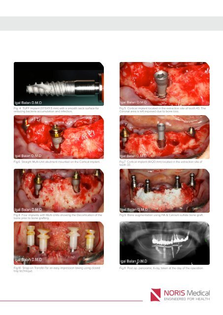
Fig. 4 TUFF Implant (37.5X11.5 mm) with a smooth neck surface for<br />
reducing bacteria accumulation and infection.<br />
Fig.5 Cortical implant located in the extraction site of tooth 43. The<br />
Coronal area is left exposed due to bone loss.<br />
Fig.6 Straight Multi-Unit abutment mounted on the Cortical implant.<br />
Fig.7 Cortical implant (4X20 mm) located in the extraction site of<br />
tooth 33.<br />
Fig.8 Four implants with Multi-Units showing the Decortication of the<br />
bone prior to bone grafting.<br />
Fig.9 Bone augmentation using HA & Calcium sulfate bone graft.<br />
Fig.10 Snap-on Transfer for an easy impression taking using closed<br />
tray technique.<br />
Fig.11 Post op. panoramic X-ray, taken at the day of the operation.