
You also want an ePaper? Increase the reach of your titles
YUMPU automatically turns print PDFs into web optimized ePapers that Google loves.
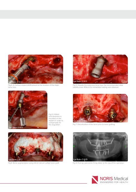
Fig 4: A Cortical implant (4x16) placed at the location of the major<br />
bone loss.<br />
Fig 5: Despite the extensive bone loss, the recommended initial<br />
stability (over 45Ncm) for immediate loading was obtained.<br />
Fig 6: A Multi-<br />
Unit abutment is<br />
mounted on the<br />
implant in order to<br />
compensate for<br />
the angulation.<br />
Fig 7: Decortication of the bone prior to bone grafting.<br />
Fig 8: Bone augmentation using HA & Calcium sulfate bone graft.<br />
Fig 9: Post op. panoramic X-ray, taken at the day of the operation.