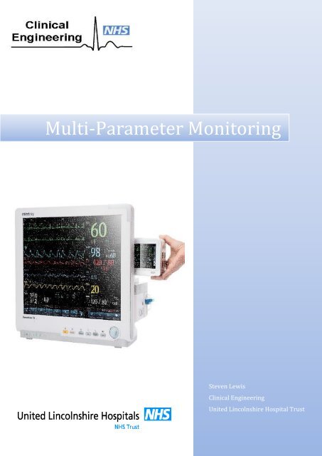
Multi-Parameter Monitoring 2

You also want an ePaper? Increase the reach of your titles
YUMPU automatically turns print PDFs into web optimized ePapers that Google loves.
<strong>Multi</strong>-<strong>Parameter</strong> <strong>Monitoring</strong><br />
Steven Lewis<br />
Clinical Engineering<br />
United Lincolnshire Hospital Trust
<strong>Multi</strong>-parameter Monitors<br />
<strong>Multi</strong>-parameter Monitors are intended to be used for monitoring, displaying, reviewing, storing<br />
and the transferring of multiple physiological parameters including, ECG, heart rate (HR),<br />
respiration (Resp), temperature (Temp), SPO 2 (pulse oxygen saturation), pulse rate (PR), noninvasive<br />
blood pressure (NIBP), invasive blood pressure (IBP), cardiac output (C.O.), airways<br />
gases such as; carbon dioxide (CO 2), oxygen (O 2), anaesthetic gas (AG).<br />
<strong>Monitoring</strong> vital signs for example; a patient’s blood pressure, pulse rate, and respiration rate is<br />
a crucial aspect of patient care in hospital. Vital signs indicate a patient’s clinical condition, are<br />
necessary to calculate national early warning scores (NEWS) and used to determine the<br />
monitoring, escalation and interventions that are required subsequently.<br />
In a hospital setting, patient monitoring is used in operating theatres, intensive care and critical<br />
care units, and many other critical and non-critical areas.<br />
Continuous multi-parameter monitoring has shown to be an effective mechanism for triggering<br />
early detection of changes in a patient’s condition by notifying the nursing staff that the patient<br />
needs attention. This is beneficial as by assessing the situation sooner and making the right<br />
clinical decision to intervene as appropriate.<br />
In addition to monitoring patients on an individual basis, the remote observation of multiple<br />
patients is possible through central patient monitoring systems. These monitoring systems are<br />
typically made up of networked machines consisting of one or more sensors, display devices,<br />
processing components, and communication links for displaying or recording the results<br />
elsewhere through the network. For portable monitors or in areas where a hard wired network<br />
is not practical wireless monitors are used, the signal is sent from the telemetry unit and picked<br />
up by aerial’s located around the hospital. This is particularly useful for areas such as Accident &<br />
Emergency and Coronary Care Unit or where multiple areas need to monitor the same patient at<br />
the same time or while a patient is being transported from one area to another.<br />
In non-critical areas the signs being monitored will predominantly be Heart Rate, SPO 2, Blood<br />
Pressure and ECG. In a critical care area such as ICU or an operating theatre, further signs will<br />
be monitored these may include; Invasive Blood Pressure (IBP), Respiration Rate and airways<br />
gases such as CO 2, O 2, and other gases that may be respired such as anaesthetic gases during a<br />
surgical procedure.<br />
Respiration Rate<br />
The function of the respiratory system is to supply adequate oxygen to the tissues and to<br />
remove the waste product carbon dioxide. This is achieved with the inspiration and expiration<br />
of air. With each breath there is a pause after expiration. The rate of respiration will vary with<br />
age and gender. A respiratory rate of 12-18 breaths per minute in a healthy adult is considered<br />
normal. Rates outside of this normal range can be classed as;<br />
<br />
<br />
<br />
<br />
<br />
Tachypnoea: the rate is regular but over 20 breaths per minute.<br />
Bradypnoea: the rate is regular but less than 12 breaths per minute.<br />
Apnoea: there is an absence of respiration for several seconds - this can lead to<br />
respiratory arrest.<br />
Dyspnoea: difficulty in breathing, the patient gasps for air.<br />
Cheyne-Stokes respiration: the breathing gets increasingly deeper then shallower,<br />
very slow and laboured with periods of apnoea. This type of breathing is often seen in<br />
the dying patient.
Hyperventilation: patients may breathe rapidly due to a physical or psychological<br />
cause, for example if they are in pain or panicking. Hyperventilation reduces the carbon<br />
dioxide levels in the blood, causing tingling and numbness in the hands; this may cause<br />
further distress. In adults, more than 20 breaths a minute is considered moderate, more<br />
than 30 breaths is severe. (Mallett & Dougherty, 2004)<br />
<strong>Multi</strong>-parameter monitors have a function to measure the respiration rate of the patient. This is<br />
the number of breaths the patient takes per minute. Most multi-parameter monitors record this<br />
by monitoring the resistance between two ECG electrodes connected to the patient. As the<br />
patient breaths in and the chest expands the resistance between the two electrodes increases<br />
and then decreases as the patient exhales and the chest contracts. Others ways to monitor<br />
respiration could be using capnography which involves CO 2 measurements, referred to as EtCO 2<br />
or end-tidal carbon dioxide concentration. The respiratory rate monitored as such is called<br />
AWRR or airway respiratory rate. Other monitors may record spirometry flow volume loops,<br />
which will show the flow, volume and time taken to inhale and exhale, or it may be monitored<br />
by simply counting the number of breaths in one minute by recording how many times the chest<br />
rises.<br />
Capnography<br />
Capnography is the measurement of carbon dioxide (CO 2) in exhaled breath; capnography gives<br />
medical professionals another tool for determining whether blood is flowing to vital organs like<br />
the heart and brain. CO 2 levels reflect cardiac output and pulmonary blood flow; as the gas is<br />
transported by the venous system to the right side of the heart and then pumped to the lungs by<br />
the right ventricles.<br />
Capnography is particularly important during surgery, where patients are continuously<br />
monitored while under anaesthesia to ensure safety. In addition to its use in surgical settings,<br />
capnography can help physicians and emergency medical personnel determine whether a<br />
patient is having a heart attack or hyperventilating. It can also help them determine whether<br />
CPR is working.<br />
The two primary methods used for measuring CO 2 in expired air are mass spectroscopy and<br />
infrared spectroscopy. In mass spectroscopy gases and vapours of different molecular weights<br />
are separated and a breakdown of what gases and percentages can be displayed.<br />
End tidal Carbon Dioxide (EtCO 2) is the partial pressure or maximal concentration of carbon<br />
dioxide at the end of an exhaled breath, which is expressed as a percentage of CO 2 or in mmHg.<br />
Infrared (IR) spectroscopy uses an EtCO 2 sensor to continuously monitor the carbon dioxide<br />
that is inspired and exhaled by the patient. It is usually presented as a graph of expiratory CO 2<br />
against time, or less commonly against expired volume.<br />
The EtCO 2 sensor consists of an infrared source, a chamber through which the gas sample<br />
passes, and a photo-detector. When the expired CO 2 passes between the beam of infrared light<br />
and photo-detector it leads to a reduction in the amount of light falling on the sensor, this is due<br />
to the principle that CO 2 absorbs infrared radiation. The absorbance is proportional to the<br />
concentration of CO 2 in the gas sample. (Physio-Control, 2013)<br />
Capnometers can be categorised based on the sensing device location. The gas samples can be<br />
analysed by mainstream or side-stream techniques.
Mainstream capnometers are in-line with the patient tubing; the housing is heated to prevent<br />
condensation. The advantage of mainstream analysis is that it gives a real-time measurement<br />
(i.e., an immediate response rate of
eat, and a waveform (a graph of pressure against time) can be displayed.<br />
The components of an intra-arterial monitoring system can be viewed as three main parts:<br />
<br />
<br />
<br />
The measuring apparatus<br />
The transducer<br />
The monitor<br />
The measuring apparatus consists of an arterial cannula connected to tubing containing a<br />
continuous column of saline. The pressure waveform of the arterial pulse is transmitted via the<br />
column of fluid, to a pressure transducer, in the case of intra-arterial monitoring the transducer<br />
consists of a flexible diaphragm which in turn moves strain gauges converting the pressure<br />
waveform into an electrical signal. Monitors amplify the output signal from the transducer, filter<br />
the noise and also display the arterial waveform in real time on a screen. They also usually give<br />
a digital numeric display of systolic, diastolic and mean arterial blood pressure (MAP).<br />
The arterial line is also connected to a flushing system consisting of a bag of saline pressurised<br />
to 300 mm/Hg via a flushing device.<br />
Fig 3 – Invasive Blood Pressure being monitored<br />
For a pressure transducer to read accurately, atmospheric pressure must be discounted from<br />
the pressure measurement. This is done by exposing the transducer to atmospheric pressure<br />
and calibrating the pressure reading to zero. A transducer should be zeroed several times per<br />
day to eliminate any baseline drift.<br />
The pressure transducer must be set at the appropriate level in relation to the patient in order<br />
to measure blood pressure correctly. This is usually taken to be level with the patient’s heart.<br />
Failure to do this results in an error due to hydrostatic pressure (the pressure exerted by a<br />
column of fluid – in this case, blood) being measured in addition to blood pressure. This can be<br />
significant – every 10cm error in levelling will result in a 7.4mmHg error in the pressure<br />
measured; a transducer too low over reads, a transducer too high under reads.<br />
IBP monitoring has numerous advantages; IBP allows continuous ‘beat-to-beat’ blood pressure<br />
monitoring. This is useful in patients who are likely to display sudden changes in blood pressure<br />
(e.g. vascular surgery), patients who require close control of blood pressure (e.g. head injured<br />
patients), or in patients receiving drugs to maintain blood pressure.<br />
The IBP technique also allows accurate blood pressure readings at very low pressures, for<br />
example in shocked patients.<br />
An invasive blood pressure reading could also allow for improvement of patient comfort if blood<br />
pressure monitoring is required for a long period of time. IBP monitoring avoids the trauma of<br />
repeated cuff inflations.<br />
Other advantages include that the arterial cannula is convenient for repeated arterial blood<br />
sampling, for instance for arterial blood gases. (Jones, 2009)
Maintenance & Service Procedures<br />
Invasive Blood Pressure<br />
This is checked using a patient simulator at various pressures, it is essential that a zeroing<br />
procedure is performed before any measurements are taken.<br />
Respiration Rate<br />
This is also checked using a patient simulator at various rates to ensure accuracy.<br />
Capnography & Airways Gases<br />
Capnograpy can be tested by attaching an ETCO 2 circuit and breathing into the airway adapter at<br />
a set rate, the rise and fall of the CO 2 graph will be displayed on the monitor. The accuracy of<br />
any measured airways gases is also to be checked by attaching a cylinder filled with various<br />
gases such as; O 2, CO 2, nitrogen and typically an anaesthetic gas at set percentages. The monitor<br />
should show the levels of the various gases, if the values displayed are not correct a calibration<br />
procedure should be carried out.
Bibliography<br />
EBME, 2014. Capnography. [Online]<br />
Available at: http://www.ebme.co.uk/articles/clinical-engineering/16-capnometrycapnography<br />
[Accessed December 2015].<br />
Fukuda Denshi UK, 2014. DS-8500 System Maintenance Manual V.09 (2014). V 9 ed. Tokyo:<br />
Fukuda Denshi.<br />
Jones, A., 2009. Physical Principles of Intra-Arterial Blood Pressure Measurements, Salford: s.n.<br />
Mallett , J. & Dougherty, L., 2004. The Royal Marsden Hospital Manual of Clinical Nursing<br />
Procedures. 6th ed. Oxford: Blackwell Science.<br />
Paramedicine, 2000. End Tidal CO2. [Online]<br />
Available at: http://www.paramedicine.com/pmc/End_Tidal_CO2.html<br />
[Accessed October 2015].<br />
Physio-Control, 2013. Lifepak® 20e Defibrillator Service/User manual.<br />
Tilakaratna, P., 2014. How Equipment Works. [Online]<br />
Available at: http://www.howequipmentworks.com/capnography/<br />
[Accessed December 2015].