Video Stacks & Endoscopy Systems

You also want an ePaper? Increase the reach of your titles
YUMPU automatically turns print PDFs into web optimized ePapers that Google loves.
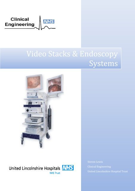
<strong>Video</strong> <strong>Stacks</strong> & <strong>Endoscopy</strong><br />
<strong>Systems</strong><br />
Steven Lewis<br />
Clinical Engineering<br />
United Lincolnshire Hospital Trust
<strong>Endoscopy</strong> <strong>Systems</strong> & <strong>Video</strong> <strong>Stacks</strong><br />
<strong>Endoscopy</strong> is a minimally invasive medical procedure directly visualising any part of the inside<br />
of the body using an endoscope, this will be used with a stack containing various equipment<br />
such as a monitor, light source, insufflator, camera, and printer all connected through an<br />
isolation transformer. These stacks are used in theatres and various clinics, such as Ear, Nose,<br />
and Throat (ENT).<br />
Endoscopes<br />
An endoscope is a long, thin, rigid or flexible tube that consists of two or three main optical<br />
cables, each of which comprises up to 50,000 separate optical fibres (made from optical-quality<br />
glass or plastic). One or two of the cables carry light down into the patient's body; another one<br />
carries reflected light (the image of the patient's body) back up to the physician's eyepiece (or<br />
into a camera, which can display it on a monitor).<br />
The optics of an endoscope are similar to those in a telescope. At the remote (distal) end, there<br />
is an objective lens, which links to one or more bendy sections of fibre-optic cable that carry the<br />
light back out of the patient's body to a second lens in an eyepiece or to a camera, which can be<br />
manipulated to adjust the focus.<br />
Endoscopes can be inserted into the body through a natural opening, such as the mouth and<br />
down the throat, or through the anus. Alternatively, an endoscope can be inserted through a<br />
small surgical cut made in the skin (known as keyhole surgery). Images of the inside of the body<br />
are relayed to a Monitor. The instrument may not only provide an image for visual inspection<br />
and photography, but may also be capable of taking biopsies or the retrieval of foreign objects.<br />
Some of the most commonly used types of endoscope include:<br />
<br />
<br />
<br />
<br />
Colonoscopes: used to examine the large intestine (colon).<br />
Gastroscopes: used to examine the oesophagus and stomach.<br />
Endoscopic Retrograde Cholangiopancreatography (ERCP): used to check for<br />
gallstones.<br />
Broncoscopes: used to examine the lungs and airways.<br />
Other types of endoscope include:<br />
<br />
<br />
<br />
Arthroscopes: used to examine joints.<br />
Hysteroscopes: used to examine the womb (uterus).<br />
Cystoscopes: used to examine the bladder.<br />
<strong>Endoscopy</strong> can involve;<br />
<br />
<br />
<br />
<br />
The Gastrointestinal Tract (GI tract): oesophagus, stomach and duodenum<br />
(esophagogastroduodenoscopy), small intestine, colon, (colonoscopy, proctosigmoidoscopy).<br />
The Respiratory Tract: the nose (rhinoscopy), the lower respiratory tract<br />
(bronchoscopy), the urinary tract (cystoscopy).<br />
The Female Reproductive System: The cervix (colposcopy), the uterus<br />
(hysteroscopy), the Fallopian tubes (Falloscopy).<br />
Normally closed body cavities (through a small incision): the abdominal or pelvic<br />
cavity (laparoscopy), the interior of a joint (arthroscopy), Organs of the chest<br />
(thoracoscopy and mediastinoscopy).
An endoscopy is normally carried out while a patient is conscious. It is not usually painful, but<br />
can be uncomfortable, so a local anaesthetic or sedative may be given.<br />
The exception is keyhole surgery, such as a laparoscopy or an arthroscopy, which are performed<br />
under general anaesthetic. (Martin, 2014)<br />
<strong>Endoscopy</strong> procedures are usually safe, and the risk of serious complications is low. Possible<br />
complications of an endoscopy include an infection in the part of the body that the endoscope is<br />
used to examine, damage to the body part which may in turn cause excessive bleeding.<br />
Monitors<br />
Monitors used on modern video stacks are high quality high definition (HD) flat panel screens,<br />
with excellent colour reproduction and a wide viewing angle. Most will have various video<br />
connections on the back such as S-<strong>Video</strong>, BNC, DVI, HDMI, etc. Some older video stacks may<br />
have a non HD flat panel monitor or a CRT monitor.<br />
Light Source & Lamps<br />
The light source may form part of a video or endoscopic stack, be a stand-alone unit often used<br />
in ENT procedures, or be attached to a microscope providing illumination to the area under<br />
investigation, the light is usually transmitted via a fibre optic cable to the lens end of a scope.<br />
High-Intensity Discharge Lamps<br />
High Pressure Sodium (HPS), Metal Halide, Mercury Vapour and Self-Ballasted Mercury Lamps<br />
are all high intensity discharge lamps (HID). Compared to fluorescent and incandescent lamps,<br />
HID lamps produce a large quantity of light from a relatively small bulb.<br />
HID lamps produce light by striking an electrical arc across tungsten electrodes housed inside a<br />
specially designed inner glass tube. This tube is filled with both gas and metals. The gas aids in<br />
the starting of the lamps, then, the metals produce the light once they are heated to a point of<br />
evaporation. High intensity discharge lamps, such as xenon, are ubiquitous within the field of<br />
endoscopy for minimally invasive surgery and diagnosis. Coupling light into a small diameter<br />
fibre bundle is difficult, so an extremely bright light source is required. Historically, the light<br />
source of choice has been the 180 to 300 W xenon lamp, which could deliver 1,000+ lumens of<br />
light at the distal end of the fibre bundle. While xenon bulbs achieve the technical requirements<br />
for endoscopy, they have a very short life, 500 - 1,000 hours, and can be expensive.<br />
LED Lighting<br />
The medical device industry is constantly changing. New technologies and products enter the<br />
market, replacing outdated or inefficient equipment. LED lighting is one of these, and there are<br />
numerous benefits in using LEDs for medical illumination applications including; longer life, less<br />
heat, dynamic control, lower energy consumption, and in many cases, lower cost. LED<br />
technology has improved significantly over the years, and is currently being integrated into<br />
medical devices, including surgical lighting, exam lights, phototherapy, and endoscopy. Ongoing<br />
advancements in LEDs that are driving the technology’s use in medical devices include<br />
improvement in light intensity, product size and weight; long-term reliability, and<br />
heat/temperature management, a line of high performance LEDs optimised to displace xenon<br />
technology for endoscopy have been developed.
Fibre Optic<br />
The light in a fiber-optic cable travels through the core by constantly bouncing from the<br />
cladding (mirror-lined walls), a principle called total internal reflection. The cladding does not<br />
absorb any light from the core, enabling the light wave to travel great distances.<br />
Fig 1 - Fibre Optic Cable<br />
Light source intensity is either adjusted manually via a dial or slider on the unit. If a camera and<br />
processor are attached they can monitor the amount of light at the scope end and adjusts the<br />
intensity automatically.<br />
Different wavelengths of light are needed for different procedures;<br />
Auto Fluorescence Imaging (AFI) is based on the detection of natural tissue fluorescence<br />
emitted by endogenous molecules (fluorophores) such as collagen, flavins, and porphyrins.<br />
After excitation by a short-wavelength light source, mucosal tissue emits a green fluorescence.<br />
A difference in the intensity of this fluorescence is seen between healthy and unhealthy tissue.<br />
These colour differences in fluorescence emission can be captured in real time during<br />
endoscopy and used for lesion detection or characterisation.<br />
Narrow Band Imaging (NBI) is a powerful optical image enhancement technology that<br />
improves the visibility of blood vessels and other structures on the bladder mucosa. This makes<br />
it an excellent tool for diagnosing bladder cancer during cystoscopy.<br />
White light is composed of an equal mixture of wavelengths. The shorter wavelengths only<br />
penetrate the top layer of the mucosa, while the longer wavelengths penetrate deep into the<br />
mucosa. NBI light is composed of just two specific wavelengths that are strongly absorbed by<br />
haemoglobin.<br />
The shorter wavelength in NBI is 415 nm light, which only penetrates the superficial layers of<br />
the mucosa. This is absorbed by capillary vessels in the surface of the mucosa and shows up<br />
brownish on the video image. This wavelength is particularly useful for detecting tumours,<br />
which are often highly vascularised. The second NBI wavelength is 540 nm light, which<br />
penetrates deeper than 415 nm light. It is absorbed by blood vessels located deeper within the<br />
mucosal layer, and appears cyan on the NBI image. This wavelength allows a better<br />
understanding of the vasculature of suspect lesions. (Olympus, 2014)<br />
Fig 2 – Absorption of Narrow Band Illumination
Insufflator<br />
An Insufflator provides distension of the required cavity for diagnostic or operative procedures.<br />
The insufflator pumps CO 2 into the cavity to a set pressure, this stretches the area being worked<br />
on making it easier to manipulate tools, assess the surgical site or perform the operative<br />
procedure. The maximum pressures of cavity’s should be observed when using an insufflator to<br />
prevent any problems, for example, Prolonged intra-abdominal pressures greater than 20mmHg<br />
should be avoided as they can cause any of the following problems;<br />
<br />
<br />
<br />
Decreased respiration due to pressure on the diaphragm.<br />
Decreased venous return.<br />
Decreased Cardiac output.<br />
CO 2 is now preferred over air as CO 2 is absorbed 150 times faster than the nitrogen in air, and is<br />
promptly eliminated via the lungs. This also means that patients are not subjected to extended<br />
discomfort from bloating and cramping.<br />
Camera System<br />
The camera on a video stack consists of two parts, the camera head, where scopes and light<br />
guides are attached, and a processer unit which feeds the image taken by the camera to a<br />
monitor, image capture device or printer.<br />
The aperture opens and closes to control how much light travels from the subject through a<br />
series of curved lens and through coloured filters focused on a digital sensor, which converts it<br />
into a digital (numerical) format. This is then processed and fed to the image capture system,<br />
monitor and printer.<br />
Image Capture System<br />
Image capture systems are image management systems that have the ability to record images<br />
and videos to their internal hard drive. They can also be connected to the hospital network<br />
where they can be integrated with PACS and other similar systems.<br />
Many image capture systems are generally windows based systems with a dedicated user<br />
interface. Very little maintenance of this system is required, at ULHT the only thing in the way<br />
service and repair we carry out is to take an image of the hard-drive at acceptance in case of any<br />
faults during its life, functional and visual checks and electrical safety testing.<br />
Isolation Transformer<br />
An isolation transformer is a transformer used to transfer electrical power from a source of<br />
alternating current (AC) power to some equipment or device while isolating the powered device<br />
from the power source, usually for safety reasons. Isolation transformers are used to protect<br />
against electric shock, to suppress electrical noise in sensitive devices, or to transfer power<br />
between two circuits which must not be connected. All true transformers are isolating as the<br />
primary and secondary are not connected physically but only by induction. However, in an<br />
isolation transformer the windings are completely insulated from each other to ensure good<br />
isolation.
In a normal supply the earth conductor is referenced to the neutral<br />
connector at source, which gives a potential voltage between live<br />
and earth of 240V. This means that touching either the live or<br />
neutral makes you part of the return path and can result in<br />
electrocution.<br />
With an isolation transformer the output voltage is not referenced<br />
to ground, so you could safely touch the live conductor and ground<br />
and not received a shock. This system is safer but if there is a fault,<br />
the operator touched both live and neutral connections or there<br />
was capacitive coupling between the secondary windings and<br />
earth. This would allow current to flow from either of the<br />
secondary connections through the operator/ patient to ground<br />
and electrocution could still occur.<br />
Maintenance & Service Procedures<br />
Endoscopes<br />
These are generally managed by the user, this includes cleaning.<br />
Cameras and Image Capture devices<br />
Cameras should have visual inspection and function test, checking image quality and focus also<br />
ensuring the various buttons work as they should.<br />
Image capture devices should also be visually inspected paying attention to the screen and case<br />
all functions should then be checked including the touch screen responds if applicable. This<br />
should then be safety tested.<br />
Light Sources<br />
A thorough visual inspection to include the case, mains lead, and the bulb for any<br />
damage/pitting should be performed. A filter clean/change and an electrical safety test are<br />
often all that is required.<br />
Light sources that used arc lamps or HID lamps require a scheduled lamp change, usually at<br />
approx. 500 hours of use.<br />
Before HID lamps are disposed of in an appropriate manner the xenon gas should be discharged.<br />
Insufflators<br />
A Visual inspection should be performed taking special care to inspect any gas connections and<br />
hoses including the pin index.<br />
A Check of output pressure and flow rates for conformity and accuracy against manufacturer’s<br />
specification. Check all controls work and that any alarms sound, including excess pressure and<br />
empty cylinder alarm.<br />
An Electrical Safety Test (EST) should then be completed to the appropriate standard.
Bibliography<br />
Blackwell Publishing, 2014. Basic Endoscopic Equipment. [Online]<br />
Available at: https://www.blackwellpublishing.com/xml/dtds/4-<br />
0/help/10003420_chapter_1.pdf<br />
[Accessed December 2015].<br />
Martin, T., 2014. Principles of Gastrointestinal <strong>Endoscopy</strong>. Surgery (Oxford), 32(3), pp. 139-144.<br />
Olympus, 2014. Olympus - Narrow Band Imaging. [Online]<br />
Available at:<br />
http://www.olympus.co.uk/medical/en/medical_systems/applications/urology/bladder/narro<br />
w_band_imaging__nbi_/narrow_band_imaging__nbi_.html<br />
[Accessed Febuary 2014].<br />
ustudy, 2011. <strong>Endoscopy</strong> Working Principle. [Online]<br />
Available at: http://www.ustudy.in/node/5066<br />
[Accessed December 2015].