Products at a Glance 2024
Overview of our portfolio from tissue dissociation to 2D/3D cell culture
Overview of our portfolio from tissue dissociation to 2D/3D cell culture

You also want an ePaper? Increase the reach of your titles
YUMPU automatically turns print PDFs into web optimized ePapers that Google loves.
More than competence for cells<br />
<strong>Products</strong><br />
<strong>at</strong> a glance<br />
Your Shortcut to <strong>Products</strong><br />
from Tissue Dissoci<strong>at</strong>ion<br />
to Cell-based Assays<br />
www.pelobiotech.com
www.pelobiotech.com<br />
Rely on & Relax<br />
Research grade products<br />
GMP grade products<br />
Welcome to your One-Stop-Cell Culture Shop,<br />
having more than competent 4 cells – th<strong>at</strong> is PELOBiotech’s outstanding characteristic. We want you to get the best<br />
and most reproducible results ever, results you can rely on and relax. We offer special cell culture media for human<br />
primary cells, stem cells and embryonic stem cells as well as special supplements for tumor stem cells. We get you the<br />
whole variety of solutions from tissue dissoci<strong>at</strong>ion to cryo-preserv<strong>at</strong>ion.<br />
Our mission is to provide high-quality cell culture products <strong>at</strong> affordable prices. Therefore, we are <strong>at</strong> the top-level of<br />
intern<strong>at</strong>ional research regarding consistent quality. We block out the Black Box esp. while developing Defined or<br />
Xeno-free Media limiting usage of FBS and other animal products. We have built up a worldwide network of<br />
competent and innov<strong>at</strong>ive partners. Our network clears your direct access to products of the future research.<br />
2
www.pelobiotech.com<br />
Our products <strong>at</strong> a glance<br />
• Tissue Dissoci<strong>at</strong>ion<br />
• Cells & Tissues<br />
Collagenases | Proteases | Enzyme Blends<br />
iPS Cells & iPS derived Cells | Adult Stem Cells (Hem<strong>at</strong>opoietic Stem<br />
Cells) | Mesenchymal Stroma/Stem Cells | Primary Cells | Organ derived<br />
Primary Cells | Blood & Bone Marrow derived Cells (MNCs & Immune<br />
Cells) | Tissue Slices | Cell and Tissue RNA<br />
• Cell Culture Media for the cells mentioned above<br />
FBS | hPL | animal origin free | defined media<br />
• Cell Culture Tools<br />
• 3D Cell Culture<br />
• Essential extras<br />
• Assays<br />
Cryo & Cold management | Cell Culture supplements |<br />
Extracellular M<strong>at</strong>rices | Cell Culture reagents | Transfection Tools |<br />
Reprogramming Tools<br />
Scaffold-free | Scaffold-based | Hydrogel-based |<br />
3D Models Ready-to use | Midifluidic<br />
Antibodies | Dyes | Proteins | Europium Chel<strong>at</strong>es |<br />
TR-FRET Reagents | Molecular biology Reagents |<br />
Small Molecules | Product Purific<strong>at</strong>ion Tools | Safety Tools<br />
Angiogenesis Assays | Cell-based Assays | Cell Purity Kit |<br />
Chimerism Assays | Metallo Quantific<strong>at</strong>ion Assays | Metastasis Assays<br />
*Illustr<strong>at</strong>ions Cre<strong>at</strong>ed with BioRender.com even if its not mentioned everywhere<br />
3
www.pelobiotech.com<br />
Tissue Dissoci<strong>at</strong>ion<br />
Tissue Dissoci<strong>at</strong>ion is an extremely important early step in experiments using primary cells. This early step already<br />
determines the quality of your cell culture and finally your results.<br />
The main question is how to isol<strong>at</strong>e cells in the best way and which criteria are the most important: Is it the cell<br />
viability, the biomarker profile or just the cell number? Our partner VitaCyte offers only highly purified tissue<br />
dissoci<strong>at</strong>ion enzymes for basic research as well as for cell therapy.<br />
CIzyme TM Tissue Dissoci<strong>at</strong>ing Enzymes<br />
Advantages of Purified Enzymes for Cell Isol<strong>at</strong>ion<br />
Problems with crude or enriched collagenase:<br />
Variable biochemical composition, reflects<br />
heterogeneity of the bacterial culture supern<strong>at</strong>ant<br />
• Heterogenous molecular forms of<br />
collagenase Lot qualific<strong>at</strong>ion often<br />
required<br />
Collagenases and Proteases<br />
Advantages of using purified enzymes:<br />
Uniform collagenase composition, by HPLC and<br />
specific CDA1 (CDA U/mg protein)<br />
• Purified enzymes; minimal contamin<strong>at</strong>ion<br />
by other enzymes & endotoxin<br />
• Enzyme formul<strong>at</strong>ion can be modified to<br />
improve cell yield, viability, function<br />
Collagenases and Proteases<br />
Applic<strong>at</strong>ions – Isol<strong>at</strong>ion of<br />
• Hep<strong>at</strong>ocytes<br />
• Islet Cells<br />
• Adipose Tissue Stem Cells<br />
• Skin Fibroblast<br />
• Skin Ker<strong>at</strong>inocytes<br />
• Cardiomyocytes<br />
• Glioblastoma<br />
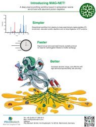
NEW <strong>Products</strong>:<br />
Recombinant Collagenase<br />
Defined and enriched DE Collagenase products.<br />
Collagenase is blended with purified neutral protease<br />
<strong>at</strong> defined activities to provide a broad spectrum of<br />
products with distinct collagenase activity.<br />
Pic: High viable<br />
cell yields: Get<br />
consistent results<br />
with our enzyme<br />
formul<strong>at</strong>ions<br />
4
U/mg DW<br />
www.pelobiotech.com<br />
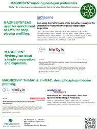
PD Collagenases<br />
Changing the paradigm.<br />
VitaCyte’s purified defined (PD) collagenase products<br />
offer a new standard of vers<strong>at</strong>ility in optimizing cell<br />
isol<strong>at</strong>ions. Enriched collagenase is blended with purified<br />
neutral protease <strong>at</strong> defined activities to provide a<br />
broad spectrum of products with distinct collagenase 2,3 to<br />
neutral protease 1 activities. This design fe<strong>at</strong>ure simplifies<br />
selection of an optimum r<strong>at</strong>io of critical enzyme activities<br />
for each cell isol<strong>at</strong>ion applic<strong>at</strong>ion. This manufacturing process minimizes<br />
enzyme variability enabling consistent control of cell isol<strong>at</strong>ions.<br />
18000<br />
16000<br />
14000<br />
12000<br />
10000<br />
8000<br />
6000<br />
4000<br />
2000<br />
0<br />
Purified defined Collagenase<br />
PD 100 PD 800<br />
CDA U/mg NP U/mg Clostripain U/mg<br />
Each PD product contains a fixed amount of protease activity (NP U 1 ), low clostripain activity 4 , and increasing amounts of<br />
collagenase activity defined by a functional collagen degrad<strong>at</strong>ion activity (CDA U) th<strong>at</strong> correl<strong>at</strong>es with the<br />
biochemical form of the enyzme 2 . The target collagenase: protease activity r<strong>at</strong>ios enables efficient<br />
explor<strong>at</strong>ion of a broad range of formul<strong>at</strong>ions.<br />
The three Cs of PD Collagenase<br />
CONSISTENT CONVENIENT COST EFFECTIVE<br />
• Fixed amount of purified protease<br />
• Low clostripain contamin<strong>at</strong>ion<br />
• Removal of the majority of<br />
contaminants in crude collagenase<br />
• • Weigh out wh<strong>at</strong> you need or<br />
reconstitute and use entire bottle<br />
• • Rapid dissolution, no clogged<br />
filters, no need for pre-centrifug<strong>at</strong>ion<br />
• • Stable for 2 years <strong>at</strong> +4°C<br />
storage, 4 years <strong>at</strong> < -20°C<br />
• • Increase productivity, minimize need<br />
for lot testing<br />
• • Purchase of large quantities of “good”<br />
lots of collagenase avoided<br />
• • Easily migr<strong>at</strong>e to purified enzyme products<br />
based on enzyme activity analysis of<br />
DE Collagenase<br />
www.pelobiotech.com<br />
5
www.pelobiotech.com<br />
Ask for PD Collagenase!<br />
The products below are defined by the total Wünsch activity per bottle, an assay th<strong>at</strong> uses a peptide substr<strong>at</strong>e to measure<br />
the activity of the collagenase c<strong>at</strong>alytic domain. This activity does not provide functional assessment of collagenase activity<br />
but it is one of the most consistent and reliable activity measures used by many collagenase suppliers. The table below<br />
provides ordering inform<strong>at</strong>ion and a cross reference chart to indic<strong>at</strong>e which PD Collagenase product may meet your needs.<br />
The Collagenase Gold product is similar to the PD Collagenase 800 but does not contain any supplemental purified<br />
protease. This product enables users to use other proteases or to purchase from VC the same protease used in the PD<br />
Collagenase products (BP Protease, c<strong>at</strong>alog # 003-1000) for cre<strong>at</strong>ion of a custom mixture.<br />
Product C<strong>at</strong>alog # #/Quantity Cross Reference<br />
PD Collagenase 100<br />
PD Collagenase 800<br />
Collagenase Gold<br />
PB- 011-<br />
1010<br />
PB-011-<br />
1050<br />
PB-011-<br />
1060<br />
1 g<br />
Worthington Type 1 or 2 Liberase TM or DH,<br />
Blendzyme 1 or 2<br />
1 g Liberase TL, Liberase DL, Liberase H I<br />
1 g Human Islets<br />
To isol<strong>at</strong>e sensitive cells such as islets, high purity and fidelity collagenase are essential due to their critical role in enzym<strong>at</strong>ic<br />
degrad<strong>at</strong>ion of the extracellular m<strong>at</strong>rix without damaging the structural and functional integrity of the cells. Successful<br />
human islet isol<strong>at</strong>ion requires the use of defined collagenase-protease enzyme mixtures, and intact class I and class II<br />
collagenase should be used to ensure maximal islet recovery <strong>at</strong> the lowest dose of enzyme. This is why Collagenase Gold<br />
is ideal for islet isol<strong>at</strong>ion. Thereby the use of these enzymes which are GMP and AOF can help with the best results. It has<br />
been found th<strong>at</strong> excess collagenase may potentially harm islets during the isol<strong>at</strong>ion process, emphasizing the need for<br />
precise dosing and high purity enzymes to avoid detrimental effects on the isol<strong>at</strong>ed cells. Furthermore, studies have<br />
demonstr<strong>at</strong>ed th<strong>at</strong> islet mass, quality, and purity are determined by the efficacy of islet isol<strong>at</strong>ion, which depends on factors<br />
such as organ procurement, collagenase digestion, and purific<strong>at</strong>ion method. This highlights the critical role of high purity<br />
collagenase in ensuring the quality and purity of isol<strong>at</strong>ed islets.<br />
References<br />
1. Breite AG, et al. Transplant<strong>at</strong>ion Proceedings 42 (2010); 2052-2054.<br />
2. McCarthy RC, et al. Transplant Proc 40 (2008); 339-342.<br />
3. Wünsch E and Hedrich H-G. Hoppe-Seyler’s Zeitschrift Physiol Chemie 333 (1963);149-151.<br />
4. Mitchell WM and Harrington WF. Methods in Enzymology (1970) 635-642.<br />
5. McCarthy RC, et al. Transplant 91 (2011) 137-145.<br />
Scan the QR code to visit the VitaCyte website to get additional protocols and product<br />
inform<strong>at</strong>ion.<br />
6
www.pelobiotech.com<br />
CIzyme TM <strong>Products</strong><br />
Product C<strong>at</strong>alog # Quantity App Quality<br />
CIzyme TM Collagenase HA PB-001-1000 2000 Wünsch units Human islets GMP<br />
CIzyme TM Collagenase HA PB-001-1050 200 Wünsch units Rodent islets GMP<br />
CIzyme TM Collagenase MA PB-001-2020 1100 Wünsch units Porcine islets GMP<br />
CIzyme TM Collagenase MA PB-001-2030 2,5 Mill CDA units Human hep<strong>at</strong>ocytes GMP<br />
CIzyme TM rCollagenase HI PB-001-4010 1600 Wünsch units Human islets<br />
GMP &<br />
AOF<br />
CIzyme TM Collagenase Gold PB-011-1060 800 Wünsch units Human islets<br />
CIzyme TM Collagenase Gold<br />
Plus<br />
PB-011-2000<br />
>1,500 FALGPA units<br />
Human islets from nondiseased<br />
organs<br />
GMP<br />
CIzyme TM Thermolysin PB-002-3000 6 mg Protease<br />
GMP &<br />
AOF<br />
CIzyme TM BP Protease<br />
PB-003-1000<br />
1,1 mill neutral protease<br />
units<br />
Neutral Protease<br />
GMP &<br />
AOF<br />
CIzyme TM BP Protease<br />
PB-003-2000<br />
2,3 mill neutral protease<br />
units<br />
Neutral Protease<br />
GMP &<br />
AOF<br />
Clostripain PB-004-2000 25 mg Protease GMP<br />
CIzyme TM RI PB-005-1030 375k CDA U, 75K NP U Rodent islets<br />
CIzyme TM AS PB-005-1090 35 W U, 280k NP U Adipose derived stem cells GMP<br />
CIzyme TM Hep<strong>at</strong>ocyte<br />
Isol<strong>at</strong>ion Kit<br />
PB-005-1010<br />
2,5 Mill CDA U,2,2 Mill NP<br />
U mg<br />
Hep<strong>at</strong>ocytes<br />
GMP<br />
PD Collagenase 100 PB- 011- 1010 1 g<br />
PD Collagenase 800 PB-011- 1050 1 g<br />
Porcine & rodent<br />
hep<strong>at</strong>ocytes<br />
Rodent dendritic cells, r<strong>at</strong><br />
cardiomyocytes, fibroblasts<br />
and porcine kidney<br />
epithelial cells<br />
7
www.pelobiotech.com<br />
Cells & Tissues<br />
iPS Cells<br />
Induced Pluripotent Stem Cells (iPSCs) have revolutionized the field of regener<strong>at</strong>ive medicine and scientific research by<br />
offering a vers<strong>at</strong>ile pl<strong>at</strong>form for studying and harnessing the potential of stem cells. These remarkable cells, pioneered<br />
by Dr. Shinya Yamanaka, are capable of differenti<strong>at</strong>ing into a wide range of cell types, mirroring the <strong>at</strong>tributes of<br />
embryonic stem cells. We offer a wide range of reprogramming tools, recombinant proteins and small molecules for this<br />
applic<strong>at</strong>ion. Recombinant proteins, synthetic transcription factors, enhance targeted reprogramming, while small<br />
molecules, such as valproic acid, optimize efficiency and maintain pluripotency. Additionally, the choice of an appropri<strong>at</strong>e<br />
co<strong>at</strong>ing for culture vessels such as recombinant Laminin is crucial in supporting iPSC <strong>at</strong>tachment, prolifer<strong>at</strong>ion, and<br />
differenti<strong>at</strong>ion.<br />
Every cell type will have a variant of integrin and thereby you will require a specific laminin for each cell type, we<br />
recommend you check out our ECM solutions i-M<strong>at</strong>rix 211, 411, 511. This integr<strong>at</strong>ed approach, including proper<br />
co<strong>at</strong>ing consider<strong>at</strong>ions, streamlines iPSC gener<strong>at</strong>ion, addressing safety concerns and making them a valuable resource<br />
for scientific and regener<strong>at</strong>ive applic<strong>at</strong>ions.<br />
• Ready-to-use iPSCs crafted from different techniques such as footprint-free StemRNA 3rd Gen Reprogramming<br />
Technology, Retrovirus & Sendai virus.<br />
• No need for specialized reprogramming expertise.<br />
• Available from both males and females, complete with donor clinical st<strong>at</strong>us.<br />
• Sourced from skin fibroblasts and blood endothelial progenitor cells.<br />
We would like to point out to you as an interested party in genetically modified cells (e.g. iPS cells, GFP/RFP<br />
tagged cells) th<strong>at</strong> you have a corresponding authorized genetic engineering facility for the storage, use and<br />
disposal in which this organism is processed.<br />
iPS cells and/or GFP/RFP expressing cells produced by infection using viruses are deemed potential hazards<br />
and are graded Biosafety Level 1.<br />
Restrictions are placed on the distribution of Biosafety Level 2 cells. Cells infected with human p<strong>at</strong>hogenic<br />
viruses, or known to release human p<strong>at</strong>hogenic viruses may be distributed only to customers who provide<br />
evidence th<strong>at</strong> they have the necessary authoriz<strong>at</strong>ion to work with p<strong>at</strong>hogens. In Germany, a permit according<br />
to §44 Infektionsschutzgesetz (IfSG) must be provided. For exceptions, see §45 IfSG. Customers from outside<br />
Germany must show a valid permit provided by their competent authorities. PELOBiotech GmbH reserves the<br />
right to decline the shipment of Biosafety Level 2 cells.<br />
8
www.pelobiotech.com<br />
GMP iPS cells<br />
GMP iPSCs are high-quality induced pluripotent stem cells derived from adult tissue biopsies, suitable for therapeutic<br />
applic<strong>at</strong>ions. They meet strict regul<strong>at</strong>ory standards to ensure their safety and effectiveness for human use.<br />
• We use StemRNA Reprogramming Technology, which is virus-free and complies with regul<strong>at</strong>ory guidelines.<br />
• Benefits include iPSCs manufactured according to ICH 5QA standards, gener<strong>at</strong>ed with footprint-free RNA<br />
reprogramming technology, and available for commercial use.<br />
• Diverse donors and over 30 years of experience in human tissue procurement.<br />
• StemRNA Reprogramming Technology produces robust iPSCs with low b<strong>at</strong>ch-to-b<strong>at</strong>ch vari<strong>at</strong>ion,<br />
elimin<strong>at</strong>ing the need for screening exogenous genes.<br />
• Our iPSC seed stocks, Master Cell Banks, and working cell banks are suitable for commercial and therapeutic<br />
applic<strong>at</strong>ions.<br />
iPS derived cells<br />
We use human iPSC technology, to cre<strong>at</strong>e a wide range of cell models and biosensor technologies. Our capabilities c<strong>at</strong>er<br />
to various applic<strong>at</strong>ions, spanning preclinical drug discovery, biobanking, in vitro diagnostics, and biomarker<br />
development.<br />
Induced pluripotent stem (iPS) cells offer several advantages and serve as a superior human disease model compared to<br />
animal cells for several reasons:<br />
• Human Relevance: iPS cells are derived from human tissues, making them more relevant for studying human<br />
diseases. This is crucial because human physiology and disease mechanisms can differ significantly from those<br />
of animals.<br />
• P<strong>at</strong>ient-Specific Modeling: iPS cells can be gener<strong>at</strong>ed from individual p<strong>at</strong>ients, allowing the cre<strong>at</strong>ion of p<strong>at</strong>ientspecific<br />
disease models. This is invaluable for studying genetic diseases and understanding the unique aspects<br />
of a p<strong>at</strong>ient's condition.<br />
• Disease Recapitul<strong>at</strong>ion: iPS cells can be differenti<strong>at</strong>ed into a variety of cell types relevant to the disease being<br />
studied, such as neurons, cardiomyocytes, or hep<strong>at</strong>ocytes. This enables researchers to closely mimic disease<br />
conditions in a dish.<br />
• Genetic Manipul<strong>at</strong>ion: iPS cells can be genetically modified to introduce disease-associ<strong>at</strong>ed mut<strong>at</strong>ions or correct<br />
genetic defects. This provides a precise way to investig<strong>at</strong>e the genetic basis of diseases.<br />
• Drug Screening: iPS-derived cells can be used for high-throughput drug screening to identify potential therapies<br />
or test drug efficacy. This is particularly important for personalized medicine.<br />
• Reduced Ethical Concerns: Using iPS cells allevi<strong>at</strong>es many ethical concerns associ<strong>at</strong>ed with the use of embryonic<br />
stem cells, which can be controversial.<br />
• Consistency: iPS cells provide a consistent and reproducible source of human cells for experiment<strong>at</strong>ion,<br />
elimin<strong>at</strong>ing genetic variability found in animal models.<br />
• Transl<strong>at</strong>ion to Clinical Applic<strong>at</strong>ions: iPS cells have the potential to be used in cell-based therapies and<br />
regener<strong>at</strong>ive medicine, making them a bridge between research and clinical applic<strong>at</strong>ions.<br />
• Longitudinal Studies: Researchers can derive iPS cells from p<strong>at</strong>ients <strong>at</strong> different stages of a disease and track<br />
the disease progression over time, which is challenging to do with animal models.<br />
• Cost and Time Efficiency: iPS cell-based research is often more cost-effective and less time-consuming than<br />
working with animal models.<br />
The l<strong>at</strong>est applic<strong>at</strong>ions of induced pluripotent stem cells (iPSCs) encompass a wide array of cutting-edge advancements<br />
in regener<strong>at</strong>ive medicine, disease modeling, drug screening, and cell therapy. These applic<strong>at</strong>ions have been made possible<br />
by the unique properties of iPSCs, which are similar to embryonic stem cells (ESCs) in terms of morphology, prolifer<strong>at</strong>ion,<br />
and gene expression profile (Okita & Yamanaka, 2008). iPSC technology has significantly enriched regener<strong>at</strong>ive medicine<br />
by introducing autologous pluripotent progenitor pools bioengineered from ordinary som<strong>at</strong>ic tissue, offering potential in<br />
disease modeling and therapeutic applic<strong>at</strong>ions (Nelson et al., 2009; Polo et al., 2010). Notably, the first clinical study using<br />
human iPSC-derived cells was initi<strong>at</strong>ed in 2014, utilizing human iPSC-derived retinal pigment epithelial (RPE) cells to tre<strong>at</strong><br />
macular degener<strong>at</strong>ion, resulting in improved vision for the p<strong>at</strong>ient (Shi et al., 2016).<br />
Recent advances in differenti<strong>at</strong>ing cells such as cardiac, neural, and skeletal muscle cells from iPSCs, as well as directly<br />
reprogramming som<strong>at</strong>ic cells in tissue regener<strong>at</strong>ion applic<strong>at</strong>ions, have been summarized and synthesized, highlighting the<br />
vers<strong>at</strong>ility of iPSCs in various therapeutic contexts (Mao et al., 2022). iPSCs have also been explored for bone<br />
regener<strong>at</strong>ion, cardiovascular disease, and as pre-clinical models for studying human disease, demonstr<strong>at</strong>ing their potential<br />
in diverse medical applic<strong>at</strong>ions (He et al., 2018; Plews et al., 2012; Huang et al., 2020).<br />
9
www.pelobiotech.com<br />
IPSCs have been investig<strong>at</strong>ed for their use in dentistry, immunotherapy, and as a tool for personalized autologous body<br />
organ transplant, showcasing their expanding role in different medical fields (Hynes et al., 2015; Jiang et al., 2013; Palomo<br />
et al., 2014). Moreover, iPSCs have been utilized for modeling inherited cardiomyop<strong>at</strong>hies, bone regener<strong>at</strong>ion, and as a<br />
pl<strong>at</strong>form for drug screening and disease modeling, further emphasizing their broad applic<strong>at</strong>ions in medicine (Karakikes et<br />
al., 2014; Tang et al., 2014; Hwang et al., 2021). The potential of iPSCs in regener<strong>at</strong>ive medicine has also been recognized<br />
through the development of liver buds and clinical-grade neural stem cells, highlighting the ongoing efforts to harness<br />
iPSCs for clinical applicability (Sekine et al., 2020; Cai et al., 2021).<br />
We offer:<br />
• Around 10,000 human disease models for preclinical drug development<br />
• Diverse models, including spheroid and organoid models, physiological barrier models, and more.<br />
• An extensive selection of iPSC deriv<strong>at</strong>ive cell types, from pericytes to cardiomyocytes, for a wide range of<br />
research applic<strong>at</strong>ions<br />
Below is a summary of the cell types we provide. Please note th<strong>at</strong> this list is not exhaustive. To explore our complete cell<br />
offerings, we encourage you to request our detailed cell list or visit pelobiotech.com for additional inform<strong>at</strong>ion.<br />
iPSC-Derived Cardiomyocytes (Heart):<br />
Cardiotoxicity screening for drug development.<br />
Studying heart diseases and genetic disorders.<br />
Regener<strong>at</strong>ive medicine for cardiac tissue repair.<br />
iPSC-Derived Hep<strong>at</strong>ocytes (Liver):<br />
Drug metabolism and toxicity testing.<br />
Modeling liver diseases and viral infections.<br />
Development of bioartificial liver devices.<br />
iPSC-Derived Hep<strong>at</strong>ic Stell<strong>at</strong>e Cells and Kupffer Cells (Liver):<br />
Investig<strong>at</strong>ing liver fibrosis and cirrhosis.<br />
Testing anti-fibrotic drugs for liver disease tre<strong>at</strong>ment.<br />
Understanding hep<strong>at</strong>ic stell<strong>at</strong>e cell biology.<br />
iPSC-Derived Neurons (Brain):<br />
Modeling neurodegener<strong>at</strong>ive diseases like Alzheimer's<br />
Drug discovery and testing for neurological disorders.<br />
Studying neural development and function.<br />
iPSC-Derived Ker<strong>at</strong>inocytes (Skin):<br />
Skin disease modeling, such as epidermolysis bullosa.<br />
Wound healing and tissue regener<strong>at</strong>ion studies.<br />
Drug testing for derm<strong>at</strong>ological conditions.<br />
iPSC-Derived Endothelial Cells:<br />
Investig<strong>at</strong>ing cardiovascular diseases and vascular disorders.<br />
Testing anti-angiogenic drugs for cancer therapy.<br />
Vascular tissue engineering and regener<strong>at</strong>ive medicine.<br />
iPSC-Derived Kidney Cells:<br />
Investig<strong>at</strong>ing renal diseases and disorders.<br />
Testing personalized medicaments for kidney health.<br />
Studying drug responses specific to kidney function<br />
iPSC-Derived Hem<strong>at</strong>opoietic Stem Cells (HSCs):<br />
Studying blood disorders like anemia and leukemia.<br />
Personalized therapies for hem<strong>at</strong>ological disorders<br />
Drug screening for hem<strong>at</strong>ological conditions.<br />
iPSC-Derived Hem<strong>at</strong>opoietic Stem Cells (HSCs):<br />
Studying blood disorders like anemia and leukemia.<br />
Developing personalized cell therapies<br />
for blood-rel<strong>at</strong>ed diseases.<br />
Drug screening for hem<strong>at</strong>ological conditions.<br />
iPSC-Derived Immune Cells:<br />
Modeling autoimmune diseases and immunodeficiencies.<br />
Developing immunotherapies and vaccines.<br />
Screening for immunomodul<strong>at</strong>ory drugs and tre<strong>at</strong>ments.<br />
10
www.pelobiotech.com<br />
Adult Stem Cells<br />
Adult stem cells, also known as som<strong>at</strong>ic or multipotent stem cells, are a unique class of undifferenti<strong>at</strong>ed cells found within<br />
various tissues and organs of the human body. Unlike embryonic stem cells, which can differenti<strong>at</strong>e into any cell type,<br />
adult stem cells are more restricted in their potential but still possess the ability to develop into a range of specialized cell<br />
types within the specific tissue or organ where they reside as shown in the figure above. However, these cells play a vital<br />
role in tissue maintenance. As you can see in the figure they form cells th<strong>at</strong> repair, and aid in regener<strong>at</strong>ion throughout an<br />
individual's life, making them a valuable resource for regener<strong>at</strong>ive medicine and research aimed <strong>at</strong> understanding and<br />
tre<strong>at</strong>ing various medical conditions. We offer the following cells:<br />
• Hem<strong>at</strong>opoietic CD34+ stem cells and Mononuclear cells to isol<strong>at</strong>e CD34 or CD113 cells<br />
• Available from both males and females, complete with donor clinical st<strong>at</strong>us.<br />
• Sourced from peripheral blood, cord blood and bone marrow.<br />
CD34 is a crucial marker for various progenitor cells, particularly hem<strong>at</strong>opoietic stem cells (HSC) and hem<strong>at</strong>opoietic<br />
progenitor cells (Sidney et al., 2014). It plays a significant role in hem<strong>at</strong>opoiesis, as evidenced by its presence in progenitor<br />
cells during hem<strong>at</strong>opoiesis, and its potential to give rise to hem<strong>at</strong>opoietic, endothelial cells, and smooth muscle cells<br />
(Kapoor et al., 2019; Bai et al., 2009). CD34 has been linked to cellular homing p<strong>at</strong>terns to the bone marrow and adhesion<br />
of cells to stromal microenvironments or sites of inflamm<strong>at</strong>ion (Kopher et al., 2010). Furthermore, CD34(+) stem cells<br />
have been found to play an important role during liver development and regener<strong>at</strong>ion (Park et al., 2015). Additionally,<br />
CD34 has been associ<strong>at</strong>ed with the regul<strong>at</strong>ion and compartmentaliz<strong>at</strong>ion of stem cells, promoting their adhesive<br />
interactions with the stromal microenvironment of the bone marrow (Healy et al., 1995). It has also been identified as a<br />
marker for hem<strong>at</strong>opoietic stem cells and non-hem<strong>at</strong>opoietic progenitors, including vascular endothelial progenitors and<br />
embryonic fibroblasts, multipotent mesenchymal stromal cells, interstitial dendritic cells, and epithelial progenitors<br />
(Soliman, 2021). Moreover, CD34 has been utilized as an important marker for labeling hem<strong>at</strong>opoietic stem cells,<br />
demonstr<strong>at</strong>ing its significance in stem cell research and applic<strong>at</strong>ions (Ye et al., 2004).<br />
Mesenchymal/Stromal Stem cells<br />
Mesenchymal stromal cells (MSCs), often called mesenchymal stem cells, are adult stem cells found in bone marrow,<br />
adipose, Wharton jelly and other tissues. Notable characteristics include:<br />
• Differenti<strong>at</strong>ion into various cell types, such as bone, cartilage, and f<strong>at</strong> cells.<br />
• Immune system modul<strong>at</strong>ion, making them valuable for tre<strong>at</strong>ing autoimmune diseases, inflamm<strong>at</strong>ory conditions,<br />
and tissue damage.<br />
• Regener<strong>at</strong>ive properties th<strong>at</strong> promote tissue repair.<br />
11
www.pelobiotech.com<br />
• Low immunogenicity, reducing the risk of immune rejection when transplanted.<br />
Additionally, we offer a wide range of MSCs in research and GMP grade from various tissue sources, including canines,<br />
monkeys, dogs, mice, and humans, with different characteristics and applic<strong>at</strong>ions, such as diseased models,<br />
conditionally immortalized cells, and those with GFP/RFP tags and cell RNA. For all of these MSCs we also offer<br />
corresponding media and tools.<br />
GMP MSCs<br />
We provide high-quality mesenchymal stromal cells (MSCs) for clinical and preclinical applic<strong>at</strong>ions, offering<br />
both GMP and research-grade options. These cells are derived from 20 years of research and clinical experience<br />
<strong>at</strong> the Karolinska Institute, ensuring their quality. Our large-scale facility in Stockholm enables cost-effective<br />
production.<br />
• Adjustable quantity and vial size per dose.<br />
• Derived from bone marrow or adipose tissue of young, healthy volunteers.<br />
• Xeno-free and <strong>at</strong> Passage 3.<br />
• Offered with intern<strong>at</strong>ional delivery in liquid nitrogen.<br />
Exosomes<br />
These vers<strong>at</strong>ile cells c<strong>at</strong>er to various research and clinical needs, with competitive pricing and delivery options<br />
for different requirements.<br />
Exosomes, small extracellular vesicles released by various cell types, have garnered significant <strong>at</strong>tention for their diverse<br />
applic<strong>at</strong>ions in both diagnostics and therapeutics. These tiny membrane-bound vesicles carry a cargo of proteins, nucleic<br />
acids, and lipids, facilit<strong>at</strong>ing intercellular communic<strong>at</strong>ion. In diagnostics, exosomes serve as potential biomarkers for<br />
various diseases, including cancer, neurodegener<strong>at</strong>ive disorders, and cardiovascular conditions. Their non-invasive n<strong>at</strong>ure<br />
and specificity make them valuable in early disease detection. Moreover, exosomes hold promise in therapeutics, acting<br />
as n<strong>at</strong>ural delivery vehicles for drugs, RNA, or therapeutic proteins. Their ability to cross biological barriers, such as the<br />
blood-brain barrier, enhances their potential for targeted drug delivery. As research continues to unveil the intric<strong>at</strong>e roles<br />
of exosomes, their applic<strong>at</strong>ions are expanding, offering innov<strong>at</strong>ive solutions in personalized medicine and regener<strong>at</strong>ive<br />
therapies.<br />
We offer exosomes produced from bone marrow mesenchymal stem cells which are CD9, CD63 and CD81 +ve,<br />
extensively tested and size and count verified by Zetaview.<br />
Also available are media (CellCor) and media supplements for exosome production using mesenchymal stem/stroma<br />
cells (see page 29-30).<br />
12
www.pelobiotech.com<br />
Primary Cells<br />
In the realm of applied research, the selection of cell cultures is a critical determinant of<br />
the quality and relevance of scientific outcomes. Our commitment to maintaining the<br />
highest standards in cell quality has resulted in one of the most extensive portfolios of<br />
primary cells in Europe. To ensure the excellence of our cells, they undergo a rigorous<br />
process before they reach you. We also offer corresponding media for all the cells or<br />
custom media tailored to your requirements and all other possible cell culture tools.<br />
• Tissue Selection: Our meticulous tissue selection guarantees the highest<br />
quality primary cells, enhancing research reliability and are generally available<br />
<strong>at</strong> a cell density >0.5 million cells per vial check on our webpage for details.<br />
• Stringent Testing: We subject every isol<strong>at</strong>ion to comprehensive testing,<br />
including popul<strong>at</strong>ion doubling time, prolifer<strong>at</strong>ion assays, and tests for specific<br />
markers, fungi, bacteria, mycoplasma, hep<strong>at</strong>itis B and C DNA, and HIV-1 DNA.<br />
• Donor Inform<strong>at</strong>ion: Certain cell types come with extensive donor d<strong>at</strong>a,<br />
enriching research with details like BMI, smoking history, gender, and medical<br />
history.<br />
• Species Diversity: Our extensive cell range spans mice, r<strong>at</strong>s, hamsters, chickens,<br />
pigs, c<strong>at</strong>s, dogs, c<strong>at</strong>tle, and humans. Human cells can also be acquired from<br />
diseased or elderly donor sources, broadening research possibilities.<br />
• Quality Control: Autom<strong>at</strong>ed liquid nitrogen control ensures consistent,<br />
high-quality cryopreserved cell b<strong>at</strong>ches, maintaining our stringent standards.<br />
Additional Services we provide:<br />
• Custom Cell Isol<strong>at</strong>ion: Researchers with specific requirements can take advantage of our custom cell isol<strong>at</strong>ion<br />
services. We offer the flexibility to work with general protocols or, if preferred, the researcher's own protocols,<br />
all <strong>at</strong> affordable prices.<br />
• Cell RNA: We offer cell RNA, providing a valuable resource for RNA sequencing work.<br />
• Tagged Cells: We provide cells tagged with markers such as GFP/RFP, facilit<strong>at</strong>ing tracking and visualiz<strong>at</strong>ion in<br />
experiments.<br />
• Immortalized Lines: Our selection includes immortalized cells, which can be advantageous for extended and<br />
consistent experiment<strong>at</strong>ion.<br />
• Cells from KO Mice: We offer cells from specific knockout (KO) mice, enabling researchers to explore the<br />
effects of specific gene disruptions.<br />
• Media and other cell culture tools: we offer corresponding media for all the cells th<strong>at</strong> we offer and various<br />
tools to aid your cell culture from co<strong>at</strong>ings, supplements to bioreactors.<br />
Our commitment to providing top-tier primary cells and rel<strong>at</strong>ed services underscores our dedic<strong>at</strong>ion to advancing the<br />
quality and relevance of your research endeavors.<br />
Heart<br />
In our diverse cell portfolio, we provide over 900 cell types. Apart from cardiomyocytes and iPS derived cardiomyocytes,<br />
we also isol<strong>at</strong>e endothelial cells, preadipocytes, myocytes, epithelial cells, and fibroblasts from different regions of the<br />
heart and from various species. A spectrum of heart-derived cell types, each with distinct characteristics, contributes to<br />
diverse realms of cardiovascular research. Cardiomyocytes, the heart's muscle cells, find applic<strong>at</strong>ions in drug screening,<br />
cardiac toxicity assessment, and investig<strong>at</strong>ions into cardiac hypertrophy and regener<strong>at</strong>ion. Endothelial cells, lining<br />
blood vessels, play a pivotal role in studies rel<strong>at</strong>ed to angiogenesis, <strong>at</strong>herosclerosis, and vascular inflamm<strong>at</strong>ion.<br />
Preadipocytes, precursors to adipocytes, are integral in research focusing on adipogenesis, obesity-rel<strong>at</strong>ed conditions,<br />
and metabolic disorders.<br />
Myocytes, whether cardiac or skeletal, are fundamental in deciphering heart function, arrhythmias, and muscular disorders.<br />
Epithelial cells, forming the outer layers of the heart and blood vessels, contribute to studies on tissue development,<br />
regener<strong>at</strong>ion, and barrier function. Finally, fibroblasts, providing structural support, are indispensable in exploring fibrosis,<br />
extracellular m<strong>at</strong>rix dynamics, and tissue repair mechanisms. This diverse array of heart-derived cells collectively propels<br />
advancements in cardiac biology, offering insights into development, function, and p<strong>at</strong>hology, with applic<strong>at</strong>ions spanning<br />
from basic research to therapeutic development and regener<strong>at</strong>ive medicine.<br />
13<br />
Scan the QR code to see our full cell list.
www.pelobiotech.com<br />
You can also check out our Heart slices for toxicology, arrythmia rel<strong>at</strong>ed work which is more transl<strong>at</strong>ionally relevant. You<br />
can read more about the use of heart slices for research in this article here.<br />
Brain<br />
Apart from isol<strong>at</strong>ed and iPS derived astrocytes, Schwann cell like cells, microglia, oligodendrocytes, sensory neurons,<br />
cortical neurons, dopamergic neurons we also isol<strong>at</strong>e endothelial<br />
cells, glioblastomas, pericytes, neuron precursor cells from different<br />
regions of the heart and from various species. Isol<strong>at</strong>ed and induced<br />
pluripotent stem (iPS) cell-derived neural cell types, such as<br />
astrocytes, Schwann cell-like cells, microglia, oligodendrocytes,<br />
sensory neurons, cortical neurons, and dopaminergic neurons, serve<br />
as invaluable tools across a spectrum of studies and applic<strong>at</strong>ions.<br />
Astrocytes, Schwann cell-like cells, microglia, and oligodendrocytes<br />
play crucial roles in various aspects of neuroscience research.<br />
Astrocytes, for instance, are involved in the regul<strong>at</strong>ion of cerebral blood<br />
flow, maintenance of synaptic environments, and modul<strong>at</strong>ion of<br />
synapse form<strong>at</strong>ion and synaptic transmission. They are also implic<strong>at</strong>ed in the glymph<strong>at</strong>ic system's ability to elimin<strong>at</strong>e waste<br />
in the brain and are essential for the maintenance of brain homeostasis and neuronal protection. Furthermore, astrocytes<br />
are involved in regul<strong>at</strong>ing neuronal synaptogenesis, maintaining blood-brain barrier integrity, and recycling<br />
neurotransmitters. Additionally, they have been shown to medi<strong>at</strong>e analogous memory in a multi-layer neuron-astrocyte<br />
network, and targeting astrocytic function may protect against brain injury induced by blood-brain barrier disruption.<br />
Schwann cell-like cells have been studied for their morphological, transcriptional, and functional differences from mouse<br />
astrocytes, and they play key roles in supporting the central nervous system structure, regul<strong>at</strong>ing synaptic functions, and<br />
maintaining brain homeostasis. Microglia, on the other hand, have been found to regul<strong>at</strong>e synapses, neuronal circuits, and<br />
behavior, and they are thought to play a pivotal role in coupling neural activity and cerebral blood flow. Oligodendrocytes,<br />
which are responsible for producing myelin in the central nervous system, have been implic<strong>at</strong>ed in the regul<strong>at</strong>ion of neuronal<br />
excitability, synaptic transmission, plasticity, and in higher cognitive functions, including the initi<strong>at</strong>ion, maintenance, and<br />
consolid<strong>at</strong>ion of memories.<br />
Additionally, isol<strong>at</strong>ed endothelial cells, glioblastomas, pericytes, and neuron precursor cells from various brain regions<br />
offer insights into angiogenesis, brain tumors, blood-brain barrier function, and neurogenesis, respectively. These diverse<br />
neural cell types facilit<strong>at</strong>e a broad range of studies, including disease modeling, drug screening, neurotoxicity testing, and<br />
elucid<strong>at</strong>ing fundamental mechanisms underlying neurological and neurodegener<strong>at</strong>ive disorders.<br />
We also offer brain tissue slices Brain slices, including brain and root ganglionic slices, are valuable tools in neuroscience<br />
research due to their ability to provide a controlled and accessible model for studying various aspects of neuroscience.<br />
Brain slices have been widely used to study neuronal differenti<strong>at</strong>ion, cell differenti<strong>at</strong>ion, and synaptic transmission. They<br />
have also been utilized to model neurodegener<strong>at</strong>ive proteinop<strong>at</strong>hies and to enhance the quality of acute slice prepar<strong>at</strong>ions,<br />
particularly for electrophysiology studies.<br />
Brain slices have been instrumental in profiling sequential sections with multiplex staining, which benefits research in life<br />
sciences. Furthermore, brain slice prepar<strong>at</strong>ions have been valid<strong>at</strong>ed for enhancing neuronal preserv<strong>at</strong>ion and overall brain<br />
slice viability, making them essential for studying metabolic changes and facilit<strong>at</strong>ing experiment<strong>at</strong>ion on age-rel<strong>at</strong>ed<br />
disorders. Moreover, root ganglionic slices, such as dorsal root ganglion neurons, have been used to study neuronal<br />
connectivity, synaptic transmission, and plasticity, as well as to perform whole-cell p<strong>at</strong>ch-clamp recordings, allowing<br />
precise measurement of cellular and synaptic properties.<br />
The dorsal root ganglion has been identified as a novel neuromodul<strong>at</strong>ory target for evoking strong and reproducible<br />
motor responses in chronic motor complete spinal cord injury. Studies have been conducted on the trigeminal ganglion<br />
morphology in human fetuses, and the trigeminal ganglion has been studied for its potential in tre<strong>at</strong>ing trigeminal<br />
neuralgia.<br />
14
www.pelobiotech.com<br />
Pancreas<br />
We extract beta cells and diseased cells from diabetic<br />
p<strong>at</strong>ients from the pancreas. Additionally, we isol<strong>at</strong>e<br />
endothelial cells, epithelial cells, fibroblasts, and PDX<br />
cancer cells from different pancre<strong>at</strong>ic regions and various<br />
species. The uses of beta cells in research are extensive<br />
and diverse. Studies have focused on understanding the<br />
factors influencing beta cell turnover, prolifer<strong>at</strong>ion, and<br />
functional st<strong>at</strong>e, particularly in the context of diabetes and<br />
regener<strong>at</strong>ive therapy.<br />
Research has explored the essential role of Nkx6.1 in<br />
maintaining the functional st<strong>at</strong>e of pancre<strong>at</strong>ic beta cells,<br />
adaptive changes in beta cell turnover during pregnancy,<br />
the rel<strong>at</strong>ionship between beta-cell mass and obesity/type 2<br />
diabetes, and the gene regul<strong>at</strong>ory network required for<br />
establishing and maintaining pancre<strong>at</strong>ic beta cell identity.<br />
Additionally, the beta cell workload hypothesis has been<br />
revisited in the context of type 2 diabetes, and<br />
m<strong>at</strong>hem<strong>at</strong>ical modeling has been used to estim<strong>at</strong>e the long lifespan and low turnover of human islet beta cells.<br />
Studies have investig<strong>at</strong>ed the prolifer<strong>at</strong>ion of sorted human and r<strong>at</strong> beta cells, targeted insulin-producing beta cells for<br />
regener<strong>at</strong>ive therapy, and explored the potential for beta cell regener<strong>at</strong>ion in aging pancre<strong>at</strong>ic beta cells. Single-cell RNA<br />
sequencing has revealed a role for reactive oxygen species and peroxiredoxins in f<strong>at</strong>ty acid-induced r<strong>at</strong> beta-cell<br />
prolifer<strong>at</strong>ion, and reciprocal modul<strong>at</strong>ion of adult beta cell m<strong>at</strong>urity by activin A and follist<strong>at</strong>in has been studied. Moreover,<br />
research has focused on beta cell regener<strong>at</strong>ion after immunological destruction in a mouse model, explored the potential<br />
for making better beta cells, and investig<strong>at</strong>ed agents inducing both alpha and beta cell prolifer<strong>at</strong>ion without affecting<br />
differenti<strong>at</strong>ion or viability. Additionally, efforts have been made to expand human beta cells, and heterogeneities of normal<br />
and stimul<strong>at</strong>ed pancre<strong>at</strong>ic beta cells have been examined. These studies collectively contribute to a comprehensive<br />
understanding of beta cell biology and its implic<strong>at</strong>ions for diabetes and regener<strong>at</strong>ive medicine.<br />
Lungs and Oral cavity<br />
Apart from isol<strong>at</strong>ed and iPS derived alveolar and<br />
bronchial epithelial cells, fibroblasts, smooth muscle<br />
cells, macrophages, endothelial cells PDX cancer cells,<br />
mesenchymal stem cells from different regions of the<br />
heart and various species. The uses of iPS-derived<br />
alveolar and bronchial epithelial cells, fibroblasts,<br />
smooth muscle cells, macrophages, endothelial cells,<br />
PDX cancer cells, and mesenchymal stem cells from<br />
different regions of the heart and various species are<br />
diverse and impactful in biomedical research. P<strong>at</strong>ientderived<br />
xenograft (PDX) models have been extensively<br />
used in cancer research to simul<strong>at</strong>e human tumor<br />
biology in vivo, aiding in the development of anticancer<br />
drugs and providing a better represent<strong>at</strong>ion of tumor<br />
heterogeneity and the tumor microenvironment.<br />
Mesenchymal stem cells (MSCs) have shown promise in the tre<strong>at</strong>ment of chronic lung diseases and ischemic heart<br />
disease, with studies demonstr<strong>at</strong>ing their potential for myocardial survival and repair, as well as their ability to<br />
differenti<strong>at</strong>e into endothelial cells, vascular smooth muscle cells, or cardiac-like myocytes when transplanted into the<br />
ischemic heart. Smooth muscle cells have been studied for their contractile properties and their associ<strong>at</strong>ion with neuroglial<br />
cells and interstitial cells of Cajal in intestinal tissue engineering. Additionally, endothelial and smooth muscle cells have<br />
been isol<strong>at</strong>ed and characterized from coronary vessels, providing insights into their contractile phenotype and potential<br />
applic<strong>at</strong>ions in vascular research.<br />
15
www.pelobiotech.com<br />
Furthermore, the establishment of living biobanks comprising p<strong>at</strong>ient-derived xenografts, primary tumor tissue, normal<br />
tissue, sera, and cancer cell lines has facilit<strong>at</strong>ed the study of tumor characteristics and mechanisms of <strong>at</strong>herosclerosis. The<br />
expression of PDX-1, a key regul<strong>at</strong>or of beta cell m<strong>at</strong>ur<strong>at</strong>ion and differenti<strong>at</strong>ion, has been investig<strong>at</strong>ed in pancre<strong>at</strong>ic<br />
cancer, shedding light on its significance in the disease. Moreover, the potential of stem cells in the tre<strong>at</strong>ment of skeletal<br />
muscle injury and disease has been explored, highlighting the therapeutic implic<strong>at</strong>ions of multipotent stem cells derived<br />
from adult bone marrow, f<strong>at</strong>, skin, periosteum, and muscle. These studies collectively demonstr<strong>at</strong>e the wide-ranging<br />
applic<strong>at</strong>ions of these cell types in advancing our understanding of various diseases and in the development of novel<br />
therapeutic str<strong>at</strong>egies.<br />
Kidney<br />
We offer a range of fibroblasts, endothelial cells, epithelial<br />
cells, mesangial cells, and podocytes from various regions of<br />
the kidney, sourced from different species. Fibroblasts play a<br />
crucial role in renal fibrosis, and their origin in renal fibrosis has<br />
been a subject of controversy, highlighting the importance of<br />
understanding their role in kidney p<strong>at</strong>hology. Endothelial cells<br />
have been studied for their involvement in angiogenesis and<br />
their response to various factors in the context of kidney<br />
diseases, providing insights into their role in kidney function and<br />
p<strong>at</strong>hology. Epithelial cells have been investig<strong>at</strong>ed for their<br />
protective role against oxid<strong>at</strong>ive cell injury, shedding light on<br />
their significance in acute kidney injury and chronic kidney<br />
diseases.<br />
Mesangial cells have been shown to particip<strong>at</strong>e in immune function in the kidney and have been studied for their role in<br />
maintaining the structure and function of the glomerulus and in the p<strong>at</strong>hogenesis of glomerular diseases. Podocytes have<br />
been studied for their involvement in renal fibrosis and their response to growth factors, providing insights into their role<br />
in kidney fibrosis and compens<strong>at</strong>ory hypertrophy. The study of bone marrow-derived cells carrying a polycystic kidney<br />
disease mut<strong>at</strong>ion in the genetically normal kidney has provided valuable insights into the f<strong>at</strong>e of these cells in the kidney,<br />
particularly following renal injury, contributing to our understanding of kidney regener<strong>at</strong>ion and disease progression.<br />
Additionally, the expression of a novel mesangium-predominant gene, Megsin, in the renal tissues of various glomerular<br />
diseases has been investig<strong>at</strong>ed, shedding light on its potential role in the p<strong>at</strong>hogenesis of kidney diseases. Moreover, the<br />
use of laser abl<strong>at</strong>ion of the zebrafish pronephros to study renal epithelial regener<strong>at</strong>ion has provided a novel tool for the<br />
study of epithelial injury in zebrafish, offering insights into kidney regener<strong>at</strong>ion and repair. These studies collectively<br />
contribute to a comprehensive understanding of kidney biology and p<strong>at</strong>hology, providing a basis for the development of<br />
novel therapeutic str<strong>at</strong>egies for kidney diseases.<br />
Intestine<br />
Smooth muscle cells have been investig<strong>at</strong>ed for their<br />
involvement in intestinal motility, peristalsis, and the<br />
p<strong>at</strong>hogenesis of intestinal fibrosis. Studies have shown th<strong>at</strong><br />
smooth muscle cells play a crucial role in the regul<strong>at</strong>ion of<br />
intestinal motility and peristalsis, and their phenotypic<br />
plasticity has been implic<strong>at</strong>ed in mechanical obstruction of the<br />
small intestine. Smooth muscle cells have been used in tissue<br />
engineering for intestinal reconstruction, exhibiting<br />
spontaneous rhythmic contraction in vitro and showing<br />
potential for functional smooth muscle bioengineering.<br />
Fibroblasts have been studied for their involvement in<br />
intestinal fibrosis, inflamm<strong>at</strong>ion, and the regul<strong>at</strong>ion of<br />
intestinal epithelial cells. Studies have shown th<strong>at</strong> fibroblasts<br />
play a role in the p<strong>at</strong>hogenesis of intestinal strictures in Crohn's<br />
disease and have been implic<strong>at</strong>ed in the secretion of inflamm<strong>at</strong>ory medi<strong>at</strong>ors from LPS-induced r<strong>at</strong> intestinal microvascular<br />
endothelial cells. Additionally, fibroblasts have been shown to modul<strong>at</strong>e colonocytes in co-culture and contribute to the<br />
development of intestinal fibrosis. Endothelial cells have been extensively studied for their involvement in intestinal<br />
microvascular development, angiogenesis, and the regul<strong>at</strong>ion of intestinal fibrosis. Studies have shown th<strong>at</strong> endothelial<br />
cells express Toll-like receptor 5 and are involved in the secretion of inflamm<strong>at</strong>ory medi<strong>at</strong>ors from LPS-induced r<strong>at</strong><br />
intestinal microvascular endothelial cells.<br />
16
www.pelobiotech.com<br />
Additionally, endothelial cells have been implic<strong>at</strong>ed in the development of intestinal fibrosis and have been shown to<br />
contribute to the p<strong>at</strong>hogenesis of necrotizing enterocolitis. Epithelial cells have been investig<strong>at</strong>ed for their involvement<br />
in intestinal inflamm<strong>at</strong>ion, barrier function, and the response to radi<strong>at</strong>ion-induced injury. Studies have shown th<strong>at</strong><br />
epithelial cells are involved in radi<strong>at</strong>ion-induced intestinal injury, with the epithelial cells being the most important target<br />
of radi<strong>at</strong>ion damage, which may subsequently induce apoptosis in endothelial cells.<br />
Scan the QR code to see our full cell list.<br />
Urethra<br />
We provide primarily epithelial cells from the urethra,<br />
and the RNA of these cells as well. The primarily<br />
epithelial cells from the urethra have been studied for<br />
their role in the development, function, and<br />
regener<strong>at</strong>ion of the urethra. These cells have been<br />
shown to express androgen receptors, and their<br />
differenti<strong>at</strong>ion has been observed in the urothelium<br />
of the prost<strong>at</strong>ic urethra and prost<strong>at</strong>ic luminal<br />
epithelial cells. Single-cell RNA sequencing has been<br />
used to obtain a complete transcriptomic profile of all<br />
epithelial cells in the mouse prost<strong>at</strong>e and urethra to<br />
identify cellular subtypes. The expression of<br />
cytoker<strong>at</strong>in 7, uroplakin, and FoxA1 has been<br />
observed in the urethral pl<strong>at</strong>e and developing urethra<br />
of the penile glans, indic<strong>at</strong>ing the presence of<br />
proteins of epithelial cells of endodermal origin.<br />
The urethral luminal epithelia have been identified as castr<strong>at</strong>ion-insensitive cells of the proximal prost<strong>at</strong>e, and they have<br />
been shown to undergo a prost<strong>at</strong>e luminal to club cell transition in benign prost<strong>at</strong>ic hyperplasia. Additionally, the urethral<br />
epithelial cells have been used in the fabric<strong>at</strong>ion of tissue-engineered bionic urethras using cell sheet technology for fullthickness<br />
urethral reconstruction. These studies collectively demonstr<strong>at</strong>e the diverse applic<strong>at</strong>ions of primarily epithelial<br />
cells from the urethra in understanding urethral development, function, and regener<strong>at</strong>ion.<br />
Stomach<br />
We extract epithelial cells, smooth muscle cells, and primary stomach cells from the stomach, sourcing them from<br />
various species. Epithelial cells from the stomach have been investig<strong>at</strong>ed for their role in the development and function<br />
of the stomach. Studies have shown th<strong>at</strong> the completeness of the stomach development is marked by the appearance of<br />
non-specific esterase activity in the stomach epithelial cells. Additionally, the adherence of lactic acid bacteria to the<br />
columnar epithelial cells of the stomach has been studied, providing insights into the microbial coloniz<strong>at</strong>ion of the<br />
stomach. Smooth muscle cells from the stomach have been studied for their involvement in gastric motility and the<br />
p<strong>at</strong>hogenesis of stomach diseases. These cells play a crucial role in the regul<strong>at</strong>ion of gastric motility and peristalsis,<br />
contributing to the digestive function of the stomach.<br />
Umbilical cord<br />
We isol<strong>at</strong>e a range of cell types, including human umbilical vein endothelial cells,<br />
various shRNA-stable transfected HUVECs, HUVECs with GFP tags in the<br />
mitochondria and plasma membrane, conditionally immortalized umbilical cord<br />
MSCs, HUVECs from arteries and veins, and whole cell RNAs.<br />
The uses of human umbilical vein endothelial cells (HUVECs) are diverse and<br />
significant in various research areas. HUVECs have been utilized in studying<br />
cytoprotective p<strong>at</strong>hways, modeling <strong>at</strong>herosclerosis, investig<strong>at</strong>ing protective effects<br />
against injury, analyzing angiogenesis and lymphangiogenesis, and exploring<br />
autophagy induction. HUVECs have been employed in the study of lact<strong>at</strong>einduced<br />
p<strong>at</strong>hways in endothelial cells, as well as in investig<strong>at</strong>ing the effects of<br />
activ<strong>at</strong>ed protein C on endoplasmic reticulum stress and apoptosis.<br />
17
www.pelobiotech.com<br />
HUVECs have been used in the cultiv<strong>at</strong>ion of cardiovascular grafts to study fibrinolytic parameters. The vers<strong>at</strong>ility of<br />
HUVECs is evident in their applic<strong>at</strong>ion in various fields such as medicine, biology, and endothelial dysfunction research.<br />
HUVECs have also been employed in the investig<strong>at</strong>ion of signaling p<strong>at</strong>hways, including the lact<strong>at</strong>e/NFkB/IL-8 p<strong>at</strong>hway,<br />
which plays a crucial role in tumor metabolism and angiogenesis. HUVECs have been utilized to study the interaction<br />
between TRPV4 and KCa2.3 in the context of hypertension tre<strong>at</strong>ment.<br />
The significance of HUVECs in research is further emphasized by their use in the study of autophagy, apoptosis, and<br />
oxid<strong>at</strong>ive stress, as well as their role in modeling <strong>at</strong>herosclerosis and investig<strong>at</strong>ing protective mechanisms against injury.<br />
GFP-tagged with liposomes and viruses HUVECs offer real-time visualiz<strong>at</strong>ion of subcellular structures, facilit<strong>at</strong>ing studies<br />
on cellular dynamics and interactions. Conditionally immortalized umbilical cord MSCs are valuable for regener<strong>at</strong>ive<br />
medicine research, exploring their potential in tissue engineering and cell-based therapies.<br />
Breast<br />
We isol<strong>at</strong>e epithelial cells, fibroblasts, endothelial cells, breast cancer cells from tumors, as well as p<strong>at</strong>ient-derived<br />
xenograft (PDX) cells, and breast cancer-associ<strong>at</strong>ed fibroblasts from breast tissue. These samples are sourced from<br />
various species. The use of epithelial cells, fibroblasts, endothelial cells, breast cancer cells from tumors, p<strong>at</strong>ient-derived<br />
xenograft (PDX) cells, and breast cancer-associ<strong>at</strong>ed fibroblasts from breast tissue are diverse and significant in various<br />
research and clinical applic<strong>at</strong>ions. Epithelial cells have been utilized in the separ<strong>at</strong>ion of breast cancer cells from peripherally<br />
circul<strong>at</strong>ing blood, particularly through the use of antibodies fixed in microchannels, which enables the isol<strong>at</strong>ion of breast<br />
cancer cells <strong>at</strong> an early stage of tumor growth and metastasis.<br />
Fibroblasts have been employed in regener<strong>at</strong>ive medicine due to their potential to provide appropri<strong>at</strong>e cross-talk and<br />
extracellular m<strong>at</strong>rix (ECM) production to maintain tissue homeostasis and enable repair. Additionally, fibroblasts have<br />
been used in the tre<strong>at</strong>ment of skin disorders, such as recessive dystrophic epidermolysis bullosa, and have been applied<br />
as a biological dressing in wound beds. Furthermore, fibroblasts have been studied for their paracrine anti-fibrotic effects<br />
on young and senescent human dermal fibroblasts, indic<strong>at</strong>ing their potential in wound repair and regener<strong>at</strong>ion.<br />
Breast cancer cells from tumors have been utilized in various studies, including the investig<strong>at</strong>ion of cancer-associ<strong>at</strong>ed<br />
fibroblasts (CAFs) inducing epithelial–mesenchymal transition (EMT) of breast cancer cells through paracrine TGF-β<br />
signaling, and the promotion of breast cancer cell malignancy via CXCL5 secretion in the tumor microenvironment.<br />
Additionally, p<strong>at</strong>ient-derived tumor cells, combined with three-dimensional culture technology, have been used to form<br />
breast cancer organoids th<strong>at</strong> resemble the in vivo tumor structure. Furthermore, p<strong>at</strong>ient-derived xenograft (PDX) cells<br />
have been employed in studying the progression of breast cancer through the regul<strong>at</strong>ion of epithelial and stromal cell<br />
signaling in the TGF-β p<strong>at</strong>hway. Breast cancer-associ<strong>at</strong>ed fibroblasts from breast tissue have been studied for their role in<br />
promoting breast tumorigenesis through the secretion of hep<strong>at</strong>ocyte growth factor and the expansion of cancer-stem like<br />
cells. Additionally, engineered human breast tissue models have been used to show th<strong>at</strong> ATR-deficient breast stromal<br />
fibroblasts enhance the growth of breast cancer cells.<br />
Ovaries<br />
We isol<strong>at</strong>e epithelial cells, fibroblasts, endothelial cells, smooth muscle cells, and ovarian cancer cells from tumors, in<br />
addition to p<strong>at</strong>ient-derived xenograft (PDX) cells and ovarian cancer-associ<strong>at</strong>ed fibroblasts from ovarian tissue. These<br />
samples are sourced from various species. Epithelial cells are crucial for studying tissue development and differenti<strong>at</strong>ion,<br />
while fibroblasts play a pivotal role in extracellular m<strong>at</strong>rix synthesis and tissue remodeling. Endothelial cells contribute to<br />
angiogenesis and vascular biology, and smooth muscle cells are integral in understanding vascular and muscular<br />
physiology. Isol<strong>at</strong>ing ovarian cancer cells from tumors facilit<strong>at</strong>es studies on cancer progression and potential therapeutic<br />
targets. PDX cells, derived from p<strong>at</strong>ient tumors and engrafted into immunocompromised mice, serve as powerful models<br />
for preclinical drug testing, allowing researchers to assess tre<strong>at</strong>ment efficacy and tumor response in a more clinically<br />
relevant context. Ovarian cancer-associ<strong>at</strong>ed fibroblasts derived from ovarian tissues contribute to the understanding of<br />
tumor microenvironment interactions.<br />
Testes and prost<strong>at</strong>e<br />
We extract epithelial cells, fibroblasts, endothelial cells, smooth muscle cells, and prost<strong>at</strong>e cancer cells from tumors, in<br />
addition to p<strong>at</strong>ient-derived xenograft (PDX) cells and prost<strong>at</strong>e cancer-associ<strong>at</strong>ed fibroblasts from prost<strong>at</strong>e tissue. These<br />
samples are sourced from various species. Additionally, total RNA from the prost<strong>at</strong>e and testes is also available. Epithelial<br />
cells are vital for studying tissue structure and function, often applied in investig<strong>at</strong>ions rel<strong>at</strong>ed to epithelial biology, wound<br />
healing, and drug response.<br />
Fibroblasts have been employed in the regul<strong>at</strong>ion of biomechanical properties of human microvascular endothelial cells,<br />
indic<strong>at</strong>ing their significance in cancer cell interactions and the tumor microenvironment. Fibroblasts have been studied for<br />
their potential role in the regul<strong>at</strong>ion of the immune and epithelial barrier responses in the prost<strong>at</strong>e. Endothelial cells play<br />
18
www.pelobiotech.com<br />
an essential role in vessel homeostasis, and their comprehensive phenotyping using flow cytometry is critical in vascular<br />
biology research. Interactions between endothelial cells and electrospun fibers have been studied for engineered vascular<br />
replacements, highlighting the importance of endothelial cell orient<strong>at</strong>ion along blood flow to increase their ability to resist<br />
shear stress. Smooth muscle cells from the testes and prost<strong>at</strong>e have been less commonly studied in the provided<br />
references, and their specific uses in research and clinical applic<strong>at</strong>ions are not as extensively documented. However, their<br />
potential role in the physiology and p<strong>at</strong>hology of the testes and prost<strong>at</strong>e may include contributions to the understanding<br />
of male reproductive health and disorders rel<strong>at</strong>ed to smooth muscle function in these organs. Prost<strong>at</strong>e cancer cells from<br />
tumors, PDX cells, and prost<strong>at</strong>e cancer-associ<strong>at</strong>ed fibroblasts are invaluable tools for prost<strong>at</strong>e cancer research, aiding in<br />
drug development, biomarker discovery, and understanding tumor-stroma interactions.<br />
Cells derived from the liver<br />
In our extensive cell portfolio, we offer over 900 cells. In addition to hep<strong>at</strong>ocytes, we isol<strong>at</strong>e endothelial cells, epithelial<br />
cells, and fibroblasts from various liver regions listed below, we also have Kupffer cells, stell<strong>at</strong>e cells, Hem<strong>at</strong>opoietic stem<br />
cells, and non-parenchymal cells. Endothelial cells are pivotal in studies rel<strong>at</strong>ed to angiogenesis, vascular biology, and<br />
drug delivery, offering insights into liver-specific vascular responses. Epithelial cells play a crucial role in understanding<br />
liver tissue regener<strong>at</strong>ion, differenti<strong>at</strong>ion, and responses to injury or disease.<br />
Hep<strong>at</strong>ocytes<br />
Discover our premium Hep<strong>at</strong>ocytes for Drug Testing: Hep<strong>at</strong>ocytes are the ideal choice for your short- and long-term drug<br />
screening and testing needs, offering several key advantages:<br />
1. Comprehensive Testing:<br />
Our hep<strong>at</strong>ocytes are extensively tested for vital cytochrome P450 (CYP) enzymes, including CYP1A2, CYP2B6,<br />
CYP2C8, CYP2C9, and CYP2C19. We also assess Phase I and Phase II metabolism as well as transporter<br />
profiling, ensuring their functionality and reliability for your experiments.<br />
2. Ample Supply:<br />
We provide hep<strong>at</strong>ocytes in generous lot sizes, delivering 5-10 million viable cells per vial, ensuring you have<br />
a consistent and plentiful source of cells for your research.<br />
3. High Cell Viability:<br />
Our hep<strong>at</strong>ocytes consistently achieve over 90% cell viability, guaranteeing robust and dependable results for<br />
your experiments.<br />
19
www.pelobiotech.com<br />
4. Detailed Donor Inform<strong>at</strong>ion:<br />
We offer in-depth donor inform<strong>at</strong>ion, including gender, age, ethnicity, BMI, cause of de<strong>at</strong>h, smoking and<br />
alcohol history, HLA typing, serology d<strong>at</strong>a, and past medical records, allowing you to choose cells th<strong>at</strong> align<br />
with your specific research requirements.<br />
5. Vers<strong>at</strong>ility in Testing:<br />
Our hep<strong>at</strong>ocytes are not only pl<strong>at</strong>eable but also 3D tested, offering vers<strong>at</strong>ility for various research applic<strong>at</strong>ions,<br />
from monolayer cultures to complex 3D experiments. Please contact us for detailed inform<strong>at</strong>ion about 3D cell<br />
culture to find out how we can help you better.<br />
Explore the intric<strong>at</strong>e and vital role of hep<strong>at</strong>ocytes in the human body's metabolism of xenobiotics and<br />
drugs by reading our article on our News section on website for in-depth inform<strong>at</strong>ion. Just scan the QR<br />
code for direct access.<br />
Scan the QR code to see our full cell list.<br />
Fibroblasts contribute to studies on liver fibrosis and tissue repair, shedding light on the cellular dynamics in hep<strong>at</strong>ic<br />
wound healing. Kupffer cells, as liver-resident macrophages, are key players in immune response studies, inflamm<strong>at</strong>ion,<br />
and liver p<strong>at</strong>hophysiology. Stell<strong>at</strong>e cells, involved in liver fibrosis, are integral for investig<strong>at</strong>ions into hep<strong>at</strong>ic stell<strong>at</strong>e cell<br />
activ<strong>at</strong>ion and its impact on liver health.<br />
Hem<strong>at</strong>opoietic stem cells extracted from the liver provide a unique perspective on hem<strong>at</strong>opoiesis and immune cell<br />
development. Non-parenchymal cells, comprising various liver cell types, enable comprehensive studies encompassing<br />
liver microenvironment, cell-to-cell interactions, and organ-specific responses to therapeutic interventions.<br />
Stell<strong>at</strong>e cells, also known as hep<strong>at</strong>ic stell<strong>at</strong>e cells, play a significant role in liver fibrosis, and their activ<strong>at</strong>ion is a key event<br />
in the development of fibrosis. They are involved in the resolution of liver injury and are the primary storage site for<br />
retinoids in the liver. Additionally, stell<strong>at</strong>e cells have been shown to express desmin, suggesting their contribution to the<br />
stell<strong>at</strong>e cell popul<strong>at</strong>ion. Furthermore, stell<strong>at</strong>e cells have been implic<strong>at</strong>ed in the regul<strong>at</strong>ion of liver fibrosis and have been<br />
shown to produce increased levels of inflamm<strong>at</strong>ory medi<strong>at</strong>ors, contributing to the fibrogenic program of liver cells. They<br />
are also involved in the turnover of extracellular m<strong>at</strong>rix, and their activ<strong>at</strong>ion is a major p<strong>at</strong>hogenic determinant of liver<br />
fibrosis. Moreover, stell<strong>at</strong>e cells have been shown to be coupled to one another by gap junctions, indic<strong>at</strong>ing their role in<br />
intercellular communic<strong>at</strong>ion. Additionally, stell<strong>at</strong>e cells have been studied in the context of liver regener<strong>at</strong>ion, and their<br />
inhibition has been shown to reduce renal fibrosis.<br />
Kupffer cells, which are liver-resident macrophages, play a crucial role in liver homeostasis, inflamm<strong>at</strong>ion, and immune<br />
regul<strong>at</strong>ion. They have been implic<strong>at</strong>ed in the p<strong>at</strong>hogenesis of wound healing and fibrosis in the liver. Kupffer cells have<br />
been shown to be involved in the regul<strong>at</strong>ion of stell<strong>at</strong>e cell activ<strong>at</strong>ion and susceptibility to fibrotic liver disease.<br />
Sinusoidal microvascular endothelial cells, which line the hep<strong>at</strong>ic sinusoids, have been studied for their involvement in<br />
liver fibrogenesis and the resolution of liver injury. They have been shown to be regul<strong>at</strong>ed by vitamin A and retinoic acid<br />
signaling, which are essential for their development and liver morphogenesis. Endothelin antagonism has been studied in<br />
the context of hep<strong>at</strong>ic fibrosis, implic<strong>at</strong>ing endothelial cells in the p<strong>at</strong>hogenesis of wound healing and fibrosis.<br />
20
www.pelobiotech.com<br />
Eyes<br />
We isol<strong>at</strong>e retinal pigment cells, endothelial cells, cryopreserved<br />
CnCePc, RbCEpCs, ker<strong>at</strong>inocytes, meshwork cells, retinal<br />
astrocytes, and various immortalized pericytes from the eyes.<br />
These samples are sourced from various species and various<br />
regions and vessels of the eye. Retinal pigment epithelial (RPE)<br />
cells play a crucial role in maintaining retinal homeostasis and<br />
visual function. They have been extensively studied for their<br />
involvement in retinal diseases, such as age-rel<strong>at</strong>ed macular<br />
degener<strong>at</strong>ion (AMD), and have been used to model retinal<br />
p<strong>at</strong>hologies in vitro. RPE cells have been shown to be involved in<br />
the regul<strong>at</strong>ion of the blood-retinal barrier (BRB) and have been<br />
used to establish cell lines and study transport functions across the<br />
inner BRB. Additionally, RPE cells have been implic<strong>at</strong>ed in the<br />
transfer of melanosomes to ker<strong>at</strong>inocytes, contributing to skin<br />
pigment<strong>at</strong>ion and melanosome distribution. Furthermore, RPE<br />
cells have been studied for their involvement in the p<strong>at</strong>hogenesis<br />
of diabetic retinop<strong>at</strong>hy and have been used to establish a human<br />
retinal pericyte line, providing a novel tool for the study of diabetic<br />
retinop<strong>at</strong>hy.<br />
Endothelial cells have been extensively studied for their role in angiogenesis, blood-brain barrier (BBB) function, and<br />
the regul<strong>at</strong>ion of vascular permeability. They have been used to establish cell lines and study the form<strong>at</strong>ion and disruption<br />
of the BBB, providing insights into CNS homeostasis and the p<strong>at</strong>hogenesis of neurological disorders. Endothelial cells have<br />
been implic<strong>at</strong>ed in the regul<strong>at</strong>ion of the blood-nerve barrier (BNB) and have been used to establish a new in vitro BNB<br />
model, contributing to the study of peripheral neurop<strong>at</strong>hy and peripheral nerve function. Cryopreserved corneal<br />
endothelial precursor cells (CnCePc) and retinal endothelial cells (RbCEpCs) have been studied for their potential in corneal<br />
and retinal tissue engineering and regener<strong>at</strong>ive medicine. They have been used to establish cell lines and study the<br />
expression of wound-healing-rel<strong>at</strong>ed genes in primary human ker<strong>at</strong>inocytes from burn p<strong>at</strong>ients, providing insights into<br />
the potential therapeutic applic<strong>at</strong>ions of these cells in wound healing and tissue regener<strong>at</strong>ion. Ker<strong>at</strong>inocytes have been<br />
extensively studied for their role in skin pigment<strong>at</strong>ion, wound healing, and skin regener<strong>at</strong>ive applic<strong>at</strong>ions.<br />
They have been used to study melanosome transfer, phagocytic activity, and the distribution p<strong>at</strong>tern of melanosomes,<br />
shedding light on their role in skin pigment<strong>at</strong>ion and melanosome transfer to ker<strong>at</strong>inocytes. Ker<strong>at</strong>inocytes have been used<br />
to establish a new immortalized human lung pericyte cell line, providing a promising tool for human lung pericyte studies<br />
and contributing to the study of lung pericyte functions in vitro. Meshwork cells, also known as trabecular meshwork,<br />
have been studied for their involvement in glaucoma and aqueous humor outflow regul<strong>at</strong>ion. They have been used to<br />
study the regul<strong>at</strong>ion of extracellular-superoxide dismutase and have been implic<strong>at</strong>ed in the regul<strong>at</strong>ion of L-arginine<br />
transport in retinal pericytes, providing insights into their role in retinal pericyte function and oxid<strong>at</strong>ive stress regul<strong>at</strong>ion.<br />
Retinal astrocytes have been studied for their involvement in neuroinflamm<strong>at</strong>ion and the blood-brain barrier (BBB).<br />
They have been used to identify distinct contributions of astrocytes and pericytes to neuroinflamm<strong>at</strong>ion and have been<br />
implic<strong>at</strong>ed in the regul<strong>at</strong>ion of the blood-brain barrier (BBB) and the maintenance of CNS homeostasis. Immortalized<br />
pericytes have been extensively studied for their involvement in the blood-brain barrier (BBB), retinal pericyte function,<br />
and the regul<strong>at</strong>ion of L-arginine transport. They have been used to study the expression of r<strong>at</strong> ABCG2 on the luminal side<br />
of brain capillaries and have been implic<strong>at</strong>ed in the regul<strong>at</strong>ion of L-arginine transport in retinal pericytes, providing insights<br />
into their role in retinal pericyte function and the blood-brain barrier (BBB).<br />
Lymph<strong>at</strong>ic system<br />
We provide fibroblasts and endothelial cells from various vessels of the lymph<strong>at</strong>ic system in different animal species.<br />
Fibroblasts have been studied for their potential role in promoting lymphangiogenesis in head and neck neoplasms,<br />
providing insight into the p<strong>at</strong>hways leading to lymph<strong>at</strong>ic vessel form<strong>at</strong>ion in each p<strong>at</strong>ient. Fibroblasts surrounding cystic<br />
lymph<strong>at</strong>ics have been found to induce the expression of Amphiregulin in lymph<strong>at</strong>ic endothelial cells, suggesting their<br />
involvement in the p<strong>at</strong>hogenesis of cystic lymphangioma. Cancer-associ<strong>at</strong>ed fibroblasts have been shown to promote<br />
21
www.pelobiotech.com<br />
lymphangiogenesis in a lymph<strong>at</strong>ic organotypic co-culture model, providing valuable insights into the mechanisms<br />
underlying lymph<strong>at</strong>ic vessel form<strong>at</strong>ion in the context of tumor microenvironment. Endothelial cells from the lymph<strong>at</strong>ic<br />
system have been extensively studied for their role in lymphangiogenesis and the development of the lymph<strong>at</strong>ic<br />
vascul<strong>at</strong>ure.<br />
Studies have demonstr<strong>at</strong>ed th<strong>at</strong> Prox1 is a master control gene in the program specifying lymph<strong>at</strong>ic endothelial cell f<strong>at</strong>e,<br />
highlighting its essential role in the induction of the lymph<strong>at</strong>ic endothelial cell phenotype. The expression of podoplanin<br />
in stromal fibroblasts has been associ<strong>at</strong>ed with high lymph<strong>at</strong>ic vessel density in triple-neg<strong>at</strong>ive, basal marker-expressing,<br />
and high-grade breast carcinomas, indic<strong>at</strong>ing its potential as a marker for lymph<strong>at</strong>ic endothelial cells and<br />
lymphangiogenesis. The differential response of lymph<strong>at</strong>ic endothelial cells to angiopoietin-1 and angiopoietin-2 has<br />
been investig<strong>at</strong>ed, providing insights into the molecular regul<strong>at</strong>ion of lymphangiogenesis and the distinct functions of<br />
angiopoietins in lymph<strong>at</strong>ic vessel development.<br />
Skeletal system<br />
We offer a variety of synovial cells, chondrocytes, osteoblasts, endothelial cells from various vessels, and tenocytes<br />
sourced from different animal species, with cell RNA from adults and fetuses all of which are extracted from various<br />
bones. Synovial cells have been studied for their modul<strong>at</strong>ory effect on cell phenotype and metabolic behavior in<br />
osteoarthritic p<strong>at</strong>ients, providing insights into the inflamm<strong>at</strong>ory and metabolomic profile of synovial fluid and its impact<br />
on synovial cell behavior. Additionally, the inflamm<strong>at</strong>ory response of synovial fibroblasts has been investig<strong>at</strong>ed, shedding<br />
light on the regul<strong>at</strong>ion of synovial cell growth by polypeptide growth factors. Chondrocytes have been extensively utilized<br />
in the study of cartilage regener<strong>at</strong>ion and the development of high-quality cartilage. Studies have focused on the potential<br />
of aged human articular chondrocytes for cartilage regener<strong>at</strong>ion, aiming to reverse chondrocyte aging and enhance<br />
cartilage regener<strong>at</strong>ion. Furthermore, the role of microRNA-224 in suppressing osteoblast differenti<strong>at</strong>ion by inhibiting<br />
SMAD4 has been investig<strong>at</strong>ed, providing insights into the molecular mechanisms underlying osteoblast differenti<strong>at</strong>ion<br />
and function.<br />
Osteoblasts have been studied for their role in bone form<strong>at</strong>ion and bone-rel<strong>at</strong>ed diseases, with a focus on the regul<strong>at</strong>ion<br />
of osteoblast prolifer<strong>at</strong>ion and differenti<strong>at</strong>ion. The knockout of the BK channel in osteoblasts has been investig<strong>at</strong>ed to<br />
determine its impact on osteoblast function and bone form<strong>at</strong>ion. The prolifer<strong>at</strong>ive actions of par<strong>at</strong>hyroid hormonerel<strong>at</strong>ed<br />
protein (PTHrP) in chondrocytes have been studied, highlighting the role of the cyclin-dependent kinase inhibitor<br />
p57Kip2 in medi<strong>at</strong>ing PTHrP-induced prolifer<strong>at</strong>ion in chondrocytes.<br />
Circul<strong>at</strong>ory system<br />
We provide a diverse array of cells from different animal species, isol<strong>at</strong>ed from various arteries and veins. Our offerings<br />
include endothelial cells, MSCs, fibroblasts, peripheral blood cells with a variety of markers, NK cells, monocytes, and<br />
blood cells. Additionally, we offer MSCs with GFP and RFP tags, as well as smooth muscle cells, with available cell RNA.<br />
Mesenchymal stem cells (MSCs) have been extensively studied and utilized in various research and clinical applic<strong>at</strong>ions<br />
due to their regener<strong>at</strong>ive and immunomodul<strong>at</strong>ory properties. MSCs have been identified and isol<strong>at</strong>ed from different<br />
sources, including the umbilical cord, bone marrow, adipose tissue, and other tissues. These cells have shown potential<br />
for use in tissue engineering, regener<strong>at</strong>ive medicine, and immunotherapy. The expression of specific surface markers and<br />
the characteriz<strong>at</strong>ion of MSCs from various sources have been the focus of numerous studies, aiming to understand their<br />
properties and potential applic<strong>at</strong>ions.<br />
Peripheral blood cells with a variety of markers have been extensively studied for their role in the immune response and<br />
as a source of hem<strong>at</strong>opoietic stem cells. These cells have been used in the study of immune cell popul<strong>at</strong>ions, including T<br />
cells, B cells, and n<strong>at</strong>ural killer (NK) cells. The expression of specific markers on peripheral blood cells has been investig<strong>at</strong>ed<br />
to understand their functions and to develop targeted therapies for various diseases. NK cells, a type of cytotoxic<br />
lymphocyte, have been studied for their role in the immune response against infected or malignant cells. These cells have<br />
been investig<strong>at</strong>ed for their potential use in cancer immunotherapy and as a tre<strong>at</strong>ment for viral infections. The expression<br />
of specific markers on NK cells has been studied to understand their activ<strong>at</strong>ion and cytotoxic functions.<br />
Monocytes, which are a type of white blood cell, have been extensively studied for their role in the immune response<br />
and as precursors to macrophages and dendritic cells. These cells have been investig<strong>at</strong>ed for their potential use in<br />
immunotherapy and as a source of antigen-presenting cells. The expression of specific markers on monocytes has been<br />
studied to understand their differenti<strong>at</strong>ion and immune functions.<br />
22
www.pelobiotech.com<br />
Muscles<br />
We provide a diverse range of skeletal muscle cells and endothelial cells from various tissues and individuals, including<br />
diseased muscle cells and osteoblasts from different species. Additionally, we offer total RNA from skeletal muscle<br />
tissues, multinucle<strong>at</strong>ed myotubes, and RNA from stimul<strong>at</strong>ed or differenti<strong>at</strong>ed osteoblasts.<br />
Skeletal muscle cells have been extensively studied for their role in muscle regener<strong>at</strong>ion, myogenesis, and the development<br />
of tissue engineering str<strong>at</strong>egies for muscle repair. These cells have been used to investig<strong>at</strong>e the molecular mechanisms<br />
underlying muscle development, regener<strong>at</strong>ion, and the p<strong>at</strong>hogenesis of muscle-rel<strong>at</strong>ed disorders. Total RNA from skeletal<br />
muscle tissues has been utilized in gene expression studies, transcriptome profiling, and the identific<strong>at</strong>ion of regul<strong>at</strong>ory<br />
p<strong>at</strong>hways involved in muscle development, function, and disease. These studies have provided insights into the molecular<br />
sign<strong>at</strong>ures of skeletal muscle tissues and the dynamic changes in gene expression during myogenesis and muscle<br />
regener<strong>at</strong>ion.<br />
Multinucle<strong>at</strong>ed myotubes, which are formed during myogenesis, have been studied for their role in muscle development,<br />
contractile function, and the regul<strong>at</strong>ion of muscle-specific gene expression. These structures have been used as an in<br />
vitro model to investig<strong>at</strong>e muscle differenti<strong>at</strong>ion, fusion, and the molecular events underlying myotube form<strong>at</strong>ion.<br />
Differenti<strong>at</strong>ed osteoblasts have been extensively utilized in the study of bone form<strong>at</strong>ion, mineraliz<strong>at</strong>ion, and the<br />
regul<strong>at</strong>ion of skeletal development. These cells have been investig<strong>at</strong>ed for their potential role in bone tissue engineering,<br />
the study of osteogenesis, and the identific<strong>at</strong>ion of molecular p<strong>at</strong>hways involved in bone remodeling and repair.<br />
Scan the QR code to see our full cell list.<br />
Custom Cell Isol<strong>at</strong>ion<br />
Researchers seeking tailored solutions can benefit from our custom cell isol<strong>at</strong>ion services. We offer the flexibility to use<br />
our standard protocols or your preferred methods, all <strong>at</strong> affordable r<strong>at</strong>es. For precise cell isol<strong>at</strong>ion, we recommend our<br />
top-quality VitaCyte enzymes, particularly when working with sensitive cells or when accur<strong>at</strong>e surface marker expression<br />
is essential, as in the case of hep<strong>at</strong>ocytes.<br />
23
www.pelobiotech.com<br />
Tissues<br />
We provide researchers with high quality human tissue samples sourced from various organs. These samples exhibit<br />
high RIN scores and are available from both healthy and diseased donors. Our extensive donor inform<strong>at</strong>ion includes<br />
details such as gender, age, ethnicity, BMI, cause of de<strong>at</strong>h, smoking and alcohol use history, HLA typing, and past<br />
medical records.<br />
Applic<strong>at</strong>ions<br />
Spinal sections encompass a wide range of uses, including target profiling and engagement, sp<strong>at</strong>ial omics analysis,<br />
functional and structural interrog<strong>at</strong>ion, and their role in CNS drug discovery. Additionally, they are crucial in studying<br />
neurotoxicity and neurop<strong>at</strong>hy, contributing significantly to the field of neurological research.<br />
Lung sections find applic<strong>at</strong>ion in studying cytokine release, gene expression, immune cell activ<strong>at</strong>ion, transcriptomics, and<br />
proteomics. They are instrumental in investig<strong>at</strong>ing airway contraction and relax<strong>at</strong>ion dynamics and the regul<strong>at</strong>ion of blood<br />
vessel constriction and dil<strong>at</strong>ion, making them vital tools for respir<strong>at</strong>ory research.<br />
Heart sections play a pivotal role in target expression and localiz<strong>at</strong>ion studies, sp<strong>at</strong>ial omics analysis, functional assays,<br />
and action potential measurements. They are essential for understanding proarrhythmia, inotropic effects, force<br />
production, vascular tone regul<strong>at</strong>ion, and the assessment of chamber-specific efficacy and safety, thereby contributing to<br />
cardiovascular research and drug development.<br />
Dorsal root ganglia sections are employed in target profiling and engagement, as well as functional and structural<br />
interrog<strong>at</strong>ion. They play a crucial role in pain and itch drug discovery, as well as research rel<strong>at</strong>ed to neurotoxicity and<br />
neurop<strong>at</strong>hy, enhancing our understanding of sensory and peripheral nervous systems.<br />
Brain sections offer valuable insights into target expression and localiz<strong>at</strong>ion, sp<strong>at</strong>ial omics, immunocytochemistry, and<br />
the applic<strong>at</strong>ion of RNAscope technology. They are indispensable in neuroscientific research, providing a found<strong>at</strong>ion for<br />
understanding neural processes and cellular interactions in the brain.<br />
24
www.pelobiotech.com<br />
Cell and Tissue RNA<br />
The use of RNA from various cells and diseased cells of individuals offers a wide range of applic<strong>at</strong>ions in research and<br />
medicine, including:<br />
• Gene Expression Profiling: Analyzing RNA from different cells allows researchers to profile gene expression<br />
p<strong>at</strong>terns, which can provide insights into the cellular functions and regul<strong>at</strong>ory mechanisms.<br />
• Disease Biomarker Discovery: Differential gene expression analysis using RNA can help identify potential<br />
biomarkers associ<strong>at</strong>ed with diseases, aiding in early diagnosis and monitoring of various conditions.<br />
• Drug Development and Testing: RNA from disease-specific cells can be used to screen potential drug<br />
candid<strong>at</strong>es, evalu<strong>at</strong>e their effectiveness, and assess their impact on specific genetic p<strong>at</strong>hways.<br />
• Precision Medicine: RNA analysis can guide the development of personalized tre<strong>at</strong>ment str<strong>at</strong>egies by<br />
understanding the genetic basis of individual diseases and tailoring therapies accordingly.<br />
• Understanding Disease Mechanisms: Investig<strong>at</strong>ing RNA from diseased cells can provide valuable inform<strong>at</strong>ion<br />
about the underlying molecular mechanisms of various conditions, which is crucial for advancing our<br />
understanding of diseases.<br />
• Functional Genomics: RNA interference (RNAi) and CRISPR-based technologies can be applied to manipul<strong>at</strong>e<br />
gene expression in cells, enabling the study of gene function and regul<strong>at</strong>ion.<br />
• Compar<strong>at</strong>ive Genomics: Comparing RNA from different cell types and st<strong>at</strong>es can help researchers better<br />
understand the genetic vari<strong>at</strong>ions and functional differences between healthy and diseased cells.<br />
• Stem Cell Research: RNA analysis is critical for characterizing the differenti<strong>at</strong>ion and pluripotency of stem cells,<br />
which is vital in regener<strong>at</strong>ive medicine and tissue engineering.<br />
• Viral and P<strong>at</strong>hogen Detection: RNA-based techniques can be used to detect the presence of viral RNA, making<br />
it a valuable tool in diagnosing infectious diseases.<br />
• Biotechnology and Biomedical Research: RNA is essential for the development of molecular tools, such as RNA<br />
vaccines, RNA-based therapeutics, and RNA sequencing (RNA-Seq) technologies.<br />
However, the production of the same involves a time-consuming and multi-step process, starting from the dissoci<strong>at</strong>ion<br />
of tissues and hoping for an adequ<strong>at</strong>e yield of cDNA. Instead of navig<strong>at</strong>ing through these time-intensive procedures th<strong>at</strong><br />
span weeks, just purchase our RNA isol<strong>at</strong>ed from different tissues and individuals with disease. We also offer RNA<br />
isol<strong>at</strong>ion kits for those who prefer to undertake the work independently.<br />
25
Cell Culture Media<br />
www.pelobiotech.com<br />
Cell Culture Media can be classified on the basis of their function<br />
• Isol<strong>at</strong>ion<br />
• Growth/Expansion<br />
• Pl<strong>at</strong>ing<br />
• Transfection<br />
• Maintenance<br />
• Differenti<strong>at</strong>ion<br />
• Starv<strong>at</strong>ion<br />
• Customized media<br />
26
www.pelobiotech.com<br />
Cell culture media play a pivotal role in establishing an environment conducive to optimal cellular growth and function.<br />
Fetal Bovine Serum (FBS), a commonly used additive, provides essential nutrients, growth factors, and hormones crucial<br />
for cell prolifer<strong>at</strong>ion. Ethical and regul<strong>at</strong>ory consider<strong>at</strong>ions prompt researchers to explore animal origin-free altern<strong>at</strong>ives,<br />
such as Human Pl<strong>at</strong>elet Lys<strong>at</strong>e (hPL), derived from human pl<strong>at</strong>elets, ensuring a consistently ethically sourced supply. To<br />
exercise even gre<strong>at</strong>er control, defined media exclude undefined components, offering precision in shaping culture<br />
conditions.<br />
Key components, including the buffer system for pH stability, carbon sources for energy, pH indic<strong>at</strong>ors for acidity level<br />
monitoring, antibiotics for contamin<strong>at</strong>ion prevention, and metabolites like amino acids, vitamins, and minerals for<br />
nutritional support, contribute to the intric<strong>at</strong>e balance. While serum-rich in nutrients introduces variability, defined media<br />
empower researchers with explicit control over each component, facilit<strong>at</strong>ing fine-tuning of the culture environment. This<br />
depth of understanding enables scientists to tailor media formul<strong>at</strong>ions to meet specific cellular requirements, fostering<br />
successful and reproducible cell culture experiments. Ensuring the sterility and quality of individual components is<br />
imper<strong>at</strong>ive for achieving GMP (Good Manufacturing Practice) quality in the final media, highlighting the importance of<br />
meticulous handling and stringent quality control measures.<br />
Fetal Bovine Serum (FBS)<br />
FBS is derived from the blood of fetal bovines and serves as a rich source of nutrients, growth factors, and hormones<br />
essential for cell culture. The production of FBS involves collecting blood from fetal calves during the slaughter process.<br />
After collection, the blood is processed to extract the serum, which is then subjected to filtr<strong>at</strong>ion and he<strong>at</strong> inactiv<strong>at</strong>ion to<br />
elimin<strong>at</strong>e potential contaminants. FBS is graded based on its origin, processing methods, and the absence of contaminants.<br />
Research-grade FBS is commonly used in basic research, while GMP (Good Manufacturing Practice) grade adheres to more<br />
stringent quality control standards, making it suitable for the production of biopharmaceuticals or clinical applic<strong>at</strong>ions.<br />
Advantages:<br />
• Rich in nutrients and growth factors, supporting a wide range of cell types.<br />
• Well-established and widely used in cell culture applic<strong>at</strong>ions.<br />
• Enhances cell <strong>at</strong>tachment, prolifer<strong>at</strong>ion, and viability.<br />
Disadvantages:<br />
• B<strong>at</strong>ch-to-b<strong>at</strong>ch variability can impact experimental consistency.<br />
• Concerns about ethical and regul<strong>at</strong>ory issues rel<strong>at</strong>ed to its production.<br />
• Potential risk of introducing contaminants like mycoplasma.<br />
Human Pl<strong>at</strong>elet Lys<strong>at</strong>e (hPL)<br />
HPL is an animal origin-free altern<strong>at</strong>ive to FBS, often chosen to address ethical concerns and regul<strong>at</strong>ory restrictions. Its<br />
production involves the collection of human pl<strong>at</strong>elets, typically obtained from donor blood. HPL grades vary based on<br />
specific quality parameters. Fibrinogen-depleted hPL elimin<strong>at</strong>es fibrinogen, reducing the risk of clot form<strong>at</strong>ion in cultures.<br />
Gamma-irradi<strong>at</strong>ed hPL undergoes irradi<strong>at</strong>ion to ensure sterility. These different grades c<strong>at</strong>er to diverse applic<strong>at</strong>ions, with<br />
researchers selecting the grade th<strong>at</strong> aligns with their specific needs, whether it be minimizing the risk of contamin<strong>at</strong>ion,<br />
ensuring sterility, or meeting regul<strong>at</strong>ory requirements. The flexibility in hPL grades allows for customiz<strong>at</strong>ion based on the<br />
desired characteristics for specific cell culture applic<strong>at</strong>ions. We also offer AB serum, A serum, BSA, HAS and other<br />
supplemented media with ECGS and BPE.<br />
Advantages:<br />
• Animal origin-free, addressing ethical and regul<strong>at</strong>ory concerns.<br />
• Consistent composition, reducing b<strong>at</strong>ch-to-b<strong>at</strong>ch variability.<br />
• Supports cell growth and expansion comparable to FBS.<br />
Disadvantages:<br />
• Costlier compared to FBS.<br />
27
www.pelobiotech.com<br />
• May require additional optimiz<strong>at</strong>ion for specific cell types.<br />
• Availability may be limited compared to FBS.<br />
•<br />
Animal Origin-Free Media<br />
Animal origin-free (AOF) media, on the other hand, prioritizes the exclusion of any components sourced directly from<br />
animals, such as serum, addressing ethical concerns, regul<strong>at</strong>ory constraints, and contamin<strong>at</strong>ion risks. While AOF media<br />
elimin<strong>at</strong>es the use of animal-derived products, it may still contain complex, chemically defined components. This broader<br />
c<strong>at</strong>egory provides an altern<strong>at</strong>ive for researchers seeking to cre<strong>at</strong>e a culture environment free of animal-origin components<br />
while allowing for flexibility in the specific formul<strong>at</strong>ion.<br />
Advantages:<br />
• Completely devoid of any animal-derived components, addressing ethical concerns and regul<strong>at</strong>ory<br />
consider<strong>at</strong>ions.<br />
• Reduced risk of introducing contaminants from animal sources, ensuring a cleaner and more controlled culture<br />
environment.<br />
• Suitable for applic<strong>at</strong>ions where the presence of animal-derived components is restricted, such as in the<br />
production of therapeutic proteins for human use.<br />
Disadvantages:<br />
• Can be more expensive compared to media containing animal-derived components.<br />
• May require additional optimiz<strong>at</strong>ion for specific cell types, as the absence of certain factors present in animalderived<br />
products may impact cell growth or function.<br />
• Availability of well-defined formul<strong>at</strong>ions for all cell types may be limited.<br />
Defined Media<br />
Defined media represent a meticulous approach to cell culture formul<strong>at</strong>ion, offering researchers precise control over every<br />
component's composition. By excluding undefined or complex components, defined media elimin<strong>at</strong>e b<strong>at</strong>ch variability,<br />
ensuring reproducibility in experimental outcomes. This level of specificity is particularly advantageous for detailed<br />
research where a clear understanding and manipul<strong>at</strong>ion of the nutritional and signaling factors influencing cell behavior<br />
are crucial. Defined media are ideal for experiments requiring consistency, allowing researchers to tailor the culture<br />
environment with exacting precision.<br />
Advantages:<br />
• Offers precise control over the composition of the media, allowing for reproducibility and consistency between<br />
b<strong>at</strong>ches.<br />
• Elimin<strong>at</strong>es b<strong>at</strong>ch-to-b<strong>at</strong>ch variability associ<strong>at</strong>ed with complex, undefined components, promoting reliability in<br />
experimental outcomes.<br />
• Ideal for research where a clear understanding and manipul<strong>at</strong>ion of individual components are crucial.<br />
Disadvantages:<br />
• More expensive compared to media containing complex, undefined components.<br />
• Optimiz<strong>at</strong>ion may be required for specific cell types, as the absence of certain factors present in undefined<br />
media might affect cell behavior.<br />
• The formul<strong>at</strong>ion process can be complex, requiring careful consider<strong>at</strong>ion of the nutritional needs of the cells.<br />
28
www.pelobiotech.com<br />
Defined media from our portfolio:<br />
C<strong>at</strong># Product Quantity Applic<strong>at</strong>ion<br />
PB-YSP002 CellCor TM CD MSC 500 ml Human MSC Expansion,<br />
GMP<br />
PB-YSP017 CellCor TM EXO CD 500 ml Human MSC expansion &<br />
Exosome production<br />
PB-YSP018<br />
CellCor TM Ker<strong>at</strong>inocytes<br />
CD AOF<br />
500 ml Culture of human<br />
ker<strong>at</strong>inocytes<br />
PB-YSP019 CellCor TM MSC CD AOF 500 ml MSC Expansion, animal<br />
origin free; RUO & GMP<br />
PB-YSP007 CellCor TM DPC CD 500 ml Culture of human dermal<br />
papilla cells<br />
PB-C-MH-350-3111<br />
PB-C-MH-360-8699<br />
PB-C-MH-895-0099-d<br />
PB-C-MH-100-2199-d<br />
PB-C-MH-100-4099-d<br />
PB-C-MH-300-1099<br />
PB-C-MH-341-7499<br />
PB-C-MH-350-0099<br />
PB-C-MH-352-0096<br />
PB-C-MH-400-9099-d<br />
PB-C-MH-442-3699-M<br />
Cellov<strong>at</strong>ions Bronchial<br />
Epithelial Cell Growth<br />
Medium Kit, defined<br />
Cellov<strong>at</strong>ions Corneal<br />
Epithelial Cell Growth<br />
Medium Kit enhanced,<br />
defined<br />
Cellov<strong>at</strong>ions Co-Culture<br />
Growth Medium AE Kit<br />
Cellov<strong>at</strong>ions Endothelial<br />
Cell Growth Medium Kit<br />
enhanced, defined<br />
Cellov<strong>at</strong>ions<br />
Microvascular Endothelial<br />
Cell Growth Medium Kit<br />
enhanced, defined<br />
Ker<strong>at</strong>inocyte Growth<br />
Medium Kit enhanced,<br />
defined<br />
Oral Epithelial Cell<br />
Growth Medium Kit,<br />
defined<br />
Airway Epithelial Cell<br />
Growth Medium Kit,<br />
defined<br />
Renal Epithelial Cell<br />
Growth Medium Kit,<br />
defined<br />
Fibroblast Growth<br />
Medium Kit defined<br />
Adipocyte Maintenance<br />
Medium Kit enhanced,<br />
defined<br />
500 ml Culture of human<br />
bronchial epithelial cells<br />
500 ml Culture of human corneal<br />
epithelial cells<br />
500 ml Co-culture of Human<br />
adipocytes and<br />
endothelial cells<br />
500 ml Culture of human<br />
endothelial cells from<br />
large vessels under FBSfree<br />
and defined<br />
conditions<br />
500 ml Culture of human<br />
endothelial cells from<br />
small vessels under FBSfree<br />
and defined<br />
conditions<br />
500 ml Culture of human<br />
ker<strong>at</strong>inocytes<br />
500 ml Culture of human oral<br />
epithelial cells<br />
500 ml Culture of human airway<br />
epithelial cells<br />
500 ml Culture of human renal<br />
epithelial cells<br />
500 ml Culture of human<br />
fibroblast<br />
500 ml Maintenance of human<br />
adipocytes<br />
29
www.pelobiotech.com<br />
Isol<strong>at</strong>ion Media<br />
Isol<strong>at</strong>ion media is crucial for extracting specific cell types from tissues or complex cell mixtures. For instance, the use of<br />
collagenase-containing media aids in breaking down the extracellular m<strong>at</strong>rix during pancre<strong>at</strong>ic tissue processing, allowing<br />
the isol<strong>at</strong>ion of primary pancre<strong>at</strong>ic islet cells.<br />
Growth/Expansion Media<br />
All-in-one growth/expansion media is designed for robust cell prolifer<strong>at</strong>ion, providing an environment optimized with<br />
growth supplements. For example, DMEM supplemented with FBS and growth factors supports the expansion of human<br />
fibroblast cells, ensuring healthy and rapid cell division.<br />
For expansion of MSCs you can use CellCor TM MSC CD AOF (available in RUO & GMP quality) or CellCor CD MSC<br />
(available only in GMP quality) media and the CellCor DPC CD for Dermal papillary cells. Using this media for MSCs and<br />
DPCs is advantageous as it has similar cell prolifer<strong>at</strong>ion as in serum media, can maintain cell identities such as morphology,<br />
genetic stability and gene expression and is consistent as compared to serum containing media.<br />
Pl<strong>at</strong>ing Media<br />
Pl<strong>at</strong>ing medium is essential for experiments involving specific cell types, such as neurons, requiring specific conditions for<br />
proper adherence. Neurobasal Medium with B27 Supplement, for instance, provides tailored nutrients to support the<br />
<strong>at</strong>tachment and growth of primary neurons on culture surfaces.<br />
Transfection Media<br />
Transfection medium is tailored for efficient introduction of genetic m<strong>at</strong>erial into cells. Using a combin<strong>at</strong>ion of<br />
Lipofectamine 3000 Transfection Reagent and Opti-MEM Reduced Serum Medium facilit<strong>at</strong>es the successful<br />
transfection of DNA or RNA into various cell lines for genetic studies.<br />
Maintenance<br />
Maintenance medium preserves cells in a specific st<strong>at</strong>e without inducing differenti<strong>at</strong>ion. It is used for maintaining<br />
mesenchymal stem cells in their undifferenti<strong>at</strong>ed st<strong>at</strong>e during prolonged culture, ensuring the stability of cell characteristics.<br />
Differenti<strong>at</strong>ion<br />
Differenti<strong>at</strong>ion medium guides cells into specialized forms and functions. We offer various differenti<strong>at</strong>ion media for MSCs<br />
and Adipocytes. Adipogenic Differenti<strong>at</strong>ion Medium induces the differenti<strong>at</strong>ion of preadipocytes into m<strong>at</strong>ure adipocytes,<br />
providing specific inducers for the adipogenic p<strong>at</strong>hway. We also offer differenti<strong>at</strong>ion media for MSCs which can help in tridifferenti<strong>at</strong>ion<br />
of MSCs and causes less senescence than traditional serum containing media.<br />
30
www.pelobiotech.com<br />
Starv<strong>at</strong>ion<br />
Starv<strong>at</strong>ion medium, devoid of serum nutrients or growth factors, is used to synchronize the cell cycle or study cellular<br />
responses under nutrient-deprived conditions. RPMI 1640 Medium without serum serves as an example, providing<br />
controlled conditions for experiments requiring a synchronized cell popul<strong>at</strong>ion.<br />
Customized media<br />
We offer customized media with or without various supplements as the use of media without phenol red, glutamine, and<br />
other supplements has been a subject of interest in various research areas. Phenol red, commonly used as a pH indic<strong>at</strong>or in<br />
tissue culture media, has been found to have significant estrogenic activity, raising concerns about its potential impact on<br />
the study of estrogen-responsive cells in culture. Glutamine, an amino acid, has been studied for its anti-f<strong>at</strong>igue effects in<br />
sports nutrition, with observ<strong>at</strong>ions on its impact on plasma glutamine levels and time to exhaustion. The role of common<br />
cell culture media supplements, including phenol red, penicillin/streptomycin, L-glutamine, and β-mercaptoethanol, has<br />
been investig<strong>at</strong>ed in the form<strong>at</strong>ion of gold nanoparticle protein corona, aggreg<strong>at</strong>ion st<strong>at</strong>e, and cellular uptake, shedding<br />
light on their influence on cellular interactions. The impact of tissue culture components, including phenol red, on energy<br />
loss in cell culture equipment has been studied, valid<strong>at</strong>ing the use of phenol-red containing media as physiologically relevant<br />
when examining light-culture system interactions.<br />
Glutamine supplement<strong>at</strong>ion has been the subject of clinical trials and system<strong>at</strong>ic reviews to evalu<strong>at</strong>e its effects on<br />
cardiometabolic risk factors and inflamm<strong>at</strong>ory markers, contributing to the understanding of its potential role in modul<strong>at</strong>ing<br />
plasma glutamine concentr<strong>at</strong>ion and inflamm<strong>at</strong>ion. Moreover, the form<strong>at</strong>ion of low-density plasma in aqueous biological<br />
media without phenol red supplement<strong>at</strong>ion has been achieved, demonstr<strong>at</strong>ing the influence of media components on<br />
plasma form<strong>at</strong>ion. Thereby for your special needs we can offer customized media if you can think it we can make it.<br />
& Cold management<br />
31
Cell Culture Tools<br />
• Cryo- and Cold-Management<br />
• Cell Culture supplements<br />
• Extracellular M<strong>at</strong>rices<br />
• Cell Culture Reagents<br />
• Transfection Tools<br />
• Reprogramming Tools<br />
• Safety Tools<br />
www.pelobiotech.com<br />
Cryo- & Cold-Management<br />
Cold- and cryo-preserv<strong>at</strong>ion are critical steps while working with tissues and<br />
cells. Noteworthy to explain the importance of optimal, reproducible, and<br />
reliable cell work in clinical trials, cell therapies and regener<strong>at</strong>ive medicine<br />
applic<strong>at</strong>ions for humans and animals. It is obvious th<strong>at</strong> basic research, drug<br />
development and compound testing will need the highest possible quality<br />
of cellular systems, too.<br />
Preserv<strong>at</strong>ion is a critical, and often underappreci<strong>at</strong>ed, step in all research<br />
and clinical projects where tissues and cells will be used. It is best to optimize<br />
this process as early as possible to ensure reliable research, predictive<br />
compound testing, effective cellular therapies and highest possible<br />
cell/tissue quality.<br />
Cells can be isol<strong>at</strong>ed via cell separ<strong>at</strong>ion (magnetic or non-magnetic procedures) or by tissue dissoci<strong>at</strong>ion enzymes<br />
(collagenase & protease). In both cases the quality of cell source will be a critical factor. Cold preserv<strong>at</strong>ion using special<br />
media which will help to keep the quality of tissues as high as possible. In many procedures cold- as well as cryo-preserv<strong>at</strong>ion<br />
will be done.<br />
Cold-Preserv<strong>at</strong>ion-Hypothermic Storage<br />
Storage of tissues and cells <strong>at</strong> low temper<strong>at</strong>ures (4-10°C) for short periods (hours up to days) is defined as hypothermic<br />
storage. In the field of regener<strong>at</strong>ive medicine cold preserv<strong>at</strong>ion is an extension of principles and technologies used for<br />
whole-organ storage.<br />
Different media are available in the market for this applic<strong>at</strong>ion which were developed in many cases long before the<br />
explosion in cell therapy and tissue engineering. New media like HypoThermosol-FRS (BioLife) or Thermo-Gold (Revive)<br />
take the effects of cold shock damage (via stress p<strong>at</strong>hways which are involved in necrosis and apoptosis) into account.<br />
Normally, cell or tissue function will be restored after 24 hours in culture <strong>at</strong> 37°C.<br />
Nevertheless, it is important to observe the post hypothermic storage viability for a week. Cells and tissues showing a high<br />
viability after warming can show a dram<strong>at</strong>ic decrease after a few days. These effects depend on tissue or cell type.<br />
32
www.pelobiotech.com<br />
Cold-Preserv<strong>at</strong>ion <strong>Products</strong>:<br />
HypoThermosol ® FRS<br />
Thermo-Gold TM<br />
HypoThermosol FRS (HTS-FRS) is an optimized<br />
hypothermic (2-8°C) preserv<strong>at</strong>ion media th<strong>at</strong> enables<br />
improved and extended preserv<strong>at</strong>ion of cells, tissues and<br />
organs. HTS-FRS is uniquely formul<strong>at</strong>ed to address the<br />
molecular-biological response of cells during the<br />
hypothermic preserv<strong>at</strong>ion process. HTS-FRS includes key<br />
ions <strong>at</strong> concentr<strong>at</strong>ions th<strong>at</strong> balance the intracellular st<strong>at</strong>e<br />
<strong>at</strong> hypothermic temper<strong>at</strong>ures. Additional components<br />
include pH buffers, energy substr<strong>at</strong>es, free radical<br />
scavengers, and osmotic/oncotic stabilizers.<br />
Thermo-Gold is a unique, p<strong>at</strong>ented, optimized<br />
cold storage and shipping media. It is designed to<br />
provide high quality of tissue or cell viability and<br />
functionality during storage <strong>at</strong> 2-8°C. This<br />
proprietary, high quality produced medium<br />
reduced the tissue and cell shock of cold storage<br />
situ<strong>at</strong>ion. Thermo-Gold includes components th<strong>at</strong><br />
scavenger for free radicals, provide pH buffering,<br />
oncotic/osmotic support, energy substr<strong>at</strong>es and<br />
electrolytes balance for plasma membrane<br />
stabiliz<strong>at</strong>ion due to membrane potential<br />
maintenance <strong>at</strong> cold storage condition.<br />
• Ready-to-use<br />
• Serum-free, protein-free<br />
• Animal component-free<br />
• cGMP manufactured with USP grade components<br />
• FDA master file<br />
• Sterility, endotoxin and cell-based tested<br />
Cryo-Preserv<strong>at</strong>ion<br />
• Ready-to-use<br />
• Serum-free, protein-free<br />
• Animal component-free<br />
• Fully defined<br />
• cGMP available<br />
• Sterility, endotoxin and cell-based tested<br />
Cryo-Preserv<strong>at</strong>ion is a process were cells or tissues are preserved using temper<strong>at</strong>ures between -80°C to -196°C applying<br />
cryoprotectants. Many protocols exist for primary cells, stem cells and cell lines. We <strong>at</strong> PELOBiotech offer a broad range of<br />
different cryo-preserv<strong>at</strong>ion media for different applic<strong>at</strong>ion and with different qualities. Many primary cells (adherent cells)<br />
are cryopreserved using the recommended culture medium plus 10-20% FBS and 10% DMSO. Reproducibility can become<br />
an issue as different FBS lots are used which normally were not tested for this applic<strong>at</strong>ion or cell type. In addition, FBS<br />
contains exosomes which will might have unwanted effects on cells and tissues.<br />
Immune cells as well as stem cells are mostly cryo-preserved in commercial cryomedia which are produced under GMP<br />
conditions. These cells are more fragile and a spontaneous differenti<strong>at</strong>ion of stem cells should be avoided.<br />
For ES and iPS cells DMSO-free cryo-media become more of interest to avoid the possible neg<strong>at</strong>ive effects of DMSO.<br />
DMSO in general is a toxic agent and can prevent the "normal" differenti<strong>at</strong>ion of stem cells. This is one reason why many<br />
groups try to develop DMSO-free cryo-media. Beside DMSO-containing cryo media we offer DMSO-free media as well as<br />
protein-free cryo media for different applic<strong>at</strong>ions.<br />
Figure 1: Process flow chart cryo-preserv<strong>at</strong>ion<br />
33
www.pelobiotech.com<br />
Media for Cryo-Preserv<strong>at</strong>ion<br />
The classical way to cryopreserve primary cells<br />
is the usage of the culture medium plus 10-<br />
20% FBS and 10% DMSO. After trypsin<strong>at</strong>ion<br />
of adherent growing cells, neutraliz<strong>at</strong>ion of<br />
the used trypsin and centrifug<strong>at</strong>ion cells are<br />
resuspended in the freezing medium (e.g.<br />
Endothelial Cell Growth Medium + 10% FBS<br />
+ 10% DMSO).<br />
As altern<strong>at</strong>ive commercially available freezing<br />
media can be used (Cryo-ROS, Cell freezing<br />
medium and others) which contain 10%<br />
DMSO. These media are available and tested<br />
for different cell types e.g. blood cells,<br />
hem<strong>at</strong>opoietic stem cells, melanocytes,<br />
macrophages, microglia and neural cells.<br />
In recent years companies approached into the<br />
market who are specialized in cryo-preserv<strong>at</strong>ion and tissue storage media. One of our partners, BioLife Solutions, Se<strong>at</strong>tle, has<br />
developed a series of DMSO containing cryo media which are produced under GMP conditions and are suitable and tested<br />
for a high number of cells. CryoStor CS 2, 5 and 10 is pre-formul<strong>at</strong>ed with USP grade DMSO of 2, 5 or 10%. The media is<br />
based on the novel HypoThermosol formula.<br />
Cryo-Preserv<strong>at</strong>ion Media<br />
CryoStor®<br />
CryoStor® CS10 is a st<strong>at</strong>e-of-the-art cryopreserv<strong>at</strong>ion medium designed to maximize postthaw<br />
cell recovery and viability in extremely low temper<strong>at</strong>ures ranging from -70°C to -<br />
196°C. This ready-to-use solution is serum-free, animal component-free, and cGMPmanufactured,<br />
ensuring a safe and protective environment for the cryopreserv<strong>at</strong>ion of a<br />
diverse array of sensitive cell and sample types. The formul<strong>at</strong>ion, containing 10% dimethyl<br />
sulfoxide (DMSO) along with other essential ingredients, is meticulously crafted with USP-grade components to minimize<br />
variability. This makes CryoStor® CS10 an ideal choice for e.g. preserving myeloma cell lines, human pluripotent stem<br />
cells, blood-derived cells.<br />
Available in various convenient form<strong>at</strong>s, CryoStor® CS10 c<strong>at</strong>ers to the cryopreserv<strong>at</strong>ion needs of B Cells, CHO Cells,<br />
Hem<strong>at</strong>opoietic Stem and Progenitor Cells, Hybridomas, Intestinal Cells, Macrophages, Mesenchymal Stem and Progenitor<br />
Cells, Monocytes, Myeloma, NK Cells, Pluripotent Stem Cells, T Cells across different species, including Human, Mouse,<br />
Non-Human Prim<strong>at</strong>e, and R<strong>at</strong>. Whether applied in Cord Blood Banking, Epithelial Cell Biology, Immunology, or Stem Cell<br />
Biology, CryoStor® CS10 stands out as an animal component-free, serum-free solution, ensuring the integrity and viability<br />
of preserved cells. We also offer the same with 2% and 5% DMSO.<br />
Cryo-Preserv<strong>at</strong>ion by Vitrific<strong>at</strong>ion<br />
Vitrific<strong>at</strong>ion is the solidific<strong>at</strong>ion of a liquid without crystalliz<strong>at</strong>ion and growth of ice. It is an ultrafast<br />
freezing method which is used for ES and iPS cells successfully. StemCell Keep is a DMSO-,<br />
serum-, protein- and xeno-free cryopreserv<strong>at</strong>ion medium for ES/iPS cells by vitrific<strong>at</strong>ion.<br />
Right: Vitrific<strong>at</strong>ion is successful<br />
Left: Recrystalliz<strong>at</strong>ion happened vitrific<strong>at</strong>ion failed)<br />
Procedure<br />
1. Just add StemCell Keep to your cells after centrifug<strong>at</strong>ion and transfer the vial within 1 minute to liquid nitrogen.<br />
2. Keep the vial for 1-2 minutes in liquid nitrogen and check the process.<br />
3. Store cells in your liquid nitrogen tank.<br />
34
www.pelobiotech.com<br />
Thawing:<br />
1. Just take the vial out of liquid nitrogen and add 1 ml warmed medium.<br />
2. Mix carefully and add cells to a vial with additional 9 ml medium.<br />
3. Seed cells to your feeder pl<strong>at</strong>e or feeder-free pl<strong>at</strong>e system.<br />
Cryo-Preserv<strong>at</strong>ion <strong>Products</strong><br />
C<strong>at</strong># Product Recommended or used for Quantity<br />
FBS containing Cryo Media<br />
PB-10002-01 Cryo-ROS Cell Lines & Primary Cells 100 ml<br />
PB-10002-01-20 Cryo-ROS Cell Lines & Primary Cells 20 ml<br />
Serum-free, Protein-free, DMSO-containing Media<br />
PB-202102 CryoStor® CS 2 Adult Stem Cells, Primary Cells, Cell Lines 100 mL bottle<br />
PB-205102 CryoStor® CS 5 Adult Stem Cells, Primary Cells, Cell Lines 100 mL bottle<br />
PB-205202 CryoStor® CS 5 Adult Stem Cells, Primary Cells, Cell Lines 100 mL bag<br />
PB-205373 CryoStor® CS 5 Adult Stem Cells, Primary Cells, Cell Lines 10 mL vial<br />
PB-210102 CryoStor® CS 10 Adult Stem Cells, Primary Cells, Cell Lines 100 mL bottle<br />
PB-210202 CryoStor® CS 10, Bag Adult Stem Cells, Primary Cells, Cell Lines 100 ml bag<br />
PB-210210 CryoStor® CS 10, Bag Adult Stem Cells, Primary Cells, Cell Lines 1 L bag<br />
PB-210373 CryoStor® CS 10 Adult Stem Cells, Primary Cells, Cell Lines 10 mL vial<br />
PB-210374 CryoStor®CS 10 Adult Stem Cells, Primary Cells, Cell Lines 16 mL vial<br />
PB-210473 CryoStor® CS 10, Syringe Adult Stem Cells, Primary Cells, Cell Lines 10 mL syringe<br />
PB-10003-01 Cryo-Gold<br />
CHO Cells, Hybridoma Cells, Adult Stem<br />
Cells, ES/iPS Cells, Primary Cells, Sperm,<br />
Oocyte, Embryo, Cell Lines<br />
100 ml<br />
PB-10003-01-20<br />
Cryo-Gold<br />
CHO Cells, Hybridoma Cells, Adult Stem<br />
Cells, ES/iPS Cells, Primary Cells, Sperm,<br />
Oocyte, Embryo, Cell Lines<br />
20 ml<br />
PB-10004-01<br />
Cryo-Jin<br />
iPS Cells, Human Hep<strong>at</strong>ocytes, Neural<br />
Cells, Islet Cells, Oocyte<br />
100 ml<br />
PB-10004-01-20<br />
DMSO-free media<br />
CPL-A1<br />
Cryo-Jin<br />
iPS Cells, Human Hep<strong>at</strong>ocytes, Neural<br />
Cells, Islet Cells, Oocyte<br />
35<br />
20 ml<br />
NutriFreez D10 Human embryonic stem cells, iPSC, MSCs 50 mL<br />
CryoScarless<br />
Prim<strong>at</strong>e ES/iPS Cells, Mesenchymal Stem<br />
Cells, Cell Lines<br />
100 ml<br />
RCHEFM002 ReproCRYO ES/iPS Cells 50 ml<br />
Vitrific<strong>at</strong>ion medium<br />
VPL-A1 StemCell Keep ES/iPS Cells 20 ml
www.pelobiotech.com<br />
Further products of interest:<br />
BloodStor®<br />
The storage of stem cells from cord blood, peripheral blood, and bone marrow is crucial due to their potential therapeutic<br />
uses in tre<strong>at</strong>ing various diseases. Cord blood, in particular, contains hem<strong>at</strong>opoietic stem cells (HSC) th<strong>at</strong> can be used to tre<strong>at</strong><br />
conditions such as leukemia, lymphoma, sickle cell disease, and other blood, bone, immune, and metabolic disorders.<br />
Cord blood stem cells are being researched for their potential in regener<strong>at</strong>ive medicine, where they may be used to induce<br />
healing and regener<strong>at</strong>e cells to restore tissues. Furthermore, peripheral blood and bone marrow are also valuable sources<br />
of stem cells for transplant<strong>at</strong>ion, with peripheral blood offering advantages such as easier and more common collection<br />
compared to bone marrow. Explor<strong>at</strong>ory analyses have indic<strong>at</strong>ed th<strong>at</strong> peripheral-blood stem cells may reduce the risk of<br />
graft failure, while bone marrow may reduce the risk of chronic graft-versus-host disease.<br />
Proper storage of these stem cells is essential to maintain their viability and functionality for future therapeutic use. Umbilical<br />
cord blood stem cells can be stored in cord blood banks for potential use in stem cell transplants, providing an "off-theshelf"<br />
source of stem cells unlike bone marrow. The ability to store umbilical cord stem cells for future use presents a<br />
significant clinical advantage. Moreover, the use of stem cells aspir<strong>at</strong>ed from bone marrow elimin<strong>at</strong>es the need for the<br />
cre<strong>at</strong>ion of a second surgical site for harvesting autogenous bone, highlighting the importance of proper storage and<br />
preserv<strong>at</strong>ion of these cells.<br />
ThawStar®<br />
ThawStar® offers reproduceable thawing of cells this is important as studies have<br />
shown th<strong>at</strong> cryopreserved cells, such as mesenchymal stromal cells (MSCs), display<br />
impaired immunomodul<strong>at</strong>ory properties and reduced therapeutic potential<br />
immedi<strong>at</strong>ely after thawing. This impairment can lead to faster complement-medi<strong>at</strong>ed<br />
elimin<strong>at</strong>ion after exposure to blood, indic<strong>at</strong>ing a compromised functionality of the<br />
cells. The cryopreserv<strong>at</strong>ion process has been found to affect the viability and<br />
metabolic function of cells, making them unsuitable for clinical use upon thawing. The alter<strong>at</strong>ion of the actin<br />
cytoskeleton due to cryopreserv<strong>at</strong>ion can also impact cell function and survival post-thawing.<br />
The susceptibility of cryopreserved MSCs to T-cell medi<strong>at</strong>ed apoptosis and their altered biodistribution in vivo after<br />
thawing further emphasize the detrimental effects of improper thawing on cell functionality. Moreover, the impact of<br />
cryopreserv<strong>at</strong>ion on cell migr<strong>at</strong>ion ability and the alter<strong>at</strong>ion of the functional competency and survival of cells due to<br />
freezing and thawing further highlight the importance of proper thawing procedures to maintain cell functionality. It<br />
is evident th<strong>at</strong> the processing, freezing, and thawing cycle can be detrimental to the cells, emphasizing the need for<br />
optimized thawing protocols to preserve cell potency and therapeutic effect. Furthermore, the use of cryopreserved<br />
products immedi<strong>at</strong>ely after thawing has been associ<strong>at</strong>ed with a higher failure r<strong>at</strong>e compared to the use of fresh cells,<br />
underscoring the critical n<strong>at</strong>ure of proper thawing procedures.<br />
Cell Culture Supplements<br />
Human and Bovine Serum <strong>Products</strong>:<br />
• HSA Solution 25% (cGMP): Human Serum Albumin (HSA) is a vers<strong>at</strong>ile product used in various cell culture<br />
applic<strong>at</strong>ions. It serves as a stabilizer for enzymes, a blocking agent in Western blots, and a component of cell<br />
culture media. Its high purity ensures consistent performance in diverse assays.<br />
• Human AB Serum, Converted from Octaplas®: This converted human AB serum is crucial for culturing primary<br />
cells, including hem<strong>at</strong>opoietic cells and stem cells. It's xeno-free and virus-inactiv<strong>at</strong>ed, making it suitable for a<br />
wide range of cell culture applic<strong>at</strong>ions.<br />
• Human Serum Albumin (HSA) 25% CSS (cGMP): Closed System Solutions (CSS) of HSA is designed for<br />
biomanufacturing applic<strong>at</strong>ions, such as the production of vaccines, therapeutic proteins, and cell therapies.<br />
• Bovine Serum Albumin (Fraction V): Bovine Serum Albumin (BSA) is a common protein supplement in cell<br />
culture media. It stabilizes enzymes and acts as a carrier protein for hormones, f<strong>at</strong>ty acids, and other<br />
compounds. BSA is often used in immunocytochemistry, ELISA, and Western blotting.<br />
36
www.pelobiotech.com<br />
• Fetal Bovine Serum (FBS): FBS is a classic cell culture supplement known for its growth-promoting properties. It<br />
is used for the cultiv<strong>at</strong>ion of a wide variety of cell types, including primary cells and established cell lines. FBS is a<br />
rich source of growth factors and nutrients.<br />
• Fetal Bovine Serum, He<strong>at</strong>-Inactiv<strong>at</strong>ed: He<strong>at</strong>-inactiv<strong>at</strong>ed FBS is tre<strong>at</strong>ed to inactiv<strong>at</strong>e complement, making it<br />
suitable for specific cell culture applic<strong>at</strong>ions where complement activity needs to be minimized.<br />
Growth Factors and Supplements<br />
• Endothelial Cell Growth Supplement (ECGS): ECGS is essential for the growth of endothelial cells and is often<br />
used in angiogenesis and vasculogenesis studies.<br />
• L-Glutamine-MAXIMUM Solution (100x): L-Glutamine is an amino acid vital for cell growth, and the<br />
MAXIMUM Solution ensures its stability in cell culture.<br />
• ITS Solution 100x: Containing insulin, transferrin, and selenium, ITS Solution enhances cell growth and is<br />
commonly used in serum-free media and bioprocessing applic<strong>at</strong>ions.<br />
• UltraGRO Advanced Cell Culture Supplement: This advanced supplement, derived from human pl<strong>at</strong>elet<br />
lys<strong>at</strong>e, promotes cell growth, expansion, and differenti<strong>at</strong>ion. It is suited for research and clinical applic<strong>at</strong>ions,<br />
including regener<strong>at</strong>ive medicine and tissue engineering.<br />
Exosome-Depleted Supplements<br />
Exosome-Depleted UltraGRO-PURE GI Research grade: A specialized supplement th<strong>at</strong> excludes exosomes for research<br />
applic<strong>at</strong>ions where exosome presence might affect the results.<br />
Xeno-Free and Defined Supplements<br />
• UltraGRO-PURE Cell Culture Supplement: A xeno-free supplement derived from human pl<strong>at</strong>elet lys<strong>at</strong>e for<br />
applic<strong>at</strong>ions where completely animal-free conditions are required.<br />
• Artificial Serum (Xeno-free): A xeno-free altern<strong>at</strong>ive to traditional serum, suitable for various cell culture<br />
experiments.<br />
• Artificial Serum (Animal-free): This animal-free supplement is ideal for researchers aiming to elimin<strong>at</strong>e<br />
animal-derived components from their culture media.<br />
• Serum-Free RocketFuel: A serum-free medium supplement th<strong>at</strong> provides essential nutrients for cell growth.<br />
• Serum-Free Recombinant RocketFuel: A serum-free and recombinant supplement designed for improved<br />
growth conditions.<br />
• BIT Admixture 100: A serum substitute th<strong>at</strong> supports cell growth while elimin<strong>at</strong>ing the need for traditional<br />
serum.<br />
• Serum Replacement Supplement (Animal-free & Defined): An animal-free, defined supplement for<br />
researchers working with highly controlled conditions, including stem cell culture and regener<strong>at</strong>ive medicine.<br />
Specific Tissue Extracts<br />
• Bovine Brain Extract (BBE): Bovine brain extract is used for specific cell culture applic<strong>at</strong>ions, particularly in the<br />
neuroscience field.<br />
• Bovine Brain Extract-Hammond (BBE-Hammond): Hammond's formula BBE is a specialized supplement used for<br />
culturing neuronal cells.<br />
• Bovine Pituitary Extract (BPE)-Hammond: BPE is used in specific cell culture applic<strong>at</strong>ions, especially for the growth of<br />
certain cell types.<br />
Miscellaneous Supplements<br />
• CultureBoost: A general-purpose cell culture supplement.<br />
• CultureBoost-R: Another cell culture supplement for promoting cell growth and health.<br />
• NEAA Cell Culture Supplement: Non-essential amino acids supplement th<strong>at</strong> supports cell culture.<br />
37
www.pelobiotech.com<br />
• B28 Neuron Culture Supplement: Specifically formul<strong>at</strong>ed for neuronal cell culture, aiding in the growth and<br />
differenti<strong>at</strong>ion of neurons.<br />
• Neural Cell Supplement: Designed for neural cell culture, particularly primary neuronal and glial cell cultures.<br />
• Fetal Bovine Serum EU approved (Cell culture tested): FBS th<strong>at</strong> complies with European Union regul<strong>at</strong>ions, ensuring<br />
quality and consistency.<br />
• ECGF Bovine (Crude Extract, cell culture grade): A crude extract of bovine endothelial cell growth factor used for<br />
promoting angiogenesis and vasculogenesis in cell culture.<br />
These products are crucial for maintaining, expanding, and experimenting with various cell types, and they find applic<strong>at</strong>ions<br />
in diverse fields such as regener<strong>at</strong>ive medicine, stem cell research, tissue engineering, drug development, and fundamental<br />
cell biology studies.<br />
Extracellular M<strong>at</strong>rices<br />
Cell Culture experiments are very often done under rel<strong>at</strong>ive strange<br />
conditions as important factors like stiffness and cell environment<br />
is not reflected in the chosen experimental designs. In the last 10<br />
years cell culture improved a lot as the cellular microenvironment,<br />
surface stiffness and physiologic culture conditions are more taken<br />
into consider<strong>at</strong>ion.<br />
Co<strong>at</strong>ing of cell culture surfaces is done in most experiments to<br />
increase cell <strong>at</strong>tachment and cell spreading. Used are un-defined<br />
mixtures of extracellular proteins derived from various sources.<br />
We offer ready-to-use ECM solutions from different sources,<br />
gel<strong>at</strong>in and purified extracellular m<strong>at</strong>rix proteins.<br />
The MicroM<strong>at</strong>rix Array gives you the chance to evalu<strong>at</strong>e the best m<strong>at</strong>rix<br />
composition for your cellular systems on a single slide. 36 different ECM mixtures<br />
are used which are plotted 9 times per square. The assay was used so far for cell<br />
adherence and stem cell differenti<strong>at</strong>ion experiments.<br />
MicroM<strong>at</strong>rix 36 |<br />
MicroM<strong>at</strong>rix 36-4Pack |<br />
C<strong>at</strong>#: PB-MM012011 – 1 slide<br />
C<strong>at</strong>#: PB-MM012011-4 – 4 slides<br />
Pic: Microm<strong>at</strong>rix <strong>at</strong> work<br />
Gel<strong>at</strong>in & Co<strong>at</strong>ing mixtures<br />
We offer various ECM mixtures to co<strong>at</strong> the culture vessels to enhance cell <strong>at</strong>tachment, cell spreading and cell growth.<br />
• Gel<strong>at</strong>in<br />
• Poly-D-Lysine & Poly-L-Lysine<br />
• Gel<strong>at</strong>in & Poly-L-Lysine<br />
• EHS-M<strong>at</strong>rix derived from mouse Engelbreth-Holm-Swalm tumor cells; the EHS m<strong>at</strong>rix is widely used for stem cells,<br />
organoid and spheroid cultures<br />
• ExtraGel an optimized and effective growth scaffold for organoid, pluripotent stem cell, and advanced 3D<br />
culture applic<strong>at</strong>ions.<br />
• Neuron Pl<strong>at</strong>ing Solution<br />
• Speed Co<strong>at</strong>ing Solution<br />
• Stem Cell M<strong>at</strong>rix XF for pluripotent stem cells<br />
38
www.pelobiotech.com<br />
Organ derived N<strong>at</strong>iveCo<strong>at</strong> TM – 2D ECM Surface Co<strong>at</strong>ings<br />
Co<strong>at</strong> any cell culture surface with N<strong>at</strong>iveCo<strong>at</strong> ECM to give your cells the full milieu of proteins and growth factors<br />
present in the n<strong>at</strong>ive tissue of interest. Available are the following products derived from healthy porcine acellular organs:<br />
• N<strong>at</strong>iveCo<strong>at</strong> Blood Vessel ECM<br />
• N<strong>at</strong>iveCo<strong>at</strong> Bone ECM<br />
• N<strong>at</strong>iveCo<strong>at</strong> Cartilage ECM<br />
• N<strong>at</strong>iveCo<strong>at</strong> Colon ECM<br />
• N<strong>at</strong>iveCo<strong>at</strong> Heart ECM<br />
• N<strong>at</strong>iveCo<strong>at</strong> Intestine ECM<br />
• N<strong>at</strong>iveCo<strong>at</strong> Kidney ECM<br />
• N<strong>at</strong>iveCo<strong>at</strong> Liver ECM<br />
• N<strong>at</strong>iveCo<strong>at</strong> Lung ECM<br />
• N<strong>at</strong>iveCo<strong>at</strong> Pancreas ECM<br />
• N<strong>at</strong>iveCo<strong>at</strong> Skin ECM<br />
• N<strong>at</strong>iveCo<strong>at</strong> Stomach ECM<br />
Just call us, if you need further inform<strong>at</strong>ion!<br />
Biom<strong>at</strong>rix Proteins<br />
Beside ECM mixtures we also offer purified biom<strong>at</strong>rix proteins derived from various sources.<br />
• Collagen Type I, II, III, IV<br />
• Fibronectin (human plasma derived; also available in GMP quality and virus inactiv<strong>at</strong>ed)<br />
• iM<strong>at</strong>rix 221 (Recombinant Laminin for Cardiomyocytes & Myoblast)<br />
• iM<strong>at</strong>rix 411 (Recombinant Laminin for Endothelial Cells)<br />
• iM<strong>at</strong>rix 511 (Recombinant Laminin for ES/iPS Cells)<br />
• Vitronectin<br />
ExtraGel – the equivalent of M<strong>at</strong>rigel ®<br />
Scilia Extragel represents a reconstituted m<strong>at</strong>rix hydrogel derived from basement membrane components sourced from<br />
mouse tumor tissues, notably abundant in extracellular m<strong>at</strong>rix proteins. This m<strong>at</strong>rix hydrogel primarily consists of laminin,<br />
collagen IV, and heparan sulf<strong>at</strong>e proteoglycans. It also incorpor<strong>at</strong>es essential growth factors such as epidermal growth<br />
factor, pl<strong>at</strong>elet-derived growth factors, nerve growth factors, basic fibroblast growth factor, transforming growth factorbeta,<br />
and insulin-like growth factors.<br />
• Rigorously tested for LDEV, fungi, bacteria, and mycoplasma, Scilia Extragel offers a convenient labor<strong>at</strong>ory<br />
solution devoid of pollutants.<br />
• Our commitment to quality is evident through strict inspections to ensure product stability, consistency, and<br />
reliable performance across b<strong>at</strong>ches.<br />
• Comp<strong>at</strong>ible with a variety of cell culture media, this hydrogel allows for vers<strong>at</strong>ile applic<strong>at</strong>ions.<br />
• With a single-b<strong>at</strong>ch manufacturing process capable of yielding up to 1L volume, Scilia Extragel guarantees a<br />
steady and substantial supply for your research needs.<br />
Hydrogels<br />
Hydrogels can be used for 2D & 3D cell culture as shown in the pictures below:<br />
Pic: Cells growing …on top of hydrogel|<br />
39<br />
… in a hydrogel (3D Cell Culture)
www.pelobiotech.com<br />
As the stiffness of plastic culture vessels is far higher even than of bones we can imagine how artificial these cultures<br />
might be. A thin layer of a hydrogel can be helpful, but the solution will be a tuneable hydrogel.<br />
Cell Culture Reagents<br />
Insulin and Insulin-Rel<strong>at</strong>ed <strong>Products</strong><br />
• rHu Insulin (cGMP): Recombinant human insulin used in cell culture, especially for studies involving glucose<br />
metabolism, diabetes, and insulin signaling p<strong>at</strong>hways.<br />
• Bovine Insulin: Insulin derived from bovine sources, often used for similar applic<strong>at</strong>ions as human insulin in cell<br />
culture.<br />
• Porcine Insulin: Insulin derived from porcine sources, employed in cell culture experiments rel<strong>at</strong>ed to diabetes,<br />
metabolism, and insulin signaling.<br />
• Insulin Solution, Human, Recombinant: A recombinant human insulin solution suitable for various cell culture<br />
and research applic<strong>at</strong>ions.<br />
Cell Culture Solutions and Buffers: Wh<strong>at</strong> about passaging?<br />
• Hanks Balanced Salt Solution without Ca2+ and Mg2+: A balanced salt solution used for cell culture<br />
applic<strong>at</strong>ions, including the maintenance and washing of cells.<br />
• HBSS (100 ml): Hank's Balanced Salt Solution in a 100 ml form<strong>at</strong>, ideal for smaller-scale cell culture experiments.<br />
• Dulbecco Phosph<strong>at</strong>e-Buffered Saline, 1X (DPBS): A common buffer for cell culture applic<strong>at</strong>ions, used for cell<br />
maintenance and experimental procedures.<br />
• PBS Buffer (1X): Phosph<strong>at</strong>e-buffered saline (PBS) <strong>at</strong> a 1X concentr<strong>at</strong>ion, widely used for cell washing and<br />
dilution in various biological assays.<br />
Chemicals and Additives<br />
• Heparin, Sodium Salt: Heparin is used as an anticoagulant in cell culture, especially when working with primary<br />
cells, stem cells, and coagul<strong>at</strong>ion studies.<br />
• Phenol Red (1ml) (minimum 10 vial order): Phenol red is a pH indic<strong>at</strong>or often added to cell culture media to<br />
visually monitor pH changes.<br />
• Phenol Red (10ml): A larger quantity of phenol red for extended cell culture experiments.<br />
• Antibiotic Antimycotic Solution (100x): A high-concentr<strong>at</strong>ion solution of antibiotics and antimycotics for<br />
preventing bacterial and fungal contamin<strong>at</strong>ion in cell cultures.<br />
• Sodium Hydroxide (100 ml): Sodium hydroxide is used for adjusting pH levels in cell culture media.<br />
• L-Glutamine: An amino acid commonly added to cell culture media to promote cell growth.<br />
• 0.155 M NaCl: A saline solution used for various biological and cell culture applic<strong>at</strong>ions.<br />
• Calcium Chloride Solution: Calcium chloride is employed in cell culture for inducing calcium-dependent<br />
processes in cells.<br />
• Lysis Buffer (5 ml): A buffer used for cell lysis and the release of cellular contents in various assays.<br />
40
www.pelobiotech.com<br />
• Depolymeriz<strong>at</strong>ion Solution (50 ml): Used to depolymerize specific cellular components and study their<br />
properties.<br />
Reagents and Supplements<br />
• Penicillin/Streptomycin Solution (1:100): A mixture of penicillin and streptomycin, commonly used to prevent<br />
bacterial contamin<strong>at</strong>ion in cell culture.<br />
• Bac-Off ® Antibiotic: A specific antibiotic solution used to prevent bacterial contamin<strong>at</strong>ion in cell cultures.<br />
• Gentamycin-Amphotericin: An antimicrobial agent th<strong>at</strong> includes gentamycin and amphotericin B, employed to<br />
prevent bacterial and fungal contamin<strong>at</strong>ion.<br />
• Pl<strong>at</strong>ing and Thawing Kit for Hep<strong>at</strong>ocytes: A specialized kit designed for pl<strong>at</strong>ing and thawing hep<strong>at</strong>ocytes,<br />
commonly used in liver cell culture experiments.<br />
• Thawing Kit for Hep<strong>at</strong>ocytes (suspension culture): A kit tailored for the thawing and culturing of hep<strong>at</strong>ocytes in<br />
suspension.<br />
Transfection tools<br />
Cytofect Transfection Kits<br />
• Cytofect Epithelial Cell Transfection Kit: Designed for efficient transfection of epithelial cells, facilit<strong>at</strong>ing the<br />
introduction of foreign genetic m<strong>at</strong>erial into these cells.<br />
• Cytofect Neuron Transfection Kit: Specialized for transfection of neurons, allowing researchers to manipul<strong>at</strong>e gene<br />
expression in neuronal cells.<br />
• Cytofect Smooth Muscle Transfection Kit: Tailored for transfection of smooth muscle cells, making it possible to<br />
study gene functions in these cell types.<br />
• Cytofect Hep<strong>at</strong>ocyte Transfection Kit: Optimized for transfection of hep<strong>at</strong>ocytes, which is valuable for liverrel<strong>at</strong>ed<br />
research and gene manipul<strong>at</strong>ion in liver cells.<br />
• Cytofect Preadipocyte Transfection Kit: Targeted <strong>at</strong> preadipocytes, enabling efficient gene transfer into these<br />
cells, essential for studying adipocyte biology.<br />
• Cytofect Endothelial Cell Transfection Kit: Developed for endothelial cells, facilit<strong>at</strong>ing the introduction of genes<br />
into these cells, which is crucial for vascular biology research.<br />
• Cytofect Fibroblast Transfection Kit: Geared towards fibroblasts, allowing gene transfer to these cells commonly<br />
used in tissue engineering and wound healing studies.<br />
• Cytofect Cell Line Transfection Kit: A vers<strong>at</strong>ile kit suitable for transfecting various cell lines, making it a valuable<br />
tool for a broad range of research projects.<br />
• Cytofect HUVEC Transfection Kit: Targeted <strong>at</strong> human umbilical vein endothelial cells (HUVECs), supporting<br />
transfection in these specific cells.<br />
Cytofect Transfection Reagents and Enhancers<br />
• Cytofect-1 (CF1) and Cytofect-2 (CF2): Transfection reagents for use with various cell types, designed to enhance<br />
the efficiency of gene delivery.<br />
• Peptide Enhancer (PE): An enhancer th<strong>at</strong> can improve the efficiency of transfection by assisting in the cellular<br />
uptake of genetic m<strong>at</strong>erial.<br />
• Viral Enhancer (VE): Enhancer designed to improve viral transfection methods for gene delivery.<br />
Cytofect Sample Kits<br />
• Cytofect Endothelial Cell Transfection Sample Kit: A sample kit for transfecting endothelial cells to evalu<strong>at</strong>e<br />
transfection efficiency and optimize transfection conditions.<br />
• Cytofect Epithelial Cell Transfection Sample Kit: Similar to the above but for epithelial cells.<br />
• Cytofect Fibroblast Transfection Sample Kit: Designed to facilit<strong>at</strong>e testing of transfection conditions in<br />
fibroblasts.<br />
41
www.pelobiotech.com<br />
• Cytofect Cell Line Transfection Sample Kit: Enables to assess transfection conditions in various cell lines.<br />
• Cytofect Skeletal Muscle Cell Transfection Sample Kit: A sample kit for the evalu<strong>at</strong>ion of transfection conditions<br />
in skeletal muscle cells.<br />
• Cytofect HUVEC Transfection Sample Kit: Suitable for testing and optimizing transfection conditions in<br />
HUVECs.<br />
• Cytofect Smooth Muscle Cell Transfection Sample Kit: Provides a sample kit for smooth muscle cell transfection<br />
optimiz<strong>at</strong>ion.<br />
• Cytofect Chondrocyte Transfection Sample Kit: Designed for the evalu<strong>at</strong>ion of transfection conditions in<br />
chondrocytes.<br />
• Cytofect Mesenchymal Stem Cell Transfection Sample Kit: Used for optimizing transfection in mesenchymal<br />
stem cells.<br />
• Cytofect Hep<strong>at</strong>ocyte Transfection Sample Kit: Supports transfection condition evalu<strong>at</strong>ion in hep<strong>at</strong>ocytes.<br />
• Cytofect Preadipocyte Transfection Sample Kit: Offers a sample kit for preadipocyte transfection optimiz<strong>at</strong>ion.<br />
• Cytofect Neuron Transfection Sample Kit: Facilit<strong>at</strong>es the evalu<strong>at</strong>ion and optimiz<strong>at</strong>ion of transfection conditions<br />
in neurons.<br />
Other Transfection Reagents<br />
• AteloGene Systemic Use and AteloGene Local Use Quick Gel<strong>at</strong>ion: Transfection reagents with specific applic<strong>at</strong>ions<br />
for systemic or local gene delivery.<br />
• ProteoCarry (Protein Transfection Reagent): Designed for transfection of proteins into cells, allowing for the<br />
delivery of functional proteins to manipul<strong>at</strong>e cellular processes.<br />
Reprogramming Tools<br />
RNA Transfection Kits and Components<br />
• Stemfect RNA Transfection Kit: A kit designed for the efficient transfection of RNA into cells, particularly useful<br />
for researchers working with RNA-based experiments.<br />
• StemRNA 3rd Gen Reprogramming Kit: A reprogramming kit tailored for the gener<strong>at</strong>ion of induced pluripotent<br />
stem cells (iPSCs) from som<strong>at</strong>ic cells. It includes key reprogramming factors to facilit<strong>at</strong>e the conversion of<br />
differenti<strong>at</strong>ed cells into pluripotent stem cells.<br />
• Oct4 mRNA, Human (20 ug): This mRNA encodes the Oct4 transcription factor, a crucial component for<br />
pluripotency induction and reprogramming of som<strong>at</strong>ic cells.<br />
• Sox2 mRNA, Human (20 ug): Encodes the Sox2 transcription factor, another essential component for cellular<br />
reprogramming and pluripotency maintenance.<br />
• c-Myc mRNA, Human (20 ug): This mRNA encodes the c-Myc transcription factor, which, when used in<br />
combin<strong>at</strong>ion with other factors, can enhance the reprogramming process.<br />
Stem Cell and Pluripotency-Rel<strong>at</strong>ed <strong>Products</strong><br />
• StemAb Alkaline Phosph<strong>at</strong>ase Staining Kit II: A kit th<strong>at</strong> helps visualize pluripotent stem cells, as alkaline<br />
phosph<strong>at</strong>ase is often used as a pluripotency marker.<br />
• hES Cell Cloning & Recovery Supplement (5 x 100 µL): This supplement is designed for the maintenance and<br />
recovery of human embryonic stem (hES) cells, essential for their growth and propag<strong>at</strong>ion.<br />
42
www.pelobiotech.com<br />
Safety Tools<br />
Research from the FDA, ATCC, and others show th<strong>at</strong> 5 % - 30 % of all cell cultures today are contamin<strong>at</strong>ed with<br />
mycoplasma. Mycoplasma contamin<strong>at</strong>ion in cell culture can have profound effects on cellular behavior, leading to skewed<br />
experimental outcomes and jeopardizing the reliability of research findings. Mycoplasma are known to alter various<br />
cellular functions, including metabolism, prolifer<strong>at</strong>ion, and gene expression. They can induce changes in cell morphology,<br />
disrupt cell cycle progression, and even trigger apoptosis. These alter<strong>at</strong>ions can confound experimental results, making it<br />
challenging to draw accur<strong>at</strong>e conclusions about the impact of specific tre<strong>at</strong>ments or conditions on cells. Mycoplasma<br />
contamin<strong>at</strong>ion can compromise the reproducibility of experiments, hindering the ability to valid<strong>at</strong>e and build upon<br />
scientific findings. By implementing stringent safety tools and practices, researchers aim to cre<strong>at</strong>e a microenvironment<br />
th<strong>at</strong> minimizes the risk of mycoplasma contamin<strong>at</strong>ion, thus ensuring the fidelity of cell culture experiments and the<br />
reliability of scientific research. Adhering to strict sterile techniques, such as using sterile pipettes, gloves, and culture<br />
hoods, further reduces the likelihood of introducing contaminants. These safety measures are imper<strong>at</strong>ive not only for<br />
maintaining the scientific validity of cell culture experiments but also for safeguarding the well-being of researchers and<br />
preventing the spread of contaminants within the labor<strong>at</strong>ory environment.<br />
Save time and ensure the integrity of your research with our meticulously cur<strong>at</strong>ed cell lines—mycoplasma, virus, and<br />
contamin<strong>at</strong>ion-free. Elev<strong>at</strong>e your experiments with the confidence th<strong>at</strong> comes from starting with pristine cells,<br />
allowing you to focus on groundbreaking discoveries r<strong>at</strong>her than constant testing. Your research deserves the purity it<br />
needs, and our cell lines deliver reliability straight out of the vial.<br />
Mycoplasma Detection and Elimin<strong>at</strong>ion Kits<br />
• BioMycoX Mycoplasma Elimin<strong>at</strong>ion Kit: Designed to elimin<strong>at</strong>e mycoplasma contamin<strong>at</strong>ion from cell cultures,<br />
ensuring the integrity of experiments and cell lines.<br />
• BioMycoX Mycoplasma PCR Detection Kit: Provides a PCR-based method for the detection of mycoplasma<br />
contamin<strong>at</strong>ion in cell cultures.<br />
• BioMycoX Mycoplasma qPCR Detection Kit: Utilizes quantit<strong>at</strong>ive PCR (qPCR) to detect and quantify<br />
mycoplasma contamin<strong>at</strong>ion.<br />
• HiSense Mycoplasma PCR Detection Kit: Specifically designed for the PCR-based detection of mycoplasma<br />
contamin<strong>at</strong>ion.<br />
• MycoQsearch Mycoplasma qPCR Detection kit EP: Offers a highly sensitive qPCR detection kit for mycoplasma,<br />
complying with European Pharmacopoeia (EP) standards.<br />
• MycoQsearchTM Plus Mycoplasma qPCR Detection kit EP (NEW_Dual Probes included for crosscontamin<strong>at</strong>ion<br />
checking): This advanced qPCR kit includes dual probes to check for cross-contamin<strong>at</strong>ion and<br />
meet EP standards.<br />
• SafeDry Mycoplasma PCR Detection Kit (8 strip x 6 ea): A mycoplasma PCR detection kit provided in an 8-<br />
strip form<strong>at</strong> for convenient use.<br />
Mycoplasma Prevention and Contamin<strong>at</strong>ion <strong>Products</strong><br />
• BioMycoX Mycoplasma Prevention Spray (1L): Designed to prevent mycoplasma contamin<strong>at</strong>ion in the<br />
labor<strong>at</strong>ory environment, providing an additional layer of protection.<br />
43
www.pelobiotech.com<br />
3D Cell Culture<br />
Welcome to your 3D One-Stop-Cell Culture Shop,<br />
Competence in cell culture is PELOBiotech's standout fe<strong>at</strong>ure. We aim to provide you with the best and most reliable results,<br />
allowing you to work with confidence and ease. At PELOBiotech, we take pride in our successful journey with 3D technologies.<br />
With one of our founders having contributed to the development and establishment of various systems now leading in the 3D<br />
industry, we are well-equipped to assist you in finding optimal solutions for your research.<br />
Th<strong>at</strong>'s why we maintain a wide array of 3D technologies, providing ready-to-use 3D cell culture models and cell culture assay<br />
kits. We are continually exploring innov<strong>at</strong>ive 3D technologies for spheroid and organoid form<strong>at</strong>ion.<br />
We offer different 3D Technologies and 3D Models<br />
• Scaffold-free<br />
• Scaffold-based<br />
• Hydrogel-based<br />
3D CoSeedis TM<br />
EZ-Seed<br />
ABLE ® Biott ® Bioreactors<br />
EZSPHERE TM Multi-Well Pl<strong>at</strong>es and Dishes (AGC ® )<br />
Organotypic Scaffolds<br />
SEEDEZ - glass microfiber scaffolds<br />
Alvetex® Scaffolds<br />
SpheroSeev<br />
• 3D Models Ready-to-Use<br />
3D Skin Model<br />
3D Airway Model<br />
• Dynamic Models<br />
Organotypic Hydrogels isol<strong>at</strong>ed from different organs<br />
PLMA photopolymerizable hydrogel, pl<strong>at</strong>elet lys<strong>at</strong>e derived<br />
M<strong>at</strong>riMix<br />
Collagen, Fibronectin<br />
Extragel an equivalent to M<strong>at</strong>rigel ®<br />
HydroSeev to improve your hydrogel culture<br />
Phenodrive<br />
Midi & Microfluidic Systems<br />
PerfusionPAL - Scaffold based Perfusion Systems<br />
Contact us today and let us show you how we can help you meet your technical<br />
and budget targets and bring your research from bench to bedside.<br />
44
www.pelobiotech.com<br />
3D Scaffold-free<br />
3D CoSeedis<br />
The leading Swiss expert in Biostandardis<strong>at</strong>ion abc biopply ag has developed the innov<strong>at</strong>ive modular multi-purpose 3D<br />
Cell Co-Culture System 3D CoSeedis TM .<br />
Biostandardiz<strong>at</strong>ion respects clinical requirements already in the stage of molecular<br />
research and therefore can safe years in therapeutic development. With this unique and easy readout system a new standard<br />
in 3D Co-Culture is set. The novel scaffold-free 3D cell co-culture system consists of a unique conical agarose m<strong>at</strong>rix array<br />
(CAMA) th<strong>at</strong> allows the form<strong>at</strong>ion of spheroidal and non-spheroidal cell aggreg<strong>at</strong>es in a highly-reproducible and consistent<br />
manner. The conically shaped microwells within the array allow precise determin<strong>at</strong>ion of aggreg<strong>at</strong>e volume and cell growth.<br />
Furthermore, the modular composition of the 3D system allows distance co-cultures and, consequently, the standardiz<strong>at</strong>ion<br />
of protocols.<br />
Key Fe<strong>at</strong>ures<br />
M<strong>at</strong>rix and Modular Set-up<br />
• Contact or distance co-cultures (fully separable)<br />
• Serum, serum-reduced or serum-free conditions possible<br />
Permeable M<strong>at</strong>rix<br />
• Allows long term 3D culture of cell aggreg<strong>at</strong>es<br />
• Efficient use of physically separ<strong>at</strong>ed feeder cells (distance co-culture)<br />
• Cultiv<strong>at</strong>ion in standard or defined media<br />
Multi-purpose Applicability<br />
• Applicable to various cell types (see Applicable Cell Systems)<br />
• Supports the form<strong>at</strong>ion of aggreg<strong>at</strong>es of spheroidal and non-spheroidal cell types<br />
• Highly reproducible cell aggreg<strong>at</strong>es<br />
• Suitable for high-throughput applic<strong>at</strong>ions<br />
Unique and easy Readout<br />
• Integr<strong>at</strong>ed method for histological analysis (H&E, IHC, ISH, etc.)<br />
3D CoSeedis is valid<strong>at</strong>ed for the following applic<strong>at</strong>ions:<br />
• 3D growth monitoring by volumetric analysis<br />
•<br />
Life/dead TM<br />
• Viability assays<br />
• HTS/HCS<br />
• Autom<strong>at</strong>ed microscopy<br />
• Immunohistochemistry and -fluorescence<br />
• RNA, DNA and protein profiling<br />
• Extracellular vesicle production/profiling (e.g. EV-free mesenchymal stromal/stem cell co-cultures)<br />
• Whole-mount staining<br />
• Colony forming assay for tumorsphere form<strong>at</strong>ion and clonogenic survival<br />
Mass production of homogenous and uniform organoids/spheroids by using 3D CoSeedis TM<br />
One of the most important topics of 3D cell culture is a smooth and robust transition of 2D cell culture to 3D. This will help<br />
to cre<strong>at</strong>e standardized workflows and protocols which are the fundament of reproducible and consistent results. abc biopply<br />
45
www.pelobiotech.com<br />
designed 3D CoSeedis TM as a robust pl<strong>at</strong>form for mass-production of highly homogeneous and uniform organoids and<br />
spheroids which makes HTS/HCS applic<strong>at</strong>ions.<br />
Available products sold together with single use or long-term licenses<br />
• 3D CoSeedis TM Chip200 (6 well form<strong>at</strong>, 200 organoids per chip), C<strong>at</strong># C200 or C200-6 (6 chips)<br />
• 3D CoSeedis TM Chip880 (6 well form<strong>at</strong>, 880 organoids per Chip), C<strong>at</strong># C880<br />
• 3D CoSeedis TM Chip680 (24 well form<strong>at</strong>, 680 organoids per chip), C<strong>at</strong># C680 or C680-6 or C680-12 (12 chips)<br />
• Accessories 3D CoSeedis TM Sp<strong>at</strong>ula , Sp<strong>at</strong>ula for m<strong>at</strong>rix handling, sterile,<br />
• Spheroid Form<strong>at</strong>ion Media<br />
• Single Use License: Get easy and fast access to our assays for in-house tests<br />
• Long-Term License: Get easily access to the assays of your choice and have your planning certainty and support<br />
for as long as your project is running<br />
• Custom-Tailored: You decide wh<strong>at</strong> you need and we offer you the best possible license terms<br />
Available services - predicting drug functionality and efficacy: accur<strong>at</strong>e, reliable and fast<br />
• 3D Cell Culture Services from cell isol<strong>at</strong>ion to 3D organoid/spheroid model using the 3D CoSeedis TM technology.<br />
The 3-in-1 Pl<strong>at</strong>e & EZ-Seed-Pl<strong>at</strong>e<br />
APRICELL Biotechnology´s EZ-Seed is a culture insert (based<br />
on Agarose) for the production of tumor spheroids.<br />
The inserts are designed for a 12 well pl<strong>at</strong>e and can be used to<br />
produce 480 monosize spheroids per 12 well pl<strong>at</strong>e. The possible spheroid size is between<br />
300-600 µm (C<strong>at</strong>#: APL-EZ-700).<br />
ABLE® Biott® Bioreactors<br />
ABLE BIOTT 3D disposable 30 ml bioreactors are designed with delta-shaped impellers and a conical shaft which offers<br />
ideal spheroid form<strong>at</strong>ion conditions for IPS cells. The bioreactors needs the Bioreactor System Controller and Motor as well<br />
as the Bioreactor Magnetic Stir System Base (30 and 100 ml).<br />
• 30 ml Bioreactor flask capacity, 5- to 10-fold cell expansion<br />
is possible (up to 50 million cells per flask)<br />
• 200-300 μm spheroids<br />
• M<strong>at</strong>erial (high-density polycarbon<strong>at</strong>e) is comp<strong>at</strong>ible with<br />
iPS cell cultiv<strong>at</strong>ion<br />
• Vessel interior is surface tre<strong>at</strong>ed for biocomp<strong>at</strong>ibility<br />
• Polypropylene screw cap contains 0.2 μm nylon membrane<br />
• Sterile<br />
• Pack of 6 bioreactors<br />
This system can be used for various purposes such as:<br />
46
www.pelobiotech.com<br />
1. Tissue Engineering Research: Benchtop fluid bioreactors are employed in tissue engineering studies to simul<strong>at</strong>e<br />
physiological conditions for the cultiv<strong>at</strong>ion of cells and tissues. These systems allow researchers to investig<strong>at</strong>e cell<br />
behavior, tissue development, and the impact of different factors on tissue growth and functionality.<br />
2. Regener<strong>at</strong>ive Medicine: Fluid bioreactors are crucial for the development of regener<strong>at</strong>ive medicine therapies. They<br />
provide controlled environments to support the growth and m<strong>at</strong>ur<strong>at</strong>ion of bio-engineered tissues, facilit<strong>at</strong>ing the<br />
cre<strong>at</strong>ion of functional substitutes for damaged or diseased tissues.<br />
3. Drug Testing and Screening: These bioreactors are utilized for drug testing and screening purposes. The 3D cell<br />
cultures in fluid bioreactors more accur<strong>at</strong>ely mimic in vivo conditions compared to traditional 2D cell cultures,<br />
allowing for better predictions of drug responses and toxicity.<br />
4. Vasculariz<strong>at</strong>ion Studies: To cre<strong>at</strong>e functional and viable tissues, vasculariz<strong>at</strong>ion is a critical factor. Fluid bioreactors<br />
enable researchers to study and optimize the vasculariz<strong>at</strong>ion process within bio-engineered tissues, improving the<br />
overall success of tissue transplant<strong>at</strong>ion.<br />
5. Stem Cell Expansion: Benchtop fluid bioreactors are used for the expansion of stem cells, providing a controlled<br />
environment to support the prolifer<strong>at</strong>ion and differenti<strong>at</strong>ion of stem cells into specific cell lineages required for<br />
tissue engineering applic<strong>at</strong>ions.<br />
6. Bioprocess Development: These bioreactors play a role in developing and optimizing bioprocesses for large-scale<br />
production of bio-engineered tissues. Understanding the requirements for scale-up and maintaining a consistent<br />
and controlled environment is crucial for successful tissue production.<br />
7. Muscle and Skeletal Tissue Engineering: Researchers use fluid bioreactors to cre<strong>at</strong>e bio-engineered muscle and<br />
skeletal tissues. The controlled fluid flow and nutrient supply in these systems contribute to the development of<br />
tissues with improved structural and functional properties.<br />
8. Cartilage Regener<strong>at</strong>ion: Benchtop fluid bioreactors are employed in the development of bio-engineered cartilage<br />
tissues. The controlled mechanical forces and nutrient supply within the bioreactor contribute to the form<strong>at</strong>ion of<br />
cartilage structures with desired properties.<br />
9. Connective Tissue Engineering: The cre<strong>at</strong>ion of connective tissues, such as tendons and ligaments, involves using<br />
fluid bioreactors to simul<strong>at</strong>e the mechanical forces and biochemical cues needed for tissue m<strong>at</strong>ur<strong>at</strong>ion and<br />
functionality.<br />
EZSPHERE TM Multi-Well Pl<strong>at</strong>es and Dishes (AGC ® )<br />
EZSPHERE multi-well pl<strong>at</strong>es and dishes (AGC ® ) are co<strong>at</strong>ed with a cell/protein repellent (SP polymer) and made from<br />
polystyrene plastic. Unique fe<strong>at</strong>ures are the micro-wells laser etched to the bottom of the pl<strong>at</strong>es or dishes. EZSPHERE is<br />
used to gener<strong>at</strong>e high numbers of spheroids. The spheroid size is limited by the dimensions of the wells.<br />
Available are dishes from 35 – 100 mm, 6 and 96 well pl<strong>at</strong>es.<br />
47
www.pelobiotech.com<br />
3D Scaffold-based<br />
Organotypic Scaffolds - only available on demand<br />
Our partner Xylyx Bio has developed TissueSpec ® ECM Scaffolds based on ECM proteins isol<strong>at</strong>ed from different porcine<br />
organs.<br />
Human organotypic scaffolds are available only on demand.<br />
Pic: The TissueSpec ® M<strong>at</strong>rix<br />
Scaffolds retain the n<strong>at</strong>ural<br />
3D structure, biomechanics<br />
and topography of n<strong>at</strong>ive<br />
porcine tissues. The<br />
scaffolds contain tissuespecific<br />
factors and<br />
architecture.<br />
Wh<strong>at</strong> are the fe<strong>at</strong>ures of TissueSpec ® M<strong>at</strong>rix Scaffold?<br />
• 3D acellular n<strong>at</strong>ive m<strong>at</strong>rix construct<br />
• Derived from normal porcine tissue<br />
• Preserves n<strong>at</strong>ive tissue architecture<br />
• Can be used in-vitro as well in-vivo<br />
• Comp<strong>at</strong>ible with fluorescence & light microscopy<br />
• Customized in size<br />
• Ready-to-use<br />
Wh<strong>at</strong> type of TissueSpec ® M<strong>at</strong>rix Scaffolds are available on demand?<br />
• Bone • Cartilage • Heart<br />
• Intestine • Kidney • Liver<br />
• Lung • Skin<br />
48
www.pelobiotech.com<br />
SEEDEZ TM<br />
Wh<strong>at</strong> is SEEDEZ TM ?<br />
Our partner Lena Biosciences offers a robust 3D cell culture system for routine, predictive testing in vitro. SEEDEZ TM is a<br />
three-dimensional cell culture scaffold made of randomly oriented, inert glass microfibers. The diameter of microfibers is<br />
carefully optimized for long-term cell growth and repe<strong>at</strong>-dose drug testing. The microfibers are arranged in such a way th<strong>at</strong><br />
cre<strong>at</strong>es tiny pores for nutrients, gases, and metabolites to pass through the scaffold allowing normal cell functions.<br />
With a simple workflow and consistent composition, SEEDEZ TM can be used with all cell types. SEEDEZ TM enables the cells<br />
to utilize their cadherin (cell-to-cell connections) and integrin (cell-to-ECM) receptors which are vital for biomimetic<br />
functions of the engineered tissue.<br />
Unlike soft hydrogel-based scaffolds SEEDEZ TM does not peel off the culture dish or get drawn into the pipette tip. When<br />
compared to less porous and more rigid scaffolds, cells perme<strong>at</strong>e SEEDEZ TM readily.<br />
How it works!<br />
At a glance:<br />
• Supports most cell types<br />
• Supports most ECMs<br />
• Consistent 3D cell distribution<br />
• Consistent cultures<br />
• Long-term culture (2 months+)<br />
• In-well assaying<br />
• Assay multiplexing<br />
• Fluorescence imaging<br />
• Easy to transfer from dish-to-dish<br />
• Cells can be recovered<br />
• Custom diameters upon request<br />
Applic<strong>at</strong>ions:<br />
• Culture complex 3D cell culture and co-culture models long-term;<br />
• Small molecule drug testing (drug metabolism, toxicity etc.);<br />
• Testing on-target efficacy/ off-target toxicity of antibody-drugconjug<strong>at</strong>es<br />
& multi-specific antibodies.<br />
SEEDEZ TM<br />
So easy to use!<br />
Just unwrap product, add cell suspension in appropri<strong>at</strong>e medium <strong>at</strong> optimized seeding density and place in incub<strong>at</strong>or to<br />
culture cells in 3D<br />
Is SEEDEZ TM comp<strong>at</strong>ible with other assays?<br />
3D tissues grown in SEEDEZ TM can be studied using a variety of standard molecular and cellular techniques. Cells can be<br />
harvested by using passaging solutions (e.g. trypsin or Accutase. DNA/RNA and proteins can be isol<strong>at</strong>ed direct out of the<br />
scaffolds.<br />
49
www.pelobiotech.com<br />
Alvetex®<br />
Wh<strong>at</strong> is Alvetex®? It is a highly porous polystyrene scaffold designed for 3D cell culture. Cells grown in<br />
Alvetex possess a n<strong>at</strong>ural tissue-like structure th<strong>at</strong> enables them to function in a more physiologically<br />
relevant manner.<br />
Alvetex 3D cell culture enables cells maintain their in vivo morphology, behavior and responsiveness<br />
within an in vitro model system.<br />
Alvetex® is a unique cellular environnement<br />
Manufactured to the highest standards of consistency each disc is engineered to a thickness of just 200 microns with pore<br />
sizes of 36-40 microns. No cell is ever further than 100 microns from the nutrient source enabling easily exchange of<br />
nutrients, gases and waste products by passive diffusion across short distances. Say goodbye to risks of foreign unknown<br />
m<strong>at</strong>erials such as proteins or cytokines of animal origin – unlike conventional m<strong>at</strong>erials used in cell culture Alvetex® is made<br />
from polystyrene and is completely inert.<br />
Advantage: Alvetex® has been designed for simple and routine use. It uses conventional cell culture plasticware. Therefore,<br />
any cell biologist can get into Alvetex 3D cell culture. Alvetex® is very easy to use! Just unwrap product, prepare scaffold,<br />
add cell suspension in appropri<strong>at</strong>e medium <strong>at</strong> optimized seeding density and place in incub<strong>at</strong>or to culture cells in 3D.<br />
Is Alvetex ® 3D cell culture comp<strong>at</strong>ible with other assays?<br />
In vitro derived 3D tissues grown on Alvetex ® can be studied using a variety of standard molecular and cellular techniques:<br />
• Tissue processing, fix<strong>at</strong>ion, embedding and sectioning<br />
• Histological staining, in situ hybridiz<strong>at</strong>ion<br />
• Bright-field microscopy and photographic imaging<br />
• Electron microscopy – both SEM and TEM<br />
• Cryost<strong>at</strong> Sectioning<br />
• Immunocytochemistry<br />
• Fluorescence microscopy, confocal, laser capture<br />
• Isol<strong>at</strong>ion of viable cells for passaging<br />
• Flow Cytometry and Cytospinning<br />
• Extraction of nucleic acid and total protein<br />
• Biochemical assays<br />
Applic<strong>at</strong>ion: A huge number of applic<strong>at</strong>ions already exist. Just check www.reinnerv<strong>at</strong>e.com for further inform<strong>at</strong>ion.<br />
50
www.pelobiotech.com<br />
SpheroSeev<br />
SpheroSeev & HydroSeev are made of recombinant bio-polymer with the strength and elasticity of spidersilk which<br />
provides mechanical support and ECM-like environment to cell for spheroid form<strong>at</strong>ion and in hydrogels. SpheroSeev is a<br />
ready-to-use solution for spheroid form<strong>at</strong>ion. Spheroids containing SpheroSeev showing an increased viability,<br />
functionality and is suitable for long-term spheroid cultures. HydroSeev is a special formul<strong>at</strong>ion to be added to hydrogels<br />
for a better performance and stability. Just contact us for protocols to improve your allig<strong>at</strong>e and/or M<strong>at</strong>rigel® (Corning)<br />
hydrogels.<br />
Advantages of using SpheroSeev:<br />
• Forms lager spheroids<br />
• Increases cell stability and viability<br />
• Enhances prolifer<strong>at</strong>ion<br />
• Enables spontaneous spheroid form<strong>at</strong>ion<br />
Simply add SpheroSeev fibers to the<br />
cells and mix well prior to seeding in a<br />
ultra-low <strong>at</strong>tachment pl<strong>at</strong>e.<br />
Fibers will integr<strong>at</strong>e into the spheroids,<br />
improving viability and functionality of<br />
the cells.<br />
Adipose derived mesenchymal<br />
stem/stroma cells (MSCs) cultured as<br />
spheroids with SpheroSeev<br />
demonstr<strong>at</strong>e a higher viability than<br />
cultured without SpheroSeev. (Greenviable<br />
cells, red – dead cells).<br />
51
www.pelobiotech.com<br />
3D Hydrogel-based<br />
Organotypic Hydrogels - TissueSpec® M<strong>at</strong>rix Hydrogel<br />
TissueSpec ® Hydrogel Kits are acellular m<strong>at</strong>rix products derived from a<br />
variety of porcine organs and tissues. Vers<strong>at</strong>ile<br />
in form<strong>at</strong>, hydrogel m<strong>at</strong>rix kits offer the m<strong>at</strong>rix components present in<br />
n<strong>at</strong>ural tissues and recre<strong>at</strong>es a physiological substr<strong>at</strong>e for 3D cell culture.<br />
TissueSpec® Hydrogel Kits have been shown to enhance cell <strong>at</strong>tachment,<br />
growth, and function in stem cell research. These<br />
kits are designed for easy applic<strong>at</strong>ion in cell culture and provide structural,<br />
mechanical, and compositional cues of the n<strong>at</strong>ive tissue<br />
microenvironment.<br />
TissueSpec ® Liver Hydrogel contains liver specific m<strong>at</strong>rix components.<br />
• Comp<strong>at</strong>ible with standard cell culture programs<br />
• Lot to lot Consistency<br />
• Easy to Use<br />
Hydrogel can be applied as a thin gel. Liver cells migr<strong>at</strong>e into<br />
hydrogel, enabling migr<strong>at</strong>ion and invasion assays.<br />
Low-density lipoprotein uptake<br />
TissueSpec® Liver Hydrogel. liver cells show higher expression of LDL<br />
receptor and secretion of fibrinogen. pics: Xylyx Bio<br />
For optimal results: Combine Microvascular Endothelial Cell Growth Medium Kit enhanced (PB-MH 100-4099) with<br />
PELOBiotech-Cells. Ask for our human and animal cell list.<br />
N<strong>at</strong>ural Hydrogels – Collagen & Fibronectin<br />
M<strong>at</strong>riMix<br />
The first M<strong>at</strong>riMix product, M<strong>at</strong>riMix (511), is composed of fibrillar collagen, recombinant human laminin-511 E8<br />
fragment, and hyaluronic acid. The types of collagen, isoforms of laminin, and the concentr<strong>at</strong>ion of each component have<br />
been customized to provide the optimal extracellular environment for your target cells. Gel<strong>at</strong>ion takes advantage of<br />
collagen's ability to form fibers <strong>at</strong> a physiological temper<strong>at</strong>ure and solvent environment.<br />
52<br />
Figure 1 Cells derived from<br />
tissues during mouse<br />
development were cultured<br />
in 3D using M<strong>at</strong>riMix. Cell<br />
organiz<strong>at</strong>ion occurred by day<br />
7 of culture.
www.pelobiotech.com<br />
Collagen based - Human/R<strong>at</strong>/Bovine<br />
We offer bovine & r<strong>at</strong> type I collagen in different concentr<strong>at</strong>ions (3-8 mg).<br />
Human pl<strong>at</strong>elet lys<strong>at</strong>e based photopolymerizable hydrogels<br />
Call us for more inform<strong>at</strong>ion (+49 89 517 286 59-0)<br />
3D ready Atelocollagen<br />
Stable three-dimensional culture (3D culture) can be performed easily just by adding your cells to <strong>at</strong>elocollagen. 3D Ready<br />
Atelocollagen is premixed with DMEM low or high glucose and can be used <strong>at</strong> once. You only need to warm the<br />
<strong>at</strong>elocollagen up with your cells.<br />
Atelocollagen is produced from Bovine dermis and contains collagen type I (95%) and type III and type IV (approx. 5%).<br />
You can use the hydrogels to culture cells within the collagen gel or on top of the hydrogel. You can easily set up cocultures<br />
by adding one cell type into the hydrogel and the second cell type will be<br />
cultured on top.<br />
Fe<strong>at</strong>ures:<br />
• Ready-to-use collagen solution for 3D cell culture<br />
• Premixed with DMEM, low or high glucose<br />
• Easy to handle: just mix with cells and warm up for gel-form<strong>at</strong>ion<br />
• Low cell sediment<strong>at</strong>ion during gel form<strong>at</strong>ion<br />
3D Models Ready-to use<br />
Scilia Extragel M<strong>at</strong>rix <strong>Products</strong><br />
Scilia Extragel M<strong>at</strong>rix is a reconstituted hydrogel formed by basement membrane components which are extracted from<br />
mouse tumor tissues. The hydrogel is rich in extracellular m<strong>at</strong>rix proteins and is mainly composed of laminin, collagen IV<br />
and heparan sulf<strong>at</strong>e proteoglycans. Various growth factors (EGF, PDGF, HGF, FGF-2, TGF-ß and IGF are also part of the<br />
hydrogel.<br />
Applic<strong>at</strong>ions:<br />
Key Benefits:<br />
• 3D cell culture - High Safety (tested neg<strong>at</strong>ive for LDEV)<br />
• Angiogenesis - Lot-to-Lot Consistency<br />
• Stem cell maintenance - High Comp<strong>at</strong>ibility<br />
• Stem cell differenti<strong>at</strong>ion - Ample Production<br />
• In vivo xenograft gener<strong>at</strong>ion<br />
<strong>Products</strong> available as standard hydrogel (8-12 mg/ml), as high concentr<strong>at</strong>ion (18-22 mg/ml), with reduced growth<br />
factors and hESC qualified. All media are also available phenol-red-free.<br />
Figure 2: Human bile duct organoids in Scilia<br />
Extragel M<strong>at</strong>rix. Organoids are imaged after<br />
being stained with DAPI (nucleus-blue), anti-<br />
ZO-1 (tight-junction protein) and Alexa Fluor<br />
647 Phalloidin (cytoskeleton protein F-c<strong>at</strong>in).<br />
Pic: Beijing Megarobo Technologies<br />
53
www.pelobiotech.com<br />
HydroSeev<br />
HydroSeev is a hydrogel supplement to overcome disadvantages of hydrogels like low stability and mechanical properties.<br />
HydroSeev is a biopolymer providing mechanical support to hydrogels like Algin<strong>at</strong>e, M<strong>at</strong>rigel ® or Silica Extragel M<strong>at</strong>rix.<br />
Just adding 0,02% HydroSeev will improve viscosity, promotes a better porosity and mechanical stability. The distribution<br />
of cells within hydrogels is improved as well.<br />
Addition of 0.02% HydroSeev to cell cultures improves: A. Cell distribution in algin<strong>at</strong>e (3.5%) hydrogel (green - HydroSeev, blue- cell<br />
clusters). B. Hydrogel viscosity is improved upon addition of HydroSeev. C. Mechanical properties of cell-seeded algin<strong>at</strong>e hydrogel are<br />
significantly improved when HydroSeev is added (left). Stress-strain curve of compressed cell-seeded algin<strong>at</strong>ehydrogel (right). Pic:<br />
SEEVIX<br />
Phenodrive<br />
Phenodrive, introduced by our partner Tissue Click, represents a revolutionary class of specialized substr<strong>at</strong>es meticulously<br />
designed for organotypic tissue cultures and organoids. This synthetic biom<strong>at</strong>erial serves as a vers<strong>at</strong>ile pl<strong>at</strong>form for<br />
culturing and testing various cell phenotypes, manifesting as spheroids and co-culture models. Its design closely mimics<br />
tissue microenvironments, encompassing stem cell niches, epithelia, cancer masses, hep<strong>at</strong>ocyte spheroids, and hypoxic<br />
conditions. Phenodrive's biomimetics seamlessly adapt to both 2D and 3D culture conditions, effortlessly accommod<strong>at</strong>ing<br />
commonly used plasticware.<br />
Key Fe<strong>at</strong>ures:<br />
• Composed of hyperbranched molecules presenting a range of bioligands.<br />
• Ordered present<strong>at</strong>ion of cell-binding molecules <strong>at</strong> high density.<br />
• Suitable for 2D and 3D culture conditions.<br />
Cell Phenotypes:<br />
Individual ECMs are tailored for various cell phenotypes, including Stem Cells, Endothelial, Neural, Epithelial, Osteoblasts,<br />
Chondrocytes, Hep<strong>at</strong>ocytes, Fibroblasts, Ker<strong>at</strong>inocytes, Pancre<strong>at</strong>ic beta cells, and Cancer cells. Different formul<strong>at</strong>ions<br />
c<strong>at</strong>er to specific cell types.<br />
54
www.pelobiotech.com<br />
Applic<strong>at</strong>ions:<br />
• PhenoDrive-Y induces the form<strong>at</strong>ion of human bone marrow stromal cells (MSC) spheroids and angiogenic<br />
sprouting of endothelial cells.<br />
• PhenoDrive-I promotes the differenti<strong>at</strong>ion of induced pluripotent stem cells (iPSC) in brain cortical neuron<br />
networks and facilit<strong>at</strong>es axonal growth in various neural cells.<br />
• PhenoDrive-R is suitable for most tissue cell phenotypes and supports the adhesion of<br />
monocytes/macrophages, mimicking their loc<strong>at</strong>ion in connective tissues.<br />
• PhenoDrive-PS induces osteoblast differenti<strong>at</strong>ion and is effective for MSC differenti<strong>at</strong>ion into osteoblasts.<br />
• PhenoDrive-U is vers<strong>at</strong>ile for most tissue cells and ensures adhesion through weak interactions with the cell<br />
glycocalyx, facilit<strong>at</strong>ing spheroid form<strong>at</strong>ion.<br />
• PhenoDrive-Q continuously consumes oxygen, cre<strong>at</strong>ing localized low oxygen tension, and is recommended for<br />
cancer cell micro-masses and chondrocytes induction.<br />
User-Friendly and Flexible:<br />
• Provided as lyophilized powder.<br />
• Easily reconstituted in ethanol or aqueous media.<br />
• Comp<strong>at</strong>ible with various plasticware and 3D scaffolds.<br />
• Gamma- and UV-stable, maintaining optical and physicochemical properties.<br />
3-in-1 Pl<strong>at</strong>e<br />
Introducing the 3-in-1 Pl<strong>at</strong>e, a revolutionary hydrogel insert<br />
seamlessly designed to fit within each well of a standard sixwell<br />
pl<strong>at</strong>e. This innov<strong>at</strong>ive insert offers a comprehensive pl<strong>at</strong>form for spheroid/organoid<br />
form<strong>at</strong>ion, extracellular m<strong>at</strong>rix integr<strong>at</strong>ion, and drug testing, all in one. The hydrogel<br />
comes pre-inserted within each well, with each insert fe<strong>at</strong>uring four quadrants capable of<br />
accommod<strong>at</strong>ing six spheroids/organoids per quadrant, enabling a total of 144 form<strong>at</strong>ions<br />
per pl<strong>at</strong>e. Notably, the quadrants are independent, allowing researchers to run four<br />
different experimental conditions concurrently on a single insert. The hydrogel itself is<br />
non-cell-adhesive, offering numerous benefits and ensuring disturbance-free experiment<strong>at</strong>ion. The 3-in-1 Pl<strong>at</strong>e<br />
revolutionizes spheroid/organoid culture, drug testing, and downstream analysis, all while maintaining optimal<br />
experimental consistency and reducing cell requirements. Explore the advantages of this groundbreaking pl<strong>at</strong>form below:<br />
• Integr<strong>at</strong>ed Pl<strong>at</strong>form: Cultiv<strong>at</strong>e, embed in an extracellular m<strong>at</strong>rix, test drugs, and perform downstream analysis<br />
without the need to remove spheroids/organoids from the pl<strong>at</strong>form.<br />
• Consistent Results: Low spheroid/organoid size variance enhances experimental consistency.<br />
• Isol<strong>at</strong>ed Conditions: One spheroid/organoid per microwell elimin<strong>at</strong>es the possibility of inter-model crosstalk,<br />
ensuring accur<strong>at</strong>e and isol<strong>at</strong>ed experimental conditions.<br />
• Disturbance-Free Media Changes: Safely change media without<br />
the risk of aspir<strong>at</strong>ing or disrupting spheroids.<br />
• Reduced Cell Requirements: Low cell requirements reduce the<br />
necessity to passage cells before experiment<strong>at</strong>ion.<br />
• Comp<strong>at</strong>ibility with Imaging Techniques: The pl<strong>at</strong>form<br />
comp<strong>at</strong>ible with in situ immunostaining and imaging.<br />
• Comprehensive IHC Analysis: Researchers can perform all steps<br />
of in situ IHC analysis without removing spheroids from the pl<strong>at</strong>form, from dehydr<strong>at</strong>ion to wax embedding<br />
and slicing.<br />
55
www.pelobiotech.com<br />
3D Skin Model<br />
Wh<strong>at</strong> is CellApplic<strong>at</strong>ions’ 3D Skin Model?<br />
It is a highly physiological, three-dimensional cellular system of Human Epidermal Ker<strong>at</strong>inocytes (HEK) for in vitro studies,<br />
offering an excellent cellular system to examine aspects of epithelial function and disease, particularly those rel<strong>at</strong>ed to skin<br />
biology and toxicology.<br />
Advantage: Basic science and pharmaceutical researchers use the 3D Skin Model to replace animal tests for the assessment<br />
of skin irrit<strong>at</strong>ion due to chemicals, skin-cream- based drugs and cosmetics.<br />
Applic<strong>at</strong>ion: CAI's Skin Model can also help avoid misclassific<strong>at</strong>ion of chemicals and skin corrosion observed in animal<br />
systems (1, 2). Other applic<strong>at</strong>ions include phototoxicity, percutaneous absorption & penetr<strong>at</strong>ion, wound healing and<br />
metabolism. The cells are grown and differenti<strong>at</strong>ed into a str<strong>at</strong>ified squamous epithelium on PCF inserts with a liquid/air<br />
interface. Contact us for more inform<strong>at</strong>ion.<br />
1) Eun & Nam, Exog Derm<strong>at</strong>ol, 2:1–5 (2003)<br />
2) York et al., Contact Derm<strong>at</strong>itis, 34:204 (1996<br />
A. Depiction of Human Skin.<br />
B. Human Epidermal Ker<strong>at</strong>inocytes (HEK) <strong>at</strong> ~80% confluency.<br />
C. Crystal Violet Staining.<br />
D. Hem<strong>at</strong>oxylin & Eosin Staining of HEK differenti<strong>at</strong>ed into str<strong>at</strong>ified squamous<br />
epithelium after 14 days.<br />
3D Airway Model<br />
Wh<strong>at</strong> is the 3D Airway Model?<br />
It is a highly physiological, three-dimensional cellular system of Human Bronchial Epithelial Cells (HBEpC).<br />
Applic<strong>at</strong>ion: It is used for in vitro examin<strong>at</strong>ion of epithelial function and disease, including airway infections, tissue repair<br />
mechanisms, signaling changes and potential tre<strong>at</strong>ments relevant to lung injuries, mechanical and oxid<strong>at</strong>ive stress,<br />
inflamm<strong>at</strong>ion, pulmonary diseases and smoking. The cells are grown and differenti<strong>at</strong>ed into a pseudostri<strong>at</strong>ed epithelium<br />
on PCF inserts with a liquid/air interface.<br />
Fig A. Depiction of Human Airway.<br />
Fig C. Crystal Violet Staining.<br />
F i g D. Hem<strong>at</strong>oxylin & Eosin Staining of HBEpC differenti<strong>at</strong>ed into<br />
pseudostri<strong>at</strong>ed epithelium after 28 days. (pic: CA)<br />
F i g B. Human Bronchial Epithelial Cell (HBEpC).<br />
The 3-D airway model is available as 12 or 24 well do-it-yourself-kit. The cells will<br />
be delivered cryopreserved.<br />
56
www.pelobiotech.com<br />
Midifluidic<br />
Midifluidic Chambers<br />
Together with our Italian partner IVTech we offer advanced cell<br />
culture systems for 3D Cell Cultures. The Midifluidic systems<br />
(Perfusion Chambers) in the classical 24 well form<strong>at</strong> can be used to<br />
perform 3D dynamic in-vitro models, following the standard<br />
protocols.<br />
IVTech is a spin-off from the University of Pisa. It was founded in<br />
January 2014 by 4 bioengineers. They have over ten years’<br />
experience in the design and development of innov<strong>at</strong>ive systems for<br />
in-vitro cell culture.<br />
We provide two different systems (LiveBox 1 & 2) with transparent<br />
glass bottoms which allow live imaging during st<strong>at</strong>ic/dynamic<br />
culture. Both systems can be connected to build up multi organ<br />
systems. Of course, we offer starter kits which include also a pump<br />
system.<br />
LiveBox 1: dynamic cell cultures. LiveBox1 is a transparent chamber<br />
designed for inter-connected dynamic cell cultures. LB1 is fe<strong>at</strong>ured<br />
with a flow inlet and outlet for the perfusion of cell culture media.<br />
LB1<br />
• single flow bioreactor<br />
• metabolic organs simul<strong>at</strong>ion<br />
LiveBox 2: for dynamic in-vitro-models of physiological barriers.<br />
LiveBox2 is transparent chamber, developed to model physiological<br />
barriers (e.g. lung and intestinal epithelium) in-vitro. LB2 is designed<br />
for inter-connected dynamic cell cultures and is equipped with two<br />
flow inlets and outlets, and a holder to house a porous membrane.<br />
• double flow bioreactor<br />
• physiological barriers simul<strong>at</strong>ion<br />
• equipped with a membrane<br />
LB2<br />
LiveFlow is a compact and low weight peristaltic pump, comp<strong>at</strong>ible with the incub<strong>at</strong>or environment. We also offer<br />
LiveFlow PRO to autom<strong>at</strong>e your invitro experiment and LivePa a pressure modul<strong>at</strong>or useful to introduce p<strong>at</strong>hological<br />
conditions, thus being one step closer to human reality.<br />
• compact and low weight peristaltic pump<br />
• 4 Independent fluidic circuits<br />
• 2 removable heads<br />
Why Advanced Cell Culture Systems:<br />
• Human organ environment simul<strong>at</strong>ion<br />
• Multi-organ models<br />
• 3D and dynamic cell cultures<br />
• Real time monitoring<br />
LiveFlow - peristaltic pump comp<strong>at</strong>ible with the chambers<br />
57
www.pelobiotech.com<br />
Applic<strong>at</strong>ions:<br />
The integr<strong>at</strong>ion of microfluidic systems with 3D cell cultures has enabled<br />
the study of angiogenesis, microcircul<strong>at</strong>ion, and vascular functions with<br />
high precision and control. These models have facilit<strong>at</strong>ed the elucid<strong>at</strong>ion<br />
of the roles of the microvascul<strong>at</strong>ure in health and disease, particularly in<br />
the context of angiogenesis and thrombosis.<br />
Microfluidic devices have been instrumental in culturing 3D cells under<br />
dynamic conditions, providing a pl<strong>at</strong>form for high-throughput drug<br />
testing on cancer spheroid models. The successful commercial applic<strong>at</strong>ion<br />
of 3D hep<strong>at</strong>ocyte in vitro models for drug testing has been linked to the<br />
development of hep<strong>at</strong>ocyte-supporting 3D microenvironments in<br />
microfluidic channels. Microfluidic pl<strong>at</strong>forms incorpor<strong>at</strong>ed with 3D<br />
microscaffolds and submicron/nanoscale topographies have shown<br />
promise for 3D cell cultures, offering a potential model for various<br />
applic<strong>at</strong>ions.<br />
Microfluidics has enabled dynamical screens with precise control over the<br />
concentr<strong>at</strong>ion, timing, and dur<strong>at</strong>ion of fluidic delivery, particularly in the<br />
context of drug screening on tumor organoids. By leveraging the small<br />
dimensions and laminar flow inherent in microfluidic systems, in vitro<br />
models have been developed to reproduce many fe<strong>at</strong>ures of the in vivo<br />
vascular microenvironment with fine sp<strong>at</strong>ial and temporal resolution.<br />
These models have provided valuable insights into the dynamics of singlecell<br />
immune responses and have facilit<strong>at</strong>ed the quantific<strong>at</strong>ion of immune<br />
dynamics <strong>at</strong> a high content level. Furthermore, microscale in vitro<br />
physiological models of the liver have been developed using microfluidic<br />
systems, enabling predictive screens for drug metabolism, enzyme<br />
induction, and assessment of acute and chronic liver toxicity arising from<br />
exposure to drugs or environmental agents.<br />
MCF7 cells are seeded into LiveBox 1<br />
Multiorganoid testing with multiple chambers,<br />
(pics: IVTech)<br />
Physiological Models:<br />
• Cornea<br />
• Liver<br />
• BBB<br />
• Intestinal Organoids<br />
• Upper Bronchial Tubes<br />
Disease Models:<br />
• Rare disease: test of a novel approach<br />
• Breast Cancer<br />
• Diabetes Tre<strong>at</strong>ment i.e. Langerhans - islet<br />
transp.<br />
Multi Organ Model:<br />
• Study of Crosstalk between<br />
normal and cancer tissues<br />
58
www.pelobiotech.com<br />
Blood-Brain-Barrier (BBB) system<br />
Realistic. Dynamic. Cell-Based Assays<br />
SynVivo offers a physiological, cell-based microchip pl<strong>at</strong>form th<strong>at</strong> provides a morphologically and biologically realistic<br />
microenvironment allowing real-time study of cellular behavior, drug delivery and drug discovery.<br />
A Microfluidic Cell-based Assay Pl<strong>at</strong>form imprinted with real microvascul<strong>at</strong>ure morphology...<br />
…cultured with cells and studied under physiological flow to provide a more realistic in vitro assay.<br />
How it works...<br />
Use Microvascular Networks to replic<strong>at</strong>e in vivo cell/particle adhesion<br />
and cell-cell or cell-particle interactions in an in vitro setting.<br />
Investig<strong>at</strong>e effects of flow and morphology for drug development and<br />
cellular research. Obtain shear-adhesion maps and bifurc<strong>at</strong>ion vs.<br />
branch adhesion in single experiment.<br />
By incorpor<strong>at</strong>ing n<strong>at</strong>ural tissue regions within the network topology,<br />
the co-culture networks allow study of cell and drug behavior <strong>at</strong> and<br />
across the interface between the vessel lumen and tissue space. The<br />
co-culture network constructs are available with several options for<br />
channel size, tissue region scaffolding, barrier design and 3D tumor<br />
models. Intended to mimic the form<strong>at</strong>ion of and transport across tight<br />
and gap junctions such as the blood-brain barrier (BBB) and other<br />
endothelial/tissue interfaces, the idealized co-culture constructs are<br />
available with several options for channel size, tissue chamber size<br />
and scaffolding, and barrier design.<br />
The Most Realistic In vitro Blood-Brain Barrier Model<br />
SynVivo’s Blood-Brain-Barrier (BBB) system recre<strong>at</strong>es the in vivo microenvironment by emul<strong>at</strong>ing a histological slice of<br />
brain tissue cells in communic<strong>at</strong>ion with endothelial cells across the BBB. SynVivo-BBB avoids limit<strong>at</strong>ions of traditional BBB<br />
models such as lack of physiological shear stress and real-time visualiz<strong>at</strong>ion capability. The SynVivo-BBB system serves as<br />
an exceptionally vers<strong>at</strong>ile pl<strong>at</strong>form, offering comprehensive investig<strong>at</strong>ion capabilities across various domains. This<br />
innov<strong>at</strong>ive system enables in-depth explor<strong>at</strong>ion of tight junction proteins, transporter proteins, drug permeability,<br />
inflamm<strong>at</strong>ion, cell migr<strong>at</strong>ion, omic changes, neurotoxicity, and neuro-oncology within the intric<strong>at</strong>e framework of the<br />
blood-brain barrier (BBB). Its adaptability extends to diverse applic<strong>at</strong>ion areas, making it an invaluable tool for basic<br />
research, drug delivery optimiz<strong>at</strong>ion, drug discovery acceler<strong>at</strong>ion, and ADME/Tox assessment. Researchers benefit from a<br />
realistic environment to unravel BBB dynamics and phenomena, optimize drug delivery str<strong>at</strong>egies, expedite drug discovery<br />
processes, and assess critical drug characteristics. The SynVivo-BBB system stands <strong>at</strong> the forefront of scientific<br />
advancements, fostering a deeper understanding of biological processes and contributing to breakthroughs in research and<br />
pharmaceutical development.<br />
59
www.pelobiotech.com<br />
PerfusionPAL<br />
Wh<strong>at</strong> is Perfusion?<br />
Perfusion describes the r<strong>at</strong>e <strong>at</strong> which blood is supplied to a given organ<br />
in the body. We believe this crucial factor can play a role in cell culture<br />
methods in order to positively impact cell survival mechanisms in vitro<br />
as well as simul<strong>at</strong>e real life growth conditions for your research.<br />
Perfusion delivers oxygen to regul<strong>at</strong>e metabolic activity of cells and<br />
promotes dissip<strong>at</strong>ion of he<strong>at</strong>. Poor perfusion may lead to cell de<strong>at</strong>h,<br />
and on a larger scale tissue necrosis. Our technology can be adjusted<br />
to deliver the specific perfusion r<strong>at</strong>e needed for your model.<br />
Wh<strong>at</strong> is Perfused Organ Panel?<br />
The POP system is a lab technology th<strong>at</strong> combines the 3D cell<br />
culturing capabilities of the SeedEZ scaffold with the advanced<br />
oxygen<strong>at</strong>ion of our Blood Substitute within a compact, perfused<br />
multi-well pl<strong>at</strong>e, PerfusionPal. The system is ideally suited for<br />
modeling interstitial perfusion th<strong>at</strong> cells in most tissues experience in<br />
vivo.<br />
Our PerfusionPal pl<strong>at</strong>e is easily perfused with a single tube th<strong>at</strong> is<br />
<strong>at</strong>tached to the base of the pl<strong>at</strong>e. The tube is flexible and has a durable<br />
metal she<strong>at</strong>h th<strong>at</strong> you can use to route the tubing between the gasket<br />
and the door of the incub<strong>at</strong>or. At the other end of the tubing is a syringe<br />
pump, with room for 2 syringes to oper<strong>at</strong>e 2 PerfusionPal pl<strong>at</strong>es <strong>at</strong><br />
once.<br />
The perfusion r<strong>at</strong>e is set by the user and can be easily paused for<br />
alter<strong>at</strong>ions, or stopped for media changes. Pumping of this blood<br />
substitute acts as a "liquid piston" th<strong>at</strong> introduces perfusion<br />
uniformly along the pl<strong>at</strong>e, without the need for separ<strong>at</strong>e pumps and<br />
tubing for each culture.<br />
pics. Lena Biosciences<br />
Applic<strong>at</strong>ions include:<br />
• Small molecule drug testing: drug metabolism, on-target efficacy & off-target toxicity<br />
• Large molecule biopharmaceutical testing: interstitial delivery mimicking in vivo conditions, on-target efficacy/ off-target<br />
toxicity of antibody-drug-conjug<strong>at</strong>es & multi-specific antibodies<br />
• Cancer research: simul<strong>at</strong>e advanced, vascularized tumors with heterogeneous cell popul<strong>at</strong>ions, extracellular m<strong>at</strong>rix, & intr<strong>at</strong>umor<br />
supply of drugs to more closely mimic a p<strong>at</strong>ient’s tumor <strong>at</strong> the time of tre<strong>at</strong>ment<br />
How does it work?<br />
Step 1:<br />
Add Blood Substitute to<br />
the bottom of the<br />
PerfusionPal tray<br />
Step 2:<br />
Insert middle component<br />
containing wells, and add<br />
media to each well<br />
Step 3:<br />
Seed cells into SeedEZ scaffold<br />
and place scaffolds into wells.<br />
Set perfusion r<strong>at</strong>e on pump and begin<br />
perfusion<br />
60
www.pelobiotech.com<br />
Benefits of using PerfusionPAL :<br />
• Superior oxygen<strong>at</strong>ion and gas exchange<br />
• Physiologically relevant interstitial perfusion<br />
• Long-term culturing, testing and analysis<br />
• In-well assaying (supern<strong>at</strong>ants can be transferred to another pl<strong>at</strong>e for reads)<br />
• Time-course analyses by sampling secretome and metabolome from wells, using ordinary micropipette<br />
• Customizable perfusion r<strong>at</strong>es<br />
• Reliable perfusion (there are no bubbles and no microchannels to clog)<br />
• St<strong>at</strong>istically independent wells<br />
• Zero dead volume in fluidics<br />
• Sterile and ready to use<br />
• Blood Substitute can be reused, and maintains purity<br />
• Blood Substitute is inert and immiscible with media, reagents, drugs etc.<br />
• Offers more precision and control compared to animal models<br />
GradientEZ<br />
GradientEZ is a qualit<strong>at</strong>ive cell migr<strong>at</strong>ion and invasion assay th<strong>at</strong> enables cells to migr<strong>at</strong>e in three dimensions as<br />
they do in living tissues. It is straightforward to use and works like a piece of paper. Simply, pipet cells in the center<br />
and add test agents to petals or lobes to model physiologically relevant, gradient-dependent p<strong>at</strong>hological<br />
conditions in 3D. Altern<strong>at</strong>ively, add invasive cancer cells to the center and normal cells to lobes. Fluorescently labeled<br />
cells or cell popul<strong>at</strong>ions added to the center or lobes may be imaged and analyzed in real time during or after cell<br />
migr<strong>at</strong>ion studies using fluorescence microscopy.<br />
Suitable for both adherent and suspension cells.<br />
How does it work?<br />
Method 1:<br />
Migr<strong>at</strong>ing or invading cells, and<br />
different cell popul<strong>at</strong>ions or test<br />
compounds may be added to<br />
the center and 6 petals or lobes<br />
Method 2:<br />
To test metast<strong>at</strong>ic potential of cancer<br />
cells in vitro, add invasive cells to the<br />
center and normal cells (e.g. brain,<br />
lung, liver, bone, adrenal, etc.) to lobes<br />
and track invasion to begin to elucid<strong>at</strong>e<br />
where they might<br />
metastasize in vivo.<br />
Benefits of using GradientEZ:<br />
• Closely mimics physiologically relevant in vivo conditions<br />
• Fits in any Petri dish, 6-well to 12-well pl<strong>at</strong>es & 6-well inserts<br />
• Test modul<strong>at</strong>ors of cell motility or invasion, in parallel, in one assay<br />
• Up to six different concentr<strong>at</strong>ions of the same agent can be tested in one well<br />
Applic<strong>at</strong>ions:<br />
• Cell migr<strong>at</strong>ion assays, cell invasion assays, chemo-invasion assays, chemotaxis assays<br />
61
www.pelobiotech.com<br />
Essential Extras<br />
• Antibodies<br />
• Dyes<br />
• Proteins<br />
• TR-FRET Reagents<br />
o Europium chel<strong>at</strong>es<br />
• Reagents<br />
• Tools<br />
• Sample Prepar<strong>at</strong>ion Tools<br />
• Safety tools<br />
62
www.pelobiotech.com<br />
Antibodies<br />
Antibodies are glycoproteins produced by plasma cells in response to foreign antigens th<strong>at</strong> illicit an inn<strong>at</strong>e immune response.<br />
Due to recombin<strong>at</strong>ion of the genes the variety of antibody responses is vast but highly specific, monoclonal antibodies are<br />
produced by clonally expanded by lymphocytes against a specific antigen and these have similar antigen binding site region<br />
binding affinity for their targets and identical downstream effects. These antibodies are often used in biochemical analysis,<br />
radioimmunotherapy, detection of various viruses and diseases. We offer various antibodies for detection of immune cells,<br />
proteins, epigenetic modific<strong>at</strong>ions, viruses, stemness and diseases.<br />
Amyloidosis is one such disease which is difficult to diagnose due to the heterogeneity of the amyloid proteins. The gold<br />
standard for amyloidosis diagnosis is tissue biopsy followed by Congo red staining and confirm<strong>at</strong>ion through characteristic<br />
apple-green birefringence under polarized light microscopy. However, biopsies may not always be feasible or accessible,<br />
and sampling errors can occur due to the focal n<strong>at</strong>ure of amyloid deposition. Additionally, immunohistochemistry,<br />
immunofluorescence, and electron microscopy can aid in subtype identific<strong>at</strong>ion.<br />
However, these techniques have limit<strong>at</strong>ions in detecting small or oligomeric forms of amyloid fibrils. Currently amYmed is<br />
the only company th<strong>at</strong> offers antibodies designed for detecting amyloidosis. The immunohistochemical classific<strong>at</strong>ion of<br />
amyloid using the amY-kit antibodies is a key offering from amYmed. Their antibody panel consists of 10 tubes with 16<br />
antibodies, covering 98% of the most common amyloidosis. The accuracy of their classific<strong>at</strong>ion method has been valid<strong>at</strong>ed<br />
through extensive studies, blind compar<strong>at</strong>ive analyses, and collabor<strong>at</strong>ions with other institutes.<br />
Comparisons with mass spectrometry have further confirmed the reliability and quality of their approach. The advantages<br />
of immunohistochemical classific<strong>at</strong>ion include its precision, simplicity, speed, and affordability. By utilizing this technique,<br />
healthcare professionals can quickly identify the correct amyloid type, even in the early stages of the disease, allowing for<br />
timely intervention and management.<br />
If you would like to know more about Amyloidosis then please take a look <strong>at</strong> our article on our website here.<br />
You can also check out articles by a leading researcher in the field of amyloidosis here.<br />
63
www.pelobiotech.com<br />
Dyes<br />
You can find a wide range of dyes for microscopy th<strong>at</strong> can be used to stain various<br />
cellular organelles, proteins, membranes, and nucleotides.<br />
There are live cell dyes available th<strong>at</strong> allow for organelle visualiz<strong>at</strong>ion with organelleselective<br />
stains (e.g. ER, mitochondria, Golgi) or function assays like, e.g. live cell<br />
tracking, labeling, cell prolifer<strong>at</strong>ion, or live dead assays.<br />
You may find a brief overview of our product range in the table below:<br />
Product<br />
NucleoSeeing<br />
CytoSeeing<br />
ERseeing<br />
LipiDye<br />
FAOBlue<br />
Thermoprobes<br />
Ap3<br />
PolyamineRED<br />
Applic<strong>at</strong>ion<br />
Live cell imaging, cell cycle studies, DNA damage assessment, environmental stress studies, drug<br />
screening, cell viability assays, cell sorting and isol<strong>at</strong>ion, cancer research, intracellular pH<br />
monitoring<br />
Our microscopy dyes provide live cell visualiz<strong>at</strong>ion without compromising cell viability. They<br />
allow tracking of cell division and migr<strong>at</strong>ion, co-staining with other probes, and are suitable<br />
for FACS or flow cytometry analysis.<br />
A permanent endoplasmic reticulum (ER) staining dye is a vers<strong>at</strong>ile tool for cellular research<br />
and diagnostics. It aids in studying ER morphology, monitoring cellular health, assessing ERrel<strong>at</strong>ed<br />
diseases, evalu<strong>at</strong>ing drug effects, diagnosing conditions, and enabling precise cell<br />
sorting.<br />
Our specialized lipid detection dye is essential for imaging live and fixed cells, particularly for<br />
staining tiny lipid droplets smaller than 1 micrometer, a task often challenging for conventional<br />
dyes like Nile Red.<br />
This unique capability is invaluable in lipid metabolism and cell biology research, enabling<br />
detailed insights into cellular processes, lipid dynamics, and lipid-rel<strong>at</strong>ed disorders like obesity<br />
and diabetes.<br />
Our f<strong>at</strong>ty acid oxid<strong>at</strong>ion detection dye simplifies live cell imaging and offers insight into singlecell<br />
metabolic activity. Its applic<strong>at</strong>ions extend to NASH research, allowing for the assessment of<br />
liver cell lipid metabolism, as well as broader metabolic studies for tre<strong>at</strong>ments and disease st<strong>at</strong>es.<br />
This dye is ideal for real-time, dynamic metabolic observ<strong>at</strong>ions and interventions in metabolic<br />
disorders.<br />
Our live polymeric thermometer dye provides real-time intracellular temper<strong>at</strong>ure insights for<br />
living cells, making it a valuable tool in fields like cancer research, neuroscience, and drug<br />
development. It allows researchers to study cellular responses to tre<strong>at</strong>ments and conditions,<br />
uncover temper<strong>at</strong>ure regul<strong>at</strong>ion mechanisms, and understand how temper<strong>at</strong>ure fluctu<strong>at</strong>ions<br />
impact cellular processes, all in a non-invasive and continuous manner.<br />
Our SHG detection dye is essential for enhancing true SHG signal detection and enabling<br />
multimodal imaging. It has applic<strong>at</strong>ions in neuroscience for visualizing neural structures, in tissue<br />
engineering for studying extracellular m<strong>at</strong>rix components and cell interactions, and in m<strong>at</strong>erial<br />
science for analyzing m<strong>at</strong>erial properties and molecular arrangements. This dye plays a crucial<br />
role in advancing research in these diverse fields.<br />
PolyamineRED is a powerful tool for the non-invasive, semi-quantit<strong>at</strong>ive detection of<br />
intracellular polyamines without the need for pre-tre<strong>at</strong>ment or cell lysis. This vers<strong>at</strong>ile dye finds<br />
applic<strong>at</strong>ions in diverse fields, including cancer research, neurobiology, and drug development.<br />
It enables the study of polyamine levels and distribution within live cells, offering insights into<br />
cancer progression, neuronal function, and the evalu<strong>at</strong>ion of potential drug candid<strong>at</strong>es targeting<br />
polyamine-rel<strong>at</strong>ed p<strong>at</strong>hways.<br />
64
www.pelobiotech.com<br />
Proteins<br />
Proteins, essential building blocks of life, are pivotal in diverse scientific applic<strong>at</strong>ions. Their structural intricacies, unveiled<br />
through techniques like X-ray crystallography, inform drug design and targeted therapies. Serving as enzym<strong>at</strong>ic c<strong>at</strong>alysts,<br />
proteins drive biochemical reactions crucial for biotechnology, medicine, and environmental science. In cell signaling,<br />
proteins regul<strong>at</strong>e physiological responses and are targets for therapeutic interventions, especially in diseases like cancer.<br />
Biotechnological advancements leverage proteins for synthesizing therapeutic agents and engineering novel functions in<br />
synthetic biology. Additionally, proteins are indispensable in diagnostics, with biomarkers aiding disease detection and<br />
monitoring. In essence, proteins are the linchpin of scientific progress, contributing to our understanding of life and<br />
propelling innov<strong>at</strong>ions across various disciplines.<br />
Recombinant Proteins<br />
Our extensive offering includes a diverse range of recombinant proteins targeting CD73, GPC3, Siglec-15, FGL1,<br />
and more. Available in form<strong>at</strong>s like Fc-fusion, 6His-tag, and Fc-tag, these proteins are sourced from various<br />
entities—human, mice, r<strong>at</strong>, rhesus, rabbit, and even viruses like SARS-CoV-2. Biotinyl<strong>at</strong>ed versions add flexibility<br />
to your research. Essential proteins like immunoglobulins and VEGFR further empower investig<strong>at</strong>ions across<br />
scientific domains.<br />
Human Proteins<br />
Recombinant human proteins play vital roles in medicine and biotechnology. Insulin manages diabetes, PDGFs aid<br />
wound healing, and growth factors like SCF, bFGF, and EGF contribute to cell prolifer<strong>at</strong>ion and tissue<br />
regener<strong>at</strong>ion. TGF-beta 1 regul<strong>at</strong>es growth and differenti<strong>at</strong>ion, ensuring high-quality standards through cGMP.<br />
Proteins like GM-CSF stimul<strong>at</strong>e white blood cell production, and cGMP-produced EPO is vital for tre<strong>at</strong>ing anemia.<br />
Enzymes<br />
Anti-lysozyme, anti-cyst<strong>at</strong>in C, and anti-gelsolin serve as valuable tools in research and diagnostics, detecting and<br />
quantifying specific proteins. Amylase detection kits (amY-Kit) assess amylase activity for insights into pancre<strong>at</strong>ic<br />
and salivary gland function. The animal-free n<strong>at</strong>ure of these proteins ensures ethical production methods, setting<br />
them apart from cGMP-produced counterparts.<br />
Growth Factors<br />
Animal-free growth factors play pivotal roles in cell growth, prolifer<strong>at</strong>ion, and differenti<strong>at</strong>ion. PDGFs aid wound<br />
healing, FLT-3L stimul<strong>at</strong>es immune cell production, and M-CSF regul<strong>at</strong>es macrophage development. Oncost<strong>at</strong>in<br />
M and TGF beta 3 find applic<strong>at</strong>ions in inflamm<strong>at</strong>ion, tissue regener<strong>at</strong>ion, and embryonic development. VEGFs are<br />
key players in angiogenesis, RANK Ligand is essential for bone remodeling, and beta NGF is involved in the nervous<br />
system. Animal-free versions ensure ethical and controlled applic<strong>at</strong>ions.<br />
Chemokines<br />
Among the diverse offerings, chemokines play a crucial role in immune responses and inflamm<strong>at</strong>ion. Chemokines<br />
guide the movement of immune cells to specific sites in the body. Examples include CCL2 (Monocyte<br />
Chemo<strong>at</strong>tractant Protein-1) and CXCL8 (Interleukin-8), which <strong>at</strong>tract monocytes and neutrophils, respectively.<br />
These chemokines are valuable tools in research, particularly in studying diseases involving immune cell<br />
recruitment. Their applic<strong>at</strong>ion extends to diagnostics, aiding in the detection and quantific<strong>at</strong>ion of specific proteins.<br />
The ethical and controlled production of these chemokines distinguishes them from cGMP-produced counterparts.<br />
Cytokines<br />
Cytokines have diverse applic<strong>at</strong>ions in immunotherapy, cancer tre<strong>at</strong>ment, and research. ILs modul<strong>at</strong>e immune<br />
responses, with IL-2 used in cancer immunotherapy. TNF-alpha is employed in cancer tre<strong>at</strong>ment and autoimmune<br />
65
www.pelobiotech.com<br />
diseases. G-CSF is crucial for recovering white blood cell counts after chemotherapy. IFNs like interferon-beta have<br />
antiviral and anti-inflamm<strong>at</strong>ory applic<strong>at</strong>ions. Animal-free versions c<strong>at</strong>er to applic<strong>at</strong>ions where the absence of<br />
animal-derived components is critical, ensuring ethical and controlled use. This distinguishes them from cGMPproduced<br />
cytokines. Reach out to us for specific details tailored to meet your research needs.<br />
TR-FRET<br />
Time-Resolved Fluorescence Resonance Energy Transfer<br />
(TR-FRET) is a powerful and vers<strong>at</strong>ile molecular assay<br />
technique used in biological research and drug discovery.<br />
TR-FRET allows scientists to study molecular interactions<br />
and events within cells or biochemical reactions with high<br />
sensitivity and precision. It is particularly valuable for<br />
measuring protein-protein interactions, receptor-ligand<br />
binding, and various other biomolecular processes.<br />
TR-FRET relies on the principles of fluorescence resonance<br />
energy transfer (FRET), in which energy is transferred from<br />
one fluorophore (the donor) to another (the acceptor)<br />
when they are in close proximity, typically within 10-100<br />
angstroms. The key distinction in TR-FRET is the measurement of the time delay between excit<strong>at</strong>ion and emission, which<br />
elimin<strong>at</strong>es short-lived background fluorescence and significantly enhances signal-to-noise r<strong>at</strong>ios.<br />
To perform TR-FRET assays, researchers require specific reagents:<br />
• Donor and Acceptor Fluorophores: These are the fluorescent molecules th<strong>at</strong> emit light when excited by an energy<br />
source. The donor fluorophore, typically a lanthanide chel<strong>at</strong>e (e.g., Europium or Terbium), is excited by a pulsed<br />
light source, and its emission is transferred to the acceptor fluorophore (e.g., XL665 or APC), resulting in a FRET<br />
signal.<br />
• Target Molecules: These are the biological components under investig<strong>at</strong>ion, such as proteins, nucleic acids, or<br />
small molecules, labeled with the donor or acceptor fluorophores. The choice of labeling str<strong>at</strong>egy depends on the<br />
specific experiment and the accessibility of the target sites.<br />
• Buffer Solutions: Appropri<strong>at</strong>e buffer solutions are used to maintain physiological conditions and optimize the<br />
stability of the labeled molecules.<br />
• Time-Resolved Detection System: This includes a fluorescence pl<strong>at</strong>e reader or a dedic<strong>at</strong>ed TR-FRET reader<br />
equipped with the capability to measure the time delay between excit<strong>at</strong>ion and emission of the fluorophores.<br />
TR-FRET has revolutionized the field of molecular biology and drug discovery by enabling the study of complex cellular<br />
processes in a high-throughput and sensitive manner. Researchers can use TR-FRET to assess biomolecular interactions,<br />
screen for potential drug candid<strong>at</strong>es, and gain insights into cellular p<strong>at</strong>hways and mechanisms with exceptional accuracy<br />
and efficiency.<br />
Europium chel<strong>at</strong>es<br />
Europium chel<strong>at</strong>es are a class of luminescent compounds widely used in various applic<strong>at</strong>ions, particularly in the field of<br />
fluorescence-based assays and diagnostics. These chel<strong>at</strong>es consist of europium ions (Eu3+) bound to organic chel<strong>at</strong>ing<br />
ligands, which enhance their stability and luminescent properties. Europium ions are known for their unique photophysical<br />
properties, including a long fluorescence lifetime, narrow emission bands, and resistance to photobleaching. These fe<strong>at</strong>ures<br />
66
www.pelobiotech.com<br />
make europium chel<strong>at</strong>es ideal for time-resolved fluorescence assays, such as Time-Resolved Fluorescence Resonance Energy<br />
Transfer (TR-FRET), where the time delay between excit<strong>at</strong>ion and emission is crucial.<br />
We offer:<br />
• Several assay kits, including bFGF aptamer and VEGF aptamer assay kits, utilize Europium chel<strong>at</strong>es for enhanced<br />
sensitivity.<br />
• QuickAllAssay offers a range of activ<strong>at</strong>ed Eu-W1024 and Eu-W8044 chel<strong>at</strong>es, along with labeling kits.<br />
• A variety of molecules, such as 2'/3'-ADP, 2'/3'-ATP, cAMP, cGMP, GDP 2'/3', 2'/3'-GTP, and 8-GTP, can be<br />
labeled with Europium for detection.<br />
• GTP-specific antibody fragments are available, as well as streptavidin labeled with Europium.<br />
• Custom assay development and labeling services, including antibodies and small molecules, are also offered.<br />
Product purific<strong>at</strong>ion<br />
Elev<strong>at</strong>e your purific<strong>at</strong>ion processes with our extensive range of labor<strong>at</strong>ory products meticulously crafted for various<br />
molecular biology and protein purific<strong>at</strong>ion applic<strong>at</strong>ions. Whether you're working on RNA or DNA purific<strong>at</strong>ion, plant DNA<br />
extraction, or IMAC metal affinity purific<strong>at</strong>ion, we have the ideal solutions to meet your needs. Our product line also<br />
includes an array of essential biochemical reagents and magnetic particles designed for efficient purific<strong>at</strong>ion, making it easier<br />
to achieve your research goals. Plus, with a range of tip boxes and Quick Pick tips, you can optimize your purific<strong>at</strong>ion<br />
workflow to enhance efficiency and precision. Discover how our purific<strong>at</strong>ion-focused products can empower your<br />
labor<strong>at</strong>ory work and help you achieve outstanding results.<br />
• Various kits and reagents are available for autom<strong>at</strong>ed nucleic acid and protein purific<strong>at</strong>ion, including total RNA and<br />
genomic DNA kits.<br />
• QuickPick series includes different kits for RNA and DNA extraction, with varying preps and buffer volumes.<br />
• Specialized kits are available for plant DNA extraction and IMAC metal affinity purific<strong>at</strong>ion.<br />
• A range of buffers and magnetic particles are offered for RNA and DNA purific<strong>at</strong>ion.<br />
• Various biochemical reagents like Streptavidin, Protein A, Protein G, and functionalized magnetic particles (Hydroxyl,<br />
Amine, Carboxyl, Chymotrypsin, HILIC, NTA, Protein A, Streptavidin, TiO2, Ti-IMAC, Trypsin, ZrO2, and Zr-IMAC)<br />
are also provided.<br />
ReSyn<br />
ReSyn ® 's ultra-capacity high performance magnetic microparticles for MS Sample prepar<strong>at</strong>ion and biosepar<strong>at</strong>ion<br />
workflows will improve the reproducibility and autom<strong>at</strong>ion. The extensive range of biochemical reagents, including<br />
Streptavidin, Protein A, Protein G, and a diverse selection of functionalized magnetic particles, opens up a world of<br />
possibilities in various scientific applic<strong>at</strong>ions. Streptavidin, known for its high binding affinity to biotin, is invaluable in<br />
assays and detection methods, facilit<strong>at</strong>ing the isol<strong>at</strong>ion and purific<strong>at</strong>ion of biotinyl<strong>at</strong>ed molecules. Protein A and Protein<br />
G are crucial for antibody purific<strong>at</strong>ion, enabling researchers to obtain highly pure antibodies for use in diagnostics and<br />
research.<br />
The functionalized magnetic particles offered by ReSyn, spanning Hydroxyl, Amine, Carboxyl, Chymotrypsin, HILIC,<br />
NTA, Protein A, Streptavidin, TiO2, Ti-IMAC, Trypsin, ZrO2, and Zr-IMAC, c<strong>at</strong>er to diverse applic<strong>at</strong>ions. Hydroxyl,<br />
Amine, and Carboxyl groups provide vers<strong>at</strong>ile surface modific<strong>at</strong>ions for specific binding interactions, while<br />
Chymotrypsin and Trypsin-functionalized particles are essential tools for protein digestion in proteomics research.<br />
Additionally, the inclusion of specific metal and resin-functionalized particles such as TiO2, Ti-IMAC, ZrO2, and Zr-<br />
IMAC expands the utility to phosphopeptide enrichment and metal affinity chrom<strong>at</strong>ography. NTA-functionalized<br />
particles are ideal for immobilizing polyhistidine-tagged proteins in affinity chrom<strong>at</strong>ography. ReSyn crafts custom<br />
particles for your unique purific<strong>at</strong>ion needs, tailor your experiments with precision and efficiency.<br />
Whether it's biomolecule purific<strong>at</strong>ion, protein digestion, or chrom<strong>at</strong>ographic separ<strong>at</strong>ions, ReSyn's comprehensive range<br />
of reagents and functionalized magnetic particles provides researchers with the tools they need to streamline and<br />
enhance their experiments across a wide spectrum of life sciences and biotechnology applic<strong>at</strong>ions.<br />
67
www.pelobiotech.com<br />
Molecular biology tools<br />
Unlock the potential of your molecular biology and PCR experiments with our wide range of reagents. These vers<strong>at</strong>ile tools<br />
are tailored to support various applic<strong>at</strong>ions, including cDNA synthesis, multiplex PCR, RT-PCR, and qPCR. Whether you<br />
need to amplify DNA, gener<strong>at</strong>e complementary DNA strands, or perform quantit<strong>at</strong>ive PCR, our selection of reagents<br />
provides the essential components to ensure the success of your molecular biology research. Explore our offerings to find<br />
the right solutions for your specific applic<strong>at</strong>ions and take your experiments to the next level. take your experiments to the<br />
next level.<br />
C<strong>at</strong>egory<br />
DNA Polymerases<br />
cDNA Synthesis and<br />
Master Mixes<br />
dNTPs<br />
qPCR and Probe<br />
Master Mixes<br />
Compounds<br />
DNA Free-Taq DNA Polymerase, DNA Free-DuoTaq Polymerase, DNA Free-DuoHotTaq<br />
Polymerase, DNA Free-HotTaq Polymerase, DNA Free-Pfu DNA Polymerase, DNA Free-<br />
Quantum Pfu DNA Polymerase<br />
CellScript All-in-One 5X 1st cDNA strand synthesis Master Mix, 2X DNA Free-Multiplex<br />
PCR Master Mix, 2X DNA Free-HotTaq Mastermix, 2X DNA Free-Multiplex PCR Master<br />
Mix, 2X DNA Free-Pfu Mastermix, 2X DNA Free-Quantum Pfu Mastermix, 2X DNA Free-<br />
Taq Mastermix, CellScript one step 2X RT-PCR Master Mix, CellScript one step RT-<br />
QGreen 2X qPCR Master Mix, CellScript one step RT-QPlex 2X Probe Master Mix,<br />
CellScript one step 2X RT-PCR PreMix<br />
DNA Free dNTPs, 10mM (each 2.5mM), DNA Free dNTPs, 40mM (each 10mM)<br />
QGreen 2X qPCR Master Mix (no ROX), QGreen 2X qPCR Master Mix (High ROX),<br />
QGreen 2X qPCR Master Mix (Low ROX), QPlex 2X Probe Master Mix<br />
68
www.pelobiotech.com<br />
Small Molecules<br />
The products listed encompass a diverse array of chemical compounds with wide-ranging applic<strong>at</strong>ions in life science<br />
research. They play pivotal roles in studying signal transduction p<strong>at</strong>hways, stem cell behavior, epigenetics, receptor<br />
biology, and cellular processes. These compounds are essential tools for gaining insights into biology, identifying<br />
therapeutic targets, and advancing our understanding of diseases, making them indispensable in the fields of regener<strong>at</strong>ive<br />
medicine, developmental biology, and cancer research.<br />
C<strong>at</strong>egory<br />
Compounds<br />
Kinase Inhibitors<br />
Y-27632 (Rock-Inhibitor), Gö 6983, BI-D1870, CHIR99021 , SU5402 , TG101348 , WH-4-<br />
023 , BIRB796 , PTK787 , JNJ-10198409 , PP1 , SB590885 , Staurosporine<br />
Stem Cell<br />
Modul<strong>at</strong>ors<br />
Stemolecule ALK5 Inhibitor , Stemolecule Thiazovivin , Stemolecule Wnt Inhibitors (IWP-2,<br />
IWP-3, IWP-4) , Stemolecule A83-01 , Stemolecule LDN-193189 , Stemolecule KAAD-<br />
Cyclopamine , Stemolecule All-Trans Retinoic Acid , Stemolecule Cyclopamine , Stemolecule<br />
Dorsomorphin , Stemolecule XAV939 , Stemolecule ec23 , Stemolecule Valproic Acid ,<br />
Stemolecule Sodium Butyr<strong>at</strong>e<br />
Epigenetic<br />
Modul<strong>at</strong>ors<br />
Trichost<strong>at</strong>in A , SAHA , HDAC Inhibitors (Sorafenib, Vorinost<strong>at</strong>) , Decitabine<br />
Receptor<br />
Modul<strong>at</strong>ors<br />
Plerixafor , VEGFR Inhibitors (Sunitinib, Vandetanib) , Sorafenib , Rosiglitazone - Ruxolitinib ,<br />
RAR Agonist (All-Trans Retinoic Acid)<br />
Cell Signaling<br />
and Cell Cycle<br />
Inhibitors<br />
GSK3 Inhibitors (CHIR99021) , MEK Inhibitors (PD0325901) , PI3K Inhibitors (GDC-0941,<br />
LY294002) Wnt P<strong>at</strong>hway Modul<strong>at</strong>ors (Wnt Inhibitors) mTOR Inhibitors (NVP-BEZ235)<br />
Kinase inhibitors, exemplified by Y-27632 and Gö 6983, are pivotal tools for exploring signal transduction p<strong>at</strong>hways, aiding<br />
researchers in unraveling cellular responses and pinpointing potential therapeutic targets across various diseases. Stem cell<br />
modul<strong>at</strong>ors, including Wnt inhibitors and RAR agonists, are essential for manipul<strong>at</strong>ing stem cell behavior and differenti<strong>at</strong>ion,<br />
thus propelling advances in regener<strong>at</strong>ive medicine and developmental biology studies. Epigenetic modul<strong>at</strong>ors like<br />
Trichost<strong>at</strong>in A and Decitabine are invaluable for epigenetic research, shedding light on gene regul<strong>at</strong>ion intricacies and<br />
potential cancer therapies. Receptor modul<strong>at</strong>ors, such as VEGFR inhibitors and Plerixafor, drive investig<strong>at</strong>ions into receptor<br />
biology and angiogenesis, expanding our comprehension of cellular communic<strong>at</strong>ion. Furthermore, cell signaling and cell<br />
cycle inhibitors like CHIR99021 and PD0325901 serve as indispensable tools for probing various cellular processes, from<br />
regul<strong>at</strong>ing the cell cycle to deciphering intracellular signaling p<strong>at</strong>hways. These products c<strong>at</strong>er to various research needs,<br />
including the study of kinase signaling, stem cell biology, epigenetics, receptor biology, and cell cycle regul<strong>at</strong>ion,<br />
providing researchers with valuable tools for their experiments and investig<strong>at</strong>ions.<br />
69
www.pelobiotech.com<br />
Assays<br />
Angiogenesis Assays<br />
Cell based Assays<br />
Cell Purity Kits<br />
Chimerism Assays<br />
Metallo Quantific<strong>at</strong>ion Assays<br />
Metastasis Assays<br />
TR-FRET Kits<br />
Angiogenesis Assays<br />
Angiogenesis assays are crucial tools for studying the form<strong>at</strong>ion of new blood vessels from existing ones. They have broad<br />
applic<strong>at</strong>ions in biomedical research, helping scientists investig<strong>at</strong>e angiogenesis mechanisms, test potential therapies, and<br />
understand its role in diseases like cancer, cardiovascular conditions, and wound healing. These assays measure endothelial<br />
cell activities, aiding in the development of tre<strong>at</strong>ments targeting angiogenesis-rel<strong>at</strong>ed disorders. These products are a series<br />
of in vitro angiogenesis tracing kits designed for specific microvascular regions in the human body, including the brain,<br />
glomerular, lymph<strong>at</strong>ic, pancre<strong>at</strong>ic, and retinal areas. They provide researchers with tools to study and trace angiogenesis<br />
processes in these distinct microvascular environments.<br />
70
www.pelobiotech.com<br />
Cell-based Assays (ECM Assays)<br />
Cell biology assays are pivotal in the field of life sciences, enabling researchers to investig<strong>at</strong>e and understand various cellular<br />
processes. Among the essential tools are the Cyto-X Solution, available in both full kits and sample sizes, which provide a<br />
colorimetric method for precise cell counting.<br />
For researchers interested in the extracellular m<strong>at</strong>rix (ECM) and its influence on cell behavior, the MicroM<strong>at</strong>rix 36 and<br />
MicroM<strong>at</strong>rix 36-4 Pack are ideal for testing the most suitable ECM for specific cell types. The screenM<strong>at</strong>rix in a 96-well<br />
form<strong>at</strong> facilit<strong>at</strong>es high-throughput screening, making it a valuable tool for cell biology studies. These assays collectively<br />
contribute to our understanding of cellular functions and the optimiz<strong>at</strong>ion of cell culture conditions.<br />
Cell Purity Kits<br />
Accumol's MTBN Purity Assessment Kit is a vers<strong>at</strong>ile tool for detecting Monocyte, T-Cell, B-Cell, and Neutrophil mRNA in<br />
genomic DNA samples isol<strong>at</strong>ed from sorted cells. This kit is specifically designed to quantify lineage-specific RNAs th<strong>at</strong> are<br />
co-purified during standard DNA isol<strong>at</strong>ion procedures. After reverse transcription and PCR amplific<strong>at</strong>ion, the resulting<br />
products can be analyzed on a sequencer or traditional gels. This test is not influenced by the presence of beads or antibodies<br />
on sorted cells' surfaces and can be applied regardless of the sorting method used. Furthermore, as it assesses purity <strong>at</strong> the<br />
nucleic acid level, it allows for delayed analysis, even months after cell sorting.<br />
Chimerism Assays<br />
Checking chimerism, the presence of two or more genetically distinct cell popul<strong>at</strong>ions within an individual, is crucial for a<br />
variety of applic<strong>at</strong>ions in the field of medicine and research. The provided Non-B Genomic Detection Kits and Non-T<br />
Genomic Detection Kits, as well as the qPCR Non-Myeloid Genomic Detection Kit, offer powerful tools for this purpose.<br />
Chimerism testing plays a vital role in clinical contexts, such as post-transplant monitoring, where assessing the proportion<br />
of donor and recipient cells is essential to evalu<strong>at</strong>e the success of organ or bone marrow transplants. It is also instrumental<br />
in identifying and tracking the presence of foreign cells in cases of graft-versus-host disease and determining the<br />
effectiveness of therapeutic interventions.<br />
In forensic science, chimerism testing is valuable for solving complex cases by distinguishing between the DNA profiles of<br />
individuals. In research, chimerism analysis helps scientists understand developmental processes, genetic mosaicism, and<br />
the dynamics of cell popul<strong>at</strong>ions within organisms.<br />
These detection kits are indispensable in these applic<strong>at</strong>ions, providing accur<strong>at</strong>e and reliable methods to assess chimerism,<br />
ultim<strong>at</strong>ely contributing to improved p<strong>at</strong>ient care, the advancement of forensic investig<strong>at</strong>ions, and a deeper understanding<br />
of biological mechanisms.<br />
Metallo-quantific<strong>at</strong>ion Assays<br />
Metallo quantific<strong>at</strong>ion assays, such as the Copper, Iron, Magnesium, and Zinc Assay kits provided, are essential tools in<br />
analytical chemistry and various scientific disciplines. These kits offer precise methods for quantifying the concentr<strong>at</strong>ion of<br />
specific metal ions in various samples, employing different detection techniques like the 3,5-DiBr-PAESA, Ferrozine, Nitroso-<br />
PSAP, Xylidyl Blue-I, and 5-Br-PAPS methods.<br />
In the field of clinical and biomedical research, metallo quantific<strong>at</strong>ion is important for understanding the role of metal ions<br />
in biological processes, such as iron in hemoglobin or copper in enzymes. Monitoring metal ion concentr<strong>at</strong>ions is also vital<br />
for diagnosing and managing various diseases, including iron-deficiency anemia and Wilson's disease.<br />
Metastasis Assays<br />
Metastasis is a critical process in cancer biology, where cancer cells spread from the primary tumor to other parts of the<br />
body, often leading to advanced disease and poor p<strong>at</strong>ient outcomes. The IN SITE Metastasis Hydrogel Kit and Metastasis<br />
Surface Co<strong>at</strong>ing Kit are instrumental tools in studying and understanding this complex phenomenon. By recre<strong>at</strong>ing a<br />
71
www.pelobiotech.com<br />
microenvironment th<strong>at</strong> mimics the conditions for metast<strong>at</strong>ic cancer cell growth, researchers can gain insights into the<br />
mechanisms and factors influencing cancer spread. This inform<strong>at</strong>ion is invaluable in developing targeted therapies, assessing<br />
tre<strong>at</strong>ment responses, and ultim<strong>at</strong>ely improving the prognosis and quality of life for cancer p<strong>at</strong>ients.<br />
Additionally, these kits have applic<strong>at</strong>ions in drug development, personalized medicine, and the investig<strong>at</strong>ion of novel<br />
therapeutic approaches to comb<strong>at</strong> metast<strong>at</strong>ic disease.<br />
Additional Assays<br />
Apoptosis Detection and Rel<strong>at</strong>ed Assays<br />
• Annexin V-APC/7-AAD Apoptosis Detection Kit: This assay allows for the identific<strong>at</strong>ion of apoptotic cells by labeling<br />
them with Annexin V and 7-AAD, distinguishing between early and l<strong>at</strong>e apoptotic cells.<br />
• Caspase-3 Activity Colorimetric Assay Kit: It measures caspase-3 activity, a key enzyme in the apoptotic p<strong>at</strong>hway.<br />
Researchers use this kit to assess apoptosis and cell de<strong>at</strong>h in various cell types.<br />
• TUNEL Apoptosis Assay: The TUNEL assay detects DNA fragment<strong>at</strong>ion, a hallmark of apoptosis, by labeling the ends<br />
of DNA fragments. It's widely used in cancer research, drug development, and understanding programmed cell de<strong>at</strong>h.<br />
Cell Senescence and Rel<strong>at</strong>ed Assays<br />
• Fluorescence Senescence β-Galactosidase Activity Assay Kit: This assay detects senescent cells, which have stopped<br />
dividing and may play a role in aging and various diseases.<br />
• Chemical Senescence β-Galactosidase Activity Assay Kit: Similar to the fluorescence-based assay, this kit is used to<br />
assess cellular senescence and its impact on cell popul<strong>at</strong>ions.<br />
72
www.pelobiotech.com<br />
Nucleic Acid and Telomere Length Assays<br />
• Absolute and Rel<strong>at</strong>ive Telomere Length Assay Kits: These kits are vital for measuring telomere length, which is linked<br />
to aging and various diseases, including cancer and cardiovascular conditions.<br />
• qRT-PCR Kits: These quantit<strong>at</strong>ive reverse transcription-polymerase chain reaction kits are used to analyze gene<br />
expression and are crucial for studying genes involved in cell cycle control and senescence.<br />
Metabolic and Redox Assays<br />
• Biopyrrin ELISA kit, Redox Assay: This assay quantifies biopyrrin, a marker for oxid<strong>at</strong>ive stress. It's applied to study<br />
oxid<strong>at</strong>ive damage in various biological systems.<br />
• Lact<strong>at</strong>e Dehydrogenase (LDH) Assay: LDH is an enzyme released during cell damage. This assay helps assess cellular<br />
cytotoxicity and damage, often used in cytotoxicity studies and assessing cell health.<br />
• Cre<strong>at</strong>inine, Urinary, ELISA Kit: Used to measure urinary cre<strong>at</strong>inine levels, this assay aids in evalu<strong>at</strong>ing kidney function<br />
and diagnosing renal diseases.<br />
Cell Viability and Prolifer<strong>at</strong>ion Assays<br />
• MTT and WST-1 Cell Viability & Prolifer<strong>at</strong>ion Assays: These assays assess cell viability, metabolic activity, and<br />
prolifer<strong>at</strong>ion. They are employed in drug screening, cytotoxicity testing, and assessing the effects of various<br />
compounds on cell health.<br />
TR-FRET<br />
Time-Resolved Fluorescence Resonance Energy Transfer (TR-FRET) is a powerful and vers<strong>at</strong>ile molecular assay technique<br />
used in biological research and drug discovery. TR-FRET allows scientists to study molecular interactions and events within<br />
cells or biochemical reactions with high sensitivity and precision. It is particularly valuable for measuring protein-protein<br />
interactions, receptor-ligand binding, and various other biomolecular processes.<br />
TR-FRET relies on the principles of fluorescence resonance energy transfer (FRET), in which energy is transferred from one<br />
fluorophore (the donor) to another (the acceptor) when they are in close proximity, typically within 10-100 angstroms. The<br />
key distinction in TR-FRET is the measurement of the time delay between excit<strong>at</strong>ion and emission, which elimin<strong>at</strong>es shortlived<br />
background fluorescence and significantly enhances signal-to-noise r<strong>at</strong>ios.<br />
To perform TR-FRET assays, researchers require specific reagents:<br />
• Donor and Acceptor Fluorophores: These are the fluorescent molecules th<strong>at</strong> emit light when excited by an energy<br />
source. The donor fluorophore, typically a lanthanide chel<strong>at</strong>e (e.g., Europium or Terbium), is excited by a pulsed<br />
73
www.pelobiotech.com<br />
light source, and its emission is transferred to the acceptor fluorophore (e.g., XL665 or APC), resulting in a FRET<br />
signal.<br />
• Target Molecules: These are the biological components under investig<strong>at</strong>ion, such as proteins, nucleic acids, or<br />
small molecules, labeled with the donor or acceptor fluorophores. The choice of labeling str<strong>at</strong>egy depends on the<br />
specific experiment and the accessibility of the target sites.<br />
• Buffer Solutions: Appropri<strong>at</strong>e buffer solutions are used to maintain physiological conditions and optimize the<br />
stability of the labeled molecules.<br />
• Time-Resolved Detection System: This includes a fluorescence pl<strong>at</strong>e reader or a dedic<strong>at</strong>ed TR-FRET reader<br />
equipped with the capability to measure the time delay between excit<strong>at</strong>ion and emission of the fluorophores.<br />
TR-FRET has revolutionized the field of molecular biology and drug discovery by enabling the study of complex cellular<br />
processes in a high-throughput and sensitive manner. Researchers can use TR-FRET to assess biomolecular interactions,<br />
screen for potential drug candid<strong>at</strong>es, and gain insights into cellular p<strong>at</strong>hways and mechanisms with exceptional accuracy<br />
and efficiency.<br />
How to order?<br />
74
www.pelobiotech.com<br />
Ordering/Requesting<br />
• You can order from PELOBiotech by phone, email, fax and mail. Please note, if you use our Online-<br />
Shop you will get an order confirm<strong>at</strong>ion with your request via email or fax, it´s not an autom<strong>at</strong>ed<br />
order. Please note, we sell only to commercial customers (B2B). We do not sell to priv<strong>at</strong>e<br />
consumers (B2C).<br />
• Online Requests are possible for all countries except Austria, Switzerland and Japan. Please order<br />
via our gre<strong>at</strong> partners.<br />
• To pay by credit card, please send your credit card inform<strong>at</strong>ion through. Please note th<strong>at</strong> a 2% fee<br />
will be added to all credit card payments. VISA and Mastercard only.<br />
• Phone customer services direct on +49 (0) 89 517 286 59 0<br />
• Email your orders to sales@pelobiotech.com or fax to +49 (0) 89 517 286 59 88<br />
Orders can be placed with us as follows:<br />
How to order?<br />
PELOBIOTECH GmbH<br />
Am Klopferspitz 19<br />
82152 Planegg / Germany<br />
Telephone: +49 (0) 89 517 286 59 0<br />
Fax: +49 (0) 89 517 286 59 88<br />
E-mail: sales@pelobiotech.com<br />
Bartelt Gesellschaft m.b.H.<br />
Neufeldweg 42<br />
8010 Graz<br />
Austria<br />
Global Call: +43-(0)316 475 328<br />
Global Fax: +43-(0)316 475 328-55<br />
Email: office@bartelt.<strong>at</strong><br />
abc biopply AG<br />
Gewerbestrasse 8<br />
CH-6330 Cham<br />
Switzerland<br />
Global Call: +41-(0)41 747 25 43<br />
Global Fax: +41-(0)41 743 25 36<br />
Email: service@biopply.com<br />
BizCom Japan, Inc.<br />
5-25-4 Higashiikebukuro, Toshima-ku<br />
Tokyo 170-0013<br />
Japan<br />
Global Call: +81-(0)3 6277 3233<br />
Global Fax: +81-(0)3 6277-3265<br />
Email: service@bizcomjapan.co.jp<br />
75
www.pelobiotech.com<br />
PELOBiotech GmbH<br />
E-Mail:<br />
info@pelobiotech.com<br />
www.pelobiotech.com<br />
Lothar Steeb, Peter Frost (CEOs)<br />
For more inform<strong>at</strong>ion<br />
Phone: +49 (0)89 517 286 59-0<br />
Fax: +49 (0)89 517 286 59-88<br />
www.pelobiotech.com<br />
2<br />
V.: <strong>2024</strong>_1.0