GolgiSeeing
GolgiSeeing is a novel and innovative fluorescent dye for selective Golgi apparatus staining. Compared to conventional ceramide-based Golgi staining reagents, GolgiSeeing does not require complicated procedures and suppresses non-specific localization to the endoplasmic reticulum.
GolgiSeeing is a novel and innovative fluorescent dye for selective Golgi apparatus staining. Compared to conventional ceramide-based Golgi staining reagents, GolgiSeeing does not require complicated procedures and suppresses non-specific localization to the endoplasmic reticulum.

Create successful ePaper yourself
Turn your PDF publications into a flip-book with our unique Google optimized e-Paper software.
Distributed by PELOBIOTECH GmbH<br />
(P) +49 89 517 286 59-0<br />
(F) +49 89 517 286 59-88<br />
Email: info@pelobiotech.com<br />
www.pelobiotech.com<br />
<strong>GolgiSeeing</strong> <br />
Catalog NO. FDV-0053<br />
Research use only, not for human or animal therapeutic or diagnostic use.<br />
This product has been commercialized under a license from the Nagoya Institute of Technology<br />
Product Background<br />
The Golgi apparatus is an organelle that plays various roles in physiological functions and is known as a central<br />
component of the protein secretory pathway. The Golgi apparatus has unique multiple-layered cisternal membrane<br />
structures, with subdivided structures such as cis-Golgi, which is responsible for reciprocal vesicular transport with<br />
the endoplasmic reticulum (ER), and trans-Golgi, which is the starting point of the secretory pathway. Since dynamic<br />
morphological changes of the Golgi apparatus are essential for secretory function and dysfunction of the Golgi<br />
apparatus has been implicated in a number of diseases, it is expected that the Golgi apparatus will be observed by<br />
live cell imaging.<br />
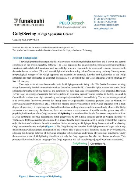
Two major methods have been used to stain the Golgi apparatus in living cells: The first is fluorescent staining<br />
using fluorescently labeled ceramide derivatives (hereafter ceramide-FL). Ceramide lipids accumulate in the Golgi<br />
apparatus during the metabolic pathway, and ceramide-FLs have been used to visualize the Golgi apparatus. However,<br />
1) The Golgi selectivity of ceramide derivatives is low, 2) Ceramide derivatives also localize to the ER, etc., and 3)<br />
Ceramide derivatives have high cytotoxicity and are quickly metabolized intracellularly. The second staining method<br />
is to overexpress fluorescent proteins by fusing them to Golgi-specific expressed proteins (such as Giantin, N-<br />
acetylgalactosaminyltransferase, etc.). While this method allows visualization of the Golgi apparatus with a high<br />
degree of specificity, it requires prior plasmid transfection, making it impossible to immediately observe the Golgi<br />
apparatus when necessary. Furthermore, there are concerns overexpression of specific marker genes may affect<br />
physiological functions of the Golgi apparatus. <strong>GolgiSeeing</strong> is a novel small molecule fluorescent reagent that utilizes<br />
a Golgi apparatus selective localization motif discovered by Dr. Shinya Tsukiji's group at Nagoya Institute of<br />
Technology. Unlike conventional ceramide-FLs, it can stain the Golgi apparatus with a simple protocol that requires<br />
only 10 minutes of addition to the culture medium. It also shows higher Golgi specificity than ceramide-FLs, allowing<br />
Golgi apparatus-focused analysis (Figure 1). The <strong>GolgiSeeing</strong> can visualize the Golgi apparatus of target cells at any<br />
desired timing without genetic manipulation and without bias to physiological functions caused by overexpression,<br />
allowing the dynamic behavior of the Golgi apparatus to be observed under more physiological conditions. Under<br />
the non-wash protocol, <strong>GolgiSeeing</strong> visualizes not only the Golgi apparatus but also the plasma membrane. This<br />
property allows simultaneous imaging of the Golgi apparatus and cell morphology (judged by plasma membrane).<br />
Figure 1. Overview image of <strong>GolgiSeeing</strong>
<strong>GolgiSeeing</strong> (original compound name; mgc 3Me FDA in Ref.1) has a unique structure consisting of fluorescein<br />
diacetate (FDA) and N-myristoylated Gly-Cys dipeptide (mgc) linked by a linker, and three amide bonds in the mgc<br />
peptide chain are all methylated (Figure 2 left, upper). <strong>GolgiSeeing</strong> functions as a Golgi apparatus-selective<br />
fluorescent probe through several processes in the cell. FDA is a modified fluorescein quenched to improve cell<br />
membrane permeability, and <strong>GolgiSeeing</strong> emits slight fluorescence before use. After <strong>GolgiSeeing</strong> penetrates the cell<br />
membrane and enters the cell, acetyl groups are removed by endogenous esterases, and the green fluorescence of<br />
fluorescein is restored (Figure 2 left, middle). The deacetylated form of <strong>GolgiSeeing</strong> transiently localizes mainly to<br />
the ER and Golgi apparatus by the effect of N-myristoyl groups and is S-palmitoylation by endogenous S-<br />
palmitoylation enzymes. The Golgi apparatus selective localization motif is formed by adding palmitic acid to the<br />
Cys side chain by enzymatic action (Figure 2 left, lower). Although the intrinsic palmitoylation modification is<br />
known to localize to the Golgi apparatus and the plasma membrane, the effect of the three methyl groups biases the<br />
equilibrium toward the Golgi, which allows <strong>GolgiSeeing</strong> to selectively visualize the Golgi apparatus (Figure 2 right).<br />
Figure 2. Principle of <strong>GolgiSeeing</strong><br />
Table Live cell Golgi apparatus imaging methods<br />
Category Genetically encoded Fluorescent probes<br />
Staining<br />
Fluorescent protein-fused Golgi<br />
apparatus marker proteins<br />
Fluorescent-labeled ceramide<br />
derivatives (Ceramide-FL)<br />
Golgi apparatus specificity High Low<br />
(High ER background)<br />
Protocol<br />
Easy<br />
Complicated<br />
(Exogenous gene transfection) (Require stepwise and<br />
temperature-controlled protocol)<br />
Time<br />
> half day<br />
(time for protein expression)<br />
Influence on physiological<br />
function<br />
High<br />
(Overexpression of exogenous<br />
proteins)<br />
<strong>GolgiSeeing</strong><br />
>1 hour 10 min<br />
High<br />
(Exogenous ceramide’s toxicity)<br />
High<br />
(Low ER background)<br />
Easy<br />
(Just addition to medium)<br />
Low<br />
[ver. 2023/08] Download the latest datasheet from www.funakoshi.co.jp (Japanese)<br />
www.funakoshi.co.jp/exports (English)
Description<br />
Catalog Number: FDV-0053<br />
Size: 0.1 mg<br />
Molecular weight: 1258.5 g/mol<br />
Solubility: Soluble in DMSO<br />
Fluorescent characteristics:<br />
Ex. 440-500 nm (maximum ~480 nm) / Em 500-560 nm (maximum ~520 nm)<br />
Compatible with conventional FITC filter set<br />
*NOTE: <strong>GolgiSeeing</strong> has fluorescein diacetate (FDA), which is quenched by two acetates and emits a slight<br />
fluorescence. After hydrolysis of two acetic groups in cells by physiological esterases, fluorescein<br />
is exposed and restores strong green fluorescence.<br />
Reconstitution and Storage<br />
Reconstitution: Stock solution recommended concentration 1 mM in 100% DMSO. Add 79 L of DMSO/vial to<br />
prepare 1 mM stock solution.<br />
Storage (powder): Store powder at less than -20 o C<br />
Storage (solution): After reconstitution in DMSO, aliquot and store at less than -20°C.<br />
Avoid repeated freeze-thaw cycles and recommend single use of each aliquot.<br />
Important notice for stability<br />
<strong>GolgiSeeing</strong> has a free-thiol group (See Figure 2 left), which is S-palmitoylated in cells and essential for Golgi<br />
apparatus localization. This thiol group is easily oxidized to form a homodimer in physiological pH buffers, and<br />
<strong>GolgiSeeing</strong>-dimer loses its Golgi apparatus localization function and may cause a background signal.<br />
<strong>GolgiSeeing</strong> working solution (such as <strong>GolgiSeeing</strong>-containing medium or buffers) should be prepared just<br />
before use. Importantly, <strong>GolgiSeeing</strong> may be gradually oxidized even in DMSO. After reconstitution of<br />
<strong>GolgiSeeing</strong> in DMSO, use within one month to maintain good staining results.<br />
How to use<br />
General procedure for Golgi apparatus-selective staining in live cells<br />
*This procedure is an example of Golgi apparatus-selective staining<br />
1. Prepare 10 M <strong>GolgiSeeing</strong> in serum-free medium such as DMEM just before use<br />
NOTE: Empirically optimize and determine the concentration of <strong>GolgiSeeing</strong> for your experiments.<br />
2. Remove the culture medium and wash cells with medium several times<br />
3. Add <strong>GolgiSeeing</strong>-containing medium to cells<br />
NOTE: Working solution of <strong>GolgiSeeing</strong> prepared in step-1 should be quickly used, as <strong>GolgiSeeing</strong> may be<br />
oxidazed to form inactive dimer in medium.<br />
4. Incubate cells at RT for 10 min<br />
NOTE: Empirically optimize incubation time and temperature for your experiments.<br />
5. Wash cells with 3 mg/ml BSA-containing medium over two times and add fresh serum-free and phenol redfree<br />
medium<br />
NOTE: <strong>GolgiSeeing</strong> is a highly hydrophobic compound and non-specifically absorbed to plastic or glass<br />
surface of imaging chambers. This non-specific absorption may cause the background signal of<br />
fluorescent images. To reduce non-specific signals, we highly recommend using a wash buffer<br />
containing BSA because BSA efficiently binds to excess <strong>GolgiSeeing</strong> and removes it from the surface<br />
of imaging chambers. 3 mg/ml BSA-containing basal medium is the preferred choice of <strong>GolgiSeeing</strong>wash<br />
buffer.<br />
6. Observe cells under live cell condition
General procedure for Golgi apparatus and plasma membrane staining in live cells<br />
*This procedure is an example of Golgi apparatus and plasma membrane staining<br />
1. Prepare 2 M <strong>GolgiSeeing</strong> in serum-free and phenol red-free medium such as DMEM just before use<br />
NOTE: Empirically optimize and determine the concentration of <strong>GolgiSeeing</strong> for your experiments.<br />
2. Remove the culture medium and wash cells with medium several times<br />
3. Add <strong>GolgiSeeing</strong>-containing medium to cells<br />
NOTE: Working solution of <strong>GolgiSeeing</strong> prepared in step-1 should be quickly used, as <strong>GolgiSeeing</strong> may be<br />
oxidazed to form inactive dimer in medium.<br />
4. Incubate cells at RT for 10 min<br />
NOTE: Empirically optimize incubation time temperature for your experiments.<br />
6. Observe cells without wash-out step under live cell condition<br />
Important Notice of Use<br />
1) <strong>GolgiSeeing</strong> will selectively localize to the Golgi apparatus through the S-palmitoylation on the free-thiol group<br />
by endogenous S-palmitoylation enzymes, as mentioned in Figure 2. Note that any drugs inhibiting S-<br />
palmitoylation may influence the Golgi apparatus-selective staining property of <strong>GolgiSeeing</strong>. Especially,<br />
alkylation reagents of the free thiol (such as maleimide, iodoacetate, etc.) will critically inhibit S-palmitoylation<br />
and are incompatible with <strong>GolgiSeeing</strong>.<br />
2) <strong>GolgiSeeing</strong> has no fixable functional groups in the molecule and is not fixed by either paraformaldehyde<br />
(PFA) or methanol. <strong>GolgiSeeing</strong> is a specialized reagent for live cell imaging applications, not compatible with<br />
fixed cell imaging and immunocytochemistry.<br />
3) Long incubation time of <strong>GolgiSeeing</strong> may increase ER-derived fluorescent signal and reduce Golgi/ER ratio.<br />
Conduct empirical optimization ranges for your experiments' incubation time and observation time course.<br />
Reference data<br />
Comparison between <strong>GolgiSeeing</strong> and a ceramide-based reagent<br />
HeLa cells were treated with 10 M <strong>GolgiSeeing</strong> or 5 M ceramide-FL (as BSA complex). In the case of <strong>GolgiSeeing</strong>,<br />
the protocol is a simple addition of <strong>GolgiSeeing</strong> into media final 10 M and incubated for 10 min. After washing<br />
cells with 3 mg/ml BSA containing media, fluorescent images were captured by confocal laser microscopy (Ex 488<br />
nm/Em 500-600 nm). On the other hand, ceramide-FL<br />
(BSA complex) -staining was performed by two<br />
protocols, simple addition or stepwise temperaturecontrolled<br />
protocol. The later protocol cells were<br />
incubated with ceramide-FL (BSA complex) for 30 min<br />
at 4 o C in HBSS, washed with ice-cold HBSS, and<br />
incubated in a fresh culture medium for an additional<br />
30 min at 37 o C. Finally, cells were washed with a fresh<br />
medium again and observed by confocal laser<br />
microscopy. This stepwise protocol requires over 1<br />
hour. The ceramide-FL probe stains not only the Golgi<br />
apparatus but also ER structure with high intensity nonspecifically.<br />
<strong>GolgiSeeing</strong> was able to visualize Golgi<br />
apparatus selectively and suppressed non-specific ER<br />
staining.<br />
[ver. 2023/08] Download the latest datasheet from www.funakoshi.co.jp (Japanese)<br />
www.funakoshi.co.jp/exports (English)
Organelle specificity<br />
HeLa cells were co-stained with <strong>GolgiSeeing</strong> and organelle<br />
markers. In the case of the Golgi apparatus marker,<br />
overexpression of mCherry-Giantin fusion protein was used. In<br />
the cases of ER, mitochondria, and lysosome, each organelle was<br />
stained by organelle-specific chemical probes. The fluorescent<br />
signal from <strong>GolgiSeeing</strong> is well corresponded with Golgi-marker<br />
and weakly overlaps with ER. However, the signals are not<br />
matched with mitochondria and lysosomes.<br />
Effect of wash-out step on plasma membrane staining<br />
HeLa cells were treated with 10 M <strong>GolgiSeeing</strong> for 10<br />
min and observed by confocal laser microscopy (Ex 488<br />
nm/Em 500-600 nm) with or without the wash step. For<br />
wash-out, 3 mg/ml BSA-containing medium was used as<br />
wash buffer. In the case of non-wash observation, the<br />
fluorescent signals of <strong>GolgiSeeing</strong> were observed in not<br />
only the Golgi apparatus but also the plasma membrane.<br />
As the Golgi apparatus is clearly distinguished from the<br />
plasma membrane, non-wash staining is useful for<br />
simultaneous observation of the Golgi apparatus and cell<br />
morphology (the shape of the plasma membrane). The<br />
wash-out step dramatically reduced the fluorescent signal<br />
from the plasma membrane.<br />
Various cell staining<br />
Four types of cultured cells (HeLa, COS-7, HEK293,<br />
and Jurkat) were treated with 10 M <strong>GolgiSeeing</strong> for 10<br />
min. After cell wash-out, cells were observed by<br />
confocal laser microscopy (Ex 488 nm/ Em 500-600<br />
nm). For all cells tested here, <strong>GolgiSeeing</strong> highly<br />
selectively stained the Golgi apparatus.
Cellular toxicity<br />
HeLa cells were seeded into a 12-well plate at 0.5 x 10 5<br />
cells/well and cultured for 48 hours with/without 10 M<br />
<strong>GolgiSeeing</strong>. After 2, 24, and 48 hours, cell numbers<br />
were assessed by a cell counter. <strong>GolgiSeeing</strong> shows<br />
little effect on cell proliferation.<br />
Application data<br />
Live cell time-lapse imaging on Brefeldin A-induced collapse of Golgi apparatus<br />
HeLa cells were seeded on glass bottom dishes and treated with 10 M <strong>GolgiSeeing</strong> for 10 min. After washing cells<br />
with 3 mg/ml BSA-containing media, the cells were cultured in brefeldin A (final 1 M in 0.1% DMSO)-containing<br />
medium or 0.1 % DMSO-containing medium as a negative control and observed under live cell condition by confocal<br />
microscopy (Ex 488 nm/Em 500-600 nm). In the brefeldin A-treated cells, the fluorescent signal from the Golgi<br />
apparatus gradually disappeared.<br />
Live cell time-lapse imaging during cell division<br />
MDCK cells were seeded on a glass bottom dish, incubated with 2.5 M <strong>GolgiSeeing</strong>, and observed for 2 hours by<br />
confocal laser microscopy (Ex 488 nm/ Em 500-600 nm) without washing step. As a non-wash procedure, the<br />
fluorescent signal of <strong>GolgiSeeing</strong> were observed not only in Golgi apparatus but also in plasma membrane. By<br />
imaging of plasma mebrane structures, cell morphology was easily observed. According to the progression of cell<br />
division, the Golgi apparatus disappeared (60 min) and was reconstituted in two daughter cells (90-120 min).<br />
[ver. 2023/08] Download the latest datasheet from www.funakoshi.co.jp (Japanese)<br />
www.funakoshi.co.jp/exports (English)
Reference<br />
1. Sawada et al.,ACS Chem. Biol., 18, 1047-1053 (2023) Palmitoylation-Dependent Small-Molecule Fluorescent<br />
Probes for Live-Cell Golgi Imaging.<br />
Disclaimer/ 免 責 事 項<br />
This product has been commercialized by Funakoshi Co., Ltd.<br />
based on the results of academic research, and the advertisement<br />
text, figures and manuals (hereinafter “Product information”) have<br />
been prepared based on published research reports on August,<br />
2023. The academic interpretation at the time of creation of the<br />
Product Information may change in accordance with future<br />
developments in the relevant research field and expansion of<br />
various scientific findings, and the latest version and certainty of<br />
the Product Information are not guaranteed. The specifications of<br />
this product and the Product Information are subject to change<br />
without notice. Please contact us for the latest information.<br />
本 製 品 は 学 術 研 究 成 果 を 基 にフナコシ 株 式 会 社 が 製 品 化 したもので、<br />
2023 年 8 月 時 点 における 公 開 研 究 報 告 を 基 に 広 告 文 章 およびマニュ<br />
アル( 以 下 、 製 品 資 料 )を 作 成 しています。 今 後 の 当 該 研 究 分 野 の 発<br />
展 および 各 種 学 術 知 見 の 拡 大 にともない、 製 品 資 料 作 成 時 の 学 術 的<br />
解 釈 が 変 更 になる 可 能 性 があり、 最 新 性 ・ 確 実 性 を 保 証 するものでは<br />
ありません。また、 本 製 品 の 仕 様 および 製 品 資 料 を 予 告 なく 変 更 する<br />
場 合 がございます。 最 新 の 情 報 に 関 しましては、 弊 社 までご 確 認 いた<br />
だきますようお 願 い 申 し 上 げます。<br />
Your Local Distributor<br />
Distributed by PELOBIOTECH GmbH<br />
(P) +49 89 517 286 59-0<br />
(F) +49 89 517 286 59-88<br />
Email: info@pelobiotech.com<br />
www.pelobiotech.com
Related products<br />
NucleoSeeing <br />
NucleoSeeing is DNA-responsive green dye for monitoring cell nucleus in live cells. As it shows low<br />
cytotoxicity and phototoxicity, it is very suitable for long-term live imaging of cell nucleus.<br />
Catalog No. FDV-0029<br />
Size 0.1 mg<br />
Features<br />
- Easy and quick procedure<br />
- Compatible with 10% FBS<br />
- Validated for both adherent cells and floating cells<br />
- Little influence on cellular functions<br />
- Ex/Em: 488 nm/520 nm (commercial FITC filters are available)<br />
ERseeing <br />
ERseeing is a novel type of ER-staining dye and shows little<br />
pharmacological effects compared with conventional<br />
glibenclamide-based ER dyes. ERseeing is irreversible<br />
staining and is compatible with medium change for long-term<br />
imaging.<br />
Catalog No. FDV-0038<br />
Size 10 nmol<br />
Features<br />
- Recommended Ex/Em: 509 nm/524 nm<br />
- Lless pharmacological effect on ER proteins<br />
- Suitable for long-term live cell imaging<br />
LipiDye II <br />
LipiDye II is a highly sensitive lipid droplet staining dye with extremely photostable property. This dye is the second<br />
generation of our previous reagent, LipiDye. This dye allows us to detect small lipid droplets (