GolgiSeeing
GolgiSeeing is a novel and innovative fluorescent dye for selective Golgi apparatus staining. Compared to conventional ceramide-based Golgi staining reagents, GolgiSeeing does not require complicated procedures and suppresses non-specific localization to the endoplasmic reticulum.
GolgiSeeing is a novel and innovative fluorescent dye for selective Golgi apparatus staining. Compared to conventional ceramide-based Golgi staining reagents, GolgiSeeing does not require complicated procedures and suppresses non-specific localization to the endoplasmic reticulum.

Create successful ePaper yourself
Turn your PDF publications into a flip-book with our unique Google optimized e-Paper software.
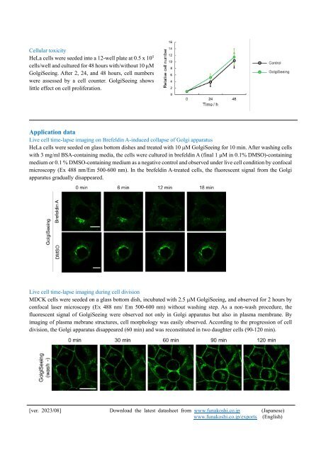
Cellular toxicity<br />
HeLa cells were seeded into a 12-well plate at 0.5 x 10 5<br />
cells/well and cultured for 48 hours with/without 10 M<br />
<strong>GolgiSeeing</strong>. After 2, 24, and 48 hours, cell numbers<br />
were assessed by a cell counter. <strong>GolgiSeeing</strong> shows<br />
little effect on cell proliferation.<br />
Application data<br />
Live cell time-lapse imaging on Brefeldin A-induced collapse of Golgi apparatus<br />
HeLa cells were seeded on glass bottom dishes and treated with 10 M <strong>GolgiSeeing</strong> for 10 min. After washing cells<br />
with 3 mg/ml BSA-containing media, the cells were cultured in brefeldin A (final 1 M in 0.1% DMSO)-containing<br />
medium or 0.1 % DMSO-containing medium as a negative control and observed under live cell condition by confocal<br />
microscopy (Ex 488 nm/Em 500-600 nm). In the brefeldin A-treated cells, the fluorescent signal from the Golgi<br />
apparatus gradually disappeared.<br />
Live cell time-lapse imaging during cell division<br />
MDCK cells were seeded on a glass bottom dish, incubated with 2.5 M <strong>GolgiSeeing</strong>, and observed for 2 hours by<br />
confocal laser microscopy (Ex 488 nm/ Em 500-600 nm) without washing step. As a non-wash procedure, the<br />
fluorescent signal of <strong>GolgiSeeing</strong> were observed not only in Golgi apparatus but also in plasma membrane. By<br />
imaging of plasma mebrane structures, cell morphology was easily observed. According to the progression of cell<br />
division, the Golgi apparatus disappeared (60 min) and was reconstituted in two daughter cells (90-120 min).<br />
[ver. 2023/08] Download the latest datasheet from www.funakoshi.co.jp (Japanese)<br />
www.funakoshi.co.jp/exports (English)