GolgiSeeing
GolgiSeeing is a novel and innovative fluorescent dye for selective Golgi apparatus staining. Compared to conventional ceramide-based Golgi staining reagents, GolgiSeeing does not require complicated procedures and suppresses non-specific localization to the endoplasmic reticulum.
GolgiSeeing is a novel and innovative fluorescent dye for selective Golgi apparatus staining. Compared to conventional ceramide-based Golgi staining reagents, GolgiSeeing does not require complicated procedures and suppresses non-specific localization to the endoplasmic reticulum.

You also want an ePaper? Increase the reach of your titles
YUMPU automatically turns print PDFs into web optimized ePapers that Google loves.
General procedure for Golgi apparatus and plasma membrane staining in live cells<br />
*This procedure is an example of Golgi apparatus and plasma membrane staining<br />
1. Prepare 2 M <strong>GolgiSeeing</strong> in serum-free and phenol red-free medium such as DMEM just before use<br />
NOTE: Empirically optimize and determine the concentration of <strong>GolgiSeeing</strong> for your experiments.<br />
2. Remove the culture medium and wash cells with medium several times<br />
3. Add <strong>GolgiSeeing</strong>-containing medium to cells<br />
NOTE: Working solution of <strong>GolgiSeeing</strong> prepared in step-1 should be quickly used, as <strong>GolgiSeeing</strong> may be<br />
oxidazed to form inactive dimer in medium.<br />
4. Incubate cells at RT for 10 min<br />
NOTE: Empirically optimize incubation time temperature for your experiments.<br />
6. Observe cells without wash-out step under live cell condition<br />
Important Notice of Use<br />
1) <strong>GolgiSeeing</strong> will selectively localize to the Golgi apparatus through the S-palmitoylation on the free-thiol group<br />
by endogenous S-palmitoylation enzymes, as mentioned in Figure 2. Note that any drugs inhibiting S-<br />
palmitoylation may influence the Golgi apparatus-selective staining property of <strong>GolgiSeeing</strong>. Especially,<br />
alkylation reagents of the free thiol (such as maleimide, iodoacetate, etc.) will critically inhibit S-palmitoylation<br />
and are incompatible with <strong>GolgiSeeing</strong>.<br />
2) <strong>GolgiSeeing</strong> has no fixable functional groups in the molecule and is not fixed by either paraformaldehyde<br />
(PFA) or methanol. <strong>GolgiSeeing</strong> is a specialized reagent for live cell imaging applications, not compatible with<br />
fixed cell imaging and immunocytochemistry.<br />
3) Long incubation time of <strong>GolgiSeeing</strong> may increase ER-derived fluorescent signal and reduce Golgi/ER ratio.<br />
Conduct empirical optimization ranges for your experiments' incubation time and observation time course.<br />
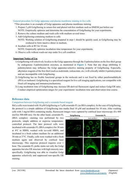
Reference data<br />
Comparison between <strong>GolgiSeeing</strong> and a ceramide-based reagent<br />
HeLa cells were treated with 10 M <strong>GolgiSeeing</strong> or 5 M ceramide-FL (as BSA complex). In the case of <strong>GolgiSeeing</strong>,<br />
the protocol is a simple addition of <strong>GolgiSeeing</strong> into media final 10 M and incubated for 10 min. After washing<br />
cells with 3 mg/ml BSA containing media, fluorescent images were captured by confocal laser microscopy (Ex 488<br />
nm/Em 500-600 nm). On the other hand, ceramide-FL<br />
(BSA complex) -staining was performed by two<br />
protocols, simple addition or stepwise temperaturecontrolled<br />
protocol. The later protocol cells were<br />
incubated with ceramide-FL (BSA complex) for 30 min<br />
at 4 o C in HBSS, washed with ice-cold HBSS, and<br />
incubated in a fresh culture medium for an additional<br />
30 min at 37 o C. Finally, cells were washed with a fresh<br />
medium again and observed by confocal laser<br />
microscopy. This stepwise protocol requires over 1<br />
hour. The ceramide-FL probe stains not only the Golgi<br />
apparatus but also ER structure with high intensity nonspecifically.<br />
<strong>GolgiSeeing</strong> was able to visualize Golgi<br />
apparatus selectively and suppressed non-specific ER<br />
staining.<br />
[ver. 2023/08] Download the latest datasheet from www.funakoshi.co.jp (Japanese)<br />
www.funakoshi.co.jp/exports (English)