DPCA2-1

Create successful ePaper yourself
Turn your PDF publications into a flip-book with our unique Google optimized e-Paper software.
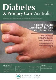
Clinical vascular screening of the foot<br />
Table 1. Medicare fees and benefits for vascular testing (Australian Government Department of Health, 2016).<br />
Test<br />
Ankle– or toe–brachial index and arterial waveform study<br />
Measurement of ankle:brachial indices and arterial waveform analysis<br />
Measurement of posterior tibial and dorsalis pedis (or toe) and brachial arterial pressures bilaterally using Doppler or<br />
plethysmographic techniques, the calculation of ankle (or toe)–brachial systolic pressure indices and assessment of arterial<br />
waveforms for the evaluation of lower extremity arterial disease, examination, hard copy trace and report.<br />
Ankle– or toe–brachial index-exercise study<br />
Exercise study for the evaluation of lower extremity arterial disease<br />
Measurement of posterior tibial and dorsalis pedis (or toe) and brachial arterial pressures bilaterally using Doppler or<br />
plethysmographic techniques, the calculation of ankle (or toe) brachial systolic pressure indices for the evaluation of lower<br />
extremity arterial disease at rest and following exercise using a treadmill or bicycle ergometer or other such equipment<br />
where the exercise workload is quantifiably documented, examination and report.<br />
Item<br />
number<br />
11610<br />
11612<br />
Fees and Medicare<br />
benefits<br />
Fee: $63.75<br />
Benefit: 75% = $47.85<br />
85% = $54.20<br />
Fee: $112.40<br />
Benefit: 75% = $84.30<br />
85% = $95.55<br />
2013; Hyun et al, 2014).<br />
Medial arterial calcification is prevalent in<br />
renal disease (An et al, 2010) and in longterm<br />
type 1 diabetes (Ix et al, 2012). It places<br />
limitations on the sensitivity of vascular<br />
pressure measurements due to the associated<br />
non-compressibility of vessels.<br />
Sensitive clinical screening methods<br />
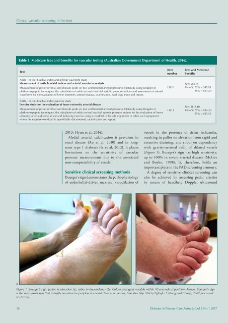
Buerger’s sign demonstrates the pathophysiology<br />
of endothelial-driven maximal vasodilation of<br />
vessels in the presence of tissue ischaemia,<br />
resulting in pallor on elevation from rapid and<br />
extensive draining, and rubor on dependency<br />
with gravity-assisted refill of dilated vessels<br />
(Figure 1). Buerger’s sign has high sensitivity,<br />
up to 100% in severe arterial disease (McGee<br />
and Boyko, 1998). It, therefore, holds an<br />
important place in the PAD screening armoury.<br />
A degree of sensitive clinical screening can<br />
also be achieved by assessing pedal arteries<br />
by means of handheld Doppler ultrasound<br />
a. b.<br />
Figure 1. Buerger’s sign: pallor in elevation (a), rubor in dependency (b). Colour change is notable within 10 seconds of position change. Buerger’s sign<br />
is the only visual sign that is highly sensitive for peripheral arterial disease screening. See also http://bit.ly/2gUqGzS (Kang and Chung, 2007 [accessed<br />
05.12.16]).<br />
18 Diabetes & Primary Care Australia Vol 2 No 1 2017