DPCA2-1

Create successful ePaper yourself
Turn your PDF publications into a flip-book with our unique Google optimized e-Paper software.
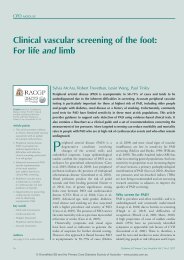
Clinical vascular screening of the foot<br />
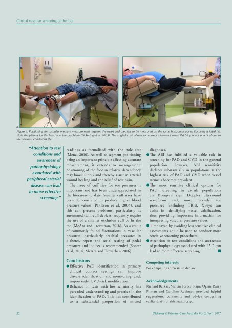
a. b.<br />
Figure 4. Positioning for vascular pressure measurement requires the heart and the sites to be measured on the same horizontal plane. Flat lying is ideal (a).<br />
Note the pillows for the head and the brachium (Pickering et al, 2005). The angled chair allows for correct alignment when flat lying is not practical due to<br />
the person’s conditions (b).<br />
“Attention to test<br />
conditions and<br />
awareness of<br />
pathophysiology<br />
associated with<br />
peripheral arterial<br />
disease can lead<br />
to more effective<br />
screening.”<br />
readings as formalised with the pole test<br />
(Menz, 2010). As well as segment positioning<br />
being an important principle affecting accurate<br />
measurement, it extends to management:<br />
positioning of the foot in relative dependency<br />
may boost supply and thereby assist in arterial<br />
wound healing and the relief of rest pain.<br />
The issue of cuff size for toe pressures is<br />
important and has been underappreciated in<br />
the literature to date. Smaller cuff sizes have<br />
been demonstrated to produce higher blood<br />
pressure values (Påhlsson et al, 2004), and<br />
this can present problems, particularly as<br />
automated twin-cuff devices frequently require<br />
the use of a smaller occlusion cuff to fit the<br />
toe (McAra and Trevethan, 2016). As a result<br />
of commonly found fluctuations in vascular<br />
pressures, particularly brachial pressures in<br />
diabetes, repeat and serial testing of pedal<br />
pressures and indices is recommended (Sonter<br />
et al, 2014; McAra and Trevethan 2016).<br />
Conclusions<br />
l Effective PAD identification in primary<br />
clinical contact settings can improve<br />
disease identification and monitoring, and,<br />
importantly, CVD-risk modification.<br />
l Reliance on tests with low sensitivity has<br />
pervaded understanding and practice in the<br />
identification of PAD. This has contributed<br />
to a substantial proportion of missed<br />
diagnoses.<br />
l The ABI has fulfilled a valuable role in<br />
screening for PAD and CVD in the general<br />
population. However, ABI sensitivity<br />
declines substantially in populations at the<br />
highest risk of PAD and CVD when vessel<br />
stenosis becomes prevalent.<br />
l The most sensitive clinical options for<br />
PAD screening in at-risk populations<br />
are Buerger’s sign, Doppler ultrasound<br />
waveforms and, more recently, toe<br />
pressures (including TBIs). X-rays can<br />
assist in identifying vessel calcification,<br />
thus providing important information for<br />
interpreting vascular pressure values.<br />
l Time saved by avoiding less sensitive clinical<br />
assessments could be used to conduct more<br />
sensitive screening procedures.<br />
l Attention to test conditions and awareness<br />
of pathophysiology associated with PAD can<br />
lead to more effective screening. n<br />
Competing interests<br />
No competing interests to declare.<br />
Acknowledgements<br />
Richard Barkas, Martin Forbes, Rajna Ogrin, Barry<br />
Pitman and Caroline Robinson provided helpful<br />
suggestions, comments and advice concerning<br />
earlier drafts of this manuscript.<br />
22 Diabetes & Primary Care Australia Vol 2 No 1 2017