DPCA2-1

Create successful ePaper yourself
Turn your PDF publications into a flip-book with our unique Google optimized e-Paper software.
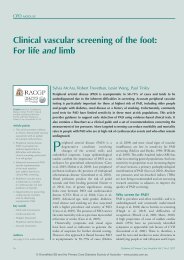
Clinical vascular screening of the foot<br />
2006; Aboyans et al, 2008; World Health<br />
Organization, 2013; Brownrigg et al, 2016),<br />
particularly when risks can be modified with<br />
interventions (Hinchcliffe et al, 2015).<br />
Prevalence, identification and<br />
classification of PAD<br />
Estimates of the prevalence of PAD vary<br />
widely from 4–57%, depending on how the<br />
disease is identified and on age and risk factor<br />
distributions in specific populations (Caro et<br />
al, 2005). In a summary statement about<br />
prevalence, Høyer et al (2013) cite evidence<br />
that more than 50% of people with PAD are<br />
asymptomatic.<br />
PAD is best known for ischaemic pain<br />
associated with intermittent claudication.<br />
However, in a large study, only 11% of people<br />
with PAD had intermittent claudication (Hirsch<br />
et al, 2001). The prevalence of pathology<br />
is similar in symptomatic and asymptomatic<br />
PAD (Diehm et al, 2009), but significant<br />
impairment of the vascular tree often exists<br />
before and without any symptoms or signs.<br />
Previously, the severity of PAD has been<br />
described and stratified using the symptoms<br />
of claudication and rest pain, then tissue<br />
death, as in the Rutherford and Fontaine<br />
classification systems (Mills et al, 2014). Due to<br />
a growing appreciation of both the prevalence<br />
and pathological significance of asymptomatic<br />
PAD, new international guidelines for vascular<br />
surgery contain recommendations that pedal<br />
risk stratification be based, instead of on<br />
symptoms, on an algorithm including foot<br />
wound status, ischaemia and infection (Mills<br />
et al, 2014).<br />
Standard CVD risk scores, such as the<br />
Framingham risk score, have low-, middleand<br />
high-risk stratification categories. The<br />
diagnostic utility of these indicators may be<br />
improved by adding non-invasive clinical pedal<br />
vascular assessment to identify asymptomatic<br />
PAD in the intermediate risk group (Greenland<br />
et al, 2001).<br />
Limitations in screening for PAD<br />
A major limitation associated with screening<br />
for PAD is that there is currently no agreement<br />
concerning the use of any single test or<br />
combination of tests to detect PAD in primary<br />
healthcare settings. People’s medical history, as<br />
well as their pulses, pedal Doppler waveforms,<br />
ABIs and TBIs, are quoted in guidelines as<br />
being strongly recommended, but there is little<br />
evidence to support their use (Hinchcliffe et<br />
al, 2015). Most clinical tests used for PAD<br />
screening have low sensitivity and therefore fail<br />
to identify a large proportion of people who have<br />
the disease (Williams et al, 2005; Brownrigg et<br />
al, 2016). Many people with PAD have no<br />
obvious visual signs, and visual signs such as<br />
skin colour, lack of hair growth, nail changes<br />
and skin atrophy are low in sensitivity for PAD<br />
detection (Williams et al, 2005; Menz, 2010).<br />
In addition to visual screening, standard clinical<br />
tests include assessment of pulses, impressions<br />
of skin temperature, capillary refilling time and<br />
possibly ABIs. These screening processes may<br />
underestimate PAD by up to 60% (Williams<br />
et al, 2005; Høyer et al, 2013). Pulse palpation,<br />
although a useful clinical skill, is not adequate<br />
as a primary screening tool for PAD due to its<br />
variable sensitivity, which declines as vascular<br />
disease states advance (McGee and Boyko,<br />
1998; Williams et al, 2005).<br />
The ABI has been the cornerstone of peripheral<br />
vascular assessment in primary care for PAD and<br />
associated CVD risk, and it is supported by four<br />
decades of evidence (Caruana et al, 2005; Rooke<br />
et al, 2011). However, when Australian GPs were<br />
surveyed for the barriers they experienced in<br />
performing vascular assessment, 58% indicated<br />
that they did not use ABIs to perform vascular<br />
assessments, with time constraints stated as the<br />
greatest barrier, followed by lack of equipment<br />
and skills (Haigh et al, 2013). This is despite<br />
Medicare rebates currently applying for both<br />
toe- and ankle-pressure studies (See Table 1).<br />
The ABI is useful for identifying CVD<br />
risk in the general population (Caruana et al,<br />
2005; Guo et al, 2008). However, its sensitivity<br />
is reduced in proportion to the degree of<br />
atherosclerosis and vascular stenosis, both of<br />
which are common in people of advanced age<br />
and those who have complications of diabetes,<br />
especially neuropathy (Aboyans et al, 2008; Xu<br />
et al, 2010; Craike et al, 2013; Formosa et al,<br />
Page points<br />
1. Estimates of the prevalence<br />
of peripheral arterial disease<br />
(PAD) vary widely from 4–57%,<br />
depending on how the disease<br />
is identified and on age and risk<br />
factor distributions in specific<br />
populations.<br />
2. A major limitation associated<br />
with screening for PAD is that<br />
there is currently no agreement<br />
concerning the use of any<br />
single test or combination of<br />
tests to detect PAD in primary<br />
healthcare settings.<br />
Diabetes & Primary Care Australia Vol 2 No 1 2017 17