FM JANUARY 2019 - digital edition

You also want an ePaper? Increase the reach of your titles
YUMPU automatically turns print PDFs into web optimized ePapers that Google loves.
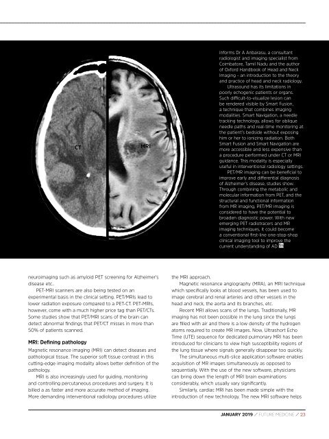
CT<br />
MRI<br />
informs Dr A Anbarasu, a consultant<br />
radiologist and imaging specialist from<br />
Coimbatore, Tamil Nadu and the author<br />
of Oxford Handbook of Head and Neck<br />
Imaging - an introduction to the theory<br />
and practice of head and neck radiology.<br />
Ultrasound has its limitations in<br />
poorly echogenic patients or organs.<br />
Such difficult-to-visualize lesion can<br />
be rendered visible by Smart Fusion,<br />
a technique that combines imaging<br />
modalities. Smart Navigation, a needle<br />
tracking technology, allows for oblique<br />
needle paths and real-time monitoring at<br />
the patient’s bedside without exposing<br />
him or her to ionizing radiation. Both<br />
Smart Fusion and Smart Navigation are<br />
more accessible and less expensive than<br />
a procedure performed under CT or MRI<br />
guidance. This modality is especially<br />
useful in interventional radiology settings.<br />
PET/MR imaging can be beneficial to<br />
improve early and differential diagnosis<br />
of Alzheimer’s disease, studies show.<br />
Through combining the metabolic and<br />
molecular information from PET, and the<br />
structural and functional information<br />
from MR imaging, PET/MR imaging is<br />
considered to have the potential to<br />
broaden diagnostic power. With new<br />
emerging PET radiotracers and MR<br />
imaging techniques, it could become<br />
a conventional first-line one-stop-shop<br />
clinical imaging tool to improve the<br />
current understanding of AD<br />
neuroimaging such as amyloid PET screening for Alzheimer’s<br />
disease etc..<br />
PET-MRI scanners are also being tested on an<br />
experimental basis in the clinical setting. PET/MRIs lead to<br />
lower radiation exposure compared to a PET-CT. PET-MRIs,<br />
however, come with a much higher price tag than PET/CTs.<br />
Some studies show that PET/MRI scans of the brain can<br />
detect abnormal findings that PET/CT misses in more than<br />
50% of patients scanned.<br />
MRI: Defining pathology<br />
Magnetic resonance imaging (MRI) can detect diseases and<br />
pathological tissue. The superior soft tissue contrast in this<br />
cutting-edge imaging modality allows better definition of the<br />
pathology.<br />
MRI is also increasingly used for guiding, monitoring<br />
and controlling percutaneous procedures and surgery. It is<br />
billed a as faster and more accurate method of imaging.<br />
More demanding interventional radiology procedures utilize<br />
the MRI approach.<br />
Magnetic resonance angiography (MRA), an MRI technique<br />
which specifically looks at blood vessels, has been used to<br />
image cerebral and renal arteries and other vessels in the<br />
head and neck, the aorta and its branches, etc.<br />
Recent MRI allows scans of the lungs. Traditionally, MR<br />
imaging has not been possible in the lung since the lungs<br />
are filled with air and there is a low density of the hydrogen<br />
atoms required to create MR images. Now, Ultrashort Echo<br />
Time (UTE) sequence for dedicated pulmonary MRI has been<br />
introduced for clinicians to view high susceptibility regions of<br />
the lung tissue where signals generally disappear too quickly.<br />
The simultaneous multi-slice application software enables<br />
acquisition of MR images simultaneously as opposed to<br />
sequentially. With the use of the new software, physicians<br />
can bring down the length of MRI brain examinations<br />
considerably, which usually vary significantly.<br />
Similarly, cardiac MRI has been made simple with the<br />
introduction of new technology. The new MRI software helps<br />
<strong>JANUARY</strong> <strong>2019</strong> / FUTURE MEDICINE / 23