FM JANUARY 2019 - digital edition

Create successful ePaper yourself
Turn your PDF publications into a flip-book with our unique Google optimized e-Paper software.
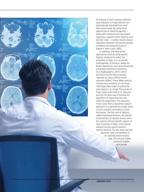
for training. In both learning methods,<br />
large datasets of image features are<br />
automatically extracted from each<br />
data point/image. By using these<br />
approaches of machine learning —<br />
along with statistical tools like logistic<br />
regression, support vector machine and<br />
decision trees — a better, feature-based<br />
separation between normal and disease<br />
conditions are achieved (Cortes C,<br />
Vapnik V. Mach Learn 1995).<br />
In radiology, the data-driven<br />
approaches work by using specific<br />
features designed to reflect the<br />
properties of data, such as density,<br />
heterogeneity of tumours, shape etc.<br />
Newer approaches are being developed<br />
using deep learning (Chartrand G,<br />
et al, Radiographics 2017), which<br />
are improving the feature-based<br />
methods by using artificial neural<br />
networks (ANNs). These ANNs work by<br />
introducing a hierarchy of non-linear,<br />
multi-layer data nodes including the<br />
pixel values in an image. Thousands of<br />
these nodes with millions of networks<br />
become the best way of training the<br />
algorithms to respond to the new<br />
inputs for diagnostics. This approach<br />
moves ways from a hypothesis-based<br />
approach to a data-driven model, which<br />
is more powerful and leads to novel<br />
discoveries. The first sets of features,<br />
called engineered features, are specific<br />
characteristics of disease tissues which<br />
are used by domain-specific experts. In<br />
case of scarcity of data, a pre-trained<br />
network can be used to perform<br />
transfer learning. For any deep learning<br />
approach, data normalisation is<br />
an essential preprocessing<br />
step. This ensures better<br />
numerical stability<br />
and quicker<br />
<strong>JANUARY</strong> <strong>2019</strong> / FUTURE MEDICINE / 29