Create successful ePaper yourself
Turn your PDF publications into a flip-book with our unique Google optimized e-Paper software.
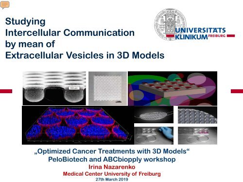
Studying<br />
Intercellular Communication<br />
by mean of<br />
Extracellular Vesicles in 3D Models<br />
„Optimized Cancer Treatments with 3D Models“<br />
PeloBiotech and ABCbiopply workshop<br />
Irina Nazarenko<br />
Medical Center University of Freiburg<br />
27th March <strong>2019</strong>
Progressing tumor attracts and re-programs<br />
stromal and immune cells<br />
Santi et al, Proteomics, 2017<br />
2 · April 4, <strong>2019</strong>
Tumor releases into microenvironment variety of<br />
different factors<br />
miRNA<br />
mRNA<br />
3 · 4. April <strong>2019</strong><br />
Schwarzenbach et al., Nature Review 2011
4 · April 4, <strong>2019</strong><br />
Emerging complexity of communication mechanisms
Publications frequency of studies investigating<br />
<strong>EVs</strong> in cancer<br />
Keywords used <strong>for</strong> PubMed search<br />
ISEV was founded<br />
in 2011 (Paris, France)<br />
2011 2012 2013 2014 2015 2016 2017 2018<br />
5 · April 4, <strong>2019</strong>
Relevance of extracellullar vesicles <strong>for</strong> liquid biopsy<br />
Modifed - from Stella Kourembanas, Annu. Rev. Physiol.<br />
2015. 77:13–27.<br />
6 · April 4, <strong>2019</strong>
Versatile EV functions in cancer<br />
Kogure et al., Journal of Biomedical Science; <strong>2019</strong><br />
7 · April 4, <strong>2019</strong>
Emerging diversity of extracellular vesicles<br />
EV<br />
Exosomes<br />
50-150 nm<br />
Microvesicles<br />
120-500 nm<br />
Large<br />
Oncosomes<br />
500-800 nm<br />
Apoptotic<br />
Bodies<br />
0.5-4 µm<br />
8 · April 4, <strong>2019</strong>
Biogenesis of extracellular vesicles: microvesicles and exosomes<br />
exosomes<br />
microvesicles<br />
Van Niel and Raposo, Nature Review 2017<br />
9 · April 4, <strong>2019</strong>
Proteomics analysis of small <strong>EVs</strong> (mostly exosomes) and large <strong>EVs</strong> (mostly<br />
microvesicles and oncosomes) show high discrepancy in protein content<br />
Micianchi et al., Oncotarget 2015<br />
10 · April 4, <strong>2019</strong>
The need of physiological in vitro models<br />
in vitro<br />
in vivo<br />
www.yogabox.de<br />
11 · April 4, <strong>2019</strong>
Development of a 3D model<br />
<strong>for</strong> EV production and analysis<br />
Requirements <strong>for</strong> EV production:<br />
‣ 3D environment in serum-free, or EV-free culture medium<br />
‣ Control of cell viability, proliferation, apoptosis and necrosis<br />
‣ High amount of <strong>EVs</strong> produced<br />
‣ Efficient and easy recovery of <strong>EVs</strong> from the 3D cell culture<br />
‣ Possibility to upscale the approach<br />
Requirements <strong>for</strong> modelling of tumor microenvironment<br />
‣ Easy analysis – microscopy, immunohistochemistry<br />
‣ Possibility of co-culture with stroma cells and other<br />
components of tumor microenvironment<br />
12 · April 4, <strong>2019</strong>
Application of 3DCoSeedis TM<br />
to produce and study <strong>EVs</strong> in 3D environment<br />
Models:<br />
Prostate cancer<br />
Breast cancer<br />
Gastrointestinal cancer<br />
13 · April 4, <strong>2019</strong>
Separation of different <strong>EVs</strong> subtypes from 2D and 3D cultures<br />
Cell Culture Supernatant<br />
depletion of cell debries<br />
800 x g and 1000 x g<br />
Centrifugation: 5000 x g <strong>for</strong> 45 min at 4°C<br />
EV5<br />
large oncosomes/<br />
apoptotic bodies<br />
Centrifugation: 12000 x g <strong>for</strong> 45 min at 4°C<br />
EV12<br />
microvesicles<br />
• Ultracentrifugation: 120000 x g <strong>for</strong> 2 h at 4°C<br />
• PEG / or other reagents-based precipitation<br />
• Ultrafiltration<br />
EV120<br />
exosomes and<br />
other small <strong>EVs</strong><br />
Serial filtration steps to remove residual EV<br />
fc fraction<br />
Krafft et al., Nanomedicine 2016<br />
Klump et al., Nanomedicine 2017
Application of 3DCoSeedis TM<br />
to produce and study <strong>EVs</strong> in 3D environment<br />
Models:<br />
Prostate cancer<br />
Breast cancer<br />
Gastrointestinal cancer<br />
15 · April 4, <strong>2019</strong>
Establishment of CWR22-RV1 cells<br />
3D culture conditions<br />
1 day,<br />
FSC<br />
2 day,<br />
FSC<br />
1 day 2 day 3 day 4 day 5 day<br />
Scale bar: 50µm<br />
Input<br />
• 10^6 cells/well<br />
• 5% (EV-depleted) FCS<br />
• 1 matrix= 950 microwells<br />
• 2 matrixes = 16 ml<br />
Liliia Paniushkina<br />
TRA<strong>IN</strong>-EV<br />
ITN-Marie Curie
Comparative analysis of different EV populations from cells<br />
derived from 2D and 3D conditions<br />
Working steps<br />
2D<br />
(225ml)<br />
3D<br />
(16ml)<br />
Cell<br />
Cult<br />
ure<br />
Sup<br />
erna<br />
tant<br />
Differential centrifugation<br />
EV5 EV12 EV120<br />
OptiPrep density gradient<br />
10<br />
fractions<br />
10<br />
fractions<br />
10<br />
fractions<br />
NTA, microBCA, DLS,<br />
qNano analysis
Cells produce more <strong>EVs</strong> under 3D condtitions<br />
***<br />
*** ***<br />
1
EV populations produced under 2D and 3D conditions differ in<br />
their distribution among Optiprep gradient fractions<br />
ug/ul<br />
particles/ml<br />
fractions
qNano measurements of EV5 and EV12<br />
Optiprep fractions from 2D and 3D cultures<br />
EV5_2D,3D<br />
EV12_2D<br />
EV12_3D<br />
NP800<br />
Size range: 250-1200nm<br />
EV5 3D fraction contains<br />
large <strong>EVs</strong> 800-900 nm<br />
NP400<br />
Size range: 250-1000nm<br />
NP400<br />
Size range: 150-1100nm<br />
EV12 3D fraction is more<br />
heterogeneous as 2D<br />
fraction
qNano measurements of EV120<br />
Optiprep fractions from 2D and 3D cultures<br />
EV120_2D<br />
EV120_3D<br />
EV120_2D,<br />
3D<br />
NP100<br />
Size range: 95-<br />
190nm<br />
NP100<br />
Size range: 75-250nm<br />
NP100<br />
Size range: 150-1100nm<br />
EV120 particles from 3D<br />
are smaller
Size distribution of EV5,12,120 vesicles<br />
from 2D and 3D cultures
To sum up<br />
‣ The 3DCoSeedis TM are applicable <strong>for</strong> the EV production in a prostate cancer<br />
model<br />
‣ 3D conditions differentially affect EV population distribution<br />
23 · April 4, <strong>2019</strong>
Application of 3DCoSeedis TM<br />
to produce and study <strong>EVs</strong> in 3D environment<br />
Models:<br />
Prostate cancer<br />
Breast cancer<br />
Richa Khanduri<br />
Gastrointestinal cancer<br />
24 · April 4, <strong>2019</strong>
Studying breast cancer small <strong>EVs</strong><br />
Cel line<br />
MDA-MB-231 BT-549 MCF7 MDA-MB-361<br />
Molecular<br />
Classification<br />
Triple negative<br />
(Claudin low)<br />
Triple negative<br />
(Claudin low)<br />
Luminal A<br />
Luminal B<br />
Tumor Type Adenocarcinoma Invasive ductal<br />
carcinoma<br />
Adenocarcinoma<br />
Adenocarcinoma<br />
Source<br />
Metastasis,<br />
Pleural Effusion<br />
Primary tumor<br />
Metastasis, Pleural<br />
Effusion<br />
Metastasis, Brain<br />
Phenotype<br />
Mesenchymal<br />
(Post-EMT)<br />
Mesenchymal<br />
(Post-EMT)<br />
Epithelial<br />
Epithelial<br />
Receptor<br />
Expression<br />
ER-, PR-<br />
HER2 low , EGFR+,<br />
ER-, PR-<br />
HER2-, EGFR+<br />
ER+, PR+/-,<br />
HER2 low , EGFR low<br />
ER+, PR+/-<br />
HER2+, EGFR+<br />
Tspan8<br />
Expression<br />
TSPAN8 -/low Tspan8- Tspan8- Tspan8+<br />
25 · April 4, <strong>2019</strong>
MDA-MB-231<br />
231-Tspan8<br />
Morphology of cells,<br />
growing under<br />
2D conditons<br />
BT-549<br />
BT-Tspan8<br />
MCF7<br />
MCF7-<br />
Tspan8<br />
26 · April 4, <strong>2019</strong>
Establishment of the 3D culture <strong>for</strong> EV production<br />
MDA-MB-231<br />
MDA-MB-231Tspan8<br />
Day 1 Day 3 Day 5 Day 7 Day 1 Day 3 Day 5 Day 7<br />
MCF7<br />
MCF7-Tspan8<br />
Day 1 Day 3 Day 5 Day 7 Day 1 Day 3 Day 5 Day 7<br />
1000 cells<br />
per microwell<br />
400 cells<br />
per microwell<br />
100 cells<br />
per<br />
microwell<br />
1000 cells<br />
per microwell<br />
400 cells<br />
per microwell<br />
100 cells<br />
per microwell<br />
27 · April 4, <strong>2019</strong>
Establishment of the 3D culture <strong>for</strong> EV production<br />
MDA-MB-231<br />
MDA-MB-231Tspan8<br />
Day 1 Day 3 Day 5 Day 7 Day 1 Day 3 Day 5 Day 7<br />
MCF7<br />
MCF7-Tspan8<br />
Day 1 Day 3 Day 5 Day 7 Day 1 Day 3 Day 5 Day 7<br />
No FBS<br />
FBS Withdrawal<br />
FBS<br />
No FBS<br />
FBS Withdrawal<br />
FBS<br />
28 · April 4, <strong>2019</strong>
Establishment of the 3D culture <strong>for</strong> EV production<br />
MDA-MB-231<br />
MDA-MB-231Tspan8<br />
Day 1 Day 3 Day 5 Day 7 Day 1 Day 3 Day 5 Day 7<br />
MCF7<br />
MCF7-Tspan8<br />
Day 1 Day 3 Day 5 Day 7 Day 1 Day 3 Day 5 Day 7<br />
No FBS<br />
FBS<br />
Withdrawal<br />
FBS<br />
No FBS<br />
FBS<br />
Withdrawal<br />
FBS<br />
29 · April 4, <strong>2019</strong>
Establishment of the 3D culture <strong>for</strong> EV production<br />
MDA-MB-361<br />
2000 cells<br />
per microwell<br />
Day 1 Day 3 Day 5 Day 7<br />
MDA-MB-231<br />
1500 cells<br />
per microwell<br />
231-Tspan8<br />
1500 cells<br />
per microwell<br />
BT-549<br />
1500 cells<br />
per microwell<br />
BT-Tspan8<br />
1500 cells<br />
per microwell<br />
MCF7<br />
2000 cells<br />
per microwell<br />
30 · April 4, <strong>2019</strong><br />
MCF7-<br />
Tspan8<br />
2000 cells<br />
per microwell
Immunohistochemistry of breast cancer cell aggregates<br />
Day 1 Day 3 Day 5 Day 7<br />
MDA-MB-231<br />
231-Tspan8<br />
MCF7<br />
MCF7-<br />
Tspan8<br />
Collaboration with Department of Pathology, P. Bronsert<br />
31 · April 4, <strong>2019</strong>
Characterisation of Extracellular Vesicles by Electron Microscopy<br />
2D<br />
3D<br />
MDA-MB-231 231-Tspan8 MDA-MB-231 231-Tspan8<br />
BT-549<br />
BT-Tspan8<br />
BT-549<br />
BT-Tspan8<br />
MCF7<br />
MCF7-Tspan8<br />
MCF7<br />
MCF7-Tspan8<br />
32 · April 4, <strong>2019</strong>
Breast cancer cells produce higher amounts<br />
of small vesicles in 3D than in 2D culture under normoxia and hypoxia<br />
A. MDA-MB-361 B.<br />
MDA-MB-231<br />
231-Tspan8<br />
<strong>EVs</strong> released per cell<br />
Normoxia<br />
Hypoxia<br />
<strong>EVs</strong> released per cell<br />
<strong>EVs</strong> released per cell<br />
<strong>EVs</strong> released per cell<br />
Normoxia<br />
Hypoxia<br />
C. D.<br />
BT-549<br />
BT-Tspan8<br />
MCF7<br />
MCF7-Tspan8<br />
Normoxia<br />
Hypoxia<br />
Normoxia<br />
Hypoxia<br />
33 · April 4, <strong>2019</strong>
Size of <strong>EVs</strong> produced by the cells, differs under 2D and 3D conditions and<br />
under normoxia and hypoxia<br />
Normoxia<br />
MDA-MB-231<br />
231-Tspan8<br />
BT-549<br />
BT-Tspan8<br />
MDA-MB-361<br />
Hypoxia<br />
MDA-MB-231<br />
231-Tspan8<br />
BT-549<br />
BT-Tspan8<br />
MDA-MB-361<br />
34 · April 4, <strong>2019</strong>
To sum up<br />
‣ The 3DCoSeedis TM are applicable <strong>for</strong> the EV production<br />
‣ 3D conditions differentially affect EV number and size in breast cancer cells<br />
35 · April 4, <strong>2019</strong>
Comprehensive analysis of the effect of 3D<br />
condititions on EV release, cargo and function in<br />
gastrointestinal cancer model<br />
Collaboration study between<br />
Medical Center University of Freiburg (Germany) and<br />
Univeristy of Porto (Portugal)<br />
36 · April 4, <strong>2019</strong>
Cell spheroids and aggregates grow under controlled conditions in<br />
microwell arrays upto 10 days<br />
Rocha et al,<br />
Advanced Science Feb. <strong>2019</strong><br />
37 · April 4, <strong>2019</strong>
Tumor cells produce higher amounts<br />
of small vesicles in 3D than in 2D culture<br />
Rocha et al,<br />
Advanced Science Feb. <strong>2019</strong><br />
38 · April 4, <strong>2019</strong>
Overall up-regulation of miRNAs and down-regulation<br />
of proteins in <strong>EVs</strong> derived from 3D cultures<br />
Rocha et al,<br />
Advanced Science Feb. <strong>2019</strong><br />
39 · April 4, <strong>2019</strong>
ARF6 signaling pathway is downregulated in 3D <strong>EVs</strong><br />
Rocha et al,<br />
Advanced Science Feb. <strong>2019</strong><br />
40 · April 4, <strong>2019</strong>
41 · April 4, <strong>2019</strong><br />
ARF6 signaling pathway is downregulated in 3D <strong>EVs</strong>
42 · April 4, <strong>2019</strong><br />
ARF6 signaling pathway is downregulated in 3D <strong>EVs</strong>
Network integrated analysis revealed<br />
a co-regulation of miRNAs and target proteins<br />
Cell<br />
EV<br />
A. Keller, Saabruecken<br />
43 · April 4, <strong>2019</strong>
Network integrated analysis revealed<br />
a co-regulation of miRNAs and target proteins<br />
miRNAs<br />
in cells<br />
Target<br />
proteins<br />
in <strong>EVs</strong><br />
Rocha et al,<br />
Manuscript in Revision<br />
44 · April 4, <strong>2019</strong>
Network integrated analysis revealed<br />
a co-regulation of miRNAs and target proteins<br />
miRNAs<br />
in cells<br />
Target<br />
proteins<br />
in <strong>EVs</strong><br />
Rocha et al,<br />
Manuscript in Revision<br />
45 · April 4, <strong>2019</strong>
3D conditions modulate functionality of <strong>EVs</strong> in a cell<br />
line-specific manner<br />
Increased uptake of 3D <strong>EVs</strong><br />
Mediation of invasive cell behavior<br />
*<br />
*<br />
46 · April 4, <strong>2019</strong>
Conclusions<br />
‣ 3DCoSeedsis TM are applicable <strong>for</strong> EV isolation, analytic and functional<br />
characterization<br />
‣ Using the 3D microwell array more <strong>EVs</strong>/cell can be isolated in a considerably more<br />
cost- and ef<strong>for</strong>ts- efficient manner than 2D culture<br />
‣ 3D environment is likely to trigger release of smaller <strong>EVs</strong> with increased amounts of<br />
certain miRNAs and decreased amounts of their target proteins<br />
‣ Functional impact of 3D environment on <strong>EVs</strong> differs between cell lines and should be<br />
individually analyzed<br />
47 · April 4, <strong>2019</strong>
https://www.extracellular-vesicles.de/<br />
48 · April 4, <strong>2019</strong>
49 · April 4, <strong>2019</strong><br />
Thery et al., JEV 2018<br />
(ca. 80 co-authors, members of the ISEV community)
International Society of Extracellular Vesicles -<br />
annual meeting ISEV<strong>2019</strong> in Kyoto, Japan<br />
German and Austrian Societies of Extracellular Vesicles -<br />
Autumn meeting and HandsOn Workshop in Freising, <strong>Munich</strong>, Germany<br />
50 · April 4, <strong>2019</strong>
Exosomes and Tumor Biology Group<br />
Our Collaborations:<br />
Freiburg:<br />
Andreas Thomsen (Radiology)<br />
Nikolas von Bubnoff (Hemathology<br />
Oncology)<br />
Marie Follo (Core Facility)<br />
National:<br />
Karlsruhe<br />
Christoph Koos (KIT)<br />
Saabruecken<br />
Andreas Keller (Medical Bioin<strong>for</strong>matics)<br />
Jena<br />
Chirstoph Kraft, Frank Garwe (IPHT);<br />
MSC/ITN Marie Curie<br />
Train-EV<br />
International:<br />
Carla Oliveira, Sara Rocha<br />
(Ipatimub, Porto)<br />
Jean-Charles Sanchez,<br />
Domitille Schvartz (Human Protein<br />
Science, University of Geneve)<br />
apc Biopply (Switzerland)<br />
51 · April 4, <strong>2019</strong>


