YSM Issue 90.4

Create successful ePaper yourself
Turn your PDF publications into a flip-book with our unique Google optimized e-Paper software.
OCTOBER 2017 VOL. 90 NO. 4 | $6.99
Welcome to Yale!<br />
The Yale Science and Engineering<br />
Association is here for you.<br />
Founded in 1914, the YSEA is one of the oldest university student/alumni<br />
organizations in the world with a focus on STEM.<br />
Whether near or far from New Haven, we help our members realize their<br />
goals and to connect in ways that strengthen the Yale science and<br />
engineering community.<br />
We are excited to be a part of your Yale journey, and we look forward to<br />
supporting you at Yale and beyond!<br />
Join us at: ysea.org
Yale Scientific Magazine<br />
VOL. 90 ISSUE NO. 4<br />
CONTENTS<br />
OCTOBER 2017<br />
NEWS 6<br />
FEATURES 25<br />
ON THE COVER<br />
22<br />
Rsearchers in the Herzon Lab at<br />
Yale have devised a total synthesis<br />
of the antibiotic pleuromutilin,<br />
opening the door to new potential<br />
antibiotics to help bacterial resistance<br />
12<br />
PUTTING THE PATCH<br />
ON RESISTANCE<br />
HOT, DENSE, AND<br />
SPINNING<br />
Just moments after the big<br />
bang, all matter existed in a state<br />
called the quark-gluon plasma.<br />
Yale professor Helen Caines and<br />
her group work with the STAR<br />
collaboration, together aiming<br />
to discover the properties of our<br />
universe this early in its history<br />
15 THE SEARCH<br />
If cancer cells can’t find the highways<br />
of the body, they can’t spread and<br />
become more lethal. A mathematical<br />
model developed by Andre Levchenko<br />
and JinSeok Park of the Yale<br />
Systems Biology Institute provides a<br />
framework to expalin cell migration<br />
behavior that can be implemented<br />
down the line to keep cells searching<br />
longer.<br />
18<br />
IN SEARCH OF LOST<br />
TIME<br />
Yale researchers repair memory<br />
deficits in Alzheimer’s mice using a<br />
drug that targets abnormal protein<br />
interactions in the brain<br />
More articles available online at www.yalescientific.org<br />
20 NANOPARTICLES<br />
FOR TRANSPLANTS<br />
Yale researchers develop a nanoparticle<br />
delivery system that releases<br />
siRNA capable of protecting ransplanted<br />
organs from rejcetion by the<br />
immune system. the nanoparticles<br />
have the potential to inhibit immune<br />
system’s recognition of transplants<br />
October 2017<br />
Yale Scientific Magazine<br />
3
q a<br />
&<br />
►BY KATHERINE HANDLER<br />
Wheat is an essential part of diets<br />
around the globe. In fact, twenty percent<br />
of the world’s total calorie consumption<br />
is from wheat alone. Thus, scientists are<br />
eager to find out how to produce it faster<br />
and more efficiently, and to do that,<br />
they’re looking back into the past.<br />
Wheat was domesticated ten thousand<br />
years ago in the present-day Middle East,<br />
when humans rapidly modified the crop’s<br />
key traits. Nowadays, we continue to produce<br />
domestic wheat. It differs from wild<br />
wheat in that it has non-shattering spikes,<br />
an adaptation that allows the plant to better<br />
retain its seeds and to be harvested<br />
more easily.<br />
Researchers at Tel Aviv University, led<br />
by Assaf Distelfeld, have been studying<br />
the genetics responsible for non-shattering<br />
spikes. Their work analyses the genome<br />
of wild emmer wheat to better link<br />
►BY SEVERYN KUSHMELIUK<br />
Have you ever noticed yourself<br />
yawning after someone else yawns,<br />
even if you’re not tired? This past<br />
August, researchers at the University<br />
of Nottingham may have figured out<br />
why this phenomenon occurs, discovering<br />
that contagious yawning is<br />
triggered by an area of the brain that<br />
is responsible for motor function: the<br />
left primary motor cortex.<br />
Reflexive yawning is considered<br />
a form of echopraxia, the automatic<br />
imitation of another person’s actions.<br />
While previous studies have<br />
shown that this form of yawning occurs<br />
in humans, chimpanzees, and<br />
dogs, this is one of the first studies<br />
to link contagious yawning and neural<br />
activity.<br />
As part of the study, the researchers<br />
directed four separate groups of<br />
How was wheat domesticated?<br />
IMAGE COURTESY OF PEXELS<br />
►The spike of a wheat plant, with its seeds still<br />
intact.<br />
the grain’s physical traits to the genes responsible<br />
for them. They found two main<br />
genes responsible for shattering spikes in<br />
wild wheat—two genes that are not functional<br />
in domesticated wheat.<br />
“The fact that we find the same mutations<br />
in every domesticated wheat genotype<br />
is amazing because it exemplifies<br />
how strong genetic bottleneck or selection<br />
can be,” Distelfeld said. Human preference<br />
for the non-shattering spike phenotype<br />
was a selective force that drove<br />
the domestication of wheat plant. But<br />
modifications to wheat may not be over,<br />
especially with these new genetic discoveries.<br />
“Now that wheat is in the ‘post-genomic<br />
era,’ many scientists will feel comfortable<br />
working on wheat instead of model<br />
plants, so wheat improvement will be<br />
faster,” Distelfeld said.<br />
Why is yawning contagious?<br />
IMAGE COURTESY OF WIKIMEDIA COMMONS<br />
►Although yawning has long been known to<br />
be contagious, researchers have only recently<br />
discovered the biological basis for why.<br />
people to watch clips of individuals<br />
yawning. Different groups were<br />
instructed to either resist yawning<br />
or let yawning occur naturally, and<br />
participants in booth groups were<br />
hooked up to an electric stimulator—in<br />
true Frankenstein style—<br />
that shocked the brain’s left primary<br />
motor cortex.<br />
The data revealed that attempts to<br />
resist yawning actually increase the<br />
urge to yawn, and that people have<br />
natural tendancy to yawn that is not<br />
affected by instructions. Most importantly,<br />
however, the researchers<br />
found that electrically stimulating<br />
the left primary motor cortex increased<br />
the likelihood of yawning,<br />
revealing that this area of the brain<br />
may play an important role in contagious<br />
yawning.
F R O M T H E E D I T O R<br />
Reaching Out<br />
Millions of Americans traveled this summer to catch a glimpse of our sun turning<br />
black. The Great American Eclipse united the nation in an awe-inspiring display of<br />
nature’s power; at one eclipse viewing, even after organizers ran out of solar glasses,<br />
people simply shared the glasses that were already passed out. They traded eclipse facts<br />
and celebrated the simple joy of witnessing a historic scientific event, one that they<br />
could tell their grandchildren about.<br />
But what if that same excitement carried to all fields of science?<br />
After all, the eclipse is simply the moon passing in front of the sun, and a total eclipse<br />
can be seen somewhere on the Earth every two years or so. How much more amazing<br />
is it when scientists are able to create materials eight hundred million times hotter than<br />
the sun (pg. 12), or when nanoparticles can be used to prevent organ rejection (pg. 20)?<br />
Science is a fundamentally human endeavor, driven by our curiosity and passion to<br />
understand the world. It is why finding diverse, long-dead microbes in ancient rocks<br />
can excite our imagination(pg. 27), and why tiny molecular motors that punch through<br />
diseases in our body can be so surprising (pg. 32). Understanding everything from auditory<br />
hallucinations (pg. 9) to distant galaxy formation (pg. 28) can send tingles down<br />
your spine and set your mind on fire. We are innately drawn to the story of science,<br />
because it is the story of humanity.<br />
Yet we tend to bury these jewels of knowledge beneath a mountain of jargon and<br />
technical language. Research to discover sterile neutrinos (pg. 34) can get bogged<br />
down in dense statistical analyses, while discoveries as elegant as the causes of twisting<br />
flowers (pg. 7) can become lost in abbreviations of genetic markers. The mission of the<br />
Yale Scientific Magazine is to make these beautiful ideas clear and accessible to everyone,<br />
because you shouldn’t need a Ph.D to appreciate the sublime beauty of massive<br />
methane craters in the Arctic sea (pg. 26).<br />
Simultaneously, we seek to train the next generation of science communicators. This<br />
issue, we are especially excited to welcome the writers, artists, photographers, and<br />
designers of the Class of 2021. Already, they have been reporting on diverse topics,<br />
ranging from reclassifying diseases (pg. 25) to biodegradable plastics (pg. 35). We are<br />
eagerly looking forwards to see their continued contributions in the next four years.<br />
As you read this issue, I hope that you will catch our infectious enthusiasm and share<br />
your own wealth of knowledge with the world as well. If we all become better science<br />
communicators, telling the world about the science that excites us, we can truly change<br />
the world.<br />
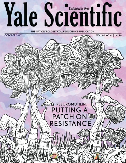
A B O U T T H E A R T<br />
Chunyang Ding<br />
Editor-in-Chief<br />
What better way to portray the potential of mushrooms in<br />
antibiotics research than to tap into the mushrooms themselves?<br />
The inspiration for this piece came from the unique<br />
shape of the mushrooms described in the cover article — the<br />
Clitopilus passeckerianus. Their pale bodies are fan-like and<br />
upward reaching, furled, and maybe even a little spooky.<br />
While tracing the details of their winding gills was laborious,<br />
it is my hope that the final illustrated product puts these<br />
notable figures of modern antibiotic research on full display.<br />
Editor-in-Chief<br />
Managing Editors<br />
News Editor<br />
Features Editor<br />
Articles Editor<br />
Online Editor<br />
Copy Editors<br />
Special Sections Editor<br />
Yale Scientific<br />
M A G A Z I N E<br />
Established in 1894<br />
OCTOBER 2017 VOL. 90 NO. 4<br />
Production Manager<br />
Layout Editor<br />
Art Editor<br />
Photography Editor<br />
Outreach Designer<br />
Publisher<br />
Operations Manager<br />
Advertising Manager<br />
Subscriptions Manager<br />
Alumni Outreach Coordinator<br />
Synapse President<br />
Synapse Vice President<br />
Outreach Coordinators<br />
Social Media Coordinator<br />
Staff<br />
Yasmin Alamdeen<br />
Sam Berry<br />
Anusha Bishop<br />
Grace Chen<br />
Ashwin Chetty<br />
Serena Cho<br />
Mary Chukwu<br />
Lukas Corey<br />
Keyi Cui<br />
Xander DeVries<br />
Victoria Dombrowik<br />
Ayah Elmansy<br />
Allie Forman<br />
Ivory Fu<br />
Kaija Gahm<br />
Lauren Gatta<br />
Hannah Geller<br />
Vivek Gopalan<br />
Advisory Board<br />
Priyamvada Natarajan<br />
Sandy Chang<br />
Kurt Zilm, Chair<br />
Fred Volkmar<br />
Stanley Eisenstat<br />
James Duncan<br />
Stephen Stearns<br />
Jakub Szefer<br />
Werner Wolf<br />
John Wettlaufer<br />
William Summers<br />
Scott Strobel<br />
Robert Bazell<br />
Craig Crews<br />
Ayaska Fernando<br />
Robert Cordova<br />
Katie Handler<br />
Jiyoung Kang<br />
Lauren Kim<br />
Theo Kuhn<br />
Severyn Kushmeliuk<br />
Andrew Kuzemczak<br />
Mindy Le<br />
Sunnie Liu<br />
Robert Luo<br />
Tanvi Mehta<br />
Diyu Pearce-Fisher<br />
Jared Peralta<br />
Miriam Ross<br />
Elizabeth Ruddy<br />
Sonia Ruiz<br />
Marcus Sak<br />
Sophia Sanchez-Maes<br />
Isabel Sands<br />
Chunyang Ding<br />
Emma Healy<br />
Sonia Wang<br />
Christine Xu<br />
Diane Rafizadeh<br />
Andrea Ouyang<br />
Kevin Biju<br />
Will Burns<br />
Amy Xiong<br />
Cheryl Mai<br />
Eileen Norris<br />
Richard Hwang<br />
Catherine Yang<br />
Natasha Zaliznyak<br />
Charlie Musoff<br />
Dawn Chen<br />
Kevin Chang<br />
Cheryl Mai<br />
Bryan Ho<br />
Krisstel Gomez<br />
Stephanie Smelyansky<br />
Jessica Trinh<br />
Wai Pan Wong<br />
Archeta Rajagoplaan<br />
Leslie Sim<br />
Anna Sun<br />
Sida Tanag<br />
Vera Villanueva<br />
Eric Wang<br />
Emma Wilson<br />
Alice Wu<br />
Cory Wu<br />
Lisa WU<br />
Jason Yang<br />
Yulan Zhang<br />
Kelly Zhou<br />
Astronomy<br />
Biological and Biomedical Sciences<br />
Chemistry<br />
Child Study Center<br />
Computer Science<br />
Diagnostic Radiology<br />
Ecology & Evolutionary Biology<br />
Electrical Engineering<br />
Emeritus<br />
Geology & Geophysics<br />
History of Science, Medicine, & Public Health<br />
Molecular Biophysics & Biochemistry<br />
Molecular, Cellular, & Developmental Biology<br />
Molecular, Cellular, & Developmental Biology<br />
Undergraduate Admissions<br />
Yale Science & Engineering Association<br />
The Yale Scientific Magazine (<strong>YSM</strong>) is published four times a year by<br />
Yale Scientific Publications, Inc. Third class postage paid in New Haven,<br />
CT 06520. Non-profit postage permit number 01106 paid for May 19,<br />
1927 under the act of August 1912. ISN:0091-287. We reserve the right<br />
to edit any submissions, solicited or unsolicited, for publication. This<br />
magazine is published by Yale College students, and Yale University<br />
is not responsible for its contents. Perspectives expressed by authors<br />
do not necessarily reflect the opinions of <strong>YSM</strong>. We retain the right to<br />
reprint contributions, both text and graphics, in future issues as well as<br />
a non-exclusive right to reproduce these in electronic form. The <strong>YSM</strong><br />
welcomes comments and feedback. Letters to the editor should be under<br />
200 words and should include the author’s name and contact information.<br />
We reserve the right to edit letters before publication. Please send<br />
questions and comments to ysm@yale.edu. Special thanks to Yale STC!
NEWS<br />
in brief<br />
Hardware Security “Fingerprints”<br />
By Ayah Elmansy<br />
PHOTOGRAPHY BY XANDER DE VRIES<br />
►DRAM provides each device its<br />
unique “fingerprint,” which can be<br />
used for electronic security.<br />
We all strive to keep our prized possessions<br />
safe. Sometimes those possessions include not<br />
just physical items, but also our digital information.<br />
Inspired to help us keep our data secure, Yale<br />
professor Jakub Szefer is researching security applications<br />
of the tiny differences between devices.<br />
Szefer is an assistant professor of electrical engineering<br />
at the Yale School of Engineering and<br />
Applied Science. Recently, he received the 2017<br />
Faculty Early Career Development Award from<br />
the National Science Foundation, which will provide<br />
funding for his research. Szefer is collaborating<br />
with his research group on this work, which<br />
includes graduate student Wenjie Xiong, as well<br />
as researchers in Germany at Technische Universität<br />
Darmstadt and Ruhr University Bochum.<br />
Szefer’s project focuses on the use of electronic<br />
devices’ unique “fingerprints” to improve computer<br />
security. At the electronic level, every computing<br />
device has distinctive hardware features,<br />
even between identical models. These differences<br />
in the devices’ digital circuits, processors, and<br />
data storage systems are known as dynamic random-access<br />
(DRAM) memory. Small variations<br />
occur when these devices are manufactured,<br />
causing slight deviations in factors such as the<br />
length of the wires of circuits or the capacitors’<br />
thicknesses. These minor deviations accumulate<br />
to give each device its unique “fingerprint,”<br />
which can be used to authenticate and identify<br />
the device.<br />
Szefer wants to explore how these fingerprints<br />
can be applied to the security of our everyday devices<br />
like smartphones, phones, computers, and<br />
embedded devices. “We hope to show that cryptographic<br />
protocols can be designed, which derive<br />
their security from not just difficult mathematical<br />
problems but also from the intrinsic<br />
properties of the hardware,” Szefer said. Through<br />
their continued efforts, Szefer and his collaborators<br />
hope to benefit both the hardware security<br />
community and consumers who use computing<br />
devices every day.<br />
IMAGE CREDIT BY ZUREKS<br />
►The researchers found notothenioids<br />
to be in jeopardy from climate change<br />
and species invasion.<br />
Something’s Fishy<br />
By Namoi D’Arbell Bobadilla<br />
It’s cold in the Antarctic Ocean, but<br />
things are heating up. Yale ecology and<br />
evolutionary biology professor Thomas<br />
Near worked with a research group to study<br />
an ancient Antarctic fish lineage called<br />
notothenioids. These fish are essential<br />
links in Antarctic food webs and are the<br />
basis for several fisheries all over the world.<br />
The researchers used DNA sequences from<br />
89 notothenioid species to reconstruct the<br />
process of how they evolved and to study<br />
their changes in habitat over the years. Their<br />
study, published in June in Nature Ecology<br />
& Evolution, showed that notothenioids are<br />
in danger from climate change and species<br />
invasion.<br />
Historically, notothenioids have survived<br />
natural warming cycles through migration,<br />
such as between the Antarctic continent<br />
and its islands. “Continents tend to produce<br />
biodiversity that emigrates to islands, but<br />
the Antarctic islands are behaving like<br />
a continent,” Near said. In other words,<br />
studies have shown that around Antarctica,<br />
the islands generate a diversity of marine<br />
life. Unfortunately, these island waters are<br />
now warming. This temperature rise allows<br />
fish that usually live in warmer water to<br />
invade and compete with native species<br />
like notothenioids. Notothenioids must<br />
now face both rising temperatures—which<br />
can alarmingly alter Antarctic marine<br />
ecosystems—and increased competition<br />
from invading species.<br />
“This polar ecosystem has probably been<br />
fairly stable for 25 to 30 million years,” Near<br />
said. However, now the climate change<br />
has brought unprecedented instability to<br />
Antarctic ecosystems and has placed its<br />
most dominant lineage, notothenioids, in<br />
jeopardy from habitat change and invasive<br />
species. Near reminds us of our duty to<br />
address such problems: “With our dramatic<br />
utilization of natural resources comes<br />
responsibility for stewardship of the planet<br />
that we’ve perturbed.”<br />
6 Yale Scientific Magazine October 2017 www.yalescientific.org
in brief<br />
NEWS<br />
The Twists and Turns of Flowers<br />
By Ashwin Chetty<br />
Have you ever taken time to enjoy the<br />
beauty of flowers? Professor Vivian Irish and<br />
postdoctoral associate Adam Saffer of Yale’s<br />
Molecular, Cellular and Developmental<br />
Biology department have done so for years,<br />
especially from a scientific perspective.<br />
They study what affects a flower’s shape<br />
and appearance. Recently, they discovered<br />
a molecule that affects the twist of flowers,<br />
which refers to the way flowers turn. In<br />
their Current Biology paper published in<br />
August, they revealed that in a specific type<br />
of flower, Arabidopsis thaliana, a substance<br />
called pectin influences the twisting of plant<br />
cells. Pectin is a common substance in the<br />
kitchen and gives jam its gelatinous quality.<br />
Saffer first looked at a mutation that caused<br />
plant cells to be short and twisted. The<br />
researchers then identified a mutated gene<br />
underlying the helical shape of these cells.<br />
This gene plays a role in the biosynthesis<br />
of a certain kind of pectin called RG-<br />
I. The researchers believe that RG-I may<br />
normally counteract a component of cell<br />
walls that causes cells to twist. However,<br />
when the mutation is present in a cell, less<br />
RG-I is is produced. RG-I inhibits twisting,<br />
so when RG-I levels are low, the unknown<br />
component is free to make cells twist. Thus,<br />
the mutation causes the beautiful helical<br />
shape of plant cells.<br />
The Irish Lab is working to better<br />
understand the role of pectin in providing<br />
cell structure, and Irish is partnering<br />
with researchers at Yale’s department of<br />
mechanical engineering and materials<br />
science to develop models to explain this<br />
left-handed twisting. By identifying a novel<br />
characteristic of pectin, Irish and Saffer<br />
have opened doors for the development of<br />
new biomaterials. While they are excited<br />
for these applications, at the end of the<br />
day, both still like to enjoy the aesthetically<br />
pleasing nature of flowers.<br />
PHOTO BY TANVI MEHTA<br />
►Pectin, the substance that gives jam<br />
its gelatinous quality, can influence the<br />
way that flowers twist.<br />
Raising a child is no easy task—what<br />
would you do if you were put in charge of<br />
raising someone else’s child? In a recent<br />
study, researchers from the Yale Department<br />
of Ecology and Evolutionary Biology<br />
explored whether males of different animal<br />
species would care for offspring that aren’t<br />
their own. The researchers studied how the<br />
energy needed to raise a child could affect<br />
males’ decisions to care for offspring.<br />
Previous theories state that males are most<br />
likely to care for children that are certain to<br />
be their own. Yet in many species, a male<br />
may take care of offspring that were not<br />
conceived by him but rather by a competing<br />
male. Postdoctoral research fellow Gustavo<br />
Requena explained the difference between<br />
his model and those of earlier theories. “In<br />
our study, we used mathematical models<br />
to emulate males’ decisions in different<br />
scenarios and ultimately address the same<br />
question but took into account a more<br />
general biological reality,” he said. This<br />
Game of Sperms<br />
By Lauren Kim<br />
model involves sperm competition games,<br />
which show how males allocate energetic<br />
resources to increase their chances<br />
for success within male-male mating<br />
competition.<br />
Factors that affect the males’ decisions<br />
include female promiscuity, maternal<br />
effort, and the difficulty of providing<br />
care to offspring. Based on these factors,<br />
researchers found that when there is more<br />
energy required, males will provide care<br />
based on relatedness to his offspring. For<br />
example, a male Arowana fish will carry<br />
eggs in his mouth to protect his offspring.<br />
However, in low-cost situations, males will<br />
provide care regardless of relation.<br />
In this way, scientists hope to provide<br />
an answer as to why males continue to<br />
provide energetically-costly care towards<br />
offspring that may not be their own. From<br />
these results, they can develop a greater<br />
understanding of different parenting<br />
patterns in nature.<br />
IMAGE COURTESY OF NATIONAL GEOGRAPHIC<br />
►Male Arowana fish carry the eggs in<br />
their mouths, protecting the offspring<br />
until they are ready to leave permanently.<br />
www.yalescientific.org<br />
October 2017<br />
Yale Scientific Magazine<br />
7
NEWS<br />
materials science<br />
HARNESSING THE SUN FOR CLEAN WATER<br />
Nanotechnology improves water purification<br />
►BY ALLIE FORMAN<br />
For you and me, obtaining safe drinking water may be as<br />
simple as walking to the nearest sink and getting a cup of tap<br />
water. But 29 percent of people worldwide do not have ready<br />
access to safe drinking water, instead getting their water from<br />
sources contaminated by feces. This leads to over half a million<br />
deaths annually. Climate change and population growth<br />
are expected to exacerbate the issue, making access to potable<br />
water even more challenging.<br />
One solution to this problem is by making salt water drinkable<br />
through desalination. But the most commonly used technique<br />
for desalination, reverse osmosis, requires large inputs<br />
of energy to boil water, making it unsuitable for places without<br />
reliable electricity.<br />
With this in mind, Yale researchers in the School of Engineering<br />
and Applied Science, in collaboration with scientists<br />
at Rice University, Arizona State University, and the University<br />
of Texas-El Paso, are working to improve an alternative<br />
solar-powered desalination method known as membrane distillation.<br />
This process has the potential to be less energy-intensive<br />
than reverse osmosis, making it suitable for underdeveloped<br />
areas and environmentally friendly.<br />
In membrane distillation, salt water and pure water are<br />
placed on opposite sides of a membrane that only allows gases<br />
to pass through. The salty side is heated, while the pure water<br />
is cooled. The difference in partial pressure causes the water on<br />
the salty side to travel through the membrane as a gas, leaving<br />
the salt and other contaminants as solids on the “dirty” side of<br />
the membrane.<br />
Membrane distillation has not been widely used for a number<br />
of reasons. Energy is required to generate and maintain<br />
the temperature difference between the impure salt water and<br />
clean freshwater reservoirs, and the process uses more energy<br />
to produce the same amount of pure water when compared to<br />
reverse osmosis. Along with their collaborators, Yale researchers<br />
Menachem Elimelech and Akshay Deshmukh have optimized<br />
a low-energy membrane distillation technique that uses<br />
solar power coupled with nanoparticle technology.<br />
Their design uses a carbon black-coated membrane that<br />
maximizes uptake of solar energy. This sets up a temperature<br />
gradient in which the salt water closest to the nanoparticle-covered<br />
membrane heats up. The process does not require<br />
an additional power source besides solar energy, and the materials<br />
are widely available.<br />
While the nanoparticle-enabled solar membrane distillation<br />
system is still in development, the scientists have demonstrated<br />
its viability with a paper recently published in the Proceedings<br />
of the National Academy of Sciences. The project is attracting<br />
attention because of its potential applications around the<br />
world, especially in areas lacking access to electricity.<br />
The improved membrane distillation design is ideal for<br />
smaller, decentralized applications, such as a rural village not<br />
connected to the grid. Unlike reverse osmosis—which necessitates<br />
a proper electrical connection for the high-pressure<br />
pumps, the new solar membrane distillation model requires<br />
very little infrastructure. “We wouldn’t use a process like this to<br />
compete with reverse osmosis in a big community,” Deshmukh<br />
said. “But for smaller stuff, which is off-grid, it could be useful<br />
because it’s using sunlight and it’s not a high-pressure process.”<br />
Membrane distillation technology can also be used to remove<br />
other contaminants besides salt from water. “This could<br />
be used in situations where the water is very dirty. For example,<br />
for waste water from industrial sites where the osmotic<br />
pressure is very high, reverse osmosis can’t be used to treat<br />
it, because the membranes can’t deal with the pressure,” said<br />
Deshmukh. He also thinks their design could be implemented<br />
in natural disaster situations, where normal water treatment<br />
infrastructure has been disrupted.<br />
Though the researchers have produced a model of the design,<br />
the technology is not yet ready to hit store shelves. The<br />
scientists now need to refine their design and market approach.<br />
While there is still work to be done, they believe that<br />
the new technology will improve many lives around the globe<br />
by providing much needed access to clean water.<br />
IMAGE COURTESY OF AKSHAY DESHMUKH<br />
►The new design utilizes a nanoparticle layer to heat salt water<br />
with solar energy.<br />
8 Yale Scientific Magazine October 2017 www.yalescientific.org
neuroscience<br />
NEWS<br />
NOW YOU HEAR ME, NOW YOU DON’T<br />
Testing susceptibility of people to hearing voices<br />
►BY SUNNIE LU<br />
PHOTOGRAPHY BY YASMIN ALAMDEEN<br />
►Dr. Powers and Dr. Corlett took MRI scans of people<br />
performing auditory tasks to understand auditory hallucinations.<br />
You hear footsteps coming down the hallway and voices chanting,<br />
“We’re coming.” A shadowy figure suddenly appears in the<br />
corner of your room, while a girl dressed up as a rat looms over<br />
your bed. Although these scenarios sound like they come from<br />
horror movies, they actually are real examples of hypnagogic hallucinations,<br />
which occur during the onset of sleep. Both having<br />
these experiences, Yale psychiatrist and neuroscientist Albert<br />
Powers and Yale neuroscientist Philip Corlett fascinated with<br />
auditory hallucinations. Their research, featured in Science, suggests<br />
that people who hear voices are more likely to experience<br />
induced hallucinations in a lab.<br />
It may seem concerning that both Powers and Corlett have<br />
experienced hallucinations while falling asleep, but these hallucinations<br />
are usually symptomatic of a neurological condition,<br />
not a psychiatric illness. However, people without a psychiatric<br />
condition can hear voices too. The two scientists wanted to figure<br />
out what produces auditory hallucinations and why some<br />
voice-hearing experiences are benign and others require medical<br />
attention.<br />
To explore these questions, they sought out four groups of<br />
test subjects: both psychotic and nonpsychotic voice-hearers<br />
and non-voice-hearers. After identifying potential subjects, the<br />
researchers separated the psychotic and nonpsychotic people<br />
using a questionnaire developed by forensic psychologists to<br />
distinguish between people who were actually experiencing hallucinations<br />
and those who only claimed to do so.<br />
After screening their subjects for hallucinations, Powers and<br />
Corlett induced auditory hallucinations in their subjects to identify<br />
whether psychotic people were more likely than non-psychotic<br />
people to hear conditioned sounds. Using a technique<br />
originally developed at Yale during the 1890s, the subjects were<br />
stimulated with a checkerboard image and a one-second long<br />
sound simultaneously and repeatedly, while getting their brains<br />
imaged by MRIs. This conditioned the subjects to associate the<br />
image with the tone. As the scientists changed the intensity of<br />
tone, sometimes turning it off, the subjects pressed a button when<br />
they thought they heard the tone, while changing the length of<br />
time they pressed the button to show their level of confidence.<br />
Many reported hearing a tone when only the checkerboard<br />
image appeared but no tone played. This inconsistency occurred<br />
more often with the two voice-hearing groups—the people with<br />
schizophrenia and self-identified clairaudient psychics. Both<br />
groups were almost five times more likely to report that they<br />
heard a nonexistent tone than the non-voice-hearing groups.<br />
Furthermore, the two non-voice-hearing groups were 28 percent<br />
more confident that they had heard the tone when no tone<br />
played. These results support a possible explanation for hallucinations.<br />
“The brain makes models for what the outside world is<br />
like,” said Corlett, noting that these models sometimes don’t always<br />
match reality. This study suggests that people hallucinate<br />
when their expectations overweigh what their senses tell them.<br />
Powers and Corlett further understood auditory hallucinations<br />
through analyzing the MRI scans collected: the parts of the<br />
brain that were responsive to the tone were active when people<br />
reported conditioned hallucinations, producing MRI scans of<br />
brains that looked like those of people actually hearing the tone.<br />
The images also revealed that both hallucinating and non-hallucinating<br />
people with psychosis exhibit abnormal brain activity in<br />
regions that monitor internal representations of reality. These results<br />
contribute to the idea that hallucinations stem from internal<br />
representations overruling actual sensory data.<br />
This study was able to distinguish not only between those who<br />
hallucinate and those who don’t, but also between psychotic and<br />
non-psychotic people. “The sooner you catch psychosis and the<br />
sooner you intervene, the better the general outcomes are,” said<br />
Powers. According to Powers, most people with the symptoms<br />
associated with increased chances of psychosis don’t even develop<br />
psychosis. The question then is, who should receive treatment?<br />
This new research may help to answer that key question<br />
by providing the basis for tests to diagnose patients who require<br />
psychiatric treatment early.<br />
“There is no one-size-fits-all anti-psychotic, so there should be<br />
different treatments for different people,” Corlett said. Although<br />
this sort of precision medicine has not existed in psychiatry so<br />
far, Corlett hopes that this study, along with future research, will<br />
lead to more personalized psychiatric treatments, which he believes<br />
would be more effective in helping people suffering with<br />
mental health issues.<br />
www.yalescientific.org<br />
October 2017<br />
Yale Scientific Magazine<br />
9
NEWS<br />
genetics<br />
MAKING CANCER CELLS NERVOUS<br />
Targeting the genes involved in nerve cells in the brain<br />
►BY DIYU PEARCE-FISHER<br />
Cancer is the second most common cause of death in the<br />
world. Almost one out of every six deaths can be traced back to<br />
this deadly disease. Science has made huge strides towards treating<br />
and curing cancer, but there are many different types of cancer,<br />
and no single cure that works for every case. Understanding<br />
the cause of each type of cancer is crucial to treating each patient,<br />
so much recent cancer research focuses on the genetic factors that<br />
cause normal cells to become cancerous.<br />
Researchers at the Yale Systems Biology Institute led a team of<br />
scientists studying the genes involved in glioblastoma, a type of<br />
brain cancer. This cancer targets star-shaped support cells in the<br />
brain called astrocytes, which serve many purposes in the nervous<br />
system. Glioblastoma is one of the deadliest types of cancer.<br />
While over two-thirds of the people diagnosed with any type of<br />
cancer make it to the five-year survival mark and beyond, the median<br />
survival time for patients diagnosed with glioblastoma is between<br />
1 and 1.5 years. The Yale-led team of researchers developed<br />
a technique enabling them to identify specific gene combinations<br />
that cause glioblastoma. They believe their approach could be applied<br />
to other types of cancer as well, which would revolutionize<br />
how we approach cancer treatment.<br />
Cancer is caused by genetic mutations. However, even if two<br />
patients have the same type of cancer, the patients may or may<br />
not have the same mutations. Prior to the Yale-led study, genome<br />
sequencing identified 223 genes with links to glioblastoma. But<br />
that also means that there are hundreds of thousands of possible<br />
combinations of genes, with certain combinations responding<br />
slightly differently to treatment. Thus, the Yale researchers wanted<br />
to clarify how these different combinations affect glioblastoma<br />
treatment.<br />
To do this, the Yale team employed the use of a technique called<br />
CRISPR. First pioneered in 2012, CRISPR is a gene editing technique<br />
that has revolutionized the biomedical sciences by enabling<br />
researchers to create cells with mutations and test the effects of the<br />
mutations. However, CRISPR does have its limitations. To use this<br />
type of CRISPR, each combination would have to be tested one<br />
by one, which would be hard to compare since the time-intensive<br />
tests would give results two to three years apart.<br />
To address this, the Yale-led team developed a novel CRIS-<br />
PR technique that is less biased, more time-efficient, and more<br />
cost-efficient. Rather than testing each individual mutation and<br />
waiting for the results of each experiment, which would exhaust<br />
both time and resources, the new technique employs a pool of viruses,<br />
each of which targets a specific gene. Infected from a pool<br />
of viruses, each mouse brain cell has the potential to have multiple<br />
mutations. Thus, the team could test for multiple combinations<br />
simultaneously. The researchers found specific combinations of<br />
mutations that could cause glioblastoma, as well as two combinations<br />
that could make the glioblastoma tumor resistant to chemotherapy.<br />
By understanding the results of different combinations of mutations,<br />
doctors will be able to screen for these mutations and decide<br />
the appropriate treatments. For instance, if they find that a patient<br />
has the combination of mutations that cause a tumor to be resistant<br />
to chemotherapy, the doctors will know not to waste time and<br />
resources on an unsuccessful chemotherapy, which could further<br />
harm or discourage the patient. Information like this is especially<br />
important when the average survival time for cancers like glioblastoma<br />
is under two years.<br />
The researchers believe that their approach can be applied to<br />
other types of cancer, which would allow doctors to give treatments<br />
specific to each case, hopefully leading to a higher chance<br />
of success. “In order to find better therapy or treatment, we need<br />
to find the therapeutic responder, or the genes that regulate or<br />
change some response. Now we have the tools—we have the experiment<br />
setting—we can simply do the same experiments but<br />
with gene therapy,” said Sidi Chen, one of the investigators on the<br />
team. By using CRISPR to create the different glioblastoma-causing<br />
gene combinations and applying specific types of therapies to<br />
see how each combination responds, the scientists may be able to<br />
figure out exactly which treatment will work for each case of cancer.<br />
In other words, targeted “cures” for cancer may soon become<br />
an applicable reality.<br />
IMAGE COURTESY OF CHEN LAB<br />
►A gioblastoma induced by new gene-editing and screening<br />
technology.<br />
10<br />
Yale Scientific Magazine October 2017 www.yalescientific.org
public health<br />
NEWS<br />
NOT SO SWEET<br />
Metabolic problems caused by artificial sweeteners<br />
►BY SERENA CHO<br />
PHOTOGRAPHY BY KELLY ZHOU<br />
►Diet drinks contain artificial sweetners that trick our brains<br />
into thinking we are getting a sweet calorie reward.<br />
Diet Coke, Vitamin Water Zero, Minute Maid Light: with<br />
the hype over healthy living and weight loss, the food industry<br />
has introduced many low-calorie drinks to the market.<br />
Most of these beverages use artificial sweeteners, which<br />
provide the same sweet taste with fewer or no calories. But<br />
what effects do the artificial sweeteners have on our body?<br />
Are they safe, after all? The hidden effects of artificial sweeteners<br />
on our body have been long disputed.<br />
According to research conducted by Yale School of Medicine<br />
scientists, the mismatch between the sweet taste and<br />
caloric value in diet beverages could explain the link between<br />
artificial sweetener use and cases of metabolic failure,<br />
such as in diabetes. Because the brain cannot register<br />
how much sugar was consumed, our body cannot properly<br />
digest and process the diet beverages.<br />
A sweet taste helps indicate the amount of energy present<br />
in a food source. “Our bodies evolved to efficiently use<br />
the energy sources available in nature,” said senior author<br />
and Yale psychiatry professor Dana Small. She continued,<br />
“Our modern food environment is characterized by energy<br />
sources our bodies have never seen before.” When a beverage<br />
tastes too sweet or not sweet enough for the number of<br />
calories it contains, the signal communicating its nutritional<br />
value can be disrupted. Our metabolism is confused, and<br />
the beverage cannot be digested properly.<br />
Small and her colleagues discovered the problems of<br />
“diet” beverages when performing an experiment on brain<br />
responses to sugar ingestion. The team observed the brain<br />
activity of fifteen healthy participants after drinking tasteless<br />
sugary drinks. Normally, when sugar is introduced to<br />
the body, the brain releases dopamine, a chemical compound<br />
that induces reward and pleasure. However, when<br />
the participants drank tasteless but sugary drinks, the<br />
amount of dopamine released was not proportional to the<br />
calories in the beverage. “In other words, the assumption<br />
that more calories trigger greater metabolic and brain response<br />
is wrong,” Small said. She was interested in pursuing<br />
this further, commenting, “A strange finding that makes no<br />
sense means that you’re going to learn something new.”<br />
Further research revealed that a sweet taste also determines<br />
how sugar is metabolized and signaled to the brain.<br />
“Calories are only half of the equation; sweet taste perception<br />
is the other half,” Small said. In fact, sweet low-calorie<br />
drinks could produce greater metabolic response and higher<br />
dopamine levels than less sugary higher-calorie drinks.<br />
In nature, when sweetness reflects the number of calories<br />
in the food, the body properly metabolizes the calories and<br />
activates the corresponding brain reward signals. However,<br />
the modern food industry introduces beverages with the<br />
kind of sweetness that our body hasn’t been exposed to before.<br />
“A calorie is not a calorie,” said Small, commenting on<br />
how the impact of these drinks on our bodies goes beyond<br />
just the number of calories. When sweetness and calories<br />
don’t align, like in artificially sweetened drinks, the beverage<br />
fails to trigger metabolism and the brain reward circuits<br />
can’t register the number of calories consumed. The metabolic<br />
failure observed here could help explain the link between<br />
artificial sweeteners and problems like diabetes.<br />
IMAGE COURTESY OF WIKIMEDIA COMMONS<br />
►Artificial sweetners confuse our metabolic systems, possibly<br />
creating mixed signals that lead to health issues like diabetes.<br />
www.yalescientific.org<br />
October 2017<br />
Yale Scientific Magazine<br />
11
WHAT IS HOT, DENSE,<br />
& spins like crazy?<br />
art by Emma Wilson<br />
by Sophia Sánchez-Maes<br />
It all started with a big bang...but then what?<br />
Our universe is 13.8 billion years old—<br />
the most remarkable detail about this fact<br />
is what it implies about its beginning. Space<br />
itself is continuously expanding, and winding<br />
back the clock shows it getting smaller<br />
and smaller, eventually shrinking down to<br />
a single point from whence it all came: life,<br />
the universe, and time itself. Squeezing the<br />
contents of the entire universe into a single<br />
point may seem unimaginably difficult, but<br />
compress matter enough, and that inward<br />
pressure is enough to break molecular bonds<br />
into atoms. Compress further and atoms<br />
themselves break into their components:<br />
protons, neutrons, and electrons. Next, the<br />
stress would break even the bonds that tie<br />
those subatomic particles together, releasing<br />
even smaller particles called quarks and gluons—two<br />
of the 13 elementary particles that<br />
constitute the most basic building blocks of<br />
matter. As carriers of the strong force, gluons<br />
are the glue that acts to bind quarks together<br />
into protons and neutrons. The strong force<br />
governs interactions between all particles<br />
with a color charge, like quarks and gluons,<br />
and is also responsible for binding together<br />
the atomic nucleus.<br />
In order to overcome intermolecular and<br />
interatomic forces and explore the properties<br />
of this primordial cosmic soup, scientists<br />
must replicate the conditions of the early<br />
universe just milliseconds after the Big Bang,<br />
which involved blistering temperatures and<br />
tightly packed matter.<br />
This August, the STAR collaboration, a<br />
group of scientists aiming to deduce the<br />
properties of the quark gluon plasma and<br />
the physics that underlay them, published<br />
their measurement of the vorticity of the<br />
quark-gluon plasma in Nature: a experiment<br />
which elucidates the unexpected fluid properties<br />
of this matter that dominated early in<br />
the universe, and is an important step to bettering<br />
our theory of the strong force.<br />
IMAGE COURTESY OF BROOKHAVEN NATIONAL LABORATORY<br />
►The tracks in this image depict the particles<br />
produced by the collision of gold ions at RHIC,<br />
as captured by the STAR detector’s Time<br />
Projection Chamber.<br />
Exploring the universe, before it was cool<br />
Researchers like Yale’s Helen Caines don’t<br />
need to journey through time to discover<br />
properties of early universe. They have only<br />
to cross the Long Island Sound to Brookhaven<br />
National Laboratory, where the Relativistic<br />
Heavy Ion Collider is cooking up the<br />
same conditions. At Brookhaven, STAR researchers<br />
focus narrow beams of gold ions<br />
(atoms which have lost their outer electrons)<br />
at one another, each traveling close to the<br />
speed of light. Such incredible speeds also<br />
give the ions incredible energy, so any headon<br />
collision between gold ions is able to<br />
dissolve the 79 protons and 118 neutrons in<br />
each gold ion into quarks and gluons, forming,<br />
for a brief instant, the quark gluon plasma,<br />
a soup of quarks and gluons which exists<br />
at extreme temperatures and densities. The<br />
Relativistic Heavy Ion Collider, along with<br />
CERN’s Large Hadron Collider in Geneva<br />
are the only facilities in the world with machines<br />
powerful enough to produce quark<br />
gluon plasma.<br />
Having proven that the produced plasma is<br />
indeed comparable to that of the primordial<br />
cosmos, scientists in the STAR collaboration<br />
moved on to also show that this material is<br />
full of unexpected surprises. For example,<br />
researchers anticipated that such a hot, energetic<br />
state of matter would behave like some<br />
sort of super gas, without the strong collectivity<br />
properties that characterize liquids,<br />
unlike gasses, which diffuse evenly.<br />
In actuality, the quark gluon plasma<br />
(QGP) behaves like a liquid, since its constituents<br />
interact more strongly than those of a<br />
gas. “We know that the gluons themselves<br />
interact, and the quarks interact via gluons,<br />
so it makes sense if they can interact very<br />
strongly with each other,” Caines said.<br />
In this series of experiments, scientists are<br />
aiming to determine the properties of the<br />
quark gluon plasma’s fluid properties. Not<br />
only is the QGP a liquid, but it has the lowest<br />
viscosity of any liquid ever encountered,<br />
meaning that unlike viscous substances like<br />
honey, it has nearly no resistance to flow.<br />
This unique property has prompted scientists<br />
to call it nearly “perfect.”<br />
Since this medium formed under such<br />
extreme temperatures and pressures, it’s<br />
12 Yale Scientific Magazine October 2017 www.yalescientific.org
particle physics<br />
FOCUS<br />
distance, it gets increasingly strong, like a<br />
spring that wishes to retract. Eventually, so<br />
much energy is expended in pulling apart<br />
this particle that another particle antiparticle<br />
pair is pulled from the vacuum to preserve<br />
net colorlessness. This means that it’s<br />
impossible to detect free quarks. This might<br />
be puzzling, since quarks in the QGP aren’t<br />
bound in mesons or nucleons, but zooming<br />
in anywhere in this soup, conditions are “locally”<br />
colorless. Since scientists can’t detect<br />
the quarks individually, the STAR collaboration<br />
scientists instead detect the particles<br />
created from the QGP as it cools, whose motion<br />
is inherited from this fluid that formed<br />
them.<br />
It’s not “just a phase”<br />
not surprising that the medium breaks other<br />
records. These record-setting properties<br />
include a low viscosity, a high temperature,<br />
and a now legendary vorticity, or swirling<br />
properties. These measurements not only<br />
help us understand this unexpected liquid<br />
phase, but also assist in our understanding of<br />
quantum chromodynamics (QCD), a theory<br />
which governs the interactions of the colored<br />
charged particles (quarks and gluons)<br />
which make up the QGP.<br />
The force is strong with this one<br />
Quarks are never observed in alone, but<br />
are always bundled in pairs or groups of<br />
three by gluons, which carry the strong<br />
force. There are six types of quark, with<br />
up and down quarks combining in groups<br />
of three to create protons and neutrons<br />
– the heaviest components of atoms. The<br />
theory governing their interactions is<br />
called quantum chromodynamics (QCD).<br />
Quarks come in three states called ‘colors.’<br />
The strong force, which governs the behavior<br />
of quarks, is incredibly unusual.<br />
Gravity, the force with which we’re most<br />
familiar, only attracts objects toward each<br />
www.yalescientific.org<br />
IMAGE COURTESY OF WIKIPEDIA`<br />
►The color charge acts such all matter must be locally white, binding quarks into combinations<br />
that preserve this colorlessness and preventing the sustained existence of free quarks.<br />
other. Electromagnetism has positive and<br />
negative (anti-positive) charges – but like<br />
gravity, their effect fades with the inverse<br />
square of the distance. Quarks and gluons<br />
have color-charge, which has nothing<br />
to do with color but for convenient<br />
metaphor. There are three colors (red,<br />
green, and blue) and three anti-colors<br />
(anti-red, anti-green, anti-blue) Any stable<br />
state must be colorless – which can<br />
only be achieved by mixing all the colors,<br />
like in protons and neutrons (made up of<br />
three quarks) – or by combining a color<br />
and anti-color, like in particles called mesons<br />
(which consist of a quark and an anti-quark).<br />
Furthermore, mass and charge<br />
are fundamental properties of particles,<br />
whereas color charge can change via gluon<br />
interactions. It is the strong force, and<br />
the color charge that mediates it, that is<br />
responsible for the interactions between<br />
quarks and gluons. By understanding<br />
the behavior of the QGP, scientists are<br />
probing these fundamental forces and<br />
properties.<br />
Try pulling a meson apart and you’ll see<br />
that the strong force operates differently<br />
in another important way: with increasing<br />
The QGP is a fluid, and is defined by superlatives.<br />
At four trillion degrees Celsius,<br />
tens of thousands of degrees hotter than a<br />
supernova, it’s the hottest thing we know<br />
in the universe. It also has the lowest viscosity,<br />
or resistance to flow, of any known<br />
fluid. Now, the STAR collaboration has also<br />
made the first measurement of its vorticity,<br />
or rotation.<br />
When gold ions collide head-on, their<br />
constituent particles combine to create the<br />
QGP; but typically the collision between<br />
ions is more glancing. In that case, region<br />
of contact, like the center of a Venn diagram,<br />
will combine to become QGP, while<br />
the outside regions will continue speeding<br />
away from each other. These glancing collisions<br />
give the resulting soup a net angular<br />
momentum, setting the QGP spinning.<br />
Scientists in the STAR collaboration<br />
aimed to detect this spin by detecting the<br />
particles generated by the plasma as it cools.<br />
October 2017<br />
IMAGE COURTESY OF WIKIPEDIA<br />
►The quark gluon plasma kicked off the start<br />
of the radiation era of the universe’s history<br />
from 10e-12 to 10e-6 seconds after the big<br />
bang, right after cosmic inflation.<br />
Yale Scientific Magazine<br />
13
FOCUS<br />
particle physics<br />
IMAGE COURTESY OF BERKELEY LABS<br />
►The color charge acts such all matter must be<br />
locally white, binding quarks into combinations<br />
that preserve this colorlessness and preventing<br />
the sustained existence of free quarks.<br />
IMAGE COURTESY OF GETTY IMAGES<br />
►This image, taken near RHIC at Brookhaven<br />
Laboratory depicts a subset of the STAR<br />
collaboration.<br />
Any angular momentum in the system will<br />
be passed on to the particles it generates,<br />
whose spins are aligned with the whirling<br />
structures from which they came. On average,<br />
those spins will indicate the net angular<br />
momentum created by the system. As<br />
Yale’s Professor Caines, spokesperson for<br />
the collaboration described, “when you collide<br />
them just a little bit edge on edge you<br />
set it spinning, and that’s basically what we<br />
measured, how fast it’s spinning collectively.<br />
We can detect that angular momentum.”<br />
The experiment used two instruments for<br />
this measurement. The first of these was the<br />
Time Project Chamber, which is filled with<br />
gas that surrounds the collision and allows<br />
scientists to track the particles released<br />
from the collision and therefore generated<br />
by the QGP. This detector measures protons<br />
created by the plasma. The second instrument,<br />
the Beam-Beam Counters, measures<br />
deflections in the paths of particles which<br />
whiz by one another and indicates the magnitude<br />
and direction of angular momentum<br />
in each collision. Correlating this measured<br />
angular momentum with aligned direction<br />
of the detected protons, STAR scientists<br />
searched for preference in direction that<br />
will indicate the vorticity of the system.<br />
This measurement indicates that the<br />
QGP is the most vortical fluid known, a<br />
measured value surprising in its magnitude.<br />
The vorticity of the QGP exceeded<br />
all predicted values – whirling faster than<br />
tornados, Jupiter’s red spot, and previous<br />
vorticity record-holder superfluid helium.<br />
This high vorticity is allowed to perpetuate<br />
by the low viscosity of the fluid – since high<br />
resistance to flow would dampen whirls prior<br />
to measurement. Vorticity is a property of<br />
the flow – it’s not intrinsic to the medium,<br />
but results from the medium and its motion.<br />
But since we know the conditions that<br />
produced the flow inside of the collider, it’s<br />
possible to gain useful information about<br />
the fluid properties. Knowing the specifics<br />
of the collision that generated it allows us to<br />
firm up our theories of how the QGP interacts<br />
and responds to forces.<br />
Changes in temperature and pressure induce<br />
phase transitions. Think briefly of ice:<br />
increasing the temperature will turn it to<br />
water, then to vapor. At 4 trillion degrees<br />
Celcius, the quark gluon plasma is very hot,<br />
which is why the strong mutual interactions<br />
of its constituent particles is puzzling. The<br />
QGP behaves more like a fluid than like a<br />
gas. Instead of expanding out evenly in all<br />
directions, it’s preferential in its flow. Is it a<br />
relic of the strong force, which governs interactions<br />
between its constituent parts? Or<br />
perhaps the extreme pressure of the cosmic<br />
soup? In future experiments, the STAR collaboration<br />
hopes to deduce the equation of<br />
state for the quark gluon plasma to resolve<br />
these questions and test theories about the<br />
strong force.<br />
This vorticity measurement will aid in<br />
improving theories of the plasma and how<br />
it affected the properties of the early universe.<br />
Most importantly, spinning charges<br />
produce magnetic fields, making this measurement<br />
an important first step in making<br />
the first measurements of the magnetic<br />
properties of the QGP, an important step<br />
in better understanding the weird physics<br />
surrounding this hot, flowy, whirling<br />
soup. Although the significance of such<br />
fundamental science isn’t always immediately<br />
clear, Yale’s Li Yi, a scientist with the<br />
collaboration sees this as exciting infinite<br />
possibility. “Maybe it sounds like science<br />
fiction, but in all of the modern world most<br />
of the technology is based on QED (quantum<br />
electro dynamics), our theory of the<br />
electromagnetic force. For example, we<br />
transfer the messages and communications<br />
through electromagnetic waves - this is all<br />
through the QED because we really understand<br />
what we can do. QCD we don’t know<br />
exactly how it works - or what we could use<br />
it for.” The STAR Collaboration’s measurements<br />
of the QGP are among the most important<br />
measurements for formulating and<br />
testing this theory.<br />
ABOUT THE AUTHOR<br />
SOPHIA SANCHEZ-MAES<br />
SOPHIA SANCHEZ-MAES is a junior Physics and Astrophysics double major<br />
in Timothy Dwight college. She works with Professor Jun Korenaga on studying<br />
the origin processes of plate tectonics, and topics in complex systems.<br />
THE AUTHOR WOULD LIKE TO THANK Professor Helen Caines and Dr. Li<br />
Yi for their incredible knowledge and willingness to share it.<br />
FURTHER READING<br />
The Star Collaboration. (2017). Global Λ hyperon polarization in nuclear<br />
collisions. Nature. Retrieved October 1, 2017.<br />
14 Yale Scientific Magazine October 2017 www.yalescientific.org
THE SEARCH<br />
Mathematical model explains<br />
diversity in cancer cell movement<br />
by Charlie Musoff | art by Anusha Bishop
FOCUS<br />
cell biology<br />
You lost your keys again. Without any idea as to where they went, you dart from room to room,<br />
pursuing each lead for just a moment before giving up and trying something else. As you search,<br />
you piece it together and suddenly remember where they are: the refrigerator. Turning away from your<br />
hamper, you make a beeline for the kitchen, and the keys are recovered within moments. The way you<br />
went about finding your keys—the indiscriminate giving way to the direct—is not unique to forgetful<br />
humans. All living things—dogs, bees, cancer cells—devise these strategies as they explore the world<br />
around them. In the last case, however, this probing can do more harm than good. The more cancer cells<br />
move in a persistent beeline, the more efficiently they will spread and the more lethal the cancer will be.<br />
A team at the Yale Systems Biology Institute<br />
led by professor Andre Levchenko<br />
and postdoctorate researcher JinSeok Park<br />
wanted to more comprehensively understand<br />
how cancer cells find their proverbial<br />
keys. To start, it is important to understand<br />
how cells interact with their direct<br />
outside environment, a network of proteins<br />
called the extracellular matrix (ECM) that<br />
cells secrete themselves. Coming into contact<br />
with a neighboring cell’s ECM triggers<br />
two signaling pathways that mediate cell<br />
movement. The balance between the different<br />
behaviors that these two pathways<br />
trigger—the cell’s polarity, so to speak—<br />
influences cells’ migration behavior, their<br />
switch from frenzy to beeline. Previous research<br />
implied that cell contact with the<br />
ECM was responsible for these different<br />
patterns, but how the cells went about this<br />
was poorly understood. Levchenko and<br />
Park derived a mathematical model to predict<br />
a given cell’s behavior based on the activity<br />
of the two principal signaling pathways,<br />
which both cements the link between<br />
cell-ECM contact and migration and, more<br />
importantly, clarifies a new avenue for cancer<br />
treatment.<br />
Migration<br />
The type of cancer called melanoma<br />
rarely kills at its origin, the skin, but rather<br />
takes lives when it attacks other organ<br />
systems, or metastasizes. Therefore, it is a<br />
disease that relies heavily on movement,<br />
a trait that made it a useful model for the<br />
team to study. The critical moment in melanoma<br />
progression happens when the cancer<br />
stops expanding across the skin and begins<br />
tunneling downward. At that point,<br />
it is only a matter of time until melanoma<br />
cells find their way to a blood or lymphatic<br />
vessel, the anatomical highways necessary<br />
for metastasis. But first, these cells have to<br />
navigate the fibers of the dermis, the layer<br />
of tissue right below the skin.<br />
These tissue fibers are like ropes that cells<br />
can pull to move around. There are three<br />
types of cell movements. When the cells<br />
rapidly pull ropes in different directions,<br />
their behavior is random; when cells alternate<br />
pulling forward and backward on the<br />
same rope, their behavior is oscillatory;<br />
and when cells consistently pull one rope in<br />
one direction, their behavior is persistent.<br />
The oscillatory pattern is a cell-specific behavior<br />
and complicates the model—once<br />
a target has been established for the cell,<br />
it continues to travel back and forth in the<br />
vicinity instead of heading straight there.<br />
Levchenko likes to think of oscillation as<br />
a cell overshooting its target and doubling<br />
back repeatedly. Regardless, if a cell is to<br />
reach a destination, persistent behavior is<br />
the most efficient, which, in the context of<br />
cancer, means a faster progression towards<br />
metastasis.<br />
Migration patterns do not happen by<br />
chance. Cells’ direct contact with the fibers<br />
of the ECM sets off two sets of signals<br />
that influence cell movement and so<br />
determine whether cells will display random,<br />
oscillatory, or persistent behavior.<br />
One of these signals is characterized by a<br />
protein called Rac1 and the other by a protein<br />
called RhoA. The two sets of signals<br />
work towards the same goal—to mediate<br />
cell movement—but perform opposite<br />
functions, as Rac1 acts as an accelerator to<br />
RhoA’s brakes. To complicate matters further<br />
RhoA functions to halt cell migration,<br />
its presence is also required for migration<br />
in the first place. This somewhat paradoxical<br />
mechanism only highlights the complexity<br />
of these networks. Research in oncology<br />
typically has focused on how cancer<br />
cells grow and replicate, but these convoluted<br />
and poorly understood processes of<br />
cell migration pushed Levchenko and Park<br />
to establish a coherent model and reconcile<br />
the three migration patterns.<br />
Polarization<br />
The model the researchers derives uses<br />
advanced mathematics to integrate these<br />
three behaviors into the same framework,<br />
an unprecedented feat. The model shows<br />
how cells’ contact with the ECM changes<br />
the balance between the levels of activity<br />
of the Rac1 and RhoA pathways, which<br />
in turn impacts migration. By plotting the<br />
two activation rates on the same axes, the<br />
researchers were able to establish discrete<br />
regions for each migration pattern. For example,<br />
if a cell has a high Rac1 activation<br />
rate and a low RhoA activation rate—a lot<br />
of accelerator and little brakes—it will display<br />
persistent behavior.<br />
The model accounts for not only the balance,<br />
but also the location of this pathway<br />
activation within a given cell. By tracking<br />
sites of high signaling activity, the researchers<br />
were able to visualize the spatial<br />
distribution of these two pathways. In<br />
randomly migrating cells, signaling was<br />
sporadic; in oscillating cells, hotspots of<br />
activity alternated between the front and<br />
the back; and in persistently moving cells,<br />
activity was high on one side and inhibited<br />
on the other. These signals are consistent<br />
with the aforementioned “ropes” of the<br />
ECM and helped Levchenko and Park affirm<br />
that their model accurately represented<br />
cell movement.<br />
This model is unique because Levchenko<br />
and Park were able to map specific populations<br />
of cells onto diagrams that were generated<br />
using the model. Within the same<br />
cancer or even the same tumor, different<br />
cells will display different behaviors, and<br />
the model accounts for these varied responses.<br />
“[Cells] all get the same piece of<br />
information, but they interpret that information<br />
in very different ways,” Levchenko<br />
said. Understanding how real cells move<br />
within and correspond to the rigid math-<br />
16 Yale Scientific Magazine October 2017 www.yalescientific.org
cell biology<br />
FOCUS<br />
ematical model was an especially rigorous<br />
step that confirmed the model’s validity.<br />
Manipulation<br />
IMAGE COUTESY OF JINSEOK PARK<br />
►Professor Andre Levchenko , director of the Yale Systems Biology Institute, worked with<br />
professor JinSeok Park on this research.<br />
Once a model had been established, the<br />
next step was to manipulate it. One determinant<br />
of cell migration is the density of<br />
the ECM. To mimic the ECM, the researchers<br />
placed artificial nanoscale posts on a<br />
cell adhesion surface. As more posts were<br />
added, the fraction of persistently migrating<br />
cells decreased. “[The cells] are getting<br />
too much input from the surface, so they<br />
are confused,” Park said. When there were<br />
fewer posts, cells seemed to perceive their<br />
organization into rows and so had stronger<br />
directional cues, but when there were many<br />
posts, the cells were less able to navigate.<br />
The proportions of the migration patterns<br />
as predicted by the model were backed up<br />
with experimental data from real melanoma<br />
cells, and as expected, the more persistent<br />
cell migration was, the farther cells<br />
migrated.<br />
Levchenko and Park went on to alter a<br />
whole host of factors in order to understand<br />
the model’s viability outside their<br />
tightly regulated conditions. When either<br />
the Rac1 or RhoA signaling pathway was<br />
perturbed, the fraction of persistently moving<br />
cells decreased, a finding that is consistent<br />
with the networks’ modulation of cell<br />
movement. Similarly, when a drug that inhibits<br />
the formation of microtubules, elements<br />
of the cytoskeleton that play a role<br />
in migration, was administered, persistent<br />
cells decreased. Lastly, the researchers<br />
tested whether using a different cell line<br />
would yield the same results. All the previous<br />
experiments were done in a cancer<br />
cell known to be particularly invasive. To<br />
determine whether the model would fit a<br />
less aggressive melanoma, it was tested in a<br />
non-invasive cell line for which fewer persistent<br />
cells were expected, as a less invasive<br />
cancer metastasizes less efficiently. Despite<br />
the change in the aggressiveness of the cancer,<br />
the model held, which suggests its application<br />
in a much broader context of cancer<br />
therapy.<br />
Humanization<br />
To expand their discovery, Levchenko<br />
and Park hope to shed further light<br />
on the role of cell-ECM communication<br />
in cancer research. Once a complete map<br />
of the key signaling networks that dictate<br />
a cancer’s behavior is understood,<br />
targeted therapies can infiltrate them. If<br />
the networks can be manipulated to limit<br />
the proportion of persistently migrating<br />
cells, then even a cancer that has begun<br />
to spread can be delayed. Prolonging the<br />
period of time before a cell makes a beeline<br />
for a blood or lymphatic vessel in this<br />
fashion is an untapped therapeutic avenue<br />
towards which this model makes great<br />
strides. On the clinical side, the model allows<br />
for much more accurate predictions<br />
of the interval of time between the onset<br />
of a primary tumor and the cancer’s metastasis,<br />
which can undoubtedly improve<br />
cancer prognosis.<br />
Cancer treatment is ultimately about patients’<br />
well-being. If this model can delay<br />
the cell search and prevent persistent cell<br />
migration, it could prolong life by months,<br />
which can make a significant difference for<br />
a highly aggressive cancer like melanoma,<br />
which has only a 15-20% 5-year survival<br />
rate in its most advanced form. Understanding<br />
how we can target the molecules<br />
that modulate cells’ search period is an<br />
important therapeutic step towards an increase<br />
in these rates. Maybe one day melanoma<br />
cells will never find their keys.<br />
ABOUT THE AUTHOR<br />
CHARLIE MUSOFF<br />
CHARLIE MUSOFF is a sophomore in Davenport College and a molecular,<br />
cellular, and developmental biology major. Besides being Yale Scientific’s<br />
Outreach Designer, Charlie enjoys running, singing with the Baker’s Dozen,<br />
and teaching with Community Health Educators.<br />
THE AUTHOR WOULD LIKE TO THANK Professors Andre Levchenko and<br />
JinSeok Park for their time and insights.<br />
FURTHER READING<br />
Holmes, William R., et al. “A Mathematical Model Coupling Polarity Signaling to<br />
Cell Adhesion Explains Diverse Cell Migration Patterns.” PLOS Computational<br />
Biology, vol. 13, no. 5, 4 May 2017, doi:10.1371/journal.pcbi.1005524.<br />
www.yalescientific.org<br />
October 2017<br />
Yale Scientific Magazine<br />
17
TURNING back the CLOCK<br />
New drug restores<br />
memory in mice<br />
with fewer side<br />
effects<br />
August would be the first patient diagnosed<br />
with Alzheimer’s Disease. After her<br />
death, Dr. Alzheimer examined her brain<br />
and reported the presence of peculiar alterations<br />
in August’s brain tissue, which would<br />
later be called plaques and tangles. These<br />
changes are now considered hallmarks of<br />
a disease that contributes to the progressive<br />
decline in cognitive function of more<br />
than five million people in the United States<br />
alone.<br />
Researchers in the Strittmatter Lab at the<br />
Yale School of Medicine used a new drug to<br />
restore the memory and learning abilities<br />
of mice with Alzheimer’s. Unlike other Alzheimer’s<br />
drugs that are sometimes linked to<br />
adverse side-effects, this drug, called SAM,<br />
specifically targets the proteins linked to the<br />
disease without affecting essential proteins<br />
in the brain.<br />
A sticky disease<br />
by WILLIAM BURNS<br />
art by SIDA TANG<br />
Over a century has passed since German physician Dr. Alois Alzheimer<br />
documented the case of August Deter, a patient whose<br />
misunderstood and senseless behavior earned her admittance at<br />
a mental institution in Frankfurt in 1901. When Dr. Alzheimer asked<br />
for her name, August responded with the now eminent phrase,<br />
“I HAVE LOST MYSELF.”<br />
Plaques are sticky clusters made up of proteins<br />
called beta-amyloid that accumulate<br />
between nerve cells in the brain. Recent evidence<br />
suggests that small aggregates of beta-amyloid<br />
plaques are more damaging than<br />
the massive plaques themselves. These aggregates,<br />
called oligomers, cluster in synaps-<br />
es—the small gaps separating neurons of the<br />
brain—and block cell-to-cell communication.<br />
When synapses are disrupted, neurons<br />
cannot send electrical signals to each other,<br />
a dysfunction that ultimately contributes to<br />
the loss of memory and brain function characteristic<br />
of Alzheimer’s disease.<br />
On a molecular level, research has shown<br />
that the interaction between two particular<br />
proteins leads to Alzheimer’s disease. These<br />
two proteins are called cell-surface glycoprotein<br />
(PrP) and metabotropic glutamate<br />
receptor 5 (mGluR5). The interaction between<br />
these two proteins relays neurotoxic<br />
signals across the brain, damages synapses,<br />
and drains the brain’s cognitive capacity. Beta-amyloid<br />
peptides play a role in this process<br />
by strengthening the disease-causing<br />
interaction between PrP and mGluR5.<br />
The catch, however, is that these proteins<br />
also serve important roles in the normal<br />
functioning of the body. GluR5, for example,<br />
is a receptor found on membranes of neurons<br />
and interacts with glutamate, a chemical<br />
messenger that relays signals across the<br />
synapses of neurons. Glutamate plays an essential<br />
role in a wide range of neural functions,<br />
including learning, memory, and synaptic<br />
plasticity, or the ability of synapses in<br />
the brain to adapt to new information and<br />
strengthen over time. The more closely neurons<br />
are “wired” together at their synapses,<br />
the more robust the brain’s ability to communicate<br />
between neurons will be.<br />
Introducing SAM<br />
Alzheimer’s drugs that block glutamate interactions<br />
with mGluR5 have been shown to<br />
trigger visual hallucinations, insomnia, and<br />
cognitive dysfunction. As a result, designing<br />
an Alzheimer’s drug that specifically targets<br />
the interaction between beta-amyloid,<br />
PrP, and mGluR5 while leaving glutamate<br />
signaling undamaged has been the goal of<br />
researchers for many years. “A few years<br />
ago, we did an experiment with a drug that<br />
blocks mGluR5, and it ameliorated some<br />
Alzheimer’s conditions, but the drug also<br />
blocks glutamate,” said professor Stephen<br />
Strittmatter. “What we really need, however,<br />
is a drug that stops Alzheimer’s but preserves<br />
the normal physiology of glutamate.”<br />
The researchers contacted the American<br />
pharmaceutical company Bristol Myers<br />
Squibb and requested a schizophrenia drug<br />
called SAM. The researchers noticed that<br />
SAM acted on the same mGluR5 pathway<br />
18 Yale Scientific Magazine October 2017 www.yalescientific.org
neuroscience<br />
FOCUS<br />
they were studying and predicted that SAM<br />
would have an effect on their Alzheimer’s<br />
mice. Encouragingly, the researchers found<br />
that SAM blocks the interaction of a beta-amyloid<br />
with PrP and mGluR5 but leaves glutamate<br />
signaling unaffected in mice. To determine<br />
this, the researchers measured the<br />
amount of calcium ions released inside the<br />
neurons, since glutamate signaling is known<br />
to be involved with the release of that ion. Because<br />
SAM did not alter calcium levels in the<br />
brain, the researchers concluded that SAM<br />
does not disturb glutamate signaling. “We<br />
found that SAM blocks one type of signaling<br />
through GluR5 but maintains another: glutamate<br />
signaling," Strittmatter said. "It stops the<br />
pathology but leaves the physiology."<br />
Like most Alzheimer’s drugs, SAM binds<br />
to PrP and mGluR5 in a way that changes<br />
the overall shape of the group. This prevents<br />
beta-amyloid peptides from sticking to the<br />
group and forming the toxic protein clusters<br />
that damage synapses. Further, the researchers<br />
showed that SAM does not alter<br />
the strength of signals transmitted between<br />
neurons in mice brains. Thus, SAM selectively<br />
targets the disease-causing mechanism of<br />
Alzheimer’s while leaving normal functions<br />
undamaged.<br />
Maze trials<br />
The researchers examined whether SAM<br />
could ameliorate memory deficits in mice<br />
models of Alzheimer’s. The mice’s first task<br />
was a water maze. “When the mice are<br />
young and they don’t yet have accumulated<br />
amyloid in the brain, they can learn tasks<br />
like how to figure out a maze, but as the mice<br />
age, accumulate amyloid, and lose synapses,<br />
they become unable to learn,” Strittmatter<br />
said. The Alzheimer’s mice, the Alzheimer’s<br />
mice treated with SAM, and the healthy<br />
mice were all given the task to navigate the<br />
same maze and find a platform submerged<br />
in a pool.<br />
For the next three days, the mice became<br />
familiarized with the layout of the maze. The<br />
researchers positioned the mice at 24 different<br />
starting points but kept the location of<br />
the platform constant in an effort to enforce<br />
learning. After the third day of training, the<br />
mice were put to the test. While the Alzheimer’s<br />
mice were slow to reach the platform,<br />
Alzheimer’s mice treated with SAM demonstrated<br />
significant improvement, arriving to<br />
the platform only a few seconds behind the<br />
healthy mice.<br />
www.yalescientific.org<br />
Then, the researchers removed the platform<br />
from the pool and observed how the<br />
mice navigated the maze. The healthy mice<br />
spent a significant amount of time swimming<br />
around the zone that the platform used<br />
to be located, showing that they had successfully<br />
learned to correlate the general location<br />
of the platform with safety. In contrast, the<br />
Alzheimer’s mice swam around the pool at<br />
random, indicating that they didn’t “remember”<br />
where the platform should have been.<br />
Mice treated with SAM exhibited restored<br />
memory and spent a comparable amount<br />
of time in the platform zone to the healthy<br />
mice. Accordingly, SAM successfully rescued<br />
memory and learning deficits in Alzheimer’s<br />
mice.<br />
Fighting instinct<br />
"Another innate behavior of mice is that<br />
they scurry around the room and jump into<br />
dark holse in the corner," said Santiago Salazar,<br />
a graduate researcher in the Strittmatter<br />
Lab and second author of the paper. This<br />
natural behavior spurred the researchers to<br />
design another experiment to test the power<br />
of SAM in recovering the learning and<br />
memory aptitudes of mice. The researchers<br />
divided a chamber into a well-lit side and a<br />
dark side. On the dark side, they plated the<br />
floor with a surface that delivered a mild<br />
shock to the mice.<br />
The mice were placed in the light chamber<br />
for 90 seconds before the door separating<br />
the light and dark chambers was opened. As<br />
expected, the mice initially jumped through<br />
the hole to reach the dark chamber only to<br />
be greeted with an electric shock. Eventually,<br />
the healthy mice and the SAM-treated mice<br />
learned to associate the aversive shock with<br />
the dark chamber. In contrast, the Alzheimer’s<br />
mice were not able to make that association<br />
as successfully. “The Alzheimer’s mice<br />
forget that they’re going to get shocked and<br />
jump through the hole much quicker than<br />
the healthy mice type and the mice treated<br />
with SAM do," Salazar said.<br />
Strittmatter, Salzaar, and other lab members<br />
are currently at work to determine the<br />
ideal dose for SAM and the longevity of the<br />
drug’s effects. If all goes well, Strittmatter<br />
hopes to take SAM to clinical trials. Designing<br />
an effective Alzheimer’s drug has been<br />
difficult, partially because there is still a lot<br />
about the disease that we still do not know.<br />
Perhaps the ability of SAM to specifically target<br />
only abnormal protein interactions without<br />
interfering with normal mechanisms in<br />
the brain will yield more promising results.<br />
October 2017<br />
IMAGE COURTESY OF SANTIAGO SALAZAR<br />
►Amyloid beta plaques in the hippocampus of a mouse brain. Blue highlights cell nuclei and<br />
green highlights the plaques. Plaques like these interfere with cell-to-cell communication in<br />
synapses of the brain.<br />
ABOUT THE AUTHOR<br />
WILLIAM BURNS<br />
WILLIAM BURNS is a sophomore Neuroscience major in Morse College.<br />
He is the copy editor for the Yale Scientific Magazine and works in<br />
Professor Forscher’s lab studying the cytoskeletal dynamics underlying<br />
neurodegenerative diseases.<br />
THE AUTHOR WOULD LIKE TO THANK Professor Strittmatter and Santiago<br />
Salazar for their time, and for sharing their passion and dedication to their<br />
research.<br />
FURTHER READING<br />
Haas, Laura et al. “Silent Allosteric Modulation of mGluR5 Maintains<br />
Glutamate Signaling While Rescuing Alzheimer’s Mouse Phenotypes.” Cell<br />
Reports, vol. 20, no. 1, 5 July 2017, pp. 76–88.<br />
Yale Scientific Magazine<br />
19
IMAGE COURTESY OF THE SALTZMAN LAB<br />
►The nanoparticles synthesized in the<br />
Saltzman lab are optimized to be low in<br />
toxicity and are capable of delivering and<br />
releasing siRNA over a few weeks.<br />
Our immune system is the most<br />
useful protection we have from<br />
the world around us—until it<br />
turns on us. While the immune system<br />
is necessary to protect us from the invasion<br />
of harmful substances, it is also<br />
responsible for numerous rejections of<br />
organ transplants every year. One out<br />
of every four kidney recipients and almost<br />
half of all heart recipients experience<br />
an organ rejection within one year<br />
of transplant. When donor organs are<br />
so few and far between—20 people die<br />
each day waiting for a transplant, having<br />
your body attack your newly transplanted<br />
organ would be unfortunate.<br />
However, scientists are working on<br />
various methods of preventing transplant<br />
rejections. One team, led by<br />
Yale professors Mark Saltzman and<br />
Jordan Pober, is attempting to sneak<br />
past the checks of the immune system.<br />
They accomplished this by removing<br />
the tags on transplanted organs<br />
that immune cells recognize<br />
and react violently to. To achieve invisibility,<br />
they deliver small interfering<br />
RNA (siRNA) to the tissue using<br />
nanoparticle vehicles. Astonishingly,<br />
human arteries pretreated with siR-<br />
NA-loaded nanoparticles exhibited<br />
an 80 percent decrease of the tag. The<br />
results are promising for lowering rejection<br />
rates of various types of organ<br />
transplants, including the most common—the<br />
kidney.<br />
Hiding from ourselves<br />
There are currently many approaches<br />
to dampening the damaging response<br />
of the immune system to transplanted organs.<br />
Some groups tackle this challenge by<br />
improving immunosuppressive therapy, a<br />
treatment in which patients take drugs that<br />
reduce the strength of the body’s immune<br />
system. But since immunosuppression cripples<br />
the entire immune system , it also leads<br />
to increased risks of infections and malignancies.<br />
There are alternative approaches,<br />
such as the modification of the graft after<br />
the transplant operation to reduce its ability<br />
to activate the immune system.<br />
Saltzman and Pober are looking at this<br />
problem in a different way. Instead of<br />
tackling the immune system itself, they<br />
want to alter the transplant so that it is<br />
invisible to the immune system. If the<br />
transplant isn’t recognized as alien material,<br />
then the immune system wouldn’t<br />
have any reason to reject it.<br />
Cells of the immune system monitor the<br />
blood for foreign agents, much like guards<br />
at a watchtower looking out for intruders.<br />
All is peaceful—until the immune system<br />
detects a protein called class II major histocompatibility<br />
complex (MHC II), found<br />
on the surface of the transplant’s endothelial<br />
cells. The memory T cell, a type of immune<br />
cell, attaches to MHC II, activating<br />
and alerting the other parts of the immune<br />
system to the foreign tissue and triggering<br />
system-wide inflammation in response.<br />
www.yalescientific.org
molecular biology<br />
FOCUS<br />
However, if researchers can somehow remove<br />
the MHC II proteins that alert the immune<br />
system to false danger, the transplanted<br />
organ may be able to avoid attack. To reduce<br />
the amount of MHC II proteins displayed, researcher<br />
can deliver molecules called small interfering<br />
RNA (siRNA) to the transplant. Normally,<br />
the MHC II proteins are translated, or<br />
produced, using a messenger RNA (mRNA) as<br />
template. mRNA is a direct copy of DNA, our<br />
genetic blueprint, and serves as the instructions<br />
for making any type of protein. siRNA<br />
works by dismantling mRNA and preventing<br />
mRNA from participating in translation and<br />
thus protein production. However, the delivery<br />
of siRNA to the transplant’s endothelial cells, a<br />
cell type lining the interior surface of blood<br />
vessels, poses some complications. Not only is<br />
siRNA unstable, but it also has a hard time permeating<br />
the cell membrane.<br />
Scientists have attempted to overcome these<br />
challenges by engineering a special delivery vehicle<br />
for these siRNA molecules. While these<br />
approaches work in test tube conditions, the<br />
highly charged delivery vehicles are often cytotoxic<br />
inside living organisms. Furthermore,<br />
they only hold and release siRNA for up to three<br />
days, while the transplant is susceptible to rejection<br />
up to several weeks after the operation.<br />
Saltzman and Pober developed a biodegradable<br />
nanoparticle called PACE that is able to get<br />
the job done. Not only does it hold more siRNA,<br />
thereby lengthening the treatment duration, but<br />
the nanoparticle also demonstrates long-lasting<br />
effects on MHC II reduction in test tubes and<br />
in living cells, without damaging the cells. The<br />
siRNA is released over a longer period of time<br />
for increased effectiveness. The resulting reduction<br />
of MHC II on the surface of endothelial<br />
cells prevents the activation of memory T cells.<br />
Without their activation, the rest of the immune<br />
system cannot further damage the endothelial<br />
cells of the transplanted organ.<br />
Furthermore, the timing of PACE delivery<br />
in relation to the transplant operation gives the<br />
approach an advantage. Before a transplant operation,<br />
the donor organ is kept perfused with<br />
blood ex vivo, that is, outside the body, using a<br />
machine. The perfusion system preserves the<br />
quality of the organ. “We recognized that if the<br />
organ is ex vivo, being perfused with blood,<br />
there is an opportunity for treatment during<br />
this period,” Saltzman said.<br />
Thus, instead of injecting the patient<br />
post-operatively with siRNA-loaded nanoparticles,<br />
healthcare providers may be able to first<br />
treat the donor organs with nanoparticles and<br />
then carry out a transplant procedure.<br />
Tuning the nanoparticles<br />
PHOTOGRAPH BY JARED PERALTA<br />
►Endothelial cells are being treated with<br />
drug-loaded nanoparticles.<br />
There are many characteristics and features<br />
of a nanoparticle that can be optimized. “Toxicity,<br />
how readily they are taken up [by endothelial<br />
cells], how slowly they release siRNA,<br />
and how much siRNA they can carry are all<br />
important,” Saltzman said.<br />
To tackle the issue of cytotoxicity, Saltzman<br />
and Pober looked at adjusting the amount of<br />
the molecule 15-pentadecanolide (PDL), a<br />
component of the PACE nanoparticles. PDL<br />
content had been previously shown to decrease<br />
cytotoxicity by neutralizing the surface<br />
charge of the nanoparticles. Nanoparticles<br />
are typically positively charged, and highly<br />
charged particles inside cells can be deadly to<br />
them. To assess these effects, they tested siR-<br />
NA-loaded PACE nanoparticles of different<br />
PDL composition, ranging from 50 to 90 percent,<br />
for cytotoxicity. As expected, very high<br />
levels of PDL in the composition of the PACE<br />
nanoparticles greatly reduced the cytotoxicity.<br />
However, extremely high levels of PDL<br />
in the composition of the PACE nanoparticles<br />
severely hindered the ability of the PACE<br />
nanoparticles to deposit siRNA and suppress<br />
MHC II. As a compromise, Saltzman and Pober<br />
chose a PACE nanoparticle with 70 percent<br />
PDL composition, which greatly reduced<br />
cytotoxicity and still suppressed more than 90<br />
percent of MHC II.<br />
The potential of their nanoparticle delivery<br />
system shines through in several key experiments.<br />
The team pretreated human arteries<br />
with siRNA-loaded nanoparticles for six<br />
hours ex vivo. They then transplanted these<br />
treated arteries into mice without functioning<br />
immune systems, so the arteries would be accepted.<br />
This led to remarkable results, reducing<br />
MHC II expression in the transplanted arteries<br />
for up to six weeks after the transplant.<br />
“We tuned the nanoparticles to slowly release<br />
the siRNA,” Saltzman said. They also demonstrated<br />
that when an ex vivo siRNA-treated<br />
human artery was transferred from a donor<br />
mouse to a recipient mouse, the artery was<br />
protected from T cell infiltration, meaning<br />
that in a mouse model, at least, the arteries<br />
could be transplanted without rejection.<br />
These findings have many future implications<br />
for organ transplant success. Already,<br />
the research team has shown the successful<br />
delivery of nanoparticles without siRNA into<br />
kidneys ex vivo. “We are initially proposing<br />
to test the benefits of reducing expression of<br />
the molecules most strongly targeted by the<br />
recipient's immune system,” said Pober. After<br />
this step, the scientists may move on to<br />
further trials to test the new technology on<br />
organ transplants. With 25,000 organs transplanted<br />
a year, the reduction of organ rejection<br />
would improve the lives of many thousands<br />
for the better.<br />
ABOUT THE AUTHOR<br />
CHERYL MAI<br />
CHERYL MAI is a junior Molecular Biophysics & Biochemistry major in<br />
Davenport College. She is the Special Sections Editor for the Yale Scientific<br />
Magazine and works in Professor Ruslan Medzhitov’s lab studying the function<br />
and regulation of sleep.<br />
THE AUTHOR WOULD LIKE TO THANK Professor Saltzman for his time<br />
and insights.<br />
FURTHER READING<br />
Kseniya Gavrilov and W. Mark Saltzman. 2012. “Therapeutic siRNA:<br />
Principles, Challenges, and Strategies.” Yale J Biol Med. 85(2): 187-200.<br />
www.yalescientific.org<br />
October 2017<br />
Yale Scientific Magazine<br />
21
FOCUS<br />
organic chemistry<br />
PUTTING A PATCH ON RESISTANCE:<br />
A total synthesis of pleuromutilin opens the door to new long-lasting antibiotics<br />
by SAM BERRY | art by JASON YANG<br />
You’ve heard the news—bacteria have been quickly evolving immunity to the<br />
antibiotics that we’ve long used to fight them off, and diseases that are now<br />
easily treatable could come back in full force. We desperately need new<br />
antibiotics—yet few pharmaceutical companies are willing to invest in developing<br />
a product that natural selection will so quickly render obsolete.<br />
That’s why a team of Yale chemists,<br />
including chemistry professor Seth<br />
Herzon, postdoctoral associate Stephen<br />
Murphy and graduate student Mingshuo<br />
Zeng, have come up with a new way to<br />
synthesize the antibiotic pleuromutilin,<br />
known to slow the process of bacterial<br />
resistance. It isn’t the antibiotic itself<br />
that was significant about the discovery—it’s<br />
been around for decades—but<br />
rather the process of creating it, which<br />
potentially will allow scientists to create<br />
more potent forms of the drug. By devising<br />
a new synthetic scheme for pleuromutilin,<br />
they have opened the doors<br />
for a new world of potential antibiotics<br />
that can provide us new weapons in the<br />
fight against resistance.<br />
Our antibiotic world<br />
We’ve all grown up in an antibiotic<br />
world. Ever since Alexander Fleming’s<br />
accidental discovery of penicillin in<br />
1927, lifespans have grown longer and<br />
many diseases across the developed<br />
world have been all but eradicated. But<br />
even as antibiotics have changed the<br />
way that we live our lives, a new threat<br />
has begun to emerge. Rampant antibiotic<br />
use promotes the survival and<br />
proliferation of bacteria with special<br />
mutations in their genes that<br />
render antibiotics ineffective.<br />
Over time, more and<br />
more strains of antibiotic-resistant<br />
bacteria begin<br />
to appear as only those with<br />
protective mutations survive rounds of<br />
antibiotic treatment.<br />
Though we desperately need<br />
new antibiotics to counter the rising<br />
threat of resistance, this isn’t<br />
happening. There are fewer and<br />
fewer new antibiotics being developed.<br />
“The reason that we<br />
have so much resistance and<br />
no new drugs is this really<br />
weird, twisted conflation<br />
of economics and<br />
evolution,” Herzon said.<br />
Antibiotics aren’t like<br />
other drugs: patients<br />
usually only use them<br />
for short periods of time, and existing<br />
ones are, in Herzon’s words, “dirt cheap.”<br />
And worst of all, antibiotics often last<br />
only a few years before resistance develops<br />
in bacteria, making the actual lifetime<br />
of an antibiotic much shorter than<br />
that of other drugs.<br />
“The result is that there’s a terrible<br />
downward economic pressure not to do<br />
this,” Herzon said of antibiotic development.<br />
And while funding has increased<br />
slightly for small biotech startups in the<br />
past few years, far more new antibiotics<br />
are needed than are currently being<br />
made.<br />
A longer-lasting antibiotic<br />
In 1951, researchers at Columbia first<br />
isolated a powdery white substance from<br />
the edible mushroom Pleurotus mutilis<br />
and showed that it had antibacterial<br />
properties, naming it pleuromutilin. It<br />
was not until 2007, just as the antibiotic-resistant<br />
bacterial strain MRSA was<br />
making a global appearance in a major<br />
outbreak, that the very first antibiotic<br />
derived from pleuromutilin, retapamulin,<br />
was approved for human use. But retapamulin<br />
is different from other antibiotics:<br />
as of 2014, no bacterial resistance<br />
against the drug has emerged during the<br />
seven years that it’s been on the market.<br />
This is a much longer lifespan than the<br />
couple of years most antibiotics last before<br />
emerging resistance makes them<br />
obsolete.<br />
This key property is likely a result of<br />
a special mechanism of action unique<br />
to this compound. Pleuromutilin and<br />
its derivatives function by binding to an<br />
important region of the ribosome, the<br />
www.yalescientific.org
organic chemistry<br />
FOCUS<br />
key player in cellular protein synthesis.<br />
This region is essential to bacterial life<br />
and its sequence is common to almost<br />
all species of bacteria, making it especially<br />
difficult for bacteria to evolve resistance.<br />
Because of this, pleuromutilins<br />
are unique among classes of antibiotics<br />
in in that they treat disease while barely<br />
fostering resistance.<br />
Modifying in the middle<br />
Scientists have been exploiting this<br />
feature of pleuromutilin derivatives by<br />
developing a large number of variants<br />
through a process known as semisynthesis.<br />
Unlike a total synthesis, in which the<br />
product is made from scratch, semisynthesis<br />
involves starting with chemical<br />
compounds isolated from natural sources<br />
and then chemically modifying them.<br />
In this case, the process has involved<br />
taking purified natural pleuromutilin<br />
and typically modifying it at single carbon<br />
atom, called C14 because of its position<br />
in the molecule. C14 is attached to<br />
a chemical group that is easily removed<br />
and replaced with other, customized<br />
groups that afford the chemical new<br />
functionality without resorting to complex<br />
synthetic methods. More than 3,000<br />
derivatives have been made by changing<br />
the chemical groups attached at only<br />
that position.<br />
However, there are many more positions<br />
that could theoretically be modified.<br />
A study showed that changing the<br />
arrangement of chemical groups at a<br />
different carbon atom, C12, can afford<br />
the molecule some degree of efficacy<br />
against a group of harder-to-treat bacteria.<br />
These bacteria are distinguished by<br />
their outer membranes and are harder to<br />
treat because this membrane blocks the<br />
uptake of many traditional antibiotics,<br />
including C14-modified pleuromutilins.<br />
While C12 modifications are theoretically<br />
possible by semisynthesis, they are<br />
much harder to access, and many other<br />
positions that could also potentially afford<br />
a wider range of effects cannot be<br />
accessed at all.<br />
“As a chemist, there’s an obvious hole<br />
here,” Herzon said. “What we don’t have<br />
is a way to modify the other positions<br />
in the molecule.” What they needed was<br />
not pleuromutilin, but a way to make<br />
pleuromutilin, and therefore a way to<br />
vastly broaden the substance’s antibiotic<br />
potential.<br />
Starting from scratch<br />
Expanding the number of possible<br />
pleuromutilin derivatives required the<br />
researchers to start from the very beginning.<br />
“What we’re doing is a total synthesis<br />
of pleuromutilin from scratch,”<br />
said Stephen Murphy, who spearheaded<br />
much of the technical work on the project.<br />
“That gives us total control of the<br />
compound, so we can explore a broader<br />
chemical space.”<br />
The individual steps of the synthesis<br />
can be modified to yield a wide variety<br />
of different products. The full synthesis<br />
process involves a total of seventeen<br />
steps, transforming easily purchased<br />
chemicals into the final product, pleuromutilin.<br />
But synthesizing the product<br />
isn’t the real goal. “What we really<br />
care about is making pleuromutilin-like<br />
structures,” Herzon said. “With this synthesis<br />
in place, we’ve identified the viable<br />
pathways to these structures.” Now<br />
that the researchers have the essential<br />
blueprints to build this molecule, they<br />
can make modifications to any part of it<br />
at any stage of the building process.<br />
They first reported their synthesis in<br />
June and, in the same report, demonstrated<br />
that the final steps of the synthesis<br />
could be easily modified to create<br />
pleuromutilin derivatives with a flipped<br />
arrangement about C12. The total synthesis<br />
allows for many more options to<br />
make antibiotic derivatives that have<br />
never been explored. Because they can<br />
IMAGE COURTSY OF WIKIMEDIA COMMONS<br />
►Antibiotic-resistant bacteria like methicillinresistant<br />
Staphylococcus aureus (MRSA),<br />
shown, provide an immediate threat to public<br />
health.<br />
begin making modifications from earlier<br />
intermediates in the process rather than<br />
working backwards from the finished<br />
product, they can now access synthetic<br />
routes to pleuromutilin-like molecules<br />
that are modified in ways that would<br />
be impossible by semisynthesis, with<br />
functions that have never been studied.<br />
Members of the Herzon Lab are well on<br />
their way to testing these new molecules<br />
for improved antibiotic properties.<br />
“Right now we’re shooting around<br />
in the dark,” Herzon said, describing<br />
the search for the next effective pleuromutilin-based<br />
antibiotic. “Once we<br />
find something more active, we can drill<br />
down and further refine structure with<br />
more precision.”<br />
ABOUT THE AUTHOR<br />
SAM BERRY<br />
SAM BERRY is a junior in Ezra Stiles majoring in Molecular Biophysics &<br />
Biochemistry. He works in the in the lab of Alanna Schepartz, where he<br />
studies protein delivery by cell-permeant miniature proteins.<br />
THE AUTHOR WOULD LIKE TO THANK Professor Herzon and Dr. Stephen<br />
Murphy for their time and enthusiasm when discussing their research.<br />
FURTHER READING<br />
Murphy, Stephen K., Mingshuo Zeng, and Seth B. Herzon. “A Modular and<br />
Enantioselective Synthesis of the Pleuromutilin Antibiotics.” Science 356, no.<br />
6341 (June 6, 2017).<br />
www.yalescientific.org<br />
October 2017<br />
Yale Scientific Magazine<br />
23
alumni<br />
FEATURE<br />
ESSAY<br />
CONTEST<br />
BREAKING THROUGH OCEAN ACIDIFICATION<br />
►BY CLARA BENADON from poolesvile high school<br />
As a Marylander, one of my favorite things to do is to make the<br />
trek up to the Chesapeake Bay. Its sparkling waters and abundant<br />
wildlife make it a prime jewel of the East Coast. Nothing can compare<br />
to the experience of paddling down the Potomac River on a<br />
sunny day, the boughs of a sycamore arching overhead.<br />
Apart from being a stunner, the Bay provides major cultural and<br />
economic benefits. Its unique way of life is perfectly encapsulated<br />
in the small towns of Smith Island, where watermen make a living<br />
from the estuary’s riches. On a recent visit, one local said, “We<br />
truly build our lives around the water.” From the individual fisherman<br />
to larger commercial operations, the Chesapeake provides<br />
$3.39 billion annually in seafood sales alone—part of a total economic<br />
value topping $1 trillion.<br />
The stability of these waters is endangered by growing ocean<br />
acidification due to absorption of carbon dioxide from the atmosphere.<br />
Acidification disintegrates the protective carbonate coverings<br />
of shellfish, killing off large amounts of oysters, mussels, and<br />
scallops. Without a thriving population of oysters, which filter the<br />
Bay, harmful pollutants run rampant. Acidity also causes low oxygen<br />
levels, hindering fish respiration. Even with survivable oxygen<br />
levels, low pH can be fatal for fish.<br />
The plummeting numbers of these Chesapeake staples make a<br />
dent on the economy. According to the Chesapeake Bay Foundation,<br />
Maryland and Virginia have suffered losses exceeding $4<br />
billion over the last three decades stemming from the decline of<br />
oyster health and distribution. High acidity stunts oyster growth,<br />
and shellfish fisheries cannot profit from the smaller shells.<br />
The losses aren’t economic alone. An estimated 2,700 species<br />
call the Bay their home, and the loss of even one species causes<br />
a ripple effect through the entire food web. According to a 2004<br />
study in Science, the survival of threatened and nonthreatened<br />
species is closely intertwined. Moreover, biodiversity keeps in<br />
check the amount of carbon dioxide in any body of water. Now,<br />
zooming out from the Chesapeake Bay, skyrocketing acidity is<br />
present in almost every aquatic biome on our planet. When pH<br />
is low, coral reefs cannot absorb the calcium carbonate that makes<br />
up their skeleton. Corals—along with snails, clams, and urchins—<br />
disintegrate. A particularly disturbing image of ocean acidification<br />
is its effect on the neurology of fish. Their decision-making skills<br />
are significantly delayed to the level where they sometimes swim<br />
directly into the jaws of predators.<br />
Economically, the UN estimates that ocean acidification will<br />
take a $1 trillion bite out of the world economy by the year 2100.<br />
This massive cost has direct human implications, including its<br />
harm on health, job security, and cultural heritage. In addition,<br />
the economies of many countries are wholly dependent upon reef<br />
-based tourism and other activities built around the water.<br />
We need a solution to our world’s rapidly acidifying oceans;<br />
solving this problem would be beneficial on an unprecedented<br />
scale. Methods that at first appeared brilliant have either been limited<br />
by their feasibility or have been rejected due to their negative<br />
side effects, ultimately prolonging the search for a solution.<br />
The method of dumping significant iron sulphate into the water<br />
is based on the principle that iron fertilizes phytoplankton, or<br />
microscopic organisms found in every body of water. The energy<br />
phytoplankton gain from the iron allows them to bloom, absorbing<br />
CO 2<br />
from the atmosphere and the ocean. When the phytoplankton<br />
die, they sink to the bottom of the ocean, locking the<br />
CO 2<br />
there for centuries. In 1988, the late oceanographer John<br />
Martin proclaimed, “Give me a half tanker of iron, and I will give<br />
you an ice age.” It is theorized that fertilizing two percent of the<br />
Southern Ocean could set back global warming by ten years.<br />
Why not implement this magic fix? A 2016 study in Nature determined<br />
that the planktonic blooms would deplete the waters of<br />
necessary nutrients. Additionally, when the large bloom dies, it<br />
would create large “dead zones,” areas devoid of oxygen and life.<br />
Side effects aside, this technique may be entirely ineffective. Carbon<br />
dioxide may simply move up the food chain when the phytoplankton<br />
are eaten and be respired back into the water. This was<br />
observed when the 2009 Lohafex expedition unloaded six tons of<br />
iron off the Southern Atlantic.<br />
Alternatively, planting kelp is less drastic. Revitalizing expansive<br />
forests of algae has proven to be effective in sucking up underwater<br />
CO 2<br />
. Kelp grows as quickly as 18 inches a day, provides a habitat<br />
for marine species, and removes nutrient pollution. Researchers<br />
from the Puget Sound Restoration Fund, who have been monitoring<br />
the capability of this process, have found that kelp forests are<br />
effective at diminishing acidification on a local scale. While planting<br />
carbonsucking species across the ocean would not be a feasible<br />
global solution, kelp forests could help solve the acidification crises<br />
found in less expansive areas.<br />
To date, there is no straightforward fix to combat ocean acidification.<br />
If a scientific breakthrough were to occur, it would perhaps<br />
be comprised of a combination of methods. However, as science<br />
continuously evolves, the key to deacidifying our oceans may well<br />
turn out to be something beyond our wildest dreams.<br />
24 Yale Scientific Magazine October 2017 www.yalescientific.org
genetics<br />
FEATURE<br />
NATURE AND NURTURE<br />
Examining genetic and environmental factors<br />
►BY ANNA SUN<br />
IMAGE COURTESY OF PIXABAY<br />
►This study’s enormous computational power unlocks new<br />
genetic and environmental connections between diseases.<br />
Imagine you have a factory that produces flaky concrete and<br />
that this concrete is shipped to construction sites across the city.<br />
You discover that one building has a crack in the roof, another<br />
has a crack in the walls, and a third has a crack in the basement.<br />
All of these incidents can be traced back to the brittle concrete<br />
produced by this one factory. Now imagine that, another day,<br />
an earthquake causes similar cracks in buildings made from<br />
smooth, unblemished concrete. What’s the difference? In this<br />
analogy, the first scenario represents the genetic contributions<br />
to diseases, while the second represents the environmental factors<br />
that affect diseases.<br />
This is how Andrey Rzhetsky, a researcher at the University<br />
of Chicago, explains the different contributions of genetics<br />
and the environment to diseases. In the past, disease classifications<br />
were largely arbitrary, based mostly on vaguely similar<br />
features or even cultural similarities. However, in a study<br />
published in Nature Genetics in August 2017, researchers from<br />
the University of Chicago, Microsoft Research, and Vanderbilt<br />
University developed a largely computational method of reclassifying<br />
diseases based on their shared genetic and environmental<br />
correlations. By analyzing data from over one-third of the<br />
U.S. population, they discovered that complex diseases might<br />
be unexpectedly more or less interconnected than we previously<br />
thought.<br />
Diseases that can be traced back to a variation in a single gene<br />
are classified as Mendelian disorders, whereas those affected by<br />
both genetic and environmental factors are complex disorders.<br />
The researchers examined vast amounts of health insurance<br />
data, ultimately deriving genetic and environmental correlations<br />
for a wide range of 29 complex diseases—including bipolar<br />
disorder, type I diabetes mellitus, asthma, migraine, irritable<br />
bowel syndrome, and cystitis/urethritis. Individual data were<br />
then grouped by families to better determine how diseases can<br />
occur non-randomly. The researchers used statistical models to<br />
estimate genetic correlations, which suggested that even seemingly<br />
unrelated complex diseases share some similarities.<br />
According to Rzhetsky, if the genetic correlation for a certain<br />
disease is low, the contribution must be mostly environmental.<br />
“This is good because we can prevent the diseases by putting<br />
patients away from the responsible environmental factors,”<br />
Rzhetsky said. For better visualization of their findings, the researchers<br />
transformed their correlations into relative distances<br />
in a refined tree model to portray the inherent similarities and<br />
differences between the selected diseases. Diseases with shorter<br />
branches in the model were more correlated and thus more<br />
similar than those separated by longer branches.<br />
As a result of the reclassification, the researchers were able to<br />
easily identify new, unexpected relationships between the complex<br />
diseases. For instance, a migraine, previously assumed to<br />
be similar to neuropsychiatric conditions, was found to have a<br />
closer distance to general immune system diseases, such as irritable<br />
bowel syndrome and cystitis/urethritis.<br />
Hallie Gaitsch, an undergraduate researcher from Yale University<br />
who also worked on this study, best portrays the significance<br />
of the research with the following scenario: no matter<br />
how many patients from different families with different genetic<br />
diseases visit a one doctor, that doctor would still not be able<br />
to grasp the links between the familial genetic and environmental<br />
contributions of these diseases. The possible onset of a<br />
complex disease could significantly increase with another related<br />
one, but comorbidity in patients could be masked for years<br />
since some diseases are assumed to have such vastly different<br />
causes and origins. Only with the enormous computational<br />
power utilized in this study would these unforeseen similarities<br />
between apparently different diseases come to light.<br />
An important implication is the potential use of pre-existing<br />
tools and treatments to treat similar diseases or perhaps even<br />
the discovery that a drug might have adverse effects on another<br />
disease. This research establishes a strong foundation in its<br />
interpretation of the intertwined relationship between genetics<br />
and the environment on complex diseases.<br />
“Our work is a hypothesis-generator for other scientists to<br />
use what we’ve shown statistically in going forth with their own<br />
experiments and seeing these new relationships,” Gaitsch said.<br />
She believes that future research could not only more deeply<br />
analyze the interconnectedness of certain diseases, but also test<br />
the efficacy of treatments on these closely related diseases.<br />
www.yalescientific.org<br />
October 2017<br />
Yale Scientific Magazine<br />
25
FEATURE<br />
geology and geophysics<br />
ARCTIC OCEAN CRATERS<br />
Sudden methane release linked to ice sheet retreat<br />
►BY THEO KUHN<br />
In the depths of the arctic Barents Sea, north of the northernmost<br />
point of Norway, the seafloor is pocked with large craters and<br />
mounds. These craters don’t have their origins in fire, but in ice. Researchers<br />
believe that they formed due to the sudden breakdown<br />
of gas hydrate—an ice-like substance found both in seafloor sediments<br />
and on land, which can rapidly transition from a solid to<br />
a gas. New research led by Karin Andreassen at the University of<br />
Norway suggests that these craters released great expulsions of gas<br />
at the end of the last Ice Age. Furthermore, the study concludes that<br />
receding ice sheets were a primary factor in drawing these gases—<br />
which mostly consist of methane, a powerful greenhouse gas—out<br />
of the seafloor and potentially releasing them into the atmosphere.<br />
Gas hydrates are substances that form when hydrocarbon gases<br />
interact with water and sediment under the appropriate conditions<br />
and crystallize. Though they are relatively simple compounds,<br />
the factors that control their stability—determining whether they<br />
remain solid or decompose into gas and liquid water—are surprisingly<br />
complex. The most important factors are pressure and temperature;<br />
the pressure must be high and the temperature must be<br />
low for the hydrate to remain solid.<br />
To develop a model for the origin of the craters, Andreassen’s<br />
team combined their observations of the seafloor with a chemical<br />
analysis of the hydrates and a model of the physical conditions that<br />
would have existed in their study area over the past 30,000 years.<br />
Their findings paint a picture in which crater formation was the direct<br />
result of deglaciation.<br />
Near the end of the last Ice Age, which occurred 17,000 years<br />
ago, more than a kilometer of solid ice lay on the seafloor at the<br />
study location. Underneath it, there was a large zone with the optimal<br />
conditions for methane hydrate formation. But within two<br />
thousand years, that ice sheet receded from the area due to the<br />
warming climate. “It happened quite fast,” Andreassen said.<br />
The loss of such a large mass of ice caused a sudden change in<br />
pressure, and, to a lesser extent, temperature, that shrank the zone<br />
in which methane hydrates were stable. As methane hydrates became<br />
unstable and decomposed, the newly-released gas collected<br />
underneath a shrinking cap of solid hydrate. Eventually, the high<br />
concentration of gas and hydrate in a dwindling area caused the<br />
seafloor to distend and fracture, further lowering the pressure and<br />
allowing the remaining hydrates to suddenly decompose. The<br />
scars of this runaway process are the craters that remain to this<br />
day.<br />
Andreassen’s model is especially intriguing because it doesn’t<br />
just connect the sudden decay of gas hydrates to ice sheets; it also<br />
suggests that glaciation helps to form large quantities of gas hydrates<br />
in the first place. Methane typically leaks towards the surface<br />
from hydrocarbon deposits that lie in the bedrock far beneath<br />
the surface—the types of formations that can be drilled into for natural<br />
gas extraction. As an ice sheet advances over these area, its immense<br />
weight forces gas upwards and towards the surface where it<br />
can form gas hydrates, and as ice sheets recede, much of this hydrate<br />
then decomposes. “It’s like a big bulldozer going back and forth,”<br />
Andreassen said. “The ice will pump the gas up from the deeper<br />
reservoirs and into the shallow surface, where it can form gas hydrates.”<br />
Methane is 25 times more potent than carbon dioxide as a greenhouse<br />
gas. Methane is currently seeping from the seafloor, but most<br />
of it doesn’t reach the atmosphere—its slow release rate allows seawater<br />
to absorb it before it breaches the surface. In contrast, the<br />
events that formed the craters are believed to have occurred abruptly.<br />
Thus, these large quantities of gas released may have been able to<br />
bubble to the surface and enter into the atmosphere.<br />
Researchers hope to find traces of these events in atmospheric<br />
records that will help them determine if the events contributed to<br />
the change in climate that occurred at the end of the last Ice Age. In<br />
the short term, Andreassen’s team hopes to understand the extent<br />
of gas hydrates and craters across the Arctic Ocean, since large areas<br />
have experienced a similar glacial history and may have undergone<br />
the same process of hydrate production. By developing a clearer<br />
picture of the history of gas hydrates in Arctic areas, researchers<br />
may be able to predict how a similar process might unfold in the<br />
future—to find ice sheets that are quickly receding, one may need<br />
not look back fifteen millennia.<br />
IMAGE COURTESY OF WIKIMEDIA COMMONS<br />
► Methane hydrate from the seafloor near Oregon. Methane<br />
hydrate can be found within sediments and sedimentary rocks,<br />
primarily within the continental shelves of the world’s oceans.<br />
26 Yale Scientific Magazine October 2017 www.yalescientific.org
geology and geophysics<br />
FEATURE<br />
ΜICROBIAL DIVERSITY<br />
How environmental niches affect biological diversification<br />
►BY JIYOUNG KANG<br />
IMAGE COURTESY OF NIAID/RML<br />
►Researchers examined the diversification of microbes by<br />
studying rock formations that are millions of years old.<br />
Have you ever marveled at the diversity of life on Earth?<br />
Over the past 500 million years, the development of new<br />
environmental niches has played a huge role in the diversification<br />
of plants and animals. These macro-organisms<br />
have colonized new continents and expanded to various<br />
habitats, developing novel evolutionary adaptations that<br />
have led to radiation of species. But what about the microbes?<br />
Even though we cannot see them with our naked<br />
eye, they have evolved for a much longer period of time:<br />
the past four billion years. A team of researchers sought<br />
out a possible explanation for their diversification in ancient<br />
rocks.<br />
Led by Eva Stüeken at University of St. Andrews, the<br />
team investigated whether the diversification of microbial<br />
populations was also prompted by the expansion of<br />
environmental niches. They studied five formations in<br />
the Neoarchean lower Fortescue Group, located in Western<br />
Australia. The depositional settings possess distinct<br />
hydrogeologic properties, meaning that they differ in<br />
what elements they are made out of and whether water<br />
constantly flows in and out of the lakes. But they were all<br />
formed about three million years ago, which assures that<br />
any diversity in microbes observed by the researchers is<br />
due to the difference in habitats and not time. To assess<br />
the biological and geological properties of the formations,<br />
they gathered samples from drill cores and measured<br />
traces of different elements.<br />
This allowed the researchers to identify the type of<br />
bacteria that lived in the region. When some bacteria<br />
eat, they change the type of carbon left in the environment,<br />
while other bacteria change the type of sulfur. If<br />
researchers found more of a certain type of carbon or<br />
sulfur, then they would be fairly certain that the corresponding<br />
bacteria was once in the area.<br />
After taking these measurements, the researchers concluded<br />
that geographic and hydrologic parameters indeed<br />
impact the diversification of microbes. For example,<br />
Mt. Roe Formation is made out of basaltic rocks that<br />
can react with water to generate methane. The researchers<br />
examined the site’s carbon isotope values and inferred<br />
the presence of methanotrophs—bacteria that use<br />
methane as their main source of energy. Similarly, they<br />
found evidence of different metabolisms present in other<br />
formations with various environmental properties. This<br />
is because distinct minerals release different types of elements,<br />
which shape the biological pattern of microbial<br />
populations as the organisms evolve to utilize different<br />
nutrients.<br />
“It is a pretty new concept that environmental diversification<br />
may have triggered microbiological diversification”,<br />
Stüeken said. In fact, the implications of the research<br />
extend even to astrobiology, in the effort to find<br />
extraterrestrial life. Based on the data, it may be important<br />
to focus on planets with diverse environments. “If<br />
you have a planet that only has a big ocean and no land<br />
masses, then it would perhaps be much more difficult to<br />
develop a diverse biosphere,” Stüeken said. Since there<br />
are fewer niche spaces, there would also be fewer types<br />
of bacteria.<br />
Naturally, there were challenges that followed the study<br />
of ancient rocks. The scientists had to work with the possibility<br />
that the rocks could have been metamorphosed<br />
from the exposure to great heat and pressure over the<br />
years, potentially skewing the data. Gathering samples<br />
was also difficult, as they are quite weathered and scarce.<br />
Still, the researchers hope to further the study by analyzing<br />
older and younger rocks with a similar method<br />
by comparing marine rocks to non-marine rocks. This<br />
work opens a new window into the field and may be an<br />
important step towards acknowledging the effect of environmental<br />
diversity on biological diversity.<br />
www.yalescientific.org<br />
October 2017<br />
Yale Scientific Magazine<br />
27
FEATURE<br />
astronomy<br />
S T A R R Y<br />
FUEL TANKS<br />
What fuels the<br />
starburst phases<br />
of galaxies?<br />
A long time ago, in galaxies far, far away, stars were<br />
churned out at unprecedented rates; over 100 solar masses<br />
were produced annually. These luminous, dusty starburst<br />
galaxies were 1000 times more common in the very early<br />
universe than they are today. But the light from these stars<br />
is only now reaching the earth, ending its multi-billion year<br />
journey through the cosmos as it reaches our telescopes.<br />
By looking out into the universe and back into the past, we<br />
gain a better understanding of how these starburst galaxies<br />
formed and how they sustained such rapid rates of star<br />
production.<br />
In the early universe, galaxies differed in their abilities to<br />
produce stars. Many of the galaxies at this time were unable<br />
to support such rapid star formation, as the process blasted<br />
away the hydrogen gas, preventing the creation of future<br />
stars. Yet others continued to produce hundreds of stars per<br />
year for periods up to 100 million years, long after other<br />
galaxies had subsided. Why did some galaxies remain fertile<br />
while others died down? Recent research suggests that we<br />
can find the answer with the help of CH+, a rare but useful<br />
cation (a positively-charged, electron-deficient molecule).<br />
Using the Atacama Large Millimeter Array (ALMA)<br />
radio telescopes in Northern Chile, astronomers studied six<br />
early starburst galaxies. They found that CH+ is abundantly<br />
present in all of them, providing insight into what set these<br />
galaxies apart from the others. CH+ is very rare and was<br />
only discovered in 1941, as it can only form in extremely cold<br />
temperatures of about 20 Kelvin and with extremely high<br />
energy inputs equivalent to 4000 Kelvin. With such a high<br />
by ELIZABETH RUDDY || art by SONIA RUIZ<br />
energy requirement, CH+ must be formed in the presence<br />
of strong ultraviolet radiation or mechanical energy. It also<br />
has an extremely short lifespan, so it cannot be transported<br />
far. Therefore, its presence in these galaxies suggests that they<br />
must have recently undergone violent energy shocks.<br />
“CH+ traces how energy flows in a galaxy. Think of plankton<br />
fluorescence which is excited by little shocks that generate<br />
turbulence in the water. When you throw a stone in, you<br />
light up trails of fluorescent plankton,” said Edith Falgarone<br />
of the École Normale Supérieure and Observatoire, where<br />
the research was conducted.<br />
Falgarone and her team of researchers studied the emission<br />
and absorption spectral lines of this CH+ cation in samples<br />
from the six starburst galaxies. Each spectral line emitted<br />
by the interstellar gas corresponds to a different compound<br />
present in the galaxy, so the CH+ spectral lines can tell us a<br />
lot about how the CH+ was formed. The lines revealed the<br />
presence of extremely turbulent hydrogen gas surrounding<br />
the galaxies, extending far outwards from the cores where<br />
stars form.<br />
The discovery of this turbulent gas elucidates how galaxies<br />
grow and how these star-forming engines are fueled.<br />
The width of the CH+ lines are broader than 1000 kilometers<br />
per second, suggesting that it was born in enormous<br />
shock waves. The researchers suggest that these motions<br />
are powered by energetic outflows originating in the core<br />
of the galaxy. These outflows exit the galaxy in such a way<br />
that leaves matter trapped within the galaxy’s gravitational<br />
pull. This culminates in vast, turbulent reservoirs of cool,<br />
28 Yale Scientific Magazine October 2017 www.yalescientific.org
low-density gas circling the galaxy, up to 30,000 light-years<br />
from the galaxy core.<br />
While it may seem that these high-powered inner-galactic<br />
winds would quench the starburst phase of the galaxy,<br />
slowing the rate of star production by blowing out much of<br />
the hydrogen gas needed to create new stars, the researchers<br />
suggest that the opposite is actually true. Transforming<br />
previous models for galaxy formation, Falgarone and her<br />
team propose that the winds actually extend the star-formation<br />
phase by feeding these vast reservoirs of “fuel” for future<br />
stars.<br />
“What we have found with CH+ is that this stellar feedback<br />
generates turbulence in the galactic environment, so energy<br />
is lost and the outward momentum of the gas is lost too.<br />
Indeed, most of the gas expelled from the galaxy eventually<br />
astronomy<br />
FEATURE<br />
gas reservoir surrounding the galaxy but would like to see<br />
some more data before making the conclusion that this<br />
reservoir is what prolongs the starburst phase. “I don’t see<br />
the jump personally between the data and the conclusions,”<br />
Arce said. “This is not to say that the results are invalid in<br />
any way —just that the beauty of the data could have perhaps<br />
been more fully presented in a longer piece.”<br />
Larson and Arce also both expressed excitement about<br />
what Falgarone’s work means for future research into star<br />
formation. We are currently seeing very exciting results<br />
coming from the ALMA telescopes. They observe in the<br />
millimeter and sub-millimeter wavelengths, so they’re useful<br />
for observing dust and molecular-level matter such as CH+.<br />
“This paper is an example of the great work people are doing<br />
with the ALMA telescopes,” Arce said.<br />
IMAGE COURTESY OF ESO<br />
►An artist’s impression of how cold hydrogen gas fuels star production in distant starburst galaxies. The turbulent gases that surround the<br />
galaxy extend far beyond outwards of the starbust core where stars are formed.<br />
falls back on it, feeding further star formation instead of<br />
quenching it,” Falgarone explains.<br />
The study has, however, raised a few questions among relevant<br />
academic circles. “The authors suggest that turbulence<br />
in the outlying molecular gas slows down its infall and prolongs<br />
star formation. To me, this seems plausible but unproven.<br />
I don’t see how the generation of strong turbulence would<br />
contribute to the fueling of a starburst; if anything, I would<br />
expect it to inhibit gas infall,” said Richard Larson of the Yale<br />
Astronomy Department.<br />
Yale professor Héctor Arce, who is currently researching<br />
star formation in the Milky Way galaxy, had similar questions<br />
about the data. According to Arce, the CH+ indicates<br />
massive outflows of gas from the center of the galaxies. He<br />
agrees that this likely means that these outflows feed a cool<br />
The work acknowledges that the mass outflow rates<br />
caused by the winds alone do not completely account for<br />
the extreme rates of star production. Something else, still<br />
unknown, is nourishing these reservoirs. Falgarone and her<br />
team suggest that perhaps this extra mass is produced by<br />
galactic mergers or possibly accretion from streams of gas<br />
that are sucked into the gravity of the galaxy.<br />
Falgarone’s work sheds light on questions that have been<br />
puzzling scientists for years—they have wondered, how did<br />
starburst galaxies come by their extra fuel? We may now<br />
have some answers, but the result also raises new questions.<br />
What causes the hot, violent winds at the centers of certain<br />
galaxies, powering the cool gas reservoirs? Why do some galaxies<br />
have them and others do not? Astronomers continue to<br />
scour the cosmos for answers.<br />
www.yalescientific.org<br />
October 2017<br />
Yale Scientific Magazine<br />
29
FEATURE<br />
molecular biology<br />
MOLECULAR SPEAK:<br />
How gut bacteria communicate with human cells<br />
by STEPHANIE SMELYANSKY |<br />
art by SUNNIE LIU<br />
More than 10,000 different bacterial species occupy the human<br />
body, outnumbering human cells ten to one. At first glance, those<br />
numbers may seem alarming, but fear not, we need these bacteria<br />
to live normal, healthy lives. These bacteria—from the ones in<br />
our stomachs to those on our skin—have a symbiotic relationship<br />
with us: we provide them with the nutrients they need to survive,<br />
and they help us digest food and defend our body against disease-causing<br />
agents. Our lives are so intertwined with these helpful<br />
bacterial species that the bacteria have perhaps learned how<br />
to speak the language of human cells. Researchers at Rockefeller<br />
University have identified gut bacteria that produce signaling<br />
molecules that can interact with human cells, opening the door<br />
for a wide variety of medical treatments for various gastrointestinal<br />
disorders.<br />
“The majority of FDA-approved drugs are either copies of, or<br />
inspired by, naturally-occurring compounds,” said Sean Brady,<br />
30 Yale Scientific Magazine October 2017 www.yalescientific.org
molecular biology<br />
FEATURE<br />
a professor at Rockefeller University whose lab conducted the<br />
study. According to Brady, bacteria are an incredibly common<br />
source for those compounds. “Most of our antibiotics and immunosuppressants<br />
come from looking at products produced by<br />
bacteria,” Brady said. Many of these drugs were found by searching<br />
soil bacteria to see if they produced molecules with antibiotic<br />
properties, which is one of the main focuses of Brady’s lab.<br />
However, in this project, the lab decided to screen the human microbiome—the<br />
set of bacteria that live within the human body—<br />
for molecules that might be able to interact directly with human<br />
cells. The crux of the idea is simple: these bacteria already live in<br />
our bodies, so researchers wanted to find out whether we can use<br />
these microbes to manipulate human physiology.<br />
To get there, the scientists had a challenge to overcome: searching<br />
through the thousands of bacterial species living in the human<br />
body to find just one or two that can “talk” to human cells.<br />
A few decades ago, researchers would have had to do so by hand,<br />
culturing every bacterial strain individually and testing it for active<br />
molecules. In the twenty-first century, however, this task is<br />
much more feasible. The researchers had already identified one<br />
bacterial molecule, commendamide, that interacted with a class<br />
of human cell receptors called G-protein coupled receptors (GP-<br />
CRs). Commendamide is an N-acyl amide, a type of biologically<br />
active organic molecule, that is often produced by bacteria. Taking<br />
advantage of this fact, the researchers used the Human Microbiome<br />
Project database to search for all of the bacterial genes<br />
in the human microbiome encoding proteins that can produce<br />
N-acyl amides like commendamide. They identified a total of 143<br />
human microbial genes that code for such proteins, of which 44<br />
were unique enough to merit testing in the lab for biological activity.<br />
The researchers determined that these 44 genes comprised<br />
six distinct classes of N-acyl amides that are naturally produced<br />
by bacteria, four of which are produced by gut bacteria.<br />
The bacterial molecules identified by the researchers are quite<br />
similar to signaling molecules already produced by the human<br />
body; their structures mimic human signaling molecules incredibly<br />
closely. Furthermore, the bacterial molecules bind to the<br />
GPCR receptors just as well as the human signaling molecules<br />
do, such that the physiological result of GPCR activation is indiscernible<br />
between the human and bacterial molecules. Brady<br />
hypothesizes that as we learn more about the chemistry of the<br />
human microbiome, we’re going to find more cases of bacterial<br />
mimicry in the future. “More and more often you’re going to find<br />
molecules that maybe aren’t identical to, but resemble, the molecules<br />
that we as humans already make to target our own receptors,”<br />
Brady said.<br />
Since the molecules these bacteria produce have such a strong<br />
effect on human physiology, the researchers decided to see if they<br />
could harness that ability for medical purposes. These commensal<br />
bacteria can interact with GPCRs, which is a lucky coincidence<br />
for researchers because GPCRs are implicated in a wide variety of<br />
metabolic disorders. These receptors are the largest and most diverse<br />
group of human cell receptors, and GPCRs make up about<br />
one third to one half of all drug targets are. Many GPCRs are<br />
located in the human gut as well, which is where the N-acyl-amide-producing<br />
bacteria were isolated from. In fact, GPCRs in the<br />
gut have been implicated in hunger, glucose absorption, and diabetes,<br />
which is exactly what the researchers decided to study.<br />
IMAGE COURTSY OF WIKIMEDIA COMMONS<br />
►An electron microscopy image of E. coli cells isolated from the<br />
human small intestine—just one of the many bacterial species living<br />
in the human gastrointestinal tract.<br />
The different N-acyl amide ligands were surveyed to see if they<br />
would bind to 240 different GPCRs. Of these, one GPCR in particular—denoted<br />
GPR119—bound the bacterial molecules particularly<br />
well. GPR119 is implicated in the regulation of glucose<br />
homeostasis, and has historically been a drug target for Type 2<br />
Diabetes. Specifically, activation of GPR119 receptors in the gut<br />
can prevent the rapid change of blood sugar levels in hyperglycemic<br />
patients. Brady and his team wanted to see that activation<br />
of GPR119 with the bacterial N-acyl amides could produce the<br />
same effects as human signaling molecules.<br />
To see if the bacterial molecules were capable of affecting<br />
GPR119 receptors strongly enough to regulate glucose levels, the<br />
researchers infected mice with E. coli engineered to produce the<br />
molecules of interest. They then measured the blood sugars of the<br />
mice. As predicted, the mice that were infected with N-acyl-amide-producing<br />
bacteria had lower blood sugar levels than mice<br />
that weren’t infected with the genetically engineered bacteria.<br />
This shows that some bacteria produce biologically active molecules<br />
that not only can interact with GPR119 in the gut, but can<br />
also regulate blood sugar in ways similar to many blood sugar<br />
medications.<br />
Like any recently published study, these findings still have a<br />
long way to go from the lab to becoming viable medical treatments<br />
for diseases like diabetes. According to Brady, human<br />
physiology is incredibly complicated, and people are still working<br />
on how to apply these findings in mice and cell culture models to<br />
the human body. But that doesn’t stop Brady and his team from<br />
thinking about ways that these bacterial molecules could be harnessed<br />
as a drug. He suggests that perhaps people can ingest the<br />
pure molecule just like any other drug. “You could also imagine<br />
introducing the organism regularly, like in a yogurt,” Brady said.<br />
This is perhaps more enjoyable than taking a pill! Whatever the<br />
mode of action, these commensal bacteria provide us with ways<br />
to manipulate the human body in new, inventive, and potentially<br />
less invasive ways in the hopes of providing new treatments for<br />
common diseases.<br />
www.yalescientific.org<br />
October 2017<br />
Yale Scientific Magazine<br />
31
FEATURE<br />
ecology<br />
Researchers led by James Tour of Rice University, Robert Pal of<br />
Durham University, and Gufeng Wang of North Carolina State<br />
University recently developed and tested nano-sized molecular<br />
machines that may hold the key to treating diseases through<br />
mechanical attacks that leave healthy cells unharmed. The original<br />
design for these molecular motors was based on work by Nobel laureate<br />
Bernard Feringa.<br />
Collaboration between these three groups of scientists lent these<br />
nanomachines biomedical applications, opening the door to an exciting<br />
new frontier in medical treatment. The researchers at Rice synthesized<br />
ten molecular motors with novel functions, the most significant<br />
being the ability to drill through the lipid bilayer membranes that<br />
encapsulate cells. The North Carolina State researchers then tested<br />
these new motors on artificial lipid vesicles, which are fluid-filled<br />
cavities surrounded by a fatty membrane, and finally, the Durham<br />
researchers conducted experiments on live cells.<br />
The key difference between the molecular motors and traditional<br />
pharmaceuticals is their method of action. “It’s a whole new mechanism<br />
of treatment. We have a mechanical effect at the molecular<br />
level,” Tour said. The molecular motors, in effect, punch holes in the<br />
membranes of cells through pure force, an attack that is less vulnerable<br />
to cellular resistance than chemical attacks that interrupt cell<br />
processes like DNA replication. Unlike chemotherapy, which uses<br />
toxic chemicals to kill tumor cells, these nanomachines can target<br />
diseased cells more directly.<br />
These molecular motors, except for the controls, each consist of<br />
a UV light-activated rotor and a larger portion of the motor that<br />
remains fixed during rotation and provides a magnetic pole around<br />
which the rotor turns. When hit with the correct wavelength of<br />
light, the motor will rotate at a frequency of up to two or three<br />
million rotations per second. If the motors have attached themselves<br />
to a cell, when UV light causes rapid spinning of the motor,<br />
it effectively drills through the cell’s lipid bilayer, causing the cell to<br />
spill its contents and die.<br />
The sheer variety of motors developed provides a testament not<br />
only to the wide array of biomedical applications that these nanomachines<br />
can have, but also to the complex molecular interactions that<br />
govern the success of molecular motors. While the main components<br />
of each molecular motor are the same, each motor can vary<br />
and size and can be modified with different chemical groups to give<br />
it new capabilities. For example, the motors can be equipped with<br />
fluorophores, which are fluorescent chemicals that allow the motor<br />
to be tracked easily inside of cells. Several of the motors were also<br />
32 Yale Scientific Magazine October 2017 www.yalescientific.org
designed with attachments that bind specifically to protein groups<br />
found on cancer cells, thus testing the ability of the nanomachines to<br />
target specific cells.<br />
To confirm whether the motors could open bilayers, the<br />
researchers tracked the release of dye from vesicles in the presence<br />
of activated motors. In the first set of experiments, the Wang<br />
group encapsulated both dye and molecular motors modified with<br />
fluorophores inside of synthetic vesicles. Application of UV light<br />
significantly decreased the fluorescence intensity within these<br />
vesicles, as the dye and molecular motors diffused out. In contrast,<br />
in the absence of motors, the vesicles released negligible amounts<br />
of dye when exposed to UV light—proving that the vesicles burst<br />
because of the motors themselves, not the UV light.<br />
Next, the Pal group performed several experiments exposing live<br />
cells to the motors. The researchers first introduced the motors to cells<br />
without UV activation. Although cells internalize some of the motors<br />
through endocytosis—a normal cellular process by which cells take<br />
up materials from their extracellular environments—the experiments<br />
showed that cells remained healthy in the presence of the inactive<br />
motors, an important consideration for potential treatments.<br />
Armed with the knowledge that the inactive nanomotors are<br />
non-toxic, the researchers then activated the motors with UV<br />
light to observe how quickly the motors would accelerate cell<br />
death. The results were remarkable: the most effective motors<br />
killed both mouse fibroblast cells (a common cell-type found in<br />
connective tissue) and prostate cancer cells 50 percent faster than<br />
UV light did alone. In absolute terms, cells died within a matter<br />
of minutes after UV-activation.<br />
Perhaps most interestingly, the researchers were able to demonstrate<br />
that molecular motors could selectively target which cells they<br />
want to kill. In the experiments—performed on mammalian cells in<br />
petri dishes—the motors were modified with protein attachments<br />
physical chemistry<br />
FEATURE<br />
and were able to target cancer cells that were overexpressing certain<br />
protein groups. The motors were able to kill only these desired targets,<br />
and cell death was completed in just two to three minutes. This finding<br />
could mark the beginning of a new kind of photodynamic, or<br />
light-based, therapy research that could offer new sources of hope for<br />
patient treatment.<br />
Still, the molecular motors are not without their challenges when<br />
it comes to translating their functions into potential treatments. For<br />
example, UV light has poor penetration of human tissue, so the labs<br />
are actively investigating alternative sources of activation that have<br />
much deeper penetration. In the meantime, the labs expect the<br />
molecular motors to become viable options for diseases that occur at<br />
or near the surface of the skin, such as eczema and melanoma. Such<br />
treatments can have a strong impact: there are about 31.6 million<br />
people in the U.S. who live with eczema, and the American Cancer<br />
Society estimates that more than 87,000 new cases of melanoma will<br />
be diagnosed in 2017 alone.<br />
The molecular motors may also have a flexible range of functions<br />
beyond killing cells, such as in drug delivery. “We are looking first at<br />
cancers because we have a good understanding of cancer cells, but any<br />
cell that you need to kill can be targeted in this way,” Tour said. “You<br />
can also use molecular motors as a treatment source. In other words,<br />
turn on the molecular motors momentarily and then drugs can enter<br />
the cell.” With these options, molecular motors have the potential to<br />
enhance everything from antibiotic delivery to gene therapy.<br />
In the future, the labs will focus on applying the molecular motors<br />
to the issues of pancreatic cancer, carcinomas and eczema, and on<br />
developing different methods of activation that reach deep tissues<br />
within the body. Indeed, these motors provide hope for targeting<br />
nearly incurable diseases like pancreatic cancer in a way that minimizes<br />
side effects. As research progresses, a safe, quick, and effective<br />
photodynamic therapy could become a reality.<br />
IMAGE COURTESY OF WIKIMEDIA COMMONS<br />
► White blood cells defend the body from disease partly by<br />
distinguishing the body’s own healthy cells from pathogens and<br />
cancerous cells. Molecular motors can similarly target diseased cells<br />
within the body and selectively destroy them.<br />
IMAGE COURTESY OF WIKIMEDIA COMMONS<br />
► Prescription drugs come in many shapes and sizes. Molecular<br />
motors may in the future work independently of or alongside such<br />
drugs to enhance treatment, while causing less damage to patient<br />
cells and fewer side effects.<br />
www.yalescientific.org<br />
October 2017<br />
Yale Scientific Magazine<br />
33
COUNTERPOINT<br />
GOOD PROSPECTS FOR NEUTRINO PHYSICS<br />
►BY ISABEL SANDS<br />
In the time it takes you to read this sentence, more than 100 billion<br />
neutrinos from the sun will pass through your fingertip. You’re not<br />
likely to notice—the neutrino is a tiny, subatomic particle with virtually<br />
no mass. It interacts with matter through two of the four fundamental<br />
forces of nature: gravity and the weak nuclear force, which causes<br />
radioactivity. However, even this gravitational interaction is feeble due<br />
to the neutrino’s infinitesimal mass. And yet, despite its imperceptible<br />
nature, this elusive particle may be the key to unlocking some of the<br />
deepest secrets of the physical universe.<br />
Neutrinos have no electric charge and belong to a class of subatomic<br />
particles called leptons. Leptons possess very little mass, do not<br />
undergo strong interactions, and are characterized as the building<br />
blocks of matter. According to our current understanding of particle<br />
physics, there are three “flavors” of neutrino: the electron, muon,<br />
and tau neutrino. Each of these uncharged leptons associates with<br />
a specific type of charged leptin—the electron neutrino matches<br />
with the electron, while the muon and tau neutrinos pair with the<br />
electron’s heavier, less stable siblings: the muon and tau particles.<br />
Some physicists, however, have hypothesized the existence of a fourth<br />
flavor: a “sterile” neutrino that only interacts with gravity, making it<br />
difficult to detect.<br />
Recently, measurements from the Daya Bay power plant in China<br />
generated excitement over the possibility of sterile neutrinos. Nuclear<br />
reactors produce a flow of electron antineutrinos, which are the<br />
antiparticles of electron neutrinos. Physicists can model this flow by<br />
understanding the reactions that occur within the nuclear reactors.<br />
But at the Daya Bay power plant, these models predicted a higher<br />
number of flowing antineutrinos than were experimentally within the<br />
IMAGE COURTESY OF YALE WRIGHT LABORATORY<br />
►The result of a US-China-Russia collaboration, the Daya Bay reactor<br />
neutrino experiment promises to shed light on the mysterious nature<br />
of neutrinos.<br />
reactor. This phenomenon, called the “reactor antineutrino anomaly,”<br />
has also been measured at other sites. Neutrinos can spontaneously<br />
change flavors in a phenomenon called “oscillation,” so is it possible<br />
that some percentage of neutrinos are oscillating into undetectable,<br />
sterile neutrinos?<br />
Careful analysis of the data from Daya Bay initially seemed to<br />
rule out sterile neutrinos. As nuclear reactors consume fuel, the<br />
elemental composition of the fuel changes. Scientists found that the<br />
degree of antineutrino deficit varies with the fuel’s composition. Thus,<br />
they suspect that these elemental changes are responsible for the<br />
antineutrino anomaly. Specifically, they hypothesize that isotopes of<br />
uranium—forms of the atom with a different atomic mass—are the<br />
primary culprits. They believe they are overestimating the number<br />
of antineutrinos produced by uranium due to the variation between<br />
isotopes. However, further analysis has revealed that uranium isotopes<br />
cannot entirely explain the antineutrino anomaly.<br />
Karsten Heeger, a Yale physics professor and the director of Yale’s<br />
Wright Laboratory, is currently working on the Daya Bay experiment.<br />
“The immediate conclusion people jumped to was, ‘Oh, this might<br />
explain everything about the reactor antineutrino anomaly!’” Heeger<br />
said. “But it does not. People have now taken a closer look and realized<br />
that two effects could be taking place, wrong nuclear physics and<br />
sterile neutrinos.” The combined effect of the two theories fits the<br />
data better than that of either hypothesis alone, which leads Heeger to<br />
believe that multiple causes contribute to the anomaly.<br />
Heeger is now leading a new, Yale-led neutrino experiment called<br />
PROSPECT at the Oak Ridge National Laboratory’s research reactor.<br />
By using only one isotope in the fuel for the reactor at Oak Ridge,<br />
PROSPECT will be able to test nuclear reactor models more precisely.<br />
“We can see if over the distance of several meters, these neutrinos<br />
oscillate into sterile neutrinos,” he said.<br />
The implications of this research are certain to be profound. “Instead<br />
of neutrinos being a byproduct, we’re now starting to see them as a<br />
way to probe into nuclear reactions,” Heeger remarked. PROSPECT<br />
will hopefully provide data that allows scientists to refine their nuclear<br />
reaction models.<br />
PROSPECT also has the potential to transform modern physics:<br />
sterile neutrinos are not included in the Standard Model, the theory<br />
that describes all known elementary particles. If PROSPECT finds<br />
evidence for sterile neutrinos, “It would revolutionize particle<br />
physics,” Heeger said. “It would require us to come up with something<br />
completely new and different.”<br />
Researchers and students at the Wright Laboratory will complete<br />
construction on PROSPECT this fall, and installation at Oak Ridge<br />
National Laboratory will take place by the end of the year. Heeger says<br />
there may be a definitive answer to the antineutrino anomaly by 2018,<br />
but for now, he remains cautious. “I have no predictions,” he said. “We<br />
need to get the data.”<br />
34 Yale Scientific Magazine October 2017 www.yalescientific.org
INN VATI N<br />
STATION<br />
Decluttering our Landfills<br />
►BY LESLIE SIM<br />
Half of the plastics we use today are used once and then<br />
left to accumulate in landfills. This statistic is symbolic of<br />
a greater issue: given our ever-increasing rate of plastic<br />
consumption, overstuffed landfills will create dire problems<br />
for future generations. And while humans eventually will<br />
have to face the consequences of plastic pollution on land—<br />
if we aren’t doing so already—plastic pieces are currently<br />
floating in our oceans, being ingested by marine animals and<br />
contaminating natural habitats.<br />
Reducing our plastic consumption is certainly one way to<br />
address this issue, but researcher Yiqi Yang and his team at the<br />
University of Nebraska-Lincoln have an even better solution:<br />
biodegradable plastics. Yang believes that his lab may have<br />
found a way to create a cost-effective biodegradable plastic<br />
that targets the textile industry, where polyester plastics are<br />
a big source of plastic consumption. Production of these<br />
plastics requires a lot of petroleum, a limited resource.<br />
Other researchers have designed biodegradable plastics in<br />
the past. For example, polylactic acid (PLA) was the first and<br />
largest-scale biopolymer produced from annually renewable<br />
resources such as corn starch and sugarcane. Biopolymers<br />
like PLA are very long molecules consisting of a biologicallyrelevant<br />
repeating subunit. However, PLA’s limitations make<br />
it ineffective for use in the textile industry: “Others have<br />
been able to find biodegradable products such as PLA, but<br />
those products cannot be used widely in the textile industry<br />
because they are easily hydrolyzed at high temperature,”<br />
Yang said. Hydrolysis refers to the degradation of a plastic<br />
by breakdown into its subunits. PLA-based plastics risk<br />
being too soft and too easily broken-down, and are thus<br />
not suitable for textile materials. While engineers have<br />
made some progress, the main problem today isn’t finding<br />
a biodegradable product, but rather one that is both costeffective<br />
for manufacturers and has the properties necessary<br />
for a quality plastic.<br />
The plastics we use daily have the opposite problem. Most<br />
plastics are too stable and stick around in landfills or natural<br />
habitats for a long time, unable to biodegrade. After some<br />
experiments, however, Yang and his team have developed<br />
a biodegradable plastic without those shortcomings. In<br />
their experiments, the team obtained two biopolymers,<br />
poly-L-lysine (PLL) and poly-D-lysine (PDL). With these<br />
biopolymers, they formed plastic fibers and employed a<br />
thermal treatment to form tighter structures with strong<br />
interactions between the polymers. This key process<br />
ultimately decreased the plastic’s sensitivity to hydrolysis,<br />
especially at high temperatures.<br />
Several experiments testing the new plastic’s properties<br />
show that Yang’s team may indeed have the key to tackling<br />
the two main problems in plastic development (hydrolysis<br />
and softness at high temperatures). However, until their<br />
materials are examined in large-scale production, they can’t<br />
say for sure whether their plastic will work on the industrial<br />
scale. The next step for Yang and his team is to test the plastic<br />
on a small scale in the textile industry. From there, they can<br />
target other industries. Nonetheless, Yang and his team have<br />
gotten a step closer to making a product that will hopefully<br />
prove useful in decreasing, if not eliminating, plastic waste.<br />
The impact of a biodegradable plastic in reducing plastic<br />
pollution is far-reaching. Currently, animals on land and in<br />
oceans are easily harmed by ingesting plastic microparticles<br />
or by getting caught in large pieces of plastic. For humans,<br />
plastic remains in our landfills for far too long, and we’re<br />
running out of space. Yang also cautions that tap water from<br />
all around the world contains microscopic pieces of plastic,<br />
and without our knowing, we could be accumulating plastic<br />
in our bodies.<br />
Since demand for plastic is growing exponentially as the<br />
human population grows, it is especially critical for us to<br />
find a sustainable resource that meets our needs without<br />
damaging our environment. As we move in the direction<br />
of finding cost-effective and useful forms of biodegradable<br />
plastic, we can rest assured that researchers like Yang and his<br />
team are on the case.<br />
www.yalescientific.org<br />
October 2017<br />
Yale Scientific Magazine<br />
35
PETER WANG (TD ‘18)<br />
A MODERN DAY RENAISSANCE-MAN<br />
►BY ERIN WANG<br />
UNDERGRADUATE PROFILE<br />
IMAGE COURTESY OF ERIN WANG<br />
►Peter has a wide variety of interests, from molecular biology to<br />
hiking through Europe.<br />
Scientists are often stereotyped as people buried deep in research papers<br />
and focused singularly on the pursuit of science—this is hardly the<br />
case for current Yale College senior Peter Wang (TD ’18). Although he<br />
is majoring in Molecular, Cellular and Developmental Biology, his interests<br />
range from photography and poetry to violin-playing and graphic<br />
design. Science first caught his attention in middle school, but he wanted<br />
to be an architect when he was younger. “I got interested in appreciating<br />
structures, appreciating how things connect and form,” Wang said.<br />
This interest steered him towards science—specifically, to biomolecules.<br />
“The complexity and sheer scale of molecules are incredible,” he said.<br />
Wang’s scientific endeavors took off at Yale. Here, he has been immersed<br />
in classes that give him more nuanced understandings of subjects.<br />
Moreover, he has been able to engage in cutting-edge RNA research:<br />
the summer after his first year at Yale, Wang began working<br />
on methods to decipher the interactions and conformations of long<br />
non-coding RNAs in Matthew Simon’s lab, where he has been ever<br />
since. These RNAs play various important roles in cells, such as regulating<br />
gene expression, so studying their structures and functions is a<br />
worthwhile endeavor. He has thrown himself into research with a fervent<br />
passion—self-evident in his current juggling of multiple independent<br />
research projects within his lab and his recent second-author publication<br />
in ACS Biochemistry.<br />
Not only is Wang passionate about studying science himself, but he<br />
also wants to make science more accessible to others. In fact, his interest<br />
in communicating biology drove him towards graphic design. “Biology,<br />
because of its complexity, relies on a lot of concepts and processes that<br />
you need very clear visual tools to help people understand,” Wang said.<br />
He believes that in many cases, art is one of the most effective ways to<br />
improve science communication to the general public.<br />
Similarly, Wang has tried to improve the accessibility of research to<br />
students at Yale. He, along with his teammates at the Yale Undergraduate<br />
Research Association (YURA), felt that students were having difficulty<br />
searching through faculty to find suitable research mentors due to different<br />
department website layouts and the fact that key information, like<br />
faculty research interests, was often difficult to find. To this end, he led<br />
a five-person team in a yearlong endeavor to build the YURA Research<br />
Database, the first of its kind at Yale. This database gives students an easy<br />
way to sort through professors and their research interests. Under Wang’s<br />
guidance, the YURA Research Database is now an invaluable tool for students<br />
looking for research at Yale, and another improved version is in the<br />
works.<br />
Wang is also an avid environmentalist. As president of the Yale Student<br />
Environmental Coalition, he helped send four Yale student observers<br />
to the 2015 United Nations Climate Change Conference in Paris.<br />
He participated in The GREEN program, through which students learn<br />
more about the environment by global exploration. Through the program,<br />
he traveled to Iceland and Hawaii—places that have adopted virtually<br />
fully renewable energy standards. From these trips, Wang learned<br />
more about how their communities maintain sustainable lifestyles.<br />
Along the way, he built friendships with locals, learned more about their<br />
cultures, and experienced the natural beauty these places provided.<br />
Outside of exploring science, Wang also enjoys exploring the world.<br />
Recently, he went on a month-long solo trek in Europe. “I had never<br />
been to Europe. I wanted to travel. I wanted to explore somewhere I’d<br />
never been,” Wang said. He plotted a ten-city path through Europe, using<br />
Airbnb to find places to stay. “That was probably the most adventurous,<br />
exciting, packed, and happy month of my life,” Wang said. The<br />
most memorable moments of his journey took place at a Zürich train<br />
station. “The sunset reflected gold on the rail tracks, and the late evening<br />
trains brought back tired home-bound people from their work.<br />
It kind of encapsulated a lot of what that trip was—catching beautiful<br />
sights, and feeling what home felt like for different people around Europe,”<br />
Wang said.<br />
Wang is currently working on applications to graduate school. In ten<br />
years, he hopes to be working in academia on exciting projects in a lab<br />
with people he enjoys being around, all the while making meaningful<br />
contributions to science. He is not certain if he will be working on<br />
non-coding RNA in the future, but he would not hesitate to investigate<br />
it if the opportunity arises.<br />
36 Yale Scientific Magazine October 2017 www.yalescientific.org
ALUMNI PROFILE<br />
SANDY CHANG (YC ‘88)<br />
BUILDINGTHENEXTCREATIVETHINKERSINSCIENCE<br />
►BY JESSICA TRINH<br />
Sandy Chang wears a lot of hats. As a professor at the Yale School of<br />
Medicine, he signs out clinical cases and runs an NIH-funded research<br />
lab, studying telomeres and their relationship to cancer and aging. He<br />
holds professorships in the departments of Laboratory Medicine, Pathology,<br />
and Molecular Biophysics and Biochemistry. Last spring, he<br />
was appointed to the role of Associate Dean for Science and Quantitative<br />
Reasoning Education at Yale College, a role that involves overseeing<br />
the quality of STEM education and administration of undergraduate research<br />
fellowships. If that wasn’t enough, he also teaches a popular freshman<br />
seminar called “Topics in Cancer Biology.” For Chang, the time he<br />
invests is worth it, as his top priority is nurturing future scientists.<br />
Born in Taiwan, Chang moved to Sarasota, Florida at the age of seven,<br />
where his love of science blossomed. namored by the vast open skies, he<br />
became interested in astronomy. Despite doing astronomy-related research<br />
in high school as part of his dream of becoming an astronomer,<br />
Chang later found himself drawn toward molecular biology. So, in 1984,<br />
he attended a college well-known for its Molecular Biophysics and Biochemistry<br />
major: Yale University.<br />
While at Yale, Chang pursued a diverse array of interests, from writing<br />
about his undergraduate research for our very own Yale Scientific<br />
Magazine to joining the fencing team. He started conducting laboratory<br />
research during the summer after his first year at Yale. Immediately, he<br />
noticed the lack of funding resources available for undergraduate students,<br />
a problem he would always remember.<br />
Mentorship was key to Chang’s success. During his time in college,<br />
Chang found no shortage of mentors, from undergraduate seniors who<br />
gave him advice on MD-PhD programs to the principal investigators<br />
of research labs who nurtured his love of scientific inquiry. Outside of<br />
class, Chang volunteered in the pediatric oncology department at the<br />
Yale New Haven Hospital, where he engaged with patients. The combination<br />
of volunteer work and scientific research led Chang to realize he<br />
couldn’t just pursue an MD or a PhD degree—he wanted both.<br />
Chang graduated from Yale in 1988 and went on to the prestigious<br />
tri-institutional Weill Cornell/Rockefeller/Sloan-Kettering MD-PhD<br />
program. Afterwards, he did his residency in clinical pathology at the<br />
Brigham & Women’s Hospital and a postdoctoral fellowship at the Dana<br />
Farber Cancer Institute, Harvard Medical School. Chang then moved to<br />
Houston, Texas to start his first real job as an assistant professor at the<br />
MD Anderson Cancer Center. In 2010, Chang received an opportunity<br />
to return to Yale as a tenured associate professor at Yale Medical School.<br />
When it comes to scientific research opportunities for students, Chang<br />
believes in leveling the playing field. He is a firm believer that anyone<br />
who desires to do research should be able to do so, regardless of their<br />
financial obligations. In particular, the First-Year Summer Research Fel-<br />
PHOTOGRAPHY BY SUNNIE LIU<br />
►Between research and serving as Dean of Science and QR, Sandy<br />
Chang still finds plenty of time to mentor undergrads.<br />
lowship provides financial support that enables students to conduct research<br />
under the supervision of a Yale faculty member. The fellowship<br />
includes a stipend that is more than enough to cover Yale’s summer income<br />
contribution, allowing students to focus on their research.<br />
A recent survey of students who completed the fellowship show<br />
promising results: over 91 percent of rising sophomores stated they<br />
would continue research in the future. Chang strongly believes that early<br />
exposure to quality research opportunities is crucial for fostering a<br />
love of science. “The second you find yourself in a research laboratory<br />
with an inspirational mentor who guides you, it’s like being bitten by the<br />
research bug—you fall immediately in love with the research you are<br />
doing,” Chang said. Funding and support from fellowships are just one<br />
important component to building foundations for those interested in<br />
pursuing research—equally important is mentorship.<br />
For instance, Chang was the first-year advisor to Cayla Broton (ES<br />
’16). Following graduation, Cayla spent a year working with Chang,<br />
making important contributions to telomere biology. She is now getting<br />
interviews to top MD/PhD programs. In addition, in his “Topics<br />
in Cancer Biology” course, Chang guides first years through reading<br />
primary research articles and crafting their own research grant proposals.<br />
These are crucial tools that he believes students will utilize long after<br />
they finish the course.<br />
Today, Chang continues to make time for his undergraduates. You will<br />
often find him having meals with students. He loves to teach, to mentor,<br />
and to foster opportunities for undergraduates to be bitten by that research<br />
bug. “It’s a privilege to help students and to watch them blossom<br />
into scientists,” Chang said.<br />
www.yalescientific.org<br />
October 2017<br />
Yale Scientific Magazine<br />
37
FEATURE<br />
documentary and book review<br />
SCIENCE IN THE SPOTLIGHT<br />
DOCUMENTARY REVIEW : CHASING CORAL<br />
►BY CORY WU<br />
A pristine bed of corals shimmers pure white, surrounded by silence.<br />
Water calmly brushes over these stagnant structures, with no other life<br />
in sight. This seemingly beautiful scene is quite deceiving, as it depicts a<br />
coral graveyard after a mass bleaching event. The vivid, vibrant colors<br />
of ordinary corals have been usurped by a sickening white, and entire<br />
schools of fish that once relied on the coral have disappeared. Over just<br />
a two-month period, an entire coral reef ecosystem has been destroyed.<br />
The culprit? An increase in the average ocean temperature by two<br />
degrees Celsius. While two degrees seems minimal, even slight increases<br />
in ocean temperature can wipe out entire reefs. Heat waves cause coral<br />
to eject their main food source, symbiotic algae, and lose their vivid<br />
colors. Thus, begins the first stage of death for the coral: coral bleaching.<br />
Jeff Orlowski, director of the award-winning documentary “Chasing<br />
Ice,” recently released a follow-up documentary called “Chasing Coral,”<br />
which focuses on a lesser-known danger of climate change: coral<br />
bleaching. The film follows a team of divers who endeavor to bring the<br />
realities of climate change and coral bleaching to the public. Viewers<br />
follow the divers as they continue the daily struggle of watching beautiful<br />
fields of coral slowly wither away.<br />
The film truly excels at the art of “show, don’t tell.” Shocking facts<br />
are coupled immediately with haunting imagery. Salient side-by-side<br />
comparisons of coral before and after bleaching in the documentary<br />
illustrate how the bustling, lively nature of coral reefs is destroyed by<br />
BOOK REVIEW: THE TELOMERASE REVOLUTION<br />
►BY ROBERT LUO<br />
Death is an inevitable consequence of the passage of time.<br />
This notion underlies much of our literature and films, and even<br />
affects the choices we make in everyday life. Death and poor<br />
health plague our futures. But what if there were an elixir that<br />
might allow us to live even longer than today’s centenarians—a<br />
life devoid of age-related illnesses, like Alzheimer’s? It might<br />
sound like something out of a science fiction novel, but some<br />
scientists and physicians argue that sometime in the future, we<br />
could turn science fiction into reality.<br />
One of these physicians is Michael Fossel, MD-PhD and a<br />
professor of clinical medicine at Michigan State University,<br />
who studies the science of aging. In his book, The Telomerase<br />
Revolution, Fossel introduces telomerase as a component in<br />
treating illnesses like heart disease and dementia—and even<br />
in reversing aging itself. Telomerase is an enzyme that repairs<br />
telomeres, the regions at the ends of chromosomes. Within the<br />
last two decades, scientists have found that the shortening of<br />
telomeres causes cellular aging, thus giving rise to the telomere<br />
theory of aging.<br />
In the book, Fossel excellently explains the elegance of this<br />
theory. He contextualizes the theory with respect to other<br />
theories of aging—vitalist, hormonal, and genetic theories,<br />
amongst others—and proposes a strong unifying argument<br />
for telomeres as a cause of aging. Poignantly, Fossel illustrates<br />
the powerful implications of the telomere theory of aging; by<br />
activating telomerase in lab mice, signs of aging can not only<br />
bleaching, leaving only foreign algae<br />
to drape the skeletal remains of the<br />
coral. “Chasing Coral” masterfully<br />
illustrates the emotional impact coral<br />
bleaching can have on people, which is<br />
particularly apparent when the divers<br />
are visibly shaken by the destruction they<br />
photograph daily.<br />
In its middle, the film’s pacing<br />
becomes somewhat sluggish. It often<br />
focuses too deeply on the methodology<br />
of capturing images of coral, instead of<br />
analyzing the process of bleaching or the<br />
methods through which corals support<br />
ecosystems. The documentary also<br />
jumps between the perspectives of multiple people too quickly at times,<br />
which can detract from the significant experiences of each person.<br />
Still, the climax and ending masterfully cinch the documentary<br />
together by reassuring viewers of the many ways we can contribute in<br />
the fight against climate change. The film’s seamless combination of<br />
informative commentary and engaging storytelling make “Chasing<br />
Coral” not only an entertaining watch, but a necessary one to understand<br />
the full threat climate change already poses to our environment.<br />
be stopped, but also reversed.<br />
Through telomerase therapy,<br />
Fossel argues, humans have the<br />
potential to not only live longer,<br />
but also live more healthily.<br />
Although Fossel explains the<br />
effects of telomere shortening on<br />
the human body, he only does so<br />
on a surface level. Those curious<br />
about mechanistic explanations<br />
will be scratching their heads<br />
searching for deeper answers.<br />
Further, Fossel goes beyond the<br />
biology of telomerase to explain<br />
the highs and lows of telomerase<br />
therapy, often muddled in politics or stalled by lack of financial<br />
support. Finally, Fossel discusses the societal implications of<br />
telomerase therapy. How will we value life when we can all live<br />
into old age? To those curious about telomerase therapy and its<br />
potentially earth-shaking implications, this book is definitely<br />
worth a read.<br />
While the full implications of the purported “telomerase<br />
revolution” are still speculative, the book ends with a<br />
commentary about what we can currently do to fight the<br />
negative effects of aging. According to Fossel, the best answers<br />
are often the most simple: good diet, exercise, and meditation.<br />
38 Yale Scientific Magazine October 2017 www.yalescientific.org
cartoon<br />
FEATURE<br />
BEFORE/AFTER<br />
►BY LISA WU<br />
Interested in writing for<br />
Contact us at<br />
ysm@yale.edu!