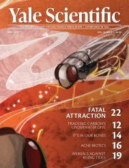
YSM Issue 90.4

Create successful ePaper yourself
Turn your PDF publications into a flip-book with our unique Google optimized e-Paper software.
FOCUS<br />
cell biology<br />
You lost your keys again. Without any idea as to where they went, you dart from room to room,<br />
pursuing each lead for just a moment before giving up and trying something else. As you search,<br />
you piece it together and suddenly remember where they are: the refrigerator. Turning away from your<br />
hamper, you make a beeline for the kitchen, and the keys are recovered within moments. The way you<br />
went about finding your keys—the indiscriminate giving way to the direct—is not unique to forgetful<br />
humans. All living things—dogs, bees, cancer cells—devise these strategies as they explore the world<br />
around them. In the last case, however, this probing can do more harm than good. The more cancer cells<br />
move in a persistent beeline, the more efficiently they will spread and the more lethal the cancer will be.<br />
A team at the Yale Systems Biology Institute<br />
led by professor Andre Levchenko<br />
and postdoctorate researcher JinSeok Park<br />
wanted to more comprehensively understand<br />
how cancer cells find their proverbial<br />
keys. To start, it is important to understand<br />
how cells interact with their direct<br />
outside environment, a network of proteins<br />
called the extracellular matrix (ECM) that<br />
cells secrete themselves. Coming into contact<br />
with a neighboring cell’s ECM triggers<br />
two signaling pathways that mediate cell<br />
movement. The balance between the different<br />
behaviors that these two pathways<br />
trigger—the cell’s polarity, so to speak—<br />
influences cells’ migration behavior, their<br />
switch from frenzy to beeline. Previous research<br />
implied that cell contact with the<br />
ECM was responsible for these different<br />
patterns, but how the cells went about this<br />
was poorly understood. Levchenko and<br />
Park derived a mathematical model to predict<br />
a given cell’s behavior based on the activity<br />
of the two principal signaling pathways,<br />
which both cements the link between<br />
cell-ECM contact and migration and, more<br />
importantly, clarifies a new avenue for cancer<br />
treatment.<br />
Migration<br />
The type of cancer called melanoma<br />
rarely kills at its origin, the skin, but rather<br />
takes lives when it attacks other organ<br />
systems, or metastasizes. Therefore, it is a<br />
disease that relies heavily on movement,<br />
a trait that made it a useful model for the<br />
team to study. The critical moment in melanoma<br />
progression happens when the cancer<br />
stops expanding across the skin and begins<br />
tunneling downward. At that point,<br />
it is only a matter of time until melanoma<br />
cells find their way to a blood or lymphatic<br />
vessel, the anatomical highways necessary<br />
for metastasis. But first, these cells have to<br />
navigate the fibers of the dermis, the layer<br />
of tissue right below the skin.<br />
These tissue fibers are like ropes that cells<br />
can pull to move around. There are three<br />
types of cell movements. When the cells<br />
rapidly pull ropes in different directions,<br />
their behavior is random; when cells alternate<br />
pulling forward and backward on the<br />
same rope, their behavior is oscillatory;<br />
and when cells consistently pull one rope in<br />
one direction, their behavior is persistent.<br />
The oscillatory pattern is a cell-specific behavior<br />
and complicates the model—once<br />
a target has been established for the cell,<br />
it continues to travel back and forth in the<br />
vicinity instead of heading straight there.<br />
Levchenko likes to think of oscillation as<br />
a cell overshooting its target and doubling<br />
back repeatedly. Regardless, if a cell is to<br />
reach a destination, persistent behavior is<br />
the most efficient, which, in the context of<br />
cancer, means a faster progression towards<br />
metastasis.<br />
Migration patterns do not happen by<br />
chance. Cells’ direct contact with the fibers<br />
of the ECM sets off two sets of signals<br />
that influence cell movement and so<br />
determine whether cells will display random,<br />
oscillatory, or persistent behavior.<br />
One of these signals is characterized by a<br />
protein called Rac1 and the other by a protein<br />
called RhoA. The two sets of signals<br />
work towards the same goal—to mediate<br />
cell movement—but perform opposite<br />
functions, as Rac1 acts as an accelerator to<br />
RhoA’s brakes. To complicate matters further<br />
RhoA functions to halt cell migration,<br />
its presence is also required for migration<br />
in the first place. This somewhat paradoxical<br />
mechanism only highlights the complexity<br />
of these networks. Research in oncology<br />
typically has focused on how cancer<br />
cells grow and replicate, but these convoluted<br />
and poorly understood processes of<br />
cell migration pushed Levchenko and Park<br />
to establish a coherent model and reconcile<br />
the three migration patterns.<br />
Polarization<br />
The model the researchers derives uses<br />
advanced mathematics to integrate these<br />
three behaviors into the same framework,<br />
an unprecedented feat. The model shows<br />
how cells’ contact with the ECM changes<br />
the balance between the levels of activity<br />
of the Rac1 and RhoA pathways, which<br />
in turn impacts migration. By plotting the<br />
two activation rates on the same axes, the<br />
researchers were able to establish discrete<br />
regions for each migration pattern. For example,<br />
if a cell has a high Rac1 activation<br />
rate and a low RhoA activation rate—a lot<br />
of accelerator and little brakes—it will display<br />
persistent behavior.<br />
The model accounts for not only the balance,<br />
but also the location of this pathway<br />
activation within a given cell. By tracking<br />
sites of high signaling activity, the researchers<br />
were able to visualize the spatial<br />
distribution of these two pathways. In<br />
randomly migrating cells, signaling was<br />
sporadic; in oscillating cells, hotspots of<br />
activity alternated between the front and<br />
the back; and in persistently moving cells,<br />
activity was high on one side and inhibited<br />
on the other. These signals are consistent<br />
with the aforementioned “ropes” of the<br />
ECM and helped Levchenko and Park affirm<br />
that their model accurately represented<br />
cell movement.<br />
This model is unique because Levchenko<br />
and Park were able to map specific populations<br />
of cells onto diagrams that were generated<br />
using the model. Within the same<br />
cancer or even the same tumor, different<br />
cells will display different behaviors, and<br />
the model accounts for these varied responses.<br />
“[Cells] all get the same piece of<br />
information, but they interpret that information<br />
in very different ways,” Levchenko<br />
said. Understanding how real cells move<br />
within and correspond to the rigid math-<br />
16 Yale Scientific Magazine October 2017 www.yalescientific.org