1 Paleoradiology: History and New Developments - Academia.dk
1 Paleoradiology: History and New Developments - Academia.dk
1 Paleoradiology: History and New Developments - Academia.dk

You also want an ePaper? Increase the reach of your titles
YUMPU automatically turns print PDFs into web optimized ePapers that Google loves.
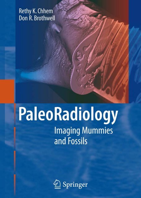
R. K. Chhem · D. R. Brothwell<br />
<strong>Paleoradiology</strong>
R. K. Chhem · D. R. Brothwell<br />
<strong>Paleoradiology</strong><br />
Imaging Mummies <strong>and</strong> Fossils<br />
With 390 Figures <strong>and</strong> 58 Tables<br />
123
Don R. Brothwell, PhD<br />
Department of Archaeology<br />
The University of York<br />
The King’s Manor<br />
York Y01 7EP<br />
UK<br />
Rethy K. Chhem, MD, PhD, FRCPC<br />
Department of Diagnostic Radiology <strong>and</strong> Nuclear Medicine<br />
Schulich School of Medicine <strong>and</strong> Dentistry<br />
University of Western Ontario<br />
London Health Sciences Centre<br />
339 Windermere Road<br />
London, Ontario<br />
N6A 5A5<br />
Canada<br />
Library of Congress Control Number: 2007936308<br />
ISBN 978-3-540-48832-3 Springer Berlin Heidelberg <strong>New</strong> York<br />
This work is subject to copyright. All rights are reserved, whether the whole or part of the<br />
material is concerned, specifically the rights of translation, reprinting, reuse of illustrations,<br />
recitation, broadcasting, reproduction on microfilm or in any other way, <strong>and</strong> storage in data<br />
banks. Duplication of this publication or parts thereof is permitted only under the provisions of<br />
the German Copyright Law of September 9, 1965, in its current version, <strong>and</strong> permission for use<br />
must always be obtained from Springer-Verlag. Violations are liable for prosecution under the<br />
German Copyright Law.<br />
Springer is a part of Springer Science+Business Media<br />
springer.com<br />
© Springer-Verlag Berlin Heidelberg 2008<br />
The use of general descriptive names, registered names, trademarks, etc. in this publication does<br />
not imply, even in the absence of a specific statement, that such names are exempt from the<br />
relevant protective laws <strong>and</strong> regulations <strong>and</strong> therefore free for general use.<br />
Product liability: The publishers cannot guarantee the accuracy of any information about<br />
dosage <strong>and</strong> application contained in this book. In every individual case the user must check such<br />
information by consulting the relevant literature.<br />
Editor: Dr. Ute Heilmann, Heidelberg, Germany<br />
Desk Editor: Meike Stoeck, Heidelberg, Germany<br />
Production: LE-TEX Jelonek, Schmidt & Vöckler GbR, Leipzig, Germany<br />
Reproduction <strong>and</strong> typesetting: Satz-Druck-Service (SDS), Leimen, Germany<br />
Cover design: WMX Design, Heidelberg, Germany<br />
Printed on acid-free paper 24/3180/YL 5 4 3 2 1 0
Foreword<br />
The Radiologist’s Perspective<br />
It is my pleasure to write the foreword to this groundbreaking text in paleoradiology.<br />
Dr. Rethy Chhem is a distinguished musculoskeletal radiologist, <strong>and</strong> he is the<br />
founder of the Paleoradiologic Research Unit at the University of Western Ontario,<br />
Canada, <strong>and</strong> the Osteoarchaeology Research Group at the National University of<br />
Singapore. His special area of paleoradiologic expertise is the Khmer civilization<br />
of Cambodia, <strong>and</strong> his contributions to radiologic <strong>and</strong> anthropologic science have<br />
built bridges between these two not always communicative disciplines.<br />
Dr. Don Brothwell is of course well known to the paleopathology community.<br />
He is something of an anthropologic <strong>and</strong> archaeologic polymath, having made<br />
important contributions to dental anthropology, the antiquity of human diet,<br />
<strong>and</strong> veterinary paleopathology, among others. His textbook, “Digging Up Bones”<br />
(Brothwell 1982), has introduced many generations of scholars to bioarchaeology,<br />
a discipline of which he is one of the founders. It is only fitting that this book is<br />
the work of a radiologist <strong>and</strong> an anthropologist, both of whom have experience in<br />
musculoskeletal imaging <strong>and</strong> paleopathology. For more than 100 years, diagnostic<br />
imaging has been used in the study of ancient disease. In fact, one of the first comprehensive<br />
textbooks of paleopathology, “Paleopathologic Diagnosis <strong>and</strong> Interpretation,”<br />
was written as an undergraduate thesis by a nascent radiologist, Dr. Ted<br />
Steinbock (Steinbock 1976).<br />
The advantages of diagnostic imaging in paleopathologic research should be<br />
intuitively obvious. Osseous <strong>and</strong> soft tissue may be noninvasively <strong>and</strong> nondestructively<br />
imaged, preserving original specimens for research <strong>and</strong> display in a museum<br />
setting. Not only will the original material, often Egyptian mummies, be preserved<br />
for future generations of researchers, but public enthusiasm will be fostered by the<br />
knowledge that we can see what is really underneath all those wrappings. Recent<br />
advances in computed multiplanar image display present novel ways to increase<br />
our underst<strong>and</strong>ing of the individuals, the processes of mummification <strong>and</strong> burial,<br />
<strong>and</strong> the cultural milieu in which these people lived. Unfortunately, although the<br />
potential of radiology has been recognized, the realization of collaborative effort<br />
has been inconsistent.<br />
The earliest use of radiography in paleopathology was in the diagnosis of specific<br />
diseases in individuals, much as it is in clinical medicine today. Egyptian mummies<br />
were radiographed as early as 1896. Comprehensive studies of mummy collections<br />
were performed in the 1960s <strong>and</strong> 1970s, culminating in the exhaustive treatise by<br />
Harris <strong>and</strong> Wente, with important contributions by Walter Whitehouse, MD, in<br />
1980 (Harris <strong>and</strong> Wente 1980). The usefulness of radiologic analysis of collections<br />
of such specimens led to the realization that diagnostic imaging has important implications<br />
in paleoepidemiology as well as in the diagnosis of individual cases.<br />
Technical innovations in radiology have paralleled progress in paleopathology.<br />
We are now able to perform per three-dimensional virtual reproductions of the<br />
facial characteristics so that mummies do not have to be unwrapped, <strong>and</strong> we can<br />
now carry out “virtual autopsies” using three-dimensional computed tomography<br />
as a guide. We are now also using modern imaging technology to go beyond pic-
VI<br />
Foreword<br />
tures. It is well established that radiologic <strong>and</strong> computed tomographic evaluation,<br />
in conjunction with physical anthropologic <strong>and</strong> orthopedic biomechanical data,<br />
may yield important biomechanical information in such studies as noninvasive<br />
measurement of the cross-sectional area of long bones to compare biomechanical<br />
characteristics in different populations such as hunter-gatherers <strong>and</strong> agriculturalists,<br />
<strong>and</strong> to study the mechanical properties of trabecular bone.<br />
This textbook represents a significant advance in the effort to engage clinical<br />
physicians, especially radiologists <strong>and</strong> paleopathologists in a dialogue. Although<br />
there have been many such attempts in the past, they have for the most part dealt<br />
with specific imaging findings to diagnose disease in specific ancient remains.<br />
Chhem <strong>and</strong> Brothwell have given us the opportunity to go beyond this type of ad<br />
hoc consultation by presenting a systematic approach to the radiologic skeletal differential<br />
diagnosis of ancient human <strong>and</strong> animal remains. However, I believe that<br />
the intent of the authors is not so much to have paleopathologists interpret these<br />
finding in a vacuum, but rather to underst<strong>and</strong> the capabilities of musculoskeletal<br />
radiologists, not only to assist with diagnosis, but also to offer information about<br />
the clinical setting in which these diseases occur <strong>and</strong> to suggest other appropriate<br />
imaging technology. For their part, musculoskeletal radiologists should be able to<br />
use this text to underst<strong>and</strong> the context in which paleopathologists work, including<br />
taphonomic change, <strong>and</strong> to appreciate the rich legacy of diagnostic imaging in biological<br />
anthropology <strong>and</strong> archaeology.<br />
Along with the authors, I hope that radiologists <strong>and</strong> biological anthropologists<br />
will use this textbook to translate both the radiologic <strong>and</strong> anthropologic idiom<br />
to better comprehend the other’s potential for collaboration. Once we establish a<br />
common language, it will be easier to solve the diagnostic problems <strong>and</strong> dilemmas<br />
we share. Doctors Chhem <strong>and</strong> Brothwell are to be congratulated for taking that<br />
important first step.<br />
Ethan M. Braunstein<br />
References<br />
Brothwell DB (1982) Digging up Bones (3rd edn). Cornell University Press, Ithaca<br />
Steinbock RT (1976) Paleopathologic Diagnosis <strong>and</strong> Interpretation. Charles C. Thomas, Springfield,<br />
Illinois, p 423<br />
Harris JE, Wente EF (1980) An X-Ray Atlas of the Royal Mummies. University of Chicago Press,<br />
Chicago, Illinois, p 403
Foreword<br />
The Anthropologist’s Perspective<br />
The study of human paleopathology has benefited from the use of radiological<br />
methods for many decades. However, the use of radiological images <strong>and</strong> interpretative<br />
insights has in earlier years tended to be limited to medical professionals with<br />
expertise <strong>and</strong> experience in interpreting radiographic images as well as having access<br />
to the necessary equipment to produce radiographs in the hospitals where they<br />
worked. As the diagnostic value of radiology in the evaluation <strong>and</strong> diagnosis of<br />
disorders in archaeological human <strong>and</strong> nonhuman remains became more apparent,<br />
plain-film radiological facilities were established in many nonmedical centers<br />
where research on these remains was a central part of their scientific endeavors.<br />
With greater access to radiographic data on paleopathological specimens, biological<br />
anthropologists became increasingly competent in interpreting these images.<br />
However, there remain very important reasons why ongoing collaboration between<br />
radiologists <strong>and</strong> biological anthropologists in the analysis of paleopathological cases<br />
continues to be a valuable contribution to science.<br />
One of the troublesome limitations of plain-film radiology is that three-dimensional<br />
anatomical features are projected onto a single plain. The inevitable superimposition<br />
that occurs can obscure important details of a radiographic image,<br />
adding to the challenge of interpretation. With the advent <strong>and</strong> widespread use of<br />
computed tomography (CT) radiological methods as an important diagnostic tool<br />
in clinical radiology, these methods began to be applied to archaeological remains.<br />
Among other advantages, CT imaging virtually eliminates the problem of superimposition.<br />
However, access to CT technology by paleopathologists, unless they<br />
are also radiologists, is often inconvenient or beyond the limited budgets of many<br />
researchers. This limitation in the use of CT imaging is changing as more facilities<br />
with CT equipment are available, including some in nonmedical research institutions.<br />
The remarkable power of CT imaging has made this mode of radiological<br />
investigation an important tool for the paleopathologist.<br />
During my collaborations with radiologists in my own research on human skeletal<br />
paleopathology during the past 40 years, several issues have been highlighted.<br />
One is the need for better specimen positioning in taking radiographs of archaeological<br />
human remains. In clinical radiology, great attention is paid to the orientation<br />
of the anatomical site to be imaged relative to the axis of the X-ray beam. Clinical<br />
radiographic technicians receive careful training in the placement of the patient to<br />
be radiographed. Positioning of paleopathological cases of disease is often a helterskelter<br />
arrangement in which little attention is paid to the anatomical relationship<br />
between multiple bones or the anatomical position relative to a living person. The<br />
emphasis is often on getting as many bones as possible on the X-ray film to save<br />
expense. Such a procedure does not lend itself to taking full advantage of the vast<br />
knowledge <strong>and</strong> experience of radiologists in the diagnosis of skeletal disorders.<br />
Another problem is that in the burial environment, soil constituents often penetrate<br />
archaeological human skeletal remains <strong>and</strong> can pose real challenges in diagnosis,<br />
particularly for those inexperienced in recognizing these infiltrates. Soil infiltrates<br />
are denser than bone <strong>and</strong> appear as sclerotic areas in radiographic images.
VIII<br />
Foreword<br />
These areas can be confused with antemortem pathology. Postmortem degradation<br />
of bone also occurs in the burial environment from both the acidic conditions<br />
commonly encountered in soil <strong>and</strong> the action of organisms, including bacteria,<br />
fungi <strong>and</strong> insect larvae, <strong>and</strong> plant roots. These destructive processes can mimic<br />
osteolytic pathological processes. Very careful attention to the fine details of the<br />
margins of destructive defects in bone is necessary to resolve the question of ante-<br />
versus postmortem destruction.<br />
In interpreting radiographs of skeletal remains curated in museums, there is the<br />
further complication of distinguishing between substances added during museum<br />
curation of archaeological remains <strong>and</strong> antemortem pathological processes. For<br />
example, the glue used to repair breaks in older museum accessions can be very<br />
dense <strong>and</strong> create an appearance of a sclerotic response or a bone tumor in a radiograph.<br />
These examples highlight the importance of collaboration between the clinical<br />
radiologist with an interest in paleopathology <strong>and</strong> the biological anthropologist<br />
in any study of archaeological remains, including mummified tissues <strong>and</strong> skeletal<br />
remains. Each discipline brings a specialized knowledge of the subject that maximizes<br />
the quality of the interpretation of radiological images from archaeological<br />
remains, both human <strong>and</strong> nonhuman.<br />
Although collaboration between radiologists <strong>and</strong> biological anthropologists is<br />
an obvious strategy, the increasing use of radiology in the study of archaeological<br />
biological tissues calls for an explicit statement regarding the use of this methodology<br />
in research. As indicated above, the radiology of archeological remains poses<br />
special problems, <strong>and</strong> these need to be identified <strong>and</strong> resolved to ensure that radiographic<br />
data on such remains is interpreted correctly. There is a very real need for<br />
an authoritative reference work that will provide the insight from both anthropology<br />
<strong>and</strong> radiology as this relates to the use of radiological methods in the study of<br />
ancient evidence of disease.<br />
I am very pleased to learn about the collaborative effort between Dr. Rethy<br />
Chhem, a skeletal radiologist, <strong>and</strong> Dr. Don Brothwell, a biological anthropologist,<br />
to produce a book on the radiology of archaeological biological tissues. Both are<br />
distinguished international authorities in their respective disciplines. In addition,<br />
both bring a depth of experience in the study of paleopathology that ensures careful<br />
coverage of the subject <strong>and</strong> new insight into the technical, theoretical, <strong>and</strong><br />
interpretative issues involved in the application of radiology to the evaluation <strong>and</strong><br />
diagnosis of abnormalities encountered in the analysis of human <strong>and</strong> nonhuman<br />
archaeological remains. I am confident that this book will be a major milestone in<br />
the study of disease in human <strong>and</strong> nonhuman archeological as well as paleontological<br />
remains.<br />
Donald J. Ortner
Preface<br />
This book arose from chance meetings <strong>and</strong> discussion between the two of us, one a<br />
radiologist <strong>and</strong> anthropologist (RC), the other a bioarchaeologist <strong>and</strong> paleopathologist<br />
(DB). The former expressed his interest in developing a scientific field that<br />
combined radiology with anthropology, especially bioarchaeology <strong>and</strong> paleopathology.<br />
The latter agreed completely that the subjects of radiographic techniques<br />
<strong>and</strong> the application of all aspects of medical imaging to the study of anthropological<br />
materials were sadly neglected. At the same time, both recognized that a publication<br />
was needed to show more clearly the considerable potential of paleoradiology.<br />
At this point, one of us (DB) expressed some uncertainty about finding the time (if<br />
not the mental strength) to contribute to the formation of this field. However, the<br />
extreme enthusiasm <strong>and</strong> persuasiveness of his friend <strong>and</strong> colleague (RC) resulted<br />
not in his withdrawal, but in discussing a joint plan of action. Such is the power of<br />
an enthusiastic colleague <strong>and</strong> a challenging project!<br />
What follows in these pages is an attempt to introduce a new field of academic<br />
study that is concerned with the value of applying X-rays to a broad range of bioanthropological<br />
materials, from human remains to other animals <strong>and</strong> even plants.<br />
We would emphasize that brought together in this way, it becomes a new field, even<br />
if components of the whole field have a much longer history. An entire chapter<br />
deals with the use of paleoradiology as a diagnosic method of ancient diseases. So<br />
in its entirety, the book is a pilot survey, an introduction to a broad-based subject<br />
that we feel is going to exp<strong>and</strong> <strong>and</strong> interest a growing number of our colleagues,<br />
spanning human <strong>and</strong> veterinary radiology, anthropology (especially bioarchaeology),<br />
zoology, <strong>and</strong> botany. It is clear that at the present time, the literature relevant<br />
to this broad discipline is highly variable, <strong>and</strong> to some extent locked away in specialist<br />
publications. There is currently a strong bias toward human remains, both<br />
skeletal <strong>and</strong> mummified. We predict that this will change, <strong>and</strong> in particular we<br />
suggest that it will be employed increasingly in the field of zooarchaeology, where<br />
considerable numbers of bones <strong>and</strong> teeth are processed annually throughout the<br />
world <strong>and</strong> increasing attention is being paid to reconstructing the health status of<br />
earlier animal populations.<br />
We sincerely hope that this introductory text on paleoradiology will stimulate<br />
interest in our colleagues, sufficient for them to ponder how they might contribute<br />
to this field in the future, or at least bring it to the notice of their colleagues or<br />
students. We do not see paleoradiology as a marginal <strong>and</strong> somewhat exotic occupation,<br />
rather one of considerable academic potential.<br />
Rethy K. Chhem <strong>and</strong> Don R. Brothwell
Acknowledgements<br />
We wish to express our great appreciation to our many friends <strong>and</strong> colleagues who<br />
have assisted in the preparation of this book in many ways. We sincerely hope that<br />
this list is complete, but if we have overlooked anyone by mistake, we ask for their<br />
forgiveness.<br />
Supporting Rethy Chhem: Gord Allan, Ian Chan, Ghida Chouraiki, Eadie<br />
Deville, Jillian Flower, Jill Friis, Bertha Garcia, John Henry, Carol Herbert, David<br />
Holdsworth, Cheryl Joe, Stephen Karlik, Karen Kennedy, Jodie Koepke, Kyle<br />
Latinis, Luy Lida, Julian Loh, Liz Lorusso, Jay Maxwell, David McErlain, Wendy<br />
McKay, El Molto, Andrew Nelson, Jeremy O’Brien, Katie Peters, Christophe Pottier,<br />
Lisa Rader, Janine Riffel, Cesare Romagnoli, Frank Rühli, Roberta Shaw, Wang<br />
Shi-Chang, Vankatesh Sudhakar, Cynthia Von Overloop, Corie Wei, Jackie Williams,<br />
Deanna Wocks, Kit M. Wong, Eric Yap, Anabella Yim, <strong>and</strong> members of the<br />
Osteoarchaeology Research Group of Singapore.<br />
Supporting Don Brothwell: Trevor Anderson, John Baker, Keith Dobney, Ben<br />
Gourley, Deborah Jaques, Simon McGrory, Theya Molleson, Naomi Mott, Sonia<br />
O’Connor, Terry O’Connor, Ian Panter, Jacqui Watson, Wyn Wheeler<br />
We wish to thank GE Healthcare Canada for their support to the <strong>Paleoradiology</strong><br />
Research Unit, <strong>and</strong> the Department of Radiology, London Health Sciences Centre<br />
<strong>and</strong> the University of Western Ontario, Canada.<br />
Finally, but by no means least, we both wish to thank Sirika Chhem <strong>and</strong> Jade<br />
Orkin-Fenster, whose hard work <strong>and</strong> commitment in York during the summer of<br />
2005 provided us with a wide range of digital radiographs for use in this book.
Contents<br />
Chapter 1<br />
<strong>Paleoradiology</strong>: <strong>History</strong> <strong>and</strong> <strong>New</strong> <strong>Developments</strong><br />
Rethy K. Chhem . . . . . . . . . . . . . . . . . . . . . . . . . . . . . . . . . . . . . . . . . . . . . . . . . . . . . . . . 1<br />
1.1 Paleoradiography . . . . . . . . . . . . . . . . . . . . . . . . . . . . . . . . . . . . . . . . . . . . . . . 2<br />
1.2 <strong>Paleoradiology</strong> of Royal Egyptian Mummies . . . . . . . . . . . . . . . . . . . . . . . 5<br />
1.2.1 1912 Thoutmosis IV . . . . . . . . . . . . . . . . . . . . . . . . . . . . . . . . . . . . . . . . . . . . . 5<br />
1.2.2 1933 Amenophis I . . . . . . . . . . . . . . . . . . . . . . . . . . . . . . . . . . . . . . . . . . . . . . . 6<br />
1.2.3 1965 Royal Mummy Collection . . . . . . . . . . . . . . . . . . . . . . . . . . . . . . . . . . . 6<br />
1.2.4 1968 Tutankhamun . . . . . . . . . . . . . . . . . . . . . . . . . . . . . . . . . . . . . . . . . . . . . 7<br />
1.2.5 1976 Ramesses II . . . . . . . . . . . . . . . . . . . . . . . . . . . . . . . . . . . . . . . . . . . . . . . . 7<br />
1.3 Paleo-CT . . . . . . . . . . . . . . . . . . . . . . . . . . . . . . . . . . . . . . . . . . . . . . . . . . . . . . 8<br />
1.4 Paleo-MRI . . . . . . . . . . . . . . . . . . . . . . . . . . . . . . . . . . . . . . . . . . . . . . . . . . . . . 9<br />
1.5 <strong>Paleoradiology</strong> <strong>and</strong> Clinical Radiology:Historical Development . . . . . . . 10<br />
References . . . . . . . . . . . . . . . . . . . . . . . . . . . . . . . . . . . . . . . . . . . . . . . . . . . . . . . . . . . . . 12<br />
Chapter 2<br />
Paleoradiologic Techniques<br />
George Saab, Rethy K. Chhem,<br />
<strong>and</strong> Richard N. Bohay . . . . . . . . . . . . . . . . . . . . . . . . . . . . . . . . . . . . . . . . . . . . . . . . . . . 15<br />
2.1 X-ray Imaging For Bioarcheology . . . . . . . . . . . . . . . . . . . . . . . . . . . . . . . . . 15<br />
2.2 Radiographic Production . . . . . . . . . . . . . . . . . . . . . . . . . . . . . . . . . . . . . . . . 16<br />
2.2.1 Equipment Overview . . . . . . . . . . . . . . . . . . . . . . . . . . . . . . . . . . . . . . . . . . . . 16<br />
2.2.2 Portable X-ray Imaging Systems . . . . . . . . . . . . . . . . . . . . . . . . . . . . . . . . . . . 16<br />
2.2.3 X-ray Factors . . . . . . . . . . . . . . . . . . . . . . . . . . . . . . . . . . . . . . . . . . . . . . . . . . . 17<br />
2.2.3.1 The Photoelectric Effect . . . . . . . . . . . . . . . . . . . . . . . . . . . . . . . . . . . . . . . . . . 17<br />
2.2.3.2 The Compton Effect . . . . . . . . . . . . . . . . . . . . . . . . . . . . . . . . . . . . . . . . . . . . . 18<br />
2.2.4 Equipment Factors . . . . . . . . . . . . . . . . . . . . . . . . . . . . . . . . . . . . . . . . . . . . . . 18<br />
2.2.4.1 Grids . . . . . . . . . . . . . . . . . . . . . . . . . . . . . . . . . . . . . . . . . . . . . . . . . . . . . . . . . . 18<br />
2.2.4.2 Radiographic Film . . . . . . . . . . . . . . . . . . . . . . . . . . . . . . . . . . . . . . . . . . . . . . 19<br />
2.2.5 Geometry Factors . . . . . . . . . . . . . . . . . . . . . . . . . . . . . . . . . . . . . . . . . . . . . . . 19<br />
2.2.5.1 Focal Spot Size . . . . . . . . . . . . . . . . . . . . . . . . . . . . . . . . . . . . . . . . . . . . . . . . . . 19<br />
2.2.5.2 Source, Object, <strong>and</strong> Film Distances . . . . . . . . . . . . . . . . . . . . . . . . . . . . . . . . 20<br />
2.2.6 St<strong>and</strong>ard Radiographic Views . . . . . . . . . . . . . . . . . . . . . . . . . . . . . . . . . . . . 21<br />
2.2.6.1 Cranial Bones . . . . . . . . . . . . . . . . . . . . . . . . . . . . . . . . . . . . . . . . . . . . . . . . . . 21<br />
2.2.6.2 Postcranial Bones . . . . . . . . . . . . . . . . . . . . . . . . . . . . . . . . . . . . . . . . . . . . . . . 21<br />
2.2.7 Optimizing Radiographic Production Factors . . . . . . . . . . . . . . . . . . . . . . 24<br />
2.2.8 Quick Troubleshooting Guide . . . . . . . . . . . . . . . . . . . . . . . . . . . . . . . . . . . . 24<br />
2.3 Image Quality . . . . . . . . . . . . . . . . . . . . . . . . . . . . . . . . . . . . . . . . . . . . . . . . . . 24<br />
2.3.1 Contrast . . . . . . . . . . . . . . . . . . . . . . . . . . . . . . . . . . . . . . . . . . . . . . . . . . . . . . . 25<br />
2.3.2 Resolution . . . . . . . . . . . . . . . . . . . . . . . . . . . . . . . . . . . . . . . . . . . . . . . . . . . . . 25<br />
2.3.3 Noise . . . . . . . . . . . . . . . . . . . . . . . . . . . . . . . . . . . . . . . . . . . . . . . . . . . . . . . . . . 25<br />
2.4 Advances in Radiography <strong>and</strong> Archiving . . . . . . . . . . . . . . . . . . . . . . . . . . . 25<br />
2.4.1 Computed Radiography . . . . . . . . . . . . . . . . . . . . . . . . . . . . . . . . . . . . . . . . . . 26
XIV<br />
Contents<br />
2.4.2 Digital Radiography . . . . . . . . . . . . . . . . . . . . . . . . . . . . . . . . . . . . . . . . . . . . . 26<br />
2.4.3 Picture Archiving <strong>and</strong> Communication Systems . . . . . . . . . . . . . . . . . . . . 26<br />
2.5 Computed Tomography . . . . . . . . . . . . . . . . . . . . . . . . . . . . . . . . . . . . . . . . . . 26<br />
2.5.1 Four Generations of CT Scanner Designs . . . . . . . . . . . . . . . . . . . . . . . . . . 27<br />
2.5.2 Spiral, Multislice, <strong>and</strong> Three-Dimensional CT . . . . . . . . . . . . . . . . . . . . . . 28<br />
2.6 Magnetic Resonance Imaging . . . . . . . . . . . . . . . . . . . . . . . . . . . . . . . . . . . . . 29<br />
2.7 Advanced Imaging Methods . . . . . . . . . . . . . . . . . . . . . . . . . . . . . . . . . . . . . . 29<br />
2.7.1 Micro-Computed Tomography . . . . . . . . . . . . . . . . . . . . . . . . . . . . . . . . . . . 29<br />
2.7.2 Coherent-Scatter CT. . . . . . . . . . . . . . . . . . . . . . . . . . . . . . . . . . . . . . . . . . . . . 29<br />
2.7.3 Stereolithography <strong>and</strong> Fused Deposition Modeling . . . . . . . . . . . . . . . . . . 30<br />
2.8 Dental Radiology . . . . . . . . . . . . . . . . . . . . . . . . . . . . . . . . . . . . . . . . . . . . . . . 30<br />
2.8.1 Technical Factors . . . . . . . . . . . . . . . . . . . . . . . . . . . . . . . . . . . . . . . . . . . . . . . 30<br />
2.8.1.2 Dental Film . . . . . . . . . . . . . . . . . . . . . . . . . . . . . . . . . . . . . . . . . . . . . . . . . . . . 30<br />
2.8.1.2 Film Exposure . . . . . . . . . . . . . . . . . . . . . . . . . . . . . . . . . . . . . . . . . . . . . . . . . . 30<br />
2.8.1.3 Film Processing . . . . . . . . . . . . . . . . . . . . . . . . . . . . . . . . . . . . . . . . . . . . . . . . . 31<br />
2.8.1.4 Digital Image Receptors . . . . . . . . . . . . . . . . . . . . . . . . . . . . . . . . . . . . . . . . . . 32<br />
2.8.1.5 Film Mounting <strong>and</strong> Storage . . . . . . . . . . . . . . . . . . . . . . . . . . . . . . . . . . . . . . 32<br />
2.8.2 Basic Anatomy of the Teeth <strong>and</strong> Jaws . . . . . . . . . . . . . . . . . . . . . . . . . . . . . . 32<br />
2.8.2.1 Basic Dental Radiography . . . . . . . . . . . . . . . . . . . . . . . . . . . . . . . . . . . . . . . . 34<br />
2.8.2.2 Occlusal Radiography . . . . . . . . . . . . . . . . . . . . . . . . . . . . . . . . . . . . . . . . . . . 36<br />
2.8.3 Specimen Imaging . . . . . . . . . . . . . . . . . . . . . . . . . . . . . . . . . . . . . . . . . . . . . . 37<br />
2.8.3.1 Imaging Intact Jaws . . . . . . . . . . . . . . . . . . . . . . . . . . . . . . . . . . . . . . . . . . . . . 37<br />
2.8.3.2 Radiography of Tooth/Bone Fragments . . . . . . . . . . . . . . . . . . . . . . . . . . . . 39<br />
2.8.3.3 Radiography of Loose Teeth . . . . . . . . . . . . . . . . . . . . . . . . . . . . . . . . . . . . . . 39<br />
2.9 The Radiographic Appearances of Some Selected Diseases<br />
of the Teeth <strong>and</strong> Jaws . . . . . . . . . . . . . . . . . . . . . . . . . . . . . . . . . . . . . . . . . . . . 39<br />
2.9.1 Dental Caries . . . . . . . . . . . . . . . . . . . . . . . . . . . . . . . . . . . . . . . . . . . . . . . . . . . 39<br />
2.9.2 Periapical Inflammatory Disease . . . . . . . . . . . . . . . . . . . . . . . . . . . . . . . . . . 40<br />
2.9.3 Periodontitis . . . . . . . . . . . . . . . . . . . . . . . . . . . . . . . . . . . . . . . . . . . . . . . . . . . 40<br />
2.9.4 Osteomyelitis . . . . . . . . . . . . . . . . . . . . . . . . . . . . . . . . . . . . . . . . . . . . . . . . . . . 42<br />
2.9.5 Pericoronal Disease . . . . . . . . . . . . . . . . . . . . . . . . . . . . . . . . . . . . . . . . . . . . . 42<br />
2.10 Applications in <strong>Paleoradiology</strong>. . . . . . . . . . . . . . . . . . . . . . . . . . . . . . . . . . . . 43<br />
2.10.1 Three-dimensional CT in Paleoanthropology . . . . . . . . . . . . . . . . . . . . . . . 43<br />
2.10.2 CT <strong>and</strong> Burials . . . . . . . . . . . . . . . . . . . . . . . . . . . . . . . . . . . . . . . . . . . . . . . . . 53<br />
2.10.3 CT <strong>and</strong> Mummies . . . . . . . . . . . . . . . . . . . . . . . . . . . . . . . . . . . . . . . . . . . . . . . 53<br />
References . . . . . . . . . . . . . . . . . . . . . . . . . . . . . . . . . . . . . . . . . . . . . . . . . . . . . . . . . . . . . 54<br />
Chapter 3<br />
The Taphonomic Process, Biological Variation, <strong>and</strong> X-ray Studies<br />
Don R. Brothwell . . . . . . . . . . . . . . . . . . . . . . . . . . . . . . . . . . . . . . . . . . . . . . . . . . . . . . . 55<br />
3.1 X-raying the Whole Range of Bioarcheological Materials . . . . . . . . . . . . . 55<br />
3.2 The Evaluation of Botanical Remains . . . . . . . . . . . . . . . . . . . . . . . . . . . . . . 56<br />
3.3 Radiological Aspects of Zooarcheology . . . . . . . . . . . . . . . . . . . . . . . . . . . . 58<br />
3.4 Positioning <strong>and</strong> Image . . . . . . . . . . . . . . . . . . . . . . . . . . . . . . . . . . . . . . . . . . . 59<br />
3.5 Taphonomic Aspects of Bones <strong>and</strong> Teeth . . . . . . . . . . . . . . . . . . . . . . . . . . . 60<br />
3.6 Measurement from X-rays . . . . . . . . . . . . . . . . . . . . . . . . . . . . . . . . . . . . . . . . 62<br />
3.7 X-raying Aspects of Growth . . . . . . . . . . . . . . . . . . . . . . . . . . . . . . . . . . . . . . 62<br />
3.8 Frozen, Dried, <strong>and</strong> Mummified Bodies . . . . . . . . . . . . . . . . . . . . . . . . . . . . 63<br />
3.9 Microradiography . . . . . . . . . . . . . . . . . . . . . . . . . . . . . . . . . . . . . . . . . . . . . . . 65<br />
3.10 Problems of Differential Diagnosis . . . . . . . . . . . . . . . . . . . . . . . . . . . . . . . . 66<br />
3.10.1 Horncore “Thumbprints” . . . . . . . . . . . . . . . . . . . . . . . . . . . . . . . . . . . . . . . . 66<br />
3.10.2 Leg Bones . . . . . . . . . . . . . . . . . . . . . . . . . . . . . . . . . . . . . . . . . . . . . . . . . . . . . . 66<br />
3.10.3 Vertebrae . . . . . . . . . . . . . . . . . . . . . . . . . . . . . . . . . . . . . . . . . . . . . . . . . . . . . . 67<br />
3.10.4 Significant Bone Loss . . . . . . . . . . . . . . . . . . . . . . . . . . . . . . . . . . . . . . . . . . . . 67<br />
3.10.5 Abnormal Cavities in Bone . . . . . . . . . . . . . . . . . . . . . . . . . . . . . . . . . . . . . . . 68
Contents<br />
3.11 Conclusion . . . . . . . . . . . . . . . . . . . . . . . . . . . . . . . . . . . . . . . . . . . . . . . . . . . . 69<br />
3.12 Summary . . . . . . . . . . . . . . . . . . . . . . . . . . . . . . . . . . . . . . . . . . . . . . . . . . . . . 70<br />
Acknowledgments . . . . . . . . . . . . . . . . . . . . . . . . . . . . . . . . . . . . . . . . . . . . . . . . . . . . . 70<br />
References . . . . . . . . . . . . . . . . . . . . . . . . . . . . . . . . . . . . . . . . . . . . . . . . . . . . . . . . . . . . 70<br />
Chapter 4<br />
Diagnostic <strong>Paleoradiology</strong>for Paleopathologists<br />
Rethy K. Chhem, George Saab,<br />
<strong>and</strong> Don R. Brothwell . . . . . . . . . . . . . . . . . . . . . . . . . . . . . . . . . . . . . . . . . . . . . . . . . . 73<br />
4.1 Introduction . . . . . . . . . . . . . . . . . . . . . . . . . . . . . . . . . . . . . . . . . . . . . . . . . . . 73<br />
4.2 The <strong>Paleoradiology</strong> Method . . . . . . . . . . . . . . . . . . . . . . . . . . . . . . . . . . . . . 74<br />
4.2.2 The Classification of Human Bones . . . . . . . . . . . . . . . . . . . . . . . . . . . . . . 76<br />
4.3 Gamuts Approach: The Tricks of the Trade . . . . . . . . . . . . . . . . . . . . . . . . 76<br />
4.3.1 The Classification of Human Joints . . . . . . . . . . . . . . . . . . . . . . . . . . . . . . . 76<br />
4.4 Bone Trauma . . . . . . . . . . . . . . . . . . . . . . . . . . . . . . . . . . . . . . . . . . . . . . . . . . 76<br />
4.4.1 The Classification of Fractures <strong>and</strong> Basics of X-ray Interpretation . . . . 77<br />
4.4.2 Differential Diagnosis . . . . . . . . . . . . . . . . . . . . . . . . . . . . . . . . . . . . . . . . . . 78<br />
4.4.3 The Healing Process <strong>and</strong> Complications of Fractures . . . . . . . . . . . . . . . 78<br />
4.5 Joint Trauma . . . . . . . . . . . . . . . . . . . . . . . . . . . . . . . . . . . . . . . . . . . . . . . . . . 80<br />
4.6 Arthropathies . . . . . . . . . . . . . . . . . . . . . . . . . . . . . . . . . . . . . . . . . . . . . . . . . 81<br />
4.6.2 Basics of X-ray Interpretation . . . . . . . . . . . . . . . . . . . . . . . . . . . . . . . . . . . . 84<br />
4.6.3 Arthropathies of the Spine <strong>and</strong> Pelvis . . . . . . . . . . . . . . . . . . . . . . . . . . . . . 87<br />
4.6.4 Arthropathies Affecting the Limbs . . . . . . . . . . . . . . . . . . . . . . . . . . . . . . . 91<br />
4.6.4.1 Osteoarthritis (Degenerative Joint Disease) . . . . . . . . . . . . . . . . . . . . . . . . 91<br />
4.6.4.2 Rotator Cuff Arthropathy . . . . . . . . . . . . . . . . . . . . . . . . . . . . . . . . . . . . . . . 92<br />
4.6.4.3 Neuropathic Arthropathy . . . . . . . . . . . . . . . . . . . . . . . . . . . . . . . . . . . . . . . 92<br />
4.7 Infection . . . . . . . . . . . . . . . . . . . . . . . . . . . . . . . . . . . . . . . . . . . . . . . . . . . . . . 93<br />
4.7.2 Basics of X-rays Interpretation . . . . . . . . . . . . . . . . . . . . . . . . . . . . . . . . . . . 93<br />
4.7.3 Differential Diagnosis . . . . . . . . . . . . . . . . . . . . . . . . . . . . . . . . . . . . . . . . . . 93<br />
4.7.4 Common Bone Infections in the Archeological Record . . . . . . . . . . . . . . . 94<br />
4.7.4.1 Pyogenic Infection . . . . . . . . . . . . . . . . . . . . . . . . . . . . . . . . . . . . . . . . . . . . . 94<br />
4.7.4.2 Syphilis <strong>and</strong> other Treponematosis . . . . . . . . . . . . . . . . . . . . . . . . . . . . . . . 94<br />
4.7.4.3 Tuberculosis . . . . . . . . . . . . . . . . . . . . . . . . . . . . . . . . . . . . . . . . . . . . . . . . . . . 96<br />
4.74.4 Leprosy . . . . . . . . . . . . . . . . . . . . . . . . . . . . . . . . . . . . . . . . . . . . . . . . . . . . . . . 98<br />
4.7.4.5 Brucellosis . . . . . . . . . . . . . . . . . . . . . . . . . . . . . . . . . . . . . . . . . . . . . . . . . . . . 98<br />
4.7.4.6 Paget’s Disease . . . . . . . . . . . . . . . . . . . . . . . . . . . . . . . . . . . . . . . . . . . . . . . . . 99<br />
4.8 Tumors . . . . . . . . . . . . . . . . . . . . . . . . . . . . . . . . . . . . . . . . . . . . . . . . . . . . . . . 99<br />
4.8.1 Classification of Bone Tumors . . . . . . . . . . . . . . . . . . . . . . . . . . . . . . . . . . . 99<br />
4.8.2 Basics of x-ray Interpretation of Bone Tumors . . . . . . . . . . . . . . . . . . . . . . 100<br />
4.8.2.1 Periosteal Reactions . . . . . . . . . . . . . . . . . . . . . . . . . . . . . . . . . . . . . . . . . . . . 101<br />
4.8.2.2 Internal Margins . . . . . . . . . . . . . . . . . . . . . . . . . . . . . . . . . . . . . . . . . . . . . . 103<br />
4.8.2.3 Matrix Patterns . . . . . . . . . . . . . . . . . . . . . . . . . . . . . . . . . . . . . . . . . . . . . . . . 104<br />
4.8.3 Common Tumors . . . . . . . . . . . . . . . . . . . . . . . . . . . . . . . . . . . . . . . . . . . . . . 105<br />
4.8.3.1 Osteochondroma or Exostosis . . . . . . . . . . . . . . . . . . . . . . . . . . . . . . . . . . . 105<br />
4.8.3.2 Enchondroma . . . . . . . . . . . . . . . . . . . . . . . . . . . . . . . . . . . . . . . . . . . . . . . . . 105<br />
4.8.3.3 Osteosarcoma . . . . . . . . . . . . . . . . . . . . . . . . . . . . . . . . . . . . . . . . . . . . . . . . . 105<br />
4.8.3.4 Paraosteal Sarcoma . . . . . . . . . . . . . . . . . . . . . . . . . . . . . . . . . . . . . . . . . . . . . 106<br />
4.8.3.5 Ewing’s Sarcoma . . . . . . . . . . . . . . . . . . . . . . . . . . . . . . . . . . . . . . . . . . . . . . . 107<br />
4.8.3.6 Chondrosarcoma . . . . . . . . . . . . . . . . . . . . . . . . . . . . . . . . . . . . . . . . . . . . . . 108<br />
4.8.3.7 Giant Cell Tumor . . . . . . . . . . . . . . . . . . . . . . . . . . . . . . . . . . . . . . . . . . . . . . 108<br />
4.8.3.8 Osteoma . . . . . . . . . . . . . . . . . . . . . . . . . . . . . . . . . . . . . . . . . . . . . . . . . . . . . . 108<br />
4.8.3.9 Fibrous Dysplasia . . . . . . . . . . . . . . . . . . . . . . . . . . . . . . . . . . . . . . . . . . . . . . 108<br />
4.8.3.10 Simple Bone Cyst . . . . . . . . . . . . . . . . . . . . . . . . . . . . . . . . . . . . . . . . . . . . . . 109<br />
4.8.3.11 Aneurysmal Bone Cyst . . . . . . . . . . . . . . . . . . . . . . . . . . . . . . . . . . . . . . . . . 109<br />
4.8.3.12 Vertebral Hemangioma . . . . . . . . . . . . . . . . . . . . . . . . . . . . . . . . . . . . . . . . . 110<br />
XV
XVI<br />
Contents<br />
4.8.3.13 Pseudotumor . . . . . . . . . . . . . . . . . . . . . . . . . . . . . . . . . . . . . . . . . . . . . . . . . . 110<br />
4.8.3.14 Metastases Versus Multiple Myeloma . . . . . . . . . . . . . . . . . . . . . . . . . . . . . 110<br />
4.9 Metabolic, Endocrine, Ecosystem Diseases,<strong>and</strong> Anemias . . . . . . . . . . . . 112<br />
4.9.1 Congenital Skeletal Diseases . . . . . . . . . . . . . . . . . . . . . . . . . . . . . . . . . . . . . 112<br />
4.9.2 Osteoporosis . . . . . . . . . . . . . . . . . . . . . . . . . . . . . . . . . . . . . . . . . . . . . . . . . . 113<br />
4.9.3 Osteomalacia <strong>and</strong> Rickets . . . . . . . . . . . . . . . . . . . . . . . . . . . . . . . . . . . . . . . 113<br />
4.9.3.1 Rickets at the Wrist . . . . . . . . . . . . . . . . . . . . . . . . . . . . . . . . . . . . . . . . . . . . . 114<br />
4.9.4 Harris Lines . . . . . . . . . . . . . . . . . . . . . . . . . . . . . . . . . . . . . . . . . . . . . . . . . . . 114<br />
4.9.5 Avascular Necrosis–Bone Infarcts . . . . . . . . . . . . . . . . . . . . . . . . . . . . . . . . 115<br />
4.9.6 Anemias . . . . . . . . . . . . . . . . . . . . . . . . . . . . . . . . . . . . . . . . . . . . . . . . . . . . . . 115<br />
References . . . . . . . . . . . . . . . . . . . . . . . . . . . . . . . . . . . . . . . . . . . . . . . . . . . . . . . . . . . . 116<br />
Chapter 5<br />
<strong>Paleoradiology</strong> in the Service of Zoopaleopathology<br />
Don R. Brothwell . . . . . . . . . . . . . . . . . . . . . . . . . . . . . . . . . . . . . . . . . . . . . . . . . . . . . . 119<br />
5.1 Congenital Abnormalities . . . . . . . . . . . . . . . . . . . . . . . . . . . . . . . . . . . . . . . 120<br />
5.2 Summary of Radiological Featuresof Congenital Abnormality . . . . . . . 121<br />
5.2.1 The Skull . . . . . . . . . . . . . . . . . . . . . . . . . . . . . . . . . . . . . . . . . . . . . . . . . . . . . 121<br />
5.2.1.1 Encephalomeningocele . . . . . . . . . . . . . . . . . . . . . . . . . . . . . . . . . . . . . . . . . 121<br />
5.2.1.2 Hydrocephalus . . . . . . . . . . . . . . . . . . . . . . . . . . . . . . . . . . . . . . . . . . . . . . . . 121<br />
5.2.1.3 Brachygnathia <strong>and</strong> Micrognathia . . . . . . . . . . . . . . . . . . . . . . . . . . . . . . . . 121<br />
5.2.1.4 Cleft Palate . . . . . . . . . . . . . . . . . . . . . . . . . . . . . . . . . . . . . . . . . . . . . . . . . . . . 121<br />
5.2.1.5 Cerebral Hernia . . . . . . . . . . . . . . . . . . . . . . . . . . . . . . . . . . . . . . . . . . . . . . . . 122<br />
5.2.2 The Postcranial Skeleton . . . . . . . . . . . . . . . . . . . . . . . . . . . . . . . . . . . . . . . . 122<br />
5.2.2.1 Dwarfism . . . . . . . . . . . . . . . . . . . . . . . . . . . . . . . . . . . . . . . . . . . . . . . . . . . . . 122<br />
5.2.2.2 Hip Dysplasia. . . . . . . . . . . . . . . . . . . . . . . . . . . . . . . . . . . . . . . . . . . . . . . . . . 122<br />
5.2.2.3 Hemivertebrae . . . . . . . . . . . . . . . . . . . . . . . . . . . . . . . . . . . . . . . . . . . . . . . . . 122<br />
5.2.2.4 Arthrogryposis . . . . . . . . . . . . . . . . . . . . . . . . . . . . . . . . . . . . . . . . . . . . . . . . 122<br />
5.2.2.5 Syndactyly . . . . . . . . . . . . . . . . . . . . . . . . . . . . . . . . . . . . . . . . . . . . . . . . . . . . 122<br />
5.2.2.6 Other Conditions . . . . . . . . . . . . . . . . . . . . . . . . . . . . . . . . . . . . . . . . . . . . . . 122<br />
5.2.3 Nutritional <strong>and</strong> Metabolic Conditions . . . . . . . . . . . . . . . . . . . . . . . . . . . . 123<br />
5.2.3.1 Osteoporosis (Osteopenia) . . . . . . . . . . . . . . . . . . . . . . . . . . . . . . . . . . . . . . 124<br />
5.2.3.2 Rickets. . . . . . . . . . . . . . . . . . . . . . . . . . . . . . . . . . . . . . . . . . . . . . . . . . . . . . . . 124<br />
5.2.3.3 Hypervitaminosis A . . . . . . . . . . . . . . . . . . . . . . . . . . . . . . . . . . . . . . . . . . . . 124<br />
5.2.3.4 Hypothyroidism . . . . . . . . . . . . . . . . . . . . . . . . . . . . . . . . . . . . . . . . . . . . . . . 124<br />
5.2.3.5 Juvenile Scurvy (Hypertrophic Osteodystrophy) . . . . . . . . . . . . . . . . . . . 125<br />
5.2.3.6 Osteodystrophia Fibrosa . . . . . . . . . . . . . . . . . . . . . . . . . . . . . . . . . . . . . . . . 125<br />
5.2.3.7 Fluorosis . . . . . . . . . . . . . . . . . . . . . . . . . . . . . . . . . . . . . . . . . . . . . . . . . . . . . . 125<br />
5.2.3.8 Harris Lines . . . . . . . . . . . . . . . . . . . . . . . . . . . . . . . . . . . . . . . . . . . . . . . . . . . 126<br />
5.3 Trauma . . . . . . . . . . . . . . . . . . . . . . . . . . . . . . . . . . . . . . . . . . . . . . . . . . . . . . . 126<br />
5.3.1 Fracture Healing . . . . . . . . . . . . . . . . . . . . . . . . . . . . . . . . . . . . . . . . . . . . . . . 127<br />
5.3.2 Infection . . . . . . . . . . . . . . . . . . . . . . . . . . . . . . . . . . . . . . . . . . . . . . . . . . . . . . 129<br />
5.3.2.1 Interdigital Necrobacillosis . . . . . . . . . . . . . . . . . . . . . . . . . . . . . . . . . . . . . . 131<br />
5.3.2.2 Vertebral Osteomyelitis . . . . . . . . . . . . . . . . . . . . . . . . . . . . . . . . . . . . . . . . . 131<br />
5.2.3.3 Actinomycosis . . . . . . . . . . . . . . . . . . . . . . . . . . . . . . . . . . . . . . . . . . . . . . . . . 132<br />
5.2.3.4 Coccidioidomycosis . . . . . . . . . . . . . . . . . . . . . . . . . . . . . . . . . . . . . . . . . . . . 132
Contents<br />
5.2.3.5 Atrophic Rhinitis . . . . . . . . . . . . . . . . . . . . . . . . . . . . . . . . . . . . . . . . . . . . . . 132<br />
5.2.3.6 Osteopetrosis . . . . . . . . . . . . . . . . . . . . . . . . . . . . . . . . . . . . . . . . . . . . . . . . . . 132<br />
5.4 The Arthropathies . . . . . . . . . . . . . . . . . . . . . . . . . . . . . . . . . . . . . . . . . . . . . 132<br />
5.4.1 Osteoarthritis . . . . . . . . . . . . . . . . . . . . . . . . . . . . . . . . . . . . . . . . . . . . . . . . . 133<br />
5.4.2 Osteochondritis Dissecans . . . . . . . . . . . . . . . . . . . . . . . . . . . . . . . . . . . . . . 133<br />
5.4.3 Legg-Perthes Disease . . . . . . . . . . . . . . . . . . . . . . . . . . . . . . . . . . . . . . . . . . . 133<br />
5.4.4 Infectious Arthritis . . . . . . . . . . . . . . . . . . . . . . . . . . . . . . . . . . . . . . . . . . . . . 133<br />
5.4.5 Rheumatoid Arthritis. . . . . . . . . . . . . . . . . . . . . . . . . . . . . . . . . . . . . . . . . . . 134<br />
5.4.6 Ankylosing Arthritis . . . . . . . . . . . . . . . . . . . . . . . . . . . . . . . . . . . . . . . . . . . 134<br />
5.4.7 Navicular Disease . . . . . . . . . . . . . . . . . . . . . . . . . . . . . . . . . . . . . . . . . . . . . . 135<br />
5.4.8 Bovine Spavin . . . . . . . . . . . . . . . . . . . . . . . . . . . . . . . . . . . . . . . . . . . . . . . . . 135<br />
5.5 Neoplasms . . . . . . . . . . . . . . . . . . . . . . . . . . . . . . . . . . . . . . . . . . . . . . . . . . . . 135<br />
5.5.1 Examples of Tumors Affecting the Skeleton . . . . . . . . . . . . . . . . . . . . . . . 137<br />
5.5.1.1 Synovial Sarcoma of Joints . . . . . . . . . . . . . . . . . . . . . . . . . . . . . . . . . . . . . . 137<br />
5.5.1.2 Benign Tumors . . . . . . . . . . . . . . . . . . . . . . . . . . . . . . . . . . . . . . . . . . . . . . . . 138<br />
5.5.1.2 Malignant Tumors . . . . . . . . . . . . . . . . . . . . . . . . . . . . . . . . . . . . . . . . . . . . . 139<br />
5.5.2 Secondary Tumors of Bone . . . . . . . . . . . . . . . . . . . . . . . . . . . . . . . . . . . . . . 139<br />
5.5.2.1 Metastatic Deposits . . . . . . . . . . . . . . . . . . . . . . . . . . . . . . . . . . . . . . . . . . . . 139<br />
5.5.2.2 Hypertrophic Pulmonary Osteoarthropathy . . . . . . . . . . . . . . . . . . . . . . . 139<br />
5.6 Oral Pathology . . . . . . . . . . . . . . . . . . . . . . . . . . . . . . . . . . . . . . . . . . . . . . . . 140<br />
5.6.1 Classifying Oral pathology . . . . . . . . . . . . . . . . . . . . . . . . . . . . . . . . . . . . . . 141<br />
5.6.1.1 The Teeth . . . . . . . . . . . . . . . . . . . . . . . . . . . . . . . . . . . . . . . . . . . . . . . . . . . . . 141<br />
5.6.1.2 The Jaw Bones . . . . . . . . . . . . . . . . . . . . . . . . . . . . . . . . . . . . . . . . . . . . . . . . . 141<br />
5.6.1.3 Comparative <strong>and</strong> Epidemiological Studies . . . . . . . . . . . . . . . . . . . . . . . . . 142<br />
5.7 Conclusions . . . . . . . . . . . . . . . . . . . . . . . . . . . . . . . . . . . . . . . . . . . . . . . . . . . 143<br />
References . . . . . . . . . . . . . . . . . . . . . . . . . . . . . . . . . . . . . . . . . . . . . . . . . . . . . . . . . . . . 143<br />
Chapter 6<br />
Normal Variations in Fossils <strong>and</strong> Recent Human Groups<br />
Don R. Brothwell . . . . . . . . . . . . . . . . . . . . . . . . . . . . . . . . . . . . . . . . . . . . . . . . . . . . . . 147<br />
6.1 Fossil Studies by Conventional Radiography . . . . . . . . . . . . . . . . . . . . . . . 147<br />
6.2 Teeth <strong>and</strong> Jaws . . . . . . . . . . . . . . . . . . . . . . . . . . . . . . . . . . . . . . . . . . . . . . . . . 149<br />
6.3 The Advent of CT . . . . . . . . . . . . . . . . . . . . . . . . . . . . . . . . . . . . . . . . . . . . . . 149<br />
6.4 The Cranial Sinuses . . . . . . . . . . . . . . . . . . . . . . . . . . . . . . . . . . . . . . . . . . . . 150<br />
6.5 The Frontal Sinuses . . . . . . . . . . . . . . . . . . . . . . . . . . . . . . . . . . . . . . . . . . . . 151<br />
6.6 Variation in Recent Populations . . . . . . . . . . . . . . . . . . . . . . . . . . . . . . . . . . 151<br />
6.7 Age <strong>and</strong> Growth . . . . . . . . . . . . . . . . . . . . . . . . . . . . . . . . . . . . . . . . . . . . . . . 151<br />
6.8 Sella Turcica Variation . . . . . . . . . . . . . . . . . . . . . . . . . . . . . . . . . . . . . . . . . . 153<br />
6.9 The Bony Labyrinth . . . . . . . . . . . . . . . . . . . . . . . . . . . . . . . . . . . . . . . . . . . . 153<br />
6.10 Variation in the Postcranial Skeleton . . . . . . . . . . . . . . . . . . . . . . . . . . . . . 153<br />
6.11 Variation in Cortical Bone . . . . . . . . . . . . . . . . . . . . . . . . . . . . . . . . . . . . . . 154<br />
6.12 Cremations . . . . . . . . . . . . . . . . . . . . . . . . . . . . . . . . . . . . . . . . . . . . . . . . . . . . 156<br />
References . . . . . . . . . . . . . . . . . . . . . . . . . . . . . . . . . . . . . . . . . . . . . . . . . . . . . . . . . . . . 156<br />
Concluding Comments<br />
Rethy K. Chhem <strong>and</strong> Don R. Brothwell . . . . . . . . . . . . . . . . . . . . . . . . . . . . . . . . . . . 159<br />
XVII
List of Contributors<br />
Don R. Brothwell, PhD (Editor)<br />
Department of Archaeology<br />
The University of York<br />
The King’s Manor<br />
York Y01 7EP<br />
UK<br />
Rethy K. Chhem, MD, PhD, FRCPC (Editor)<br />
Department of Diagnostic Radiology <strong>and</strong> Nuclear Medicine<br />
Schulich School of Medicine <strong>and</strong> Dentistry<br />
University of Western Ontario<br />
London Health Sciences Centre<br />
339 Windermere Road<br />
London, Ontario<br />
N6A 5A5<br />
Canada<br />
Richard N. Bohay, DMD, MSc, MRCD<br />
Schulich School of Medicine <strong>and</strong> Dentistry<br />
University of Western Ontario<br />
London Health Sciences Centre<br />
339 Windermere Road<br />
London, Ontario<br />
N6A 5A5<br />
Canada<br />
Ethan M. Braunstein, MD<br />
Radiology Department<br />
Mayo Clinic<br />
Scottsdale<br />
5777 East Mayo Boulevard<br />
AZ 8505 Phoenix<br />
Arizona, USA<br />
Donald J. Ortner, PhD<br />
Smithsonian Institution<br />
Washington 20560<br />
District of Columbia, USA<br />
George Saab, MD, PhD<br />
Department of Diagnostic Radiology <strong>and</strong> Nuclear Medicine<br />
Schulich School of Medicine <strong>and</strong> Dentistry<br />
University of Western Ontario<br />
London Health Sciences Centre<br />
339 Windermere Road<br />
London, Ontario<br />
N6A 5A5<br />
Canada,
Chapter 1<br />
<strong>Paleoradiology</strong>:<br />
<strong>History</strong> <strong>and</strong> <strong>New</strong> <strong>Developments</strong><br />
Rethy K. Chhem<br />
“… by far the greatest technical advance was made<br />
when radiology began to be used in the examination<br />
of anthropological <strong>and</strong> paleontological materials.” …<br />
“The Roentgenological examination, moreover, has<br />
the great advantage in that it permits the investigator<br />
to examine bones without destroying them <strong>and</strong> to<br />
inspect mummies without unwrapping them.”<br />
(Sigerist 1951)<br />
<strong>Paleoradiology</strong> is the study of bioarcheological materials<br />
using modern imaging methods, such as x-ray<br />
radiography, computed tomography (CT), magnetic<br />
resonance imaging (MRI), <strong>and</strong> micro-CT. The first<br />
x-ray study of human <strong>and</strong> animal mummies was performed<br />
by Koenig in 1896 (Koenig 1896), but to the<br />
best of my knowledge, the terms “paleoradiology”<br />
<strong>and</strong> “paleoradiologist” were coined much later by<br />
Notman, a radiologist at the Park Nicollet Medical<br />
Center in Minneapolis, in his article in the American<br />
Journal of Roentgenology in 1987 (Notman et<br />
al. 1987). Although “paleoradiology” etymologically<br />
means “ancient radiology,” it is clear that when used<br />
within the context of paleopathology, the term defines<br />
without any confusion, the applications of x-ray tests<br />
to bioarcheological materials. Notman, in collaboration<br />
with a pathologist <strong>and</strong> an anthropologist from<br />
the University of Alberta, Canada, used radiographic<br />
investigation to study paleopathological lesions in<br />
two frozen sailors from the Franklin expedition<br />
(1845–1848) who perished in the Canadian arctic. As<br />
part of the investigation, Notman <strong>and</strong> his colleagues<br />
correlated the x-ray images of the two sailors with the<br />
results found at autopsy. Early publications on x-ray<br />
studies of mummies <strong>and</strong> skeletal remains include descriptive<br />
techniques, anatomy, <strong>and</strong> some of the paleopathology<br />
results.<br />
The development of paleoradiology has, to a large<br />
extent, been dependent on the parallel development<br />
of radiology <strong>and</strong> medical imaging technology. Unfortunately,<br />
for the last 100 years, the lack of input<br />
from radiologists, particularly those with expertise in<br />
skeletal pathology, has hampered the development of<br />
a sound scientific foundation for diagnostic methods<br />
to assist paleopathology studies.<br />
The availability of CT scanners in the early 1970s<br />
<strong>and</strong> the ongoing development of CT methods in the<br />
subsequent decades provided better visualization<br />
of the anatomy <strong>and</strong> of paleopathological lesions in<br />
mummies <strong>and</strong> in ancient skeletal remains. At the present<br />
time, the newer generations of CT scanners with<br />
their three-dimensional (3D) <strong>and</strong> surface rendering<br />
capabilities can create a 3D face reconstruction, or<br />
whole-body reconstructions of mummies. These have<br />
become extremely useful for anthropological studies<br />
museum displays <strong>and</strong> have attracted tremendous media<br />
attention. Despite these achievements, however,<br />
the role of CT in detecting ancient diseases is still not<br />
well established, largely due to a lack of clear diagnostic<br />
protocols. Most publications of CT deal with<br />
image acquisition of a whole body of a mummy, but<br />
without any tailored protocol designed to study a specific<br />
skeletal disease (O’Brien et al. 2007).<br />
This textbook is an attempt to lay these crucial<br />
foundations for the development of a scientific method<br />
for diagnostic paleoradiology, providing paleopathology<br />
studies with the structure to develop as an<br />
evidence-based discipline. This is the approach of the<br />
<strong>Paleoradiology</strong> Research Unit at the Department of<br />
Diagnostic Radiology <strong>and</strong> Nuclear Medicine within<br />
the Schulich School of Medicine <strong>and</strong> Dentistry at the<br />
University of Western Ontario in London, Ontario<br />
(Canada). This endeavor is facilitated by the presence<br />
of renowned experts in medical imaging science at the<br />
Robarts Research Institute, a private medical research<br />
institute affiliated with the University of Western Ontario<br />
<strong>and</strong> located within the university campus.<br />
Despite their laudable effort to acquire some radiological<br />
knowledge, many paleopathologists use what<br />
radiologists call “Aunt Minnie’s” approach, which is<br />
to compare x-rays of a specimen with radiological<br />
images from textbooks to establish the final diagnosis.<br />
This approach has led to many errors in the interpretation<br />
of x-ray images simply because most x-ray<br />
patterns are not specific <strong>and</strong> a thorough differential<br />
diagnosis has not been discussed.<br />
There is a widespread belief that radiologists who<br />
interpret x-rays of dry bone specimens are prone to<br />
mistakes because of the lack of underst<strong>and</strong>ing of ta-<br />
1
2 Chapter 1 <strong>Paleoradiology</strong>: <strong>History</strong> <strong>and</strong> <strong>New</strong> <strong>Developments</strong><br />
phonomic changes. This is, in my opinion, a logical<br />
fallacy, as once radiologists are made aware of these<br />
pitfalls, they will be integrated within the differential<br />
diagnosis of authentic paleopathological lesions. Who<br />
better to study disease than those who are intimately<br />
involved in its diagnosis? Therefore, the main reason<br />
for underdevelopment of paleoradiology is most likely<br />
that the paths of anthropologists <strong>and</strong> radiologists<br />
rarely cross. Bringing experts from these two separate<br />
scientific fields would, without any doubt, allow the<br />
establishment of evidence-based paleopathology.<br />
This kind of close collaboration between clinical<br />
radiologists, medical imaging scientists, anatomists,<br />
pathologists, <strong>and</strong> bioanthropologists has allowed an<br />
intense cross-fertilization of ideas <strong>and</strong> forms a very<br />
strong interdisciplinary approach for the development<br />
of scientific paleoradiology <strong>and</strong> paleopathology<br />
disciplines at the University of Western Ontario. The<br />
imaging facility at Robarts Research Institute, in particular,<br />
has some of the most advanced medical imaging<br />
technology available, including multislice CT,<br />
micro-CT, high-field MRI, <strong>and</strong> magnetic resonance<br />
(MR) spectroscopy, all of which are useful for enhancing<br />
paleoradiological studies.<br />
This chapter reviews the history <strong>and</strong> development<br />
of paleoradiology from its pioneering years to the present,<br />
when advanced medical imaging technology is<br />
used to investigate biological materials from archeological<br />
settings. The most recent developments of methods<br />
in paleoradiology are also reviewed, using the<br />
anatomical-clinical model (Boyer et al. 2003; Chhem<br />
2006; Chhem <strong>and</strong> Ruhli 2004; Chhem et al. 2004),<br />
together with a radiological–pathological correlation<br />
model (Chhem et al. 2006; Notman et al. 1987). Radiological–pathological<br />
methods, as used in the clinical<br />
setting, are essential for a rational <strong>and</strong> objective interpretation<br />
of radiological findings in paleopathology,<br />
while keeping in mind any pitfalls caused by taphonomic<br />
changes.<br />
Finally, we describe the ongoing advanced imaging<br />
investigation being carried out by our <strong>Paleoradiology</strong><br />
Research Unit on some unique materials from the Royal<br />
Ontario Museum (ROM). The ROM has allowed<br />
members of our team access to its rare <strong>and</strong> precious<br />
collection of Egyptian mummies, among them the famous<br />
3200-year-old mummified brain of Nakht, for<br />
which the first historical CT scans were performed on<br />
September 27, 1976.<br />
1.1<br />
Paleoradiography<br />
The first documented paleopathological studies were<br />
recorded more that two centuries ago (Esper 1774)<br />
(Fig. 1.1). Much later, soon after the discovery of x-<br />
rays by Roentgen in November 1895, but well before<br />
the official establishment of radiology as a medical<br />
specialty, x-ray study was used for nonmedical purposes<br />
to evaluate mummies of both humans <strong>and</strong> other<br />
animals, as well as to image ancient skeletal remains<br />
<strong>and</strong> hominid fossils (Böni et al. 2004; Koenig 1896)<br />
(Fig. 1.2). These studies were carried out primarily in<br />
Europe <strong>and</strong> in the USA (Albers-Schoenberg 1904; Culin<br />
1898; Dedekin 1896; Eder <strong>and</strong> Valenta 1896; Elliot<br />
Smith 1912; Gardiner 1904; Gocht 1911; Gorjanovic-<br />
Kramberger 1902; Holl<strong>and</strong> 1937; Koenig 1896; Londe<br />
1897; Petrie 1898; Salomon 1921) (Figs. 1.3–1.5). In<br />
those early stages of x-ray technical development, radiological<br />
studies were performed on mummies for<br />
several reasons. X-ray images of the contents <strong>and</strong> the<br />
wrapping often were taken to distinguish authentic<br />
mummies from fakes, to evaluate the bone age, to detect<br />
skeletal diseases, <strong>and</strong> to search for burial goods.<br />
The most common geographic origins of mummies<br />
were Egypt <strong>and</strong> Peru, which served as materials for<br />
the first monography of paleoradiology that was published<br />
in 1930 (Moodie 1930) (Fig. 1.6). Occasionally,<br />
an x-ray study was performed to evaluate bones <strong>and</strong><br />
Fig. 1.1. Cover page of the first book on paleopathology
a b<br />
a<br />
b<br />
teeth in paleolithic human fossils (Gorjanovic-Kramberger<br />
1902). These early x-ray studies <strong>and</strong> results<br />
were published in French, German, or English in diverse<br />
scholarly journals (Böni 2004).<br />
A historical review of the literature published<br />
within the first 25 years after the discovery of x-rays by<br />
Roentgen showed that paleoradiological studies were<br />
1.1 Paleoradiography<br />
Fig. 1.2. a Koenig: Radiography<br />
of an Egyptian<br />
human mummy (1896).<br />
Reprinted with permission<br />
from Thieme, <strong>New</strong> York.<br />
b Koenig: Radiography<br />
of an Egyptian cat (1896).<br />
Reprinted with permission<br />
from Thieme <strong>New</strong> York<br />
Fig. 1.3. a Londe: Mummy’s<br />
forearm (1897). b Londe:<br />
Radiography of a mummy’s<br />
forearm<br />
conducted by scientists from diverse backgrounds,<br />
including physicians <strong>and</strong> physicists, simply because<br />
there were no “radiologists” trained yet at that stage<br />
of x-ray development. For more information on this<br />
subject, a good source is Böni <strong>and</strong> his colleagues, who<br />
published a general review of the early history of paleoradiology<br />
(Böni et al. 2004) (Table 1.1).<br />
3
4 Chapter 1 <strong>Paleoradiology</strong>: <strong>History</strong> <strong>and</strong> <strong>New</strong> <strong>Developments</strong><br />
a<br />
b<br />
Fig. 1.5 Petrie: Radiography of the lower leg of a mummy<br />
(1898)<br />
Fig 1.4. a Londe: Fake<br />
mummy (1897). b Londe:<br />
Fake mummy<br />
Fig. 1.6 Cover first book on x-ray study of mummies. Reprinted<br />
with permission from Field Museum Press, Fieldiana
Table 1.1. Early paleoradiology studies of mummies/skeletal remains/fossils<br />
Author Year Study subject Site<br />
1.2<br />
<strong>Paleoradiology</strong> of Royal Egyptian Mummies<br />
Because there is a widespread public <strong>and</strong> academic<br />
fascination with Egyptology, this section gives a detailed<br />
review of the history of paleoradiology of the<br />
royal Egyptian mummies, which has helped to shed<br />
light on the lives of ancient Egyptian rulers. The following<br />
section is a review of the literature related<br />
to the paleoradiology of royal Egyptian mummies<br />
(Chhem 2007) (Table 1.2).<br />
1.2.1<br />
1912 Thoutmosis IV<br />
The first x-ray study of a royal Egyptian mummy was<br />
performed on Thoutmosis IV in 1903 by Dr. Khayat,<br />
an Egyptian radiologist. Thoutmosis IV was the 8th<br />
Pharaoh of the 18th Dynasty of Egypt, who ruled<br />
from 1400 to 1390 BC. The x-ray study provided the<br />
following information:<br />
“…The left ilium (which was exposed in the embalming-incision)<br />
<strong>and</strong> the upper of the tibia (exposed<br />
in the broken right leg) was made, <strong>and</strong> other parts of<br />
the body were examined by means of the Roentgenrays”<br />
(Elliot Smith 1912, p 44). “The epiphysis of the<br />
crest of the ilium was in process of union being united<br />
in the front but still free behind. This seemed to<br />
indicate that the body was that of a man of not more<br />
than 25 years…...in Piersol’s Human Anatomy, which<br />
was published three years after (in 1907) my report on<br />
1.2 <strong>Paleoradiology</strong> of Royal Egyptian Mummies<br />
Koenig 1896 Human <strong>and</strong> cat mummies Frankfurt, Germany<br />
Holl<strong>and</strong> 1896 Bird mummy Liverpool, UK<br />
Dedekind 1896/97 Egyptian mummies Vienna, Austria<br />
Londe 1897 Egyptian mummies<br />
Fake mummy<br />
Paris, France<br />
Leonard 1898 Peruvian mummies Philadelphia, USA<br />
Petrie 1898 Egyptian mummies London, UK<br />
Gorjanovic-Kramberger 1901 Hominid fossil Vienna, Austria<br />
Gardiner 1904 Egyptian mummies London, UK<br />
Albers-Schoenberg 1905 Egyptian mummies Hamburg, Germany<br />
Elliot Smith 1912 Egyptian mummies Cairo, Egypt<br />
Salomon 1921 Peruvian mummy Berlin, Germany<br />
Table 1.2. Published x-ray studies on Egyptian royal mummies<br />
Thoutmosis IV 1912 Elliot-Smith<br />
Amenophis I 1933 Derry<br />
Ramesses II 1976 Bucaille et al. a<br />
1979 Massare a<br />
1985 Bard et al.<br />
2004 Chhem et al. a<br />
Tutankhamun 1971 Harrisona 1972 Harrison <strong>and</strong> Abdalla a<br />
1976 Bucaille et al. a<br />
2003 Boyer et al. a<br />
2006 Shafik et al. b<br />
Royal mummies 1972 Harris <strong>and</strong> Weeks<br />
1980 Harris <strong>and</strong> Wente<br />
1988 Braunstein et al. a<br />
a Peer-reviewed journals<br />
b Abstract<br />
this mummy was written…” (Elliot Smith 1912, p 44).<br />
“In the skiagrams of this mummy, which were taken<br />
by Dr. Khayat in 1903, the epiphysis of the vertebral<br />
border of the scapula appears to be separate….but so<br />
far as it goes appearances support the low estimate<br />
of age, even if we accept Testut’s date for the union<br />
of this epiphysis, …..<strong>and</strong> thereby extend the limit to<br />
28 years. Judging from the texture of the bones as revealed<br />
by the x-rays, one would be inclined to admit<br />
that Thoutmosis IV might possibly have been even<br />
older than this.” (Elliot Smith 1912, p 45).<br />
5
6 Chapter 1 <strong>Paleoradiology</strong>: <strong>History</strong> <strong>and</strong> <strong>New</strong> <strong>Developments</strong><br />
1.2.2<br />
1933 Amenophis I<br />
Amenophis I (also known as Amenhotep I) was the<br />
2nd Pharaoh of the 18th Dynasty, who is generally<br />
thought to have ruled for 20 years between 1526 <strong>and</strong><br />
1506 BC. His mummy was found by Victor Loret in<br />
1898 in the Deir el-Bahri cache in the mortuary temple<br />
of Queen Hatshepsut in the Valley of the Kings.<br />
An x-ray study was done at the Cairo Museum on Saturday<br />
January 30, Tuesday February 2nd, <strong>and</strong> Thursday<br />
February 4th, 1932, after removal of the mummy<br />
from the coffin <strong>and</strong> cartonnage. Dr. Douglas Derry<br />
used x-ray findings to assess the age of the mummy:<br />
“The body proved to be that of an adult man. It is not<br />
possible to assign an age, except to say that all epiphyses<br />
are completely united <strong>and</strong> he is therefore above<br />
25 years of age. So far as the teeth could be seen, they<br />
would were not unduly worn, nor are there any signs<br />
indicating advanced age such as loss of teeth or rarefaction<br />
of any of the bones, so that this king may have<br />
been about 40–50 years of age” (Derry 1933, p 47).<br />
In his further evaluation of the x-ray study, Derry<br />
reported the following findings. “The cranial cavity<br />
appears to contain a diffuse mass, but whether this<br />
is the remains of the brain <strong>and</strong> membranes or whether<br />
it represents linen packing introduced by way<br />
of the nose, cannot be definitely decided, as the photographs<br />
do not show the condition of the ethmoid”<br />
(Derry 1933, pp 46–47). “The body has suffered considerable<br />
damage probably at the h<strong>and</strong>s of the thieves.<br />
The right arm is bent at the elbow <strong>and</strong> the forearm is<br />
lying across the abdomen. There is a small amulet on<br />
the middle of the right arm, <strong>and</strong> towards the lower<br />
end of the arm there are two or three beads…”. “The<br />
body cavity both chest <strong>and</strong> abdomen probably contains<br />
linen package” (Derry 1933, p 48).<br />
1.2.3<br />
1965 Royal Mummy Collection<br />
In the spring of 1965, a team from the University of<br />
Michigan, in collaboration with Alex<strong>and</strong>ria University<br />
in Egypt, was invited to undertake a paleoradiological<br />
study of skulls of ancient Nubian populations<br />
who lived near the Nile River. The Michigan project<br />
focused mostly on craniofacial variation studies. The<br />
radiological equipment used included a portable xray<br />
cephalometer using ytterbium-169 isotope with a<br />
half-life of 32.5 days, which allowed the equipment to<br />
be totally independent of a power source.<br />
Following this first Nubian project, the Egyptian<br />
Department of Antiquities invited the same team, led<br />
by Dr. James E. Harris, Chairman of the Department<br />
of Orthodontics at the University of Michigan, to<br />
conduct an x-ray survey of the royal mummy collection<br />
of the Egyptian Museum. The project started in<br />
December 1967 with a radiographic study limited to<br />
the royal mummies’ skulls. At that time, x-ray images<br />
of mummies were taken while they were still lying<br />
within their glass cases, to prevent any possible damage.<br />
However, the glass was found to contain lead,<br />
which severely degraded the images. In 1968, permission<br />
was given to remove the glass cases so that the<br />
mummies could be x-rayed in their wooden coffins,<br />
which resulted in far fewer artifacts than those caused<br />
by the glass cases. At this time the ytterbium source<br />
was replaced by a conventional x-ray machine using<br />
90 kV, <strong>and</strong>, in addition to skull studies, a whole-body<br />
radiographic evaluation of the complete collection of<br />
royal mummies from the middle kingdom to the Roman<br />
period was performed.<br />
The st<strong>and</strong>ard x-ray protocol included lateral <strong>and</strong><br />
frontal views of the skull, the thorax, the pelvis, <strong>and</strong><br />
the lower limbs. The data obtained during those multiple<br />
expeditions to Egypt form the basis of the publication<br />
of the Atlas of Royal Mummies by Harris<br />
<strong>and</strong> Wente (1980). The Atlas focused primarily on<br />
craniofacial variations <strong>and</strong> dental malocclusion, underst<strong>and</strong>ably,<br />
as the analysis of the data was conducted<br />
by a team of academic dental surgeons. The main<br />
limitation of this study was the lack of a specific x-ray<br />
protocol designed to study specific skeletal regions,<br />
as whole-body radiography was obtained for the survey.<br />
In lieu of a thorough analysis of x-ray data, apart<br />
from the study of craniofacial variations, there was a<br />
limited radiological inventory made available to potential<br />
mummy scientists, which was described in the<br />
preface of the Atlas. The preface stated that the reader<br />
was provided with “copies of x-rays from which he<br />
may draw his own conclusions <strong>and</strong> interpretations”<br />
(Harris <strong>and</strong> Wente 1980). This approach, although<br />
laudable, did not offer appropriate x-ray data for a<br />
paleopathological study of any of the royal Egyptian<br />
mummies. In addition, these data were not validated<br />
in the peer-reviewed literature until 1988 when 12<br />
royal mummies were selected for paleopathological<br />
studies using x-rays as methods for disease detection<br />
(Braunstein et al. 1988). However, in 1973 the data<br />
were collated in a scholarly textbook, which became<br />
generally popular. Interestingly, this book entitled<br />
“X-raying the Pharaohs” (Harris <strong>and</strong> Weeks 1973)<br />
shed light on the context in which the radiological<br />
study of royal mummies was conducted, as revealed<br />
in the following quotes.<br />
“We arrived at the museum each morning at<br />
9 o’clock <strong>and</strong>, after signing the guard’s register, proceeded<br />
upstairs to Gallery 52 where the mummies<br />
were displayed. While some of the staff began the task<br />
of setting the x-ray unit on its tripod, adjusting the<br />
transformers, <strong>and</strong> loading the film cassettes, two of
us would decide on the mummies to be x-rayed that<br />
day. After a museum official <strong>and</strong> a guard arrived to<br />
oversee the work, the museum riggers would take one<br />
of the huge display cases, slide it into a narrow passage<br />
in the crowded room, <strong>and</strong> remove the leaded glass<br />
lid. Inside, the pharaoh lay in a solid oak coffin, covered<br />
with linen……..”.<br />
“The x-rays were taken, usually six to eight of<br />
each mummy, <strong>and</strong> the films rushed to a make-shift<br />
darkroom in a nearby hotel for developing. If they<br />
were acceptable, the workmen carefully returned the<br />
mummy to its case, sealed it, <strong>and</strong> prepared to bring<br />
another pharaoh to the unit. It was slow work <strong>and</strong> the<br />
average was four mummies a day…….”.<br />
“During the third season, a complete set of headto-toe<br />
x-rays of each of the royal mummies was obtained.<br />
Some revealed important material for physicians<br />
<strong>and</strong> Egyptologists – information on pathological conditions,<br />
artifacts, <strong>and</strong> unusual techniques of mummification……”<br />
“The expedition was confronted by two problems<br />
that first season. There was only a short time left after<br />
the problems of moving equipment from Aswan,<br />
<strong>and</strong> only frontal <strong>and</strong> lateral x-rays of the pharaohs’<br />
heads were obtained. Head-to-toe x-rays would have<br />
been preferable, since many of the mummies had never<br />
been unwrapped <strong>and</strong> held the possibility of revealing<br />
pathological conditions <strong>and</strong>, perhaps, artifacts.<br />
More importantly, the museum staff was justifiably<br />
concerned about the mummies, which, after 3000 years,<br />
were in fragile condition, <strong>and</strong> they asked that the<br />
x-raying be done through the glass display cases <strong>and</strong><br />
no attempt be made to move the mummies about.<br />
This added greatly to the exposure time necessary to<br />
obtain prints, <strong>and</strong>, after several tests, it was discovered<br />
that the glass used in the cases was leaded. The<br />
x-rays penetrated with 3- to 5-minute exposures, but<br />
the resulting prints were generally foggy <strong>and</strong> lacking<br />
detail…….”<br />
“In spite of the rather poor quality of the prints,<br />
everything went smoothly. The members of the expedition<br />
did not disrupt the work of the museum,<br />
nothing was damaged, <strong>and</strong> even the foggy x-rays proved<br />
interesting. The director of the museum, Henry<br />
Riad, realizing the problems the leaded glass cases<br />
had caused, <strong>and</strong> pleased with the results, invited the<br />
expedition to return the following year, when, he promised,<br />
the cases could be opened <strong>and</strong> clearer x-rays<br />
made” (Harris <strong>and</strong> Weeks 1973).<br />
1.2.4<br />
1968 Tutankhamun<br />
The almost intact tomb of this young Pharaoh of the<br />
18th Dynasty was found by Howard Carter’s team<br />
in 1922 in the Valley of the Kings. In 1968, a team<br />
led by R.G. Harrison was granted permission to xray<br />
the mummy. Because the permission to remove<br />
the mummy from the tomb to Luxor hospital was<br />
not granted, an old portable x-ray machine (manufactured<br />
in 1930) was used. Parameters for exposure<br />
were selected based on a trial-<strong>and</strong>-error approach,<br />
<strong>and</strong> a set of test films was developed in a bathroom<br />
of the Winter Palace Hotel in Luxor. Once the quality<br />
of test films become acceptable, the rest of the x-rays<br />
films were sent to Liverpool for development.<br />
Radiography of the skull provided information<br />
about the teeth that allowed an estimation of the age<br />
of Tutankhamun at between 18 <strong>and</strong> 22 years, although<br />
x-rays of the limbs also allowed an estimation of bone<br />
age, which suggested the age at death was 18 years.<br />
Radiographs of the thorax showed that he had not<br />
died of tuberculosis. Radiography of the abdomen<br />
<strong>and</strong> pelvis demonstrated mummification materials,<br />
but no evidence of any diseases. Finally, the estimation<br />
of the height based on measurements of the x-ray<br />
of the limbs showed that Tutankhamun’s stature was<br />
5 ft 6 in.<br />
Radiography of the skull also showed two bony<br />
fragments in the skull cavity. The first may come<br />
from the ethmoid, but in a publication in 1971, Harrison<br />
stated that: “This piece of bone is fused with the<br />
overlying skull <strong>and</strong> this could be consistent with a depressed<br />
fracture, which has healed. This could mean<br />
that Tutankhamun died of a brain hemorrhage caused<br />
by a blow to his skull from a blunt instrument” (Harrison<br />
1971). Using these same set of x-rays of the skull,<br />
Boyer et al. have more recently dismissed this murder<br />
hypothesis, because a review of skull <strong>and</strong> cervical xrays<br />
did not bring any convincing evidence to support<br />
proposed “theories of a traumatic or homicidal<br />
death” (Boyer et al. 2003). A recent CT study (Shafik<br />
et al. 2006) confirmed the radiological results by Boyer<br />
et al. that had already demonstrated post-mortem<br />
fracture of Tutankhamun’s skull, overturning the<br />
homicide theory of the king. It took almost 30 years<br />
to correct an erroneous medical diagnosis caused by<br />
poor interpretation of simple skull x-rays (Boyer et<br />
al. 2003; Harrison 1971). This is yet another example<br />
of the importance of having x-rays of mummies read<br />
<strong>and</strong> interpreted by an expert in the field, who in this<br />
instance would be a trained pediatric radiologist (Boyer<br />
et al. 2003).<br />
1.2.5<br />
1976 Ramesses II<br />
1.2 <strong>Paleoradiology</strong> of Royal Egyptian Mummies<br />
Ramesses II, the third Pharaoh of the 19th Dynasty,<br />
ruled from around 1279 to 1213 BC. His original<br />
tomb was in the Valley of the Kings, but his mummy<br />
7
8 Chapter 1 <strong>Paleoradiology</strong>: <strong>History</strong> <strong>and</strong> <strong>New</strong> <strong>Developments</strong><br />
was moved to Deir el-Bahri, where it was discovered<br />
in 1881. More recently, on the recommendation of Dr.<br />
Bucaille, a French surgeon, the mummy of Ramesses<br />
II was sent to France for scientific study <strong>and</strong> arrived<br />
in Paris in September 26, 1976 (1 day later, the world’s<br />
first CT of a mummy was performed in Toronto,<br />
Canada) <strong>and</strong> was returned to Cairo on May 10, 1977.<br />
Before its trip to Paris, this mummy was x-rayed in<br />
early 1976 in Cairo by Bucaille <strong>and</strong> his collaborators.<br />
The results of that study were presented at the annual<br />
meeting of the “Société Française de Radiologie in<br />
Paris April 26, 1976 (Bucaille et al. 1976). The mummy<br />
of Ramesses II was again radiographed at the Musée<br />
de l’Homme in Paris <strong>and</strong> the results were published<br />
in a monograph almost a decade later in 1985 (Bard<br />
et al. 1985). A xeroradiographic study of the mummy<br />
of Ramesses II was also carried out on December 20,<br />
1976 <strong>and</strong> the results published in Brussels in French<br />
(Massare 1979) (Fig. 1.7). Massare, the sole author of<br />
the paper, claimed that Ramesses II suffered from<br />
ankylosing spondylitis, but in 2004 this diagnosis of<br />
spinal inflammatory disease was refuted by Chhem<br />
<strong>and</strong> his colleagues, based on a study of unpublished<br />
<strong>and</strong> limited x-ray materials provided by Fauré, one of<br />
the three radiologists involved in x-raying <strong>and</strong> interpreting<br />
the films in 1976 at the Musée de l’Homme<br />
in Paris. Chhem’s alternative diagnosis was a diffuse<br />
idiopathic skeletal hyperostosis (Chhem et al. 2004).<br />
CT of the spine <strong>and</strong> sacroiliac joints would represent<br />
the gold st<strong>and</strong>ard with which to validate the diagnosis<br />
of diffuse idiopathic skeletal hyperostosis as established<br />
by this limited set of x-ray studies. Beyond spi-<br />
Fig. 1.7. Ramesses II. Xeroradiography, lateral skull <strong>and</strong> upper<br />
cervical spine (courtesy of Dr. Massare)<br />
nal disease, the x-ray study shows other findings such<br />
as calcification of the intracranial carotid arteries,<br />
periodontal abscesses, <strong>and</strong> a rotator cuff arthropathy<br />
(Bard et al. 1985; Massare 1979).<br />
1.3<br />
Paleo-CT<br />
CT also uses x-rays, but records many images from<br />
different angles that are stacked together to show<br />
cross-sections of body tissues <strong>and</strong> organs. CT can<br />
provide much more detailed information than x-ray<br />
films, giving images of soft tissues <strong>and</strong> blood vessels<br />
as well as bone. In August 1974, two cerebral hemispheres<br />
were retrieved from an autopsy performed<br />
at the Medical Science Building at the University of<br />
Toronto, by an international multidisciplinary team<br />
sponsored by the Academy of Medicine, the University<br />
of Toronto, the Royal Ontario Museum in Toronto,<br />
<strong>and</strong> the Paleopathology Association in Detroit, Michigan<br />
(Hart et al. 1977). The first CT scan of Egyptian<br />
mummy material was performed on September 27,<br />
1976 at the Hospital of Sick Children in Toronto on<br />
the preserved <strong>and</strong> desiccated brain of Nakht, a 14year-old<br />
weaver who died 3200 years ago in Egypt<br />
(Lewin <strong>and</strong> Harwood-Nash 1977a, b) (Fig. 1.8). A<br />
CT study of another Egyptian mummy (Djemaetesankh)<br />
was also performed from head to the hips<br />
(Harwood-Nash 1979). During the same period, the<br />
mummy of the famous Egyptian pharaoh Ramesses<br />
II was sent for a scientific investigation at the Musée<br />
de L’Homme, Paris. X-rays <strong>and</strong> xeroradiography<br />
studies were conducted by a French team of three radiologists:<br />
Drs. M. Bard, C. Fauré, <strong>and</strong> C. Massare.<br />
However, no CT investigation was performed during<br />
this project, which was begun in 1976, but during the<br />
same period, a mummy’s brain <strong>and</strong> another mummy<br />
Fig. 1.8. First computed tomography (CT) of a mummified<br />
Egyptian brain (from ICRS Medical Science 1977)
were evaluated by CT in Toronto. The French team<br />
had missed a historical opportunity <strong>and</strong> the Toronto<br />
team became the first in the world to perform a CT<br />
scan of Egyptian mummies. The CT investigation included<br />
not only the study of a naturally mummified<br />
brain, but also whole-body imaging of the mummy.<br />
CT helps to assess not only the mummy’s anatomy<br />
without the need of unwrapping, but also in the detection<br />
of amulets or paleopathological lesions. As the<br />
Toronto team used a first-generation CT scanner, the<br />
image resolution was poor. The overall morphology of<br />
the cerebral hemispheres <strong>and</strong> the ventricular outline<br />
were identified, the demarcation between the white<br />
<strong>and</strong> grey matters was faint, <strong>and</strong> a few post-mortem<br />
lacunae were identified. The thickness of each slice<br />
was 12 mm (Harwood-Nash 1979).<br />
a<br />
a<br />
Fig. 1.9. a CT head. b CT abdomen. Both reprinted with permission<br />
from Lippincott, Williams, <strong>and</strong> Wilkins<br />
From this pioneering work to the current period,<br />
CT has been used to investigate many other mummies.<br />
Also, as CT technology has developed over<br />
time, the applications have exp<strong>and</strong>ed considerably<br />
(Table 1.3). More recently, micro-CT has been used to<br />
investigate mummy’s materials (Fig. 1.10) <strong>and</strong> fossils<br />
(Chhem 2006; McErlain et al. 2004). Note that CT<br />
<strong>and</strong> micro-CT have been used on many bioarcheological<br />
materials other than mummies <strong>and</strong> skeletal<br />
remains. Although this subject is beyond the scope<br />
of this chapter, there are other sources for this information<br />
(Hohenstein 2004; McErlaine et al. 2004; Van<br />
Kaik <strong>and</strong> Delorme 2005).<br />
1.4<br />
Paleo-MRI<br />
Magnetic resonance imaging (MRI) provides very<br />
detailed anatomical images of organs <strong>and</strong> tissues<br />
throughout the body without using x-rays, but MRI<br />
works by magnetizing the protons of the water mol-<br />
Table 1.3. A 30-year-history of advanced medical imaging of<br />
mummies: milestones<br />
Author Year Study subject<br />
Lewin <strong>and</strong><br />
Harwood-Nash<br />
1977 CT mummy’s brain<br />
(Nakht)-September 27,<br />
1976<br />
Harwood-Nash 1979 CT mummy’s brain <strong>and</strong><br />
whole-body<br />
Marx <strong>and</strong> D’Auria 1988 3D Skull/Face<br />
Magid et al. 1989 3D Entire skeleton<br />
Nedden et al. 1994 CT Guided Stereolithography<br />
Head<br />
Yardley <strong>and</strong> Rutka 1997 CT ENT (ear-nose)<br />
Melcher et al. 1997 CT dentition-3D<br />
Ciranni et al. 2002 CT skeleton/h<strong>and</strong>-<br />
tailored for arthritis<br />
Hoffman et al. 2002 3D/Virtual fly-through<br />
“endoscopy”<br />
Ruhli et al. 2002 CT-guided biopsy<br />
Cesareni et al. 2003 3D-Virtual removal<br />
of wrapping<br />
1.4 Paleo-MRI<br />
McErlain et al. 2007 Micro-CT of mummy’s<br />
brain (Nakht)<br />
Karlik et al. 2007 MR imaging <strong>and</strong> MR<br />
spectroscopy of<br />
mummy’s brain (Nakht)<br />
9
10 Chapter 1 <strong>Paleoradiology</strong>: <strong>History</strong> <strong>and</strong> <strong>New</strong> <strong>Developments</strong><br />
a<br />
b<br />
ecules within the body. As mummies are generally<br />
desiccated, MRI usually is not an efficient method<br />
to obtain the best images. Despite this, a few attempts<br />
were made in the early 1980s to investigate<br />
mummies using MRI. On July 23, 1983, an Egyptian<br />
mummy from the Minneapolis Institute of Arts was<br />
transported by a private jet to St Mary’s Hospital,<br />
Rochester, Minnesota (USA) for MRI <strong>and</strong> CT studies.<br />
A body coil was used consisting of two saddle<br />
coils. Several sequences were carried out, including<br />
spin echo, inversion recovery, <strong>and</strong> free induction<br />
decay. Despite the diversity of pulse sequences used,<br />
it proved impossible to obtain an MRI signal or image<br />
(Notman et al. 1986). On June 21, 1991, another<br />
Egyptian mummy from the Egyptian Gallery of the<br />
Oriental Institute of the University of Chicago Hospitals<br />
was submitted for an MRI examination. Once<br />
again, no MRI images could be produced. MRI study<br />
has also been attempted in Minneapolis, Buffalo,<br />
<strong>and</strong> other medical centers with little success (Kircos<br />
<strong>and</strong> Teeter 1991). The first successful MRI study of<br />
mummies was conducted by Piepenbrink et al. at<br />
the University of Goettingen Institute of Anthropology<br />
in 1986. However, to obtain the image of a foot<br />
of a Peruvian child mummy, the authors rehydrated<br />
the samples with 20% aqueous solution of acetone<br />
for 18 days (Piepenbrink et al. 1986). This invasive<br />
method would not be approved by many museum<br />
curators, for whom the preservation of fragile <strong>and</strong><br />
precious, mummified material is of utmost importance.<br />
The first successful MRI study of an ancient<br />
brain was actually performed in 1986 using a 0.15-T<br />
resistive magnet unit (Doran et al. 1986). The 8000year-old<br />
brain was recovered from a swampy pond in<br />
Central Florida. MRI was possible because the brain<br />
was preserved in an aqueous environment. This MRI<br />
study allowed the identification of several anatomical<br />
structures, such as the occipital <strong>and</strong> frontal lobes,<br />
the cingulate gyrus, <strong>and</strong> the lateral ventricles. However,<br />
no MRI study of a desiccated mummy’s brain<br />
had ever been conducted in the past until May 24,<br />
2006, when the paleoradiological team of the Schulich<br />
School of Medicine <strong>and</strong> Dentistry at the University<br />
of Western Ontario obtained both MR images<br />
(Fig. 1.11) <strong>and</strong> spectra of a desiccated 3200-year-old<br />
brain from a 15-year-old Egyptian male (Karlik et al.<br />
2007). This technological achievement may warrant<br />
the replacement of the term “paleoradiology” by “paleoimaging,”<br />
although, given the underdevelopment<br />
of paleoradiology <strong>and</strong> the rarity <strong>and</strong> limitations of<br />
MRI study of bioarcheological materials, it might be<br />
wise to stick to “paleoradiology,” at least for now.<br />
1.5<br />
<strong>Paleoradiology</strong> <strong>and</strong> Clinical Radiology:<br />
Historical Development<br />
Fig. 1.10. a Micro-CT of<br />
Nakht’s brain. b Micro-CT<br />
of Nakht’s brain with maximum<br />
intensity projection<br />
The use of x-rays dramatically exp<strong>and</strong>ed after their<br />
discovery by Roentgen in 1895. In 1897, Walsh published<br />
a book on the role of Roentgen rays in medical<br />
work, discussing the diagnostic possibility offered<br />
by x-rays in detecting diseases of bone including<br />
trauma, infection, tumor, congenital diseases, <strong>and</strong><br />
rheumatoid arthritis. The first edition was followed<br />
by three others soon after. In the same period, a textbook<br />
focusing exclusively on “Disease in Bone <strong>and</strong> its<br />
Detection by X-rays” was written by W.H. Shenton in<br />
London, UK (Shelton 1911). This small didactic book<br />
described with great detail the radiographic findings<br />
in inflammatory bone diseases, tuberculosis, osteoarthritis,<br />
tumor, <strong>and</strong> osteomalacia. In the USA, Leon-
ard of Philadelphia acknowledged that “the advent of<br />
the Roentgen method into the diagnosis of fractures<br />
has created the highest st<strong>and</strong>ard by which the results<br />
obtained in the treatment of fracture must be judged”<br />
(cited by Walsh 1907, p 186). Golding Bert urged “that<br />
radiography should be regarded as a subsidiary agent<br />
to diagnosis, <strong>and</strong> its evidence in cases of doubt <strong>and</strong><br />
difficulty should be received with caution, <strong>and</strong> only<br />
after due interpretation by someone whose experience<br />
warrants his speaking with authority” (cited by<br />
Walsh 1907, p 198). Skiagraphy was coined in April<br />
1896 by a 24-year-old medical student, Sydney D.<br />
Rowl<strong>and</strong>, who served as editor of the world’s first<br />
radiological journal called “Archives of Clinical Skiagraphy,”<br />
which was devoted to new photography in<br />
medicine <strong>and</strong> surgery. In the preface of that first journal<br />
on radiology, Rowl<strong>and</strong> wrote: “the object of this<br />
publication is to put on record in permanent form,<br />
some of the most striking applications of the new<br />
photography to the needs of medicine <strong>and</strong> surgery.<br />
The progress of this new Art has been so rapid that,<br />
although Prof. Roentgen’s discovery is only a thing of<br />
yesterday, it has already taken its place among the approved<br />
<strong>and</strong> accepted aids to diagnosis…the first essays<br />
were of a rough <strong>and</strong> ready character; week after<br />
week, however, improvements have been made in the<br />
practical applications of the Art which I venture to<br />
call skiagraphy; <strong>and</strong>, at the present time, we are in the<br />
1.5 <strong>Paleoradiology</strong> <strong>and</strong> Clinical Radiology: Historical Development<br />
Fig. 1.11. Magnetic resonance<br />
imaging of Nakht’s<br />
brain<br />
position to obtain a visible image of every bone <strong>and</strong><br />
joint in the body…” (Burrows 1986).<br />
This brief review of the literature on radiology in<br />
the early phase of development of x-ray technology<br />
shows there was a considerable corpus of literature<br />
on radiology of bone pathology, indicating the availability<br />
of radiological expertise that paleopathologists<br />
could have used to enhance their attempts to detect<br />
skeletal lesions in mummies or dry bone specimens<br />
from archeological settings. The lack of actual interaction<br />
between radiologists <strong>and</strong> anthropologists/paleopathologists<br />
still plagues the methods <strong>and</strong> practice of<br />
paleopathology 100 years after the discovery of x-rays<br />
by Roentgen. One hundred <strong>and</strong> ten years after the first<br />
x-ray study of bioarcheological material performed by<br />
Koenig on a human <strong>and</strong> an animal Egyptian mummy,<br />
<strong>and</strong> despite the publication of paleoradiological articles<br />
in numerous <strong>and</strong> diverse scientific journals, there<br />
is still no single didactic paleoradiology book available<br />
to teach both the method <strong>and</strong> diagnostic approach<br />
of this discipline. Hence our endeavor to produce this<br />
book to fill this gap.<br />
Although Harris et al. published two unique books<br />
on x-ray studies of royal Egyptian mummies, their<br />
purpose was not to teach paleoradiology, but instead<br />
to offer a kind of radiographic database of mummies<br />
for researchers in the field of Egyptology <strong>and</strong><br />
mummy science (Harris <strong>and</strong> Weeks 1973; Harris <strong>and</strong><br />
11
12 Chapter 1 <strong>Paleoradiology</strong>: <strong>History</strong> <strong>and</strong> <strong>New</strong> <strong>Developments</strong><br />
Wente 1980). The Atlas of Egyptian <strong>and</strong> Peruvian<br />
Mummies, which was published by Moodie using the<br />
material from the Field Museum, Chicago, was the<br />
first book exclusively dedicated to x-rays of mummies<br />
(Moodie 1930). Again, the purpose of this atlas was<br />
to offer an inventory of human <strong>and</strong> animal mummies<br />
from the museum collection. The two classic textbooks<br />
on paleopathology (Aufderheide <strong>and</strong> Rodriguez-Martin<br />
1998; Ortner 2003) contain a collection<br />
of ancient skeletal materials with pathologic lesions.<br />
However, x-ray images represent less than 5–10% of<br />
the total number of illustrations contained in these<br />
textbooks. Of all textbooks published on paleopathology,<br />
only one offers a didactic approach to diseases<br />
in the ancient skeleton using the “medical diagnosis”<br />
paradigm, which is that published by R.T. Steinbock<br />
in 1976 (Steinbock 1976).<br />
Finally, after a review of the historical development<br />
of radiology for the last 100 years, we can define<br />
paleoradiology as a method that is used to investigate<br />
mummies <strong>and</strong> ancient skeletal remains. There is still<br />
the potential to exp<strong>and</strong> the discipline to include the<br />
radiological exploration of other biological materials<br />
such as botanical fossils. According to Chhem<br />
(2006) paleoradiology can be divided into two main<br />
domains: anatomical paleoradiology, which covers<br />
morphological study <strong>and</strong> is useful for mummies <strong>and</strong><br />
hominid fossils, <strong>and</strong> diagnostic paleoradiology. Borrowing<br />
from Ruffer’s definition for paleopathology<br />
(Ruffer 1921), I would state that “diagnostic paleoradiology<br />
can be defined as the science of radiological<br />
detection of diseases, which can be demonstrated in<br />
human <strong>and</strong> animal remains of ancient time.”<br />
From this historical review, there is a need to make<br />
paleoradiology a more formal scientific discipline<br />
defined by sound methodology that should not be<br />
confused with techniques (Louiseau-Williams 2002),<br />
such as radiography, CT, micro -CT), or postprocessing<br />
of images such as 3D-surface-rendering. According<br />
to Louiseau-Williams, method is “the mode of<br />
argument that a researcher uses to reach explanatory<br />
statements.” Lack of methodology, or at best weak<br />
methodology in paleoradiology has led to much confusion<br />
in this discipline for the last 100 years since<br />
its origin <strong>and</strong> early development. Considering this<br />
historical background, our book calls for increased<br />
collaboration between radiologists, paleopathologists<br />
<strong>and</strong> bioarcheologists, as well as other basic scientists<br />
in order to establish a solid evidence-based study of<br />
ancient human, other animal, <strong>and</strong> plant remains.<br />
References<br />
Albers-Schoenberg HE (1904) Röntgenbilder einer seltenen<br />
Koncherkrankung. Muench Med Wschr 51:365<br />
Aufderheide AC, Rodriguez-Martin C (1998) The Cambridge<br />
Encyclopedia of Human Pathology. Cambridge University<br />
Press, Cambridge<br />
Bard M, Fauré C, Massare C (1985) Etude radiologique. In:<br />
Balout L, Rouet C (eds) La Momie de Ramses II. Edition<br />
Recherche Sur les Civilizations, Paris, pp 68–75<br />
Böni T, Ruhli FJ, Chhem RK (2004) <strong>History</strong> of paleoradiology:<br />
early published literature, 1896–1921. Can Assoc Radiol J<br />
55:203–210<br />
Boyer RS, Rodin EA Grey TC, Connoly RC (2003) The skull<br />
<strong>and</strong> cervical spine radiographs of Tutankhamen: a critical<br />
appraisal. Am J Neuroradiol 24:1142–1147<br />
Braunstein EM, White SJ, Russell W, Harris JE (1988) Paleoradiologic<br />
evaluation of the Egyptian royal mummies. Skeletal<br />
Radiol 17:348–352<br />
Bucaille M, Kassem K, Meligy RL, Manialawiy M, Ramsiys A,<br />
Fauré C (1976) Interêt actuel de l’étude radiologique des<br />
momies pharaoniques. Ann Radiol 19:5, 475–480<br />
Burrows EH (1986) Pioneers <strong>and</strong> early years: a history of British<br />
radiology. Colophon, Alderney<br />
Chhem RK (2006) <strong>Paleoradiology</strong>: imaging disease in mummies<br />
<strong>and</strong> ancient skeletons. Skeletal Radiol 35:803–804<br />
Chhem RK (2007) Paleoradiological studies of Royal Egyptian<br />
mummies: history, development <strong>and</strong> future challenges.<br />
Abstract, VI World Congress on Mummy Studies, 20–24<br />
February, Lanzarote<br />
Chhem RK, Ruhli FJ (2004) <strong>Paleoradiology</strong>: current status <strong>and</strong><br />
future challenges. Can Assoc Radiol J 55:198–199<br />
Chhem RC, Schmit P, Fauré C (2004) Did Ramesses II have<br />
ankylosing spondylitis? A reappraisal. Can Assoc Radiol J<br />
55:211–217<br />
Chhem RK, Woo JKH, Pakkiri P, Stewart E, Romagnoli C,<br />
Garcia B (2006) CT imaging of wet specimens from a pathology<br />
museum: how to build a “virtual museum” for radiopathological<br />
correlation teaching. HOMO J Comp Hum<br />
Biol 57:201–208<br />
Ciranni R, Garbini F, Neri E, Melai L, Giusti L, Fornaciari G<br />
(2002) The “Braids Lady” of Arezzo: a case of rheumatoid<br />
arthritis in a 16th century mummy. Clin Exp Rheumatol<br />
20:745–752<br />
Culin S (1898) An archaeological application of the Roentgen<br />
rays. Bulletin of the Free Museum of Science Department<br />
of Archaeology <strong>and</strong> Palaeontology, University of Pennsylvania<br />
4:182–183<br />
Dedekind A (1896) A novel use for the Roentgen rays. Br J Photogr<br />
131<br />
Derry DE (1933) An X-ray examination of the mummy of King<br />
Amenophis I. Trans ASAE 34:47–48<br />
Doran GH, Dickel DN, Ballinger Jr WE, Agee OF, Laipis PJ,<br />
Hauswirth WW (1986) Anatomical, cellular <strong>and</strong> molecular<br />
analysis of a 8000-yr-old human brain tissue from the<br />
Windover arcaheological site. Nature 323:803–806<br />
Eder JNL, Valenta E (1896) Versuche ueber Photographie mittlest<br />
der Roentgen’schen Strahlen von Regierungsrath Dr.<br />
J.M. Eder und E. Valenta. Herausgegeben mit Genehmigung<br />
des k. k. Ministeriums für Cultus und Unterricht von<br />
der k. k. Lehr- und Versuchsanstalt für Photographie und<br />
Reproductions-Verfahren, Wien. R. Luchner (W. Mueller),<br />
Wien<br />
Elliot Smith G (1912) The Royal Mummies. Catalogue General<br />
des Antiquites Egyptiennes du Musée du Caire, 1912. L’Ins-
titut Français d’Archeologie Orientale, Le Caire. Reprinted<br />
in 2000, Duckworth, London<br />
Esper JF (1774) Description des Zoolites Nouvellement Découvertes<br />
d’Animaux Quadrupedes Inconnus et des Cavernes<br />
qui les Renferment de Même que de Plusieurs Autres Grottes<br />
Remarquables qui se Trouvent dans le Margraviat de<br />
Bareith au Delà des Monts. (Translated from German by<br />
Isenflamm JF) Nuremberg, Knorr GW, 1774<br />
Gardiner JH (1904) Radiographien von Mumien. The London<br />
Roentgen Society. 7. IV. Fortschr a. d. Gebiet der Roentgenstr<br />
7:133<br />
Gocht H (1911) Die Roentgen-Literatur. Zugleich An hang z<br />
Gochts H<strong>and</strong>buch der Roentgen-Lehre. F. Enke, Stuttgart<br />
Gorjanovic-Kramberger K (1901, 1902) Der palaeolithische<br />
Mensch und seine Zeitgenossen aus dem Diluvium von<br />
Krapina in Kroatien. Mittheilungen der Anthropologischen<br />
Gesellschaft in Wien 1901, 31:164–97 (4 plates); 1902,<br />
32:189–216 (4 plates)<br />
Harris JE, Weeks KR (1973) X-raying the Pharaohs. Charles<br />
Scribner, <strong>New</strong> York<br />
Harris JE, Wente EF (1980) An X-ray Atlas of the Royal Mummies.<br />
University of Chicago University Press, Chicago<br />
Harrison RG (1971) Post mortem on two pharaohs: was Tutankhamen’s<br />
skull fractured? Buried <strong>History</strong> 4:114–129<br />
Harrison RG, Abdalla AB (1972) The remains of Tutankhamun.<br />
Antiquity XLVI:8–18<br />
Hart GD, Cockburn A, Millet NB, Scott JW (1977) Autopsy of<br />
an Egyptian Mummy, Can Med Assoc J 117:1–10<br />
Harwood-Nash DC (1979) Computed tomography of ancient<br />
Egyptian mummies, J Comput Assist Tomogr 3:768–773<br />
Hoffman H, Torres WE, Ernst RD (2002) <strong>Paleoradiology</strong>: advanced<br />
CT in the evaluation of nine Egyptian mummies.<br />
Radiographics 22:377–385<br />
Hohenstein P (2004) X-ray imaging for palaeontology. Br J Radiol<br />
77:420–425<br />
Holl<strong>and</strong> T (1937) X-rays in 1896. Liverpool Medico-Chir J<br />
45:61<br />
Karlik SJ, Bartha R, Kennedy K, Chhem RK (2007) MRI <strong>and</strong><br />
multinuclear NMR spectroscopy of a 3200 year old Egyptian<br />
mummy brain. Am J Roentgenol (in press)<br />
Kircos LT, Teeter E (1991) Studying the mummy of Petosiris: a<br />
preliminary report. <strong>New</strong>s Notes Orient Inst 131:1–6<br />
Koenig W (1896) 14 Photographien von Roentgen-Strahlen<br />
aufgenommen im Physikalischen Verein zu Frankfurt a.<br />
M. Leipzig: Johann Ambrosius Barth<br />
Lewin PK, Harwood-Nash DC (1977a) X-ray computed axial<br />
tomography of an ancient Egyptian brain. ICRS Med Sci<br />
5:78<br />
Lewin PK, Harwood-Nash DC (1977b) Computerized axial<br />
tomography in medical archaeology. Paleopathol <strong>New</strong>sl<br />
17:8–9<br />
Londe A (1897) Les rayons Roentgen et les momies. La Nature<br />
25:103–105<br />
Louiseau-Williams D (2002) The Mind in the Cave. Thames<br />
<strong>and</strong> Hudson, London<br />
Magid D, Bryan BM, Drebin RA, Ney D, Fishman EK (1989)<br />
Three-dimensional imaging of an Egyptian mummy. Clin<br />
Imag 13:239–240<br />
Marx M, D’Auria SH (1988) Three-dimensional CT reconstructions<br />
of an ancient human Egyptian mummy. AJR Am<br />
J Roentgenol 150:147–149<br />
References<br />
Massare C (1979) Anatomo-radiologie et vérité historique : a<br />
propos du bilan xéroradiographique de Ramsès II. Bruxelles-Medical<br />
59:163–170<br />
McErlain DD, Chhem RK, Bohay RN, Holdsworth DW (2004)<br />
Micro-computed tomography of a 500-year-old tooth:<br />
technical note. Can Assoc Radiol J 55:242–245<br />
McErlain DD, Chhem RK, Granton P, Leung A, Nelson A,<br />
White C, Holdsworth D (2007) Micro-computed tomography<br />
imaging of an Egyptian mummy brain. Abstract,<br />
VI World Congress on Mummy Studies, 20–24 February,<br />
Lanzarote<br />
Melcher AH, Holowka S, Pharoah M, Lewin PK (1997) Noninvasive<br />
computed tomography <strong>and</strong> three-dimensional reconstruction<br />
of the dentition of a 2,800-year-old Egyptian<br />
mummy exhibiting extensive dental disease. Am J Phys<br />
Anthropol 103:329–340<br />
Moodie RL (1930) Roentgenologic studies of Egyptian <strong>and</strong> Peruvian<br />
mummies. In: Laufer B (ed) Anthropology Memoirs<br />
of the Field Museum, Vol III. Field Museum of Natural <strong>History</strong>,<br />
Chicago<br />
Nedden DZ, Knapp R, Wicke K, Judmaier W, Murphy WA Jr,<br />
Seidler H, Platzer W (1994) Skull of a 5300-year-old mummy:<br />
reproduction <strong>and</strong> investigation with CT-guided stereolithography.<br />
Radiology 193:269–272<br />
Notman DN, Anderson L, Beattie OB, Amy R (1987) Arctic<br />
paleoradiology: portable radiographic examination of two<br />
frozen sailors from the Franklin expedition (1845–1848).<br />
Am J Roentgenol 149:347–350<br />
Notman NH, Tashjian J, Aufderheide AC, Cass OW, Shane OC<br />
III, Berquist TH, Gedgaudas E (1986) Modern imaging <strong>and</strong><br />
endoscopic biopsy techniques in Egyptian mummies. Am J<br />
Roentgenol 146:93–96<br />
O’Brien J, Battista J, Romagnoli C, Chhem RK (2007) CT imaging<br />
of human mummies: a critical review of the literature<br />
(1979–2005). Abstract, VI World Congress on Mummy<br />
Studies, 20–24 February, Lanzarote<br />
Ortner DG (2003) Identification of Pathological Conditions<br />
in Human Skeletal Remains, 2nd edition. Academic Press,<br />
Amsterdam<br />
Petrie WMF (1898) Deshasheh, 1897. Fifteen Memoirs of the<br />
Egypt Exploration Fund. The Offices of the Egypt Exploration<br />
Fund, London<br />
Piepenbrink H, Frahm J, Haase A, Matthaei D (1986) Nuclear<br />
magnetic resonance imaging of mummified corpses. Am J<br />
Phys Anthropol 70:27–28<br />
Ruffer MA (1921) Studies of Paleopathology of Egypt. University<br />
of Chicago Press, Chicago<br />
Ruhli FJ, Hodler J, Böni T (2002) Technical note: CT-guided<br />
biopsy: a new diagnostic method for paleopathological research.<br />
Am J Phys Anthropol 117:272–275<br />
Salomon F (1921) Roentgenbild eines peruanischen Mumientiels.<br />
Fortschr Roentgenstr 28:309–310<br />
Shafik M, Selim A, Eisheik E, Abdel Fattah S, Amer H, Hawas<br />
Z (2006) The first multidetector CT study of royal mummy:<br />
King Tutankhamen. Abstract, Radiological Society of<br />
North America, Nov 26–Dec 1, 2006<br />
Shenton WH (1911) Disease in Bone <strong>and</strong> its Detection by X-<br />
Rays. MacMillan, London<br />
Sigerist HE (1951) A <strong>History</strong> of Medicine. Oxford University<br />
Press, <strong>New</strong> York<br />
13
14 Chapter 1 <strong>Paleoradiology</strong>: <strong>History</strong> <strong>and</strong> <strong>New</strong> <strong>Developments</strong><br />
Steinbock RT (1976) Paleopathological Diagnosis <strong>and</strong> Interpretation:<br />
Bone Diseases in Ancient Populations. Charles<br />
C. Thomas, Springfield<br />
Van Kaik G, Delorme S (2005) Computed tomography in various<br />
fields outside medicine. Eur Radiol 15:D74–81<br />
Walsh D (1907) The Röntgen Rays in Medical Work, 4th edition.<br />
William Wood, <strong>New</strong> York<br />
Yardley M, Rutka J (1997) Rescued from the s<strong>and</strong>s of time: interesting<br />
otologic <strong>and</strong> rhinologic findings in two ancient<br />
Egyptian mummies from the Royal Ontario Museum.<br />
J Otolaryngol 26:379–383
Chapter 2<br />
Paleoradiologic Techniques<br />
George Saab, Rethy K. Chhem, <strong>and</strong> Richard N. Bohay<br />
2.1<br />
X-ray Imaging For Bioarcheology<br />
X-ray imaging is used in three main types of human<br />
bioarcheological investigations. The first deals with<br />
the identification of anatomical structures that allow<br />
the determination of the stature, age at death,<br />
<strong>and</strong> gender. The second is to identify diseases in ancient<br />
skeletal remains <strong>and</strong> mummies. The last is the<br />
study of hominid fossils embedded in a burial matrix<br />
(Chhem <strong>and</strong> Ruhli 2004). In order to achieve these<br />
goals, bioarcheologists may need to undertake several<br />
steps. Bioarcheological materials can be submitted<br />
first to an x-ray investigation, <strong>and</strong> high-quality images<br />
can be obtained. The images are stored either on<br />
the traditional x-ray films or, more recently, on digital<br />
data supports. The ideal image analysis will be performed<br />
by radiologists with not only a qualification<br />
in musculoskeletal pathologies, but also equipped<br />
with an adequate <strong>and</strong> working knowledge of ancient<br />
bioarcheological materials. Alternatively, there can be<br />
collaboration between bioarcheologist <strong>and</strong> radiologist.<br />
These steps underline the need for adequate xray<br />
equipment <strong>and</strong> appropriate qualification in paleoradiology<br />
(Chhem 2006). X-ray studies have also been<br />
used to evaluate cultural material from archeological<br />
settings (Lang <strong>and</strong> Middleton 1997).<br />
This chapter provides a general description of<br />
conventional <strong>and</strong> advanced imaging techniques suitable<br />
for bioarcheological applications. These include<br />
analogue <strong>and</strong> digital radiography, imaging physics,<br />
digital archiving, recent developments in computed<br />
tomography (CT), novel imaging methods, <strong>and</strong> threedimensional<br />
specimen reconstruction techniques. A<br />
section on dental radiography has also been included.<br />
The imaging physics principles contained herein are<br />
not meant to be comprehensive, but rather to elucidate<br />
simple radiographic production factors that produce<br />
the best possible images. These factors exploit two<br />
important distinctions between bioarcheological <strong>and</strong><br />
medical imaging applications: (1) the specimens do<br />
not move <strong>and</strong> (2) the total x-ray dose is less of a concern<br />
than it would be with a living subject. Emphasis<br />
is given to those technical factors that can be cont-<br />
rolled without costly upgrades. Care has been taken<br />
to avoid the use of physics <strong>and</strong> mathematical jargon<br />
in order to make this chapter accessible to readers without<br />
a background in radiology <strong>and</strong> physics.<br />
X-ray equipment is available either in a hospital<br />
radiology department or in an anthropology department.<br />
In the former, one faces a few challenges,<br />
including the lack of specialized staff for taking radiographs<br />
of bioarcheological materials, but also the<br />
competition with clinical services. However, this<br />
is where one can have access to more advanced <strong>and</strong><br />
costly imaging procedures such as CT scanning. Hospital<br />
x-ray equipments have also been used successfully<br />
to image 1-million-year-old slate fossils from the<br />
Devonian era (Hohenstein 2004). These plates of slate<br />
measure around 35 mm in thickness <strong>and</strong> contain a<br />
large variety of fossilized specimens including sponges,<br />
jellyfish, coral, mollusks, worms, <strong>and</strong> arthropods.<br />
The role of x-ray was to identify the fossilized animals,<br />
<strong>and</strong> to guide their exposure <strong>and</strong> preparation for paleontological<br />
study. Conversely, some x-ray equipment<br />
already available in an anthropology department may<br />
have a few limitations. Some types of x-ray equipment<br />
designed to study small specimens may not allow the<br />
study of an entire mummy or a large bone such as the<br />
femur or pelvis. In either department, mastering key<br />
concepts in x-ray imaging will help bioarcheologists<br />
to obtain the highest-quality image from their specimens.<br />
Beyond hospital facilities, a research imaging<br />
center offers the most cutting-edge technology (micro-CT<br />
scan <strong>and</strong> others) for the radiological assessment<br />
of bioarcheological materials. From this short<br />
review, bioarcheologists are facing technical, scientific,<br />
medical, <strong>and</strong> financial issues. Access to x-ray<br />
facilities, especially advanced imaging tests, represents<br />
the first challenge. Finding the expert to read<br />
<strong>and</strong> interpret the findings is also a major challenge.<br />
Diagnostic errors are common in paleopathology not<br />
only when x-rays are read by a radiologist with no<br />
specialized qualification in musculoskeletal pathologies,<br />
but also when the reader has no knowledge of the<br />
taphonomic processes that have altered the physical<br />
characteristics of skeletal specimens relative to those<br />
of the live clinical model. This stresses the value of a<br />
2
16 Chapter 2 Paleoradiologic Techniques<br />
multidisciplinary approach to the radiological study<br />
of bioarcheological materials mentioned in the preface<br />
of this book.<br />
2.2<br />
Radiographic Production<br />
2.2.1<br />
Equipment Overview<br />
Reduced to its elemental form, the x-ray imaging system<br />
consists of a high-voltage electrical supply, an xray<br />
tube containing a cathode <strong>and</strong> anode, <strong>and</strong> an image<br />
receptor (Fig. 2.1). The high-voltage power supply<br />
includes a series of transformers that amplify the<br />
electric inputs to meet the voltage requirements of the<br />
imaging system. This establishes a voltage across one<br />
end of the x-ray tube to the other. The peak kilovoltage,<br />
or kVp, can be manually set by the radiographer<br />
(Bushong 2004).<br />
The x-ray tube is usually encased in an oil bath <strong>and</strong><br />
lead housing. It is essentially a vacuum with two principle<br />
components: the cathode <strong>and</strong> the anode. The<br />
former consists of a coil of tungsten wire, much like<br />
Fig. 2.1. A schematic of x-ray imaging system components. A<br />
power supply creates a voltage (peak kilovoltage, kVp) across<br />
the ends of an x-ray tube. This directs an electron beam from<br />
the cathode toward the anode. The electron beam produces xrays<br />
that pass through the specimen to be captured at the image<br />
receptor, which is converted into a radiographic image<br />
the filament of a household light bulb, surrounded<br />
by a focusing cup. Most x-ray tube cathodes actually<br />
have two separate filaments, each with an associated<br />
focusing cup. When the x-ray system is initiated, a<br />
current is passed through the tungsten filament. The<br />
high resistance of the filament causes it to heat with<br />
increasing current until it begins to boil off electrons<br />
from its constituent atoms. This process is called<br />
thermionic emission, <strong>and</strong> the product is a cloud of<br />
electrons. These electrons (or tube current) are accelerated<br />
across the tube by the kilovoltage <strong>and</strong> focused<br />
onto the anode by the focusing cup. Like the kVp,<br />
both the x-ray tube current in milliamps (mA) <strong>and</strong><br />
the exposure time in seconds (s) are controlled by the<br />
radiographer.<br />
The anode usually consists of a tungsten target<br />
mounted on a rotating surface, which is bombarded<br />
by electrons from the x-ray tube current. The rotating<br />
design allows a greater surface area to interact with<br />
the incoming electrons. This allows much higher tube<br />
currents to be used without damaging the target, as<br />
the heat is distributed along the entire target area.<br />
These interactions between electrons <strong>and</strong> the tungsten<br />
target produce x-rays. The area on the anode<br />
from which the x-rays are produced is the focal spot.<br />
X-ray tubes are usually equipped with two filaments<br />
<strong>and</strong> have two focal spots, one big <strong>and</strong> one small, for<br />
reasons that will become clear later in this chapter. Xrays<br />
are produced at the anode by two principle processes:<br />
Bremsstrahlung radiation <strong>and</strong> characteristic<br />
x-ray production (Johns <strong>and</strong> Cunningham 1984). The<br />
former is named for the German word for braking,<br />
producing x-rays with a continuous range of energies,<br />
with the maximum energy rays equal to the selected<br />
kVp. Characteristic x-rays occur at discrete energy levels.<br />
Characteristic x-rays result when an outer shell<br />
electron drops to fill the vacancy created when an<br />
inner shell electron is knocked from the atom. Both<br />
processes of x-ray production contribute to the x-ray<br />
emission spectrum (Fig. 2.2).<br />
The anode is oriented to direct all of the produced<br />
x-rays towards the object or specimen to be imaged. A<br />
fraction of x-rays will emerge through the object, to<br />
be captured by an image receptor, which is any medium,<br />
analogue or digital, that converts incident x-rays<br />
into an image.<br />
2.2.2<br />
Portable X-ray Imaging Systems<br />
There are currently several types of small, self-contained,<br />
shielded imaging systems available on the<br />
market that are well suited for bioarcheological applications.<br />
They are user-friendly <strong>and</strong> easy to maintain.<br />
These systems come with small focal spot sizes (20 µm
Fig. 2.2. The x-ray emission spectrum produced at the anode.<br />
The maximum energy of the x-rays in kiloelectron volts (keV)<br />
is equivalent to the peak kilovoltage (kVp) of the x-ray system.<br />
Characteristic x-rays occur at discrete energy levels, when<br />
electrons of the inner shells of atoms at the anode are ejected<br />
by the electron beam (the x-ray tube current)<br />
is typical) <strong>and</strong> capacity for low peak kilovoltages,<br />
ranging from 10 to 40 kV, to optimize image quality.<br />
Optimization of these parameters is discussed in the<br />
subsequent sections of this chapter. These systems<br />
are designed with imaging areas in the range of 4.4–<br />
8.8 cm 2 , appropriate for imaging small specimens.<br />
2.2.3<br />
X-ray Factors<br />
For the purpose of this discussion, x-ray factors include<br />
the kVp, tube current (mA) <strong>and</strong> exposure time<br />
(s). These can be adjusted by the radiographer to modify<br />
the quantity <strong>and</strong> energies of the produced x-rays,<br />
thereby affecting the appearance of the final image<br />
(Bushong 2004). Increasing the tube current increases<br />
the amount of x-rays at each energy level, whereas<br />
increasing the kVp increases both the amount of xrays<br />
at each energy level as well as the maximum x-ray<br />
energy (Fig. 2.3).<br />
Higher-energy x-rays penetrate the object <strong>and</strong> register<br />
on the image receptor. Lower-energy x-rays do<br />
not contribute to the image <strong>and</strong> only add unnecessary<br />
radiation to the object, which is especially concerning<br />
for medical applications. Fortunately, the lower-energy<br />
x-rays can be selectively filtered in a process called<br />
beam hardening. The filtration occurs within the material<br />
of the x-ray tube itself <strong>and</strong> with additional layers<br />
of aluminum or copper placed in the path of the x-ray<br />
beam.<br />
2.2 Radiographic Production<br />
Fig. 2.3 Alterations in the x-ray emission spectrum. Characteristic<br />
x-rays have not been included in this figure, for simplicity.<br />
The shape of the spectrum (solid line) is affected by an increase<br />
in tube current (mA, dashed line), <strong>and</strong> an increase in kVp (dotted<br />
line)<br />
Why not maximize the kVp to produce high-energy<br />
x-rays, as shown in Fig. 2.3, <strong>and</strong> therefore produce<br />
a more penetrating beam? If the x-rays had sufficiently<br />
high energy, they would all penetrate the object regardless<br />
of its composition resulting in an image that<br />
would be completely homogenous <strong>and</strong> not very useful.<br />
This is discussed in more detail in section 2.3.1.<br />
The problem with high kVp also has to do with the<br />
way the x-rays interact with the object being imaged.<br />
X-rays interact with matter in many different ways,<br />
but only two are relevant for image production. These<br />
interactions are the photoelectric effect, which improves<br />
image quality, <strong>and</strong> Compton scattering, which<br />
reduces image quality.<br />
2.2.3.1<br />
The Photoelectric Effect<br />
The photoelectric effect is a fundamentally important<br />
interaction between x-rays <strong>and</strong> matter (Fig. 2.4).<br />
It was first described in 1905 by Albert Einstein, who<br />
was recognized for this work with the 1921 Nobel<br />
Prize in physics. The photoelectric effect occurs when<br />
an incoming x-ray with energy equal to or slighter<br />
greater than that of a tightly bound inner shell electron<br />
of an atom, <strong>and</strong> an electron (referred to as a photoelectron)<br />
is ejected (Johns <strong>and</strong> Cunningham 1984).<br />
This leaves a vacancy in an inner electron shell that<br />
is quickly filled by an electron from an outer electron<br />
shell to stabilize the atom. The shift in energy levels<br />
from an outer to an inner shell causes excess energy<br />
to be emitted in the form of secondary x-rays, or in<br />
some instances the ejection of another electron called<br />
an Auger electron.<br />
The photoelectric effect naturally requires incident<br />
x-rays to have at least as much energy as the binding<br />
energy of the electrons in the inner shell. The proba-<br />
17
18 Chapter 2 Paleoradiologic Techniques<br />
Fig. 2.4. The photoelectric effect. a An incident x-ray is absorbed<br />
by an inner shell electron (e – ) of an atom of the object<br />
being imaged. This causes an electron, called a photoelectron,<br />
to be ejected. b Secondary characteristic x-ray radiation is<br />
released after an electron from an outer electron shell drops<br />
down to fill the vacancy left by the photoelectron. The photoelectric<br />
effect produces the image contrast necessary for radiographic<br />
imaging<br />
bility that the photoelectric effect will occur (which<br />
we want to maximize) is proportional to the atomic<br />
number of the absorbing material (which we cannot<br />
control) <strong>and</strong> inversely proportional to the x-ray energy<br />
(which we can control with kVp). This is a nonlinear<br />
relationship; modest decreases in kVp will produce<br />
large increases in the probability of this interaction,<br />
resulting in the image contrast necessary for image<br />
formation (Johns <strong>and</strong> Cunningham 1984).<br />
2.2.3.2<br />
The Compton Effect<br />
The next interaction between x-rays <strong>and</strong> matter significant<br />
for x-ray imaging is the Compton effect,<br />
sometimes referred to as “Compton scattering”. This<br />
phenomenon was first measured by Arthur Compton<br />
in 1922, earning him the 1927 Nobel Prize in physics.<br />
The Compton effect occurs between incident xrays<br />
<strong>and</strong> lower-energy electrons, which reside in the<br />
outermost shells of an atom (Johns <strong>and</strong> Cunningham<br />
1984) (Fig. 2.5). The incident x-ray collides with the<br />
electron, knocking it out of the atom, which is henceforth<br />
referred to as a Compton, or recoil, electron.<br />
The incident x-ray has transferred some of its energy<br />
to the Compton electron, but continues along a path<br />
anywhere from 0° to 180° from its original trajectory.<br />
Its energy decreases as a result <strong>and</strong> is now scattered,<br />
reducing the quality of the radiographic image.<br />
The probability of the Compton effect occurring<br />
is proportional to the number of outer electron shells<br />
in the atom. The probability is also inversely proportional<br />
to the kVp, as was the case with the photoe-<br />
Fig. 2.5. The Compton effect. An incident x-ray interacts with<br />
an atom <strong>and</strong> ejects an electron from an outer shell. The electron<br />
is now called a Compton electron. The incident x-ray becomes<br />
a scattered x-ray, which continues at a different path<br />
with lower energy. The Compton effect reduces x-ray image<br />
quality<br />
lectric effect. The difference is that the relationship<br />
between the Compton effect <strong>and</strong> kVp is linear, thus<br />
small decreases in kVp produce small increases in xray<br />
scatter.<br />
In summary, minimizing kVp increases the probability<br />
of the photoelectric effect, thereby producing<br />
differences in attenuation between anatomic structures<br />
in the object being imaged. Minimizing kVp also<br />
increases the probability of the Compton effect, which<br />
produces x-ray scattering to reduce image quality.<br />
Fortunately the increase in the photoelectric effect<br />
exceeds the increase in the Compton effect. Therefore,<br />
in considering both of these interactions, the radiographer<br />
is advised to select a kVp that is adequate<br />
for object penetration but kept at a minimal level.<br />
2.2.4<br />
Equipment Factors<br />
In addition to x-ray factors, various components of<br />
the x-ray imaging system itself influence the appearance<br />
of radiographs. These include grids <strong>and</strong> radiographic<br />
film.<br />
2.2.4.1<br />
Grids<br />
Scattered x-rays reduce image quality. The most common<br />
physical means of reducing scatter is a device<br />
called a grid. The grid is placed between the object<br />
<strong>and</strong> the image receptor to filter scattered x-rays so<br />
they cannot contribute to the image. Grids consist<br />
of alternating sections of material that x-rays cannot<br />
penetrate (radiopaque strips) <strong>and</strong> material through<br />
which x-rays can easily pass (radiolucent interspaced
material strips). The strips are arranged to transmit<br />
only the x-rays directed toward the receptor that have<br />
not been scattered. One disadvantage of using a grid<br />
for medical applications is increased x-ray dose to the<br />
patient, as a higher tube currents <strong>and</strong> exposure times<br />
are required to make up for the x-rays lost to the grid.<br />
A disadvantage applicable to bioarcheological applications<br />
is the production of grid lines on the radiographic<br />
film, caused by the absorption of x-rays by the<br />
grid. This is a shortcoming that can be minimized by<br />
use of a reciprocating grid, which moves back <strong>and</strong><br />
forth rapidly throughout the x-ray exposure, thereby<br />
decreasing grid lines (Bushong 2004).<br />
2.2.4.2<br />
Radiographic Film<br />
An image receptor is any medium that converts incident<br />
x-rays into an image. Film remains the most<br />
commonly used image receptor in radiography, although<br />
it is largely being replaced by computed <strong>and</strong><br />
digital radiography (CR <strong>and</strong> DR, respectively) in the<br />
hospital environment (see sections 2.4.1 <strong>and</strong> 2.4.2, respectively).<br />
Most radiographic film consists of a base,<br />
which causes the film to be rigid, <strong>and</strong> an emulsion<br />
layer on both sides. The emulsion layer is a mixture<br />
of gelatin <strong>and</strong> silver halide crystals; this is the part of<br />
the film that creates the image. The main purpose of a<br />
dual emulsion film is to limit patient dose, a consideration<br />
less important for bioarcheological specimens.<br />
Most radiographic films are used in conjunction with<br />
an intensifying screen, a sheet of crystals of inorganic<br />
salts (phosphors) that emit fluorescent light when excited<br />
by x-rays. This serves to intensify the effect of<br />
x-rays during exposure of the radiographic film. Figure<br />
2.6 is a schematic cross-section of a screen-film<br />
image receptor. For portability <strong>and</strong> durability, these<br />
are usually permanently mounted in cassettes (Bushong<br />
2004).<br />
When selecting a film-screen combination for bioarcheological<br />
radiography, it is important to consider<br />
the film speed. The faster the speed of the film, the<br />
thicker it will be, allowing for improved x-ray absorption<br />
<strong>and</strong> reduction in the necessary x-ray dose. However,<br />
this benefit is not as critical for bioarcheological<br />
specimens <strong>and</strong> it comes at the expense of resolution,<br />
therefore slow speed (thinner) film is optimal. Other<br />
important parameters to consider are single-emulsion<br />
films, to maximize resolution, <strong>and</strong> uniform small<br />
crystal size in the intensifying screen to provide high<br />
contrast <strong>and</strong> maximum resolution.<br />
2.2 Radiographic Production<br />
Fig. 2.6. Simplified cross-section of a screen-film image receptor<br />
system. The phosphor screens serves to absorb x-rays <strong>and</strong><br />
emit visible light photons, which is recorded on the film emulsions.<br />
The emulsion-base-emulsion layers comprise the film<br />
Proper h<strong>and</strong>ling <strong>and</strong> storage of radiographic film<br />
is very important. Film should be kept free of dirt,<br />
<strong>and</strong> bending <strong>and</strong> creasing films should be strictly<br />
avoided. The film is sensitive to light <strong>and</strong> radiation,<br />
so it must be stored <strong>and</strong> h<strong>and</strong>led in the dark, away<br />
from sources of radiation, such as the x-ray imaging<br />
system. The storage area should also be dry <strong>and</strong> cool,<br />
preferably less than 20°C (68 F).<br />
2.2.5<br />
Geometry Factors<br />
The geometric arrangement of x-ray equipment is an<br />
important determinant for image quality. Geometry<br />
factors include the size of the focal spot <strong>and</strong> its distance<br />
from the object <strong>and</strong> image receptor.<br />
2.2.5.1<br />
Focal Spot Size<br />
Most general x-ray tubes are equipped with a small<br />
<strong>and</strong> a large focal spot. Recall from section 2.2.1 that<br />
the focal spot is the x-ray source, the area on the anode<br />
where the electron beam interacts to produce xrays.<br />
A small focal spot provides greater image detail<br />
than its large counterpart because it casts the smallest<br />
penumbra, which is the area of blur at the edge of<br />
the image (Schueler 1998) (Fig. 2.7). One might wonder<br />
when a large focal spot would be ever required.<br />
It is used because the greater surface area for x-ray<br />
production minimizes heat production <strong>and</strong> the risk<br />
19
20 Chapter 2 Paleoradiologic Techniques<br />
of the anode cracking or “pitting.” Furthermore, a<br />
large focal spot enables the shortest possible exposure<br />
time to reduce blurring caused by patients who<br />
cannot remain still because of breathing problems<br />
or dyskinesia. This is not an issue for bioarcheological<br />
applications; a small focal spot should be selected<br />
with care not to pit or damage the anode by excessive<br />
use.<br />
2.2.5.2<br />
Source, Object, <strong>and</strong> Film Distances<br />
As illustrated in Fig. 2.7a, even a small focal spot is<br />
not a precise point source. It has a finite area <strong>and</strong> will<br />
therefore cast an image penumbra, the area of blur at<br />
the edge of an image (Schueler 1998). The penumbra,<br />
along with image magnification, can be minimized<br />
by decreasing the distance from the object to the im-<br />
Fig. 2.7. A comparison<br />
between small (a) <strong>and</strong> large<br />
(b) focal spots of x-ray<br />
production. The larger focal<br />
spot increases image blurring<br />
at the edges, called the<br />
penumbra, decreasing image<br />
detail<br />
Fig. 2.8. The effects of focal<br />
spot, object, <strong>and</strong> image receptor<br />
positioning on image<br />
quality. a A slight penumbra<br />
is cast by a small focal spot.<br />
b The penumbra can be<br />
reduced when the distance<br />
between the object <strong>and</strong> the<br />
image receptor is minimized<br />
<strong>and</strong> the distance between<br />
the focal spot <strong>and</strong> the image<br />
receptor is maximized. This<br />
also reduces image magnification
age receptor, <strong>and</strong> increasing the distance between the<br />
focal spot <strong>and</strong> the image receptor, as illustrated in<br />
Fig. 2.8.<br />
2.2.6<br />
St<strong>and</strong>ard Radiographic Views<br />
Bioarcheological specimens are often fragments, or<br />
isolated but intact bones, either cranial or postcranial.<br />
Postcranial bones refer to any bone other than<br />
those of the skull, which fall into four broad categories:<br />
long bones, short bones, flat bones, <strong>and</strong> irregular<br />
bones.<br />
2.2.6.1<br />
Cranial Bones<br />
Despite the extensive use of CT in documenting skull<br />
pathology in clinical situations, conventional radiography<br />
of the skull is still widely performed today.<br />
Obtaining adequate radiographs can be difficult because<br />
of the complexity of skull anatomy, particularly<br />
when one is investigating fragmented bioarcheological<br />
specimens. Positioning a skull for st<strong>and</strong>ard radiographic<br />
views is often described in terms of the orbitomeatal<br />
line, sometimes denoted as the radiographic<br />
baseline. This refers to the conceptual line from the<br />
2.2 Radiographic Production<br />
Table 2.1. St<strong>and</strong>ard radiographic views of the skull. OML Orbitomeatal line, IOML infraorbitomeatal line, IR image receptor<br />
Radiographic view Skull OML position Central x-ray orientation Structures viewed best<br />
Posteroanterior modified<br />
Caldwell<br />
Anteroposterior modified<br />
Caldwell<br />
Lateral Interpupillary line perpendicular<br />
to the IR<br />
Midsagittal line parallel<br />
the IR<br />
outer canthus of the eye to the center of the external<br />
auditory meatus (Ballinger 1982).The most common<br />
skull views are listed in Table 2.1 <strong>and</strong> illustrated in<br />
Figs. 2.9–2.14. These have been described previously<br />
in great detail in numerous texts <strong>and</strong> the interested<br />
reader is referred to the References section for a selection.<br />
A modified Caldwell is described rather than the<br />
original method because the modification is angled<br />
to further decrease the superimposition of the petrous<br />
ridges, which obscure the orbits. Anteroposterior<br />
views, as opposed to posteroanterior or modified<br />
Caldwell views, are selected when one wants to magnify<br />
the frontal structures, such as the orbits, which<br />
would be situated furthest from the image receptor.<br />
Recall that these magnified structures also have an<br />
increased penumbra <strong>and</strong> therefore increased blurring,<br />
as explained in section 2.2.5.2.<br />
2.2.6.2<br />
Postcranial Bones<br />
Postcranial bones are classified as: long bones, including<br />
the femur, tibia, fibula, ulna, humerus, phalanges,<br />
<strong>and</strong> metacarpals; short bones, including the tarsals <strong>and</strong><br />
carpals; flat bones, including the ribs, sternum, scapula,<br />
<strong>and</strong> skull bones; <strong>and</strong> irregular bones, including the vertebrae,<br />
<strong>and</strong> facial bones. These are imaged individually,<br />
in frontal <strong>and</strong> lateral projections (Ballinger 1982).<br />
20° from vertical Perpendicular to the IR Orbits, frontal bone, anterior<br />
structures, frontal<br />
sinuses, nasal septum<br />
20° from vertical Perpendicular to the IR Magnified orbits, posterior<br />
view of the skull,<br />
frontal sinuses, nasal<br />
septum<br />
Perpendicular to the IR Sella turcica, dorsum sellae,<br />
clivus<br />
Waters 37°–40° to the IR perpendicular to the IR Maxillary sinus, orbits,<br />
zygomatic arches<br />
Towne’s Perpendicular to the IR Caudad 60° to the IR Occipital bone, dorsum<br />
sellae, petrous ridges,<br />
foramen magnum<br />
Basal IOML parallel to the IR perpendicular to the IR Petrous bones, m<strong>and</strong>ible,<br />
zygomatic arches,<br />
ethmoid sinuses, foramen<br />
magnum<br />
21
22 Chapter 2 Paleoradiologic Techniques<br />
Fig. 2.9. Posteroanterior<br />
modified Caldwell view:<br />
positioning <strong>and</strong> resulting<br />
radiograph. The straight<br />
arrow signifies the direction<br />
of the central x-ray <strong>and</strong> the<br />
dotted line represents the<br />
orbitomeatal line, as described<br />
in Table 2.2. Images<br />
courtesy of Mr. John Henry<br />
Fig. 2.10. Anteroposterior<br />
modified Caldwell view:<br />
positioning <strong>and</strong> resulting<br />
radiograph. The straight<br />
arrow signifies the direction<br />
of the central x-ray <strong>and</strong> the<br />
dotted line represents orbitomeatal<br />
line, as described<br />
in Table 2.2. Images courtesy<br />
of Mr. John Henry<br />
Fig. 2.11. Lateral skull view:<br />
positioning <strong>and</strong> resulting<br />
radiograph. The straight<br />
arrow signifies the direction<br />
of the central x-ray,<br />
the double line represents<br />
the interpupillary line, <strong>and</strong><br />
the dotted line represents<br />
the midsagittal line, as described<br />
in Table 2.2. Images<br />
courtesy of Mr. John Henry
2.2 Radiographic Production<br />
Fig. 2.12. Waters skull view:<br />
positioning <strong>and</strong> resulting<br />
radiograph. The straight<br />
arrow signifies the direction<br />
of the central x-ray <strong>and</strong> the<br />
dotted line represents the<br />
orbitomeatal line, as described<br />
in Table 2.2. Images<br />
courtesy of Mr. John Henry<br />
Fig. 2.13. Towne’s skull view:<br />
positioning <strong>and</strong> resulting radiograph.<br />
The straight arrow<br />
signifies the direction of the<br />
central x-ray <strong>and</strong> the dotted<br />
line represents the orbitomeatal<br />
line, as described in<br />
Table 2.2. Images courtesy<br />
of Mr. John Henry<br />
Fig. 2.14. Basal skull view:<br />
positioning <strong>and</strong> resulting<br />
radiograph. The straight<br />
arrow signifies the direction<br />
of the central x-ray <strong>and</strong><br />
the dotted line represents<br />
the infraorbitomeatal line,<br />
as described in Table 2.2.<br />
Images courtesy of Mr. John<br />
Henry<br />
23
24 Chapter 2 Paleoradiologic Techniques<br />
2.2.7<br />
Optimizing Radiographic Production Factors<br />
The previous sections described how radiographers<br />
can optimize a wide variety of factors that influence<br />
appearance of the x-ray image. These included x-ray,<br />
equipment, <strong>and</strong> geometry factors; they are summarized<br />
for easy reference in Table. 2.2.<br />
2.2.8<br />
Quick Troubleshooting Guide<br />
There will be times when x-ray systems fail. It is therefore<br />
a good idea to keep all of the service documentation<br />
in an easily accessible place. A troubleshooting<br />
flowchart is provided in Fig. 2.15 for quick reference.<br />
If all else fails, the best thing to do is to contact the<br />
manufacturer directly.<br />
2.3<br />
Image Quality<br />
Radiographic images are said to be of high quality<br />
when they reproduce precisely the structures <strong>and</strong><br />
Table 2.2. X-ray imaging variables to optimize radiographs<br />
for bioarcheological applications<br />
Radiographic production<br />
factors<br />
X-ray factors:<br />
mAs (miliamperes ×<br />
seconds)<br />
High<br />
Setting<br />
kVp (peak kilovoltage) High enough for object<br />
penetration, otherwise as<br />
low as possible<br />
Equipment factors:<br />
Grid Reciprocating<br />
Screen–film combination Slow speed, single emulsion,<br />
uniform crystal size in<br />
the intensifying screen<br />
Geometry factors:<br />
Focal spot Small<br />
Source-to-image distance Maximized<br />
Object-to-image distance Minimized<br />
Fig. 2.15. Quick troubleshooting<br />
guide for equipment<br />
failure
composition of the object being imaged. There are<br />
many aspects to image quality, but here we discuss<br />
three of primary importance: contrast, resolution,<br />
<strong>and</strong> noise.<br />
2.3.1<br />
Contrast<br />
Radiographic contrast is the difference in density between<br />
areas on an image. It is directly dependent on<br />
two separate factors: image receptor contrast <strong>and</strong> subject<br />
contrast. The former refers to the inherent properties<br />
of the film <strong>and</strong> processing factors. The latter<br />
pertains to the composition of the object to be imaged.<br />
X-rays do not penetrate an object equally because of<br />
differences in object density <strong>and</strong> atomic structure, resulting<br />
in images with good subject contrast. However,<br />
if the kVp were increased, more of the high-energy<br />
x-rays would be produced, decreasing the differences<br />
in the x-ray penetration throughout the object. Thus,<br />
the primary controller of subject contrast is kVp. This<br />
should be kept adequate for object penetration but set<br />
at a minimal level (Bushong 2004).<br />
2.3.2<br />
Resolution<br />
Radiographic resolution can be described by many<br />
different <strong>and</strong> complex methods (Bushong 2004). Perhaps<br />
the simplest way is to consider resolution as the<br />
distinction between adjacent high-contrast structures<br />
on an image. Resolution is closely related to sharpness,<br />
which describes the abruptness between borders<br />
of adjacent high-contrast structures within an image.<br />
Resolution can be estimated by imaging an object of<br />
parallel line bars with alternating radiopaque <strong>and</strong> radiolucent<br />
lines. This object can be described in terms<br />
of line pairs per millimeter (lp/mm). For most diagnostic<br />
imaging examinations, a minimum resolution<br />
of 2.5 lp/mm is required, <strong>and</strong> preferably 5.0 lp/mm.<br />
The latter is in the range of st<strong>and</strong>ard films/screens<br />
used in hospital radiology departments.<br />
Images with low resolution or sharpness are described<br />
as “blurry.” One unavoidable cause of image<br />
blurriness is the structure of the object being imaged.<br />
Anatomical features that do not have well-defined<br />
edges, such as rounded objects, produce blurry edges<br />
on radiographs. Subject motion can also cause blurring,<br />
which could be problematic in bioarcheological<br />
imaging if the specimens roll or shift during the<br />
exposure. Another cause of blurriness is the selected<br />
film speed, described in section 2.2.4.2, <strong>and</strong> the penumbra<br />
effect described in section 2.2.5. The latter can<br />
be reduced by controlling geographic factors: incre-<br />
asing the distance between the x-ray source <strong>and</strong> the<br />
image, while minimizing the distance between the<br />
object <strong>and</strong> the image receptor (Fig. 2.8).<br />
2.3.3<br />
Noise<br />
Noise is an undesirable characteristic of image quality<br />
that causes an image to appear textured or grainy.<br />
Noise can be caused by inherent properties of the image<br />
receptor. It could also originate from quantum<br />
mottle, a term given to the r<strong>and</strong>om interactions that xrays<br />
have with the image receptor (Bushong 2004). The<br />
radiographer can reduce the noise caused by quantum<br />
mottle by increasing exposure (tube current multiplied<br />
by exposure time, mAs) to increase the number of xrays<br />
that produce the image (Fig. 2.3), <strong>and</strong> decreasing<br />
the kVp so as to maintain the same density on the film.<br />
Noise is a greater problem with digital systems, as it<br />
is with any electrical system. Noise can be controlled<br />
with special software or by increasing the signal. The<br />
signal, determined by the kVp <strong>and</strong> mAs, is often quoted<br />
as a signal-to-noise ratio.<br />
2.4<br />
Advances in Radiography <strong>and</strong> Archiving<br />
2.4 Advances in Radiography <strong>and</strong> Archiving<br />
Radiographs have been recorded on film for over a<br />
century. Coupled with image intensifier screens <strong>and</strong><br />
chemical processing after exposure, radiographic<br />
film has provided high-quality images that can be<br />
easily viewed on light boxes <strong>and</strong> archived. Despite<br />
the advent of digital imaging modalities such as CT<br />
<strong>and</strong> magnetic resonance imaging (MRI), radiography<br />
has not lost its importance or relevance, <strong>and</strong> has remained<br />
predominantly film-based. The reliance on<br />
film, however, is decreasing as DR methods challenge<br />
traditional screen-film image receptors. DR images<br />
equal or surpass the quality of film, <strong>and</strong> the highspeed<br />
electronic networks that are integral parts of<br />
healthcare <strong>and</strong> research infrastructure facilitate the<br />
transition <strong>and</strong> storage of digital media (Gallet <strong>and</strong><br />
Titus 2005). The digital image receptors include CR,<br />
introduced in the early 1980s <strong>and</strong>, more recently, DR.<br />
Both CR <strong>and</strong> DR are briefly compared in Table 2.3.<br />
Although the image resolution of each is similar but<br />
less than that of film (5–8 lp/mm for screen-film at<br />
400 ASA speed), the ability to “window” or dynamically<br />
alter the contrast of the image greatly enhances<br />
image quality <strong>and</strong> therefore facilitates interpretation.<br />
The specifications of the monitor used to display the<br />
images are very important, including brightness,<br />
amount of ambient light, size of display, number of<br />
gray scales, <strong>and</strong> monitor resolution.<br />
25
26 Chapter 2 Paleoradiologic Techniques<br />
Table 2.3. A comparison between computed radiography (CR) <strong>and</strong> digital radiography (DR)<br />
IR type Processing procedure Processing time Typical resolution<br />
CR Exposure of cassette, transfer to CR reader, transfer to network 30–45 s 3–5 lp/mm<br />
DR Exposure of digital panel, images immediately available 5–20 s 3–5 lp/mm<br />
2.4.1<br />
Computed Radiography<br />
CR is a process of producing digital radiographs using<br />
a storage phosphor plate in the x-ray cassette rather<br />
than film. These storage phosphors store the energy<br />
from the x-rays. During processing, the phosphor is<br />
stimulated with a laser, causing it to emit light. The<br />
light is captured <strong>and</strong> converted into electrical signals.<br />
Instead of the chemical image development necessary<br />
for film-based radiography, a CR reader extracts the<br />
electrical information to produce a digital image (Gallet<br />
<strong>and</strong> Titus 2005).<br />
CR is relatively easy to implement. It is fully compatible<br />
with existing x-ray equipment designed for<br />
film processing. The main disadvantage is that CR<br />
requires several steps for processing: the exposed cassette<br />
is brought to the CR reader, data is transferred to<br />
a computer, <strong>and</strong> then the cassette is erased. This takes<br />
approximately half the time of conventional radiography<br />
methods.<br />
2.4.2<br />
Digital Radiography<br />
As CR was growing in popularity, a new step in digital<br />
imaging became available: DR. DR technology<br />
consists of flat-panel detectors with integrated image<br />
readout. Unlike CR, where an exposed cassette has to<br />
be physically brought to the CR reader, DR provides<br />
rapid access to digital images. Images can be viewed<br />
as they are being exposed, with a quality comparable<br />
to CR. DR excels for applications where speed <strong>and</strong> image<br />
quality are paramount, <strong>and</strong> its use is rapidly becoming<br />
more widespread (Gallet <strong>and</strong> Titus 2005).<br />
2.4.3<br />
Picture Archiving <strong>and</strong> Communication Systems<br />
Digital images must be stored <strong>and</strong> archived. Picture<br />
Archiving <strong>and</strong> Communication Systems (PACS) is a<br />
software <strong>and</strong> computer server method for image storage<br />
<strong>and</strong> retrieval that has the potential to eliminate ra-<br />
diographic film. Digital images from all imaging modalities,<br />
including radiography, CT, ultrasound, MRI,<br />
<strong>and</strong> nuclear medicine, are transferred through the<br />
computer network to a PACS server, which archives<br />
the images in a local drive (De Backer et al. 2004). A<br />
copy of the data is also stored on a separate archive.<br />
Whenever a stored imaging study is requested, all relevant<br />
prior imaging is also immediately available. In<br />
addition to being a robust archival system, there are<br />
numerous advantages for image viewing. The software<br />
has many features for image analysis, including<br />
image contrast windowing, measurement tools,<br />
<strong>and</strong> three-dimensional image reconstructions for<br />
tomographic data. With proper security access <strong>and</strong><br />
software, images can be retrieved from virtually anywhere<br />
worldwide, greatly facilitating international<br />
collaboration <strong>and</strong> peer-to-peer information access.<br />
This is particularly beneficial to scientific disciplines<br />
such as bioarcheology, where multidisciplinary involvement<br />
is essential.<br />
2.5<br />
Computed Tomography<br />
The main limitation of radiography is that all the<br />
structures of the object are superimposed onto a<br />
single image plane, where extraneous structures may<br />
obscure important findings. Godfrey Hounsfield <strong>and</strong><br />
Allan Cormack independently described an imaging<br />
technique to overcome this limitation. They shared<br />
the 1979 Nobel Prize in Physiology <strong>and</strong> Medicine for<br />
the development of CT. Unlike conventional radiography,<br />
CT produces distinct images from multiple<br />
planes (or “cuts”) through the object (Bushong 2004).<br />
Accordingly, the word “tomography” originates from<br />
the Greek word “tomos” meaning “to cut.”<br />
The original CT scanners of the early 1970s had a<br />
small bore (gantry aperture) designed solely for head<br />
scanning. In 1976, large-bore, whole-body scanners<br />
were introduced, <strong>and</strong> CT became widely available for<br />
medical applications in 1980. Typical bore sizes are<br />
in the range of 70 cm, although some manufacturers<br />
now offer CT scanners with bores up to 90 cm at an<br />
increased price. Large bores may ease the sensation
Table 2.4. Comparison of the various computed tomography (CT) design types<br />
CT design Principle Source Detector<br />
First generation Rotate-translate Pencil-beam Single 5 min<br />
Second generation Rotate-translate Fan-beam Multiple, linear array 30 s<br />
Third generation Rotate Fan-beam Rotating curvilinear array
28 Chapter 2 Paleoradiologic Techniques<br />
Fig. 2.18. A second-generation CT scanner. Like its predecessor,<br />
this design utilizes translation <strong>and</strong> rotation steps, but the<br />
fan-beam source <strong>and</strong> multiple detectors permit larger rotation<br />
increments <strong>and</strong> therefore faster scan times<br />
Fig. 2.19. A third-generation CT scanner. The translation step<br />
used in previous scanners is unnecessary in this design, as the<br />
source/detector pair rotates 360 º around the specimem<br />
rotation increments can be used, 5 º or more, reducing<br />
the required number of translations, <strong>and</strong> consequently<br />
the scan time. The disadvantage to this design is<br />
that fan-beam radiation made it more difficult to<br />
correct for scatter, thus reducing image quality. This<br />
problem was addressed by the next generation of CT<br />
scanners.<br />
Third-generation CT scanners (Fig. 2.19) also use<br />
a fan-beam source of radiation <strong>and</strong> multiple detectors,<br />
but in this design the detectors are arranged in<br />
a curvilinear array. This arrangement of detectors<br />
allows scatter to be reduced. There are more detectors<br />
<strong>and</strong> a greater fan-beam angle compared to the<br />
second-generation system, allowing radiation to encompass<br />
the entire cross-sectional area of the object.<br />
The translation step of image acquisition is no longer<br />
necessary. Instead, the source/detector pair simply<br />
rotates around the object. Once again, the scan time<br />
was greatly reduced since the previous generation.<br />
One potential disadvantage is the creation of ring artifacts,<br />
when each detector provides overlapping x-ray<br />
information, causing an apparent ring formation du-<br />
Fig. 2.20. A fourth-generation CT scanner. Multiple detectors<br />
completely encircle the specimen <strong>and</strong> only the fan-beam x-ray<br />
source rotates around the specimen<br />
ring the reconstruction. However, ring artifacts only<br />
occur when individual detectors are not properly calibrated,<br />
<strong>and</strong> are therefore easily corrected.<br />
Fourth-generation CT scanners (Fig. 2.20) differ<br />
from the previous generation by having a stationary<br />
circular array of detectors completely encircling the<br />
object. Only the fan-beam x-ray source moves. This<br />
design is resistant to the ring artifacts that plague the<br />
third-generation scanners. One disadvantage is that<br />
the larger angle of source x-rays causes this design<br />
to be somewhat more sensitive to scatter than the<br />
previous generation. Both third- <strong>and</strong> fourth-generation<br />
CT scanners provide similar image quality <strong>and</strong><br />
speed.<br />
2.5.2<br />
Spiral, Multislice, <strong>and</strong> Three-Dimensional CT<br />
The first four generations of CT scanners acquire information<br />
for a single axial image at a time. The table<br />
(or couch) on which the object to be imaged is placed<br />
must be repositioned in a stepwise fashion to obtain<br />
data for subsequent images.<br />
Spiral CT scanners have a fan-beam x-ray source<br />
<strong>and</strong> operate the same way as third-generation scanners.<br />
The distinction is that while the x-ray source is<br />
rotating in a circular fashion, the couch is simultaneously<br />
moving through the plane of the x-ray beam<br />
(Fig. 2.21). The amount the couch moves relative to<br />
the speed of x-ray source rotation is described by the<br />
term “pitch.” Therefore the term “spiral” refers to<br />
the way the data is acquired, not the path of the xray<br />
tube. The spiral CT design allows for more rapid<br />
scanning, within a breath-hold for medical imaging<br />
applications (Bushong 2004).<br />
If the number of detector arrays is doubled, two<br />
spiral sets of data (slices) can be acquired in the<br />
same amount of time previously required for a single<br />
slice. This is the configuration of a two-slice spiral<br />
CT scanner. It decreases the scan time by a factor of
Fig. 2.21. Spiral CT scanning. The imaging data is obtained in<br />
the same fashion as a fourth-generation CT scanner, but the<br />
couch that the specimen rests on moves during the data acquisition,<br />
greatly improving the scan time<br />
two, or allows twice the volume to be acquired in the<br />
same amount of time. Note that the term slice refers<br />
to the entire set of data, not a single transverse image,<br />
or “cut” through the object. Since the first multislice<br />
scanners were introduced in the early 1990s, developers<br />
have been working to push the limits of slice acquisition<br />
by continuing to add more detector arrays.<br />
Sixty-four-slice CT scanners are currently commercially<br />
available <strong>and</strong> this will surely continue to improve.<br />
Using advanced image reconstruction algorithms,<br />
the images can be reformatted into three-dimensional<br />
surface-rendered images that can be rotated.<br />
2.6<br />
Magnetic Resonance Imaging<br />
In 1946, Felix Bloch of Stanford University <strong>and</strong> Edward<br />
Purcell from Harvard University, independently<br />
demonstrated that radiofrequency energy can excite<br />
hydrogen protons, elevating them to a higher-energy<br />
state. After a short time the protons return to their<br />
resting energy state, releasing electromagnetic energy<br />
in the process. This energy could be detected <strong>and</strong> recorded<br />
in what became known as nuclear magnetic<br />
resonance (NMR) spectroscopy (Nishimura 1996).<br />
Two decades later, Paul Lauterbur of <strong>New</strong> York State<br />
University produced the first magnetic resonance<br />
images of two test tubes (Lauterbur 1973). This was<br />
achieved by employing a magnetic field gradient to<br />
provide spatial information about the protons, thereby<br />
progressing from the single dimension of NMR<br />
spectroscopy to the two-dimensional MRI. In the late<br />
1970s <strong>and</strong> early 1980s, several institutions <strong>and</strong> a rapidly<br />
growing list of manufactures began to produce<br />
images of in vivo human anatomy.<br />
Today, MRI is nearly ubiquitous in hospital radiology<br />
departments, with numerous advantages over<br />
other imaging techniques including excellent softtissue<br />
resolution, variable contrast depending on the<br />
technique employed, <strong>and</strong> unlike CT, MRI does not<br />
use ionizing radiation. Lauterbur was recently recognized<br />
for the discovery of MRI with the Nobel Prize<br />
for Physiology or Medicine in 2003.<br />
2.7<br />
Advanced Imaging Methods<br />
Bioarcheological specimen imaging is by no means<br />
limited to radiography <strong>and</strong> conventional CT. <strong>New</strong><br />
imaging techniques are being developed to provide a<br />
growing number of tools to evaluate the specimens.<br />
2.7.1<br />
Micro-Computed Tomography<br />
One line of research focused on improving CT image<br />
resolution is the development of micro-CT<br />
(Holdsworth <strong>and</strong> Thorton 2002). These machines<br />
generally operate under the same physical principles<br />
as conventional CT scanners <strong>and</strong> are commercially<br />
available. The term “micro” indicates that the pixels<br />
on the resulting digital images are in the micrometer<br />
range. These systems have small imaging bores, as<br />
they were originally designed to study bone architecture<br />
<strong>and</strong> density in small animals. Depending on<br />
the bore size, these scanners are well suited for the<br />
study of individual bony specimens up to the size of<br />
an adult femur. The actual specifications can vary<br />
from system to system, but the smallest scanners will<br />
accept specimens from 1 to 4 cm in diameter, <strong>and</strong> can<br />
adjust its field of view <strong>and</strong> pixel spacing (by changing<br />
geometric magnification) to produce images with<br />
nominal pixel spacing between 11 <strong>and</strong> 50 μm. Other<br />
scanners are suited for specimens between 4 <strong>and</strong> 7 cm<br />
in diameter, producing pixel spacing between 25 <strong>and</strong><br />
100 μm. The largest micro-CT scanners accept specimens<br />
up to 14 cm diameter, with 100-μm pixel spacing.<br />
Any specimens bigger than this would have to be<br />
imaged in a regular clinical multislice CT. Micro-CT<br />
has been used to study ancient teeth <strong>and</strong> showed great<br />
anatomical details (McErlain et al. 2004).<br />
2.7.2<br />
Coherent-Scatter CT<br />
2.7. Advanced Imaging Methods<br />
For conventional imaging applications, scatter is an<br />
undesirable interaction between x-rays <strong>and</strong> matter<br />
because it degrades image quality. However, new imaging<br />
techniques are being developed based primarily<br />
on the scatter phenomenon (Batchelar <strong>and</strong> Cunningham<br />
2002). Coherent-scatter CT (CSCT) produces<br />
cross-sectional images based on the low-angle (
30 Chapter 2 Paleoradiologic Techniques<br />
<strong>and</strong> image intensifier are used to acquire scatter patterns<br />
under first-generation CT geometry. Following<br />
complex data processing, quantitative maps of bonemineral<br />
content are obtained. This system will accept<br />
specimens up to 4 cm in diameter. CSCT may soon<br />
prove to be an important tool for densitometry of bioarcheological<br />
specimens.<br />
2.7.3<br />
Stereolithography <strong>and</strong> Fused Deposition Modeling<br />
Advances are also being made in the way that the imaging<br />
data can be utilized. A set of CT imaging data<br />
for a bioarcheological specimen can be used to create<br />
a three-dimensional plastic representation. This<br />
is the principle behind stereolithography, also known<br />
as three-dimensional layering. A software algorithm<br />
divides the imaging data into thin layers <strong>and</strong> this<br />
information is output to a laser that acts on a tank<br />
filled with liquid photopolymer, a clear plastic that<br />
is sensitive to ultraviolet light. When the laser shines<br />
on the photopolymer, the plastic hardens. The laser<br />
thereby reproduces the specimen, layer by layer, until<br />
the model is complete.<br />
Fused deposition modeling is an alternate method<br />
of three-dimensional printing that also recreates the<br />
specimen one layer at a time. Plastic material is supplied<br />
to a nozzle, which can move in horizontal <strong>and</strong><br />
vertical directions under computer control. Layers<br />
are created as the nozzle is heated to melt the plastic,<br />
which hardens instantly after extrusion from the<br />
nozzle. Both stereolithography <strong>and</strong> fused deposition<br />
modeling allow access to the internal structures of bioarcheological<br />
specimens without damaging or even<br />
touching them (Recheis et al. 1999).<br />
2.8<br />
Dental Radiology<br />
Methods of imaging the teeth <strong>and</strong> jaws of specimens<br />
will depend on whether the specimen is part of an intact<br />
skull, a fragment of tooth <strong>and</strong> bone, or simply a<br />
collection of loose teeth. In all instances, radiography<br />
will require an x-ray generator <strong>and</strong>, depending on<br />
whether the system is film or digitally based, a chemical<br />
processor or computer. This section will describe<br />
the basic technical factors in dental radiography,<br />
dental anatomy, <strong>and</strong> basic dental radiographic techniques<br />
of the teeth <strong>and</strong> jaws. An introduction to the<br />
radiographic appearance of dental caries, periodontal<br />
disease, periapical disease, <strong>and</strong> some abnormalities<br />
associated with impacted teeth are described at the<br />
end of this section.<br />
2.8.1<br />
Technical Factors<br />
2.8.1.2<br />
Dental Film<br />
Dental film is available in various sizes <strong>and</strong> film<br />
speeds (Fig. 2.22). Unlike medical radiography, where<br />
most x-ray film is exposed with the use of intensifying<br />
screens, dental radiographs are exposed directly<br />
with x-rays. The purpose of the intensifying screen is<br />
to emit light when excited by x-ray photons. Because<br />
the screens are more sensitive to x-rays than the film,<br />
less radiation is required to create the image when a<br />
screen/film combination is used. However, this combination<br />
also results in decreased image resolution<br />
because the light emitted by the screen spreads as it<br />
travels toward the film, resulting in a larger area on<br />
the film being exposed than if x-rays were used alone<br />
(Fig. 2.23). Currently, dental x-ray film is available in<br />
D, E, <strong>and</strong> F speeds. Faster film is preferred for patient<br />
imaging because of the decreased radiation exposure.<br />
However, the slower ultra-speed, or D-speed, film has<br />
smaller silver halide crystals <strong>and</strong> is recommended<br />
when patient exposure is not a factor. Dental film is<br />
also available in single- or double-film packets. The<br />
use of double-film packets will provide two films with<br />
one exposure. The components of the x-ray film packet<br />
are arranged specifically <strong>and</strong> it is essential that the<br />
front (unmarked, white) surface of the film is placed<br />
adjacent to the material to be imaged. Within the<br />
packet, the film is surrounded by a light-protective<br />
black paper sleeve. Behind the film <strong>and</strong> paper sleeve<br />
is a thin, lead foil, which serves to decrease patient<br />
exposure <strong>and</strong> backscatter radiation, which would decrease<br />
the clarity of the image (Fig. 2.24).<br />
2.8.1.2<br />
Film Exposure<br />
Dental film is usually exposed with a dental x-ray<br />
generator (Fig. 2.25), which has a fixed rectangular,<br />
or more often round, collimator, 6 cm in diameter.<br />
The role of the cone is to control the distance from<br />
the x-ray source to the skin in living patients. Dental<br />
units are usually fixed to the wall or ceiling of the<br />
room they occupy <strong>and</strong> regulations usually exist regarding<br />
placement of the generator <strong>and</strong> the type <strong>and</strong><br />
size of barriers protecting personnel from x-ray exposure.<br />
Most modern dental x-ray generators have set<br />
kilovoltage (typically 60–70 kV) <strong>and</strong> milliamperage<br />
(typically 7–8 mA). Unlike medical x-ray equipment,<br />
the anode is stationary. Exposure time is variable,<br />
<strong>and</strong> depending on the unit ranges from a fraction<br />
of a second to a few seconds. Exposure time may be
Fig. 2.22. Intra-oral dental x-ray film sizes: 0 (a), 1 (b), 2 (c),<br />
<strong>and</strong> 4 (d)<br />
Fig. 2.23. a Image of the maxilla produced with size 4 (occlusal)<br />
dental film. b Image of the maxilla produced using a<br />
screen/film cassette. Image sharpness is improved using the<br />
nonscreen dental x-ray film (a)<br />
Fig. 2.24. Arrangement of the contents of the dental x-ray film<br />
packet. a Outer packet cover; b lead foil; c black paper wrap<br />
surrounding the x-ray film; d the x-ray film<br />
measured in seconds or pulses (1 s=60 pulses). Older<br />
x-ray units often allowed variation in mA <strong>and</strong> kVp.<br />
In simple terms, exposure time <strong>and</strong> mA essentially<br />
a<br />
have the same effect on the image density (blackness),<br />
where kVp primarily affects contrast. The ability to<br />
adjust kVp can be desirable when there is a need to<br />
change the contrast. High kVp techniques produce<br />
low-contrast images (many levels of gray) while low<br />
kVp technique produces high contrast images (few<br />
levels of gray between black <strong>and</strong> white). In imaging<br />
bone <strong>and</strong> teeth, high-contrast is often more desirable<br />
than low contrast, which is preferred in soft-tissue<br />
imaging. Modern dental x-ray generators have constant<br />
potential circuits that produce a more uniform,<br />
higher-energy x-ray beam throughout the exposure,<br />
resulting in decreased exposure times. These units<br />
describe the kilovoltage in terms of kV rather than<br />
kVp. Mobile dental x-ray generators are available <strong>and</strong><br />
operate using a st<strong>and</strong>ard electrical supply. Mobile<br />
lead barriers are also available to protect personnel<br />
from occupational exposure. When barriers are not<br />
present, personnel should position themselves at least<br />
6 feet (approx. 2 m) away from the x-ray generator<br />
<strong>and</strong> at an angle of between 90 º <strong>and</strong> 135 º to the x-ray<br />
beam.<br />
2.8.1.3<br />
Film Processing<br />
2.8. Dental Radiology<br />
Fig. 2.25. a Permanent wall-mounted dental x-ray unit. b Mobile<br />
dental x-ray unit<br />
Automatic dental film processors or manual processing<br />
will be required for dental film. Medical processors<br />
cannot h<strong>and</strong>le the small film size <strong>and</strong> films will<br />
be lost within the processor unless attached to a larger<br />
“lead” film (Fig. 2.26). This method of processing is<br />
not ideal because the portion of the dental film attached<br />
to the lead will be unprocessed. Automatic<br />
dental film processors (Fig. 2.27) have the advantage<br />
of automatic control of time <strong>and</strong> temperature <strong>and</strong><br />
produce dry films that can be h<strong>and</strong>led immediately.<br />
Meticulous maintenance of processors is essential to<br />
good image quality. Film processing requires com-<br />
b<br />
31
32 Chapter 2 Paleoradiologic Techniques<br />
Fig. 2.26. Small dental film in a medical x-ray film processor<br />
can be accomplished by taping the dental x-ray film to a larger<br />
lead film with rubber tape. a Dental x-ray film. b Rubber tape.<br />
c Lead film<br />
Fig. 2.27. Permanent, plumbed dental x-ray processor located<br />
in a dedicated dark room<br />
mercially obtained chemical developer <strong>and</strong> fixer, <strong>and</strong><br />
these must be changed <strong>and</strong> replenished regularly in<br />
order to obtain quality images. Depleted chemicals,<br />
<strong>and</strong> high or low temperature or time processing will<br />
negatively affect the quality of the image. In addition,<br />
most automatic processors employ a roller system<br />
to transport the radiographs through the processor.<br />
These rollers must be cleaned regularly in order<br />
to avoid films from becoming marked <strong>and</strong> soiled by<br />
contacting soiled rollers. Automatic processors must<br />
be connected to an electrical source <strong>and</strong> most must<br />
be plumbed. Where darkroom facilities are unavailable,<br />
a daylight loader attachment can be added to the<br />
processor, which permits film processing under daylight<br />
conditions. Manual processing is less costly <strong>and</strong><br />
more mobile than an automatic processor. With meticulous<br />
technique, the images produced with manual<br />
processing will be of high quality. However, it is much<br />
more time consuming than automatic processing <strong>and</strong><br />
films must be air dried in a dust free environment,<br />
which takes additional time after processing is complete.<br />
Manual processing is described in various dental<br />
radiography textbooks.<br />
2.8.1.4<br />
Digital Image Receptors<br />
Digital image receptors are also available in dental<br />
imaging. The most common systems include photostimulable<br />
phosphor (PSP) plates (Fig. 2.28a), chargecouple<br />
devices (CCDs; Fig. 2.28b), <strong>and</strong> complimentary<br />
metal oxide semiconductor (CMOS) receptors.<br />
The diagnostic quality of the images is comparable<br />
to x-ray film. Advantages of digital imaging include<br />
postacquisition image manipulation, creating multiple<br />
high-quality copies, <strong>and</strong> eliminating the need for<br />
chemical processing. The disadvantages include cost,<br />
limited size of sensors, <strong>and</strong> nondicom proprietary<br />
software, which makes image sharing problematic<br />
in some applications. Also, CCD <strong>and</strong> CMOS sensors<br />
are thicker <strong>and</strong> inflexible, which can make sensor<br />
placement more difficult in intact jaws. For CCD <strong>and</strong><br />
CMOS technology, the sensor is attached directly to<br />
a computer, or in wireless applications the computer<br />
must be in the vicinity of the sensor. The PSP plates<br />
are most similar to dental radiographic film in that<br />
they are thin <strong>and</strong> flexible. The exposed plates are taken<br />
to a central scanner that is attached to a computer.<br />
Digital images can also be created from dental film<br />
using a scanner with a transparency adapter.<br />
2.8.1.5<br />
Film Mounting <strong>and</strong> Storage<br />
There are various methods of organizing <strong>and</strong> storing<br />
dental radiographs (Fig. 2.29). Commercially available<br />
film mounts are available in a variety of groupings<br />
based on typical dental patient examinations.<br />
Film mounts are opaque <strong>and</strong> allow for rapid removal<br />
<strong>and</strong> replacement of a film. Often, however, when different<br />
film sizes <strong>and</strong> number of films are used the<br />
ideal grouping may not be available. Radiographs<br />
can also be secured onto acetate film sheets with<br />
tape. This system has the advantage of allowing various<br />
size film <strong>and</strong> numbers to be organized in various<br />
presentations. It is important to secure both top<br />
<strong>and</strong> bottom edge of the film to the acetate to avoid<br />
film loss. Coin envelopes can also be used for storing<br />
radiographs that are not needed for examination. Of<br />
course digital images are stored electronically <strong>and</strong> it<br />
is always advisable to maintain remote backup.<br />
2.8.2<br />
Basic Anatomy of the Teeth <strong>and</strong> Jaws<br />
The upper jaw is called the maxilla <strong>and</strong> the lower jaw is<br />
the m<strong>and</strong>ible. The permanent dentition is composed<br />
of 32 teeth – 8 incisors, 4 cuspids, 8 bicuspids, <strong>and</strong> 12<br />
molars. The primary dentition has 20 teeth – 8 inci-
a<br />
Fig. 2.28. a Photostimulable phosphor plate digital dental x-ray system.<br />
b Front <strong>and</strong> side view of a size-2 charge-couple dental sensor<br />
Fig. 2.29. Dental film mounting systems. The top right shows<br />
processed x-ray films taped to an acetate sheet. The various<br />
plastic <strong>and</strong> cardboard film mounts that are available for various<br />
sizes of dental film are also shown<br />
sors, 4 cuspids, <strong>and</strong> 8 molars. The stage of development<br />
of the dentition can be used to estimate subject<br />
age. Each tooth has a crown, which is the functional<br />
portion of the tooth that is exposed to the oral environment,<br />
<strong>and</strong> the root(s), which are attached to the<br />
alveolar bone of the jaws through periodontal ligament<br />
fibers. Figure 2.30 shows the basic radiographic<br />
anatomy of a permanent tooth. The central portion<br />
of the tooth contains the dental pulp, which is composed<br />
primarily of loose connective tissue <strong>and</strong> vascular<br />
tissue. In dry specimens this space will be empty.<br />
The dentine, a substance that has about the same ra-<br />
b<br />
2.8. Dental Radiology<br />
Fig. 2.30. Radiographic anatomy of a permanent tooth<br />
diographic density as bone, surrounds the pulp, <strong>and</strong><br />
overlying the dentine is the enamel. The enamel terminates<br />
at the cemento-enamel junction, which is located<br />
just above the normal terminal position of the<br />
alveolar bone, called the alveolar crest. Each root of<br />
a tooth is composed of a central root canal extending<br />
from the pulp chamber <strong>and</strong> which is surrounded<br />
by dentine. The outer surface of the root is covered<br />
by a thin bone-like substance called cementum. The<br />
cementum is not evident radiographically except in<br />
cases of excessive deposition, called hypercementosis,<br />
a relatively common variation.<br />
There are two common systems of tooth identification.<br />
In the American system, each tooth is numbered sequentially<br />
from 1 to 32, beginning at the maxillary right<br />
third molar. With the international system, the jaws are<br />
divided into four quadrants. The maxillary right is quadrant<br />
one, the maxillary left is quadrant two, the m<strong>and</strong>ibular<br />
left is quadrant three, <strong>and</strong> the m<strong>and</strong>ibular right<br />
is quadrant four. Within each quadrant, the teeth are<br />
numbered from one to eight beginning at the central<br />
incisor (Fig. 2.31). With the international system the<br />
primary quadrants are numbered five through eight<br />
<strong>and</strong> each tooth within the quadrant is numbered one<br />
through five, beginning with the central incisor. Primary<br />
teeth can also be identified by lower-case alphabet<br />
letters a–e. The surfaces of the teeth <strong>and</strong> jaws can also<br />
be identified (Fig. 2.32). The chewing surface of molars<br />
<strong>and</strong> bicuspids is called the occlusal surface, whereas the<br />
biting surface of anterior teeth is called the incisal surface.<br />
The surface adjacent to the cheek is called buccal,<br />
the surface adjacent to the tongue is lingual for lower<br />
teeth <strong>and</strong> palatal for upper teeth. The anterior surface of<br />
a tooth is mesial <strong>and</strong> the posterior surface is distal.<br />
The incisor, cuspid, <strong>and</strong> bicuspid teeth usually<br />
have only one root, with the exception of the maxil-<br />
33
34 Chapter 2 Paleoradiologic Techniques<br />
a b<br />
Fig. 2.32. Terminology describing surface of the teeth <strong>and</strong> jaws<br />
lary first bicuspid, which usually has a buccal <strong>and</strong> palatal<br />
root. The maxillary molars typically have three<br />
roots – a large palatal root, <strong>and</strong> the smaller mesiobuccal<br />
<strong>and</strong> distobuccal roots. Most m<strong>and</strong>ibular molars<br />
have a mesial root <strong>and</strong> a larger distal root.<br />
Primary teeth can be described in terms similar<br />
to the permanent teeth. In general, primary teeth are<br />
smaller than their permanent successor <strong>and</strong> the enamel<br />
thickness is relatively thinner than in permanent<br />
teeth. This fact leads to a more rapid progression of<br />
dental caries in primary teeth as compared to permanent<br />
teeth <strong>and</strong> to the finding that primary teeth, near<br />
exfoliation, often show severe dental attrition.<br />
The anatomy of the dentition is complex <strong>and</strong> beyond<br />
the scope of this book. Textbooks of dental anatomy<br />
are extremely valuable in the identification of<br />
teeth.<br />
2.8.2.1<br />
Basic Dental Radiography<br />
Fig. 2.31. Tooth numbering<br />
systems. a American;<br />
b International<br />
Before describing some simple techniques for imaging<br />
the teeth <strong>and</strong> jaws of paleontological <strong>and</strong> archeological<br />
specimens, a basic description of the techniques<br />
used in routine dental radiography is described.<br />
There are three basic dental radiography techniques:<br />
the bitewing radiograph, the periapical radiograph,<br />
<strong>and</strong> the occlusal radiograph.<br />
Bitewing Radiography<br />
In bitewing radiography, the front surface of the xray<br />
film is placed adjacent <strong>and</strong> parallel to the lingual<br />
(palatal) surface of the maxillary <strong>and</strong> m<strong>and</strong>ibular<br />
crowns of the teeth. The central ray of the x-ray beam<br />
is directed perpendicular to the radiograph. A bitewing<br />
tab is used to hold the x-ray film behind the<br />
teeth (Fig. 2.33). Two types of bitewing tabs are available;<br />
the most versatile is the stick-on type, which<br />
permits the film to be orientated in the usual horizontal<br />
fashion or vertically. The sleeve-type bitewing<br />
tab can only be used with a horizontal orientation<br />
of the film. When periodontal bone loss is present,<br />
a vertical bitewing is recommended, as it is more<br />
likely to capture the alveolar crest of the maxillary<br />
<strong>and</strong> m<strong>and</strong>ibular teeth. In patients, the film is held<br />
in place by the patient, who occludes on a bitewing
a b<br />
c d<br />
tab. The bitewing radiograph shows the crown of the<br />
maxillary <strong>and</strong> m<strong>and</strong>ibular posterior teeth as well as a<br />
small portion of the alveolar bone nearest the cemento-enamel<br />
junction. The radiograph is usually made<br />
with size 2 film in adults <strong>and</strong> size 1 in children. The<br />
primary purpose of the radiograph is to examine the<br />
teeth for caries <strong>and</strong> to assess the periodontal status.<br />
A typical bitewing series is composed of an anterior<br />
<strong>and</strong> posterior bitewing radiograph made on each side.<br />
An ideal series will capture the distal of the canine<br />
<strong>and</strong> the distal surface of the most posterior tooth in<br />
the arch. The occlusal plane is centered horizontally<br />
<strong>and</strong> runs parallel to the top <strong>and</strong> bottom edge of the<br />
film. Ideally, there should be no overlap of the mesial<br />
<strong>and</strong> distal surfaces of adjacent teeth. In fact, due to<br />
the anatomy it is possible to create an “open” contact<br />
between the teeth radiographically, even when a tight<br />
contact exists anatomically. Fig. 2.34 shows examples<br />
of horizontal <strong>and</strong> vertical bitewing radiographs.<br />
Periapical Radiography<br />
Periapical radiography captures the entire crown,<br />
root, <strong>and</strong> surrounding bone (Fig. 2.35). They are generally<br />
made with size 1 or 2 film. While bitewings<br />
are usually only made for posterior teeth, where interproximal<br />
caries detection is dependent on radiographic<br />
methods, periapical radiographs are made of<br />
both posterior <strong>and</strong> anterior teeth. For adults, posterior<br />
teeth are imaged using a horizontally orientated size-<br />
2 film. Anterior teeth are imaged using a vertically<br />
placed size-1 film. Often the shape of the palate is too<br />
2.8. Dental Radiology<br />
Fig. 2.33. Bitewing radiography.<br />
a Sleeve <strong>and</strong> stick-on<br />
bitewing tabs. b View of position<br />
of film <strong>and</strong> tab from<br />
below. c View of x-ray cone<br />
from the side. d View of the<br />
x-ray cone from the front<br />
narrow to allow the placement of the larger size-2 film<br />
in the anterior region without bending the edges of<br />
the film. When possible, it is ideal to maintain parallelism<br />
(parallel technique) between the film <strong>and</strong> the<br />
long axis of the teeth. In the maxilla <strong>and</strong> the anterior<br />
m<strong>and</strong>ible in particular, the parallel technique requires<br />
that the film be situated further from the teeth.<br />
In some instances, the palate or floor of mouth does<br />
not allow parallel placement without losing the apices<br />
of the teeth on the image. In these circumstances,<br />
the bisecting angle technique should be employed. In<br />
this method the radiograph is purposely placed at an<br />
angle to the long axis of the teeth with the coronal<br />
edge of the film nearer the teeth <strong>and</strong> the apical edge<br />
of the film further from the teeth. The central ray of<br />
the x-ray beam is directed at an angle perpendicular<br />
to the bisector made by the angle between the long<br />
axis of the tooth <strong>and</strong> the x-ray film (Fig. 2.36). Distortion<br />
(unequal magnification of different parts of<br />
the structures) is more common with this method.<br />
Distortion causing a decrease in the vertical dimension<br />
of the teeth <strong>and</strong> jaws is termed foreshortening,<br />
while an increased length is termed elongation. Films<br />
are held in place with either plastic or Styrofoam film<br />
holders, which the patient bites on to hold the film in<br />
place (Fig. 2.37). A complete periapical series of the<br />
dentition typically includes: eight posterior periapical<br />
radiographs made with horizontally placed size-2<br />
film (the premolar view captures the distal surface of<br />
the canine <strong>and</strong> the molar view captures the last molar<br />
in the quadrant), three vertically placed size-1 films<br />
35
36 Chapter 2 Paleoradiologic Techniques<br />
a b<br />
a b<br />
Fig. 2.36. Periapical radiography techniques<br />
of the anterior m<strong>and</strong>ibular teeth (centered over the<br />
left canine, incisors, <strong>and</strong> right canine), <strong>and</strong> five vertically<br />
placed size-1 films of the anterior maxillary<br />
teeth (centered over the left canine, left lateral incisor,<br />
central incisors, right lateral incisor, <strong>and</strong> right<br />
canine).<br />
2.8.2.2<br />
Occlusal Radiography<br />
Fig. 2.34. a Horizontal<br />
bitewing radiograph made<br />
using a sleeve bitewing tab.<br />
b Vertical bitewing radiograph<br />
made using a stick-on<br />
bitewing tab<br />
Fig. 2.35. a Posterior periapical<br />
radiograph made with a<br />
size-2 x-ray film. b Anterior<br />
periapical radiograph made<br />
with a size-1 x-ray film<br />
Occlusal radiography is a technique where the largest<br />
of the intra-oral film is placed parallel to the occlusal<br />
plane of the jaws. There are two types of occlusal<br />
radiographs – true occlusal views <strong>and</strong> st<strong>and</strong>ard occlusal<br />
views. The true occlusal view shows the teeth<br />
<strong>and</strong> jaws at a 90 º angle to the st<strong>and</strong>ard periapical view.<br />
St<strong>and</strong>ard occlusal views are basically periapical images<br />
of the teeth <strong>and</strong> jaws using a large film <strong>and</strong> a bisecting<br />
angle technique. For all of the occlusal views,<br />
the x-ray film is placed against the occlusal plane <strong>and</strong><br />
the central ray of the x-ray beam is directed toward<br />
the center of the film. X-ray cone position can be confirmed<br />
by ensuring the lines marking the top <strong>and</strong> side<br />
of the x-ray cone bisect the front edge <strong>and</strong> the side
edge of the x-ray film, respectively. A positive angulation<br />
of the central ray of the x-ray beam means that<br />
the x-ray cone is situated above the occlusal plane <strong>and</strong><br />
angled downward toward the teeth. A negative angulation<br />
means the x-ray cone is situated below the occlusal<br />
plane <strong>and</strong> angled upward toward the teeth.<br />
True Maxillary Occlusal (Vertex View)<br />
The value of this radiograph in evaluation of the dental<br />
assessment is minimal in situations where all the<br />
teeth are erupted, but can be useful in assessing the<br />
position of unerupted <strong>and</strong> impacted teeth in the maxilla.<br />
It is also a useful view for assessing the buccal<br />
cortex of the maxilla (Fig. 2.38).<br />
True M<strong>and</strong>ibular Occlusal<br />
The true m<strong>and</strong>ibular occlusal view (Fig. 2.39) will be<br />
useful in evaluating the buccal <strong>and</strong> lingual cortices of<br />
the m<strong>and</strong>ible as well as the position of unerupted teeth<br />
<strong>and</strong> impacted teeth. In patients, this radiograph is often<br />
used to assess the soft tissue of the floor of the mouth<br />
where calcifications within the subm<strong>and</strong>ibular salivary<br />
gl<strong>and</strong> duct can be observed radiographically. Usually,<br />
the film is placed so that the long axis of the film is<br />
bisected by the midsagittal plane. However, in some<br />
cases it is desirable to place the long axis of the film anteroposteriorly<br />
<strong>and</strong> image the left <strong>and</strong> right sides using<br />
separate films. This is particularly true when disease is<br />
present that has exp<strong>and</strong>ed the buccal cortex.<br />
The next three views are best thought of as large<br />
format, periapical radiographs using a bisecting angle<br />
technique. These views are particularly useful when<br />
the anatomy <strong>and</strong> bone surrounding the teeth is of<br />
interest <strong>and</strong> a larger coverage area is desired. These<br />
films are also useful in situations when the maxilla<br />
<strong>and</strong> m<strong>and</strong>ible are articulated <strong>and</strong> opening is limited.<br />
Anterior Maxillary Occlusal<br />
This view (Fig. 2.40a) can be used to assess the location<br />
of impacted canines <strong>and</strong> posterior teeth as well as<br />
assess the maxillary buccal cortex. The x-ray beam is<br />
typically angled at +65 º to the occlusal plane.<br />
Lateral Maxillary Occlusal<br />
This view (Fig. 2.40b) can be used to localize impacted<br />
teeth in the anterior maxilla as well as assess the<br />
alveolar process <strong>and</strong> maxillary sinus. The x-ray beam<br />
is angled +60 º to the occlusal plane.<br />
Anterior M<strong>and</strong>ibular Occlusal<br />
This view (Fig. 2.41) is similar to the anterior m<strong>and</strong>ibular<br />
periapical view, but provides an image of the<br />
entire tooth <strong>and</strong> anterior m<strong>and</strong>ible. The x-ray beam is<br />
angled – 55 º to the occlusal plane.<br />
2.8.3<br />
Specimen Imaging<br />
2.8.3.1<br />
Imaging Intact Jaws<br />
2.8. Dental Radiology<br />
Fig. 2.37. Periapical radiography.<br />
Red rope wax<br />
on a Styrofoam holder to<br />
stabilize the film <strong>and</strong> holder<br />
in position. Modeling putty<br />
can be used to position <strong>and</strong><br />
stabilize the jaw<br />
Dental imaging will usually involve periapical <strong>and</strong><br />
occlusal views. Periapical views will use size-1 film<br />
37
38 Chapter 2 Paleoradiologic Techniques<br />
a<br />
b<br />
Fig. 2.38. Maxillary vertex<br />
occlusal view<br />
Fig. 2.39. True m<strong>and</strong>ibular<br />
occlusal view<br />
Fig. 2.40. a Anterior maxillary<br />
occlusal view; the x-ray<br />
tube angle is +65 º . b Lateral<br />
maxillary occlusal view; the<br />
x-ray tube angle is +60˚
for anterior views, size 2 for posterior views, <strong>and</strong> for<br />
small jaws, a small size-0 film can be used. Modeling<br />
putty is useful for stabilizing the skull or m<strong>and</strong>ible.<br />
Plastic or Styrofoam bite blocks are used for holding<br />
the dental film. In order to stabilize the film, white<br />
orthodontic or red rope wax can be adapted to the<br />
Styrofoam film holder <strong>and</strong> pressed onto the occlusal<br />
or incisal edges of the teeth. When it is possible to<br />
place the film within 20 º of parallel to the teeth or alveolar<br />
process, a parallel technique should be used. If<br />
it is not possible, the bisecting angle technique should<br />
be employed. In both instances, the film <strong>and</strong> holder<br />
can be stabilized by adapting an appropriate thickness<br />
of wax (thin as possible) to the film holder <strong>and</strong><br />
gently pressing it onto the incisal or occlusal surfaces<br />
of the teeth.<br />
Occlusal techniques are simple to accomplish by<br />
adapting a small amount of orthodontic wax onto the<br />
front surface of the occlusal film <strong>and</strong> the teeth. The<br />
skull should be stabilized with modeling putty so that<br />
the film is parallel to the floor.<br />
Bitewing radiographs can be made by articulating<br />
the skull <strong>and</strong> m<strong>and</strong>ible <strong>and</strong> stabilizing them with<br />
elastics <strong>and</strong> wax. It is most convenient to stabilize the<br />
skull with modeling putty, keeping the occlusal plane<br />
parallel to the floor.<br />
2.8.3.2<br />
Radiography of Tooth/Bone Fragments<br />
Often occlusal or size-2 film will be most useful for<br />
imaging fragments of tooth <strong>and</strong> bone (Fig. 2.42). The<br />
film should be placed on a flat surface <strong>and</strong> the specimen<br />
situated so that the long axis of the teeth or alveolar<br />
process is parallel to the front surface of the film.<br />
Red rope wax is used to stabilize the fragment.<br />
2.8.3.3<br />
Radiography of Loose Teeth<br />
Imaging of loose teeth can be accomplished as for<br />
tooth <strong>and</strong> bone fragments (Fig. 2.43). In addition to<br />
st<strong>and</strong>ard images through the buccal-lingual surface,<br />
2.9. The Radiographic Appearances of Some Selected Diseases of the Teeth <strong>and</strong> Jaws<br />
individual teeth can be imaged through the mesialdistal<br />
surface to provide right-angle views.<br />
The imaging of individual, loose teeth is simple<br />
<strong>and</strong> straight forward. Like photography, it will likely<br />
be desirable to make multiple exposures of the same<br />
specimen with variable exposure factors. Loose teeth<br />
can be radiographed in two planes to provide the typical<br />
mesial-distal view as well as buccal-lingual views.<br />
Ideal image geometry is simple to achieve by placing<br />
the film onto a flat surface <strong>and</strong> temporarily attaching<br />
the tooth in the desired orientation to the film using<br />
some dental rope or orthodontic wax. The size of the<br />
film will depend upon the requirements of the project.<br />
Multiple teeth will fit onto an occlusal-size radiograph<br />
<strong>and</strong> can be exposed together, or teeth can be<br />
imaged individually with smaller periapical film.<br />
2.9<br />
The Radiographic Appearances of Some Selected<br />
Diseases of the Teeth <strong>and</strong> Jaws<br />
There are many texts dedicated to oral <strong>and</strong> maxillofacial<br />
pathology <strong>and</strong> radiology that can provide a<br />
detailed <strong>and</strong> comprehensive description of the many<br />
diseases that can affect the teeth <strong>and</strong> bone. The purpose<br />
of this section is to introduce the reader to the<br />
radiographic appearance of dental caries, periodontal<br />
disease, periapical disease, <strong>and</strong> pericoronal disease.<br />
2.9.1<br />
Dental Caries<br />
Fig. 2.41. Anterior m<strong>and</strong>ibular<br />
occlusal view. The angle<br />
between the x-ray film <strong>and</strong><br />
the tube is -55 º<br />
Dental caries is a disease of the teeth that results in<br />
demineralization of the enamel <strong>and</strong> dentine <strong>and</strong><br />
eventual cavitation of the tooth. Caries can occur on<br />
any tooth surface, but is more common on the occlusal<br />
<strong>and</strong> proximal (mesial <strong>and</strong> distal) surfaces of teeth.<br />
The smooth surfaces of teeth are more resistant to decay.<br />
Radiographically, the disease appears as areas of<br />
decreased radiographic density within the enamel <strong>and</strong><br />
dentine. While dental caries can be detected visually<br />
39
40 Chapter 2 Paleoradiologic Techniques<br />
a b c<br />
on most of the tooth surfaces, small lesions occurring<br />
at or just below the contact area of the mesial <strong>and</strong> distal<br />
surfaces of teeth may only be seen with bitewing<br />
radiographs. Radiographs can also demonstrate the<br />
depth of caries that are evident visually on the occlusal<br />
surface (Fig. 2.44). There are many causes for decreased<br />
radiographic density on the tooth including<br />
anatomical variations, developmental defects in the<br />
enamel (hypoplasia), wear (attrition), <strong>and</strong> fracture.<br />
Carious lesions are usually identified by tooth number<br />
<strong>and</strong> tooth surface.<br />
2.9.2.<br />
Periapical Inflammatory Disease<br />
As the carious lesion advances or traumatic tooth fracture<br />
exposes the dental pulp to the oral environment,<br />
bacteria will eventually reach the dental pulp <strong>and</strong> cause<br />
pulpal inflammation <strong>and</strong> eventual pulp necrosis. The<br />
infection will eventually go beyond the tooth through<br />
the apical foramen of the roots <strong>and</strong> invade the periodontal<br />
ligament space <strong>and</strong> surrounding bone. At this<br />
point, radiographic signs will become evident indicating<br />
infection of the periapical tissues. Early signs of<br />
disease include a widening of the periodontal ligament<br />
space around the apex of the root <strong>and</strong> a loss of definition<br />
of the lamina dura. Eventually, frank bone loss will<br />
occur, creating a radiolucent lesion of bone around the<br />
root apex. This is called a rarefying osteitis. Occasionally,<br />
in more chronic conditions, a zone of increased<br />
bone density will surround the rarefying osteitis or the<br />
widened periodontal ligament space <strong>and</strong> this is termed<br />
sclerosing osteitis (Fig. 2.45)<br />
2.9.3.<br />
Periodontitis<br />
Fig. 2.42. Tooth/bone fragment<br />
stabilized with red<br />
rope wax on occlusal film<br />
Fig. 2.43. a Loose teeth stabilized on occlusal film with small pieces of red rope wax. b Images of loose teeth showing typical<br />
mesial-distal projection. c Images of same teeth rotated 90 º showing buccal-lingual projection<br />
The radiographic appearance of healthy periodontal<br />
bone shows an alveolar crest that is located about<br />
2.0 mm apical to an imaginary line that would connect<br />
the cemento-enamel junctions of adjacent teeth. The<br />
first stage of periodontal disease is gingivitis, which<br />
is inflammation of the gingival tissue in response to<br />
chronic accumulation of plaque <strong>and</strong> calculus around<br />
the teeth. If left untreated, the inflammation <strong>and</strong> infection<br />
will eventually extend to the alveolar bone<br />
causing destruction of the bone supporting the roots<br />
of the teeth. The two most common patterns of periodontal<br />
bone loss are horizontal bone loss <strong>and</strong> angular<br />
(or vertical) bone loss (Fig. 2.46).
a b c<br />
d<br />
Fig. 2.44. a Advanced interproximal carious lesion at 15 mesial<br />
<strong>and</strong> early dentine lesion at 14 distal. b Early dentine lesion at<br />
16 mesial, early enamel lesion at 15 distal, late enamel lesion<br />
at 15 mesial, early dentine lesion at 14 distal, <strong>and</strong> early enamel<br />
lesion at 14 mesial. c Late dentine lesion at 12 distal. Note the<br />
Fig. 2.45. a Periapical inflammatory bone loss (rarefying osteitis)<br />
at the distal root of the first molar. b Rarefying osteitis at<br />
the mesial <strong>and</strong> distal roots of the second molar <strong>and</strong> distal root<br />
of the first molar. c Rarefying osteitis of the second premo-<br />
e<br />
a b c<br />
f<br />
2.10. Applications in <strong>Paleoradiology</strong><br />
spread of carious lesion within dentine. d Occlusal <strong>and</strong> mesial<br />
caries at 46. e Occlusal caries in dentine at 26 <strong>and</strong> severe carious<br />
destruction of tooth 36. f Enamel caries at 31 mesial <strong>and</strong> 41<br />
mesial. g Buccal or palatal smooth surface caries at 13<br />
lar <strong>and</strong> first molar. Note the root resorption at the first molar,<br />
which is occasionally seen associated with periapical inflammation<br />
a b c<br />
Fig. 2.46. a Appearance of normal periodontal bone. Note that<br />
the alveolar crests are within 2.0 mm of the cemento-enamel<br />
junctions. b Calculus deposits around the cervical area of the<br />
teeth <strong>and</strong> horizontal bone loss. Note the loss of sharp defini-<br />
tion of the alveolar crest <strong>and</strong> bone loss between the roots of the<br />
molars (furcation involvement). c A vertical bone defect on the<br />
mesial surface of the left central incisor (21)<br />
g<br />
41
42 Chapter 2 Paleoradiologic Techniques<br />
2.9.4.<br />
Osteomyelitis<br />
In some cases, localized bone inflammation around<br />
the roots of a necrotic tooth or periodontally involved<br />
teeth will extend more generally into the<br />
bone <strong>and</strong> bone marrow <strong>and</strong> produce an osteomyelitis.<br />
There are various forms of the disease depending<br />
on the virulence of the disease <strong>and</strong> the resistance<br />
of the host. In more acute forms, the radiographic<br />
appearance will be one of patchy bone destruction<br />
that extends beyond the teeth. In chronic cases, it<br />
is common to see a diffuse increase in bone density<br />
around the teeth (Fig. 2.47). Other radiographic<br />
features of osteomyelitis include the presence of sequesta,<br />
which appear as isl<strong>and</strong>s of radiopaque bone<br />
surrounded by radiolucent b<strong>and</strong>s, <strong>and</strong> a typical form<br />
of periosteal reaction that looks like thin laminations<br />
of bone over a cortical surface. This finding<br />
is more commonly found in the young. It is also<br />
possible to see enlargement of the bone producing<br />
asymmetry from left to right. Osteomyelitis of the<br />
maxilla is extremely uncommon, although localized<br />
Fig. 2.47. Osteomyelitis of the m<strong>and</strong>ible. Note areas of periapical<br />
<strong>and</strong> periodontal bone destruction, <strong>and</strong> below the roots of<br />
the teeth a sclerotic bone pattern that accentuates the appear-<br />
Fig. 2.48. a Normal dental follicle of developing second molar<br />
(note early development of third molar follicle with no tooth<br />
formation evident). b Partially impacted third molar with en-<br />
bone inflammation due to periapical inflammation<br />
<strong>and</strong> periodontitis is common.<br />
2.9.5<br />
Pericoronal Disease<br />
The most common teeth to be impacted are the third<br />
molars, followed by the maxillary canines. Impacted<br />
teeth are surrounded by a uniform, thin radiolucent<br />
b<strong>and</strong> called the follicle space (Fig. 2.48a). This is surrounded<br />
by a thin sclerotic zone, the follicle cortex.<br />
In situations where the tooth is partially erupted,<br />
inflammation <strong>and</strong> infection can occur around the<br />
crown; this is termed pericoronitis. Radiographically,<br />
the follicle can appear enlarged <strong>and</strong> the follicle cortex<br />
will become less apparent (Fig. 2.48b). The dentigerous<br />
cyst is a developmental cyst that can arise from<br />
the dental follicle surrounding the crown of the impacted<br />
teeth. Although the lesion is benign, it can<br />
become large <strong>and</strong> cause local destruction of bone, expansion<br />
of cortical bone, <strong>and</strong> displacement of teeth<br />
(Fig. 2.48c).<br />
ance of the radiolucent inferior alveolar nerve canal. Bone sequestrum<br />
located between the roots of the second molar<br />
larged dental follicle <strong>and</strong> loss of follicle cortex typical of pericoronitis.<br />
c Enlarged follicle around a bony impacted third<br />
molar, indicating dentigerous cyst formation
2.10<br />
Applications in <strong>Paleoradiology</strong><br />
A list of applications in paleoradiology can be found<br />
in Tables 2.5 <strong>and</strong> 2.6, <strong>and</strong> are illustrated in Figs. 2.49–<br />
2.73.<br />
2.10.1<br />
Three-dimensional CT in Paleoanthropology<br />
For the last decade, three-dimensional CT has played<br />
an increasing role in the evaluation of hominid fossils<br />
(Mafart et al. 2002). This advanced technology,<br />
which combines multislice CT <strong>and</strong> imaging computer<br />
software, has allowed an innovative approach to the<br />
nondestructive study of those precious <strong>and</strong> rare materials.<br />
The range of indications for three-dimensional<br />
CT imaging includes the evaluation of skull intraos-<br />
Table 2.5. <strong>Paleoradiology</strong> domains<br />
1. Anatomical paleoradiology<br />
Tool for the study of anatomy of:<br />
skeletal remains<br />
mummies <strong>and</strong> bog bodies<br />
hominid fossils<br />
2. Diagnostic paleoradiology<br />
Tool for detecting ancient diseases (paleopathology)<br />
Table 2.6. Paloradiology in bioarcheology<br />
Human skeletal <strong>and</strong> teeth remains<br />
Animal skeletal <strong>and</strong> teeth remains<br />
Mummies: human <strong>and</strong> other animal<br />
Bog bodies<br />
Hominid fossils<br />
Paleobotany<br />
Paleontology<br />
Soil matrix<br />
Cremation urns<br />
Skeletons in jars<br />
Fig. 2.49. M<strong>and</strong>ible with unerupted teeth (Prei Khmeng, Cambodia,<br />
2000 years old). a, b X-rays. c Micro-CT image<br />
a<br />
b<br />
c<br />
2.10. Applications in <strong>Paleoradiology</strong><br />
43
44 Chapter 2 Paleoradiologic Techniques<br />
a<br />
b<br />
Fig. 2.50. Iron age breadroll, York (Courtesy Prof Brothwell)<br />
Fig. 2.52. Human forearm bone within an elephant tusk (Phum<br />
Snay, Cambodia, 2000 years old). a, b Specimens. c X-ray<br />
a<br />
b<br />
a<br />
b<br />
c<br />
Fig. 2.51. a Fossilized gallstone.<br />
b X-ray of fossilized gallstone<br />
(Courtesy Prof Brothwell)
a<br />
b<br />
c<br />
Fig. 2.53. a–c Cremated bones within an urn (APSARA-Siemreap,<br />
Cambodia 16th century). a Specimen. b, c X-ray<br />
Fig. 2.54. Egyptian bird’s mummy<br />
2.10. Applications in <strong>Paleoradiology</strong><br />
Fig. 2.55. Egyptian bird’s mummy containing eggs instead of<br />
a bird!<br />
45
46 Chapter 2 Paleoradiologic Techniques<br />
a<br />
b<br />
Fig. 2.56. a Fossilized tree bark. b X-rays of fossilized tree bark<br />
shown in a<br />
a<br />
b<br />
c<br />
Fig. 2.57. Skeleton with bone matrix. a Specimen. b CT of tibia<br />
within soil matrix. c Three-dimensional CT of the hip (Prei<br />
Khmeng, Cambodia, 2000 years old)
a b<br />
c<br />
d<br />
a b c d<br />
Fig. 2.59. Bones in a jar. a Specimen. b Scout view. c, d CT images (Cardamoms, Cambodia, 16th century)<br />
2.10. Applications in <strong>Paleoradiology</strong><br />
Fig. 2.58. Skeleton of a child<br />
in soil matrix. a Specimen.<br />
b X-ray of specimen.<br />
c Skull specimen. d CT<br />
images of the skull specimen<br />
(Prei Khmeng, Cambodia,<br />
2000 years old)<br />
47
48 Chapter 2 Paleoradiologic Techniques<br />
a<br />
b<br />
a<br />
Fig. 2.61. CT of an elbow from an Egyptian Mummy<br />
c<br />
d<br />
a b<br />
Fig. 2.60. Peruvian mummy. a Scout<br />
view. b X-ray. c CT of the knees.<br />
d Three-dimensional CT of the head<br />
<strong>and</strong> shoulders (Courtesy of Professor<br />
Villari)<br />
Fig. 2.62. Wrist of an Egyptian mummy. a Sagittal CT image.<br />
b Coronal CT image
a<br />
a<br />
c<br />
b<br />
c<br />
b<br />
d e<br />
d<br />
2.10. Applications in <strong>Paleoradiology</strong><br />
Fig. 2.63. H<strong>and</strong>-wrist<br />
tendons. a Specimen. b<br />
Axial CT scan. c Three-dimensional<br />
CT of the flexor<br />
tendons<br />
Fig. 2.64. Experimental<br />
mummification of a cat.<br />
a, b: Specimen. c, d: CT<br />
images<br />
49
50 Chapter 2 Paleoradiologic Techniques<br />
a<br />
b<br />
c<br />
Fig. 2.65. a–c CT of the temporal bone of a clinical case. Normal<br />
anatomy<br />
a b<br />
Fig. 2.66. a, b Three-dimensional CT of a normal lumbar spine<br />
(courtesy of General Electric)<br />
a<br />
b<br />
c<br />
Fig. 2.67. Cretaceous dinosaur’s hatched egg, from China<br />
(courtesy of Dr. Yap). a Specimen. b, c CT images
a<br />
b<br />
c<br />
d<br />
c<br />
Fig. 2.68 a–d. Micro-CT of a tooth (Cardamoms, Cambodia<br />
16th century)<br />
a<br />
b<br />
2.10. Applications in <strong>Paleoradiology</strong><br />
Fig. 2.69. Micro-CT of a fossilized guinea pig. a Two-dimensional<br />
micro-CT. b Three-dimensional micro-CT<br />
a<br />
c<br />
Fig. 2.70. Infant humerus. a Two-dimensional micro-CT.<br />
b Three-dimensional micro CT<br />
51
52 Chapter 2 Paleoradiologic Techniques<br />
a<br />
b<br />
c<br />
Fig. 2.71. A 3200-year-old Egyptian natural mummy’s brain.<br />
a Specimen (Courtesy of the Royal Ontario Museum). b Micro-CT.<br />
c Professor Holdsworth (left) <strong>and</strong> Professor Chhem<br />
(right)<br />
a b<br />
Fig. 2.72. a MicroCT of trabeculae in normal vertebral body.<br />
b MicroCT of trabeculae that are rare, thick <strong>and</strong> vertical in<br />
vertebral body with hemangioma (Specimen Courtesy of Dr<br />
El Molto)<br />
seous cavities such as sinuses <strong>and</strong> the semicircular canals<br />
of the inner ear, as well as morphometric analysis<br />
(Table 2.7). This technology helps in the creation of<br />
a virtual endocast of hominid brains, separating it<br />
from its rock matrix without inducing any damage<br />
to the original specimen. The use of imaging digital<br />
data leads to a virtual representation of fossils that al-<br />
Fig. 2.73. Egyptian natural mummy’s brain (3200 years old).<br />
a, b Specimen (Courtesy of Royal Ontario Museum). c, d MRI<br />
a<br />
b<br />
c<br />
d
Table 2.7. Three-dimensional (3D) CT imaging in paleoanthropology<br />
Intraosseous cavities of skulls such as sinus <strong>and</strong> semi<br />
circular canals of the inner ear<br />
Post-cranial skeletons<br />
Morphometric analysis<br />
Virtual endocast of hominid brains<br />
Virtual representation of fossils<br />
Study of ontogeny, phylogeny <strong>and</strong> diagenesis<br />
lows the study of ontogeny (individual development),<br />
phylogeny (speciation), <strong>and</strong> diagenesis (fossilization).<br />
Radiological studies of archeological soil have been<br />
used to evaluate the stratigraphic boundaries <strong>and</strong><br />
their precise locations, to detect signs of disturbance,<br />
to highlight mixing <strong>and</strong> redeposition, <strong>and</strong> variation<br />
in sediment variation of density. This technique had<br />
been proven useful in the analysis of paleoenvironment<br />
in order to elucidate the depositional <strong>and</strong> postdepositional<br />
context of samples, <strong>and</strong> finally to guide<br />
subsampling selection. There is no doubt that CT<br />
would add tremendous information <strong>and</strong> precision to<br />
x-ray techniques of imaging archeological soils in the<br />
near future (Zollikofer <strong>and</strong> Ponce de Leon 2005).<br />
2.10.2<br />
CT <strong>and</strong> Burials<br />
When the bones have been cremated or treated then<br />
preserved in jars, x-ray study helps in the analysis of<br />
the content of those burial urns. The ritual storage of<br />
skeletons in large jars is widespread among prehistoric<br />
populations, especially in Southeast Asia. The jars are<br />
usually preserved in remote areas of the jungle or in<br />
mountain caves. The content of the jars can be easily<br />
studied by visual inspection. However, CT scan will<br />
help in documenting the relative positions of each bone<br />
within those jars, as the digital data collected will remain<br />
a precious spatial document of bone distribution<br />
within the container, even after the bones have been<br />
removed for anthropological study. The precise position<br />
of those individual bones may be of great value<br />
to cultural anthropologists interested in the study of<br />
mortuary rituals in the future. CT is also useful for<br />
the study of skeletons in their soil matrix without any<br />
need to remove the soil (Chhem et al. 2004). Finally,<br />
x-ray <strong>and</strong> CT have been used for archeological soil <strong>and</strong><br />
sediment profile analysis (Butler 1992).<br />
Table 2.8. <strong>Paleoradiology</strong> in mummy studies<br />
Presence of a human or animal skeleton<br />
Forgeries<br />
Age of the mummy<br />
Mummification process<br />
Paleopathology<br />
Table 2.9. CT <strong>and</strong> mummies: indications<br />
Whole-body (+3D CT-surface rendering)<br />
Museum display<br />
Gross anatomy/<br />
Anthropological<br />
Media industry = public<br />
education <strong>and</strong> entertainment<br />
Database<br />
2.10. Applications in <strong>Paleoradiology</strong><br />
Limited scanning time: Royal Mummies<br />
Imaging software R & D<br />
Frozen Mummies<br />
Others<br />
Region of interest (two-dimensional, multiple planes)<br />
Hypothesis-driven<br />
research<br />
Paleopathology a<br />
a Do not use 3D process because it will defeat the purpose of<br />
the CT scan, which was to detect lesions inside the bone. The<br />
3D technique will hide lesions underneath the bone surface<br />
2.10.3<br />
CT <strong>and</strong> Mummies<br />
CT study of mummies, especially those from Egypt,<br />
is extremely popular among both scholars <strong>and</strong> the lay<br />
public (Hoffman et al. 2002). CT has been used for<br />
the evaluation of the content of the mummies, without<br />
the need to unwrap them. The cross-sectional<br />
images <strong>and</strong> three-dimensional capability of the new<br />
generation of multislice CT scans have allowed the<br />
creation of not only mummies’ images in two <strong>and</strong><br />
three dimensions, but also facial reconstructions,<br />
which are extremely appealing for museum display.<br />
Beyond this main goal aimed at lay public education,<br />
CT had also been used in many scientific studies of<br />
mummies, including evaluation of the skeleton <strong>and</strong><br />
53
54 Chapter 2 Paleoradiologic Techniques<br />
dry tissues, the age of the mummy, the mummification<br />
process, the identification of burial goods, <strong>and</strong><br />
a few paleopathological studies (Tables 2.8 <strong>and</strong> 2.9).<br />
Among these studies, the role of CT in the detection<br />
ancient diseases is yet to be validated. A review of the<br />
scientific literature on CT evaluation of mummies<br />
that has been published since the first CT of Egyptian<br />
mummy was performed by Dr. Harwood-Nash in Toronto<br />
in September 1976 (Harwood-Nash 1979) has<br />
shown a methodology that needs much refinement,<br />
especially in terms of accuracy in diagnosing ancient<br />
skeletal <strong>and</strong> dental pathologies.<br />
References<br />
Ballinger PW (1982) Merrill’s Atlas of Radiographic Positions<br />
<strong>and</strong> Radiologic Procedures, 5th edition. Mosby, St. Louis<br />
Batchelar DL, Cunningham IA (2002) Material-specific analysis<br />
using coherent-scatter imaging. Med Phys 29:1651–<br />
1660<br />
Bushong SC (2004) Radiologic Science for Technologists, 8th<br />
edition. Elsiver Mosby, St. Louis<br />
Butler S (1992) X-radiograpahy of archaeological soil <strong>and</strong> sediment<br />
profiles. J Archaeol Sci 19:151–161<br />
Chhem RK (2006) <strong>Paleoradiology</strong>: imaging disease in mummies<br />
<strong>and</strong> ancient skeletons. Skeletal Radiol 35:803–804<br />
Chhem RK, Ruhli FJ (2004) <strong>Paleoradiology</strong>: current status <strong>and</strong><br />
future challenges. Can Assoc Radiol J 55:198–199<br />
Chhem RK, Venkatesh SK, Wang SC, Wong KM, Ruhli FJ,<br />
Siew EP, Latinis K, Pottier C (2004) Multislice computed<br />
tomography of two 2000-year-old skeletons in a soil matrix<br />
from Angkor, Cambodia. Can Assoc Radiol J 55:235–241<br />
De Backer AI, Mortele KJ, De Keulenaer BL (2004) Picture<br />
archiving <strong>and</strong> communication system – Part one: Filmless<br />
radiology <strong>and</strong> distance radiology. JBR-BTR 87:234–241<br />
Gallet J, Titus H (2005) CR/DR systems: what each technology<br />
offers today; what is expected for the future. Radiol Manage<br />
27:30–36<br />
Harwood-Nash DC (1979) Computed tomography of ancient<br />
Egyptian mummies. J Comput Assist Tomogr 3:768–773<br />
Hoffman H, Torres WE, Ernst RD (2002) <strong>Paleoradiology</strong>: advanced<br />
CT in the evaluation of nine Egyptian mummies.<br />
Radiographics 22:377–385<br />
Hohenstein P (2004) X-ray imaging for palaeontology. Br J Radiol<br />
77:420–425<br />
Holdsworth DW, Thorton MW (2002) Micro-CT in small animal<br />
<strong>and</strong> specimen imaging. Trends Biotech 20:s1–s6<br />
Johns HE, Cunningham JR (1984) The Physics of Radiology,<br />
4th edition. Charles C. Thomas, Springfield, IL<br />
Lang J, Middleton A (1997) Radiography of Cultural Material.<br />
Butterworth Heineman, Oxford<br />
Lauterbur PC (1973) Image formation by induced local interactions:<br />
examples employing nuclear magnetic resonance.<br />
Nature 242:190–191<br />
Mafart B, Delingette H, Gerard Subsol; International Union<br />
of Prehistoric <strong>and</strong> Protohistoric Sciences (2002) Three-dimensional<br />
Imaging in Paleoanthropology <strong>and</strong> Prehistoric<br />
Archaeology. British Archaeology Reports. Archaeopress,<br />
Oxford<br />
McErlain DD, Chhem RK, Bohay RN, Holdsworth DW (2004)<br />
Micro-computed tomography of a 500-year-old tooth:<br />
technical note. 55:242–245<br />
Nishimura DG (1996) Principles of Magnetic Resonance Imaging.<br />
Stanford University Press, Palo Alto, CA<br />
Recheis W, Weber GW, Schafer K, Knapp R, Seidler H, zur<br />
Nedden D (1999) Virtual reality <strong>and</strong> anthropology. Eur J<br />
Radiol 31:88–96<br />
Schueler BA (1998) Clinical applications of basic X-ray physics<br />
principles. Radiographics 18:731–744<br />
Zollikofer C P, Ponce de Leon MS (2005) Virtual Reconstruction:<br />
A Primer in Computer-Assisted Paleontology <strong>and</strong><br />
Biomedicine. John Wiley Sons, <strong>New</strong> York
Chapter 3<br />
The Taphonomic Process, Biological Variation,<br />
<strong>and</strong> X-ray Studies<br />
Don R. Brothwell<br />
3.1<br />
X-raying the Whole Range<br />
of Bioarcheological Materials<br />
While calcified tissue is rightly considered to most<br />
benefit from radiographic study, it is important to<br />
keep in mind that other biological materials may be<br />
explored using this technique. Indeed, the potential<br />
for its application to the study of ancient organic remains<br />
has yet to be fully realized. For this reason, it<br />
is appropriate to review here, as broadly as possible,<br />
the different kinds of bioarcheological materials that<br />
can potentially be radiographed. Having said that, it<br />
should be acknowledged that the advent of fine digital<br />
imaging, <strong>and</strong> especially computed tomography (CT)<br />
scanning, may transform the prospects for studying<br />
small organisms <strong>and</strong> smaller pieces of bioarcheological<br />
material.<br />
Clearly the three main divisions of biology that<br />
need to be considered are plant remains, invertebrates,<br />
<strong>and</strong> vertebrates. These are not of equal importance<br />
in terms of their variety <strong>and</strong> preservation at<br />
archeological sites, but they all deserve some consideration<br />
nevertheless. It is also essential to include<br />
a consideration of taphonomic <strong>and</strong> diagenetic factors,<br />
as the chances of preservation <strong>and</strong> the degree of<br />
preservation can vary considerably from site to site<br />
(Lyman 1984, 1994). There are very few simple rules<br />
when it comes to the preservation of bioarcheological<br />
remains. Variation in alkalinity <strong>and</strong> acidity are not<br />
well correlated with the degree of preservation. Microradiography<br />
has been one means of assessing the<br />
histological integrity or changes occurring in buried<br />
bones (van Wagenen <strong>and</strong> Asling 1958). Alkaline calcareous<br />
“petrification” of soft tissue in cave sediments<br />
<strong>and</strong> acidic preservation in peat bogs demonstrate the<br />
need to be prepared to accept many different states of<br />
decay. Muds <strong>and</strong> silts may penetrate deep into plant<br />
remains <strong>and</strong> bones, so that the radiographic image<br />
may be very different indeed to what it is like in life.<br />
Natural mummification (drying) may not preserve all<br />
kinds of tissue to the same degree, so the x-ray images<br />
may again be distorted from the real-life appearance.<br />
Ritual mummification of the kind best known from<br />
Egypt may change tissues in other ways, especially<br />
with the application of such substances as natron<br />
(hydrated sodium carbonate from natural evaporite<br />
deposits), resins, <strong>and</strong> bitumen. The hardened tissues<br />
may produce unusual radiographic shadows, <strong>and</strong> in<br />
the case of the intervertebral discs, cause changes that<br />
look remarkably like alkaptonuric arthritis.<br />
In the case of wood, marine wood borers can cause<br />
a complex of tunnels into which silt may drift, <strong>and</strong> on<br />
l<strong>and</strong>, woodworm, <strong>and</strong> death-watch beetles can transform<br />
ancient timber. Similarly, bone can be seriously<br />
damaged <strong>and</strong> changed by insects, <strong>and</strong> for instance at<br />
least one species of bee can destroy bone to the extent<br />
that the ragged holes simulate malignant metastatic<br />
deposits. Rodents are also well known for damaging<br />
bone, <strong>and</strong> circular damage produced by them has<br />
even been misidentified as human surgical intervention.<br />
Finally, there are the natural processes affecting<br />
bone <strong>and</strong> other tissues. Localized natural erosions<br />
may look remarkably like osteolytic activity, while<br />
some degree of mineral replacement (as part of a gradual<br />
fossilization process) can result in an apparently<br />
increased bone density.<br />
All of these matters have to be considered when<br />
applying radiological techniques to bioarcheological<br />
material (Douglas <strong>and</strong> Williamson 1972, 1975; Ely<br />
1980 Morgan 1988; Østergard 1980). As yet, perhaps<br />
the most neglected aspect of x-raying in archeology<br />
<strong>and</strong> anthropology is at the field level. This is as yet<br />
totally neglected, <strong>and</strong> perhaps this is because most<br />
situations dem<strong>and</strong> action in the laboratory <strong>and</strong> not<br />
on site. But there are situations where portable radiography,<br />
of the kind used by veterinary specialists,<br />
could be an advantage at an excavation. For instance,<br />
it could have been useful after the discovery of the<br />
Lindow bog body, to have inserted film under the<br />
small exposed area of the body in the bog, in order<br />
to try <strong>and</strong> see the extent <strong>and</strong> orientation of the body<br />
still in the peat. This would have needed “trial <strong>and</strong> error”<br />
radiographing, as the density of the body <strong>and</strong> the<br />
peat are very similar. In special situations, radiography<br />
on site might assist in determining what a deposit<br />
contained. From my own experience, I know that it<br />
would have been useful to x-ray a “slice” of midden<br />
3
56 Chapter 3 The Taphonomic Process, Human Variation, <strong>and</strong> X-ray Studies<br />
deposit at an Orkney site, where fin rays were still in<br />
correct relationships, whereas attempts to excavate<br />
them resulted in their disintegration. Similarly, plant<br />
remains within fine water-borne deposits can be usefully<br />
x-rayed before attempts are made to clear <strong>and</strong><br />
remove the fragile structures.<br />
3.2<br />
The Evaluation of Botanical Remains<br />
From the publication evidence, it would seem that few<br />
botanists or archeologists consider the radiographic<br />
techniques have any value to their disciplines. It can<br />
certainly be said that in comparison with its clear value<br />
in studying vertebrate remains, x-rays have more<br />
limited application to plant remains. But nevertheless,<br />
there are a variety of situations where it has proved<br />
useful to identify plant material, consider structural<br />
aspects, or on the evidence of x-rays to consider conservation<br />
measures.<br />
Unless soft plant remains have been “invaded” by<br />
other elements that assist in their preservation, it is<br />
likely that only the harder <strong>and</strong> more durable plant tissues<br />
will mainly remain in archeology. Taphonomic<br />
factors may obscure detail or differentially erode or<br />
damage plant remains. In the case of marine timber,<br />
these can be quite rapidly attacked by “wood-boring”<br />
organisms, including the infamous shipworm (Teredo,<br />
a bivalve mollusc). The shipworm settles on wood<br />
as very small larvae, initially creating only a tiny hole<br />
in the timber surface. They then grow <strong>and</strong> excavate<br />
into the wood, producing a large series of internal<br />
tunnels. Fortunately for radiography, the tunnels are<br />
lined with calcium carbonate, which shows up relatively<br />
well in x-rays (Fig. 3.1). Not all ancient marine<br />
wrecks suffer damage from wood-destroying organisms,<br />
<strong>and</strong> the 17th-century Swedish warship, the<br />
Wasa, appears to have escaped serious decay, possibly<br />
because of the microenvironment in Stockholm harbor,<br />
where it capsized. However, as a conservation<br />
measure, it is clearly a good policy to x-ray samples of<br />
timbers <strong>and</strong> other wood objects from marine sites as<br />
a check <strong>and</strong> precautionary measure.<br />
Waterlogged sites in particular have produced a<br />
variety of timbers used in different ways, <strong>and</strong> selected<br />
ones have been of considerable value in building up an<br />
important dendrochronological database. While sections<br />
can often be cut (Fig. 3.2) in such wood samples,<br />
CT scans provide a precise <strong>and</strong> nondestructive means<br />
of counting growth rings (Kuniholm 2001; Tout et al.<br />
1979). It could also provide accurate information on<br />
fluctuations in growth-ring thickness, linked to environmental<br />
factors over time.<br />
A very different kind of tubular structure of plant<br />
origin was found at a Roman site in York (UK). Depo-<br />
Fig. 3.1. X-ray of an experimental wood sample, showing damage<br />
from marine wood borers (simulating ancient shipworm<br />
destruction). Moss x-ray 1. Courtesy of Karla Graham <strong>and</strong><br />
English Heritage<br />
sits on this site were rich in iron <strong>and</strong> the result was numerous<br />
ferruginous tubular structures, up to 5–10 cm<br />
in length. But these were not Roman objects, as suspected,<br />
but when x-rayed were seen to be rotted tree<br />
roots that had attracted iron oxide from the surrounding<br />
soil. Archeological objects of different kinds can<br />
benefit from radiographic evaluation of the botanical<br />
material. This may produce very unexpected finds,<br />
as in the x-ray of the Neolithic “Iceman’s” calf leather<br />
belt pouch, which revealed tinder fungus, flint tools,<br />
<strong>and</strong> a bone awl (Figs. 3.3 <strong>and</strong> 3.4). Similarly, an x-ray of<br />
the retoucheur revealed a worked antler point driven<br />
deep into the medullary canal of the limewood grip.<br />
In addition, CT scanning revealed the contents of the<br />
quiver – 14 arrow shafts, in this fur bag, stiffened by an<br />
attached hazel rod (Egg et al. 1953) (Fig. 3.5).
Fig. 3.2. Radiograph of growth lines in medieval waterlogged<br />
wood from York<br />
Fig. 3.3. The Iceman’s belt pouch of calf leather. Courtesy K.<br />
Spindler<br />
Fig. 3.4. Contents of the belt pouch, as shown by x-ray, including<br />
fungus, bone awl, <strong>and</strong> flint tool. Courtesy of K. Spindler<br />
With the advent of digitally enhanced x-rays, it is<br />
now possible to enlarge <strong>and</strong> obtain much better detail<br />
of ancient flax fabrics, for instance. Indeed, the finer<br />
structures of any ancient objects of material culture<br />
made from plants, from fish traps to chariot wheels,<br />
can potentially be further revealed by x-raying. Without<br />
deconstructing the object, radiographs allow a<br />
3.2. The Evaluation of Botanical Remains<br />
Fig. 3.5. Computed tomography (CT) scan revealing the contents<br />
of Iceman’s quiver. Courtesy K. Spindler<br />
closer evaluation of the carpentry in wooden objects,<br />
hidden tenons, dowelling, <strong>and</strong> so forth. Sediment deposits<br />
<strong>and</strong> corrosion, under normal visual examination,<br />
can obscure the detail that x-rays can once more<br />
reveal.<br />
The study of ancient waterlogged lacquerware provides<br />
a good example of how xeroradiography has<br />
been applied successfully in the past to the study of<br />
archeological objects. In the case of a series of early<br />
Chinese finds, they presented various conservation<br />
problems, although they appeared visually to be in<br />
good condition (Jackson <strong>and</strong> Watson 1995). There<br />
was no sign of fungal colonization, but the water content<br />
varied considerably, as did the lacquer surfaces.<br />
Low-energy x-rays were used, with exposures around<br />
60 kV, 3 mA for 10 s. Investigation by xeroradiography<br />
was found to be ideal. Under-surface detail, including<br />
repairs to the objects, were clearly revealed.<br />
This technique is clearly of great value in the study of<br />
lacquerware (Fig. 3.6).<br />
The other category of plant material that deserves<br />
radiographic consideration is food remains. In<br />
particular, the internal detail of poorly defined or<br />
consolidated cereal or other plant deposits, bread, or<br />
even coprolites, could potentially be revealed by xray<br />
(Figs. 3.7 <strong>and</strong> 3.8). Breads may still reveal cereal<br />
fragments or inorganic inclusions, while sedimentcovered<br />
or consolidated cereal grain or other stored<br />
food might yield more information from radiographs.<br />
Also, some coprolites are rich in coarse, but mastica-<br />
57
58 Chapter 3 The Taphonomic Process, Human Variation, <strong>and</strong> X-ray Studies<br />
Fig. 3.6. Xeroradiographic image of ancient Chinese lacquerware,<br />
with internal detail clearly visible. Courtesy of Jacqui<br />
Watson, English Heritage<br />
Fig. 3.7. Iron Age bread roll (burnt), x-rayed to show the inner<br />
granular detail<br />
ted plant debris, <strong>and</strong> x-rays provide an ideal method<br />
of scanning such material before any specimens are<br />
selected for further laboratory analysis.<br />
3.3<br />
Radiological Aspects of Zooarcheology<br />
Paleontologists have made use of radiographic techniques<br />
more enthusiastically than zooarcheologists<br />
in the past. Indeed, in some instances, it has been a<br />
highly successful tool in revealing the less commonly<br />
seen finer anatomy of organisms. For instance, from<br />
fine shale of the Ordovician <strong>and</strong> Devonian periods,<br />
the pyrite-preserved soft parts of trilobites within the<br />
shale were revealed by x-ray (Robison <strong>and</strong> Kaesler<br />
1987). An ideal x-ray of a fossil dem<strong>and</strong>s a balance<br />
Fig. 3.8. Ancient coprolites, containing some contrasting<br />
structures<br />
between sufficient penetration through the fossil <strong>and</strong><br />
matrix (needing high voltage) <strong>and</strong> sufficient light/<br />
dark contrasts (needing a low kV <strong>and</strong> a long exposure<br />
time). Depending on the lithology, fossil hydroids,<br />
graptolites, fish, <strong>and</strong> other vertebrate species have<br />
been revealed successfully by this technique (Harbersetzer<br />
1994; Longbottom 2005). Heavily fossilized<br />
skeletal material can present special problems with<br />
regard to obtaining sufficient detail of the internal<br />
structures, especially if the interior of the bone is invaded<br />
by siliceous deposits (Fig. 3.9).<br />
Although it is mainly vertebrate remains that have<br />
received radiographic study in archeology, it should<br />
be mentioned that there is clearly potential when considering<br />
invertebrate remains, for the extraction of<br />
information by means of x-rays. There is certainly a<br />
need for colleagues specifically working with invertebrates<br />
to consider the potential value of radiographic<br />
techniques, <strong>and</strong> perhaps undertake experimental<br />
investigations in order to evaluate what might be<br />
achieved with some kinds of archeological material.<br />
For instance, would it be possible, by means of digital<br />
x-rays, to detect not only insect damage, but also the<br />
actual animals buried within other organic remains?<br />
For instance, insect damage to bone might be confirmed<br />
radiographically by detecting the insect remains<br />
deep within the calcified tissue. However, experiments<br />
would be needed to establish that insect structures<br />
could be identified within bone or other tissue.<br />
Similarly, insect damage to mummified remains is
Fig. 3.9 Fossil bone from South Africa with detail obscured by<br />
siliceous deposits<br />
not usually confirmed by the internal detection of the<br />
invertebrate species.<br />
By far the most important group are the molluscs,<br />
the shipworm already being mentioned. As yet, there<br />
has been little use of x-rays in the study of the internal<br />
structure of shells, <strong>and</strong> nothing in archeology, although<br />
the study of shell growth lines by CT scanning<br />
could be achieved without sectioning. The application<br />
of radiography to the analysis of vertebrate remains<br />
is, however, a very different story.<br />
Although calcified tissues preserve relatively well<br />
on archeological sites, <strong>and</strong> detailed studies on these<br />
bones <strong>and</strong> teeth have been undertaken for many years,<br />
the radiographic investigation of these finds have<br />
been surprisingly neglected. This is in part due to the<br />
general unavailability of x-ray machines, but more<br />
particularly because radiography has not been seen as<br />
a worthwhile research tool. However, the situation is<br />
changing <strong>and</strong> departments of archeology, museums,<br />
<strong>and</strong> major archeological trusts, increasingly have access<br />
to x-ray facilities on a day-to-day basis.<br />
In the past, some of us have had to make use of a<br />
portable x-ray machine, of the kind used in veterinary<br />
work. This has the advantage of being movable to museum<br />
stores or even into field conditions (if there is an<br />
energy source; Clutton-Brock et al. 1980). Setting up<br />
a portable machine within buildings is likely to result<br />
in some concern if the protection screening does not<br />
totally contain any radiation scatter. It is not enough<br />
for the person operating the machine to wear a lead<br />
rubber protection apron. The ideal, in our experience,<br />
is to have available a lead rubber sheet for below the<br />
specimens to be x-rayed, as well as a clamp-held lead<br />
rubber sheet (fixed in a circular or squared shape) as<br />
a vertical surround. The x-ray head would then be directed<br />
down centrally into this protected area, producing<br />
minimal scatter.<br />
However, if an x-ray machine can remain in a fixed<br />
laboratory position, <strong>and</strong> the usual range of objects<br />
3.4. Positioning <strong>and</strong> Image<br />
for x-ray are no longer than a human femur, then the<br />
Faxitron cabinet x-ray system is a compact <strong>and</strong> ideal<br />
model. The machine is versatile – with an ideal kV<br />
range of 10–120 kV <strong>and</strong> 3 mA – <strong>and</strong> can not only take<br />
x-rays of biological materials in general (including<br />
mummified tissue <strong>and</strong> fabrics), but also of soil cores,<br />
ceramics, <strong>and</strong> metals.<br />
In establishing an x-ray machine for the first time,<br />
<strong>and</strong> if a digital facility has not been made available,<br />
it is of course necessary to consider the purchase of<br />
developing chemicals <strong>and</strong> a range of accessory equipment.<br />
These will include a viewing illuminator, cassettes,<br />
processing tanks, <strong>and</strong> frames. Safe lights are<br />
not recommended, since (unlike photographic film)<br />
x-ray film should be developed in complete darkness.<br />
Medical x-ray film is not recommended (unless live<br />
animals are being x-rayed) because the control is<br />
poor <strong>and</strong> details can be blurred. Industrex AX <strong>and</strong> SR<br />
films from Kodak give far superior results, particularly<br />
the finer-grained SR film, which can be used in<br />
slide mounts to project clear images of fish vertebrae<br />
<strong>and</strong> mummified tissue for measurement <strong>and</strong> diagnostic<br />
purposes. Positioning aids should include radiotranslucent<br />
pads of cotton wool <strong>and</strong> plastic foam,<br />
for example.<br />
3.4<br />
Positioning <strong>and</strong> Image<br />
As in all radiography (Douglas <strong>and</strong> Williamson 1972,<br />
1975), in x-raying bioarcheological materials, we aspire<br />
to obtain a good portrayal of the structures being<br />
examined. Unlike the radiography of modern biological<br />
specimens, however, we have the challenging task<br />
of differentiating between antemortem biological reality<br />
<strong>and</strong> the taphonomic <strong>and</strong> burial factors that can<br />
result in a range of visual artifacts. Burial can result<br />
in changes in contrast that may dem<strong>and</strong> some experimenting<br />
with exposure factors. There may also be<br />
postmortem erosions or internal silting (through areas<br />
of damage or nutrient foramina), which can simulate<br />
extra ossification (Fig. 3.9). On the other h<strong>and</strong>,<br />
calcified tissue can be perfectly preserved, as shown<br />
by the micro-CT scanning of a prehistoric guinea pig<br />
m<strong>and</strong>ible (Fig. 3.10).<br />
The positioning of skeletal material depends upon<br />
the relevance of the x-rays to a particular investigation<br />
(Figs. 3.11 <strong>and</strong> 3.12). If, for instance, the x-rays<br />
are to explore cranial sinus variation, or the cortical<br />
tissue thickness of a long bone, or the st<strong>and</strong>ard cranial<br />
measurements of dried or mummified dogs, then<br />
every effort should be made to position the specimens<br />
in st<strong>and</strong>ard lateral or anteroposterior (or other) orientations.<br />
If, however, the x-ray is to investigate specific<br />
abnormality or pathology, it may well be necessary<br />
59
60 Chapter 3 The Taphonomic Process, Human Variation, <strong>and</strong> X-ray Studies<br />
to position each specimen uniquely in order to bring<br />
out the maximum detail. In the case of a complex<br />
structure such as the skull, it is important to remember<br />
that the superimposition of bone contours from<br />
the left <strong>and</strong> right halves can confuse, if not conceal,<br />
detail that the x-ray is in fact intended to highlight.<br />
For example, possible healed injury to the zygomatic<br />
arch <strong>and</strong> maxilla may be revealed by a dorsoventral<br />
x-ray (Fig. 3.13), but not by a simple lateral x-ray. However,<br />
an angular positioning of the cranium, relative<br />
to the x-ray film, might bring out further detail of the<br />
trauma. Whether employing st<strong>and</strong>ard x-ray positions<br />
(lateral, dorsoventral, anteroposterior), or otherwise,<br />
it is important to become familiar with radiographic<br />
skeletal anatomy before attempting interpretations of<br />
abnormality. Building up an archive of normal <strong>and</strong><br />
pathological bone radiographs of different species is<br />
invaluable in this respect. It is also useful to tabulate,<br />
for reference purposes, the kV <strong>and</strong> timing needed to<br />
produce good x-rays of various skeletal materials.<br />
Fig. 3.10. Micro-CT scan of a guinea pig m<strong>and</strong>ible from Colombia<br />
(supplied by Professor Chhem)<br />
3.5<br />
Taphonomic Aspects of Bones <strong>and</strong> Teeth<br />
The complex nature of the interaction between the<br />
variables that help to prevent or assist the preservation<br />
of vertebrate remains after death is now well described<br />
(Lyman 1984, 1994). Thus taphonomy needs<br />
only a brief consideration in relation to the radiography<br />
of skeletal remains. The reason for consider-<br />
Fig. 3.12. Diagram of the dog skull in dorsoventral orientation,<br />
<strong>and</strong> the x-ray detail revealed in this position. (Modified from<br />
Douglas <strong>and</strong> Williamson, 1972.)<br />
Fig. 3.11 The need for radiographic<br />
positioning, as seen<br />
for example in the lateral<br />
positioning of the dog skull.<br />
After Douglas <strong>and</strong> Williamson<br />
(1972)
ing this matter at all is because site influences on the<br />
preservation of bones <strong>and</strong> teeth are highly variable<br />
<strong>and</strong> have an effect on the quality of the radiograph<br />
obtained on archeological specimens. The most ideal<br />
environments, ensuring better than usual preservation<br />
of vertebrate remains, are found in arid or cold<br />
environments. Frozen mammoths <strong>and</strong> other Pleistocene<br />
species recovered from sites in Alaska <strong>and</strong> Siberia<br />
are examples of how well preserved prehistoric<br />
remains can be.<br />
In temperate climates, such as in northern Europe,<br />
<strong>and</strong> in tropical environments, bones <strong>and</strong> teeth<br />
may become eroded, demineralized (becoming radiotranslucent),<br />
or show varying degrees of mineral<br />
replacement (becoming radio-opaque). Nevertheless,<br />
following some experimentation in terms of kVs <strong>and</strong><br />
exposure timing, the most problematic material is<br />
likely to yield to radiography. For instance, the human<br />
remains from Broken Hill (Kabwe), Zambia, are<br />
impregnated with lead <strong>and</strong> zinc ores, but can still be<br />
penetrated by x-rays. Well-fossilized bones from the<br />
Tor <strong>New</strong>ton cave in the south-west of Engl<strong>and</strong> give<br />
satisfactory results, as exemplified by the broken <strong>and</strong><br />
restored cave-bear radius (Fig. 3.14). Ironically, in my<br />
experience, a pathological bone from Iron Age Danebury<br />
presented more of a radiographic problem. The<br />
bone appears to have been fractured, with massive callus<br />
formation. But this whole area has been impregnated<br />
with inorganic sediment, which has greatly reduced<br />
the contrasts within the specimen (Fig. 3.15).<br />
Generally, bones <strong>and</strong> teeth, whether recent or subfossil,<br />
need kVs <strong>and</strong> exposure times within a relatively<br />
narrow range of values. Smaller mammals, small fish,<br />
<strong>and</strong> birds usually have far thinner bones, <strong>and</strong> this<br />
needs to be taken into account when deciding on the<br />
kV <strong>and</strong> timing. In the case of birds, there are some<br />
exceptions to this rule, for the large flightless species<br />
can display a very robust build, especially in the long<br />
bones of the legs (Fig. 3.16).<br />
3.5 Taphonomic Aspects of Bones <strong>and</strong> Teeth<br />
Fig. 3.13. Slightly angled positioning of an iron Age dog skull,<br />
to explore by x-ray an area of healed damage to the right orbital<br />
area (arrowed) Fig. 3.14. Relatively good detail of a restored fossil cave-bear<br />
radius<br />
Fig. 3.15. Deformed Iron Age long-bone fragment, with x-ray<br />
detail obscured by impregnated sediments<br />
Fig. 3.16. The robust structure of a Moa femur, compared to the<br />
fragile nature of a chicken bone<br />
61
62 Chapter 3 The Taphonomic Process, Human Variation, <strong>and</strong> X-ray Studies<br />
3.6<br />
Measurement from X-rays<br />
As yet, very few radiographic studies have been undertaken<br />
on nonhuman vertebrate species, with a view<br />
to comparing different population samples. There is<br />
certainly a need to begin to establish st<strong>and</strong>ards for<br />
within-group means <strong>and</strong> variation of the kind, for instance,<br />
revealed for the human h<strong>and</strong> (Clauser 1962)<br />
or vertebral column (Todd <strong>and</strong> Pyle 1928). Variation<br />
in dry human bones have also been explored, both in<br />
terms of cortical thickness <strong>and</strong> sinus size <strong>and</strong> shape<br />
(Brothwell et al. 1968). In other mammals, x-rays<br />
could assist in the measurement of cranial angles, for<br />
instance (Grigson 1975).<br />
In the study of Holocene mammals, Horowitz <strong>and</strong><br />
Smith (1990, 1991) provide good examples of the ways<br />
in which the radiography <strong>and</strong> measurement of normal<br />
bones may assist in the full evaluation of change<br />
through time. By x-raying a series of caprine metacarpals,<br />
for instance, <strong>and</strong> taking comparable cortical<br />
thickness measurements of goat <strong>and</strong> sheep through<br />
the Holocene, they were able to demonstrate a secular<br />
trend in the reduction of cortical bone thickness.<br />
In further studies, these researchers were able to demonstrate<br />
cortical thickness changes at Jericho <strong>and</strong><br />
other Near Eastern sites, suggesting secular changes<br />
after the Neolithic <strong>and</strong> perhaps especially through<br />
the Bronze Age. Factors such as intensive milking in<br />
females <strong>and</strong> size selection in males could contribute<br />
to this variation. From a methodological point of<br />
view, it is important to keep in mind that in thicker<br />
long bones to be measured, the time <strong>and</strong> kV needed<br />
to reveal the inner contour of the cortical bone may<br />
fade out the outer alignment of the bone. For this reason,<br />
the outer bone surface in the region where any<br />
measurements are to be taken should be marked with<br />
lead crayon or with a taped lead strip (or even pins).<br />
An alternative could be to support the bone on foam<br />
pads, simulating soft tissue.<br />
As well as variation in bone thickness, there is<br />
clearly other variation that has been poorly explored<br />
so far. How much variation (indicative of regional,<br />
gender, or age differences) is there in the extent<br />
or nature of trabecular tissue at the articular ends of<br />
bones? Similarly, what variation is to be seen in the<br />
extent or complexity of sinus systems (especially in<br />
terms of sexual dimorphism, maturity, or regionalvariety<br />
differences)?<br />
In the case of chickens, it would be valuable to<br />
have far more information, revealed by radiography,<br />
of the inner aspects of long bones of the legs, indicating<br />
calcium storage (as medullary bone) before egg<br />
laying begins (Fig. 3.17). By means of CT scans, it is<br />
of course possible to reconstruct whole skeletons on<br />
screen, where no comparative dry bone specimens are<br />
Fig. 3.17. Bone sections of chicken femora, which could be<br />
studied nondestructively by x-ray. The laying animal (left) has<br />
considerable amounts of internal medullary bone. Courtesy<br />
Dr. K. Simkiss<br />
available. An example of this can be seen in the CT<br />
reconstruction of a turtle skeleton, required in order<br />
to study ancient turtle remains (Frazier 2005).<br />
Further measurement related to radiographic variation<br />
in bones is concerned with the recording of<br />
ancient bone density differences, developed in the<br />
past two decades (Lyman 1984). CT scanning has<br />
been shown to be ideally suited to the investigation<br />
of a range of archeological materials, from fossils to<br />
more recent faunal specimens (Lam et al. 1998). Bone<br />
density variation can be used to consider taphonomic<br />
questions, such as whether species representation is<br />
influenced by bone density <strong>and</strong> its affect on differential<br />
survival (Lam et al. 1999, 2003). Bone density studies<br />
of this kind are further needed in relation to agegroup<br />
composition within species <strong>and</strong> to fluctuating<br />
environmental stress.<br />
3.7<br />
X-raying Aspects of Growth<br />
While the determination of age from the skeleton <strong>and</strong><br />
dentition may be determined from “external” details<br />
such as bone size, degree of maturity <strong>and</strong> union of<br />
epiphyses, or the extent of the eruption of attrition of<br />
the teeth, there are circumstances where radiography<br />
may provide valuable additional information. It need<br />
hardly be mentioned that developing teeth within the<br />
jaws can be readily revealed by lateral radiographs<br />
(Fig. 3.18). Similarly, epiphyses that are only partially<br />
united to the diaphysis may be assessed with regard<br />
to the degree of closure by recourse to x-rays. Projecting<br />
radiographs of archeological fish vertebrae onto<br />
a screen can make it easier to count the growth rings<br />
<strong>and</strong> thus age the individual. Digital x-rays are even<br />
better for this task.<br />
While the radiographic ageing of human adults<br />
has now received some study <strong>and</strong> review (Sorg et al.<br />
1989), there is a need for far more detailed studies
Fig. 3.18. X-rays of immature pig (left) <strong>and</strong> sheep jaws (right),<br />
showing variable internal age-related detail of tooth development<br />
of a similar kind on other mammals. As it is easier<br />
to x-ray living populations of domesticates, it is to<br />
be hoped that studies will eventually be initiated on<br />
possible age-related change, as mentioned previously.<br />
We also need growth information on wild species, as<br />
exemplified by a comparison of a neonate cave bear,<br />
with the x-ray of a neonate brown bear (Abel <strong>and</strong> Kyrle<br />
1931) (Fig. 3.19).<br />
One might hope that in the future, the interests of<br />
the zooarcheologist are combined with the biologist<br />
in order that research designs may include questions<br />
perhaps of special relevance to investigating past populations.<br />
Having said that, the radiographic study<br />
of skeletal growth provided by Whenham, Adam,<br />
<strong>and</strong> Moir (Wenham et al. 1986) on development in<br />
fetal red deer provides precise data of the kind that<br />
we need more of in archeology. Mention of this study<br />
serves to emphasize this continual need to relate<br />
modern data to the resolution of problems in the past,<br />
<strong>and</strong> indeed, there are now studies on a growing range<br />
of species, from Rhesus monkeys to horses <strong>and</strong> cattle<br />
(Brown et al. 1960; MacCallum et al. 1978; van Wagenen<br />
<strong>and</strong> Asling 1958; Wenham et al. 1969, 1986).<br />
With this kind of information for reference, ancient<br />
material can be aged more accurately (Kratochvil et<br />
al. 1988). It might also be noted here that studies on living<br />
species may have relevance to the interpretation<br />
of abnormal growth in the past (Rudali 1968).<br />
3.8 Frozen, Dried, <strong>and</strong> Mummified Bodies<br />
Fig. 3.19. Neonate cave-bear bones (right) compared with the xray<br />
of a modern neonate brown bear (left; after Abel <strong>and</strong> Kyrle<br />
1931.)<br />
A final point regarding growth is that it is clearly<br />
related to the degree of sexual dimorphism. Part of<br />
the final growth differentials leading to sexual dimorphism<br />
in parts of the skeleton may in fact result<br />
in changes to the internal architecture of bones, <strong>and</strong><br />
this matter has yet to be assessed.<br />
3.8<br />
Frozen, Dried, <strong>and</strong> Mummified Bodies<br />
For well over two centuries the arid conditions of<br />
Egypt, Peru, <strong>and</strong> other regions of the world have<br />
produced bodies of interest to archeology. Somewhat<br />
unusually, even part of the second woolly rhinoceros<br />
found frozen at Starunia in Pol<strong>and</strong> was x-rayed<br />
for detail of the foot (Fig. 3.20) (Novak et al. 1930).<br />
Some of the bodies, representing humans <strong>and</strong> other<br />
vertebrate species, have naturally dried after burial.<br />
Others, especially many from Egypt, have been submitted<br />
to mummification procedures – usually by the<br />
application of natron. After removing various inner<br />
organs <strong>and</strong> embalming all the soft tissues, the Ancient<br />
Egyptians wrapped the bodies of kings, queens,<br />
<strong>and</strong> special dignitaries, as well as a variety of ritual<br />
animals (cats, birds, shrews, even baby crocodiles <strong>and</strong><br />
snakes).<br />
63
64 Chapter 3 The Taphonomic Process, Human Variation, <strong>and</strong> X-ray Studies<br />
Fig. 3.20. X-ray of the foot of a frozen <strong>and</strong> well-preserved woolly<br />
rhinoceros from Pol<strong>and</strong>. After Novak <strong>and</strong> colleagues (1930)<br />
Soon after the discovery by Röntgen late in 1895,<br />
there was world interest in the potential use of x-rays<br />
(Holl<strong>and</strong> 1896 – quoted by David 1978), both in clinical<br />
work <strong>and</strong> other forms of examination. As early<br />
as 1896, the fine detail of a frog skeleton had been described<br />
(Bétoulières 1961) <strong>and</strong> Dr. Thurston Holl<strong>and</strong><br />
of Liverpool investigated several bundles, including a<br />
bird. It is also recorded that in May 1897, Dr. Charles<br />
Leonard, using an x-ray machine built at the University<br />
of Pennsylvania (USA), was able to obtain radiographs<br />
of a Mochica body from Pachacamac, Peru<br />
(see Fiori <strong>and</strong> Nunzi 1995). Very soon afterwards,<br />
Flinders Petrie published x-rays of a Fifth Dynasty<br />
Egyptian body from Deshasheh (Petrie 1898). The<br />
bones displayed evidence of Harris lines, bone pathology<br />
whose significance was not recognized for<br />
some decades after. The radiographs were remarkably<br />
good, considering the rudimentary nature of these<br />
first x-ray machines <strong>and</strong> the slow-exposure glass photographic<br />
plates that were used.<br />
Peru has continued to produce the dried bodies<br />
of various species, especially human, <strong>and</strong> to a lesser<br />
extent dogs, guinea pigs, camelids, <strong>and</strong> certain other<br />
species. In the case of some bodies, the dried tissue<br />
<strong>and</strong> hair can at times obscure bone detail, but generally<br />
the conformation of the bones <strong>and</strong> teeth is good,<br />
<strong>and</strong> both size <strong>and</strong> shape variation can be studied from<br />
such material.<br />
In early Egypt the preservation of ritual animals<br />
could, on occasion be as good as for humans, (although<br />
some of the x-rayed ibis <strong>and</strong> birds of prey from<br />
Saqqara, excavated in 1994, appear to have been allowed<br />
to rot before mummification) but, as in the case<br />
of human mummies, the external wrappings are detrimental<br />
to the actual study of the vertebrate remains<br />
– except by radiography. The investigation of all such<br />
mummies by x-rays is a worthwhile goal for the future,<br />
<strong>and</strong> some nonhumans are at last being studied<br />
in this way (Ikram <strong>and</strong> Isk<strong>and</strong>er 2002). Positioning<br />
of mummy bundles is a methodological problem that<br />
remains to be resolved. The difficulty is knowing how<br />
best to position the bundles in relation to the x-ray<br />
film, in order to get the best views of the inner skeleton<br />
(in terms of identifying species, noting pathology,<br />
<strong>and</strong> possibly taking measurements for comparative<br />
purposes). The nature of the problem is illustrated by<br />
the Egyptian bird mummy from the Petrie Collection<br />
at University College London. The body, which appears<br />
to be damaged in the middle, also appears to be<br />
headless. But the head <strong>and</strong> neck are curved onto the<br />
body between the (long) wings (Figs. 3.21 <strong>and</strong> 3.22).<br />
At times, the radiography of mummy bundles reveals<br />
special surprises. For example, during an examination<br />
of bird mummy bundles at UCL, one specimen<br />
turned out to be three eggs, even though it was<br />
wrapped to look like an adult bird (Fig. 3.23). A supposed<br />
cat mummy has been revealed by x-ray to be<br />
a fake, <strong>and</strong> was simply a large nonfeline bone made<br />
up to look like a cat mummy (Pahl 1968). The linen<br />
wrappings on such Egyptian mummified bundles<br />
have been found to be virtually radiotranslucent, even<br />
when covered in resin, <strong>and</strong> can be ignored when setting<br />
the time <strong>and</strong> kV. However, the presence of s<strong>and</strong><br />
<strong>and</strong> mud with the body is a common problem <strong>and</strong><br />
can obscure parts of the skeleton. Therefore, frontal<br />
<strong>and</strong> profile radiographs should be taken as a matter<br />
of course. Given good detail, species identification is<br />
far more possible, as for instance in the case of a fish<br />
mummy identified on bone evidence as Nile cat fish<br />
(Eutropius niloticus; Leek 1976).<br />
Others who have x-rayed animal mummies have<br />
had surprises. In one instance, what appeared to be a<br />
crocodile from the external appearance, in fact turned<br />
out to be a collection of four crocodile skulls arranged<br />
one after the other to simulate a complete body. Another<br />
miniature bundle was shown by radiography to<br />
be a small gerbil (David 1978), while a “dog” mummy<br />
turned out to be two sections of a human long bone.<br />
The coffins constructed for animal mummies are<br />
usually in the shape of the animals preserved, but the<br />
actual body size within the coffin may turn out to be a<br />
poor fit (even allowing for shrinkage with dehydrati-
Fig. 3.21. X-ray of a naturally dried recent bird, to show how<br />
well the skeletal structures can appear<br />
Fig. 3.22. X-ray of an Egyptian bird mummy, where interpretation<br />
is made difficult owing to body positioning for mummification<br />
Fig. 3.23. X-ray of a bird mummy (as apparent from the external<br />
wrappings) that was in fact three eggs<br />
on). For instance, the Late Period young cat (No. 9303)<br />
in Manchester Museum (UK) turned out on x-ray to<br />
be much smaller than the adult-sized coffin (Fleming<br />
et al. 1980). As well as the contents, museums may<br />
also be surprised at how much damage can be suffered<br />
by the skeleton within a mummified package through<br />
repeated h<strong>and</strong>ling <strong>and</strong> transportation. Even though<br />
the external surface appears undamaged, the bones<br />
inside are very fragile <strong>and</strong> do not “give” as easily as<br />
the linen wrappings. An x-ray record should be kept<br />
of such items to check on the degree of deterioration;<br />
this is especially important if the package is dissected<br />
at a later date. Records need to be kept of measurements<br />
of animals seen in x-ray. By measurement, it<br />
has been shown that a series of young Nile crocodiles<br />
displayed similar body sizes, suggesting that these<br />
immature animals were kept <strong>and</strong> used at specific sizes<br />
for ritual purposes (Owen 2001).<br />
3.9<br />
Microradiography<br />
3.9 Microradiography<br />
Although interest in magnified radiographs extends<br />
back to the turn of the last century, the 1950s saw the<br />
most significant advances, which then extended over<br />
the next four decades. Fine-grained emulsions contributed<br />
to these developments, as well as to the eventual<br />
development of special microfocal x-ray tubes.<br />
While Britain made a significant contribution to<br />
this field of radiography, it should be acknowledged<br />
that the potential application of microradiography to<br />
archeological studies was also being explored elsewhere.<br />
Osteon appearances were, for instance, being<br />
revealed in prehistoric human bone from a variety<br />
of periods <strong>and</strong> sites (Magdalenien, Neolithic, Bronze<br />
Age, Hallstatt, <strong>and</strong> so on). The magnification of ×40<br />
provided very good evidence of osteon sizes <strong>and</strong> remodeling,<br />
as well as postmortem cracks <strong>and</strong> erosions<br />
(Baud <strong>and</strong> Morganthaler 1956). The evaluation of the<br />
histology of calcified tissues <strong>and</strong> of diagenetic processes<br />
could clearly be assisted by such procedures, but<br />
there were also other possibilities.<br />
Using equipment developed in particular by Buckl<strong>and</strong>-Wright<br />
(1976, 1980, 1989) <strong>and</strong> based on initial designs<br />
by Cosslett <strong>and</strong> Nixon (1952), a range of bone <strong>and</strong><br />
tooth specimens was studied. In each case, there was a<br />
clear advantage in obtaining a much enlarged image.<br />
The specimens included sections of mammal bones as<br />
well as skeletal elements from small mammals <strong>and</strong> amphibians.<br />
In the bone sections, patterns of remodeling<br />
during growth could be clearly seen. The investigation<br />
of small mammal bones was related to my interest in<br />
resolving the problem of the differential identification<br />
of species from individual bones (<strong>and</strong> the possibility<br />
65
66 Chapter 3 The Taphonomic Process, Human Variation, <strong>and</strong> X-ray Studies<br />
that activity-related differences might be revealed in<br />
the internal architecture of some bones).<br />
The final reason for exploring microradiographic<br />
techniques was in order to consider their value in detecting<br />
small-scale pathology. This has been mentioned<br />
previously (Baker <strong>and</strong> Brothwell 1980), <strong>and</strong> does<br />
allow vertebrate paleopathology to extend to small<br />
species. It obviously permitted small-scale pathological<br />
lesions to be viewed in much better detail even<br />
within larger bones (e.g., microfractures in osteoporotic<br />
trabecular bone). Small metastatic deposits, for<br />
instance, could not only be viewed in enlarged form,<br />
but considered more precisely in terms of raggedness<br />
of contour <strong>and</strong> shape. Joel Blondiaux <strong>and</strong> colleagues<br />
(Blondiaux et al. 1994) demonstrated the value of microradiography<br />
in the study of the concentric remodeling<br />
of bones changing shape as a result of leprosy.<br />
All of this is now being replaced by the advent of digital<br />
radiography, which is proving to be an excellent<br />
technique.<br />
3.10<br />
Problems of Differential Diagnosis<br />
The importance of radiographs in the correct interpretation<br />
of animal pathology needs little emphasis<br />
(Andrews 1985; Kold 1986; Morgan 1988). However,<br />
it would seem useful in this chapter to review a few<br />
cases in order to provide evidence of the varying extent<br />
to which x-rays may assist in answering diagnostic<br />
<strong>and</strong> research problems. Pathology will be exp<strong>and</strong>ed<br />
on in Chapter 6.<br />
3.10.1<br />
Horncore “Thumbprints”<br />
It is well known to those working on horncores, especially<br />
of sheep, that abnormal narrowing may occur<br />
along regions of the horncore, giving depressions<br />
reminiscent of thumbprints pressed into clay. As<br />
yet, their etiology has not been finally resolved, although<br />
the problem has been investigated in several<br />
zooarcheology laboratories (Albarella 1995). In x-ray<br />
(Fig. 3.24), there is no evidence of internal crushing<br />
damage to the bone tissue <strong>and</strong> there is no deflection<br />
of the frontal sinus extending into the horncore. In<br />
this case, it does not therefore provide any clues to<br />
the etiology of the condition. On a technical point,<br />
the white mass in Fig. 3.24 is plasticine, used to support<br />
the skull in the optimal position for the radiography<br />
of the horncore. Whenever possible, props of<br />
this kind should be of a material that does not show<br />
up on x-rays. Alternatively, for some bone positioning,<br />
such props may be marginal to the x-ray <strong>and</strong> can<br />
Fig. 3.24. Abnormal horncore of a sheep, displaying narrowing<br />
as a result of “thumbprints”<br />
Fig. 3.25 The quality of x-rays in two specimens that originally<br />
formed part of the quick scanning of a relatively large number<br />
of fragments<br />
thus be cut out of the final illustration for publication.<br />
3.10.2<br />
Leg Bones<br />
The two specimens shown in Fig. 3.25 formed part<br />
of a group of six specimens placed on a Kodak Industrex<br />
AX ready pack film, as part of a quick scanning<br />
procedure for a series of bone pathology from<br />
the Southampton University (UK) faunal unit. In the
case of a dog tibia with a bowed shaft, the question<br />
was whether this anomaly was the result of genetics,<br />
rickets or trauma. Bone remodeling is minor, but extends<br />
to the medullary surface, <strong>and</strong> there are no obvious<br />
changes to the articular ends or Harris lines. In<br />
view of these points, it seems likely that the pathology<br />
results from a greenstick fracture rather than a state<br />
of malnutrition. In the case of the sheep metopodial,<br />
there are two aspects to consider. Within the marrow<br />
cavity are lighter masses extending along half of the<br />
shaft. Had there been a single mass of this kind in the<br />
shaft of a human long bone, the differential diagnosis<br />
would have had to include a possible infarction, but<br />
these multiple masses within the sheep bone are quite<br />
certainly the result of inorganic silty material being<br />
intruded into the bone through cracks or foramina.<br />
What is pathological, <strong>and</strong> becomes clearer in x-ray, is<br />
a swelling near the mid-shaft. This is situated on only<br />
one side, <strong>and</strong> consists of cortical bone exp<strong>and</strong>ing internally<br />
into the medullary cavity, as well as externally.<br />
Within this expansion is an area of less dense bone.<br />
The most likely explanation of this pathology seems<br />
to be that the animal suffered a restricted injury to the<br />
leg, resulting in moderate infection, with bone damage<br />
<strong>and</strong> necrosis in the region of the trauma. Healing<br />
occurred, resulting in new bone formation surrounding<br />
the original damaged area.<br />
3.10.3<br />
Vertebrae<br />
The two pig vertebrae shown next in x-ray (Fig. 3.26)<br />
are also from early Southampton. Additional radiographic<br />
views were taken of these <strong>and</strong> other vertebrae,<br />
but only one displayed marked pathology. While<br />
the upper vertebra is a normal bone for comparison,<br />
the other visibly displays a marked irregularity to the<br />
vertebral body. In x-ray, there is clearly deep variation<br />
in the degree of bone density, confirming that this is a<br />
severe osteomyelitis.<br />
3.10.4<br />
Significant Bone Loss<br />
Pathological processes may result in significant bone<br />
loss, either locally or throughout the skeleton. In our<br />
own species, postmenopausal osteoporosis immediately<br />
comes to mind, but other species may show various<br />
kinds of bone loss. In fact, severe osteoporosis has<br />
been noted in a sheep from Portchester (Fig. 3.27) <strong>and</strong><br />
might be the result of bad over-winter conditions followed<br />
by pregnancy <strong>and</strong> then death.<br />
In contrast to the previous specimens, the femur<br />
from the leg of the second Lindow bog body (Brot-<br />
3.10 Problems of Differential Diagnosis<br />
Fig. 3.26. Photocopy of a radiograph of two Saxon Southampton<br />
pig vertebrae, one being normal <strong>and</strong> the other displaying<br />
osteomyelitis of the vertebral body (arrowed)<br />
Fig. 3.27. X-ray of a sheep femur from Portchester, Engl<strong>and</strong>,<br />
with severe osteoporotic bone loss <strong>and</strong> cortical thinning<br />
hwell <strong>and</strong> Bourke 1995) displays considerable bone<br />
loss <strong>and</strong> reduced density as a result of post-mortem<br />
changes while buried in the acid peat (Fig. 3.28). The<br />
acidity of deposits is clearly a factor that should not be<br />
forgotten when considering variation in bone density,<br />
while burial on chalk can result in a deeply etched<br />
surface. In the case of bones that are demineralized to<br />
any extent, pressure on them during burial can cause<br />
noticeable distortions in shape. For instance, wooden<br />
67
68 Chapter 3 The Taphonomic Process, Human Variation, <strong>and</strong> X-ray Studies<br />
stakes pressed into the Danish Huldremose bog body<br />
caused severe deformity of a forearm (Fig. 3.29) <strong>and</strong> a<br />
femur (Brothwell et al. 1990). Skulls can deform like<br />
deflated footballs.<br />
Fig. 3.28. Partly decalcified foot bones from the Huldremose<br />
bog body, showing poor bone density<br />
Fig. 3.29. X-ray of the thorax area of the Danish Huldremose<br />
bog body, displaying postmortem changes of the forearm<br />
3.10.5<br />
Abnormal Cavities in Bone<br />
Abnormal cavities within bone can develop for<br />
various reasons, <strong>and</strong> their interpretation will be influenced<br />
by their position in relation to the area of<br />
skeleton or dentition. They may not always be obvious<br />
on archeological bone since postmortem erosion<br />
can mask an osteolytic lesion. Radiographs can show<br />
whether or not a sclerotic lining exists, thus indicating<br />
a pathological condition that would otherwise<br />
have been missed. If there is the possibility that there<br />
is apical infection (an abscess) at one or more tooth<br />
positions – even without clear external evidence – it<br />
is advisable to radiograph the jaw. Cysts, neoplastic<br />
processes <strong>and</strong> trauma with infection may all lead to<br />
considerable bone destruction <strong>and</strong> remodeling. In an<br />
Iron Age pig m<strong>and</strong>ible from Danebury (Fig. 3.30),<br />
there is considerable bone destruction posterior to the<br />
canine, which in lateral view can be seen to extend<br />
under part of the posterior dentition. In the case of a<br />
Saxon phalanx from Southampton, an infection was<br />
obvious at the proximal joint, but only in x-ray can<br />
Fig. 3.30. Radiographic detail of a Danebury Iron Age pig,<br />
showing severe inflammatory changes within the jaw<br />
Fig. 3.31. Photocopy of a radiograph of a Saxon phalanx with a<br />
well-defined internal abscess
the large abscess cavity be seen deep within the bone<br />
(Fig. 3.31).<br />
3.11<br />
Conclusion<br />
In concluding this chapter, it is obvious that radiographs<br />
greatly enhance the prospects for a more reli-<br />
3.11 Conclusion<br />
able <strong>and</strong> full evaluation of ancient plant <strong>and</strong> animal<br />
remains. Clearly not all bioarcheological remains<br />
can be studied by radiography, <strong>and</strong> selection must be<br />
undertaken. The nature of the material <strong>and</strong> the archeological<br />
problems posed may indicate what specimens<br />
are especially worthy of x-raying. It is worth<br />
emphasizing that it is often possible to scan quite a<br />
number of specimens arranged close together, as seen<br />
in Fig. 3.32. This is clearly demonstrated by Gejvàll<br />
Fig. 3.32. An example of the<br />
radiographic scanning of<br />
a series of bone fragments,<br />
carried out to confirm or<br />
otherwise any possible<br />
abnormalities<br />
69
70 Chapter 3 The Taphonomic Process, Human Variation, <strong>and</strong> X-ray Studies<br />
Fig. 3.33. Posterior occipital fragment in x-ray of an ancient<br />
cow skull, which displays on the surface some antimortem<br />
holes; etiology as yet unknown<br />
(1969) in assembling a selection of animal paleopathology<br />
from the prehistoric site of Lerna in Greece.<br />
Costs are always a matter for consideration, <strong>and</strong> multiple-specimen<br />
x-rays are far more economical <strong>and</strong><br />
quite sufficient for the basic scanning of significant<br />
numbers of specimens.<br />
It should be mentioned that zooarcheologists can<br />
see dry-bone pathology that extends beyond the normal<br />
range of cases normally seen by veterinary colleagues<br />
on the living. In such instances, it is especially<br />
important to have x-rays available for discussion <strong>and</strong><br />
tentative diagnosis. For instance, there are quite a<br />
number of cases of ancient bovid skulls displaying on<br />
the external surface of the occipital bone (at the posterior<br />
nuchal aspect) one, or often more than one, rounded<br />
perforation (Brothwell et al. 1996), but as yet only<br />
preliminary radiographic studies have been undertaken<br />
(Fig. 3.33). Yet, the chances of a correct diagnosis<br />
rests on assessing the external apertures in relation to<br />
the connecting internal sinus complex, which means<br />
in turn the need to “close in” on the perforations by<br />
digital radiography or local CT scanning.<br />
Exotic species are not to be excluded when considering<br />
species representation <strong>and</strong> animal health, for<br />
even ancient societies kept tamed or caged wild forms.<br />
As a result of poor feeding, conditions such as rickets<br />
<strong>and</strong> nutritional secondary hyperparathyroidism can<br />
occur (Porter 1986).<br />
Finally, it should be noted that even with apparently<br />
“normal” bone it is advisable to routinely x-ray<br />
important specimens before subjecting them to any<br />
destructive tests, in order to reveal any hidden pathologies<br />
<strong>and</strong> provide a record for future reference.<br />
3.12<br />
Summary<br />
While in the past, radiography has been neglected by<br />
archeological botanists <strong>and</strong> zooarcheologists, there is<br />
now clear evidence of its potential in assisting in the<br />
evaluation of basic structures, of growth <strong>and</strong> aging,<br />
normal variation, <strong>and</strong> in the complex field of vertebrate<br />
paleopathology. There will be little excuse in<br />
the future for ignoring this technique in the field of<br />
bioarcheology. In particular, it appears clear that digital<br />
radiography <strong>and</strong> CT scanning may transform the<br />
quality of information derived from such investigations.<br />
Acknowledgments<br />
I would like to express my considerable appreciation<br />
to Naomi Mott, who worked with me at University<br />
College London (UK) on aspects of x-raying both the<br />
skeletal <strong>and</strong> mummified remains of various species of<br />
vertebrate, <strong>and</strong> Jacqui Watson for considerable help<br />
<strong>and</strong> advice on botanical aspects.<br />
References<br />
Abel O, Kyrle G (1931) Die drachenhöhle bei Mixnitz. Speläol<br />
Monogr 7–9:1–953<br />
Albarella U (1995) Depressions on sheep horncores. J Archaeol<br />
Sci 22:699–704<br />
Andrews AH (1985) Osteodystrophia fibrosa in goats. Vet<br />
Annu 25:226–230<br />
Baker J, Brothwell D (1980) Animal diseases in archaeology,<br />
Academic Press, London<br />
Baud C-A, Morganthaler PW (1956) Recherches Sue le Degré<br />
de minéralisation de l’os humin fossile par la méthode microradiographique.<br />
Arch Suisses d’Anthropol Gén 21:79–<br />
86<br />
Bétoulières P (1961) Les débuts de la radiologie à Montpellier.<br />
Monspel Hippocrat 13:23–28<br />
Blondiaux J, Duvette J-F, Vatteoni S, Eisenberg L (1994) Microradiographs<br />
of leprosy from an osteoarchaeological<br />
context. Int J Osteoarchaeol 4:13–20<br />
Brothwell DR, Bourke JB (1995) The human remains from<br />
Lindow Moss 1987–8. In: Turner RC, Scaife RG (eds) Bog<br />
Bodies, <strong>New</strong> Discoveries <strong>and</strong> <strong>New</strong> Perspectives. British<br />
Museum Press, London, pp 52–58<br />
Brothwell DR, Molleson T, Metreweli C (1968) Radiological<br />
aspects of normal variation in earlier skeletons: an exploratory<br />
study. In: Brothwell DR (ed) The Skeletal Biology<br />
of Earlier Human Populations. Pergamon, London, pp<br />
149–179<br />
Brothwell DR, Liversage D, Gottlieb B (1990) Radiographic<br />
<strong>and</strong> forensic aspects of the female Huldremose body. J Dan<br />
Archaeol 9:157–178<br />
Brothwell D R, Dobney K, Ervynck A (1996) On the causes of<br />
perforations in archaeological domestic cattle skulls. Int J<br />
Osteoarchaeol 6:472–487<br />
Brown WAB, Christofferson PV, Massler M, Weiss MB (1960)<br />
Postnatal tooth development in cattle. Am J Vet Res 21:7–<br />
34<br />
Buckl<strong>and</strong>-Wright JC (1976) The micro-focal x-ray unit <strong>and</strong> it<br />
application to bio-medical research. Experientia 32:1613–<br />
1615
Buckl<strong>and</strong>-Wright JC (1980) Qualitative <strong>and</strong> quantitative assessment<br />
of tissue organization in normal <strong>and</strong> diseased<br />
organs. In: Ely RV (ed) Microfocal Radiography. Academic<br />
Press, London, pp 147–196<br />
Buckl<strong>and</strong>-Wright JC (1989) A new high-definition microfocal<br />
x-ray unit. Br J Radiol 62:201–208<br />
Clauser CE (1962) X-ray anthropometry of the h<strong>and</strong>. Technical<br />
Report No. AMRL-TDR-111, Wright Patterson Air<br />
Force Base, Ohio<br />
Clutton-Brock J, Dennis Bryan K, Armitage P, Jewell P A<br />
(1990) Osteology of the Soay sheep. Bull Br Mus Nat Hist<br />
(Zool) 56:1–56<br />
Cosslett VE, Nixon WC (1952) An experimental x-ray shadow<br />
microscope. Proc R Soc Ser B, 140:422–431<br />
David R (1978) Mysteries of the Mummies. Book Club Associates,<br />
London<br />
Douglas SW, Williamson HD (1972) Principles of veterinary<br />
radiography. Bailliere Tindall, London<br />
Douglas SW, Williamson HD (1975) Veterinary radiological<br />
interpretation. Heinemann, London<br />
Egg M, Goedecker-Ciolek R, Waateringe WG-V, Spindler<br />
K (1993) Die gletschermumie vom ende der steinzeit aus<br />
den Ötztaler Alpen Jahrbuch des Römisch-Germanischen<br />
Zentral-museums 39 Mainz<br />
Ely RV (1980) The evolution of microfocal x-ray equipment.<br />
In: Ely RV (ed) Microfocal radiography. Academic Press,<br />
London, pp 1–41<br />
Fiori MG, Nunzi MG (1995) The earliest documented applications<br />
of x-rays to examination of mummified remains <strong>and</strong><br />
archaeological materials. J R Soc Med 88:67–69<br />
Fleming S, Fishman B, O’Connor D, Silverman D (1980) The<br />
Egyptian Mummy, Secrets <strong>and</strong> Science. H<strong>and</strong>book 1. University<br />
Museum, Philadelphia<br />
Frazier J (2005) Marine turtles – the ultimate tool kit: a review<br />
of worked bones of marine turtles. In: Luik H, Choyke AM,<br />
Batey CE, Lougas L (eds) From Hooves to Horns, From<br />
Mollusc to Mammoth. ICAZ, Tallin, pp 359–382<br />
Gejvall N-G (1969) The Fauna: Volume 1, Lerna, a Preclassical<br />
Site in the Argolid. American School of Classical Studies at<br />
Athens, Princeton<br />
Grigson C (1975) The craniology <strong>and</strong> relationships of four species<br />
of Bos: II, basic craniology, Bastaurus L. Proportions<br />
<strong>and</strong> angles. J Archaeol Sci 2:109–128<br />
Harbersetzer J (1994) Radiography of fossils. In: Leiggi P, May<br />
PJ (eds) Vertebrate Palaeontological Techniques, Vol 1.<br />
Cambridge University Press, Cambridge, pp 329–339<br />
Horwitz LK, Smith P (1990) A radiographic study of the extent<br />
of variation in cortical bone thickness in Soay sheep. J Archaeol<br />
Sci 17:655–664<br />
Horwitz LK, Smith P (1991) A study of diachronic change in<br />
bone mass of sheep <strong>and</strong> goats. Jericho (Tel-Es-Sultan) Archaeozoologia<br />
4:29–38<br />
Ikram S, Isk<strong>and</strong>er N (2002) Catalogue general of Egyptian<br />
antiquities in the Cairo Museum: non-human mummies.<br />
Supreme Council of Antiquities Press, Cairo<br />
Jackson T, Watson J (1995) Conservation of waterlogged Chinese<br />
lacquerware from the warring states period 475–221<br />
BC. The Conservator 19:45–51<br />
Kold SE (1986) The incidence <strong>and</strong> treatment of bone cysts in<br />
the equine stifle joint. Vet Annu 26:187–194<br />
References<br />
Kratochvil Z, Cerveny C, Stinglova H, Lukas J (1988) Determining<br />
age of medieval cattle by x-ray examination of<br />
metapodia. Pamatky Archaeol 79:456–461<br />
Kuniholm PI (2001) Dendrochronology <strong>and</strong> other applications<br />
of tree-ring studies in archaeology. In: Brothwell DR,<br />
Pollard AM (eds) H<strong>and</strong>book of Archaeological Sciences.<br />
Wiley, Chichester, pp 35–46<br />
Lam YM, Chen X, Marean CW, Frey CJ (1998) Bone density<br />
<strong>and</strong> long bone representation in archaeological faunas:<br />
comparing results from CT <strong>and</strong> photon densitometry. J Archaeol<br />
Sci 25:559–570<br />
Lam YM, Chen X, Pearson OM (1999) Intertaxonomic variability<br />
in patterns of bone density <strong>and</strong> the differential representation<br />
of bovid, cervid, <strong>and</strong> equid elements in the archaeological<br />
record. Amer Ant 64:343–362<br />
Lam YM, Pearson OM, Marean CW, Chen X (2003) Bone<br />
density studies in zooarchaeology. J Archaeol Sci 30:1701–<br />
1708<br />
Leek FF (1976) An ancient Egyptian mummified fish. J Egypt<br />
Archaeol 62:131–33<br />
Longbottom A (2005) The use of x-rays in palaeontology. Set<br />
in Stone. NHM (Palaeo) <strong>New</strong>sletter 3:8–10<br />
Lyman RL (1984) Bone density <strong>and</strong> differential survivorship of<br />
fossil classes. J Anthropol Archaeol 3:259–299<br />
Lyman RL (1994) Vertebrate taphonomy. Cambridge University<br />
Press, Cambridge<br />
MacCallum FJ, Brown MP, Goyal HO (1978) An assessment<br />
of ossification <strong>and</strong> radiological interpretation in limbs of<br />
growing horses. Br Vet J 134:366–374<br />
Morgan J P (1988) Radiology of skeletal disease, principles of<br />
diagnosis in the dog. Iowa state University Press, Ames<br />
Novak J, Panow E, Tokarski J, Szafer W, Stach J (1930) The<br />
second woolly rhinoceros (Coelodonta antiquitatis Blum)<br />
from Starunia, Pol<strong>and</strong>. Bull Int Acad Polon Sci Lett 3:1–47<br />
Østergard M (1980) X-ray diffractometer investigations of<br />
bones from domestic <strong>and</strong> wild animals. Amer Ant 45:59–<br />
63<br />
Owen LM (2001) A radiographic study of thirty-nine animal<br />
mummies from Ancient Egypt. B.Sc. dissertation, University<br />
of York, UK<br />
Pahl WM (1986) Radiography of an Egyptian “cat mummy”,<br />
an example of the decadence of the animal worship in the<br />
late dynasties. Ossa 12:133–140<br />
Petrie WMF (1898) Deshasheh. Memoir of the Egypt Exploration<br />
Fund, London<br />
Piepenbrink H, Schutkowski H (1987) Decomposition of skeletal<br />
remains in dry desert soil. A roentgenological study.<br />
Hum Evol 2:481–491<br />
Porter FJ (1986) Radiology of exotic species. Vet Annu 26:372–<br />
378<br />
Robison RA, Kaesler RL (1987) Phylum Arthropoda. In:<br />
Boardman RS, Cheetham AH, Rowell AJ (eds) Fossil Invertebrates.<br />
Blackwell, London, pp 205–269<br />
Rudali G (1968) Experimental production of hyperostosis<br />
frontalis interna in mice. Israel J Med Sci 4:1230–1235<br />
Sorg MH, Andrews RP, Iscan MY (1989) Radiographic aging<br />
of the adult. In: Iscan MY (ed) Age Markers in the Human<br />
Skeleton. Thomas, Springfield, IL, pp 169–193<br />
Todd TW, Pyle SI (1928) A quantitative study of the vertebral<br />
column by direct <strong>and</strong> roentgenoscopic methods. Am J Phys<br />
Anthropol 12:321–338<br />
71
72 Chapter 3 The Taphonomic Process, Human Variation, <strong>and</strong> X-ray Studies<br />
Tout RE, Gilboy WB, Clark AJ (1979) The use of computerised<br />
x-ray tomography for the non-destructive examination of<br />
archaeological objects. Proceedings of the 18th International<br />
Symposium on Archaeometry <strong>and</strong> Archaeological<br />
Prospection, 14–17 March 1978, Bonn, pp 608–616<br />
Wagenen van G, Asling CW (1958) Roentgenographic estimation<br />
of bone age in the Rhesus monkey (Macaca mulatta).<br />
Am J Anat 103:163–186<br />
Wenham G, McDonald I, Elsley FW (1969) A radiographic<br />
study of the development of the skeleton of the foetal pig. J<br />
Agric Sci Camb 72:123–130<br />
Wenham G, Adam CL, Moir CE (1986) A radiographic study<br />
of skeletal growth <strong>and</strong> development in fetal red deer. Br Vet<br />
J 142:336–349
Chapter 4<br />
Diagnostic <strong>Paleoradiology</strong><br />
for Paleopathologists<br />
Rethy K. Chhem, George Saab, <strong>and</strong> Don R. Brothwell<br />
4.1<br />
Introduction<br />
4.1.1<br />
Diagnostic <strong>Paleoradiology</strong><br />
Paleopathology is the study of ancient disease processes<br />
in skeletal remains using a spectrum of methods<br />
consisting of gross observation <strong>and</strong> radiological, paleohistopathological,<br />
biochemical, isotope, <strong>and</strong> DNA<br />
studies. Each of these tests carries both advantages <strong>and</strong><br />
limitations, <strong>and</strong> almost all require the irreversible destruction<br />
of the specimen. In contrast, x-ray study is an<br />
appealing option because it can be performed without<br />
any significant damage to the specimen. According to<br />
Ragsdale, “Radiographs can be thought of as gross photographs<br />
of whole lesions <strong>and</strong> contribute much toward<br />
the orthopedic pathologist’s goal of underst<strong>and</strong>ing the<br />
origin, structure, <strong>and</strong> mechanism of skeletal diseases”,<br />
<strong>and</strong> “Maceration of residual gross material produces<br />
bone specimens that match radiographs precisely <strong>and</strong><br />
are relevant to other fields, for example paleopathology…”<br />
(Chhem 2006; Ragsdale 1993). This underlines<br />
the important role of specimen radiological studies<br />
in both clinical <strong>and</strong> paleopathological investigations.<br />
Whether the radiation causes any alteration of the<br />
genetic materials of the specimen has yet to be documented<br />
scientifically. The accurate detection of ancient<br />
skeletal lesions faces several challenges, one of which is<br />
the lack of any clinical history, upon which the diagnostic<br />
radiologist customarily relies. An additional difficulty<br />
in establishing the final diagnosis is the impossibility<br />
of having any laboratory tests performed, such<br />
as blood, urine, or any other body fluid tests.<br />
Of all of the medical tests available to the clinician,<br />
x-rays are the most appropriate first-line procedure<br />
for the diagnostic approach to skeletal lesions. X-ray<br />
study may bring a wealth of information on bone <strong>and</strong><br />
joint diseases, by allowing the “visualization” of the<br />
internal structure of the bones, without the inevitable<br />
alteration <strong>and</strong>/or destruction of the specimen. Despite<br />
their nondestructive properties, radiological studies<br />
are unfortunately still underutilized in the evaluation<br />
of ancient bones.<br />
Obtaining a good-quality x-ray study of a dry skeletal<br />
specimen is technically easy. The main limitation<br />
lies in the interpretation of the radiological findings.<br />
The fundamental problem here is to identify the most<br />
qualified scientist <strong>and</strong> expert to carry out this radiological<br />
evaluation. The professionals who perhaps most<br />
meet the required criteria are skeletal radiologists, as<br />
their medical background equips them with the scientific<br />
<strong>and</strong> medical skills to diagnose diseases of the<br />
bones <strong>and</strong> joints using x-rays. However, a skeletal radiologist<br />
with no working knowledge of physical anthropology<br />
or bioarcheology may face difficulties in<br />
interpreting diseases in ancient skeletal specimens.<br />
Skeletal radiologists probably will need to learn about<br />
the taphonomic <strong>and</strong> diagenesis processes of bone <strong>and</strong><br />
teeth in order to avoid false diagnoses of diseases that<br />
can be explained by postmortem alterations. Radiologists<br />
must take note of the bioarcheological data of<br />
the x-rayed specimens, which include the age at death,<br />
gender, stature, ethnic group or human population,<br />
geographic location, <strong>and</strong> archeological context. Close<br />
collaboration with an anthropologist <strong>and</strong> familiarity<br />
with the main issues related to paleopathology would<br />
certainly enhance the skeletal radiologist’s skills in<br />
establishing bone <strong>and</strong> joint diseases with confidence<br />
<strong>and</strong> high accuracy.<br />
The nature of ancient skeletal remains makes them<br />
quite different from the skeleton in a live patient, <strong>and</strong><br />
these differences will alter the accuracy <strong>and</strong> validity<br />
of any diagnostic test. The establishment of accurate<br />
terminology is the key to a valid radiological approach<br />
to the differential diagnosis (also called a gamut) of<br />
skeletal lesions. In medicine, the differential diagnosis<br />
means starting with the displayed signs <strong>and</strong> symptoms,<br />
<strong>and</strong> then trying to differentiate between the<br />
potential diseases or conditions associated with those<br />
signs <strong>and</strong> symptoms to find the correct diagnosis.<br />
The availability of valid <strong>and</strong> accurate terminology not<br />
only enhances the quality of paleopathological diagnosis,<br />
but also facilitates communication between experts<br />
from diverse backgrounds, who are working in<br />
bioarcheology. The basic key radiological patterns of<br />
the skeletal system already exist <strong>and</strong> have been used<br />
extensively <strong>and</strong> accurately in clinical situations, <strong>and</strong><br />
4
74 Chapter 4 Diagnostic <strong>Paleoradiology</strong> for Paleopathologists<br />
most can certainly be applied to paleoradiological<br />
studies. However, some basic radiological patterns<br />
are not applicable to dry specimens. For example,<br />
joint effusion <strong>and</strong> soft-tissue abnormalities no longer<br />
exist in dry specimens, depriving the skeletal radiologist<br />
of crucial clues that would ordinarily help them<br />
to establish a diagnosis in live specimens. Obviously,<br />
st<strong>and</strong>ard clinical radiology is not a perfect approach<br />
to paleoradiology <strong>and</strong> paleopathology; however, diagnostic<br />
radiological methods represent one of the few<br />
“gold st<strong>and</strong>ards” available <strong>and</strong> are superb tools in the<br />
diagnosis of bone <strong>and</strong> joint diseases.<br />
Despite the current development of medical imaging<br />
technology, no technique (such as, for example,<br />
computed tomography – CT, plain films, <strong>and</strong> magnetic<br />
resonance imaging – MRI) will ever establish<br />
itself as a perfect gold st<strong>and</strong>ard, simply because some<br />
of the human tissues associated with live bones, such<br />
as bone marrow, tendons, <strong>and</strong> muscles, are desiccated<br />
or have disappeared due to the process of taphonomy.<br />
Another fundamental fact is that current radiological<br />
procedures are extremely sensitive in detecting bone<br />
<strong>and</strong> joint abnormalities, but they are not always specific.<br />
“Pathognomonic” patterns are rare <strong>and</strong> only apply<br />
to a few skeletal lesions, including fracture, nonossifying<br />
fibroma <strong>and</strong> osteochondroma (radiologists<br />
“Aunt Minnie diagnostic”), but in the vast majority<br />
of cases it is wise to offer a differential diagnosis that<br />
includes two to three lesions because most skeletal<br />
lesions share common radiological patterns. Finally,<br />
clinical radiologists propose their differential diagnosis<br />
in light of the clinical history they have been<br />
provided along with the knowledge of patient age.<br />
Often, the two to three differential diagnoses radiologists<br />
offer are the result of a process initiated by a<br />
detailed analysis of basic radiological patterns, followed<br />
by a cognitive synthesis of what the radiological<br />
patterns combined could represent, <strong>and</strong> finally narrowed<br />
down to the most likely diagnoses. The possibility<br />
of pseudopathology, a well-known concept in<br />
paleopathology, must be emphasized <strong>and</strong> considered<br />
systematically if one wishes to avoid diagnostic error.<br />
In conclusion, because of the nature of dry specimens,<br />
a precise diagnosis is often impossible. In some<br />
cases, paleoradiologists may establish a diagnosis that<br />
includes broad categories of skeletal diseases rather<br />
than offering a specific diagnosis, which even in clinical<br />
situations is impossible to achieve without the<br />
use of additional tests such as biopsy with histological<br />
<strong>and</strong>/or bacteriological studies.<br />
This chapter presents a logical approach to the diagnosis<br />
of skeletal lesions using plain radiography, <strong>and</strong><br />
offers some general basic principles on the x-ray interpretation<br />
of bone lesions for bioarcheologists with<br />
no medical or radiological backgrounds. The gamut<br />
approach is a well-known diagnostic method used by<br />
radiologists in proposing a differential diagnosis for<br />
pathologic lesions as detected by x-ray study (Chapman<br />
<strong>and</strong> Nakielny 2003; Resnick <strong>and</strong> Kransdorf<br />
2005). This rigorous method, which is fully validated<br />
in clinical situations, may be applied to the diagnosis<br />
of skeletal lesions from archeological specimens. Despite<br />
the numerous differences between the dry bone<br />
specimens obtained from ancient skeletal remains<br />
<strong>and</strong> skeletal lesions in live specimens, the clear use of<br />
established radiological terminology to describe basic<br />
x-ray patterns is invaluable in enabling paleoradiologists<br />
to adopt a more evidence-based approach.<br />
There are few paleopathology textbooks available<br />
that deal with a wide spectrum of skeletal diseases.<br />
It is not our intention to thoroughly review all skeletal<br />
pathologies, but instead we offer the readers a<br />
systematic <strong>and</strong> logical approach to different groups<br />
of common pathologies that are found in skeletons<br />
from the archeological records. Finally, the question<br />
of “cause of death” has been thoroughly studied in the<br />
forensic anthropology literature. It is not the purpose<br />
of this book. Instead, this chapter seeks to address the<br />
medical discussion of paleopathology, which has so<br />
often been neglected in the anthropological <strong>and</strong> osteoarcheological<br />
literature.<br />
4.2<br />
The <strong>Paleoradiology</strong> Method<br />
4.2.1<br />
General Principles<br />
for X-ray Interpretation<br />
The most important first step for paleopathologists,<br />
<strong>and</strong> others who wish to use paleoradiologic techniques,<br />
is to identify <strong>and</strong> collaborate with a skeletal<br />
radiologist who is keen <strong>and</strong> interested in the history of<br />
diseases. However, this is very much a two-way street.<br />
The paleopathologist or bioarcheologist should make<br />
the radiologist aware of taphonomic changes that affect<br />
the nature of dry specimens, directing them to<br />
key textbooks dealing with that topic. In return, the<br />
bioarcheologist should take advantage of the skeletal<br />
radiologist’s skills <strong>and</strong> medical background to<br />
apply them to dry specimens (Aufderheide <strong>and</strong> Rodriguez-Martin<br />
1998; Brothwell <strong>and</strong> S<strong>and</strong>ison 1967;<br />
Ortner 2003). In North America, the qualification of<br />
a skeletal radiologist is obtained through a long period<br />
of training in musculoskeletal pathology, which<br />
includes 4 years of undergraduate studies in sciences,<br />
then 4 years in medical school, followed by 5 years of<br />
residency in radiology with an additional fellowship<br />
year in skeletal imaging.<br />
For nonradiologists, another important key to appropriate<br />
interpretation is to read relevant textbooks
of skeletal radiology that are recommended to radiology<br />
residents <strong>and</strong> skeletal radiology fellows during<br />
their training (Brower <strong>and</strong> Flemmings 1997; Edeiken<br />
et al. 1980; Forrester <strong>and</strong> Brown 1987; Freiberger<br />
et al. 1976; Griffith 1987; Helms 2005; Keats 1988;<br />
Resnick <strong>and</strong> Kransdorf 2005; Schmorl <strong>and</strong> Junghans<br />
1956). It is important to re-emphasize here that the<br />
best x-ray study for detecting <strong>and</strong> establishing the<br />
differential diagnosis of skeletal lesions in dry bone<br />
specimens is conventional radiography (i.e., plain xray<br />
images). In most cases, CT is only needed to clarify<br />
changes already seen on x-ray by suppressing the<br />
superimposition of bony structures in complex anatomical<br />
areas such the spine, base of skull or pelvis,<br />
or soil matrix that may hide some part of the lesion.<br />
Finally, it is essential to establish clear communication<br />
between radiologist <strong>and</strong> bioarcheologist in order<br />
to clarify <strong>and</strong> determine the terminology used in radiological<br />
reports.<br />
The paleoradiological method is utilized toward<br />
the establishment of an accurate diagnosis of bone<br />
lesions, based on the appropriate interpretation of<br />
radiological findings on a dry specimen as shown<br />
on x-ray studies of acceptable quality (Tables 4.1 <strong>and</strong><br />
4.2). It is essential to obtain the best x-ray image of<br />
the specimen. The technical requirements have been<br />
discussed elsewhere in this book. The next important<br />
task for the radiologist is not only to identify the basic<br />
x-ray patterns of the bony lesion, but also to distinguish<br />
them from normal anatomical variants. For this<br />
purpose, radiologists usually consult the Dr. Keats’<br />
Atlas of normal anatomical variants, which is widely<br />
used (Keats 1988). In addition, the paleoradiologist<br />
must be aware of the taphonomic changes that may<br />
simulate authentic bone disease (Table 4.3). The confounding<br />
factors during the x-ray pattern recognition<br />
process include soil matrix, other material deposits<br />
on the bone surface, <strong>and</strong>/or any other postmortem<br />
bone <strong>and</strong> joint alteration (Brothwell <strong>and</strong> S<strong>and</strong>ison<br />
1967; Rogers <strong>and</strong> Waldron 1995; Ruhli et al. 2004;<br />
Steinbock 1976a).<br />
All bone lesions can be categorized as destructive,<br />
formative, or more commonly, a combination of<br />
both (Resnick <strong>and</strong> Kransdorf 2005). Their shape, size,<br />
number, <strong>and</strong> location within the bone <strong>and</strong> their distribution<br />
within the skeleton are important considerations<br />
in the differential diagnosis.<br />
When discussing a differential diagnosis in clinical<br />
situations, the number of lesions provides clues to the<br />
final diagnosis. Therefore, distinguishing a solitary lesion<br />
from multiple lesions is essential <strong>and</strong> can be achieved<br />
easily by interrogating the patient or by doing a nuclear<br />
medicine bone scan. It is much more challenging<br />
when dealing with dry bones, as the collection does not<br />
always include an entire skeletal assemblage. In addition,<br />
a few bones from a complete assemblage may be of<br />
4.2 The <strong>Paleoradiology</strong> Method<br />
Table 4.1. The paleoradiological method of diagnosis<br />
1. Obtain the best x-ray image of the specimen<br />
2. Identify the lesion<br />
3. Analyze systematically the basic x-ray patterns of<br />
the lesion<br />
4. Combine relevant basic x-ray patterns<br />
5. Determine if the x-ray abnormality is a normal<br />
anatomical variant<br />
6. Determine if the x-ray abnormality is the result of<br />
taphonomic alteration<br />
7. Discuss the differential diagnosis<br />
8. Always discuss pseudopathology<br />
9. Suggest the final diagnosis from the broad category<br />
of bone <strong>and</strong> joint diseases<br />
Table 4.2. Lesions within individual bones: basic x-ray patterns<br />
1. Radiolucent, radiodense, or mixed<br />
2. Shape<br />
3. Size<br />
4. Solitary or multiple<br />
5. Location within the individual bone<br />
6. Distribution within the skeleton<br />
Table 4.3. Dry bones: factors affecting x-ray pattern recognition<br />
Bones Quality of postmortem preservation<br />
Taphonomic alteration<br />
Soil matrix or other materials on bone<br />
surface<br />
No soft tissues<br />
Joints Anatomical alignment<br />
Disarticulated<br />
Skeleton Complete assemblage<br />
Incomplete<br />
Mixed with bone from other skeletons<br />
Background<br />
information<br />
No clinical history available<br />
No laboratory tests available<br />
too poor quality to be radiographed, <strong>and</strong> this limitation<br />
must be kept in mind in paleoradiology diagnoses.<br />
Another point of importance is the difference between<br />
ancient skeletal remains <strong>and</strong> mummies’ bones.<br />
In contrast to ancient skeletal remains where the set<br />
75
76 Chapter 4 Diagnostic <strong>Paleoradiology</strong> for Paleopathologists<br />
of bones may be incomplete, most mummies present<br />
with a complete skeleton, covered by desiccated soft<br />
tissues. The advantage of having a complete skeleton<br />
is, however, offset by the difficulty in reading the xrays,<br />
due to the size of the skeleton, the presence of the<br />
wrapping, <strong>and</strong> the position of the arms, particularly<br />
when they are crossed on the chest, which leads to the<br />
superimposition of bone over bone. In these cases, CT<br />
imaging, with its cross-sectional ability, may play a<br />
major role in bony evaluation.<br />
4.2.2<br />
The Classification of Human Bones<br />
The classification of bones (Table 4.4) is essential to<br />
underst<strong>and</strong> bone functions <strong>and</strong> lesions, because parameters<br />
such as the shape <strong>and</strong> structure are due to<br />
genetic, metabolic, <strong>and</strong> mechanical factors. Bones act<br />
as levers (involved in trauma), storage for calcium<br />
(altered in rickets <strong>and</strong> osteomalacia), <strong>and</strong> bone marrow<br />
space (disturbed by infection, tumor or anemia).<br />
Beyond the pathogenesis, classification provides clear<br />
terminology that allows effective communication between<br />
experts from diverse backgrounds collaborating<br />
in paleopathology studies.<br />
4.3<br />
Gamuts Approach: The Tricks of the Trade<br />
The essential concepts that have been addressed in<br />
this chapter include the analysis of basic x-ray patterns,<br />
radiopathological correlation, <strong>and</strong> the target<br />
approach to arthritis <strong>and</strong> the gamut of bone <strong>and</strong> joint<br />
Table 4.4. Classification of human bones<br />
Bone classification Examples<br />
Long bones Femur<br />
Tibia<br />
Fibula<br />
Ulna<br />
Humerus<br />
Phalanges<br />
Metacarpals<br />
Short bones Tarsals<br />
Carpals<br />
Flat bones Ribs<br />
Sternum<br />
Scapula<br />
Skull bones<br />
Irregular bones Vertebrae<br />
Facial bones<br />
diseases (Chapman <strong>and</strong> Nakielny 2003; Resnick <strong>and</strong><br />
Kransdorf 2005; Rogers <strong>and</strong> Waldron 1989, 1995;<br />
Rogers et al. 1987). Finally, one must underst<strong>and</strong> that<br />
the use of clear terminology is essential for efficient<br />
communication between paleoradiologists <strong>and</strong> paleopathologists<br />
in order to establish the foundation of<br />
an evidence-based approach to paleopathology, without<br />
which the study of ancient skeletal diseases, both<br />
at the individual <strong>and</strong> population levels, will be heavily<br />
flawed.<br />
Instead of a long <strong>and</strong> narrative text, tables of differential<br />
diagnosis or gamuts are provided to assist the<br />
paleopathologist who may have little or no medical<br />
background, in underst<strong>and</strong>ing the principles behind<br />
radiological interpretation of bone diseases as shown<br />
on x-rays images. Given the numerous patholologies<br />
that affect the skeleton, the list of gamuts provided<br />
in this chapter is indeed not exhaustive. Readers<br />
who are willing to further explore the capability of<br />
diagnostic paleoradiology are invited to consult the<br />
numerous basic textbooks listed in the bibliography<br />
below.<br />
4.3.1<br />
The Classification of Human Joints<br />
Classification of joints helps to establish terminology<br />
based on their anatomical structure, shape, <strong>and</strong> function.<br />
Joints are divided into three main categories.<br />
Synarthrosis joints are seen in skull sutures, syndesmosis<br />
at the ankle, <strong>and</strong> gomphosis of the teeth <strong>and</strong><br />
bone sockets. Amphiarthroses are joints that are connected<br />
by ligaments or elastic cartilage like the discovertebral<br />
joint. Diarthroses are synovial joints of the<br />
limbs <strong>and</strong> the spine (uncovertebral, facet, <strong>and</strong> costovertebral<br />
joints).<br />
Cross-sectional imaging (i.e., CT) modalities can<br />
depict the joints in the axial, coronal, <strong>and</strong> sagittal planes.<br />
Perhaps the easiest ways to visualize these are:<br />
axial images divide the anatomy into upper <strong>and</strong> lower<br />
parts, coronal divide the anatomy into front <strong>and</strong> back<br />
parts, <strong>and</strong> sagittal divide the anatomy into left <strong>and</strong><br />
right parts. In addition, advanced imaging software<br />
can reconstruct high-resolution imaging data into<br />
three-dimensional views.<br />
4.4<br />
Bone Trauma<br />
X-ray examination is the first test used for detecting<br />
trauma in paleopathology <strong>and</strong> in a clinical context<br />
(Crawford Adams <strong>and</strong> Hamblen 1999; Galloway<br />
1999; Griffith 1987; Resnick <strong>and</strong> Kransdorf 2005;<br />
Schultz 1991; Steinbock 1976a).
4.4.1<br />
The Classification of Fractures<br />
<strong>and</strong> Basics of X-ray Interpretation<br />
X-ray study is the best first-line test to diagnose <strong>and</strong><br />
classify fractures (Table 4.5). The first step is to define<br />
a fracture as incomplete or complete. A fracture<br />
is complete when there is a complete discontinuity of<br />
the bone. In a closed or simple fracture, the skin is intact.<br />
When the fracture communicates with the outside<br />
environment by breaking through the skin, it is<br />
called an open or compound fracture. This type of<br />
fracture is much more prone to infection <strong>and</strong> is associ-<br />
Table 4.5. Fracture terminology in a clinical context<br />
Main categories Acute<br />
Stress<br />
Pathologic<br />
Anatomic site For long bones, divide the shaft into<br />
thirds<br />
St<strong>and</strong>ard anatomic terminologgy<br />
use (surgical neck of the humerus,<br />
lateral condyle)<br />
Pattern of fracture Simple (two fragments)<br />
Direction of fracture<br />
line a<br />
Apposition <strong>and</strong><br />
alignment b<br />
Comminuted<br />
(more than two fragments)<br />
Transverse<br />
Spiral<br />
Oblique<br />
Longitudinal<br />
Displacement (medial, lateral, posterior,<br />
anterior, dorsal, volar)<br />
Angulation (medial, lateral, posterior,<br />
anterior, dorsal, volar)<br />
Rotation (internal, external)<br />
Distraction (separated fragments)<br />
Avulsion (fragment separated by<br />
pull of a ligament/tendon)<br />
a Does not apply to paleoradiology if the fracture occurred in<br />
the perimortem period because postmortem changes may further<br />
displace the two fragments of the fractures<br />
b Most patterns do not apply to paleoradiology. Alignment applies<br />
only if the fracture heals premortem in a wrong position,<br />
called malunion<br />
4.4 Bone Trauma<br />
ated with severe morbidity <strong>and</strong> high mortality if left<br />
untreated. The next diagnostic step is to describe the<br />
site of the fracture. It is important to use anatomical<br />
terms for fracture description, such as femoral neck,<br />
base of the metatarsal, waist of the scaphoid, ulnar<br />
styloid, or medial malleolus. A long bone is divided in<br />
proximal, middle, <strong>and</strong> distal thirds. Following these<br />
first two steps, one must describe the fracture lines. In<br />
a transverse fracture, the line is perpendicular to the<br />
long axis of the cortices. In oblique fractures, the line<br />
runs obliquely to the cortex axis. A spiral fracture is<br />
an oblique fracture that spans the circumference of<br />
the bone. A comminuted fracture has more than two<br />
Fig. 4.1. Stress (insufficiency)<br />
fracture at the<br />
proximal tibia <strong>and</strong> distal<br />
fibula: clinical case<br />
77
78 Chapter 4 Diagnostic <strong>Paleoradiology</strong> for Paleopathologists<br />
bony fragments. Displacement is described by defining<br />
the position of the distal fragment with respect to the<br />
proximal one. Angulation is defined in degrees varus<br />
or valgus with respect to the midline. Compression<br />
fracture is used in spinal trauma. Depressed fracture<br />
can be used for the tibial plateau or skull fracture.<br />
Fracture in children should be classified differently,<br />
as children are not “small adults,” but have a distinctive<br />
skeletal trauma pattern. Greenstick fractures<br />
are seen in children between 5 <strong>and</strong> 12 years old. One<br />
cortex is broken, while the other is bent. Torus fracture<br />
is an impaction fracture seen in young children. It<br />
occurs most commonly in the distal radius (Aegerter<br />
1975; Schultz 1991).<br />
Beyond these two main groups, fractures can be<br />
divided into three types: (1) acute fracture, (2) stress<br />
fracture (Figs. 4.1 <strong>and</strong> 4.2), including fatigue fracture<br />
(abnormal stress on normal bone; e.g., military recruits),<br />
<strong>and</strong> insufficiency fracture (normal stress on<br />
abnormal bone; e.g., elderly with osteoporosis), <strong>and</strong><br />
(3) pathologic fracture, where there is underlying<br />
bone pathology.<br />
4.4.2<br />
Differential Diagnosis<br />
The major problem here is not to differentiate a fracture<br />
from any other types of bone lesion, but instead<br />
to identify the three types of fractures from the ar-<br />
Fig. 4.2. Compression fracture in osteoporosis. Prehistoric<br />
Amerindian. Berkeley collection<br />
cheological record: antemortem (before death), perimortem<br />
(near the time of death), <strong>and</strong> postmortem<br />
(after death) fractures (Roberts <strong>and</strong> Manchester<br />
2005) (Table 4.6). Distinguishing antemortem from<br />
peri- <strong>and</strong> postmortem is easy because of evidence of<br />
healing (i.e., presence of a callus in antemortem fractures).<br />
Differentiating perimortem (no callus formation<br />
yet) from postmortem fractures is much more<br />
challenging, <strong>and</strong> sometimes impossible (Lovell 1997).<br />
The analysis of the site, pattern, <strong>and</strong> direction of the<br />
fracture line is essential, as is knowledge of fracture<br />
mechanisms. In most cases, postmortem fractures<br />
have sharp margins <strong>and</strong> do not fit with any well-identified<br />
types described in the classification used in the<br />
clinical context. The description of the mechanisms<br />
<strong>and</strong> types of fractures is beyond the scope of this<br />
book. Readers are urged to consult the various orthopedic<br />
surgery <strong>and</strong> forensic pathology textbooks listed<br />
in the bibliography. A consultation with an orthopedic<br />
surgeon, forensic pathologist, <strong>and</strong> musculoskeleletal<br />
radiologist is essential to interpret challenging<br />
fractures in archeological settings.<br />
4.4.3<br />
The Healing Process <strong>and</strong> Complications of Fractures<br />
The healing process is divided into five successive<br />
histologic stages: hematoma, cellular proliferation,<br />
callus, consolidation, <strong>and</strong> remodeling (Table 4.7).<br />
Table 4.6. Antemortem versus perimortem versus postmortem<br />
fracture<br />
1. Antemortem Sign of healing: callus with good<br />
alignment or malunion<br />
2. Perimortem Fracture line (spiral, greenstick,<br />
depressed pattern)<br />
3. Postmortem Many small fragments, no callus<br />
formation, no clear pattern or<br />
fracture line<br />
Table 4.7. Tubular bone healing process<br />
1. Hematoma<br />
2. Cellular proliferation<br />
3. Callus: woven bone<br />
4. Consolidation: lamellar bone<br />
5. Remodeling
The hematoma is contained by surrounding muscles,<br />
fascia, <strong>and</strong> skin. Subperiosteal <strong>and</strong> endosteal cellular<br />
proliferation starts at the early phase, beginning from<br />
the deep surface of the periosteum <strong>and</strong> progressing<br />
toward the fracture. It is during this stage that repair<br />
tissue is formed around each fracture fragment, <strong>and</strong><br />
then gradually progresses to bridge the gap between<br />
the two fragments. This immature bone, called osteoid<br />
or woven bone, forms a solid bone mass in <strong>and</strong><br />
around the fracture site, which is called the primary<br />
callus. At this stage the callus become visible on xrays.<br />
During the consolidation stage, the primary<br />
callus is transformed into lamellar bone under the<br />
combined action of osteoblasts <strong>and</strong> osteoclasts. The<br />
remodeling stage is the last step of fracture healing.<br />
Bone is strengthened in the lines of stress; the rest<br />
is resorbed. A fracture in children heals within 4–<br />
6 weeks. In adults the period is longer <strong>and</strong> may take<br />
up to 6 months. Radiological criteria for evidence of<br />
fracture healing are a callus that bridges the fracture<br />
site with continuity of trabecular bone across the<br />
fracture (Figs. 4.3–4.5).<br />
Healing of cancellous bone is different form that<br />
of tubular bone. The repair process occurs between<br />
the two bone surfaces without the need of callus formation.<br />
There are two main types of complications of<br />
fractures: fracture-related <strong>and</strong> injury-related. Fracture-related<br />
complications include infection, delayed<br />
union, malunion, nonunion, or avascular necrosis.<br />
Injury-related complications include life-threatening<br />
injuries to major vessels, injury to the viscera <strong>and</strong>/or<br />
neurovascular structures, or fat embolism. All of these<br />
complications were lethal before the era of modern<br />
medicine.<br />
Depending on their etiology, site, <strong>and</strong> concomitant<br />
injuries, fractures carry a variable degree of morbidity<br />
(Table 4.8) that may lead to death without proper<br />
a b c<br />
4.4 Bone Trauma<br />
Fig. 4.3 a, b,c. Callus formation. a Hematoma, cellular proliferation (neither the hematoma nor the cellular proliferation is seen on<br />
x-rays). b Consolidation. The callus is seen on x-rays, but granulation tissues are not. c Callus <strong>and</strong> bone remodeling<br />
a<br />
b<br />
Fig. 4.4 a, b. Fracture of the<br />
third metacarpal. Sequence<br />
of a healing process. a Day<br />
1; b day 14; c day 28; d day<br />
42<br />
79
80 Chapter 4 Diagnostic <strong>Paleoradiology</strong> for Paleopathologists<br />
Fig 4.5. Old <strong>and</strong> healed fracture of the metacarpal<br />
modern treatment. In ancient times, vascular complications<br />
of fractures such as acute ischemia, hemorrhagic<br />
shock, or fat embolism were fatal. However,<br />
without proper treatment, some fractures may heal,<br />
while others may result in complications such as nonunion,<br />
delayed union, malunion, limb shortening, or<br />
avascular necrosis, which allow the patient to survive<br />
but with permanent morbidity (Figs. 4.6–4.9). Infection<br />
is an important cause of morbidity <strong>and</strong> will be<br />
discussed later in this chapter (Milgram 1990; Schultz<br />
1991). Other trauma to bone includes foreign body<br />
(Fig. 4.10), trepanation (Fig. 4.11), heterotopic bone<br />
formation (the term myositis ossificans is a misnomer;<br />
Fig. 4.12), <strong>and</strong> amputation (Fig. 4.13).<br />
Table 4.8. Complications of fracture<br />
Immediate Sign of healing: callus with good<br />
alignment or malunion<br />
Acute ischemia a<br />
Hemorrhagic shock a<br />
Fat embolism a<br />
Delayed Infection b<br />
Injury to local structures: nerves,<br />
muscles, tendons a<br />
Delayed union b<br />
Nonunion b<br />
Malunion b<br />
Avascular necrosis b<br />
Limb shortening b<br />
Ischemic contracture c<br />
Regional/local osteoporosis c<br />
a<br />
Not seen in paleoradiology. Only fractures that have had no<br />
time to heal will be seen<br />
b<br />
Seen in paleoradiology<br />
c<br />
Not obvious<br />
4.5<br />
Joint Trauma<br />
Traumatic joint injuries can be divided into three<br />
types: dislocation, subluxation, <strong>and</strong> diastasis. A dislocation<br />
is a complete disruption of the joint with loss<br />
of contact between the articular cartilages. A subluxation<br />
is a partial loss of contact of opposing articular<br />
surfaces. Diastasis is a traumatic separation of a fibrocartilage<br />
joint, such as the symphysis pubis (Aegerter<br />
1975; Griffith 1987; Resnick <strong>and</strong> Kransdorf 2005;<br />
Schultz 1991; Steinbock 1976a). This clinical definition<br />
of joint injuries may not be applied perfectly in<br />
paleopathology, as joints may be altered significantly<br />
by taphonomic changes when one tries to assess a<br />
complete skeletal assemblage in situ in the soil matrix.<br />
Once the bones are removed, the joints “disappear” as<br />
the bones are collected separately. However, old <strong>and</strong><br />
neglected joint dislocation may alter the structures of<br />
opposing articular surfaces that may be detected by<br />
both observation <strong>and</strong> x-ray study.
a<br />
b<br />
Fig. 4.7 a, b. Healed fracture of the proximal humerus with<br />
exuberant callus<br />
a b<br />
Fig. 4.8 a, b. a Supracondylar fracture of the femur/malunion:<br />
two-dimensional (2D) computed tomography (CT). b Supracondylar<br />
fracture of the femur/malunion: three-dimensional<br />
(3D) CT. a <strong>and</strong> b: specimen from 2000-year-old Prei Khmeng,<br />
Angkor, Cambodia<br />
4.6<br />
Arthropathies<br />
4.6.1<br />
Classification of Arthropathies<br />
A fundamental underst<strong>and</strong>ing of the classification<br />
of arthropathies is necessary for paleopathologists,<br />
4.6 Arthropathies<br />
Fig. 4.9. Fracture of the midshaft of the humerus: nonunion<br />
with pseudoarthrosis. Prehistoric Amerindian Berkeley collection<br />
who struggle to bring sound methodology in order to<br />
develop an “evidence-based” discipline, which is essential<br />
in supporting the trend toward a population<br />
study of ancient diseases (Roberts <strong>and</strong> Manchester<br />
81
82 Chapter 4 Diagnostic <strong>Paleoradiology</strong> for Paleopathologists<br />
Fig. 4.10. Arrowhead through the spinal canal <strong>and</strong> vertebral<br />
body. Prehistoric Amerindian, Illinois (courtesy of the late Dr.<br />
Morse)<br />
a b<br />
Fig. 4.11 a, b. Skull with trepanation (courtesy of Professor Villari)<br />
2005; Rogers et al. 1987, Rogers <strong>and</strong> Waldron 1995;<br />
Steinbock 1976a). The major problem in the current<br />
approach to bone <strong>and</strong> joint diseases is the lack of a<br />
clear <strong>and</strong> st<strong>and</strong>ard terminology agreement between<br />
experts from diverse backgrounds. To address this<br />
crucial issue, we propose here a classification based<br />
on broad categories of arthropathies used in clinical<br />
situations (Brower <strong>and</strong> Flemmings 1997; Edeiken et<br />
al. 1980; Freiberger et al. 1976; Resnick <strong>and</strong> Kransdorf<br />
2005) (Table 4.9). Similar to the diagnostic approach<br />
suggested by Rogers et al. in the interpretation<br />
of dry joint specimens, skeletal radiologists first<br />
identify the basic patterns of x-ray changes in joint<br />
disorders, <strong>and</strong> then assess the distribution of those<br />
basic abnormalities within a joint, <strong>and</strong> then the entire<br />
skeleton (Rogers <strong>and</strong> Waldron 1995). For instance,<br />
basic abnormalities of the knee will be described at<br />
each knee compartment (i.e., the femoropatellar <strong>and</strong><br />
the lateral <strong>and</strong> medial femorotibial compartments).<br />
a<br />
b<br />
Fig. 4.12 a, b. Heterotopic bone formation (myositis ossificans).<br />
Samrong Sen, prehistoric Cambodia<br />
Following this step, the other knee will be evaluated<br />
to determine if there is symmetric or asymmetric involvement.<br />
Finally, other joints of the entire skeleton<br />
in the extremities, spine, <strong>and</strong> pelvis are checked for<br />
any abnormalities. Like many other bone <strong>and</strong> joint<br />
disorders, x-ray study is the most accurate first-line<br />
test to be performed. Also, as suggested by Rogers<br />
<strong>and</strong> Waldron (1995), “….the advice of an experienced<br />
skeletal radiologist is absolutely crucial in drawing<br />
conclusions from the films which are taken.” As erosion<br />
is a major radiological criterion in the differential<br />
diagnosis of arthropathies, its definition should<br />
be stated clearly upfront. “Erosive arthropathy” is a<br />
term widely used in the paleopathology literature, but<br />
it should be applied with caution as in clinical, pathological,<br />
<strong>and</strong> radiological paradigms, this may mean<br />
an arthropathy with erosions, which is not specific,<br />
in contrast to “erosive osteoarthritis,” which is a welldefined<br />
clinical entity.
Fig 4.13. Amputation of the distal forearm<br />
4.6 Arthropathies<br />
Table 4.9. A classification of bone <strong>and</strong> joint diseases based on<br />
broad categories of arthropathies, used in clinical situations<br />
Type Subtype Examples<br />
Degenerative Primary Age-related<br />
Secondary Post-trauma<br />
Inflammatory Seropositive Rheumatic arthritis,<br />
scleroderma, systemic<br />
lupus erythematosus,<br />
dermatomyositis<br />
Metabolic Crystal deposition<br />
Seronegative Ankylosing spondylitis,<br />
Reiter’s<br />
syndrome, psoriasis,<br />
inflammatory bowel<br />
disease<br />
Other depositional<br />
Infectious Bacterial,<br />
viral, fungal<br />
Neuropathic<br />
(Charcot’s<br />
joint)<br />
Gout, CPPD, hydroxyapatite<br />
Hemochromatosis,<br />
Wilson’s disease,<br />
hemophilia<br />
Endocrine Acromegaly<br />
Osteomyelitis, septic<br />
arthritis, Tuberculosis<br />
Diabetes, tabes<br />
dorsalis, spinal cord<br />
injury<br />
It is important to reiterate that effective <strong>and</strong> clear<br />
communication is essential between experts dealing<br />
with paleopathology. Another term that creates much<br />
confusion in the literature is “exostosis” to define an<br />
ossified enthesopathy. The latter term is much more<br />
appropriate when dealing with arthropathies, while<br />
the former term actually st<strong>and</strong>s for a benign tumor,<br />
osteochondroma. Also, “periostitis” means inflammation<br />
of the periosteum. It is therefore appropriate<br />
to use this term in bone infection or periostitis associated<br />
with seronegative arthropathies such as ankylosing<br />
spondylitis, psoriatic arthritis, Reiter’s disease,<br />
or arthritis associated with inflammatory bowel disease.<br />
However, x-ray films cannot determine inflammation<br />
of the periosteum, as they are not specific.<br />
To prevent any confusion, it is wiser to use the term<br />
“periosteal reactions,” which is more appropriate in<br />
paleoradiology. It also can be applied to other bone<br />
disorders that are not classified as inflammation or<br />
infection such as, for example, callus in fracture, eosinophilic<br />
granuloma, <strong>and</strong> Ewing tumor. Finally, the<br />
many “phytes” of the spine must be defined approp-<br />
83
84 Chapter 4 Diagnostic <strong>Paleoradiology</strong> for Paleopathologists<br />
riately because they alter the differential diagnosis of<br />
spine disorders. This issue will be discussed later in<br />
this chapter.<br />
4.6.2<br />
Basics of X-ray Interpretation<br />
In clinical situations, x-ray interpretation follows<br />
the ABCs: alignment, bony mineralization, cartilage<br />
space <strong>and</strong> soft tissues (Forrester <strong>and</strong> Brown 1987).<br />
This systematic approach allows for an accurate diagnosis<br />
of arthropathies based on the assessment of<br />
plain x-ray films (Brower <strong>and</strong> Flemmings 1997; Forrester<br />
<strong>and</strong> Brown 1987; Resnick <strong>and</strong> Kransdorf 2005)<br />
(Table 4.10). Unfortunately, the ABCs are not entirely<br />
appropriate when applied to paleoradiology, as dry<br />
skeletons from archeological records have lost their<br />
soft tissues <strong>and</strong> joint alignment, <strong>and</strong> so the first <strong>and</strong><br />
last parameters are inapplicable. Also, because taphonomic<br />
processes may have altered the chemical<br />
composition of the bones, extreme caution should be<br />
used when analyzing bone mineralization, <strong>and</strong> cartilage<br />
space assessment is also limited for these same<br />
reasons. To further confuse the diagnosis, soil matrix<br />
may have collected within the joint space, leading<br />
to a widening of that space. In summary, the logical<br />
approach to arthropathies widely used in clinical radiology<br />
has some limitations when applied to paleopathology,<br />
which should be taken into consideration<br />
when one is involved in paleoradiological diagnosis.<br />
What then to do with the dry joint? An alternative<br />
method is to base the x-ray interpretation on the<br />
so-called “target area approach,” which assesses the<br />
distribution of basic x-ray abnormalities within each<br />
joint of the skeleton. This approach, first proposed<br />
by Resnick, allows quite an accurate diagnosis of<br />
arthropathies when combined with the analysis of<br />
basic pattern changes (Resnick <strong>and</strong> Kransdorf 2005)<br />
(Figs. 4.14–4.23). However, this approach is possible<br />
only if the entire skeletal assemblage is complete,<br />
which is not always the case in paleopathology. When<br />
a single joint is abnormal, then the gamut of diagnostic<br />
considerations would fall under the category of<br />
monoarthropathies (infection, gout, or posttraumatic<br />
osteoarthritis). If there are multiple joints involved,<br />
then the approach includes differential diagnoses in<br />
the polyarthropathy category. In this situation, all<br />
joints must be assessed (i.e., the peripheral joints <strong>and</strong><br />
those of the spine). For this approach to be valid, it is<br />
essential to be able to identify <strong>and</strong> recognize the basic<br />
x-rays patterns seen in inflammatory, degenerative,<br />
<strong>and</strong> metabolic arthropathies (Tables 4.11–4.14). Indeed,<br />
there is always the possibility of overlap between<br />
these x-ray patterns.<br />
Table 4.10. Basic x-ray patterns in joint diseases<br />
Bone formation Osteophytes (margin of<br />
articular cartilage)<br />
Syndesmophytes (annulus<br />
fibrosis/spine)<br />
Enthesophytes (ligament <strong>and</strong><br />
fascia)<br />
Subchondral bone sclerosis<br />
Periosteal reactions<br />
Bone destruction Subchondral cyst<br />
Joint space narrowing a<br />
Malalignment a<br />
Periarticular soft-tissue swelling a<br />
a Not applicable to paleoradiology<br />
Table 4.11. Degenerative joint disease<br />
1. Subchondral cyst<br />
2. Subchondral sclerosis<br />
3. Osteophytes<br />
4. Joint space narrowing a<br />
a Not applicable to paleoradiology<br />
Table 4.12. Inflammatory arthritis (limbs)<br />
1. Articular erosions<br />
2. Bony ankylosis<br />
3. Periosteal reactions<br />
4. Enthesopathy<br />
5. Periarticular osteoporosis a<br />
6. Joint space narrowing a<br />
7. Subluxation a<br />
a Not applicable to paleoradiology<br />
Articular erosions (central/<br />
marginal)<br />
Periarticular erosions<br />
Table 4.13. Inflammatory arthritis (spine/pelvis)<br />
1. Syndesmophytes<br />
2. Squaring of vertebral body<br />
3. Sacroiliitis
Fig 4.14. Normal synovial joint<br />
Fig. 4.15. Osteoarthritis<br />
Fig. 4.16. Rheumatoid arthritis<br />
4.6 Arthropathies<br />
Fig. 4.17. Crystal-induced arthropathy (gout <strong>and</strong> others)<br />
Fig. 4.18. Target approach. Wrist: osteoarthritis<br />
85
86 Chapter 4 Diagnostic <strong>Paleoradiology</strong> for Paleopathologists<br />
Fig. 4.19. Target approach. Knee: osteoarthritis<br />
Table 4.14. Bony overgrowth of the spine: spinal arthropathies<br />
Syndesmophytes Ankylosing spondylitis<br />
Psoriatic arthropathy<br />
Reiter’s disease<br />
Inflammatory bowel diseases<br />
Enthesophytes Diffuse idiopathic skeletal<br />
hyperostosis<br />
Osteophytes Spondylosis deformans<br />
Degenerative disc disease<br />
Fig. 4.20. Target approach. H<strong>and</strong>: rheumatoid arthritis Fig. 4.21. Target approach. Wrist: rheumatoid arthritis, ankylosing<br />
spondylitis, psoriatic arthritis, Reiter’s syndrome, <strong>and</strong><br />
gouty arthritis
Fig. 4.22. Target approach. H<strong>and</strong>: ankylosing spondylitis, psoriatic<br />
arthritis, Reiter’s syndrome, osteoarthritis, <strong>and</strong> Gouty<br />
arthritis<br />
4.6.3<br />
Arthropathies of the Spine <strong>and</strong> Pelvis<br />
4.6 Arthropathies<br />
There are two main types of degenerative process in<br />
the spine (Resnick <strong>and</strong> Kransdorf 2005; Schmorl <strong>and</strong><br />
Junghans 1956), which can cause acute <strong>and</strong> chronic<br />
back pain. Spondylosis deformans is a degenerative<br />
change of the “annulus fibrosus” of the intervertebral<br />
disc with osteophyte formation at the anterior margin<br />
of the vertebral body. It is defined as a bony outgrowth<br />
from the point of attachment of the annulus fibrosis at<br />
the margin of the vertebral body, which initially develops<br />
horizontally but then turns vertically. Osteophyte<br />
formation is the result of Sharpey fiber tears with disruption<br />
of the normal connection between the disc <strong>and</strong><br />
the vertebral body. The major x-ray sign of spondylosis<br />
deformans is a spinal osteophyte, with preservation of<br />
the disc space. This osteophyte is much less exuberant<br />
than the bony overgrowth seen in diffuse idiopathic<br />
skeletal hyperostosis (DISH) called enthesophytes,<br />
which is described later in this section (Fig. 4.24).<br />
Degenerative disc disease involves the central portion<br />
of the disc called the nucleus pulposus. X-ray findings<br />
include disc-space narrowing, osteophytes, <strong>and</strong><br />
sometimes a Schmorl’s node, which is an intraosseous<br />
displacement of discal material through a defect<br />
of the cartilaginous endplate <strong>and</strong> subchondral plate<br />
of the vertebral body. A Schmorl’s node is caused by<br />
repetitive trauma, which produces a herniation of the<br />
nucleus pulposus through a weak point of the endplate.<br />
This weak spot is the result of a combination of<br />
three factors:<br />
1. The cartilaginous endplate is thinner at the passage<br />
of the former notochord that has resorbed.<br />
2. Presence of a few residual vascular canals of the<br />
discal vessels that have resorbed.<br />
3. Presence of minute multiple spots of bone necrosis<br />
in the area (Schmorl <strong>and</strong> Junghans 1956).<br />
Fig. 4.23. Target approach. Knee: rheumatoid arthritis, ankylosing<br />
spondylitis, psoriatic arthritis, Reiter’s syndrome, <strong>and</strong><br />
gouty arthritis Fig. 4.24. Normal anatomy of the lumbar spine: lateral view<br />
87
88 Chapter 4 Diagnostic <strong>Paleoradiology</strong> for Paleopathologists<br />
DISH is defined as an ossification of entheses that<br />
includes ligament <strong>and</strong> tendon attachment over the<br />
bone. The most common site of enthesopathy is the<br />
spine, but it can also affect any other entheses of the<br />
skeleton. Unlike ankylosing spondylitis, where it is<br />
called syndesmophyte, there is no ankylosis (stiffening<br />
or fusion of a joint) of the synovial part of the sacroiliac<br />
joints or facet joints, <strong>and</strong> DISH is most often<br />
asymptomatic (Table 4.15; Fig. 4.25). Histologic study<br />
shows ossification of the anterior longitudinal ligament,<br />
paravertebral connective tissues <strong>and</strong> the annulus<br />
fibrosus. In a thorough review of the literature on<br />
paleopathology, Rogers et al. showed that many specimens<br />
previously reported as ankylosing spondylitis<br />
are actually examples of DISH, as this latter spine disorder<br />
is commonly interpreted as ankylosing spondylitis<br />
(Rogers et al. 1987; Rogers <strong>and</strong> Waldron 1995).<br />
a<br />
b<br />
Fig. 4.25 a, b. Enthesophyte, diffuse idiopathic skeletal hyperostosis<br />
(DISH; courtesy of Dr. El Molto)<br />
The most striking example of this medical error in<br />
mummy studies was the case of Ramsesses II (Chhem<br />
et al. 2004) (Fig. 4.26–4.29).<br />
Seronegative spondyloarthropathies are mainly<br />
a disease of the enthesis of the pelvis, spine <strong>and</strong> extremities<br />
(Fig. 4.30–4.32). This broad group includes<br />
ankylosing spondylitis, psoriatic arthritis, Reiter’s<br />
disease, <strong>and</strong> arthritis associated with inflammatory<br />
bowel disease. They involve primarily the axial skeleton,<br />
namely the spine <strong>and</strong> the sacroiliac joints, but<br />
they can also affect the extremities. In summary, a<br />
seronegative spondylarthropathy is a group of polyarthropathies<br />
made of a combination of sacroiliitis,<br />
spondylitis, <strong>and</strong> peripheral arthritis. Although there<br />
Table 4.15. Diffuse idiopathic skeletal hyperostosis – Resnick’s<br />
criteria<br />
1. Ossification of the anterior longitudinal ligament<br />
over four contiguous vertebral bodies<br />
2. No facet joint fusion, no sacroiliitis, no sacroiliac<br />
fusion<br />
3. Disc space is preserved a<br />
a Not applicable in paleoradiology<br />
Table 4.16. X-ray patterns of seronegative spondylarthropathies<br />
Sacroiliac Articular erosions<br />
(iliac side first then sacral side)<br />
Subchondral bone sclerosis<br />
Ankylosis<br />
Spine Squaring: erosion of the superior <strong>and</strong><br />
inferior corners of the anterior margin<br />
of the vertebral body, which becomes<br />
“straight”<br />
Shiny corner: increased density of the<br />
anterior corners of the vertebral body<br />
due to osteitis<br />
Syndesmophytes<br />
Spinal fusion<br />
Extremities Erosion: central or marginal<br />
Bone proliferations:<br />
– along the shaft: periosteal reactions<br />
– across the joint: ankylosis<br />
– along the entheses: enthesopathy<br />
– at the edge of marginal erosions<br />
Preservation of bone density a<br />
a Difficult to confirm in dry specimens
a<br />
c<br />
d<br />
a b<br />
Fig. 4.27 a, b. DISH at upper thoracic spine in dry specimen.<br />
Thick enthesophytes. Facet joints are not fused<br />
b<br />
e<br />
a b<br />
Fig. 4.26 a–e. Ramesses II.<br />
DISH (reprinted with permission<br />
from CAR Journal, June<br />
2004). a Enthesophyte at<br />
C3–C4. Facet joints are normal.<br />
b Close-up x-ray of upper<br />
cervical spine. c Anteroposterior<br />
view of abdomen. Normal<br />
dry anterior longitudinal ligament<br />
of lumbar spine. d Close<br />
up view of lumbar spine. e<br />
Pelvis: sacroiliac joints are not<br />
fused. Ossified enthesopathy<br />
of the insertion of the tendons<br />
of the rectus femoris (best seen<br />
on the right side)<br />
Fig. 4.28 a, b. DISH at cervical spine in dry specimen<br />
4.6 Arthropathies<br />
89
90 Chapter 4 Diagnostic <strong>Paleoradiology</strong> for Paleopathologists<br />
Fig. 4.29. DISH in clinical case. Thick enthesophyte – the facet<br />
joints are not fused<br />
Fig. 4.30. Ankylosing spondylitis of the cervical spine. Clinical<br />
case. Facet joints are fused. Thin syndesmophyte along the<br />
anterior aspect of the spine<br />
Fig. 4.31. Prehistoric Amerindian. Crable site. Ankylosing<br />
spondylitis in a dry spine specimen. Note the thin syndesmophyte<br />
along the anterior aspect of the spine <strong>and</strong> fusion of the<br />
facet joints (courtesy of the late Dr. Morse)<br />
a<br />
b<br />
Fig. 4.32 a, b. a Normal sacroiliac joints. b Sacroiliitis of left<br />
sacroiliac joint in ankylosing spondylitis (clinical case)
are no pathognomonic x-ray patterns, the combination<br />
of clinical <strong>and</strong> radiological patterns may help<br />
in the differential diagnosis of each of these four<br />
spondylarthropathies (Brower <strong>and</strong> Flemmings 1997;<br />
Resnick <strong>and</strong> Kransdorf 2005) (Table 4.16). Psoriatic<br />
arthritis is associated with skin disease (psoriasis).<br />
Reiter’s disease can be sexually transmitted or is associated<br />
with a postdysentery, infectious “enteropathic”<br />
arthropathy with gastrointestinal <strong>and</strong> liver diseases<br />
(Crohn’s disease, ulcerative colitis, Whipple’s<br />
disease, or biliary cirrhosis). Again, the prevalence of<br />
spondylarthropathy is lower in the archeological records<br />
than it was previously described because of the<br />
confusion with DISH (Chhem et al. 2004; Rogers <strong>and</strong><br />
Waldron 1995).<br />
4.6.4<br />
Arthropathies Affecting the Limbs<br />
Rheumatoid arthritis (Brower <strong>and</strong> Flemmings 1997)<br />
is a disease of the synovium that typically affects the<br />
h<strong>and</strong> <strong>and</strong> wrist joints. This distribution represents<br />
a major challenge as many small joints of the h<strong>and</strong>s<br />
may have disappeared due to postmortem changes<br />
(Rogers <strong>and</strong> Waldron 1995; Rogers et al. 1987).<br />
Articular erosions are common, with a symmetrical<br />
distribution. The earliest site of erosion is at the<br />
“bare area,” which is the segment of the epiphysis that<br />
is not covered with articular cartilage, but is still located<br />
within the joint capsule. Early erosions are very<br />
subtle at the beginning of the disease <strong>and</strong> should be<br />
searched for carefully, <strong>and</strong> it is here that posterior-anterior,<br />
lateral, <strong>and</strong> oblique views of the h<strong>and</strong> <strong>and</strong> wrist<br />
will be most useful. The three other cardinal signs<br />
of rheumatoid arthritis that are observed in clinical<br />
cases are joint-space narrowing, soft-tissue swelling,<br />
<strong>and</strong> periarticular osteoporosis. Joint-space narrowing<br />
<strong>and</strong> soft-tissue swelling are, of course, lacking in dry<br />
joint specimens, <strong>and</strong> periarticular osteoporosis may<br />
be extremely difficult to confirm in dry bones, since<br />
density depends on the interaction with the chemical<br />
nature of the soil matrix. Alignment of joints in<br />
the context of paleopathology is extremely difficult to<br />
evaluate as taphonomic changes have altered the joint<br />
anatomy. Therefore, the erosions <strong>and</strong> their distribution<br />
remain the best criteria for the establishment of<br />
the diagnosis of rheumatoid arthritis. However, they<br />
must be differentiated from the pseudoerosion caused<br />
by taphonomic processes of diverse origin. The other<br />
sites of involvement of rheumatoid arthritis are the<br />
feet, hip, <strong>and</strong> knee (Brower <strong>and</strong> Flemmings 1997;<br />
Resnick <strong>and</strong> Kransdorf 2005; Rogers <strong>and</strong> Waldron<br />
1995).<br />
Gouty arthritis is classified as a crystal-induced<br />
arthropathy as a result of the deposition of urate crys-<br />
a b<br />
tals in the joint. It is the oldest arthropathy described<br />
in the literature <strong>and</strong> was initially called podagra<br />
(Brower <strong>and</strong> Flemmings 1997), which means “attack<br />
of the foot.” X-ray study is usually normal during the<br />
early phase. Radiological findings manifest 6–8 years<br />
after the initial attack (Brower <strong>and</strong> Flemmings 1997;<br />
Resnick <strong>and</strong> Kransdorf 2005; Rogers <strong>and</strong> Waldron<br />
1995; Rogers et al. 1987). Multiple bone erosions are<br />
the radiological hallmark of the disease (Fig. 4.33).<br />
They include articular (central <strong>and</strong> marginal) or periarticular<br />
erosions with a sclerotic margin, with an<br />
“overhanging edge” in advanced <strong>and</strong> typical cases.<br />
Bone density is preserved, but this criterion is difficult<br />
to assess in dry joints from archeological records.<br />
The most typical site of involvement is the first metatarsophalangeal<br />
joint, followed by the other joints of<br />
the feet <strong>and</strong> h<strong>and</strong>s. Gout tophi, chalky depositions of<br />
urates around the joints, typical in clinical cases, have<br />
long disappeared in dry bone <strong>and</strong> joints.<br />
4.6.4.1<br />
Osteoarthritis (Degenerative Joint Disease)<br />
4.6 Arthropathies<br />
Fig. 4.33 a, b. a Gouty arthritis of the wrist. Radiocapitate joint<br />
(clinical case). b Gouty arthritis at the interphalangeal joints.<br />
Articular <strong>and</strong> periarticular erosions<br />
Primary osteoarthritis is a disease of chondrocyte<br />
degeneration with no pre-existing joint disease, associated<br />
trauma or bone deformity. Secondary osteoarthritis<br />
is the result of the aging process, body weight,<br />
91
92 Chapter 4 Diagnostic <strong>Paleoradiology</strong> for Paleopathologists<br />
trauma, <strong>and</strong> anatomy variation. The clinical history<br />
covers years to decades rather than days or months<br />
in contrast to inflammatory arthritis. Clinical symptoms<br />
include stiffness <strong>and</strong> swelling that may change<br />
with the weather conditions. There is no joint motion<br />
restriction in early disease. Stiffness arises at the late<br />
stage of osteoarthritis.<br />
Advanced pathological findings include osteophytes,<br />
subchondral bone cysts, <strong>and</strong> eburnation, which<br />
indicates bone necrosis. Radiological findings reflect<br />
the pathological changes; osteophytes appear as bone<br />
formations at the edge of the articular surface. Subchondral<br />
cysts are radiolucent, well-defined lesions<br />
developing in the subchondral bone. Eburnation is an<br />
area of sclerosis of the subchondral bone (Aufderheide<br />
<strong>and</strong> Rodriguez-Martin 2003; Milgram 1990).<br />
a b<br />
Fig. 4.34 a, b. Severe osteoarthritis, most likely secondary to an<br />
unknown arthropathy – with protrusio acetabuli<br />
a b<br />
Fig. 4.35 a, b. Neuropathic joint of the knee (courtesy of Professor<br />
Vacher-Lavenu)<br />
Osteoarthritis is a common disease in skeletons<br />
from the archeological record. Primary arthritis commonly<br />
involves the hip (Fig. 4.34), knees, <strong>and</strong> base of<br />
the thumb joints. Any osteoarthritis occurring at the<br />
other joints of the appendicular skeleton should be<br />
evaluated for a pre-existing lesion such as trauma or<br />
inflammatory arthritis.<br />
Osteoarthritis of the spine involves synovial joints,<br />
including the apophyseal, costovertebral, <strong>and</strong> transversovertebral<br />
joints. Radiological findings are the<br />
same as those described in the appendicular skeleton,<br />
namely osteophytes, subchondral cysts, <strong>and</strong> sclerosis.<br />
Joint-space narrowing is not a reliable finding in dry<br />
skeletons because of common taphonomic misalignment<br />
of the joints.<br />
4.6.4.2<br />
Rotator Cuff Arthropathy<br />
This is a secondary arthritis of the glenohumeral joint<br />
that occurs following a chronic rotator cuff tendonopathy<br />
<strong>and</strong> tear (Fig. 4.36). Radiological signs include osteoarthritis<br />
of the glenohumeral joint <strong>and</strong> osteophytes<br />
at the humeral greater tuberosity <strong>and</strong> acromion. Here<br />
again, evaluation of the joint space is unreliable because<br />
of postmortem changes at the joint space.<br />
4.6.4.3<br />
Neuropathic Arthropathy<br />
Neuropathic arthropathy is the result of advanced<br />
diabetes mellitus, syringomyelia, vitamin B12 deficiency<br />
or chronic alcoholism, leprosy, spina bifida,<br />
tertiary syphilis, or trauma to the spinal cord. Clinical<br />
symptoms are extremely variable, but loss of sensory<br />
function around the affected joint is the main<br />
common factor. Pathologic findings include multiple<br />
periarticular fractures of different ages leading to<br />
a b<br />
Fig. 4.36 a, b. Rotator cuff arthropathy in a 500-year-old scapula<br />
from the Cardamom Mountains, Cambodia
fragmentation of the bones <strong>and</strong> misalignment of the<br />
joints. X-ray findings include fragmentation of bones<br />
at the joint, with many loose bodies within the joint<br />
space, <strong>and</strong> misalignment (Fig. 4.35). Multiple joints<br />
may be involved in advanced stages of the condition.<br />
Sclerosis of the surface of the bone, which borders the<br />
joint, may occur (Milgram 1990).<br />
4.7<br />
Infection<br />
4.7.1<br />
Osteomyelitis in Clinical Settings<br />
Bone infections are called osteomyelitis. The acute<br />
<strong>and</strong> hematogenous form occurs more commonly in<br />
children. Clinical symptoms include fever, local pain<br />
<strong>and</strong> warmth, restriction of joint motion, irritability,<br />
<strong>and</strong>/or failure to feed. Older children <strong>and</strong> adults may<br />
complain of pain <strong>and</strong> local swelling, but fever may be<br />
absent. When infection affects the spine, back pain is<br />
the most common symptom. Before the era of antibiotics,<br />
severe forms of osteomyelitis developed into<br />
septicemia, which often led to death.<br />
Acute osteomyelitis occurs commonly in children.<br />
The clinical findings include pain, restriction of<br />
joint motion, fixed posture, swelling, erythema, local<br />
warmth, fluctuance of soft tissues, <strong>and</strong> sometimes<br />
pus drainage through a fistula. Irritability <strong>and</strong> failure<br />
to feed are common. Fever may not be present. Before<br />
the era of antibiotics, acute osteomyelitis often became<br />
chronic, with an indolent course <strong>and</strong> episodes of<br />
fever <strong>and</strong> draining sinus tracts. Cancer could then<br />
arise in those chronic sinus tracts.<br />
In severe <strong>and</strong> active chronic osteomyelitis, pus may<br />
drain continuously through a fistula for many years<br />
<strong>and</strong> decades, <strong>and</strong> sequestra may be discharged periodically<br />
through the skin. Long asymptomatic periods<br />
may be followed by recurrent pain, swelling, <strong>and</strong><br />
drainage. Heel ulcers <strong>and</strong> sacral decubitus ulcers are<br />
common sites of chronic osteomyelitis. Typical chronic<br />
osteomyelitis is caused by Staphylococcus aureus.<br />
However, advanced leprosy or syphilis may also lead<br />
to chronic bone infection with swelling, <strong>and</strong> skin ulcers<br />
with pus drainage (Milgram 1990; Resnick <strong>and</strong><br />
Kransdorf 2005).<br />
4.7.2<br />
Basics of X-rays Interpretation<br />
The x-ray patterns depend on multiple factors including<br />
the age of the patient, the virulence of the pathogens,<br />
<strong>and</strong> the stage <strong>and</strong> severity of the infection. However,<br />
the basic x-ray patterns have two main forms:<br />
Table 4.17. Osteomyelitis<br />
Acute osteomyelitis<br />
Chronic osteomyelitis<br />
bone destruction <strong>and</strong> bone formation(Tables 4.17 <strong>and</strong><br />
4.18). There are a few radiological findings that are<br />
highly specific for chronic osteomyelitis: the presence<br />
of a sequestrum, involucrum, <strong>and</strong>/or cloaca. The<br />
presence of these x-ray findings correlate well with<br />
the gross pathological findings of chronic bone infection.<br />
4.7.3<br />
Differential Diagnosis<br />
Lysis<br />
Periosteal reactions<br />
Lysis<br />
Sclerosis<br />
a Nonspecific radiological term<br />
b Histopathological term<br />
c More common in children<br />
Table 4.18. Spondylodiscitis<br />
1. Lysis<br />
2. Sclerosis<br />
3. Spinal alignment<br />
Periosteal reaction a (periostitis b )<br />
Involucrum c (periosteal reactions:<br />
live bone)<br />
Sequestrum c (dead bone)<br />
Cloaca: defect in the cortex (more<br />
common in adults)<br />
4. Disc space (not reliable in dry specimens)<br />
4.7 Infection<br />
Periosteal reactions <strong>and</strong> areas of lysis or sclerosis are<br />
radiological patterns that are not specific to infection.<br />
The differential diagnoses include tumor or pseudotumors<br />
(Chapman <strong>and</strong> Nakielny 2003; Helms 2005;<br />
Resnick <strong>and</strong> Kransdorf 2005). When the lesion is<br />
solitary, <strong>and</strong> especially when it is lytic, the diagnostic<br />
consideration should be that of bone tumor until<br />
proven otherwise. Bone tumors are discussed elsewhere<br />
in this chapter.<br />
93
94 Chapter 4 Diagnostic <strong>Paleoradiology</strong> for Paleopathologists<br />
4.7.4<br />
Common Bone Infections in the Archeological Record<br />
This section deals with a few types of bone infections<br />
selected mostly for their significance in shedding the<br />
light on the history of diseases <strong>and</strong> migration of past<br />
populations, at the exclusion of their real prevalence.<br />
The most common bone infections described in paleopathology<br />
literature are osteomyelitis caused by S.<br />
aureus, syphilis, tuberculosis, leprosy, <strong>and</strong> brucellosis<br />
(Roberts <strong>and</strong> Manchester 2005; Rogers <strong>and</strong> Waldron<br />
1989; Steinbock 1976a).<br />
4.7.4.1<br />
Pyogenic Infection<br />
In the preantibiotic era acute osteomyelitis could not<br />
be cured, <strong>and</strong> evolved into a chronic phase, if the patient<br />
survived. Therefore, the presence of a chronic<br />
osteomyelitis in the archeological record may testify<br />
to the survival of the fittest (Roberts <strong>and</strong> Manchester<br />
2005). Pyogenic osteomyelitis was the result of poor<br />
hygiene, malnutrition, or change in diet during shift<br />
from hunter-gatherer status to sedentary agriculture<br />
or during the industrial revolution in Great Britain<br />
(Steinbock 1976a). It is most commonly caused by S.<br />
aureus.<br />
Radiological patterns of chronic pyogenic osteomyelitis<br />
include osteolysis, osteosclerosis, thick periosteal<br />
reaction (involucrum), defect in the cortex<br />
(cloaca), <strong>and</strong> sequestrum, which is a dead bone (Resnick<br />
<strong>and</strong> Kransdorf 2005) (Fig. 4.37).<br />
The establishment of the nature of the infective<br />
agent cannot be determined by using x-ray patterns.<br />
In clinical situations, the identification of the germ is<br />
established by microbiological study of blood samples,<br />
pus or materials from bone biopsy. This study of<br />
ancient DNA has been proven useful in the diagnosis<br />
of causal pathogens of osteomyelitis in skeletal material<br />
from the archeological record (Greenblatt <strong>and</strong><br />
Spigelman 2003). The diagnosis of a typical osteomyelitis<br />
is challenging. Differential diagnosis should<br />
include bone tumors such as osteosarcoma or Ewing’s<br />
sarcoma.<br />
Brodie’s abscess is a single or multiple radiolucent<br />
pus collection within the bone seen during subacute<br />
or chronic osteomyelitis. They affect trabecular bones<br />
<strong>and</strong> may occur in the metaphysis, or rarely the<br />
epiphysis, of the bone. Sclerosing osteitis of Garré<br />
is a chronic infection that affects the cortical bone.<br />
Radiologically, osteitis of Garré <strong>and</strong> Brodie’s abscess<br />
should be differentiated from osteoid osteoma, a benign<br />
bone tumor (Fig. 4.38).<br />
4.7.4.2<br />
Syphilis <strong>and</strong> other Treponematosis<br />
Fig. 4.37. Chronic osteomyelitis.<br />
Note a small<br />
cloaca at the junction<br />
between the middle third<br />
<strong>and</strong> distal third of the<br />
tibia. Prehistoric Amerindian<br />
from the Berkeley<br />
collection<br />
Syphilis <strong>and</strong> the other treponematoses are a popular<br />
subject of intense debate in the paleopathology literature,<br />
where several theories have been proposed to explain<br />
the origin <strong>and</strong> evolution of the disease (Ortner<br />
2003; Powel <strong>and</strong> Cook 2005; Roberts <strong>and</strong> Manchester<br />
2005; Steinbock 1976b). The natural history of treponematosis<br />
will not be discussed in this section. The<br />
focus will be on the radiological diagnosis of syphilis,<br />
which is often neglected in paleopathological studies.
Radiological imaging represents a first-line investigative<br />
test in both congenital <strong>and</strong> acquired syphilis<br />
(Milgram 1990; Resnick <strong>and</strong> Kransdorf 2005) (Tables<br />
4.19 <strong>and</strong> 4.20). Syphilis involves the bone in the<br />
later stages of disease (i.e., stage 3). Inguinal adenopathies<br />
<strong>and</strong> chancre appear at stage 1. Hematogenous<br />
spread occurs at stage 2. Syphilis is caused by spiro-<br />
a b<br />
Fig. 4.38 a, b. Focal thickening of the tibial shin. This x-ray pattern<br />
is not specific. Differential diagnosis includes chronic osteomyelitis,<br />
stress fracture, or osteoid osteoma<br />
Fig. 4.39. Treponematosis in a postmedieval skull (London,<br />
British Museum of Natural <strong>History</strong> Collection)<br />
Table 4.19. Congenital syphilis<br />
4.7 Infection<br />
chetes, namely Treponema pallidum, which is sexually<br />
transmitted. Spirochetes cause vasculitis, which<br />
triggers intense new bone formation, which appears<br />
as areas of sclerosis or periosteal reaction, <strong>and</strong> also<br />
some areas of bone necrosis associated with granulation<br />
tissue formation called gummata (Aegerter 1975;<br />
Milgram 1990; Steinbock 1976a). Bone formation can<br />
involve solitary or multiple sites in the skeleton, but is<br />
rarely widespread. Skeletal syphilis occurs commonly<br />
at the cranial vault (Fig. 4.39), nasal bones, maxilla,<br />
<strong>and</strong> m<strong>and</strong>ible as well as the long bones (tibia, clavicle,<br />
h<strong>and</strong>s <strong>and</strong> feet; Fig. 4.40 <strong>and</strong> 4.41). X-ray patterns are<br />
not pathognomonic <strong>and</strong> include sclerotic <strong>and</strong> lytic lesions,<br />
periosteal reactions (saber shin at the tibia is<br />
classical; Fig. 4.41) <strong>and</strong> dactylitis at the bones of the<br />
fingers. Round, radiolucent areas are foci of active infection<br />
or gumma, which are a form of caseous necrosis<br />
distinct from that of tuberculosis. It is impossible<br />
to radiologically distinguish syphilis from yaws. In<br />
keeping with the underlying pathologic process, acquired<br />
syphilis appears as periotitis (inflammation/<br />
infection of the periosteum), osteitis (inflammation/<br />
infection of the cortex), <strong>and</strong> osteomyelitis (infection<br />
of the bone <strong>and</strong> bone marrow; Resnick <strong>and</strong> Kransdorf<br />
2005). Their radiological patterns reflect the underlying<br />
histopathological changes. Periostitis would be<br />
displayed radiographically as periosteal reactions, osteitis<br />
as thickening of cortical bone, <strong>and</strong> osteomyelitis<br />
as a combination of the x-ray findings mentioned<br />
above associated with multiple radiolucencies in the<br />
Osteochondritis Metaphyseal transverse radiolucent<br />
b<strong>and</strong> or erosion<br />
Metaphyseal alternating longitudinal<br />
radiolucent <strong>and</strong> sclerotic b<strong>and</strong><br />
(“celery stalk”)<br />
Osteomyelitis Radiolucent <strong>and</strong> sclerotic areas, periosteal<br />
reactions<br />
Periostitis Periosteal reaction<br />
Table 4.20. Acquired syphilis: radiological patterns<br />
Osteitis a<br />
Cortical thickening b , increased<br />
density b<br />
Osteomyelitis a Radiolucent b <strong>and</strong> sclerotic b areas,<br />
periosteal reactions b<br />
Periostitis a<br />
a Pathological terms<br />
b Radiological terms<br />
Periosteal reactions b<br />
95
96 Chapter 4 Diagnostic <strong>Paleoradiology</strong> for Paleopathologists<br />
Fig. 4.40. Treponematosis of the tibia. “Saber shin” pattern.<br />
Prehistoric Amerindian from the Berkeley collection<br />
cortex <strong>and</strong> trabecular bones. Congenital syphilis in<br />
the neonate <strong>and</strong> the young infant results in osteochondritis,<br />
diaphyseal osteomyelitis <strong>and</strong> periostitis<br />
(Resnick <strong>and</strong> Kransdorf 2005). It is important to<br />
stress here that even in clinical situations, x-ray studies<br />
can readily detect bony changes caused by treponematosis,<br />
but it is not able to determine the causative<br />
agent of the infection, which must be diagnosed by a<br />
laboratory test. Indeed there are some reports in the<br />
literature that support the role of DNA testing in the<br />
identification of the specific infectious etiologies resulting<br />
in bone infection. DNA testing destroys the<br />
bone <strong>and</strong> so is not widespread as yet; more study is<br />
needed to validate the feasibility <strong>and</strong> accuracy of this<br />
test (Herman <strong>and</strong> Hummel 2003).<br />
4.7.4.3<br />
Tuberculosis<br />
There are four main pathological forms of tuberculosis<br />
of the skeletal system: spondylitis, spondylodiscitis,<br />
osteomyelitis, arthritis <strong>and</strong> dactylitis (Resnick<br />
<strong>and</strong> Kransdorf 2005) (Table 4.21; Figs. 4.42–4.46).<br />
a<br />
Fig. 4.41. Treponematosis<br />
of the distal<br />
tibia. Prehistoric<br />
Amerindian from<br />
the Berkeley collection<br />
Fig. 4.42 a, b. Spondylodiscitis at L1–L2 (courtesy of Dr. Haddad)<br />
b
a b<br />
Fig. 4.43 a, b. Tuberculous spondylodiscitis at T10–T11 with<br />
parasinal abscess (CT, clinical case; courtesy Professor<br />
Wang)<br />
Fig. 4.44. Tuberculosis of the spine. Byzantyne period, Saracane,<br />
Turkey<br />
Table 4.21. Tuberculosis of the skeleton: subtypes<br />
Spine Limbs<br />
Spondylitis<br />
Spondylodiscitis<br />
Osteomyelitis<br />
Arthritis<br />
Dactylitis (spina ventosa)<br />
In tuberculous spondylitis, the initial infection can<br />
occur at the vertebral body or the posterior elements<br />
of the vertebra. Infection of the vertebral body leads<br />
to its progressive destruction <strong>and</strong> collapse. It may also<br />
spread through the endplate to destroy the intervertebral<br />
disc. Radiological study shows the following pat-<br />
a<br />
4.7 Infection<br />
Fig. 4.45 a, b. Spondylitis. Nonspecific x-ray pattern. Differential<br />
diagnosis includes staphylococcus, tuberculosis, or brucellosis.<br />
The final diagnosis lies on the identification of ancient<br />
DNA of the causal microbial agent<br />
Fig. 4.46. Tuberculous dactylitis or spina ventosa in a child<br />
(clinical case, courtesy of Professor Wang)<br />
terns: radiolucent lesions of variable size within the<br />
vertebral body, narrowing of the disc space, collapse<br />
of the vertebral body, <strong>and</strong> ultimately an acute kyphosis<br />
centered on the collapsed vertebra.<br />
b<br />
97
98 Chapter 4 Diagnostic <strong>Paleoradiology</strong> for Paleopathologists<br />
The same bone destruction can be seen at the pedicle,<br />
lamina, <strong>and</strong> spinous process. Although erosion<br />
of the anterior part of the vertebral body has been suggested<br />
as a possible sign of tuberculous spondylitis, it<br />
is extremely difficult to distinguish tuberculosis from<br />
spinal infection caused by Staphylococcus. In the clinical<br />
setting, the final diagnosis is established using<br />
microbiological data. It is possible that mycobacterial<br />
DNA study may aid in the diagnosis of skeletal tuberculosis<br />
in dry bone specimens (Herman <strong>and</strong> Hummel<br />
2003; Zink et al. 2001).<br />
Multiple tubercles are formed in mycobacterial osteomyelitis,<br />
which present on x-rays as multiple, relatively<br />
well-defined radiolucent lesions in the medullary<br />
cavity of the cortex of long tubular bones such the<br />
tibia or fibula (Resnick <strong>and</strong> Kransdorf 2005). Pathological<br />
study actually shows foci of osteolysis filled with<br />
infectious granulomas called tubercles. They may be<br />
surrounded by a thin sclerotic rim <strong>and</strong> associated<br />
with periosteal reactions. They are commonly located<br />
in the metaphysis of long bones in children. Indeed,<br />
these radiological patterns are not specific. Differential<br />
diagnosis includes other types of osteomyelitis <strong>and</strong><br />
tumors such as metastasis or multiple myeloma.<br />
Tuberculous arthritis characteristically appears<br />
as a triad of findings that includes periarticular osteoporosis,<br />
gradual narrowing of the joint space, <strong>and</strong><br />
marginal erosions (Resnick <strong>and</strong> Kransdorf 2005). The<br />
two former signs are not reliable in paleopathology<br />
because taphonomic changes can alter the density of<br />
the dry bone specimen. Also, joint space is not always<br />
preserved due to postmortem changes. Therefore,<br />
only marginal erosions should be retained as a sign<br />
for tuberculous arthritis. However, marginal erosions<br />
are not specific as they may be present in any<br />
inflammatory <strong>and</strong> infectious arthritis. Also, bony ankylosis<br />
<strong>and</strong> bone proliferation are rare in tuberculous<br />
arthritis (Rogers <strong>and</strong> Waldron 1995).<br />
Tuberculous dactylitis (Fig. 4.46) occurs in the<br />
short tubular bones of the h<strong>and</strong>s <strong>and</strong> feet <strong>and</strong> is much<br />
more common in children. X-rays show an expansion<br />
of the entire bone called “spina ventosa”. When they<br />
are bilateral, it should be distinguished from syphilis<br />
(Resnick <strong>and</strong> Kransdorf 2005).<br />
In summary, x-ray findings may suggest skeletal<br />
tuberculosis. The gold st<strong>and</strong>ard that allows the establishment<br />
of tuberculosis is the extraction of microbacterial<br />
DNA from skeletal remains (Haas et al. 2000b).<br />
4.74.4<br />
Leprosy<br />
Leprosy is an infectious disease caused by Mycobacteria<br />
leprae that affects the skin, mucosa, <strong>and</strong> the peripheral<br />
nerves (Milgram 1990; Resnick <strong>and</strong> Kransdorf<br />
2005). The incubation period is extremely long<br />
<strong>and</strong> is estimated to be between 3 <strong>and</strong> 6 years. M. leprae<br />
infections are divided into four forms: lepromatous,<br />
tuberculoid, diphormous, <strong>and</strong> intermediate (Resnick<br />
<strong>and</strong> Kransdorf 2005). Clinical symptoms include<br />
malaise, lethargy, fever, skin manifestations, <strong>and</strong> adenopathies<br />
(Milgram 1990; Resnick <strong>and</strong> Kransdorf<br />
2005). Musculoskeletal lesions are infection caused<br />
by the pathogen <strong>and</strong> neuropathic joint secondary to<br />
nerve damage. Bone infections manifest as periostitis<br />
(infection of the periosteum), osteitis (infection of the<br />
cortex), <strong>and</strong> osteomyelitis (infection of spongiosa <strong>and</strong><br />
bone marrow). Bone infections are located to the face,<br />
h<strong>and</strong>s <strong>and</strong> feet (metaphysis; Figs. 4.47 <strong>and</strong> 4.48), tibia<br />
<strong>and</strong> fibula (periosteum). The radiological patterns<br />
include osteolysis <strong>and</strong> periosteal reactions, but rarely<br />
a<br />
sclerosis (Table 4.22). Septic arthritis x-ray patterns<br />
are those of subacute infection as seen in tuberculosis.<br />
As for any bone infections, x-ray patterns are not<br />
specific for the pathogen. However, the distribution<br />
of bone lesions within the skeleton is key to the diagnosis<br />
(Manchester 2002). In the clinical situation,<br />
the diagnosis of leprosy is established on the basis of<br />
histological changes <strong>and</strong> the identification of the bacterium.<br />
b<br />
Histopathological studies have been suggested as<br />
an accurate test for determining the diagnosis of leprosy<br />
(Schultz <strong>and</strong> Roberts 2002). The extraction of<br />
M. leprae DNA is the best evidence with which to establish<br />
an unequivocal diagnosis of leprosy in skeletal<br />
remains from archeological settings (Haas 2000a).<br />
The second musculoskeletal manifestation of leprosy<br />
is neuropathic joint due to denervation, which<br />
includes bone resorption <strong>and</strong> joint destruction, similar<br />
to those caused by syphilis, diabetes mellitus, <strong>and</strong><br />
syringomyelia. This affects the h<strong>and</strong>, wrist, ankle,<br />
<strong>and</strong> feet joints (Resnick <strong>and</strong> Kransdorf 2005). Other<br />
radiographic findings include osteopenia, atrophy, increased<br />
radiodensity, hyperostosis <strong>and</strong> insufficiency<br />
fractures (Cooney et al. 1944; Esguerra-Gomez <strong>and</strong><br />
Acosta 1948; Faget <strong>and</strong> Mayoral 1994) (Table 4.22). A<br />
comprehensive study of medieval leprosy in Europe<br />
had been thoroughly undertaken by Moller-Christensen<br />
(1961).<br />
4.7.4.5<br />
Brucellosis<br />
Brucellosis most commonly affects the lumbar spine<br />
(Fig. 4.45). It is caused by a bacterium called Brucella,<br />
which used to be common in cows <strong>and</strong> was transmitted<br />
to humans through the ingestion of infected milk.<br />
Radiologically, spondylodiscitis of the spine caused<br />
by Brucella is not very different from spondylodiscitis<br />
due to tuberculosis or S. aureus, although some suggests<br />
that a “parrot beak” osteophyte at the anterosuperior<br />
aspect of the vertebral body may be a char
acteristic pattern (Resnick <strong>and</strong> Kansdorf 2005). X-ray<br />
signs of spondylodiscitis include disc-space narrowing<br />
(not valid in dry specimens), irregularity of the<br />
vertebral endplates, <strong>and</strong> radiolucent areas within the<br />
vertebral body or posterior arch. Some sclerosis may<br />
accompany the bone destruction. Overall, there are<br />
no definite specific radiological signs for Brucello-<br />
Fig. 4.47 a, b. Leprosy in the foot. Cannington, Dark Age<br />
Fig. 4.48. Leprosy at the toes. Dakhleh, Egypt, Roman Period<br />
(courtesy of Dr. El Molto)<br />
Table 4.22. Radiological patterns of leprosy<br />
1. Osteopenia<br />
2. Radiolucent areas<br />
3. Resorption of phalanges<br />
4. Atrophy<br />
5. Increased density<br />
6. Hyperostosis<br />
7. Insufficiency fractures<br />
sis of the spine. Molecular study with extraction of<br />
Brucella DNA is the single best test with which to establish<br />
a firm diagnosis of spondylodiscitis caused by<br />
this bacterium.<br />
4.7.4.6<br />
Paget’s Disease<br />
It has been suggested that Paget’s disease (Chapman<br />
<strong>and</strong> Nakielny 2003; Milgram 1990; Resnick <strong>and</strong><br />
Kransdorf 2005) may be caused by an indolent virus<br />
infection, but this hypothesis is yet to be confirmed.<br />
Paget’s disease generally affects the elderly, but may<br />
sometimes present as early as 35 years of age. Clinical<br />
features vary considerably depending on the location<br />
of the lesion. It is asymptomatic in most cases,<br />
especially when it affects the sacrum or pelvis. Paget’s<br />
of the skull leads to deformity <strong>and</strong> sometimes headaches,<br />
hearing impairment, <strong>and</strong>/or dental malocclusion.<br />
Involvement of the spine leads to neurological<br />
symptoms due to spinal stenosis, with acute paraplegia<br />
occurring in some severe cases. Paget’s of the long<br />
bones causes bone pain <strong>and</strong> bowing. Complications<br />
include fracture <strong>and</strong> arthropathies. Groin pain while<br />
walking is seen in Paget’s of the hip <strong>and</strong> pelvis. Malignant<br />
transformation is seen in less than 1% of cases.<br />
The diagnosis is essentially made radiographically<br />
(Resnick <strong>and</strong> Kransdorf 2005; Roberts <strong>and</strong><br />
Manchester 2005) (Tables 4.23 <strong>and</strong> 4.24). The x-ray<br />
findings are variable depending on the stage of the<br />
disease: active, intermediate, or inactive. In the active<br />
stage, bone resorption predominates <strong>and</strong> occurs<br />
in the skull <strong>and</strong> long bones. In the intermediate stage,<br />
there is a mixture of lytic <strong>and</strong> sclerotic lesions. In<br />
the inactive stage, the lesions appear mainly sclerotic<br />
(Fig. 4.49). Overall, there will be a softening <strong>and</strong> deformity<br />
of the bone.<br />
4.8<br />
Tumors<br />
4.8.1<br />
Classification of Bone Tumors<br />
4.7 Infection<br />
In clinical medicine, diagnosis of bone tumors using<br />
x-ray is the most exciting <strong>and</strong> challenging intellectual<br />
exercise for a skeletal radiologist. In contrast<br />
to “other tumors,” when the pathologist has the final<br />
say, in bone tumor they always correlate the histologic<br />
findings with those provided by x-ray studies in order<br />
to prevent misdiagnosis (Table 4.25). The “tumor<br />
board,” which includes skeletal radiologists, pathologists,<br />
orthopedic surgeons, <strong>and</strong> cancer specialists, is<br />
the ideal place to discuss the diagnosis of bone tumors<br />
99
100 Chapter 4 Diagnostic <strong>Paleoradiology</strong> for Paleopathologists<br />
(Chhem <strong>and</strong> Ruhli 2004). Although bone tumors are<br />
rare in archeological settings, the logical approach for<br />
the establishment of the diagnosis of these lesions is<br />
worth the description.<br />
Table 4.23. Paget’s disease x-ray findings<br />
Lytic Area of radiolucency with sharp margin in<br />
the skull <strong>and</strong> long bones (focal osteoporosis,<br />
bone resorption or fatty transformation of<br />
bone marrow)<br />
Sclerotic Long bones: Thickening of trabeculae,<br />
endosteal, cortex, <strong>and</strong> periosteal reaction<br />
involving the epiphysis<br />
Table 4.25. Classification of bone tumors (Lichstenstein 1972)<br />
Cartilage derivation Benign: osteochondroma, enchondroma,<br />
chondroblastoma<br />
Malignant: chondrosarcoma<br />
Bone derivation Benign: osteoma, osteoid osteoma,<br />
osteoblastoma<br />
Connective tissue<br />
derivation<br />
Hematopoietic<br />
origin<br />
Skull: thickening of both skull tables with<br />
“cotton wool” areas<br />
Spine: ivory vertebra <strong>and</strong> “picture frame”<br />
vertebra with increased AP diameter of the<br />
vertebral body<br />
Pelvis: thickening of the trabeculae <strong>and</strong><br />
iliopectineal line<br />
Mixed A combination of the lytic <strong>and</strong> sclerotic<br />
findings<br />
Table 4.24. Ivory vertebra<br />
1. Metastases<br />
2. Paget (+ increase AP diameter of vertebral body)<br />
3. Lymphoma<br />
4. Infection<br />
Malignant: osteosarcoma<br />
Benign: nonossifying fibroma<br />
Malignant: fibrosarcoma<br />
Multiple myeloma<br />
Leukemia<br />
Lymphoma<br />
4.8.2<br />
Basics of x-ray Interpretation of Bone Tumors<br />
There are three important facts to keep in mind when<br />
one interprets x-ray results of bone tumors (Lichstenstein<br />
1972) (Tables 4.26–4.34):<br />
1. The x-ray pattern is not the image of the tumor itself.<br />
2. The x-ray pattern is the image of bone destruction<br />
by the tumor <strong>and</strong> the bone reaction to confine it.<br />
3. The x-ray pattern is the result of imbalance between<br />
bone destruction caused by tumor <strong>and</strong> adjacent<br />
bone formation<br />
Fig. 4.49. Paget’s disease of the left tibia
4.8.2.1<br />
Periosteal Reactions<br />
The periosteum is a cellular layer that demarcates<br />
bone from the surrounding soft tissue (Ragsdale et al.<br />
1981). It is composed of two layers: an inner cambium<br />
layer <strong>and</strong> a dense outer fibrous layer. The inner endosteal<br />
surface is attached to the underlying cortical<br />
bone by Sharpey fibers, which become increasingly<br />
Table 4.26. Common radiologically “aggressive-looking” lesions<br />
1. Malignant primary bone tumors<br />
2. Metastasis (malignant secondary tumors)<br />
3. Osteomyelitis<br />
4. Eosinophilic granuloma<br />
Table 4.27. Bone destruction <strong>and</strong> formation in skull<br />
Bone destruction in skull Metastasis<br />
Multiple myeloma<br />
Paget<br />
Infection<br />
Fibrous dysplasia<br />
Eosinophilic granuloma<br />
Trauma<br />
Trepanation<br />
Bone formation in skull Osteoma<br />
Osteochondroma (ear)<br />
Paget<br />
Meningioma<br />
Destruction of facial sinuses Leprosy<br />
Trauma (mummification)<br />
Fibrous dysplasia<br />
Tumors<br />
Table 4.28. Classification of bone tumors according to biological activity<br />
Internal margins Periosteal reactions Biologic activity Diagnosis<br />
Geographic<br />
IA Solid Slow<br />
nonaggressive<br />
IB Solid Slow<br />
nonaggressive<br />
IC Interrupted<br />
Shell<br />
Moth-eaten<br />
II Interrupted<br />
Shell<br />
Permeative<br />
Enchondroma, chondroblastoma, chondromyxoid<br />
fibroma, osteoblastoma, bone cyst,<br />
fibrous dysplasia<br />
Same as IA, giant cell tumor<br />
Intermediate Same as IA <strong>and</strong> IB undergoing malignant<br />
transformation, bone sarcomas<br />
Intermediate Small, round-cell lesions, bone sarcomas<br />
III Lamellated fast Same as II<br />
Table 4.29. Solitary lytic bone lesions: x-ray patterns, checklist<br />
X-ray patterns Types<br />
Location: epiphysis, metaphysis, diaphysis I A, B, C,<br />
II <strong>and</strong><br />
Cortical, trabecular bone or both<br />
Shape, size, margins, transition zone<br />
III<br />
Solitary or small <strong>and</strong> multiple<br />
Mineralized matrix<br />
(osteoid or cartilaginous)<br />
Cortical destruction or “expansion”<br />
Periosteal reactions or not<br />
Table 4.30. Type IA/IB lesions<br />
1. Enchondroma<br />
2. Fibrous dysplasia<br />
3. Simple bone cyst<br />
4. Nonossifying fibroma<br />
5. Eosinophilic granuloma<br />
Table 4.31. Type IC,<br />
II lesions<br />
4.8 Tumors<br />
1. Osteosarcoma<br />
2. Ewing tumor<br />
3. Chondrosarcoma<br />
4. Giant cell tumor<br />
5. Lytic metastasis<br />
6. Multiple myeloma<br />
7. Osteomyelitis<br />
8. Eosinophilic granuloma<br />
101
102 Chapter 4 Diagnostic <strong>Paleoradiology</strong> for Paleopathologists<br />
Table 4.32. Type III lesions<br />
1. Ewing tumor<br />
2. Chondrosarcoma<br />
3. Osteomyelitis<br />
Table 4.33. Bone tumors: diagnosis based on prevalence, number,<br />
<strong>and</strong> location within individual bone<br />
Two most common secondary tumors<br />
Metastases: most common<br />
Multiple myeloma: second most common<br />
Three most common primary bone tumors<br />
Osteosarcoma: most common<br />
Ewing tumor: second most common<br />
Chondrosarcoma: third most common<br />
Most common site within individual bones<br />
Epiphyseal lesions Chondroblastoma<br />
Giant cell tumor<br />
Metaphyseal lesions Osteochondroma (exostosis)<br />
Osteosarcoma<br />
Ewing<br />
Chondrosarcoma<br />
Osteomyelitis<br />
Eosinophilic granuloma<br />
Fibrous dysplasia<br />
Simple bone cyst<br />
Aneurysmal bone cyst<br />
Diaphyseal lesions Ewing<br />
Chondrosarcoma<br />
Osteomyelitis<br />
adherent as the skeleton matures. The periosteum of<br />
adult bone is only a few cells thick <strong>and</strong> is predominantly<br />
fibrous.<br />
Many types of irritation to bone, including benign<br />
<strong>and</strong> malignant tumors, trauma, inflammation, or infection,<br />
will cause the periosteum to react by laying<br />
new bone in a characteristic pattern. These periosteal<br />
reactions can be continuous or interrupted (Fig. 4.50).<br />
The former often accompanies a benign tumor or<br />
fracture healing. These nonaggressive processes give<br />
the periosteum adequate time to lay down continuous<br />
new bone, which may appear thick or wavy. When the<br />
cortex is interrupted, the bone contour may appear<br />
widened. This is called a “shell” pattern. Interrupted<br />
periosteal reactions usually indicate a more aggressive<br />
lesion such as a malignant tumor or infection. The<br />
speed of progression does not provide the periosteum<br />
adequate time for consolidation. The periosteum<br />
Table 4.34. Bone tumor: most common skeletal sites<br />
1. Nonossifying fibroma: metadiaphyseal regions of<br />
the tibia <strong>and</strong> distal femur<br />
2. Simple bone cyst: proximal femur/humerus<br />
3. Enchondroma: small bones of h<strong>and</strong>s/feet<br />
4. Osteochondroma: distal femur or proximal<br />
humerus<br />
5. Giant cell tumor: distal femur, proximal tibia or<br />
humerus<br />
6. Osteosarcoma: “close to knee” (proximal tibia<br />
<strong>and</strong> distal femur); “away from elbow” (proximal<br />
humerus <strong>and</strong> distal radius)<br />
7. Fibrous dysplasia: femur, tibia, ribs<br />
a b c<br />
Fig. 4.50 a–d. Types of periosteal reaction. a Unilamellar <strong>and</strong> continuous. b Continuous. c Solid <strong>and</strong> continuous. d Disrupted<br />
“Codman’s triangle”
eacts by inducing bone formation perpendicular to<br />
the surface of the cortex, appearing spiculated (“hairon-end,”<br />
“sunburst”), or the bone formation may be<br />
parallel to the surface with a lamellated appearance<br />
(“onion-skin” when the lamellated reactions are continuous,<br />
“Codman angle” when they are interrupted).<br />
It must be kept in mind that there are always exceptions;<br />
some aggressive processes will induce continuous<br />
periosteal reactions, <strong>and</strong> benign processes may<br />
at times produce an interrupted appearance. Another<br />
important caveat is that continuous <strong>and</strong> interrupted<br />
periosteal reactions are not mutually exclusive; both<br />
types may occur in a single complex lesion. The most<br />
important concept to underst<strong>and</strong> from this section is<br />
that the term “aggressive” lesion is only an expression<br />
of the lesion’s biological activity, whether the lesion is<br />
a tumor, an infection or a trauma. The terms “benign”<br />
<strong>and</strong> “malignant” are defined histologically. Hence, an<br />
aggressive type of periosteal reaction is a radiological<br />
term, <strong>and</strong> does not necessarily equate with malignant<br />
bone tumor.<br />
4.8.2.2<br />
Internal Margins<br />
Lytic lesions of osseous structures are those that cause<br />
a focal loss of bone <strong>and</strong> therefore appear radiolucent<br />
relative to normal bone. The border surrounding a<br />
lytic lesion is called the internal margin, or zone of<br />
a b c<br />
d e<br />
4.8 Tumors<br />
transition (Madewell et al. 1981). Internal margins<br />
are important because, like periosteal reactions, they<br />
provide information about the aggressiveness of the<br />
lesion. Well-defined lesions tend to be nonaggressive,<br />
whereas those with indistinct borders are more likely<br />
to be aggressive. Again, there are always exceptions;<br />
some entities, such as bone metastases, can have various<br />
appearances.<br />
Internal margins of lytic lesions are classically<br />
described in terms of three destruction patterns: geographic,<br />
moth-eaten, or permeative (Fig. 4.51; Tables<br />
4.31–4.32). Geographic lesions (type I) are solitary<br />
<strong>and</strong> lytic. They may have a narrow zone of transition,<br />
either sclerotic (IA) or absent (IB), indicating slow<br />
growth. Geographic lesions with ill-defined margins<br />
(IC) indicate a more rapid growth pattern <strong>and</strong> are<br />
therefore more likely to be aggressive, with the underst<strong>and</strong>ing<br />
that the term “aggressive” as used here characterizes<br />
only the biological activity. Malignant bone<br />
tumors, bone infection, <strong>and</strong> eosinophilic granulomas<br />
can appear aggressive. As mentioned previously in the<br />
section 4.8.2.1 on periosteal reactions, an important<br />
point is that not all aggressive, solitary bone lesions<br />
are malignant tumors. Moth-eaten lesions (type II),<br />
as the name implies, appear as multiple, small, rounded<br />
lesions of variable size that arise separately, rather<br />
than from the edge of a main central lesion <strong>and</strong> then<br />
coalesce. They occur in both cancellous <strong>and</strong> cortical<br />
bones. Permeative lesions (type III) are also multip-<br />
Fig. 4.51 a–e. Types of lytic<br />
bone lesions (clinical cases).<br />
a Lytic lesion type IA. b<br />
Lytic lesion type IB. c Lytic<br />
lesion type IC. d Lytic lesion<br />
type II. e Lytic lesion type<br />
III<br />
103
104 Chapter 4 Diagnostic <strong>Paleoradiology</strong> for Paleopathologists<br />
le oval or linear lucent lesions, but occur mostly in<br />
the cortex. Both types II <strong>and</strong> III are characteristic of<br />
aggressive processes. Distinguishing between them is<br />
not essential because they have the same differential<br />
diagnoses. It is far more important to distinguish between<br />
less aggressive (IA, IB), intermediate (IC), <strong>and</strong><br />
more aggressive (II, III) lesions.<br />
4.8.2.3 Matrix Patterns<br />
Bone tumors are composed of a collection of collagen<br />
fibers <strong>and</strong> crystalline salts called the matrix (Sweet et<br />
al. 1981). There are many different types of matrices,<br />
but only two can be clearly identified on radiographs:<br />
osteoid <strong>and</strong> chondroid matrices (Fig. 4.52). The cellular<br />
difference between the two pertains to how readily<br />
the crystalline salts precipitate on the collagen fibers.<br />
Osteoid matrix tumors include osteoid osteoma, osteoblastoma,<br />
bone isl<strong>and</strong>, <strong>and</strong> osteosarcoma. These<br />
tumors usually mineralize in a way that results in<br />
radiographic patterns described as dense, homogenous,<br />
or cloudlike. Chondroid matrix tumors include<br />
enchondroma, chondroblastoma, chondrosarcoma,<br />
<strong>and</strong> chondromyxoid fibroma. These tumors may not<br />
a b c<br />
Fig. 4.52 a, b, c. Types of tumor matrix. a fibrous. b chondroid.<br />
c osteoid<br />
a b c<br />
mineralize, or will do so with a radiographic pattern<br />
of arcs or circles. Other osseous lesions are said to<br />
have a cellular matrix, neither osteoid nor chondroid.<br />
Fibrous matrix cannot be distinguished as such on<br />
x-ray, but rather appears as a nonspecific radiolucent<br />
area. Fibrous dysplasia is one such example, typically<br />
having a diffuse ground-glass appearance.<br />
The radiographic examination of a solitary lesion<br />
should follow the checklist given in Table 4.29. The<br />
systematic analysis of the basic x-ray patterns of solitary<br />
lytic lesions is an extremely important step in<br />
the logical approach to the differential diagnosis.<br />
In addition to the radiographic features, the type of<br />
lytic lesions (IA, IA, IC, II, <strong>and</strong> III) <strong>and</strong> the location<br />
<strong>and</strong> number of the lesions within the bone are key<br />
to the differential diagnosis of solitary bone lesions<br />
(Fig. 4.53). By correlating the radiographic features<br />
with the age at death of the specimen (age of patient<br />
in clinical cases), one can narrow the gamut of diagnoses<br />
to one, two, or at the most three probable bone<br />
diseases. This is the way skeletal radiologists cognitively<br />
process their approach to bony lesions in daily<br />
practice. Clinical radiologists are, however, fortunate<br />
in being able to incorporate other data, unavailable to<br />
paleoradiologists, such as clinical examination, clinical<br />
history, <strong>and</strong> laboratory data to accurately establish<br />
the final diagnosis. Despite the wealth of clinical data,<br />
<strong>and</strong> the availability of advanced imaging tests such as<br />
CT <strong>and</strong> MRI, some lesions may require further investigation<br />
with surgical biopsy, <strong>and</strong> histological or bacterial<br />
evaluation to reach the right diagnosis. All of<br />
this demonstrates the challenges faced by paleoradiologists<br />
<strong>and</strong> paleopathologists in establishing the final<br />
diagnosis of paleopathological specimens without the<br />
advantages enjoyed by clinical radiologists. While the<br />
advice of a skeletal radiologist in the interpretation of<br />
x-rays of the pathological specimen is recommended,<br />
this chapter is designed to assist paleopathologists in<br />
Fig. 4.53 a, b, c. Summary of x-ray patterns of lytic bone lesions. a, b Types IA <strong>and</strong> IB indicate nonaggressive bone destruction,<br />
whereas types IC, II, <strong>and</strong> III indicate an aggressive process. “Aggressiveness” reflects only the biological activity of the lesion,<br />
not the histologic nature of the lesion. Nonaggressive lesions may represent a malignant tumor, while an aggressive lesion is not<br />
always a malignant tumor (it can represent infection or eosinophilic granuloma). c Continuous (nonaggressive) <strong>and</strong> interrupted<br />
(aggressive) periosteal reactions
their efforts to evaluate bony lesions when a skeletal<br />
radiologist is not available.<br />
4.8.3<br />
Common Tumors<br />
The purpose of this section is not to provide a thorough<br />
review of skeletal tumors as diagnosed by x-ray<br />
study, but instead to describe the clinical <strong>and</strong> radiological<br />
profiles of common skeletal lesions as reported<br />
in the physical anthropology <strong>and</strong> paleopathology<br />
literature (Chapman <strong>and</strong> Nakielny 2003; Lichtenstein<br />
1972; Resnick <strong>and</strong> Kransdorf 2005). Readers interested<br />
in further exp<strong>and</strong>ing the scope <strong>and</strong> depth of their<br />
knowledge in skeletal tumors are advised to read the<br />
relevant textbooks listed in the references.<br />
4.8.3.1<br />
Osteochondroma or Exostosis<br />
They are solitary in most cases (Fig. 4.54). Multiple<br />
exostosis is a rare dominant autosomal hereditary<br />
disease (Fig. 4.55). Malignant transformation is rare<br />
in solitary exostosis, but may occur in multiple hereditary<br />
exostosis. Exostoses cease development<br />
when the epiphyseal plates close. The tip of an exostosis<br />
points away from the growth plate as the result<br />
of muscle pull during skeletal growth <strong>and</strong> development.<br />
The cortex <strong>and</strong> medulla of the osteochondroma<br />
are continuous with the host bone, as demonstrated<br />
on x-rays films. While there is no serious disability<br />
a<br />
b<br />
Fig. 4.54 a, b. Osteochondroma or exostosis of the femur: clinical<br />
case<br />
4.8 Tumors<br />
associated with osteochondromas because they are<br />
slow-growing tumors, they can sometimes be painful<br />
as they enlarge, causing neurological symptoms when<br />
nerves are compressed, <strong>and</strong> difficulty walking when<br />
the tumor arises from the ankle <strong>and</strong>/or foot. In clinical<br />
cases, 80% occur in patients less than 21 years old.<br />
There is no gender predominance. Differential diag-<br />
a<br />
b<br />
Fig. 4.55 a, b. a Familial hereditary multiple exostosis. Clinical<br />
case. b 3D CT: familial multiple hereditary exostosis. Clinical<br />
case. “Beautiful 3D” adds no value to the diagnosis already<br />
achieved using x-ray. Courtesy of GE<br />
105
106 Chapter 4 Diagnostic <strong>Paleoradiology</strong> for Paleopathologists<br />
Fig. 4.56. “Feline” spur: normal variant, simulating osteochondroma<br />
(exostosis) or ossified enthesopathy<br />
noses include ossified enthesopathy <strong>and</strong> feline spur, a<br />
normal anatomical variant (Fig. 4.56).<br />
4.8.3.2<br />
Enchondroma<br />
Enchondroma is a well-defined, nonaggressive, osteolytic<br />
medullary lesion with a lobulated contour with<br />
calcifications of the matrix (Fig. 4.57) that occurs<br />
commonly in the h<strong>and</strong>s <strong>and</strong> feet. Large enchondromas<br />
cause endosteal erosion, cortical expansion, <strong>and</strong> thickening.<br />
A pathologic fracture may complicate large enchondroma<br />
(Resnick <strong>and</strong> Kransdorf 2005). There is<br />
no gender predilection, <strong>and</strong> endochondroma usually<br />
occurs at between 10 <strong>and</strong> 50 years of age. These lesions<br />
are asymptomatic <strong>and</strong> rarely painful. They are often<br />
discovered incidentally in clinical situations where xray<br />
examination was performed for trauma.<br />
Fig. 4.57. Enchondroma of the first metatarsal. Clinical case<br />
4.8.3.3<br />
Osteosarcoma<br />
Osteosarcoma is a malignant tumor of the bone developing<br />
most often in males between 10 <strong>and</strong> 25 years<br />
of age, with a second peak at around 60 years. Clinical<br />
features include months of pain <strong>and</strong> swelling,<br />
weight loss, anemia, <strong>and</strong>/or pathologic fracture. Osteosarcoma<br />
may occur anywhere in the long or flat<br />
bones, <strong>and</strong> without treatment, lung metastasis occurs<br />
in 80% of cases within 3 years, leading rapidly to<br />
death. Pneumothorax may occur spontaneously <strong>and</strong><br />
may be the initial manifestation of lung metastasis.<br />
Clinically, patients may present with acute chest pain<br />
<strong>and</strong>/or shortness of breath. Radiographic patterns<br />
include mixed osteolysis <strong>and</strong> sclerosis. Osteolysis occurs<br />
at the metaphysis of long bones <strong>and</strong> extends to<br />
the cortex, then produces an aggressive type of peri-
a<br />
b<br />
Fig. 4.58 a–c. Osteosarcoma of the femur: dry specimen. Dupuytren Museum (courtesy Professor Vacher-Lavenu)<br />
a b c<br />
osteal reaction such as Codman’s triangle (Resnick<br />
<strong>and</strong> Kransdorf 2005) (Figs. 4.58 <strong>and</strong> 4.59).<br />
4.8.3.4<br />
Paraosteal Sarcoma<br />
Paraosteal sarcoma is a malignant bone tumor that<br />
originates on the external surface of the bone. It is a<br />
very rare tumor. The mean age of distribution is ap-<br />
c<br />
4.8 Tumors<br />
Fig. 4.59 a–c. Osteosarcoma<br />
of the femur: a wet specimen,<br />
b 2D CT, c 3D CT. the<br />
“Beautiful 3D CT” does not<br />
add any value to the diagnosis<br />
already established by<br />
2D CT<br />
proximately 25 years old. Clinical symptoms include<br />
swelling, a mass, a dull aching pain, or local tenderness.<br />
X-rays show an irregular, lobulated, dense tumor<br />
developing around a long bone. The most common location<br />
is around the knee region. Other locations are<br />
the femur, tibia, humerus, <strong>and</strong> fibula. Radiologically,<br />
this rare malignant tumor must be distinguished<br />
from heterotopic bone formation (myositis ossificans;<br />
Huvos 1991) (Fig. 4.60).<br />
107
108 Chapter 4 Diagnostic <strong>Paleoradiology</strong> for Paleopathologists<br />
a b<br />
c<br />
Fig. 4.60 a–c. Paraosteal sarcoma of the femur: dry specimen.<br />
Dupuytren Museum (courtesy of Professor Vacher-Lavenu)<br />
4.8.3.5<br />
Ewing’s Sarcoma<br />
This is an aggressive tumor of the bone that involves<br />
the tubular <strong>and</strong> innominate bones as well as the spine.<br />
It is most common in males between 10 <strong>and</strong> 25 years<br />
of age. Pain is the most common symptom, typically<br />
lasting for months to 1 year before treatment is<br />
sought in clinical cases, because the pain has become<br />
increasingly severe <strong>and</strong> persistent. Fever <strong>and</strong> anemia<br />
may also be associated features, <strong>and</strong> symptoms can<br />
vary depending on the tumor location. For example,<br />
there is stiffness <strong>and</strong> pain when the tumor arises near<br />
the hip joint. X-rays patterns include a moth-eaten<br />
osteolysis of the trabecular bone with poorly defined<br />
<strong>and</strong> variable cortex <strong>and</strong> periosteal reactions. They appear<br />
as laminated, “onion-skin,” or multiple str<strong>and</strong>s<br />
a<br />
Fig. 4.61 a, b. Giant cell tumor of the tibia. Clinical case<br />
that are vertical to the cortex (Resnick <strong>and</strong> Kransdorf<br />
2005). In clinical situations, a significant periosseous<br />
soft-tissue mass is usually present. This sign is irrelevant<br />
in paleoradiology, as soft tissues are long gone<br />
in dry bone specimens.<br />
4.8.3.6<br />
Chondrosarcoma<br />
This is an osteolytic lesion with endosteal erosion,<br />
cortical thickening, <strong>and</strong> periosteal reaction. It occurs<br />
most commonly in the humerus <strong>and</strong> femur of<br />
males between 30 <strong>and</strong> 60 years of age, but the lesion<br />
can typically take several years to clinically manifest.<br />
Symptoms include dull aching local pain with episodes<br />
of exacerbation occurring over a 6-months to<br />
several-years period. The matrix contains scattered<br />
“ring <strong>and</strong> arc,” “popcorn,” or “dot <strong>and</strong> comma” calcifications<br />
(Chapman <strong>and</strong> Nakielny 2003; Resnick <strong>and</strong><br />
Kransdorf 2005).<br />
4.8.3.7<br />
Giant Cell Tumor<br />
It is an osteolytic lesion of the epiphysis that may extend<br />
to the metaphysis <strong>and</strong> involves the subchondral<br />
bone. The lytic lesion contains a delicate trabecular<br />
pattern. The margin is either well defined, without<br />
any peripheral sclerosis (Type IB) or ill defined<br />
(Type IC) (Fig. 4.61). They occur in patients aged<br />
20–40 years, when the growth plate is closed (Resnick<br />
<strong>and</strong> Kransdorf 2005).<br />
4.8.3.8<br />
Osteoma<br />
Osteoma is a benign bone tumor that is characterized<br />
by bony excrescences usually arising in membranous<br />
b
Fig. 4.62. Skull from Ancient Egypt: osteoma of the temporal<br />
bone<br />
bones. They have well-defined margins <strong>and</strong> may be<br />
sessile or pedunculated. Most osteomas are painless,<br />
slowly enlarging hard lumps that can arise from the<br />
orbit, the vault of the skull (Fig. 4.62), <strong>and</strong> occasionally<br />
around the external auditory canal. X-rays show a<br />
well-defined sclerotic margin (Huvos 1991).<br />
4.8.3.9<br />
Fibrous Dysplasia<br />
Fibrous dysplasia is not a tumor. It is included in this<br />
section because its x-ray pattern simulates a nonaggressive<br />
bone tumor (Table 4.35). Fibrous dysplasia<br />
is described as bone lesions of unknown etiology by<br />
Jaffe <strong>and</strong> Lichtenstein (Lichtenstein 1972; Milgram<br />
1990) that may affect a single or multiple bones. Extraskeletal<br />
manifestations may also occur. Histologic<br />
studies show a predominantly fibrous matrix separating<br />
the osseous trabeculae. Fibrous dysplasia is usually<br />
asymptomatic; becoming painful only when there<br />
is a stress or complete fracture through the tumor.<br />
Bowing <strong>and</strong> deformity of the extremities are common<br />
in advanced cases. The most common finding is the<br />
so called “ground-glass appearance” on radiography<br />
(Fig. 4.63).<br />
a b<br />
Table 4.35. Fibrous dysplasia<br />
1. Geographic lesion with the sclerotic margin (IA)<br />
2. Oval shaped<br />
3. Endosteal resorption<br />
4. No cortical rupture, no periosteal reactions<br />
5. Metaphyseal or diaphyseal<br />
6. Bone deformity (“Shepherd’s crook of the proximal<br />
femur/bubbly appearance of the rib)<br />
7. Leontiasis ossea (deformity of the facial bones)<br />
8. Mixed type lesion of the skull<br />
4.8.3.10<br />
Simple Bone Cyst<br />
4.8 Tumors<br />
Fig. 4.63 a, b. a Fibrous dysplasia of the tibia. Clinical case. b<br />
Fibrous dysplasia of the femoral neck. Clinical case. c Fibrous<br />
dysplasia of the rib. Clinical case<br />
This is the most common benign “tumor” of the appendicular<br />
skeleton in children. It is most often asymptomatic<br />
<strong>and</strong> may only become painful when a<br />
pathologic fracture occurs. The cyst develops at the<br />
metaphysis of long bones with more than 50% at the<br />
109
110 Chapter 4 Diagnostic <strong>Paleoradiology</strong> for Paleopathologists<br />
proximal humerus. The other sites of simple bone<br />
cysts are the femur, tibia, calcaneum, or pelvis. X-ray<br />
studies show a radiolucent lesion with fine skeletal<br />
margins in contact with the growth plate. The cyst<br />
may contain a few septae. In skeletal remains, the<br />
fluid content of the cyst may dry up. In that situation,<br />
the cyst itself appears completely radiolucent<br />
<strong>and</strong> displays a gaseous density (Resnick <strong>and</strong> Kransdorf<br />
2005).<br />
4.8.3.11<br />
Aneurysmal Bone Cyst<br />
This is a multiloculated cystic cavity containing<br />
blood. Aneurysmal bone cysts are either primary or<br />
secondary to a benign <strong>and</strong> malignant tumor. Eighty<br />
percent of aneurysmal bone cysts occur before the<br />
age of 20 years. In clinical cases, a history of trauma<br />
is present in three-quarters of the cases. The cyst develops<br />
at the metaphysis of the long bone, at the posterior<br />
arch of the vertebrae, or in the pelvic bones.<br />
X-ray patterns include a radiolucent lesion containing<br />
septae. It is commonly expansile, leading to a significant<br />
thinning of the cortical bone (Fig. 4.64). In<br />
some cases aneurysmal bone cysts may extend to the<br />
adjacent soft tissues with aggressive periosteal reaction<br />
that may simulate a malignant tumor (Biesecker<br />
et al. 1970).<br />
4.8.3.12 a b<br />
Vertebral Hemangioma<br />
This is a benign neoplasm formed by proliferating<br />
blood vessels (Aufderheide <strong>and</strong> Rodriguez-Martin<br />
1998; Ortner 2003). It is most common in the thoracic<br />
spine <strong>and</strong> is present in 11% of spinal autopsy series<br />
(Schmorl <strong>and</strong> Junghans 1971). d It affects individuals<br />
after the fourth decade of life <strong>and</strong> is more common<br />
among c females than males (Pastushyn et al. 1998).<br />
Hemangiomas are usually asymptomatic. Exceptionally,<br />
pathologic fracture can occur <strong>and</strong> may lead<br />
a b c<br />
to cord compression. In these cases, the entire vertebral<br />
body is involved with extension to the posterior<br />
arch through the pedicles (Laredo et al. 1986). In dry<br />
specimens, x-ray patterns of hemangioma include:<br />
a focal area of coarsening of the vertical trabeculae<br />
within the vertebral body, expansion of the cortex is<br />
present when the hemangioma develops beyond the<br />
margins of the vertebral body. The so-called “honeycomb”<br />
or “corduroy” pattern is enhanced by the<br />
replacement of bone marrow by ambient air that has<br />
infiltrated the interstices, separating vertical trabeculae<br />
(Fig. 4.65).<br />
Fig. 4.64 a–d. Aneurysmal bone cyst: dry specimen. Dupuytren<br />
Museum (courtesy of Professor Vacher-Lavenu)<br />
Fig. 4.65 a–c. Vertebral hemangioma<br />
T12/L1 (a axial<br />
view of T12). Roman Period<br />
(circa 150–400 A.D.), Dakheh<br />
Oasis, Egypt (courtesy<br />
of Dr. El Molto)
Fig. 4.66. Heniation pit: normal variant. Samrong Sen, prehistoric<br />
Cambodia<br />
a<br />
Fig. 4.67 a, b. a Nonossifying fibroma of femur. Pathognomonic<br />
on x-rays. Clinical case. b Nonossifying fibroma: CT<br />
Fig. 4.68 a, b. a Giant bone isl<strong>and</strong>, simulating a metastasis. b<br />
Giant bone isl<strong>and</strong> of iliac bone: CT<br />
b<br />
Fig. 4.69. Metastasis versus multiple myeloma. Prehistoric<br />
Amerindian from the Berkeley collection<br />
a b a<br />
b<br />
4.8 Tumors<br />
Fig. 4.70. Sclerotic skeletal metastases from prostate cancer<br />
(courtesy of Dr. Friedman)<br />
Fig. 4.71 a, b. Sclerotic metastases of the spine (“ivory vertebra”):<br />
x-ray, anteroposterior view. Clinical case<br />
111
112 Chapter 4 Diagnostic <strong>Paleoradiology</strong> for Paleopathologists<br />
Fig. 4.72. Skull x-ray: prehistoric Amerindian from the Berkeley<br />
collection. Metastases versus multiple myeloma<br />
4.8.3.13<br />
Pseudotumor<br />
These are characteristic <strong>and</strong> must be differentiated<br />
from authentic tumors (Figs. 4.66–4.68). In any<br />
doubt, readers must systematically consult the “Keats”<br />
(Keats 1988).<br />
4.8.3.14<br />
Metastases Versus Multiple Myeloma<br />
Skeletal metastases are the most common secondary<br />
bone tumor. They occur in the advanced stage of a<br />
primary cancer from other regions of the body. They<br />
can be either sclerotic or lytic (Figs. 4.69–4.72). The<br />
main criterion for the establishment of diagnosis of<br />
skeletal metastases is their multiplicity. Multiple lytic<br />
metastases must be differentiated from multiple myeloma,<br />
which is a malignant tumor of plasma cells.<br />
A description of clinical <strong>and</strong> radiological patterns of<br />
skeletal metastases <strong>and</strong> multiple myeloma is beyond<br />
the scope of this chapter. As a rule, multiple lytic lesions<br />
of the skeleton suggest either a metastases or<br />
multiple myeloma. Finally, not all dry-bone tumors<br />
can be diagnosed based on x-rays – beyond their aggressiveness<br />
(Fig. 4.73). Care must be taken to exclude<br />
taphonomic changes like insect bites (Fig. 4.74).<br />
4.9<br />
Metabolic, Endocrine, Ecosystem Diseases,<br />
<strong>and</strong> Anemias<br />
This section discusses the paleoradiological method<br />
in the diagnosis of selected diseases that have been<br />
Fig. 4.73. Skull x-ray: Ancient Egypt. “Aggressive” tumor. No<br />
specific histologic diagnosis is possible with this x-ray study<br />
alone<br />
Fig. 4.74. Taphonomic Nubian skull: insect bites<br />
Table 4.36. Skull: abnormal size <strong>and</strong> shape<br />
1. Craniosynostosis (plagiocephaly, brachycephaly, dolichocephaly,<br />
scaphocephaly, trigonocephaly)<br />
2. Platybasia: rickets, osetomalacia, Paget, fibrous dysplasia,<br />
osteogenesis imperfecta<br />
3. Metabolic: rickets, hyperthyroidism, hypophosphatasia<br />
4. Congenital: Morquio’s <strong>and</strong> Hurler’s syndromes<br />
5. Hematological: thalassemia, sickle cell<br />
6. “Cultural”: Maya’s skull
detected in skeletal remains (Table 4.36). The description<br />
of the pathogenesis of each of these diseases<br />
is beyond the scope of this book.<br />
4.9.1<br />
Congenital Skeletal Diseases<br />
The diagnosis of congenital skeletal lesions is usually<br />
established exclusively by plain-film radiography.<br />
Some diagnoses are straightforward: for example, tarsal<br />
or carpal coalition, or abnormal skull shape, while<br />
others are more difficult. The diagnosis of congenital<br />
skeletal disease is challenging even for pediatric radiologists<br />
with experience in skeletal dysplasia.<br />
The analysis of x-ray studies starts with a methodic<br />
evaluation of basic radiological findings, such as<br />
the shape, size, <strong>and</strong> the exact location of the lesions<br />
(i.e., epiphysis, diaphysis, metaphysis). The pattern<br />
of the distribution gives clues to the diagnosis. These<br />
features are combined <strong>and</strong> then correlated with the<br />
“catalogue” of skeletal dysplasia published in the “Radiology<br />
of Syndromes, Metabolic Disorders, <strong>and</strong> Skeletal<br />
Dysplasias” book (Lachman <strong>and</strong> Hooshang Taybi<br />
1996). If the final diagnosis cannot be established after<br />
this careful approach, then the final option would<br />
be to contact members of the International Skeletal<br />
Dysplasia Society (http://www.isds.ch/ISDSframes.<br />
html) for advice.<br />
4.9.2<br />
Osteoporosis<br />
Osteoporosis is a skeletal disease that leads to decreased<br />
bone mass. Early osteoporosis starts at the<br />
spine, pelvis, sternum <strong>and</strong> ribs, while in advanced<br />
osteoporosis abnormal findings are seen in the extremities.<br />
Paleopathologists consider the diagnosis of<br />
osteoporosis when the bones are lighter in terms of<br />
weight compared to similar specimens from the same<br />
archeological setting. Visual observation of the specimen<br />
is normal in most cases. Only a cut surface shows<br />
Table 4.37 X-rays of osteoporosis<br />
1. Decrease bone density (consider taphonomic changes)<br />
2. Thinning <strong>and</strong> rarefaction of bone trabeculae<br />
3. Thinning of cortical bone (second metacarpal)<br />
4. Anterior wedging of the vertebral body<br />
5. “Fish” vertebra (biconcave vertebral body)<br />
6. Insufficiency fracture (stress fracture)<br />
4.9 Metabolic, Endocrine, Ecosystem Diseases, <strong>and</strong> Anemia<br />
a thinning of the cortex <strong>and</strong> rarefaction of trabecular<br />
bone.<br />
Radiological findings vary depending of the anatomical<br />
location (Table 4.37). The main x-ray pattern<br />
is a decrease in bone density, described as osteopenia<br />
in the radiological literature, to characterize any skeletal<br />
disturbance that has led to decreasing bone mass<br />
(osteoporosis) <strong>and</strong>/or demineralization (osteomalacia).<br />
The second common pattern is a rarefaction of<br />
bone trabeculae, which become thinner (Helms 2005;<br />
Ortner 2003; Resnick <strong>and</strong> Kransdorf 2005; Steinbock<br />
1976a).<br />
In the spine, severe osteoporosis leads to a compression<br />
fracture of the vertebral body that may affect<br />
the endplates <strong>and</strong>/or the anterior aspect of the vertebral<br />
body. Anterior wedging <strong>and</strong> “fish vertebra” are<br />
the two most common radiological features associated<br />
with vertebral compression fracture. Fracture of the<br />
vertebral body in the setting of osteoporosis occurs<br />
most commonly at the midthoracic or thoracolumbar<br />
spine (Fig. 4.2). For any fracture cephalad to T7, other<br />
causes such as metastasis or multiple myeloma should<br />
considered in the differential diagnosis (Resnick <strong>and</strong><br />
Kransdorf 2005). Osteoporosis of the long bones demonstrates<br />
thinning of the cortex <strong>and</strong> trabecular bones.<br />
Also, resorption of the endosteal surface of the<br />
cortex leads to a widening of the medullary cavity.<br />
Biparietal thinning is not a reliable sign of osteoporosis,<br />
because it is also associated with normal<br />
developmental growth in young individuals (Ortner<br />
2003). Insufficiency fracture is a stress fracture that<br />
occurs in a pathologically weakened bone <strong>and</strong> is a<br />
typical complication of severe osteoporosis. A quantitative<br />
method to assess osteoporosis is to perform<br />
an x-ray study of the h<strong>and</strong>s to measure the combined<br />
thickness of the cortex at the level of the midshaft of<br />
the second metacarpal. The same radiological findings<br />
used clinically may be applied to specimens<br />
from the archeological record, assuming that taphonomic<br />
changes are taken into consideration. For example,<br />
postmortem changes attributed to burials <strong>and</strong><br />
soil conditions may affect the density of the bones<br />
(Roberts <strong>and</strong> Manchester 2005; Steinbock 1976a).<br />
In summary, osteopenia is not a reliable radiological<br />
sign for osteoporosis, as many confounding<br />
factors may affect bone density; however, thinning of<br />
the cortex of long bones <strong>and</strong> compression fractures of<br />
osteopenic vertebral bodies are often associated with<br />
osteopenia <strong>and</strong>/or osteoporosis.<br />
4.9.3<br />
Osteomalacia <strong>and</strong> Rickets<br />
Osteomalacia is a metabolic bone disease where the<br />
osteoid matrix fails to mineralize properly due to an<br />
113
114 Chapter 4 Diagnostic <strong>Paleoradiology</strong> for Paleopathologists<br />
Table 4.38. Causes of osteomalacia<br />
1. Vitamin D deficit (nutritional, deficit of sunlight)<br />
2. Intestinal malabsorption<br />
3. Chronic renal disease<br />
Table 4.39. X-rays of osteomalacia<br />
1. Intracortical tunneling<br />
2. Blurred <strong>and</strong> coarsened trabecular pattern<br />
3. Blurring of vertebral end plates<br />
4. Generalized osteopenia<br />
5. Pseudofracture (femoral neck, pubis, axillary<br />
border of the scapula, radius, <strong>and</strong> ulna)<br />
6. True fracture<br />
7. Bone deformity<br />
Table 4.40. X-rays of clinical rickets<br />
1. Osteopenia at metaphysis<br />
2. Enlargement of growth plate<br />
3. Cupping of metaphysis<br />
4. Bone spurs at the metaphysis<br />
5. Cupping at the anterior aspect of the ribs<br />
Table 4.41. X-rays of hyperparathyroidism<br />
1. Osteopenia<br />
2. Subperiosteal resorption (radial side of metacarpals,<br />
sacroiliac joints, distal clavicle)<br />
3. Brown tumors<br />
abnormality of vitamin D metabolism (Milgram 1990;<br />
Ortner 2003; Resnick <strong>and</strong> Kransdorf 2005; Steinbock<br />
1976a) (Table 4.38). Rickets is the same basic disorder<br />
but occurs in the growing child (Milgram 1990). Radiological<br />
findings of osteomalacia vary depending<br />
on the stage <strong>and</strong> severity of the disease <strong>and</strong> include<br />
intracortical tunneling, blurred <strong>and</strong> coarsened trabecular<br />
patterns, blurring of vertebral end plates, <strong>and</strong><br />
generalized osteopenia (Table 4.39). Complications<br />
are the result of severe bone weakening <strong>and</strong> include<br />
bone deformity <strong>and</strong> pseudofracture (of the femoral<br />
neck, pubis, axillary border of scapula, radius, <strong>and</strong>/or<br />
ulnar bones; Aegerter 1975; Griffith 1987). Rickets is<br />
termed “a disease of civilization,” where the “crowd-<br />
ed, overhanging houses of the city would block out<br />
any sunlight that managed to penetrate the barrier<br />
of industrial smoke” (Roberts <strong>and</strong> Manchester 2005).<br />
Radiographic signs of rickets are: osteopenia at the<br />
metaphysis, enlargement of the growth plate, cupping<br />
of the metaphysis, bone spurs at the metaphysis, <strong>and</strong><br />
cupping at the anterior aspect of the ribs (Tables 4.40<br />
<strong>and</strong> 4.41).<br />
X-rays of the wrist in a child with typical radiological<br />
signs of rickets include widening of the growth<br />
plate (not a reliable sign in dry specimens), fraying<br />
<strong>and</strong> cupping of the metaphysis, bone spurs extending<br />
from the metaphysis (Fig. 4.75).<br />
4.9.4<br />
Harris Lines<br />
Harris lines of arrested growth are extremely well<br />
known to paleopathologists. They are thin, dense<br />
lines running transversely across the shaft of long<br />
bones, most commonly involving the femur, tibia, <strong>and</strong><br />
radius (Harris 1930; Roberts <strong>and</strong> Manchester 2005).<br />
Harris lines are not specific for any particular disease.<br />
In most cases they reflect nutritional deficiencies <strong>and</strong><br />
childhood diseases.<br />
4.9.5<br />
Avascular Necrosis–Bone Infarcts<br />
Necrosis of bone is the result of the obliteration of<br />
the blood supply to the bone (Milgram 1990; Resnick<br />
<strong>and</strong> Kransdorf 2005). Cell necrosis in bone cannot be<br />
demonstrated by x-ray study; however, the reaction of<br />
bone to that ischemic process is manifested on radiographs.<br />
Bone necrosis can be induced by a multitude<br />
of causes that include trauma, hemoglobinopathies,<br />
pancreatitis, dysbaric conditions, alcohol consumption,<br />
<strong>and</strong> Gaucher’s disease. Necrosis can occur in<br />
the epiphysis, metaphysis, <strong>and</strong> diaphysis. The radiological<br />
findings depend on the stage of the disease. In<br />
the early stage, the x-ray study is completely normal.<br />
As the disease progresses, the x-ray shows osteopenia,<br />
cystic areas, <strong>and</strong> bone sclerosis, particularly at<br />
the femoral head, which is the most common site of<br />
bone necrosis. In the advanced stage, there is a collapse<br />
of the subchondral bone of the femoral head<br />
separated from the underlying bone by a crescentshaped<br />
radiolucency. In the final stage, there is a narrowing<br />
of the joint space <strong>and</strong> development of secondary<br />
osteoarthritis. Necrosis of the diaphysis appears<br />
as a shell-like calcification of the geographic necrotic<br />
area. It should be differentiated from enchondroma,<br />
where calcifications are clustered in the center of the<br />
lesion.
a<br />
b<br />
Fig. 4.75 a, b. a Rickets at the wrist. X-rays of the wrist in a child<br />
with typical radiological signs of rickets, which include widening<br />
of the growth plate (not a reliable sign in a dry specimen),<br />
fraying <strong>and</strong> cupping of the metaphysis, <strong>and</strong> bone spur<br />
extending from the metaphysis. Clinical case (courtesy of Dr.<br />
Oudjhane). b Rickets at the knees. X-rays are similar to those<br />
described at the wrist. Clinical case (courtesy of Dr. Oudjhane)<br />
4.9.6<br />
Anemias<br />
Chronic hemolytic anemia is a popular subject in<br />
paleopathology literature. Its diagnosis was often<br />
linked to the presence of cribra orbitalia or porotic<br />
hyperostosis, which is defined as a “porotic or sievelike<br />
appearance of bony orbital roofs” (Welker cited<br />
by Walper et al. 2004). The presence of cribra orbita-<br />
Table 4.42. Possible causes of cribra orbitalia<br />
Anemia<br />
Iron deficiency<br />
Sickle cell disease<br />
Thalassemia<br />
Hereditary spherocytosis<br />
Vitamin B12, vitamin C deficiency<br />
Osteitis<br />
Taphonomic changes<br />
Table 4.43. “Hair-on-end”<br />
Hemolytic anemia<br />
Tumors<br />
4.9 Metabolic, Endocrine, Ecosystem Diseases, <strong>and</strong> Anemia<br />
Thalassemia<br />
Sickle cell anemia<br />
Spherocytosis<br />
Elliptocytosis<br />
Hemangioma<br />
Meningioma<br />
Metastasis<br />
lia is not specific for anemia, as it may be associated<br />
with osteoporosis, inflammation of the orbital roof,<br />
or post mortem erosions (Walper et al. 2004). Cribra<br />
orbitalia has been associated with other types of anemia<br />
including sickle cell disease, thalassemia, <strong>and</strong><br />
hereditary spherocytosis (Caffey 1937) (Table 4.42).<br />
Deficiencies in iron, vitamin B12, <strong>and</strong> vitamin C<br />
have also been suggested as causes of cribra orbitalia<br />
(Ortner 2003; Stuart-Macadam 1992). In physical anthropological<br />
studies, the diagnosis of cribra orbitalia<br />
is established mainly on visual inspection of the dry<br />
skull, <strong>and</strong> radiological study has rarely been used to<br />
assist in the diagnosis. The pathogenesis of cribra orbitalia<br />
has been addressed thoroughly in the paleopathology<br />
literature, but there is no current consensus.<br />
Further investigation including imaging (especially<br />
CT <strong>and</strong> micro-CT), DNA, <strong>and</strong> histopathology may<br />
be helpful to solve this issue. X-ray studies of chronic<br />
hemolytic anemia include a thickening of the skull,<br />
a “hair-on-end” pattern, erosion of the outer table<br />
of the skull, small facial sinuses (Aksoy et al. 1966;<br />
Reimann et al. 1975; Sebes <strong>and</strong> Diggs 1979; Steinbock<br />
1976b) (Table 4.43; Figs. 4.76 <strong>and</strong> 4.77). A recent study<br />
did report that CT was useful in displaying the bony<br />
changes seen in cribra orbitalia (Exner et al. 2004),<br />
but the study failed to address the challenging issue of<br />
determining the exact cause of those bony changes at<br />
the orbital roof. In the future, data obtained through<br />
micro-CT study may shed some light on both the pathology<br />
<strong>and</strong> the pathogenesis of cribra orbitalia.<br />
115
116 Chapter 4 Diagnostic <strong>Paleoradiology</strong> for Paleopathologists<br />
a<br />
c d<br />
Fig. 4.77. Chronic anemia: Maya skull. Micro-CT of a skull<br />
fragment showing a “hair-on-end” pattern with thickening of<br />
the diploe <strong>and</strong> erosion of the outer table<br />
References<br />
Aegerter E (1975) Orthopedics Diseases, 4th edn. Saunders,<br />
Philadelphia<br />
Aksoy M, Camli N, Erdem S (1966) Roentgenographic bone<br />
changes in chronic iron deficiency anemia. Blood 27:677–<br />
686<br />
b<br />
Fig. 4.76 a–d. Chronic anemia:<br />
Maya skull. a Coronal<br />
micro-CT. b Axial micro-<br />
CT. c 3D CT d 3D close-up<br />
on cribra orbitalia at the left<br />
orbit<br />
Aufderheide A, Rodriguez-Martin C (1998) The Cambridge<br />
Encyclopedia of Human Paleopathology. Cambridge University<br />
Press, Cambridge<br />
Biesecker JL, Marcove RC, Huvos AG, Miké V (1970) Aneurysmal<br />
bone cysts. A clinical, pathological study of 66 cases.<br />
Cancer 26:615–625<br />
Brothwell DR, S<strong>and</strong>ison AT (1967) Diseases in antiquity: a<br />
survey of the diseases, injuries, <strong>and</strong> surgery of early populations.<br />
Charles C. Thomas, Springfield<br />
Brower AC, Flemmings DJ (1997) Arthritis in Black <strong>and</strong> White.<br />
Saunders, Philadelphia<br />
Caffey J (1937) The skeletal changes in the chronic hemolytic<br />
anemias. Am J Roentgenol Radium Ther Nucl Med 37:293–<br />
324<br />
Chapman S, Nakielny R (2003) Aids to Radiological Differential<br />
Diagnosis, 4th edn. Saunders, Edinburgh<br />
Chhem RK (2006) <strong>Paleoradiology</strong>: imaging disease in mummies<br />
<strong>and</strong> ancient skeletons. Skeletal Radiol 35:803–804<br />
Chhem RK, Ruhli FJ (2004) <strong>Paleoradiology</strong>: current status <strong>and</strong><br />
future challenges. Can Assoc Radiol J 55:198–199<br />
Chhem RK, Schmit P, Fauré C (2004) Did Ramesses II really<br />
have ankylosing spondylitis? A reappraisal. Can Assoc Radiol<br />
J 55:211–217<br />
Cooney JP, Comdr EH, Crosby MC (1944) Absorptive bone<br />
changes in leprosy. Radiology 42:14
Crawford Adams J, Hamblen DL (1999) Outline of Fractures<br />
Including Joint Injuries, 11th edn. Churchill Livingstone,<br />
Edinburgh<br />
Edeiken J, Freiberger RH, Jacobson HG, Martel W, Norman A,<br />
Resnick DL, Steinbach HL (1980) Bone Disease (Third Series)<br />
Syllabus. Professional Self-Evaluation <strong>and</strong> Continuing<br />
Program. American College of Radiology, Chicago<br />
Esguerra-Gomez G, Acosta E (1948) Bone <strong>and</strong> joint lesions in<br />
leprosy: a radiologic study. Radiology 50:619–631<br />
Exner S, Bogush G, Sokiranski R (2004) Cribra orbitalia visualized<br />
in computed tomography. Ann Anat 186:169–172<br />
Faget GH, Mayoral A (1944) Bone changes in leprosy: a clinical<br />
<strong>and</strong> roentgenologic study of 505 cases. Radiology 42:1–13<br />
Forrester M, Brown JC (1987) The Radiology of Joint Disease.<br />
Saunders, Philadelphia<br />
Freiberger RH, Edeiken J, Jacobson HG, Norman A (1976)<br />
Bone Disease (Second Series) Professional Self-Evaluation<br />
<strong>and</strong> Continuing Program. American College of Radiology,<br />
Chicago<br />
Galloway A (1999) Broken bones: anthropological analysis of<br />
blunt force trauma. Charles C. Thomas, Springfield<br />
Greenblatt C, Spiegelman M (eds) (2003) Emerging Pathogens.<br />
Oxford University Press, Oxford<br />
Griffith HJ (1987) Basic Bone Radiology, 2nd edn. Appleton<br />
Lange, Norwalk<br />
Haas CJ, Zink A, Palfi G, Szeimies U, Nerlich AG (2000a) Detection<br />
of leprosy in ancient human skeletal remains by<br />
molecular identification of Mycobacterium leprae. Am J<br />
Clin Pathol 114:428–436<br />
Haas CJ, Zink A, Molnar E, Szeimies U, Reischl U, Marcsik<br />
A, Ardagna Y, Dutour O, Pálfi G, Nerlich AG (2000b) Molecular<br />
evidence for different stages of tuberculosis in ancient<br />
bone samples from Hungary. Am J Phys Anthropol<br />
113:293–304<br />
Harris HA (1930) Lines of arrested growth in the long bones in<br />
childhood: the correlation of histological <strong>and</strong> radiographic<br />
appearances in clinical <strong>and</strong> experimental conditions. Br J<br />
Radiol 18:622–640<br />
Helms AC (2005) Fundamentals of Skeletal Radiology, 3rd<br />
Edn. Philadelphia, PA<br />
Herman B, Hummel S (2003) Ancient DNA can identify<br />
disease elements. In: Greenblatt C, Spiegelman M (eds)<br />
Emerging Pathogens. Oxford University Press, Oxford, pp<br />
143–150<br />
Huvos AG (1991) Bone Tumors. Diagnosis, Treatment, <strong>and</strong><br />
Prognosis, 2nd edn. Saunders, Philadelphia<br />
Keats TE (1988) An Atlas of Normal Developmental Roentgen<br />
Anatomy. Year Book Medical Publishers, Chicago<br />
Lachman RS, Hooshang Taybi H (1996) Radiology of Syndromes,<br />
Metabolic Disorders, <strong>and</strong> Skeletal Dysplasias, 4th<br />
edn. Mosby, St. Louis<br />
Laredo JD, Reizine D, Bard M, Merl<strong>and</strong> JJ (1986) Vertebral<br />
Hemangiomas: Radiologic Evaluation. Radiology 161:183–<br />
189<br />
Lichtenstein L (1972) Bone Tumors. Mosby, Saint Louis<br />
Lovell NC (1997) Trauma in Paleopathology. Yearb Phys Anthropol<br />
40:139–170<br />
Madewell JE, Ragsdale BD, Sweet DE (1981) Radiologic <strong>and</strong><br />
pathologic analysis of solitary bone lesions: part II – internal<br />
margins. Radiol Clin North Am 19:715–747<br />
Manchester K (2002) Infection bone changes of leprosy. In:<br />
Roberts CA, Lewis ME, Manchester K (eds) The Past <strong>and</strong><br />
References<br />
Present of Leprosy: Archaeological, Historical, Paleopathological<br />
<strong>and</strong> Clinical Approaches. British Archaeological<br />
Reports International Series 1054, Archaeopress, Oxford<br />
pp 69–72<br />
Milgram JW (1990) Radiologic <strong>and</strong> Histologic Pathology of<br />
Non Tumorous Diseases of Bones <strong>and</strong> Joints. Vol 1 <strong>and</strong> Vol<br />
2. Northbrook Publishing, Northbrook<br />
Moller-Christensen V (1961) Bone Changes in Leprosy.<br />
Munksgaard, Copenhagen<br />
Ortner D J (2003) Identification of Pathological Conditions in<br />
Human Skeletal Remains, 2nd edn. Academic Press, London<br />
Pastushyn, AI, Slin’ko E, Mirzoyeva GV (1998) Vertebral<br />
Hemangiomas: Diagnosis, management, natural history<br />
<strong>and</strong> clinicopathological correlates in 86 patients. Surg Neurol<br />
50:535–547<br />
Powel ML, Cook DC (2005) The Myth of Syphilis: The Natural<br />
<strong>History</strong> of Treponematosis in North America. University of<br />
Florida Press, Gainesville<br />
Ragsdale BD (1993) Morphologic analysis of skeletal lesions:<br />
correlative of imaging studies <strong>and</strong> pathologic findings. Adv<br />
Pathol Lab Med 6:445–490<br />
Ragsdale BD, Madewell JE, Sweet DE (1981) Radiologic <strong>and</strong><br />
pathologic analysis of solitary bone lesions. Part II – periosteal<br />
reactions. Radiol Clin North Am 19:749–783<br />
Reimann F, Kayhan V, Talati U, Grokmen (1975) X-ray <strong>and</strong><br />
clinical study of the nose, sinuses <strong>and</strong> maxilla in patients<br />
with severe iron deficiency diseases. Laryngol Rhinol Otol<br />
54:880–890<br />
Resnick D, Kransdorf MJ (2005) Bone <strong>and</strong> Joint Imaging. Elsevier<br />
Saunders, Philadelphia<br />
Roberts C, Manchester K (2005) The Archaeology of Disease,<br />
3rd edn. Cornell University Press, Ithaca<br />
Rogers J, Waldron T (1989) Infections in paleopathology: the<br />
basis of classification according to most probable cause. J<br />
Archaeol Sci 16:611–625<br />
Rogers J, Waldron T (1995) A field guide to joint disease in<br />
archaeology. John Wiley, Chichester<br />
Rogers J, Waldron T, Dieppe P, Watt I (1987) Arthropathies<br />
in paleopathology: the basis of classification according to<br />
most probable cause. J Archaeol Sci 14:179–193<br />
Ruhli FJ, Chhem RK, Boni T (2004) Diagnostic paleoradiology<br />
of mummified tissue: interpretation <strong>and</strong> pitfalls. Can Assoc<br />
Radiol J 55:218–227<br />
Schmorl G, Junghans H (1956) Clinique Radiologie de la Colonne<br />
Vertebrale Normale et Pathologique. Doin Cie, Paris<br />
Schmorl G, Junghans H (1971) The Human Spine in Health<br />
<strong>and</strong> Disease, 2nd edn. Grune <strong>and</strong> Stratton, <strong>New</strong> York<br />
Schultz RJ (1991) The Language of Fractures, 2nd edn. Williams<br />
<strong>and</strong> Wilkins, Baltimore<br />
Schultz M, Roberts CA (2002) Diagnosis of leprosy in skeletons<br />
from an English later medieval hospital using histological<br />
analysis. In: Roberts CA, Lewis ME, Manchester<br />
K (eds) The Past <strong>and</strong> Present of Leprosy: Archaeological,<br />
Historical, Paleopathological <strong>and</strong> Clinical Approaches.<br />
British Archaeological Reports International Series 1054.<br />
Archaeopress, Oxford, pp 89–104<br />
Sebes JI, Diggs LW (1979) Radiographic changes of the skull in<br />
sickle cell anemia. AJR Am J Roentgenol 132:373–377<br />
Steinbock RT (1976a) Paleopathological Diagnosis <strong>and</strong> Interpretation:<br />
Bone Diseases in Ancient Human Populations.<br />
Charles C. Thomas, Springfield<br />
117
118 Chapter 4 Diagnostic <strong>Paleoradiology</strong> for Paleopathologists<br />
Steinbock RT (1976b) Hematologic disorders – the anemias.<br />
In: Steinbock RT (ed) Paleopathological Diagnosis <strong>and</strong><br />
Interpretation: Bone Diseases in Ancient Human Populations,<br />
1st edn. Charles C. Thomas, Springfield, pp 213–248<br />
Stuart-Macadam P (1992) Porotic hyperstosis: a new perspective.<br />
Am J Phys Anthropol 87:39–47<br />
Sweet DE, Madewell JE, Ragsdale BD (1981) Radiologic <strong>and</strong><br />
pathologic analysis of solitary bone lesions: part III – matrix<br />
patterns. Radiol Clin North Am 19:785–814<br />
Walper U, Crubezy E, Schultz M (2004) Is cribra orbitalia with<br />
anemia? Analysis <strong>and</strong> interpretation of cranial pathology<br />
in Sudan. Am J Phys Anthropol 123:333–339<br />
Zink A, Haas CJ, Reischl U, Szeimies U, Nerlich AG (2001)<br />
Molecular analysis of skeletal tuberculosis in an ancient<br />
Egyptian population. J Med Microbiol 50:355–366
Chapter 5<br />
<strong>Paleoradiology</strong> in the Service<br />
of Zoopaleopathology<br />
Don R. Brothwell<br />
Prehistorians <strong>and</strong> medical historians have been interested<br />
in finding <strong>and</strong> describing evidence of diseases<br />
in human remains since the 19th century, but in contrast<br />
animal remains have been neglected. Admittedly,<br />
ancient nonhuman pathology was brought into reviews<br />
by Moodie (1923) <strong>and</strong> Pales (1930), <strong>and</strong> a larger<br />
study appeared by 1960 (Tasnádi 1960). Alas, the latter<br />
work by Tasnádi (1960), “Az Osállatok Pathologiája,”<br />
appeared only in Hungarian <strong>and</strong> has been largely<br />
ignored. So it was not until 1980 that a general introductory<br />
work appeared on “Animal Diseases” (Baker<br />
<strong>and</strong> Brothwell 1980), which is slowly being updated<br />
by other findings of pathology <strong>and</strong> new discussions<br />
(Brothwell 1995; Luff <strong>and</strong> Brothwell 1993).<br />
Clearly, we now need more evidence of zoopaleopathology,<br />
both generally in actual pathological<br />
remains, <strong>and</strong> by applying veterinary <strong>and</strong> molecular<br />
techniques to their investigation. X-rays provide one<br />
important line of investigation, producing conventional<br />
radiographs or computed tomography (CT) scans,<br />
although the latter are only slowly being applied to<br />
zooarcheological material. Recently, for instance, a<br />
series of Egyptian animal mummies were CT scanned,<br />
to reveal basic details on the animals <strong>and</strong> possible<br />
evidence of abnormality. Similarly, pathological<br />
bones of mammoth from an Upper Pleistocene site at<br />
Lynford in East Anglia (UK), were scanned to reveal<br />
more about the pathology <strong>and</strong> to check for possible<br />
fragments of stone tools being used in hunting by<br />
contemporary Ne<strong>and</strong>erthalers (Brothwell et al. 2006)<br />
(Figs. 5.1 <strong>and</strong> 5.2). In this chapter, an attempt is made<br />
to review, albeit briefly, the range of pathology, especially<br />
of bones <strong>and</strong> teeth, which can be revealed or<br />
confirmed by radiographic study. It is clearly important<br />
to base diagnoses on the same criteria employed<br />
by veterinary radiologists on living species, but at<br />
the same time to be aware that archeological material<br />
brings with it special diagnostic problems. In particular,<br />
broken <strong>and</strong> incomplete material is usual <strong>and</strong><br />
postmortem changes can occur. Nevertheless, it is<br />
hoped the following will show that differential x-raysupported<br />
diagnoses are possible.<br />
Fig. 5.1. Computed tomography (CT) scan section of rib pathology<br />
in a mammoth from Lynford, Engl<strong>and</strong><br />
Fig. 5.2. CT scan detail of pathology in the caudal vertebral<br />
area of a mammoth<br />
5
120 Chapter 5 <strong>Paleoradiology</strong> in the Service of Zoopaleopathology<br />
5.1<br />
Congenital Abnormalities<br />
Inborn defects in skeletal development can vary<br />
considerably from total skeletal modification to<br />
very minor changes to one or a few bones. The reason<br />
for the error in bone growth may be genetic, of<br />
unknown etiology, or of intrauterine origin, including<br />
infections such as bovine viral diarrhea, which<br />
can stunt growth. In archeological material, unless<br />
the whole skeleton is preserved, diagnosis is more<br />
likely to have to be on incomplete material <strong>and</strong> even<br />
damaged skeletal elements. In such situations, radiological<br />
detail can often assist in underst<strong>and</strong>ing<br />
the nature of the defect, <strong>and</strong> in revealing its internal<br />
detail.<br />
While congenital abnormalities (Johnson <strong>and</strong><br />
Watson 2000) in earlier domestic or wild species have<br />
an interest in terms of the general biology of these<br />
species, any noticeably recurring anomalies could<br />
potentially be indicating inbreeding. In domestic species,<br />
such evidence gives information on the nature<br />
of animal practices in such communities. The abnormalities<br />
can in these cases take the form of rib shortening,<br />
sternal variation, hydrocephaly, jaw reduction,<br />
<strong>and</strong> vertebral malformation (Chai 1970). In terms of<br />
the general occurrence of skeletal defects, not all oc-<br />
cur to the same degree in domestic groups (Table 5.1).<br />
Those listed are only a select list, <strong>and</strong> other species<br />
may display other skeletal defects. Angular limb deformities<br />
may be prevalent in llamas, as are polydactyly,<br />
syndactyly, <strong>and</strong> malformations of the m<strong>and</strong>ible<br />
(Fowler 1989).<br />
Some congenital defects will be much more obvious<br />
than others, even when only a single bone is<br />
present. When the element is fragmentary, radiographic<br />
details may help to clarify the anomaly <strong>and</strong> confirm<br />
the diagnosis. Dwarfism in archeological cattle,<br />
when extreme, is difficult to identify, <strong>and</strong> x-rays can<br />
assist in confirming the bone <strong>and</strong> anomaly (Fig. 5.3).<br />
Polydactyly or monodactyly would be easy to identify<br />
provided the distal cannon joint was intact (Leipold<br />
1997). It is important to remember that one male who<br />
carries a dominant mutant gene that influences skeletal<br />
defects may cause significant disease in a population<br />
(both now <strong>and</strong> in the past). An example of this<br />
is seen in the occurrence of osteogenesis imperfecta<br />
(Holmes et al. 1964), where one ram with the gene<br />
affected a series of lambs in the flock. Radiographically,<br />
the long bones of the young animals displayed<br />
abnormal structure, with generally thin, poorly mineralized<br />
cortical bone. The skull was similarly affected,<br />
with the central areas of the frontal <strong>and</strong> parietal<br />
bones almost completely radiolucent.<br />
Table 5.1. Selected skeletal conditions of congenital origin in domestic species (modified from Fowler 1989). Where the cell is left<br />
blank, there was no information available. Y inheritance confirmed, S inheritance suspected, U etiology unknown<br />
Condition Bovine Equine Ovine Caprine Porcine Canine Feline<br />
Ankylosis (carpus) U<br />
Arthrogryposis Y Y Y Y<br />
Femur shortened Y<br />
Hemivertebrae S Y U<br />
Metacarpal shortening U Y<br />
Polydactyly Y U Y Y S Y Y<br />
Scoliosis U U<br />
Syndactyly Y U U Y<br />
Encephalomeningocele U U<br />
Hydrocephalus U U S Y Y<br />
Brachygnathia (m<strong>and</strong>ible) Y Y Y Y U Y<br />
Micrognathia Y U<br />
Brachygnathia (maxilla) U Y<br />
Cleft palate Y U Y U Y<br />
Dwarfism Y Y Y Y Y U
Fig. 5.3. X-rays of longitudinally reduced long bones of a “bulldog”<br />
calf<br />
5.2<br />
Summary of Radiological Features<br />
of Congenital Abnormality<br />
The following summary outlines the range of congenital<br />
variation in the skeleton, some of which has<br />
already been noted in archeological cases, <strong>and</strong> others<br />
that could be identified in the future (albeit in fragmentary<br />
remains).<br />
5.2.1<br />
The Skull<br />
5.2.1.1<br />
Encephalomeningocele<br />
Encephalomeninogocele is seen as a defective cranial<br />
formation, medially(Fig. 5.4). The rounded cranial<br />
defect may be represented archeologically by only<br />
part of the defective skull. In x-ray, the margins of the<br />
defect can be seen to be rounded normal bone.<br />
5.2.1.2<br />
Hydrocephalus<br />
Enlargement of the brain box (Fig. 5.5) with much exp<strong>and</strong>ed<br />
(<strong>and</strong> possibly thinned) frontal, parietals, <strong>and</strong><br />
occipital plates. As fragments, change in the enlarged<br />
<strong>and</strong> remodeled bones might still be demonstrated.<br />
5.2.1.3<br />
Brachygnathia <strong>and</strong> Micrognathia<br />
This is retarded development of the jaws, singly or<br />
upper <strong>and</strong> lower together. In fragments, external <strong>and</strong><br />
internal detail may indicate smaller bone size.<br />
5.2.1.4<br />
Cleft Palate<br />
The incomplete formation of the palate in the midline<br />
(Fig. 5.6). The margins of the defect should appear<br />
a<br />
b<br />
5.2 Summary of Radiological Features of Congenital Abnormality<br />
Fig. 5.4 a,b. Medial opening in the upper posterior aspect of the<br />
cranial vault as a result of a meningocele<br />
Fig. 5.5. Radiographic detail of a calf with hydrocephaly, showing<br />
an enlarged brain area<br />
Fig. 5.6. Calf with complete absence of palate (no bone between<br />
the teeth)<br />
121
122 Chapter 5 <strong>Paleoradiology</strong> in the Service of Zoopaleopathology<br />
radiographically as smooth normal bone. Survival<br />
chances are not good with this condition.<br />
5.2.1.5<br />
Cerebral Hernia<br />
This condition, which has been reported in a Roman<br />
chicken skull (Brothwell 1979), is distinctive, especially<br />
in lateral x-ray. The endocranial cavity is clearly<br />
enlarged in medial contour, with the anterior area<br />
exp<strong>and</strong>ed <strong>and</strong> even perforating the external surface<br />
(Fig. 5.7).<br />
5.2.2<br />
The Postcranial Skeleton<br />
5.2.2.1<br />
Dwarfism<br />
Various forms are characterized by growth reduction,<br />
especially of longitudinal growth in the long bones.<br />
Dyschondroplasia in a humerus is known from a case<br />
at the Knap of Howar in Scotl<strong>and</strong> (Baker <strong>and</strong> Brothwell<br />
1980). The bulldog calf anomaly results in a more<br />
extreme reduction in growth.<br />
5.2.2.2<br />
Hip Dysplasia<br />
Major femur head <strong>and</strong> pelvic acetabulum defects are<br />
seen as a result of hip joint maldevelopment. Ultimately,<br />
the femur head may ride out of the acetabulum<br />
<strong>and</strong> form a “secondary joint” on the ilium. The<br />
acetabulum may be poorly formed <strong>and</strong> shallow, <strong>and</strong><br />
following femoral dislocation can show secondary remodeling<br />
(Fig. 5.8). At least two cases are described<br />
archeologically. It is most likely to occur in dog remains<br />
(Murphy 2005).<br />
5.2.2.3<br />
Hemivertebrae<br />
Abnormal wedged “half-vertebrae” are one form<br />
of congenital abnormality, <strong>and</strong> can cause scoliosis<br />
(anomalous side-to-side curvature) of the spine.<br />
Fragmentary vertebrae, especially if also fused, may<br />
not clearly show the condition, <strong>and</strong> x-ray may assist<br />
in diagnosis. A case has been described from Iron Age<br />
Danebury in Engl<strong>and</strong> (Brothwell 1995).<br />
5.2.2.4<br />
Arthrogryposis<br />
A congenital condition causing persistent flexure <strong>and</strong><br />
contracture of a joint. Malformation of the bones of<br />
Fig. 5.7. Roman chicken skulls in dorsoventral view, the one on<br />
the left displaying an exp<strong>and</strong>ed endocranial (brain) area, indicating<br />
cerebral hernia. X-ray courtesy of T. <strong>and</strong> S. O’Connor<br />
Fig. 5.8. Severe hip dysplasia in a dog. Note the poorly formed<br />
acetabulum<br />
the joint occurs. Trauma must be distinguished, as<br />
well as other arthropathies.<br />
5.2.2.5<br />
Syndactyly<br />
This is known to occur in various species <strong>and</strong> affects<br />
mainly the distal digits of the foot, which can be totally<br />
fused (Fig. 5.9). In eroded <strong>and</strong> incomplete archeological<br />
cases, radiographic detail may help to confirm<br />
the diagnosis. In the case of a pig example from a British<br />
site (Osbourne House), the distal phalanges were<br />
well fused. Other bones of the distal limbs can also<br />
be fused, as in, for instance, the third <strong>and</strong> fourth carpals<br />
of an ancient pig from Garton Slack in Yorkshire<br />
(Baker <strong>and</strong> Brothwell 1980).<br />
5.2.2.6<br />
Other Conditions<br />
There are several other congenital defects that have<br />
been noted in archeological material (Figs. 5.10–5.12),<br />
including bifid ribs, enlarged foramina (in various<br />
bones), <strong>and</strong> nontraumatic clefts in joints. However,
Fig. 5.9. Syndactyly in a calf. Modified from Bargai et al (1989)<br />
the previous descriptions at least provide a survey of<br />
the degree of variation, <strong>and</strong> the fact that x-rays often<br />
assist in making a firm diagnosis. Clearly, there is a<br />
need to underst<strong>and</strong> the degree of normal variation in<br />
order to identify a congenital abnormality.<br />
5.2.3<br />
Nutritional <strong>and</strong> Metabolic Conditions<br />
Archeologists have a long-term interest in the environment,<br />
changes through time, <strong>and</strong> its varying impact<br />
on the growth <strong>and</strong> the well being of earlier human<br />
populations, <strong>and</strong> their associated domesticates.<br />
Besides the minor influences of diet, temperature,<br />
<strong>and</strong> altitude on growth, where animals are subject to<br />
5.2 Summary of Radiological Features of Congenital Abnormality<br />
Fig. 5.10. Conjoined ribs in a young calf (congenital)<br />
Fig. 5.11. Internal detail of a two-headed calf, dorsoventral view<br />
more ecological extremes, skeletal changes may extend<br />
into the pathological, with several well-defined<br />
conditions resulting when the environmental stresses<br />
continue for long enough. In all cases, radiographic<br />
study may assist in diagnosis, especially if the material<br />
is incomplete.<br />
123
124 Chapter 5 <strong>Paleoradiology</strong> in the Service of Zoopaleopathology<br />
Fig. 5.12. Congenital deformity in vertebrae; comparing a recent<br />
case (top) with a Iron Age case (bottom)<br />
Fig. 5.13. Medieval sheep femur with osteoporotic bone loss<br />
<strong>and</strong> thin cortical bone of the shaft<br />
5.2.3.1<br />
Osteoporosis (Osteopenia)<br />
Osteoporosis (Fig. 5.13) is a condition that may result<br />
from several more specific diseases such as hyperparathyroidism,<br />
starvation, parasitism, hypothyroidism,<br />
<strong>and</strong> anemia (Dennis 1989). The result of this is<br />
that the bone is normal but deficient in amount. There<br />
may be cortical thinning <strong>and</strong> a reduction in bone<br />
density. The medullary cavities will also appear wider<br />
than normal if the animal has been osteoporotic for<br />
some time before death. Harris (growth arrest) lines<br />
may confirm long-term environmental stress. Orbital<br />
cribra, in species other than humans, appear to<br />
be rare, although it could possibly be associated with<br />
renal failure <strong>and</strong> its consequences in cats. Limited regional<br />
osteoporosis may occur if, for instance, there<br />
is long-term disuse of a limb after trauma. In cattle,<br />
osteoporosis usually develops in growing calves, <strong>and</strong><br />
some of the fractures seen may in fact be a sequel to<br />
osteoporosis (Greenough et al. 1972). Senile osteoporosis<br />
can occur in old animals, but seems unlikely to<br />
be a common cause, as in the past, few would have<br />
been allowed to reach senility. In young animals, osteoporosis<br />
may also be associated with rickets, <strong>and</strong><br />
can result from placing newly weaned animals on<br />
poor pasture.<br />
5.2.3.2<br />
Rickets<br />
This skeletally damaging condition is seen in young<br />
animals <strong>and</strong> results from a deficiency of dietary<br />
phosphorus or vitamin D. While uncommon in<br />
farming communities today, it may not have been<br />
in the past. Seen in x-ray, the growth plates of the<br />
young animal are wider than normal, so that the diaphyseal<br />
ends appear to be splayed out. There is also<br />
a ragged margination <strong>and</strong> epiphyseal irregularity. In<br />
the later stages, the poorly mineralized bone allows<br />
long bones to bend in weight bearing. Rickets can<br />
occur at any age during growth. The equivalent in<br />
the adult animal is osteomalacia, with decalcification<br />
of the bone matrix. It is likely to occur particularly<br />
in pregnant <strong>and</strong> milking cows, <strong>and</strong> particularly<br />
in areas of poor soil conditions. The radiographic<br />
evidence is the same as in rickets. The extreme form<br />
of nutritional disease, beyond osteomalacia, is seen<br />
in secondary hyperparathyroidism, where cranial<br />
thickening may also occur (du Boulay <strong>and</strong> Crawford<br />
1968).<br />
5.2.3.3<br />
Hypervitaminosis A<br />
This has been well described in cats (Baker <strong>and</strong><br />
Hughes 1968), but is known to at least occur in dogs<br />
<strong>and</strong> humans (Fig. 5.14). It is particularly the result of<br />
animals being fed on a diet that is rich in liver. The<br />
excess vitamin A stimulates the formation of subperiosteal<br />
new bone, <strong>and</strong> can be massively developed on<br />
the limb <strong>and</strong> vertebral joints. Some bone remodeling<br />
could occur as a result of limb immobility, but has not<br />
been described. A differential diagnosis would need<br />
to exclude in particular massive osteoarthrosis of the<br />
limbs <strong>and</strong> spondylosis of the spine.<br />
5.2.3.4<br />
Hypothyroidism<br />
While there is a congenital form described in horses,<br />
dogs, <strong>and</strong> cats, it is rare, <strong>and</strong> the usual form is due<br />
to iodine shortage in the environment. Only in the<br />
severe form of cretinism could this be recognized archeologically,<br />
<strong>and</strong> it might depend on the amount <strong>and</strong><br />
region of the skeleton remaining. This thyroxine defi-
Fig. 5.14. Extensive union of bones at joints in a cat, due to vitamin<br />
A poisoning<br />
ciency results in dwarfism, with delayed maturation,<br />
short <strong>and</strong> broad long bones with thick cortices, vertebral<br />
changes, <strong>and</strong> kyphosis. However, the animals<br />
would probably die very young.<br />
5.2.3.5<br />
Juvenile Scurvy (Hypertrophic Osteodystrophy)<br />
Currently, this condition is only seen in large breeds<br />
of dogs, but could have a very different history in certain<br />
species. Long bones are especially affected in the<br />
young animals (Fig. 5.15). The cause appears to be<br />
uncertain, although lack of vitamin C is one factor.<br />
Radiographically, the ends of the diaphyses may display<br />
radiolucent areas <strong>and</strong> regions of increased opacity.<br />
Subperiosteal hemorrhage may occur, followed by<br />
the ossification of one or more ossifying hematomas.<br />
In primates <strong>and</strong> guinea pigs, true scurvy can occur,<br />
with both skeletal <strong>and</strong> dental pathology resulting.<br />
5.2.3.6<br />
Osteodystrophia Fibrosa<br />
This is another nutritional deficiency condition, <strong>and</strong><br />
can follow osteoporosis. It is well noted in ruminants,<br />
but also appears in horses, carnivores, <strong>and</strong> pigs (Andrews<br />
1985). In the past it was known as “big head,”<br />
especially in horses, where it was associated with<br />
bran feeding. While its pathogenesis appears to be<br />
complex, the calcium/phosphorus balance is critical.<br />
It can be seen as an extension of secondary hyperparathyroidism,<br />
<strong>and</strong> appears to be most rapidly developing<br />
in goats. A progressive swelling occurs in the<br />
m<strong>and</strong>ible, <strong>and</strong> to a lesser extent the maxillae, nasals,<br />
zygomatic bones, <strong>and</strong> frontal area. There may also be<br />
lingual angulation of the cheek teeth, which may also<br />
become loose.<br />
5.2 Summary of Radiological Features of Congenital Abnormality<br />
Fig. 5.15. Evidence of scurvy in a dog (uncommon). The proximal<br />
humerus shows an early stage ossifying hematoma. The<br />
distal femur displays a smooth swelling, indicating a much<br />
older bleeding <strong>and</strong> ossification<br />
Fig. 5.16. Cortical bone changes resulting from chronic fluorosis.<br />
Cow metatarsals, showing dense bone in x-ray (right).<br />
After Jubb et al. (1985)<br />
5.2.3.7<br />
Fluorosis<br />
While this is seen in industrial society as a pollution<br />
problem, in fact fluorine in the environment can be<br />
high naturally. In young animals, fluorine may be<br />
combined with skeletal mineral in all areas, but later<br />
may be concentrated. Subperiosteal hyperostoses can<br />
be bilateral <strong>and</strong> extensive. In dry-bone pathology, the<br />
bone reaction looks surprisingly like hypertrophic<br />
pulmonary osteopathy (Fig. 5.16). Articular surfaces<br />
are normal, although mineralization may occur in<br />
tendons <strong>and</strong> ligaments near joints (Jubb et al. 1985).<br />
125
126 Chapter 5 <strong>Paleoradiology</strong> in the Service of Zoopaleopathology<br />
During the development of the teeth, mottled areas<br />
of enamel may form, <strong>and</strong> in severe degrees of fluorosis<br />
this tissue can be chalky <strong>and</strong> opaque, leading<br />
to anomalous wear. Radiologically, the subperiosteal<br />
new bone appears to be dense <strong>and</strong> opaque, with a<br />
coarsened trabecular pattern.<br />
5.2.3.8<br />
Harris Lines<br />
While not a distinct pathological entity, mention<br />
should be made here of radio-opaque transverse trabeculae<br />
that have been noted in the bones of various<br />
mammals, particularly humans. They have become<br />
known as Harris lines because they were first<br />
discussed by the anatomist H.A. Harris (1931), <strong>and</strong><br />
can be seen in some x-rays as lines at right angles to<br />
the long axis of long bones. They also occur in other<br />
bones, <strong>and</strong> in the pig have been noted where there is<br />
considerable growth <strong>and</strong> remodeling at the angle of<br />
the m<strong>and</strong>ibular ramus. Their etiology is debatable,<br />
but could be associated with nutritional stress <strong>and</strong> infection<br />
during growth (Platt <strong>and</strong> Stewart 1962).<br />
Other examples of environmental stress are given<br />
in Figs. 5.17–5.19<br />
Fig. 5.17. Environmental stress resulting in defective horn<br />
growth in a Roman calf from Vindol<strong>and</strong>a, Engl<strong>and</strong>. Underlying<br />
bone removed<br />
Fig. 5.18. Femur <strong>and</strong> tibia from an ancient Egyptian baboon,<br />
probably indicating osteomalacia. Such cases deserve radiographic<br />
evaluation. After Moodie (1923)<br />
Fig. 5.19. Femur <strong>and</strong> tibia of a young dog, with poor bone formation<br />
caused by secondary (nutritional) hyperparathyroidism<br />
5.3<br />
Trauma<br />
Injuries to the skeleton, usually showing quite advanced<br />
states of repair, are relatively common in<br />
bird <strong>and</strong> mammal remains. However, it is difficult
in zooarcheological material to detect perimortal<br />
fractures; indeed it is suspected that fractured<br />
bones displaying very early stages of healing are also<br />
all too often missed. The situation is not made any<br />
easier by the fact that such trauma evidence is often<br />
incomplete, <strong>and</strong> may be butchered food residues. In<br />
the case of possible well-healed fractures, it is first<br />
of all important to establish by x-ray that the swelling<br />
is traumatic <strong>and</strong> not osteomyelitic or neoplastic.<br />
Radiography will also establish the degree of overlap<br />
in the fractured bone, or the angular relationships<br />
between the broken pieces. In the case of vertebral<br />
bodies, wedging may be due to compressional collapse,<br />
or alternatively infection leading to vertebral<br />
collapse. In various respects then, radiographic evaluation<br />
is essential to the full underst<strong>and</strong>ing of the<br />
trauma.<br />
Unlike trauma in modern veterinary situations, ancient<br />
examples of injury do not include those caused<br />
by cars, racing, or gunshot (except in postmedieval<br />
times). Nevertheless, in past societies, horse kicks,<br />
male dominance fighting, simple accidents, <strong>and</strong> human<br />
hunting <strong>and</strong> farm brutality can all result in skeletal<br />
trauma. The big question is to what extent can the<br />
various causes be identified? The anatomical position<br />
of the fracture, the extent of the breakage <strong>and</strong> displacement,<br />
the degree of healing, even the inclusion<br />
of metallic or other fragments, may all provide clues<br />
useful to a reconstruction of the traumatic event. An<br />
example of the differences that can be shown to occur<br />
is seen in samples of cranial trauma in wild boar <strong>and</strong><br />
domestic dogs (Fig. 5.20), where the former display<br />
brain-box injuries, <strong>and</strong> the dogs, orbital <strong>and</strong> snout lesions<br />
(Baker <strong>and</strong> Brothwell 1980).<br />
A good review of fracture types <strong>and</strong> healing, as<br />
revealed in x-ray, is provided by Robert Toal <strong>and</strong> Sally<br />
Mitchell (Toal <strong>and</strong> Mitchell 2002). In zooarcheology,<br />
it is not usual to provide detailed descriptions<br />
of trauma, but supported by x-rays, this is important<br />
to do. The range of fractures may be summarized as<br />
follows:<br />
1. Incomplete, “greenstick,” fissure or fatigue fractures.<br />
As the terms imply, these may show a partial<br />
break with bending, a longitudinal fissuring<br />
without displacement, or bone-stressed microfracturing.<br />
These are not easy to identify.<br />
2. Complete simple fractures show a break fully<br />
through the bone. The fracture line may be transverse,<br />
oblique or spiral.<br />
3. Comminuted fractures display multiple lines, extending<br />
from one plane.<br />
4. Segmented fractures show more dispersed fragmentation.<br />
5. Impacted, or crushing, fractures are most usual<br />
in vertebrae.<br />
6. Depressed fractures are seen where there is collapse<br />
of bone in the skull.<br />
7. Avulsion fractures are usually linked to traction<br />
stress, with fragmentation usually.<br />
8. Chip fractures: these are probably not of importance<br />
zooarcheologically.<br />
5.3.1<br />
Fracture Healing<br />
5.3 Trauma<br />
Fig. 5.20. Distribution of<br />
traumatic lesions in samples<br />
of early pig <strong>and</strong> dog skulls,<br />
to show differences in position.<br />
X-rays would have revealed<br />
the extent of internal<br />
damage. After Baker <strong>and</strong><br />
Brothwell (1980)<br />
The quality of healing <strong>and</strong> the time taken to heal are<br />
factors of interest to zooarcheologists for two reasons.<br />
First, if the fracture is well healed <strong>and</strong> in good<br />
alignment, in some situations it might suggest good<br />
husb<strong>and</strong>ry in farming communities, <strong>and</strong> care for the<br />
traumatized animal. Care is needed in interpretation<br />
here, as Adolf Schultz (Schultz 1939) described<br />
some surprisingly well-healed fractures in a series<br />
of wild primates. Second, the time taken to produce<br />
the degree of healing in a relatively “new” fracture,<br />
is suggestive of the amount of special care the animal<br />
received, or the time the animal was allowed to<br />
127
128 Chapter 5 <strong>Paleoradiology</strong> in the Service of Zoopaleopathology<br />
live before eventually being put down. The pathology<br />
found at Iron Age Danebury (Brothwell 1995) suggested<br />
strongly the intentional care of animals that<br />
might have been better dispatched. It should be noted<br />
that healing times of fractures can only be sensibly<br />
estimated if there is no associated pathology, such as<br />
secondary nutritional hyperparathyroidism, or at the<br />
site of a destructive neoplasm. Some degree of movement<br />
at the fracture may also deter healing, although<br />
a callus may form but not unite the bone ends. As a<br />
rough guide to healing, it should be noted that within<br />
10 days, some bone end rounding <strong>and</strong> demineralization<br />
will have occurred. Within 1 month, a mature<br />
callus will be clearly forming around the fracture<br />
ends <strong>and</strong> sides. After 3 months, the fracture should<br />
be united, with callus surface remodeling. Times will<br />
be extended to some degree in old animals.<br />
In Figs. 5.21–5.27, a range of archeological fracture<br />
cases are shown, showing various degrees of healing<br />
distributed over the skeleton. Some, such as fractures<br />
to the pelvis <strong>and</strong> m<strong>and</strong>ibular ramus are surprising,<br />
<strong>and</strong> raise the question of the quality of husb<strong>and</strong>ry in<br />
these early societies. There is probably more literature<br />
on zooarcheological fractures than on any other aspect<br />
of ancient animal health. In the case of a fossil<br />
Fig. 5.21. Fractured cow tibia, with early callus formation (arrowhead).<br />
Iron Age<br />
hyena described by Vlcek <strong>and</strong> Benes (1974), x-rays<br />
provided further internal information on the nature<br />
of the trauma <strong>and</strong> the area behind the eyes <strong>and</strong> extending<br />
along the region of the sagittal crest. Deformities<br />
to the temporal muscles had influenced skull morphology<br />
<strong>and</strong> resulted in eventual asymmetry (Fig. 5.28).<br />
The value of underst<strong>and</strong>ing vertebral trauma as revealed<br />
by x-rays in modern horses enables a better<br />
underst<strong>and</strong>ing of trauma in ancient horses (Jeffoctt<br />
<strong>and</strong> Whitwell 1976) (Fig. 5.29). Udrescu <strong>and</strong> van Neer<br />
(2005) have raised the interesting question of the possible<br />
intervention of human groups in the treatment<br />
of domestic species in the past. They review a range<br />
of fractures in domestic <strong>and</strong> wild species, employing<br />
radiography to some extent, <strong>and</strong> conclude that there<br />
is as yet no good evidence to support this hypothesis.<br />
Fig. 5.23. Cow rib fragment, with healed fracture<br />
Fig. 5.24. Young sheep with a fractured but healing pelvis (iliac<br />
blade; arrowhead), Danebury<br />
Fig. 5.22. Cow cannon bone<br />
with restricted cortical expansion<br />
(arrowhead), possibly<br />
indicating an ossifying<br />
hematoma. Dragonby, UK
Fig. 5.25. Horse ribs displaying minor trauma <strong>and</strong> soft tissue<br />
ossification (arrowheads)<br />
Fig. 5.26. Young pig m<strong>and</strong>ible, with healing fracture. Danebury<br />
Iron Age<br />
5.3.2<br />
Infection<br />
5.3 Trauma<br />
Osteitis, periostitis, <strong>and</strong> osteomyelitis are caused by<br />
the deposition <strong>and</strong> growth of microorganisms in<br />
bone. The three terms indicate the extent of the infection,<br />
from a restricted subperiosteal reaction to the<br />
total involvement of bone. The microbes arrive as the<br />
result of a direct skeletal injury, by infection of neighboring<br />
tissues, or via the circulation of the blood. The<br />
infection may be very restricted or widespread in the<br />
skeleton. Radiographic evaluation is critical to a careful<br />
differential diagnosis, <strong>and</strong> in particular, neoplastic<br />
changes need to be excluded. Septic arthritis, the<br />
involvement of joints, has been considered with other<br />
arthropathies.<br />
In zooarcheological material, periostitis is seen<br />
more commonly than more extensive osteomyelitis.<br />
It probably affects immature bovids more than other<br />
species (Bargai et al. 1989). Examples of the variable<br />
radiographic changes that can be seen in zooarcheological<br />
material are shown in Figs. 5.31–5.38. The following<br />
are more specific conditions that can occur,<br />
<strong>and</strong> where radiography is of diagnostic value.<br />
Fig. 5.27. Medieval dog<br />
ulna <strong>and</strong> radius, displaying<br />
fracture <strong>and</strong> healing<br />
Fig. 5.28. Trauma to a fossil<br />
hyena skull. (After Vlcek<br />
<strong>and</strong> Benes, 1974.)<br />
129
130 Chapter 5 <strong>Paleoradiology</strong> in the Service of Zoopaleopathology<br />
Fig. 5.29. Vertebral trauma in modern horses, as a guide to the<br />
interpretation of ancient examples. a Crush fractures involving<br />
T13+14 in a young filly. b Multiple fractures from a fall in a<br />
young gelding. After Jeffcott <strong>and</strong> Whitwell (1996)<br />
Fig. 5.30. Phalangeal union caused by long term infection<br />
(“foul-in-the-foot”)<br />
Fig. 5.31. Severe bone changes caused by actinomycosis. Modern<br />
cow m<strong>and</strong>ible<br />
Fig. 5.32. Recent pelvis displaying changes caused by coccidioidomycosis<br />
(arrowhead)<br />
Fig. 5.33. Retarded snout growth in a young pig, indicating<br />
atrophic rhinitis
5.3.2.1<br />
Interdigital Necrobacillosis<br />
Also known as “foul-in-the-foot,” this is initially a<br />
soft-tissue infection that can be caused by several microbes.<br />
It can have a high incidence in dairy cattle,<br />
affecting various ages. Predisposing factors include<br />
st<strong>and</strong>ing in dung <strong>and</strong> mud, yards with sharp stones<br />
or stubble, <strong>and</strong> possibly hereditary factors. Lesions to<br />
the interdigital space allow microbes to enter the foot.<br />
Eventually the distal bones of the foot may become<br />
involved (Fig. 5.30), <strong>and</strong> there can be union of bones<br />
<strong>and</strong> a general appearance of osteomyelitis.<br />
Fig. 5.34. Chicken long bones with dense extra bone resulting<br />
from osteopetrosis<br />
5.3.2.2<br />
Vertebral Osteomyelitis<br />
5.3 Trauma<br />
It should be mentioned here that whereas in human<br />
groups, a destructive osteomyelitis of the vertebrae<br />
is most commonly associated with tuberculosis. In<br />
young pigs, cattle, <strong>and</strong> lambs, it is more likely to be<br />
another microbe, including Brucella suis <strong>and</strong> Corynbacterium<br />
pyogenes. As yet (May 2005), there is little<br />
discussion in the archeological literature.<br />
Fig. 5.36. Horse from Saxon Southampton, first considered to<br />
display spavin, but more likely to have a joint infection<br />
Fig. 5.37. Pig scapula showing marked septic arthritis at the<br />
glenoid fossa<br />
Fig. 5.35. Young dog with<br />
forelimb osteomyelitis <strong>and</strong><br />
sequestrum (arrowhead).<br />
After Morgan (1988)<br />
131
132 Chapter 5 <strong>Paleoradiology</strong> in the Service of Zoopaleopathology<br />
Fig. 5.38. Osteomyelitis of the left m<strong>and</strong>ibular body of a Danebury<br />
horse<br />
5.2.3.3<br />
Actinomycosis<br />
Although it is claimed to have been noted in ancient<br />
cattle jaws, no certain cases of actinomycosis, also<br />
known as “lumpy jaw,” have yet been identified (Jensen<br />
<strong>and</strong> Mackey 1979). This noncontagious osteomyelitis<br />
produces a characteristic granulomatous mass<br />
of new bone (Fig. 5.31), usually in the m<strong>and</strong>ible. Today,<br />
there appears to be no breed, gender, or age bias.<br />
Radiographically, the swollen bone mass appears to<br />
be filled with well defined “soap bubbles” (multiple<br />
osteolytic foci).<br />
5.2.3.4<br />
Coccidioidomycosis<br />
Unlike the previous mycotic infection, the new bone<br />
in this condition is relatively dense <strong>and</strong> extends from<br />
the cortical surface. In x-ray, it can appear somewhat<br />
spicular in form (Maddy 1958; Morgan 1988), <strong>and</strong><br />
a differential diagnosis must take into account neoplasms<br />
<strong>and</strong> other forms of infection (Fig. 5.32). It appears<br />
to have a predilection for dogs. Geographically,<br />
animals of the southern United States <strong>and</strong> Mexico are<br />
most likely to be affected (Fink 1985). The microbe<br />
appears to prefer dogs <strong>and</strong> humans, but is noted also<br />
in pigs, sheep, horses, cats, <strong>and</strong> rodents.<br />
5.2.3.5<br />
Atrophic Rhinitis<br />
This is a well-known condition in pigs, but can occur<br />
in other species, especially cats <strong>and</strong> dogs, but in<br />
these other species it is a far less severe disorder. The<br />
most common lesion is the atrophy of the turbinates<br />
in the nose, not easily recognizable in archeological<br />
material. There may be an associated osteoarthrosis<br />
of the temporom<strong>and</strong>ibular joint. Most noticeable in a<br />
percentage of the cases is a retarded snout (Fig. 5.33),<br />
sometimes with lateral twisting <strong>and</strong> distortion. Xrays<br />
of the whole skull, or even just the snout, may<br />
reveal inner anomalies <strong>and</strong> the relative shortening of<br />
the snout. Genetic factors may influence the degree<br />
of development of the disease (Kennedy <strong>and</strong> Moxley<br />
1958; Penny <strong>and</strong> Mullen 1975).<br />
5.2.3.6<br />
Osteopetrosis<br />
Numerous archeological chicken bones display pathology<br />
the result of infection by avian leucosis viruses<br />
(Fig. 5.34). The disorder mainly affects the long<br />
bones, <strong>and</strong> is the result of excessive osteoblast proliferation.<br />
In x-rays, there is clearly gross thickening,<br />
with the dense cortical bone extending inwards into<br />
the medullary cavity, as well as outwards along much<br />
of the shaft (Payne 1990). The virus appears to have<br />
arrived in Britain during Roman times, <strong>and</strong> spread<br />
geographically in chicken populations by the medieval<br />
period (Brothwell 2002).<br />
5.4<br />
The Arthropathies<br />
In zooarcheology, there is rarely the possibility of<br />
viewing joints intact. Thus it is not possible to evaluate,<br />
for instance, the degree of narrowing of the joint<br />
space or the positioning of one bone against another<br />
within the joint. On the other h<strong>and</strong>, the pathology of<br />
the dry bone may be very clear, <strong>and</strong> radiographic detail<br />
can add significantly to the joint interpretation.<br />
Classification of joint disease is not completely the<br />
same for humans <strong>and</strong> other species, but there is considerable<br />
overlap (Pedersen et al. 2000). Joint changes<br />
linked to gout, as well as diffuse idiopathic skeletal<br />
hyperostosis, appear to be distinctly human conditions<br />
(Rogers et al. 1987). Overall, the radiographic<br />
changes indicative of some form of arthropathy in<br />
ancient material, would particularly include (Allan<br />
2002):<br />
1. Anomalous bone in the joint area.<br />
2. Decreased or increased subchondral<br />
bone opacity.<br />
3. Subchondral bone cyst formation.<br />
4. Altered perichondral bone opacity.<br />
5. Perichondral bone proliferation.<br />
6. Mineralization of joint soft tissues.<br />
7. Intra-articular calcified bodies.<br />
8. Joint malformation.<br />
The extent to which these signs are represented depends<br />
on the nature of the subchondral bone loss,<br />
which may be smooth <strong>and</strong> restricted or may be ragged<br />
<strong>and</strong> irregular <strong>and</strong> result in considerable bone destruction.<br />
While opacity can vary noticeably, it must<br />
be remembered that postmortem changes “ especially
if demineralizing ” may modify the true picture. Perichondral<br />
bone proliferation may produce osteophytes<br />
of very variable size, <strong>and</strong> in some cases union with<br />
the next vertebra. Joint malformation <strong>and</strong> displacement<br />
are the end stages, especially in osteoarthritis,<br />
where bone on bone movement <strong>and</strong> eburnation can<br />
result in much bone loss <strong>and</strong> marginal remodeling<br />
(Figs. 5.39–5.45).<br />
5.4.1<br />
Osteoarthritis<br />
Also known as degenerative joint disease or osteoarthrosis,<br />
this is by far the commonest form of joint disease<br />
in mammalian species, <strong>and</strong> to some extent is age<br />
related. The vertebral column <strong>and</strong> larger joints are<br />
most noticeably affected (Fig. 5.39). The development<br />
of rims of new bone marginal to joints is typical, with<br />
tongue-like osteophytes (osteophytosis) characterizing<br />
vertebral changes. Added to these changes are<br />
joint-surface pitting <strong>and</strong> remodeling, together with<br />
eburnation (polishing from bone-on-bone movement).<br />
X-rays will assist in discriminating between<br />
this <strong>and</strong> other arthropathies. While in zooarcheologi-<br />
Fig. 5.39. Vertebral osteophyte development in osteoarthritis<br />
(slight-medium)<br />
Fig. 5.40. Probable subchondral cysts in a cow pelvis of Iron<br />
Age date<br />
cal material, vertebral changes are most common, it<br />
does occur noticeably in the stifle joint, especially in<br />
dogs (Tirgari <strong>and</strong> Vaughan 1975). The value of x-rays<br />
in differential diagnosis is to reveal any unexpected<br />
internal detail <strong>and</strong> perhaps to discriminate between<br />
osteophyte development <strong>and</strong> the florid projections of<br />
bone that can occur in stages of suppurative arthritis<br />
(Doige 1980).<br />
Enthesophyte formation, linked to the ossification<br />
of soft-tissue insertions, is also distinctive.<br />
Subchondral cyst formation may also be associated<br />
with osteoarthritic changes (Fig. 5.41). In the field<br />
of zooarcheology, it should be noted that there is<br />
currently special interest in collecting data on osteoarthritis<br />
from the point of view of its relevance in<br />
indicating the use of animals for riding or traction<br />
purposes (Johannsen 2005; Levine et al. 2005). Such<br />
evidence would be relevant in considering the antiquity<br />
of such animals as horses, cattle, <strong>and</strong> camelids<br />
for such purposes.<br />
5.4.2<br />
Osteochondritis Dissecans<br />
This is the result of abnormal cartilage development<br />
or cartilage trauma, leading to the death of a cartilage<br />
“isl<strong>and</strong>.” This material degrades the joint <strong>and</strong><br />
may partly calcify, as well as causing remodeling of<br />
bone at the joint surface. An x-ray will establish the<br />
internal limitations of the changes. Osteochondrosis<br />
appears to be a variant, <strong>and</strong> a more generalized skeletal<br />
disturbance (May 1989), resulting from defective<br />
endochondral ossification. There is a need for a critical<br />
survey of these lesions in mammals.<br />
5.4.3<br />
Legg-Perthes Disease<br />
This may be generally restricted to canids (especially<br />
small dogs today) <strong>and</strong> humans, <strong>and</strong> results in an aseptic<br />
necrosis of the femoral head. This leads to collapse<br />
<strong>and</strong> remodeling of the femur head <strong>and</strong> neck, with<br />
compensatory changes to the pelvic acetabulum.<br />
5.4.4<br />
Infectious Arthritis<br />
5.4 The Arthropathies<br />
Any loss of a smooth joint surface should be questioned<br />
as potentially of infectious origin. There will be<br />
osteolucent signs of bone destruction, with increasing<br />
osseous opacity (Fig. 5.41). Joint surfaces can become<br />
highly irregular <strong>and</strong> can collapse, with subsequent<br />
deformity. Areas of florid (nonosteophytic) bone may<br />
133
134 Chapter 5 <strong>Paleoradiology</strong> in the Service of Zoopaleopathology<br />
a b<br />
Fig. 5.42. Navicular bone radiograph, showing small multiple<br />
invaginations due to the progressive changes in navicular disease<br />
Fig. 5.43. Modern example of early stage spavin in a horse<br />
occur near the joint surface. Radiographic appraisal<br />
can reveal the depth <strong>and</strong> extent of the changes.<br />
5.4.5<br />
Rheumatoid Arthritis<br />
This erosive polyarthritis affects mainly the joints<br />
of the extremities, <strong>and</strong> is reported in dogs <strong>and</strong> cats,<br />
although very uncommonly. It is a progressive condition,<br />
with bone changes in the later stages of the<br />
disease. Radiographic changes are important in confirming<br />
the condition, <strong>and</strong> include decreased bone<br />
opacity at the affected joints, with variable bone destruction<br />
<strong>and</strong> cyst formation. There can also be mushrooming<br />
of the articular margins in the metacarpals<br />
<strong>and</strong> metatarsals in the advanced state, together with<br />
some degree of joint subluxation <strong>and</strong> luxation. Additional<br />
changes more similar to osteoarthritis may also<br />
occur (Allan 2002). While some forms of arthropathy<br />
have been described in ancient remains, this particular<br />
condition has yet to be identified.<br />
5.4.6<br />
Ankylosing Arthritis<br />
Fig. 5.41 a, b. a Young calf<br />
with septic arthritis. Joint<br />
destruction (arrows) <strong>and</strong><br />
soft tissue calcification both<br />
occur (arrowheads; after<br />
Bargai et al. 1989). b Healing<br />
after septic arthritis in<br />
a recent calf (after Douglas<br />
<strong>and</strong> Williamson 1975)<br />
This is really a composite arthritis, probably etiologically<br />
complex. It is seen in a variety of mammals,<br />
large <strong>and</strong> small, <strong>and</strong> has been described in various<br />
zooarcheological remains, especially of the horse. In<br />
advanced stages, additional bone may seem to “flow”
Fig. 5.45. Cow vertebral bodies, displaying large osteophytes.<br />
Dragonby, UK<br />
over the surfaces of the vertebral bodies, uniting several<br />
of the bones. There can also be the welding together<br />
of the neural arches, both involving the articular<br />
facets <strong>and</strong> the bases of the neural spines (Stecher<br />
<strong>and</strong> Goss 1961).<br />
5.4.7<br />
Navicular Disease<br />
The navicular bone is in fact the sesamoid of the<br />
third phalanx of the horse (Fig. 5.42). As in the case<br />
of other sesamoids at other joints, its job is to minimize<br />
friction at the “coffin joint.” Trauma in this area<br />
may lead to inflammation of the navicular <strong>and</strong>, if it<br />
spreads into the joint, long-term lameness may result<br />
(Adams 1979). Evidence of disease in this small bone<br />
may therefore indicate that the horse was no longer a<br />
healthy <strong>and</strong> useful riding <strong>and</strong> traction animal. The<br />
bone is not only small, but also complex in shape,<br />
<strong>and</strong> only by radiography can the internal changes to<br />
the bone be seen. There is still debate as to the cause<br />
of the changes <strong>and</strong> the nature of the pathology, but<br />
5.5 Neoplasms<br />
Fig. 5.44. Severe osteoarthritis<br />
in the elbow joint of a<br />
dog. Much extra bone<br />
there are clear bone changes nevertheless. It is usually<br />
bilateral in the forelimb, <strong>and</strong> only occasional in the<br />
hindlimb. The major radiographic changes are shown<br />
in Fig. 5.42, <strong>and</strong> basically consist of increasing invaginations<br />
<strong>and</strong> remodeling into the distal border, leading<br />
to the development of cyst-like lesions.<br />
5.4.8<br />
Bovine Spavin<br />
This is a special form of osteoarthritis that can<br />
progress to become an ankylosing arthropathy (Adams<br />
1979). It usually affects the proximal end of the<br />
third metatarsal <strong>and</strong> third <strong>and</strong> central tarsal bones<br />
(Fig. 5.43). Some joint variation can occur. The cause<br />
is usually due to poor conformation at the hocks, or of<br />
trauma. By radiography, the extent of the actual joint<br />
changes <strong>and</strong> joint ankylosis can be assessed. While<br />
usually a condition of horses, it is regarded by some<br />
as having an equivalent in cattle, where it involves<br />
primarily the central, second, <strong>and</strong> third tarsal bones.<br />
While not uncommon in horses, a similar condition<br />
in cows is mostly commonly seen among bullocks<br />
(Greenough et al. 1972). The causes in this species<br />
may be abnormal conformation <strong>and</strong> limb stance, as<br />
well as excessive stress.<br />
5.5<br />
Neoplasms<br />
(Figs. 5.46–5.55)Relatively little is known of the incidence<br />
of tumors in wild populations of vertebrates,<br />
but as in human groups, domestic species have been<br />
studied in far greater detail. Also, as in our species,<br />
it is the malignant forms that are the special concern<br />
of veterinarians. For a good general reference text on<br />
this disease group, “Tumours in Domestic Animals”<br />
is recommended (Moulton 1990). While most cytological<br />
features cannot be assessed in archeological<br />
135
136 Chapter 5 <strong>Paleoradiology</strong> in the Service of Zoopaleopathology<br />
material, it is nevertheless possible to tentatively identify<br />
benign from malignant tumors (Table 5.2). Both<br />
forms may lead to the removal or increase in bone,<br />
but there are usually other morphological characteristics<br />
that will distinguish the two types (Table 5.2).<br />
Benign tumors are structurally well differentiated,<br />
slowly growing, <strong>and</strong> without metastases, although<br />
they can become malignant. Malignant forms are<br />
usually highly invasive <strong>and</strong> expansive, with continuous<br />
destructive growth <strong>and</strong> the formation of secondaries.<br />
Examples of both benign <strong>and</strong> malignant tumors<br />
are given in Table 5.3, but it must be emphasized that<br />
accuracy of diagnosis is greatly reduced when considering<br />
incomplete, dry-bone pathology. Moreover, as<br />
postmortem burial influences may erode bones <strong>and</strong><br />
teeth, causing pseudopathology, it is important to distinguish<br />
antemortem changes from diagenetic changes.<br />
Characteristic changes seen in radiographs may<br />
assist in establishing the identity of genuine tumors<br />
(Ling et al. 1974), as indeed may the occurrence of<br />
new bone in the form of a Codman’s triangle (Douglas<br />
<strong>and</strong> Williamson 1975) (Fig. 5.46). Because of the<br />
difficulties of a differential diagnosis in archeological<br />
material, it is especially important to describe the pa-<br />
Table 5.3. Some tumors that can produce bone changes in domestic<br />
species<br />
Benign Malignant<br />
Osteoma<br />
Chondroma<br />
Osteochondroma<br />
Giant cell tumor<br />
Histiocytoma<br />
Hemangioma<br />
Fibroma<br />
Hemangioma<br />
Odontoma<br />
Lysis<br />
Periosteal reaction<br />
Transition zone<br />
Cortical disruption<br />
Rate of change<br />
Osteosarcoma<br />
Chondrosarcoma<br />
Synovial sarcoma<br />
Liposarcoma<br />
Histiocytic sarcoma<br />
Hemangiosarcoma<br />
Fibrosarcoma<br />
Myeloma (multiple)<br />
Melanoma, malignant<br />
Meningioma<br />
Metastatic (secondary deposits)<br />
can result from mammary<br />
adenocarcinoma, etc.<br />
Benign → Malignant/aggressive<br />
Geographic Moth-eaten Permeative<br />
Smooth Irregular, speculated<br />
Sharp, distinct,<br />
short<br />
thology as accurately as possible, both in morphological<br />
<strong>and</strong> metrical terms, supported by the radiographic<br />
evidence. The latter technique may be essential in establishing<br />
the occurrence of metastatic deposits, seen<br />
usually in bone as irregular zones of destruction at a<br />
surface level or deep within bone. Domestic species<br />
do not equally develop tumors, <strong>and</strong> in dogs, some<br />
modern breeds are far more susceptible than others.<br />
The occurrence of metastases also varies, <strong>and</strong> in dogs<br />
may result in 17% of bone changes (Moulton 1990;<br />
Owen 1969).<br />
Probably in all domestic species, if not also in<br />
wild forms, the frequency of benign <strong>and</strong> malignant<br />
tumors increases with age. Age at slaughter will of<br />
Amorphous<br />
Indistinct, long<br />
None Loss of cortex<br />
No change or slow Rapid<br />
Fig. 5.46. Diagram of Codman’s triangle, of neoplastic relevance<br />
(after Douglas <strong>and</strong> Williamson 1975)<br />
Table 5.2. Summary of x-ray<br />
contrasts between benign<br />
<strong>and</strong> malignant tumors<br />
(modified from Bains 2006)
course influence the frequencies found. Where animals<br />
achieve old age, as in dogs, cats, <strong>and</strong> horses, the<br />
chances of noting tumors may be increased. In sheep,<br />
goats, cattle, <strong>and</strong> pigs, which have a more restricted<br />
life expectancy, the chances of noting tumors may be<br />
lower. However, some tumors strike at an earlier age,<br />
for instance nephroblastoma in young pigs <strong>and</strong> skin<br />
histiocytoma in dogs.<br />
A wide range of species is susceptible to tumors<br />
of the skin, with melanomas occurring particularly<br />
where pigmentation is heavy. In contrast, primary<br />
pulmonary tumors are rare in domestic species. Ingestion<br />
of bracken in some countries results in a high<br />
incidence of esophageal <strong>and</strong> stomach cancer in cattle.<br />
Mammary tumors are frequent in dogs, less so in<br />
cats, <strong>and</strong> uncommon in other species. Tumors of the<br />
nervous system are generally uncommon in domesticates.<br />
Some of these tumors produce secondaries<br />
in the skeleton. Specific tumors of bone also occur,<br />
<strong>and</strong> in dogs the osteosarcoma is most frequent (Owen<br />
1969). It is important to keep in mind that there may<br />
be a hereditary background to tumor development, at<br />
least in some species; purebred <strong>and</strong> inbred dogs display<br />
relatively high frequencies. Bovine <strong>and</strong> equine<br />
breeds are affected to a lesser degree. Do inbred varieties<br />
of domestic species in prehistory display equally<br />
enhanced frequencies of tumors? The diagnosis of<br />
neoplasms in domesticates from the past is clearly an<br />
important part of biological reconstruction, with radiologic<br />
aspects being of considerable importance.<br />
5.5.1<br />
Examples of Tumors Affecting the Skeleton<br />
In contrast to our growing knowledge of tumors in<br />
earlier human remains, there is an urgent need for<br />
more neoplastic evidence in other species. Some types,<br />
such as osteosarcoma in dogs (Fig. 5.47), may well be<br />
present in the numerous ancient dog remains, but as<br />
yet eludes diagnosis. Others may be geographically<br />
restricted, as in the case of carcinoma of the horn core<br />
in cattle, which is mainly restricted to India today.<br />
5.5.1.1<br />
Synovial Sarcoma of Joints<br />
While a number of tumors may be sited at joints, this<br />
malignant tumor, which especially affects dogs, can<br />
be bone destructive. The major weight-bearing joints<br />
of the legs are usually affected, <strong>and</strong> lameness follows.<br />
Joint destruction can be extensive, <strong>and</strong> cystic cavities<br />
become well defined at the joints. The discrete borders<br />
produced by the osteolysis contrast with bone<br />
destruction produced by osteomyelitis, but can be<br />
confused with arthritic changes. In contrast to the<br />
5.5 Neoplasms<br />
Fig. 5.47. Distribution within the dog skeleton of a sample of<br />
osteosarcoma cases. Modified from Ling et al. (1974)<br />
above pathology, specific tumors of bone in mammals<br />
are variable from species to species. In dogs, osteosarcomas<br />
are far more common than benign conditions,<br />
whereas in cattle <strong>and</strong> horses, benign tumors<br />
exceed sarcomas (Pool 1990). In comparison with<br />
other organ systems, however, neoplasms of bone are<br />
not common, which means that the chances of detecting<br />
tumors in archeological material are not great.<br />
They do occur, however, <strong>and</strong> clearly it is important<br />
137
138 Chapter 5 <strong>Paleoradiology</strong> in the Service of Zoopaleopathology<br />
to be aware of their skeletal pathology in order not to<br />
miss the evidence in zooarcheological material. Radiographic<br />
evidence, in single or multiple views, may<br />
provide critical evidence of changes at the borders of<br />
lesions or remodeling deep within the architecture<br />
of bones. Bone changes may vary from destructive<br />
(lytic) to proliferative (sclerotic). Radiographically,<br />
the margin of a benign tumor is likely to be smoother<br />
<strong>and</strong> more well defined, while an aggressive malignant<br />
neoplasm is likely to produce ragged, poorly defined<br />
borders <strong>and</strong> irregular new bone. The x-rays should<br />
confirm the nature of the bone changes as neoplastic,<br />
but of course the exact nature of the tumor may<br />
remain a very tentative diagnosis. While the zooarcheologist<br />
should be proficient in at least separating<br />
out pathology from the overall bone sample, <strong>and</strong> even<br />
suggesting the possibility of a neoplasm, discussion<br />
with veterinary colleagues is ideal prior to publication.<br />
Only the zooarcheologist will have experience<br />
of identifying pathology from fragmented bone, <strong>and</strong><br />
of the variable pseudopathology that can occur as a<br />
result of different burial environments.<br />
5.5.1.2<br />
Benign Tumors<br />
Although in human paleopathology, reports of benign<br />
tumors, especially osteomas, are not uncommon,<br />
there is far less evidence in other species. Admittedly,<br />
in a small sample of neoplasm records for sheep, 2.6%<br />
were benign osteoma (Marsh 1965), but in a larger<br />
pooled sample the incidence was far smaller.<br />
Osteoma<br />
Smooth, dense <strong>and</strong> usually solitary bone growth,<br />
ranging from small mounds to substantial masses.<br />
They appear to favor herbivores, <strong>and</strong> especially occur<br />
in regions of the skull. Radiographically, they are<br />
dense bone, merging into the normal cortical bone.<br />
They can grow in skull sinus spaces, so that without<br />
breakage or x-rays, they can be missed. Occasionally,<br />
they can be massive (Fig. 5.48), or multiple, as in the<br />
case of an equine cannon bone from Westbridge Friary<br />
(Fig. 5.49).<br />
Osteochondroma<br />
These are single or multiple endochondral ossifications,<br />
usually found in dogs <strong>and</strong> to a lesser extent in<br />
horses, but other species can be involved. In x-rays,<br />
these benign tumors display clear contours, with<br />
spongy bone grading into the normal bone (Fig. 5.50).<br />
These tumors can become malignant. It should be<br />
noted here that enchondromas <strong>and</strong> chondromas,<br />
both cartilage-linked benign tumors, result in bone<br />
lesions, but are uncommon. Also, in contrast to humans,<br />
hemangiomas are rare in domesticates, but if<br />
Fig. 5.48. CT image through the nasal area of a cat, to show<br />
a large benign osteoma in the zygomatic area. After Johnson<br />
<strong>and</strong> Watson (2000)<br />
Fig. 5.49. Multiple osteomas on an equine cannon bone from<br />
Westbridge Friary, UK. Only CT scans would provide structural<br />
detail. Drawing by Clare Thawley)<br />
suspected, then the radiographic picture should be<br />
of osteolytic damage, sometimes with an expansive<br />
periosteal response in the skull.
Fig. 5.50 a, b. Osteochrondroma on the scapula of a recent dog<br />
5.5.1.2<br />
Malignant Tumors<br />
Destructive <strong>and</strong> life-threatening tumors are the most<br />
significant from the point of view of interpretation in<br />
zooarcheology.<br />
Osteosarcoma<br />
This is a primary bone tumor that is especially common<br />
in dogs. For this reason, it would be very interesting<br />
to find cases in earlier dog varieties. Radiographic<br />
appearances show a poorly delimited lesion<br />
with no sclerotic border at the margins. The original<br />
cortical surface is replaced by a periosteal response<br />
that can produce a considerable mass of spicular bone<br />
in a “sunburst” pattern (Fig. 5.51), although this is a<br />
variable feature <strong>and</strong> can be obscured by postburial<br />
changes. There is some overlap in the radiographic<br />
appearance of osteosarcoma, chondrosarcoma, <strong>and</strong><br />
fibrosarcoma of bone, but osteosarcoma is most likely<br />
to occur in early dogs, as the other forms are relatively<br />
rare. This tumor does not cross joints.<br />
Multilobular Tumor of Bone<br />
These progressively malignant, evenly contoured,<br />
solitary tumors have yet to be described archeologically,<br />
but are the commonest bone tumor of the dog’s<br />
skull, <strong>and</strong> thus could well appear in zooarcheological<br />
samples. The x-rays may display nodular lesions that<br />
can grow to a substantial size <strong>and</strong> display increased<br />
radiodensity <strong>and</strong> a somewhat granular texture.<br />
Myelomatous Tumors<br />
These are malignant tumors initially of the bone marrow.<br />
They are essentially multicentric lytic lesions,<br />
sometimes with a degree of bone proliferation. A<br />
possible case has been described in a Roman chicken<br />
from Lankhills in Winchester, Engl<strong>and</strong> (UK), where<br />
modest bone proliferation of new bone in the sacrum<br />
is associated with multiple lytic lesions in the pelvic<br />
basin (O’Connor <strong>and</strong> O’Connor 2006), as revealed by<br />
digital radiography (Fig. 5.52).<br />
Other Tumors Affecting Bone<br />
Although other neoplasms that produce bone pathology<br />
are described in the veterinary literature, such as<br />
giant cell tumor <strong>and</strong> liposarcoma, other varieties tend<br />
to be rare or sufficiently uncommon as to exclude<br />
them from discussion here.<br />
5.5.2<br />
Secondary Tumors of Bone<br />
There is a range of tumors that originates in soft tissues,<br />
but may secondarily affect bones. Also, benign<br />
forms of bone tumor can be transformed into malignant<br />
forms.<br />
5.5.2.1<br />
Metastatic Deposits<br />
Malignant melanomas, mammary carcinoma, <strong>and</strong><br />
other forms, may by hematogenous metastasis result<br />
in the development of both osteolytic <strong>and</strong> osteoblastic<br />
tumors in the skeleton, but this is not common<br />
(Fig. 5.53–5.55). Unfortunately, these secondaries do<br />
not produce a pathognomonic radiographic appearance.<br />
All one can do in the case of zooarcheological<br />
material is note any multiple ragged lytic lesions or<br />
indistinctive additional bone growths <strong>and</strong> attempt<br />
to discriminate these lesions from those other tumor<br />
types described here.<br />
5.5.2.2<br />
Hypertrophic Pulmonary Osteoarthropathy<br />
5.5 Neoplasms<br />
Hypertrophic pulmonary osteoarthropathy (Marie’s<br />
disease) in dogs, as in humans, may show distinctive<br />
bone deposits that are associated with pulmonary metastases<br />
(Fig. 5.54). The bone changes can also occur<br />
as a result of other lung disease, so it is not pathognomonic<br />
to lung malignancy. Indeed, one of the best archeological<br />
cases, in a prehistoric dog from Canada,<br />
was shown to have mycobacterial aDNA in the bone<br />
lesion, suggesting pulmonary tuberculosis in this case<br />
(Bathurst <strong>and</strong> Barta 2004). The radiographs of such<br />
cases display a patchy skeletal spread of subperiosteal<br />
new bone, which can thicken out in an “onion peel”<br />
layering. Excavations in Colchester, Engl<strong>and</strong> (UK),<br />
have produced two pig bones that are also suggestive<br />
of this condition (Luff <strong>and</strong> Brothwell 1993).<br />
139
140 Chapter 5 <strong>Paleoradiology</strong> in the Service of Zoopaleopathology<br />
Fig. 5.52. Roman chicken pelvis, displaying multiple lytic lesions,<br />
indicating myelomatous tumors. Courtesy of Sonia <strong>and</strong><br />
Terry O’Connor<br />
5.6<br />
Oral Pathology<br />
One of the most commonly occurring fields of pathology<br />
in zooarcheology is concerned with the jaws <strong>and</strong><br />
teeth, <strong>and</strong> is often caused by diet. It may be thought<br />
that abnormal conditions of this part of the skull<br />
would be perfectly obvious <strong>and</strong> not in need of more<br />
Fig. 5.51. Irregular, but<br />
somewhat spicular bone<br />
development, on the humerus<br />
of a dog, caused by an<br />
osteosarcoma<br />
Fig. 5.53. Dog m<strong>and</strong>ible with regional destruction, caused by a<br />
metastatic carcinoma<br />
Fig. 5.54. Layered subperiosteal new bone in a dog with Marie’s<br />
disease<br />
detailed radiographic study. However, use of x-rays<br />
may often clarify the extent of the pathology, especially<br />
in the interior of jaws, as for instance in the case<br />
of deeply impacted teeth, or apical abscesses or early<br />
actinomycosis (Figs. 6.56–6.59).
Fig. 5.55. Proximal humerus of a young dog, with bone destruction<br />
caused by a chondrosarcoma. After Morgan (1988)<br />
5.6.1<br />
Classifying Oral pathology<br />
5.6.1.1<br />
The Teeth<br />
Where teeth are of normal size <strong>and</strong> shape in zooarcheological<br />
material, then radiography has little value.<br />
But there are many anomalies (Miles <strong>and</strong> Grigson<br />
2003) where x-rays may reveal more detail. These can<br />
be summarized as follows:<br />
1. Abnormal position of teeth, buried partly or completely<br />
in the jaw. In the case of total burial, this<br />
can be missed, but in all cases where a tooth appears<br />
to be congenitally absent, an x-ray check is<br />
called for.<br />
2. Teeth may be deformed or even fused together,<br />
<strong>and</strong> the extent of the deformity at root level requires<br />
radiography.<br />
3. Angled impaction of one tooth against another<br />
may need clarification as to the cause.<br />
4. Severe caries, severe attrition or crown trauma,<br />
may result in root fragments remaining within<br />
the jaw <strong>and</strong> well below the gum line.<br />
5. Because caries is uncommonly found in nonhuman<br />
teeth, estimating the extent of the decalcification<br />
by x-ray would be worth while. However, it<br />
is important to be aware of pseudocaries, caused<br />
by postmortem diagenetic factors.<br />
6. Severe wear leading to pulp exposure usually leads<br />
to apical infection, which may only be detected in<br />
radiographs.<br />
5.6.1.2<br />
The Jaw Bones<br />
5.6 Oral Pathology<br />
In young animals, the jaws are usually in a healthy<br />
state, unless trauma <strong>and</strong> related infection have left<br />
their mark. The other exception is craniom<strong>and</strong>ibular<br />
osteopathy of dogs, as yet not noted in archeological<br />
material (Johnson <strong>and</strong> Watson 2000) (Fig. 5.56). This<br />
is a distinctive proliferative bone disease occurring in<br />
young animals <strong>and</strong> producing a swelling especially at<br />
the base of the m<strong>and</strong>ibular body. There is increased<br />
bone opacity in the swollen area. It may be bilateral<br />
but not symmetrical. Other more common conditions<br />
may be listed as follows.<br />
1. Periodontal disease consists of the infection of<br />
various tissues that support or are associated<br />
with the teeth (DeBowes 2000) (Fig. 5.57). This in<br />
particular involves the alveolar bone surrounding<br />
the tooth roots, which displays resorption<br />
<strong>and</strong> recession, exposing the roots <strong>and</strong> causing increasing<br />
looseness of teeth. This is to some extent<br />
wear- <strong>and</strong> age-related. It can also be influenced by<br />
calculus (tartar) development, which can stimulate<br />
local or massive periodontal changes. Microtrauma<br />
from coarse cereal foods may also cause<br />
periodontal infection <strong>and</strong> eventual tooth loss.<br />
The extent of the alveolar bone destruction, especially<br />
between the teeth <strong>and</strong> into deeper parts of<br />
the tooth socket, can best be revealed by x-ray.<br />
2. Apical infection. As a sequel to severe caries,<br />
trauma, or as a result of attrition-related pulp exposure,<br />
infection may be established in the bone<br />
surrounding the root apex. Bone destruction results<br />
in a rounded bony cavity, an abscess, <strong>and</strong><br />
finally leads to loss of one or more teeth.<br />
3. Apical infection (root abscess). These may not be<br />
obvious without an x-ray of the jaw. When teeth are<br />
lost, the abscess chamber may be examined visually,<br />
but can often extend under teeth still in place.<br />
141
142 Chapter 5 <strong>Paleoradiology</strong> in the Service of Zoopaleopathology<br />
Fig. 5.57. Sheep m<strong>and</strong>ible displaying ante-mortem tooth loss,<br />
tooth drifting, <strong>and</strong> some alveolar recession<br />
4. Antemortem tooth loss (Fig. 5.58). This follows<br />
usually from the processes discussed above. However,<br />
it can be the result of accidental trauma, <strong>and</strong><br />
it deserves to be noted that poor h<strong>and</strong>ling by humans<br />
in earlier cultures might well have caused<br />
tooth loss.<br />
5.6.1.3<br />
Comparative <strong>and</strong> Epidemiological Studies<br />
As oral pathology is one of the most commonly occurring<br />
fields of pathology in both recent <strong>and</strong> ancient<br />
material, diagnosis <strong>and</strong> recording of the conditions<br />
might potentially enable comparison between sam-<br />
Fig. 5.56. The enlarged lower<br />
jaw of a dog with craniom<strong>and</strong>ibular<br />
osteopathy.<br />
After Johnson <strong>and</strong> Watson<br />
(2000)<br />
ples of different periods <strong>and</strong> regions. The range of abnormalities<br />
<strong>and</strong> the different jaw <strong>and</strong> tooth positions<br />
that can be affected are illustrated by two Pleistocene<br />
American lions (Panthera leo atrox) from the Yukon<br />
territory (Beebe <strong>and</strong> Hull<strong>and</strong> 1988). Between them,<br />
the specimens display evidence of chronic periodontitis,<br />
osteomyelitis, congenital absence of incisors,<br />
<strong>and</strong> antemortem loss of a lower canine. Not only did<br />
x-rays reveal tooth root detail, but indicated clearly<br />
that in one m<strong>and</strong>ible, the original cortex of the m<strong>and</strong>ibular<br />
body was intact, even though the area was<br />
swollen. Also, in the other m<strong>and</strong>ible, the socket for<br />
the right canine is completely infilled with bone.<br />
Periodontal infections <strong>and</strong> tooth loss are common<br />
in the majority of mammals when they become old<br />
<strong>and</strong> tooth wear is severe, as evidenced by great apes<br />
<strong>and</strong> sheep (Dean et al. 1992; <strong>New</strong>ton <strong>and</strong> Jackson<br />
1984). In view of the commonness of such pathology,<br />
perhaps especially in cave bears in the Pleistocene, it<br />
is surprising that detailed surface <strong>and</strong> radiographic<br />
studies have not been undertaken on bears (Bachofen-Echt<br />
1931; Moodie 1923) <strong>and</strong> other ancient species,<br />
but this remains an interesting research challenge<br />
for the future.<br />
Precise evaluation of oral pathology dem<strong>and</strong>s radiographic<br />
checks for another reason. There is clearly<br />
regional variation in the frequencies of these pathologies,<br />
as exemplified by studies on wild pigs from Israel<br />
(Horwitz <strong>and</strong> Davidovitz 1992). This may be partly<br />
due to inbreeding, but some pathologies are also influenced<br />
by environmental contrasts.<br />
The regular occurrence of oral pathology cannot<br />
be too strongly emphasized. A visual <strong>and</strong> radiographic<br />
examination of the jaws of 581 recent adult<br />
culled ewes, revealed that only two were considered<br />
to be normal (Richardson et al. 1979). X-rays revealed<br />
bone rarefaction, poor root development, <strong>and</strong> pro-
Fig. 5.59. Iron Age sheep m<strong>and</strong>ible, displaying tooth impaction<br />
<strong>and</strong> the drifting of teeth. Dragonby<br />
found cemental hyperplasia. Yet other m<strong>and</strong>ibular<br />
pathology was seen on a Scythian horse, which displayed<br />
additional bone “excrescences,” interpreted as<br />
the result of chronic mechanical irritation from harnessing<br />
(Bokonyi 1968).<br />
The need to develop a methodology to record oral<br />
pathology as precisely as possible has been discussed<br />
to some extent for instance in relation to “broken<br />
mouth” <strong>and</strong> periodontal disease in sheep (Spence et<br />
al. 1980). Similarly, recording methods are discussed<br />
in relation to ancient remains by Levitan (1985) <strong>and</strong><br />
Baker <strong>and</strong> Brothwell (1980). Clearly, radiographic<br />
studies deserve an input into any such methodology.<br />
5.7<br />
Conclusions<br />
I have tried to draw together in this chapter a range<br />
of diseases where radiography has a part to play in<br />
aspects of study <strong>and</strong> diagnosis. Textbooks on veterinary<br />
pathology <strong>and</strong> radiology are often not as comprehensive<br />
as they could be, <strong>and</strong> it is hoped that this<br />
chapter has no intentional pathology or species bias,<br />
except that it has excluded humans from considering<br />
mammals. The literature on veterinary skeletal <strong>and</strong><br />
dental pathology is vast, <strong>and</strong> it has been necessary to<br />
strictly control the bibliographic list. Our hope is that<br />
this work is comprehensive enough to be used as an<br />
introductory reader in the field of paleoradiology, including<br />
veterinary paleopathology.<br />
The radiography of archeological material has both<br />
advantages <strong>and</strong> disadvantages. We don’t normally<br />
have the whole skeleton, <strong>and</strong> rarely a well-preserved<br />
mummified body with other tissue. On the other<br />
h<strong>and</strong>, the dry-bone pathology, combined with x-rays,<br />
can be very revealing, even when bones are incomplete.<br />
Most importantly, this is a largely unexplored field<br />
with great potential.<br />
References<br />
References<br />
Fig. 5.58. Occlusal view of<br />
a sheep m<strong>and</strong>ible, displaying<br />
tooth loss <strong>and</strong> apical<br />
abscessing. Medieval<br />
Adams OR (1979) Lameness in Horses, 3rd edn. Lea <strong>and</strong><br />
Feibiger, Philadelphia<br />
Allan G (2002) Radiographic signs of joint disease, In: Thrall<br />
DE (ed) Textbook of Veterinary Diagnostic Radiology 4th<br />
edn. Saunders, Philadelphia, pp 187–207<br />
Andrews AH (1985) Osteodystrophia fibrosa in goats. Vet Annual<br />
25:226–230<br />
Bachofen-Echt A (1931) Abnorme zahnstellung bei keifern von<br />
Ursus deningeri aus Mosbach. Palaeobiologica 4:345–351<br />
Baines E (2006) Clinically significant developmental radiological<br />
changes in the skeletally immature dog: 1. long bones.<br />
Practice 28:188–199<br />
Baker JR, Hughes IB (1968) A case of deforming cervical spondylosis<br />
in a cat associated with a diet rich in liver. Vet Rec<br />
81:44–45<br />
Baker J, Brothwell D (1980) Animal Diseases in Archaeology.<br />
Academic Press, London<br />
Bargai U, Pharr JW, Morgan JP (1989) Bovine Radiology. Iowa<br />
State University Press, Ames<br />
Bathurst RR, Barta JL (2004) Molecular evidence of tuberculosis<br />
induced hypertrophic osteopathy in a 16th-century<br />
Iroquoian dog. J Archaeol Sci 31:1–9<br />
Beebe BF, Hull<strong>and</strong> TJ (1988) M<strong>and</strong>ibular <strong>and</strong> dental abnormalities<br />
of two Pleistocene American lions (Panthera leo<br />
atrox) from Yukon territory. Can J Vet Res 52:468–472<br />
Bokonyi S (1968) Mecklenburg Collection, Part 1: Data on Iron<br />
Age Horses of Central <strong>and</strong> Eastern Europe Bull 26, Peabody<br />
Museum, Harvard University, Cambridge<br />
Boulay GH du, Crawford MA (1968) Nutritional bone disease<br />
in captive primates. Symp Zool Soc Lond 21:223–236<br />
Brothwell D (1979) Roman evidence of a crested form of domestic<br />
fowl, as indicated by a skull showing associated cerebral<br />
hernia. J Archaeol Sci 6:291–293<br />
Brothwell D (1995) The special animal pathology. In: Cunliffe<br />
B (ed) Danebury, An Iron Age Hillfort in Hampshire, 6: A<br />
143
144 Chapter 5 <strong>Paleoradiology</strong> in the Service of Zoopaleopathology<br />
Hillfort Community in Perspective. CBA Research Report<br />
102, CBA, York, pp 207–233<br />
Brothwell D (2002) Ancient avian osteopetrosis: the current<br />
state of knowledge. Acta Zool Cracov 45:315–318<br />
Brothwell D, Schreve D, Boismier W (2006) On the palaeopathology<br />
of mammoths, with special reference to cases from<br />
Lynford, Engl<strong>and</strong>. Quat Int (in press)<br />
Chai CK (1970) Effect of inbreeding in rabbits: skeletal variations<br />
<strong>and</strong> malformations. J Hered 61:3–8<br />
Dean MC, Jones ME, Pilley JR (1992) The natural history of<br />
tooth wear, continuous eruption <strong>and</strong> periodontal disease in<br />
wild shot great apes. J Hum Evol 22:23–39<br />
DeBowes LJ (2000) Dentistry: periodontal aspects. In: Ettinger<br />
SJ, Feldman EC (eds) Textbook of Veterinary Internal<br />
Medicine: Diseases of the Dog <strong>and</strong> Cat, 5th edn. Saunders,<br />
Philadelphia, pp 1127–1142<br />
Dennis R (1989) Radiology of metabolic bone disease. Vet Annual<br />
29:195–206<br />
Doige CE (1980) Pathological changes in the lumbar spine of<br />
boars. Can J Comp Med 44:382–389<br />
Douglas SW, Williamson HD (1975) Veterinary Radiological<br />
Interpretation. Heinemann, London<br />
Fink MT (1985) Coccidioidal bone proliferation in the pelvis<br />
(os coxa) of canids. In: Merbs CT, Miller RJ (eds) Health<br />
<strong>and</strong> Disease in the Prehistoric Southwest. Arizona State<br />
University, Tempe, pp 324–339<br />
Fowler ME (1989) Medicine <strong>and</strong> Surgery of South American<br />
Camelids. Iowa State University Press, Ames<br />
Greenough PR, MacCallum FJ, Weaver AD (1972) Lameness<br />
in Cattle. Oliver <strong>and</strong> Boyd, Edinburgh<br />
Harris HA (1931) Lines of arrested growth in the long bones in<br />
childhood. Br J Radiol 4:561–588, 622–640<br />
Holmes JR, Baker JR, Davies ET (1964) Osteogenesis imperfecta<br />
in lambs. Vet Rec 76:980–984<br />
Horwitz LK, Davidovitz G (1992) Dental pathology of wild<br />
pigs (Sus scrofa) from Israel. Israel J Zool 38:111–123<br />
Jeffcott LB, Whitwell KE (1976) Fractures of the thoracolumbar<br />
spine of the horse. Proceedings of the 22nd Annual<br />
Convention of the American Association of Equine Practitioners,<br />
Dallas, pp 1–12<br />
Jensen R, Mackey DR (1979) Diseases of Feedlot Cattle. 3rd<br />
edn. Lea <strong>and</strong> Feibiger, Philadelphia<br />
Johannsen NN (2005) Palaeopathology <strong>and</strong> Neolithic cattle<br />
traction: methodological issues <strong>and</strong> archaeological perspectives.<br />
In: Davies J, Fabis M, Maint<strong>and</strong> I, Richards M,<br />
Thomas R (eds) Diet <strong>and</strong> Health in Past Animal Populations:<br />
Current Research <strong>and</strong> Future Directions. Oxbow,<br />
Oxford, pp 39–51<br />
Johnson KA, Watson AD (2000) Skeletal diseases. In: Ettinger<br />
SJ, Feldman EC (eds) Textbook of Veterinary Internal Medicine:<br />
Diseases of the Dog <strong>and</strong> Cat, 2, 5th edn. Saunders,<br />
Philadelphia, pp 1887–1916<br />
Jubb KV, Kennedy PC, Palmer N (1985) Pathology of Domestic<br />
Animals. 3rd edn. Academic Press, London<br />
Kennedy B, Moxley JE (1980) Genetic factors influencing atrophic<br />
rhinitis in the pig. Anim Prod 30:277–283<br />
Leipold HW (1997) Congenital defects of the musculoskeletal<br />
system. In: Greenough PR, Weaver AD (eds) Lameness in<br />
Cattle. Saunders, Philadelphia, pp 79–85<br />
Levine MA, Whitwell KE, Jeffcott LB (2005) Abnormal thoracic<br />
vertebrae <strong>and</strong> the evolution of horse husb<strong>and</strong>ry. Archaeofauna<br />
14:93–109<br />
Levitan B (1985) A methodology for recording the pathology<br />
<strong>and</strong> other anomalies of ungulate m<strong>and</strong>ibles from archaeological<br />
sites. In: Fieller NRJ, Gilbertson DD, Ralph NG<br />
(eds) Palaeobiological Investigations. BAR (Internat) 266,<br />
Oxford, pp 41–54<br />
Ling GV, Morgan JP, Pool RR (1974) Primary bone tumors in<br />
the dog: a combined clinical, radiographic, <strong>and</strong> histologic<br />
approach to early diagnosis. J Am Vet Med Assoc 165:55–<br />
67<br />
Luff R, Brothwell D (1993) Health <strong>and</strong> welfare. In: Luff R (ed)<br />
Animal Bones from Excavations in Colchester, 1971–85.<br />
Colchester Archaeological Report, 12: Colchester Archaeological<br />
Trust, Colchester, pp 101–126<br />
Maddy KT (1958) Disseminated coccioidomycosis of the dog. J<br />
Am Vet Med Assoc 132:483–489<br />
Marsh H (1965) <strong>New</strong>som’s Sheep Diseases. 3rd edn. Williams<br />
<strong>and</strong> Wilkins, Baltimore<br />
May C (1989) Osteochondrosis in the dog: a review. Vet Ann<br />
29:207–216<br />
Mays SA (2005) Tuberculosis as a zoonotic disease in antiquity.<br />
In: Davies J, Fabis M, Mainl<strong>and</strong> I, Richards M, Thomas<br />
R (eds) Diet <strong>and</strong> Health in Past Animal Populations: Current<br />
Research <strong>and</strong> Future Directions. Oxbow, Oxford, pp<br />
125–134<br />
Miles AEW, Grigson C (2003) Colyer’s Variations <strong>and</strong> Diseases<br />
of the Teeth of Animals. Cambridge University Press,<br />
Cambridge<br />
Moodie RL (1923) Paleopathology: an Introduction to the<br />
Study of Ancient Evidences of Disease. University of Illinois<br />
Press, Urbana<br />
Morgan JP (1988) Radiology of skeletal disease – principles of<br />
diagnosis in the dog. Iowa State University Press, Ames<br />
Moulton JE (ed) (1990) Tumors in domestic animals, 3rd edn.<br />
University of California Press, Berkeley<br />
Murphy EM (2005) Animal palaeopathology in prehistoric<br />
<strong>and</strong> historic Irel<strong>and</strong>: a review of the evidence. In: Davies J,<br />
Fabis M, Mainl<strong>and</strong> I, Richards M, Thomas R (eds) Diet <strong>and</strong><br />
Health in Past Animal Populations: Current Research <strong>and</strong><br />
Future Directions. Oxbow, Oxford, pp 8–23<br />
<strong>New</strong>ton JE, Jackson C (1984) The effect of age on tooth loss <strong>and</strong><br />
the performance of masham ewes. Anim Prod 39:421–425<br />
O’Connor T, O’Connor S (2006) Digitising <strong>and</strong> image processing<br />
radiographs to enhance interpretation in avian palaeopathology.<br />
In: Grupe G, Peters J (eds) 2005 Feathers, grit<br />
<strong>and</strong> symbolism, birds <strong>and</strong> humans in the ancient Old <strong>and</strong><br />
<strong>New</strong> Worlds. Leidorf, Rahden, pp 69–82<br />
Owen LN (1969) Bone Tumours in Man <strong>and</strong> Animals. Butterworth,<br />
London<br />
Pales L (1930) Paléopathologie et pathologie comparative.<br />
Masson, Paris<br />
Payne LN (1990) Leukosis/sarcoma group. In: Jordan FTW<br />
(ed) Poultry diseases, 3rd edn. Bailliere Tindall, London,<br />
pp 106–115<br />
Pedersen NC, Morgan JP, Vasseur PB (2000) Joint diseases of<br />
dogs <strong>and</strong> cats. In: Ettinger SJ, Feldman EC (eds) Textbook<br />
of Veterinary Internal Medicine, 2, Diseases of the Dog <strong>and</strong><br />
Cat. 5th edn. Saunders, Philadelphia, pp 1862–1886<br />
Penny RHC, Mullen PA (1975) Atrophic rhinitis of pigs: abattoir<br />
studies. Vet Rec 96:518–521<br />
Platt BS, Stewart RJC (1962) Transverse trabeculae <strong>and</strong> osteoporosis<br />
in bones in experimental protein-calorie deficiency.<br />
Br J Nutr 16:483–496
Pool RR (1990) Tumors of bone <strong>and</strong> cartilage. In: Moulton JE<br />
(ed) Tumors in Domestic Animals, 3rd edn. University of<br />
California Press, Berkeley, pp 157–230<br />
Richardson C, Richards M, Terlecki S, Miller WM (1979) Jaws<br />
of adult culled ewes. J Agric Sci Camb 93:521–529<br />
Rogers J, Waldron T, Dieppe P, Watt I (1987) Arthropathies<br />
in palaeopathology: the basis of classification according to<br />
most probable cause. J Arch Sci 14:179–193<br />
Schultz AH (1939) Notes on diseases <strong>and</strong> healed fractures of<br />
wild apes. Bull Hist Med 7:571–582<br />
Spence JA, Aitchison GU, Sykes AR, Atkinson PJ (1980) Broken<br />
mouth (premature incisor loss) in sheep: the pathogenesis<br />
of periodontal disease. J Comp Path 90:275–292<br />
Stecher RM, Goss LJ (1961) Ankylosing lesions of the spine. J<br />
Am Vet Med Assoc 138:248–255<br />
Tasnadi Kubacska A (1960) Az Osállatok Pathologiája. Medicina<br />
Konyukiado, Budapest<br />
Tirgari M, Vaughan LC (1975) Arthritis of the canine stifle<br />
joint. Vet Rec 18:394–399<br />
References<br />
Toal RL (2002) The navicular bone. In: Thrall DE (ed) Textbook<br />
of Veterinary Diagnostic Radiology, 4th edn. Saunders,<br />
Philadelphia, pp 295–305<br />
Toal RL, Mitchell SK (2002) Fracture healing <strong>and</strong> complications.<br />
In: Thrall DE (ed) Textbook of Veterinary Diagnostic<br />
Radiology, 4th edn. Saunders, Philadelphia, pp 161–178<br />
Udrescu M, Van Neer W (2005) Looking for human therapeutic<br />
intervention in the healing of fractures of domestic<br />
animals. In: Davies J, Fabis M, Mainl<strong>and</strong> I, Richards M,<br />
Thomas R (eds) Diet <strong>and</strong> Health in Past Animal Populations;<br />
Current Research <strong>and</strong> Future Directions, Oxbow,<br />
Oxford, pp 24–33<br />
Vlcek E, Benes J (1974) Über eine schädelassymetrie der<br />
höhlenhyäne Crocuta spelaea (Goldfuss), die als folge<br />
einer unilateralen schädelverletzung entst<strong>and</strong>en ist. Lynx<br />
15:31–44<br />
145
Chapter 6<br />
Normal Variations in Fossils<br />
<strong>and</strong> Recent Human Groups<br />
Don R. Brothwell<br />
What is normality in terms of variation seen in fossil<br />
remains or the large skeletal samples excavated from<br />
sites of the last 10,000 years? There tends to be an assumption<br />
that we know what the boundaries of normality<br />
are, <strong>and</strong> thus “abnormality” presents no problems<br />
in terms of its differentiation. But this seems to be<br />
a matter for some debate, <strong>and</strong> intrapopulation studies<br />
on biological variation in skeletal <strong>and</strong> dental remains<br />
are by no means common. In particular, variation revealed<br />
by radiographic study is so far poorly reported<br />
in the literature. Moreover, it is probably true to say<br />
that variation in fossil humans has been especially neglected,<br />
<strong>and</strong> this applies to revealing <strong>and</strong> confirming<br />
pathology as well as establishing normal variation. A<br />
few examples will establish the ways in which x-rays<br />
could have assisted in extending our paleontological<br />
knowledge. Take for instance the East African skulls<br />
KNM-ER 406, KNM-ER 1470, <strong>and</strong> KNM-ER 1813<br />
(Leakey et al. 1978), x-rays <strong>and</strong> computed tomography<br />
(CT) scans would have provided important extra<br />
information about cranial thickness, size <strong>and</strong> shape<br />
of frontal, maxillary, <strong>and</strong> mastoid sinuses, <strong>and</strong> perhaps<br />
even information on the basicranial angle <strong>and</strong><br />
size <strong>and</strong> shape of the sella turcica. In the same way, xrays<br />
were needed to fully appreciate the morphology<br />
<strong>and</strong> degree of breakage <strong>and</strong> distortion of the Arago<br />
XXI skull (de Lumley 1981).<br />
In the case of complete <strong>and</strong> unbroken skulls, other<br />
aspects of the inner architecture could potentially be<br />
revealed by radiography. For instance, Tobias (1968)<br />
has shown differences in the venous sinus grooves in<br />
the posterior cranial fossa of Australopithecus boisei<br />
<strong>and</strong> Swartkrans 859, which could be identified radiographically.<br />
Yet again, another form of enquiry to<br />
benefit from the application of radiological techniques<br />
would be cranial deformation. In the case of the Kow<br />
Swamp series (Thorne 1971), I believe that lateral xrays<br />
would have greatly assisted in revealing clearly the<br />
extent of bone modification, as well as providing information<br />
on cranial thickness <strong>and</strong> facial conformation.<br />
Could the inner architecture as revealed by x-rays<br />
also assist in deciding on the correct head positioning<br />
of fossil skulls? In the case of the Chinese Mapa cranial<br />
fragment (Woo <strong>and</strong> Peng 1959), additional in-<br />
formation on the endocranial surface of the frontal<br />
<strong>and</strong> its sinus system could have helped to establish the<br />
correct orientation into the Frankfort horizontal. As<br />
regards the postcranial skeleton, accuracy of measuring<br />
the femoral neck-shaft angle would be better<br />
achieved from x-rays, especially as so often the fossil<br />
remains can be damaged (as in the Qafzeh-Skhul material;<br />
Trinkaus 1993).<br />
Finally, of course, all fossil pathology should be xrayed.<br />
For instance the pitting on the Krapina cranial<br />
fragment 34.12 (Radovčić et al. 1988) deserves further<br />
exploration, if possible by CT scan. Could the changes<br />
be postmortem? This is another aspect of human<br />
paleontological studies that could be assisted by radiological<br />
evaluation, <strong>and</strong> in the case of the Swanscombe<br />
cranial bones, the nature of the postmortem<br />
bone damage (Fig. 6.1) was clearly revealed in a series<br />
of x-rays (Le Gros Clark 1964).<br />
6.1<br />
Fossil Studies by Conventional Radiography<br />
Generally, except for the teeth <strong>and</strong> jaws, radiological<br />
studies on fossil hominins have progressed far<br />
Fig. 6.1. Damage to the right parietal bone of the Swanscombe<br />
skull, as revealed by x-ray (Le Gros Clark 1964)<br />
6
148 Chapter 6 Normal Variations in Fossils <strong>and</strong> Recent Human Groups<br />
too slowly, <strong>and</strong> only in recent years has the situation<br />
started to change. Gorjanović-Kramberger (1906)<br />
was a pioneer in x-raying Ne<strong>and</strong>erthal material from<br />
Krapina. The Canadian anatomist Davidson Black<br />
(1929) was similarly aware of the value of x-rays, although<br />
limiting his studies in this case to the jaws<br />
(Fig. 6.2). Further x-ray studies on this Homo erectus<br />
material were published later by Weidenreich (1935)<br />
<strong>and</strong> others, but again only a restricted amount was<br />
radiographed. Surprisingly little then followed on H.<br />
erectus <strong>and</strong> was published, although the Trinil femora<br />
received detailed attention <strong>and</strong> somewhat surprisingly<br />
displayed no features that could be used to distinguish<br />
them from modern femora (Day <strong>and</strong> Molleson<br />
1973).<br />
The Ne<strong>and</strong>erthal centenary celebration of 1956<br />
resulted in the publication of a series of specialist papers<br />
(von Koenigswald 1958), including a comparative<br />
study of the Rhodesian (Fig. 6.3), Florisbad <strong>and</strong><br />
Saldanha skulls (Singer 1958). This included lateral<br />
x-rays of the three specimens, <strong>and</strong> it appeared that the<br />
bones of the Florisbad calotte were much thicker than<br />
the other two. This raises the question again of the<br />
significance of cranial thickness in fossil hominins,<br />
including some erectines <strong>and</strong> Solo specimens. This is<br />
very likely to indicate environmental stress (resulting<br />
in anemia) rather than being a useful paleontological<br />
trait. Regrettably, the earlier x-rays of the Solo skulls<br />
(Jacob 1967) did not show internal detail very clearly,<br />
owing to the degree of mineralization (Fig. 6.4), <strong>and</strong><br />
Fig. 6.2. X-rays of a Chinese Homo erectus m<strong>and</strong>ible (1), compared<br />
with the Piltdown m<strong>and</strong>ible (2), Heidelberg jaw (3), a<br />
recent Chinese (4), <strong>and</strong> a female adult orangutan (5). From<br />
Black (1929)<br />
there is certainly a need to CT scan all the fossil hominins,<br />
a task already being gradually carried out.<br />
During this period, Professor J.S. Weiner undertook<br />
to x-ray as many human fossils as he could gain<br />
access to. Unfortunately, these were never published<br />
as an atlas of hominin radiographs, <strong>and</strong> no critical<br />
analysis appears to have been made. The project did,<br />
however, help to stimulate interest in the potential<br />
data that could be derived from such x-rays. A more<br />
detailed study was undertaken on the highly fossilized<br />
Rhodesian (Kabwe) skull (Price <strong>and</strong> Molleson 1974),<br />
supporting the diagnosis of an antemortem squamous<br />
temporal lesion, but doubting mastoid infection<br />
in life. X-rays also supported pathological diagnosis<br />
of a fossil parietal from Cova Negra (trauma with infection)<br />
<strong>and</strong> another parietal from Lazaret, with bone<br />
changes possibly indicative of a meningioma (Lumley-Woodyear<br />
1973). Trinkaus (1983) similarly employs<br />
x-rays to explore the nature of the pathology seen<br />
in the Shanidar Ne<strong>and</strong>erthalers.<br />
Fig. 6.3. Lateral view of the Rhodesian skull<br />
Fig. 6.4. Lateral view of Solo 10, as taken by J.S. Weiner, showing<br />
heavy fossilization. Courtesy of the Trustees, Natural <strong>History</strong><br />
Museum, London
In the Taung Diamond Jubilee symposium proceedings,<br />
one section was entitled “new shadow picture<br />
beyond Röntgen’s wildest visions.” The pioneer studies<br />
helped to introduce paleontology to the merits of<br />
CT, <strong>and</strong> considered such questions as cranial capacity<br />
estimates from matrix-filled skulls (Conroy <strong>and</strong> Vannier<br />
1985), the assessment of intracranial morphology<br />
(Zonneveld <strong>and</strong> Wind 1985), <strong>and</strong> temporal bone<br />
structure <strong>and</strong> variation (Wind <strong>and</strong> Zonneveld 1985).<br />
6.2<br />
Teeth <strong>and</strong> Jaws<br />
The value of x-rays in the evaluation of oral, <strong>and</strong> especially<br />
dental variation, was appreciated early in<br />
paleontological studies. Not only were the jaws of H.<br />
erectus being radiographed (Black 1929; Weidenreich<br />
1935), but distinctive features of the Ne<strong>and</strong>erthalers<br />
were clearly visible by x-rays. Thus, excellent detail<br />
of the jaws of the Gibraltar child were produced<br />
(Fig. 6.5), showing clearly the well-developed taurodonty<br />
in the erupted molars (Buxton 1928). Since<br />
then, Kallay (1963) <strong>and</strong> others such as Skinner <strong>and</strong><br />
Sperber (1982) have successfully used radiography to<br />
reveal differences in the internal structure of teeth,<br />
from enamel thickness to pulp chamber size (Fig. 6.6).<br />
Unfortunately, the nature of fossilization in some material,<br />
such as the hominins of Hadar, Sterkfontein,<br />
Kromdraai, Taung, Swartkrans, <strong>and</strong> other sites has<br />
produced in some cases a chalky <strong>and</strong> poorly differentiated<br />
tissue appearance. The degree of development<br />
of deciduous <strong>and</strong> permanent teeth can usually be assessed<br />
(as in Swartkrans SK63 for instance).<br />
In contrast to the commonly occurring taurodonty<br />
of the Ne<strong>and</strong>erthalers, the pulp chambers of H. erectus,<br />
as exemplified by the Ternifine jaws (Arambourg<br />
1963) <strong>and</strong> Nariokotome youth (Brown <strong>and</strong> Walker<br />
1993), appear to be more comparable to modern proportions<br />
(Fig. 6.7). This is also the case with advanced<br />
Upper Paleolithic communities, as for instance in<br />
the Le Placard, Solutrean jaws (Skinner <strong>and</strong> Sperber<br />
1982).<br />
6.3<br />
The Advent of CT<br />
Perhaps because it represents new technology, CT<br />
scanning has attracted far more attention in paleontology<br />
than the older conventional x-ray techniques.<br />
In fact there is an argument for a combination of old<br />
<strong>and</strong> new. For instance, Harris lines have never been<br />
reviewed in a broad range of fossil hominins, but their<br />
detection could be assisted by a combination of old<br />
<strong>and</strong> new techniques. In my own experience of exam-<br />
6.3 The Advent of CT<br />
Fig. 6.5. The teeth of the Gibraltar Ne<strong>and</strong>erthal child, as revealed<br />
by x-ray (as shown by Buxton 1928)<br />
a<br />
b<br />
Fig. 6.6. Examples of Krapina Ne<strong>and</strong>erthal (a) <strong>and</strong> Solutream<br />
teeth (b) in x-rays<br />
ining curated x-rays, La Chapelle aux Saints, Pavil<strong>and</strong>,<br />
Wadjak, Shanidar 3, <strong>and</strong> Tabun 1, appear to show no<br />
lines. But there could be old partial lines in Krapina<br />
material, as well as Spy 2 <strong>and</strong> Rhodesian postcranials,<br />
so there is a good case for the specific study of<br />
Harris lines in fossil material. Other anomalies in<br />
fossil material of course also need investigation, <strong>and</strong><br />
149
150 Chapter 6 Normal Variations in Fossils <strong>and</strong> Recent Human Groups<br />
Fig. 6.7. Tracings of the Nariokotome subadult (middle) compared<br />
with m<strong>and</strong>ibles KNM-34 (top) <strong>and</strong> KNM-ER 992 (bottom).<br />
From Brown <strong>and</strong> Walker (1993)<br />
the possibility of Scheuermann’s disease in the Afar<br />
australopithecine vertebrae (Cook et al. 1983) would<br />
benefit from CT investigation. The application of CT<br />
scanning to the study of the anomalous Singa skull<br />
was certainly able to reveal that the right temporal<br />
bone lacks the structures of the bony labyrinth (Spoor<br />
et al. 1998).<br />
Overall scanning of skulls <strong>and</strong> postcranial bones<br />
will in the future clearly be part of the methodology of<br />
investigating fossil material. As Seidler <strong>and</strong> colleagues<br />
also point out, the stereolithography of fossil skulls<br />
also assists in the comparison of external <strong>and</strong> endocranial<br />
morphology, where there is more variability<br />
in structural relationships than has been appreciated<br />
in the past (Seidler et al. 1997). Also, three-dimensional<br />
techniques enable the selective reconstruction<br />
of only parts of a fossil, as in the Broken Hill skull,<br />
where both external <strong>and</strong> some internal detail are associated<br />
together (Spoor <strong>and</strong> Zonneveld 1999). In the<br />
same way, three-dimensional information was provided<br />
on the partial australopithecine skull MLD 37/38<br />
Fig. 6.8. Computed tomography (CT) reconstruction of the<br />
australopithecine MLD 37/38, cleaned of matrix, <strong>and</strong> showing<br />
a transverse sinus groove (1), sigmoid sinus groove (2), <strong>and</strong><br />
jugular foramen (3). From Neubauer et al. (2004)<br />
(Fig. 6.8) by Neubauer et al. (2004). The new CT study<br />
of the Le Moustier Ne<strong>and</strong>erthal teenager would also<br />
suggest that patterns of differential growth in the<br />
face by adolescence is distinctive in this fossil group<br />
(Thompson <strong>and</strong> Illerhaus 2000).<br />
6.4<br />
The Cranial Sinuses<br />
Skull pneumatization has variable functions in mammals,<br />
<strong>and</strong> from the point of view of human evolution,<br />
the frontal, sphenoid, maxillary, <strong>and</strong> mastoid sinus<br />
systems have not had equal importance. The mastoid<br />
process, if not the temporal sinus system in general,<br />
has increased in size during hominin evolution, <strong>and</strong><br />
is associated with the balance of the head. As Koppe<br />
<strong>and</strong> Nagai (1999) point out, the maxillary sinuses are<br />
influenced in their development by factors of diet,<br />
masticatory stress, craniofacial growth, dental variables,<br />
<strong>and</strong> malocclusion. Certainly there are big size<br />
differences, <strong>and</strong> the maxillary sinus volume in the<br />
Broken Hill skull is twice that of modern humans<br />
(Spoor <strong>and</strong> Zonneveld 1999). As yet there is only limited<br />
comparative fossil information. While less can<br />
be said of the sphenoid sinus, it is interesting that in<br />
the Swanscombe occipital, the basioccipital displays<br />
the posterior extension of the sphenoid sinus, which<br />
raises the question of sinus correlations, <strong>and</strong> thus<br />
does a large sphenoid sinus suggest that the frontal<br />
sinus system was equally large (influencing the form<br />
of the supraorbital area)? Clearly experimental stud
ies could help to resolve some of the questions related<br />
to variation.<br />
6.5<br />
The Frontal Sinuses<br />
By far the most is known about sinus variation in the<br />
frontal bone. Expansion into the frontal from the nasal<br />
area occurs particularly in the teenage years <strong>and</strong> is<br />
a sexually dimorphic characteristic. There are probably<br />
family differences (Szilvássy 1982), <strong>and</strong> in any<br />
careful evaluation of this kind, it is important that<br />
the orientation of the skull (or head) is correct <strong>and</strong><br />
the same (Schüller 1943). This is also important if the<br />
sinus size <strong>and</strong> shape has forensic relevance (Krogman<br />
1962).<br />
In terms of evolutionary variation, the frontal sinuses<br />
in Pleistocene fossils vary considerably. This is seen<br />
even within one group, such as the Ne<strong>and</strong>erthalers<br />
(Fig. 6.9), only part of the variation being explainable<br />
in terms of sexual dimorphism (Vlček 1967). In a brief<br />
evaluation of 19 Middle- <strong>and</strong> Upper-Pleistocene fossils,<br />
I found that only 3 (15.8%) displayed very small or<br />
small frontal sinuses, while an equal number (42.1%)<br />
had medium to large sinuses. Six (31.6%) had marked<br />
asymmetry in sinus size. In the case of Holocene populations,<br />
the surface areas of the frontal sinuses, as<br />
viewed in anteroposterior x-rays, was highly variable<br />
<strong>and</strong> did not seem to be associated with supraorbital<br />
size <strong>and</strong> prominence (Brothwell et al. 1968). Buckl<strong>and</strong>-Wright<br />
(1970) considered sinus variation in early<br />
British populations from the Bronze Age through to<br />
medieval times, <strong>and</strong> again was able to demonstrate<br />
differences between the groups, perhaps influenced<br />
both by genetic <strong>and</strong> environmental factors.<br />
6.6<br />
Variation in Recent Populations<br />
It can be seen from the previous discussion on sinus<br />
variation, that some radiological investigations<br />
extend from earlier hominins <strong>and</strong> the possible evolutionary<br />
relevance of structural differences, to the<br />
microevolutionary or environmentally determined<br />
variation of Holocene peoples. In the latter groups,<br />
we are dealing with modern physical appearance, but<br />
nevertheless with intra- <strong>and</strong> intergroup distinctiveness.<br />
Some fields are relatively well researched, such<br />
as dental development, eruption, <strong>and</strong> pathology, as<br />
also is orthodontic variation, although in both cases,<br />
there is a need for far more regional <strong>and</strong> ethnic investigations,<br />
<strong>and</strong> the impact of environmental stress on<br />
the rate of progress of skeletal <strong>and</strong> dental features. In<br />
the case of tribal collections, these are being increas-<br />
6.7 Age <strong>and</strong> Growth<br />
Fig. 6.9. Ne<strong>and</strong>erthal frontal sinus variation, as illustrated by<br />
Vlček (1967), from x-rays. Gi = Gibraltar, N = Ne<strong>and</strong>ertal, Ci =<br />
Cicero, LCh = La Chapelle, KE = Krapina E, KC = Krapina C,<br />
LQ = Quina, LF = La Ferrassie. Original Vlček<br />
ingly returned to local communities, who are being<br />
given the power to curate or destroy material that<br />
will never be available again. Such is the wisdom of<br />
our political masters, that they show little regard for<br />
the long-term scientific value of such skeletal material.<br />
The only good result of such “repatriation” is that<br />
there has been a need to consider the radiographic recording<br />
of large series of skeletal remains (Fairgrieve<br />
<strong>and</strong> Bashford 1988) <strong>and</strong> develop protocols that will<br />
take into consideration all aspects of variation (Bruwelheide<br />
et al. 2001).<br />
6.7<br />
Age <strong>and</strong> Growth<br />
Even before the discovery of x-rays, it was known<br />
that tooth development <strong>and</strong> eruption could be used<br />
as a guide to age determination in children (Saunders<br />
1837). By means of radiography, more precise information<br />
could be obtained on teeth, <strong>and</strong> over the decades<br />
a considerable literature has grown in relation<br />
to this topic (Bang 1989). Its relevance clearly extends<br />
well beyond the dental profession <strong>and</strong> dental ageing<br />
has been applied to numerous forensic cases, as well<br />
as age assessment of children from earlier populations.<br />
In comparison with dental attrition <strong>and</strong> root<br />
151
152 Chapter 6 Normal Variations in Fossils <strong>and</strong> Recent Human Groups<br />
translucency measurements, dental development<br />
remains one of the most reliable of ageing methods,<br />
though of course restricted in age range (Whittaker<br />
2000). As regards ethnic <strong>and</strong> regional variation in<br />
tooth development, although there is sufficient evi-<br />
Fig. 6.10. Cancellous tissue changes in relation to age, as proposed<br />
by Acsádi <strong>and</strong> Nemeskéri (1970), employing x-rays<br />
dence to conclude that both within-group <strong>and</strong> regional<br />
(genetic/environmental) differences occur<br />
(El-Nofely <strong>and</strong> Iscan 1989), far more detail is needed,<br />
especially in relation to earlier populations. And<br />
while Upper Paleolithic communities may display<br />
eruption timing relatively similar to modern populations,<br />
Ne<strong>and</strong>erthals, H. erectus <strong>and</strong> other fossil hominins<br />
may show modified times of development <strong>and</strong><br />
eruption. Radiological studies of skeletal growth have<br />
also helped to provide st<strong>and</strong>ards by which to evaluate<br />
ethnic variation as well as considering past populations.<br />
While data from dry bone specimens have been<br />
particularly valuable (Ubelaker 1989), x-rays can be<br />
useful in defining the age of some adults (Sorg et al.<br />
1989). In particular, the degree of epiphyseal union<br />
<strong>and</strong> the extent of older adult demineralization (in<br />
postmenopausal osteoporosis). Changes in the inner<br />
architecture at the articular ends of long bones in relation<br />
to age (Fig. 6.10) were schematized by Acsadi<br />
<strong>and</strong> Nemeskéri (1970) <strong>and</strong> further radiographic studies<br />
have since been carried out (Walker <strong>and</strong> Lovejoy<br />
1985). It can be concluded that radiographic evaluation<br />
is of value, but that single age indicators are not<br />
as accurate as a combination of multiple factors.<br />
It seems likely that further investigations of craniofacial<br />
growth, <strong>and</strong> the suitability of different orientations<br />
of the skull will assist in the correct comparisons<br />
of this part of the skeleton, both of living<br />
peoples <strong>and</strong> those in the past. While external cranial<br />
morphology is still the main area studied in paleontology<br />
<strong>and</strong> archeology, inner cranial morphology is<br />
increasingly seen to be variable <strong>and</strong> worthy of study<br />
(Schuster <strong>and</strong> Finnegan 1977). Moss (1971) using information<br />
on normal <strong>and</strong> Down’s syndrome Danes<br />
(Kisling 1966), shows that the inner cephalometric<br />
points employed in the orientation of radiographs<br />
Fig. 6.11. Radiographic<br />
contours <strong>and</strong> cephalometric<br />
positioning of two Danish<br />
groups: a Down’s syndrome<br />
males; b normal males. Left<br />
contours in the nasion-sella<br />
orientation. Right contours are<br />
positioned on the endocranial<br />
base. Relationships between<br />
groups change in emphasis as<br />
a result. Modified from Moss<br />
(1971)
can noticeably change other cranial relationships<br />
(Fig. 6.11).<br />
Further ethnic studies of the kind carried out in<br />
Australia <strong>and</strong> Peru will certainly need radiographic<br />
support. Brown’s study (Brown 1973) revealed significant<br />
differences in the inner cranial morphology<br />
of tribal Australian samples. While the Peruvian<br />
study did not include x-rays, it raises again the importance<br />
of this technique in the full evaluation of<br />
human biological variation (Pawson et al. 2001). The<br />
two communities involved were high-altitude groups,<br />
one being in a mining area <strong>and</strong> showing more rapid<br />
skeletal development. However, differences possibly<br />
resulting from socioeconomic factors may have also<br />
been influenced by high altitude hypoxia, <strong>and</strong> these<br />
complex influences on growth dem<strong>and</strong> the comparison<br />
of high <strong>and</strong> low altitude groups, both now <strong>and</strong> in<br />
the past.<br />
6.8<br />
Sella Turcica Variation<br />
The pituitary fossa is surrounded by part of the sphenoid,<br />
<strong>and</strong> is complex in shape. It is best seen in lateral<br />
x-ray, when it appears as a rounded cavity in the endocranial<br />
base, with an upper projection of the anterior<br />
clinoid process <strong>and</strong> at the back the posterior<br />
clinoid process. The cavity <strong>and</strong> surrounding bone is<br />
variable in normal shape, <strong>and</strong> may be further modified<br />
by disease, especially intracranial tumors. Some<br />
variation can be the result of ligament ossification<br />
in relation to the clinoid processes. Sella bridging is<br />
viewed by some as a nonmetric cranial trait (Hauser<br />
<strong>and</strong> De Stefano 1989) <strong>and</strong> there may be genetic factors<br />
involved (Saunders <strong>and</strong> Popovich 1978). Regional incidences<br />
of sella bridging range from 3.9% (Japanese<br />
male sample) to 34.9% in Canadian Iroquois. Little<br />
data are available on fossil hominins, except for brief<br />
comment (Washburn <strong>and</strong> Howell 1952), <strong>and</strong> there<br />
are few studies of more recent archeological material<br />
(Burrows et al. 1943). Further studies on this cranial<br />
region, <strong>and</strong> indeed on the area of the basicranial axis<br />
in general, could clearly be revealing.<br />
6.9<br />
The Bony Labyrinth<br />
The temporal bone is no less neglected than the sphenoid<br />
in studies on earlier populations. The labyrinth,<br />
which is roughly of adult size at birth, is enclosed<br />
within the petrous bone of the temporal. In the primates,<br />
the size of the semicircular canals <strong>and</strong> the cochlea<br />
can be correlated with body mass (Spoor <strong>and</strong><br />
Zonneveld 1999). Currently, the structures are best<br />
6.10 Variation in the Postcranial Skeleton<br />
revealed by CT scans, when morphology is sufficiently<br />
clear to enable measurements to be taken. This<br />
enabled the bony labyrinth of Ne<strong>and</strong>erthalers to be<br />
compared with other hominins, with the result that<br />
their closest affinities appeared to be with European<br />
Middle Pleistocene fossils, while Holocene groups appear<br />
to be more closely related to Asian <strong>and</strong> African<br />
H. erectus groups (Spoor et al. 2003).<br />
Of a different level of enquiry is the detection of<br />
evidence of otosclerosis in ancient temporals. This<br />
disabling condition is today far more common in<br />
females, with progressive deafness usual. Genetic<br />
factors appear to be involved, <strong>and</strong> ethnic differences<br />
occur. A detailed study of a large sample of native<br />
American skulls revealed no evidence of otosclerosis,<br />
but further ethnic studies in relation to the past are<br />
needed (Gregg et al. 1965). In contrast to the otosclerosis<br />
evidence, mastoid sinus changes suggestive of<br />
infection were clearly in evidence.<br />
6.10<br />
Variation in the Postcranial Skeleton<br />
During growth <strong>and</strong> living into old age, the skeleton<br />
reacts to a great variety of stresses. These may be superimposed<br />
over early established, even intrauterine,<br />
asymmetries. The nature of these asymmetries has by<br />
no means been well studied, <strong>and</strong> although of no great<br />
clinical relevance, it is of interest to skeletal biologists<br />
researching earlier populations. For instance, if a proportion<br />
of medieval battle victims are considered to<br />
have been well-trained archers, what skeletal features<br />
would help to indicate which of the bodies are the<br />
archers? Where military or sports training is carried<br />
out carefully, then bone <strong>and</strong> soft-tissue responses will<br />
be unnoticed. However, where there are training errors,<br />
growth anomalies, environmental problems or<br />
inadequate equipment, then overuse injuries can occur<br />
(Hutson 1990) <strong>and</strong> must have done in the past.<br />
The radiology of such injuries is well known today<br />
(Bowerman 1977).<br />
Occupational or activity-related changes in the<br />
skeleton have been increasingly discussed over the<br />
past two decades (Capasso et al. 1999; Eckhardt 2000;<br />
Larsen 1997). Skeletal features as divergent as mastoid<br />
hypertrophy, costoclavicular sulcus, humeral hypertrophy<br />
<strong>and</strong> asymmetry, anconeus enthesopathy,<br />
<strong>and</strong> pilasterism, to name a few, have been discussed.<br />
Unfortunately, radiographic support for much of this<br />
research is limited, <strong>and</strong> there is a real need for specific<br />
radiological studies on the living <strong>and</strong> controlled extrapolation<br />
to peoples of the past. Radiological studies<br />
on industrial biomechanical stress provides sufficient<br />
evidence to argue in favor of more investigations of<br />
this kind. The increasing occurrence with time, of<br />
153
154 Chapter 6 Normal Variations in Fossils <strong>and</strong> Recent Human Groups<br />
Fig. 6.12. Effect of duration of exposure in motor-saw operators, on the development of pseudocysts (a) <strong>and</strong> chronic atrophy<br />
(b) in the carpal area. After Horváth (1980)<br />
pseudocysts <strong>and</strong> chronic atrophy in the carpal bones<br />
of motor-saw operators (Fig. 6.12), is the kind of x-ray<br />
evidence we need more of (Horváth 1980). A different<br />
approach to the study of both body form <strong>and</strong> activity<br />
was presented by Tanner (1964) in a consideration<br />
of Olympic athletes. Radiological comparisons of<br />
the athletes, in terms of muscle <strong>and</strong> bone dimensions<br />
at specific points in the body, revealed considerable<br />
variation in bone widths for the arms <strong>and</strong> legs<br />
(Fig. 6.13). Now that CT scanning is available, there<br />
is considerable potential for exploring the small-scale<br />
surface modifications, such as the enthesopathies, in<br />
far more detail.<br />
6.11<br />
Variation in Cortical Bone<br />
The cortex of bones can vary in two major respects,<br />
namely, in thickness from the medullary cavity to the<br />
outer bone surface, or in terms of bone density as recorded<br />
for instance as dry weight or ash weight per<br />
cubic centimeter. Generally there is increase in both<br />
variables during childhood growth <strong>and</strong> commonly<br />
changes in later adult life, especially at a postmenopausal<br />
period in females. A range of diseases, from<br />
chronic anemia to muscular dystrophy, can affect<br />
cortical bone at various life stages.<br />
Studies on cortical bone have produced a vast literature,<br />
as reviewed by Virtama <strong>and</strong> Helelä (1969)<br />
<strong>and</strong> others. Hormonal factors, nutrition, disease, <strong>and</strong><br />
genetic (ethnic) factors are all influences to be taken<br />
into account. Physical activity can play a part in delaying<br />
cortical reduction. In the evaluation of density<br />
at a postmortem level, taphonomic factors must be<br />
taken into account (Bell et al. 1996), as these can significantly<br />
modify true density.<br />
The degree of age variation in cortical thickness<br />
for normal individuals is shown in Fig. 6.14 (Virta-<br />
Fig. 6.13. Arm bone widths in relation to calf bone (tibia) widths<br />
in Olympic athletes. Modified from Tanner (1964)<br />
ma <strong>and</strong> Helelä 1969). Variation in a medieval series<br />
of femora from Winchester, Engl<strong>and</strong> (UK) is shown<br />
in Fig. 6.15 (Brothwell et al. 1968). It is now known<br />
that modeling changes occur during the adult period,<br />
with probably a similar pattern in both genders<br />
(Feik et al. 2000), so further studies on archeological<br />
series must take account of age in their analyses.<br />
CT scanning has provided a further method of<br />
analyzing the structural <strong>and</strong> biomechanical variation<br />
seen in both fossil <strong>and</strong> more recent populations (Bridges<br />
et al. 2000). Interest in age-related cortical bone<br />
loss now spans four decades, <strong>and</strong> includes studies on<br />
prehistoric Amerindian series (Carlson et al. 1976;<br />
Perzigian 1973) <strong>and</strong> early historic Europeans (Mays<br />
2006; Mays et al. 2006).
6.11 Variation in Cortical Bone<br />
Fig. 6.14. Cortical thickness variation in a normal European population. a Sites of measurements taken by Virtama <strong>and</strong> Helelä<br />
(1969). b Transverse diameters (right) for various skeletal positions for males <strong>and</strong> females<br />
155
156 Chapter 6 Normal Variations in Fossils <strong>and</strong> Recent Human Groups<br />
Fig. 6.15. Variation in cortical thickness of the upper femur<br />
shaft in a medieval population from Winchester, Engl<strong>and</strong><br />
(UK). All adults<br />
6.12<br />
Cremations<br />
Finally, mention should be made as regards the<br />
value of x-rays in the study of cremations. While<br />
measurements of cortical bone are of no value because<br />
of bone shrinkage <strong>and</strong> deformity, there is still<br />
value in exploring the pathology present in some<br />
cremated material. In particular, healed fractures,<br />
Harris lines, <strong>and</strong> even apical infection of the jaws<br />
could be revealed by radiographic techniques. In the<br />
case of cremations packed into funerary containers,<br />
CT scans enable the skeletal remains to be analyzed<br />
in situ (Fig. 6.16). The advantage of this is that bone<br />
in many cremations is fissured <strong>and</strong> fragile, <strong>and</strong> its<br />
removal from cremation urns results in bone disintegration.<br />
A CT scan may also indicate the extent<br />
Fig. 6.16. CT scan view of bone within cremation vessels, as<br />
studied by Anderson <strong>and</strong> Fell (1995)<br />
of bone preserved, so that in some instances it may<br />
not be worth spending further time on its analysis<br />
(Anderson <strong>and</strong> Fell 1995).<br />
References<br />
Acsádi G, Nemeskéri J (1970) <strong>History</strong> of Human Life Span <strong>and</strong><br />
Mortality. Akadémiao Kiadó, Budapest<br />
Anderson T, Fell C (1995) Analysis of Roman cremation vessels<br />
by computerized tomography. J Archaeol Sci 22:609–617<br />
Arambourg C (1963) Le gisement de Ternifine, II L’Atlanthropus<br />
mauritanicus. Arch L’Inst Paleo Humaine, Mem 32. Masson,<br />
Paris, pp 37–190<br />
Bang G (1989) Age changes in teeth: developmental <strong>and</strong> regressive.<br />
In: Iscan MY (ed) Age Markers in the Human<br />
Skeleton. Thomas, Springfield, pp 211–235<br />
Bell LS, Skinner MF, Jones SJ (1996) The speed of post mortem<br />
change to the human skeleton <strong>and</strong> its taphonomic significance.<br />
Forensic Sci Int 82:120–140<br />
Black D (1929) Preliminary note on additional Sinanthropus<br />
material discovered in Choukoutien during 1928. Bull Geol<br />
Soc China 8:15–20<br />
Bowerman JW (1977) Radiology <strong>and</strong> Injury in Sport. Appleton-Century-Crowfts,<br />
<strong>New</strong> York<br />
Bridges PS, Blitz JH, Solano MC (2000) Changes in long bone<br />
diaphyseal strength with horticultural intensification in<br />
west-central Illinois. Am J Phys Anthropol 112:217–238<br />
Brothwell DR, Molleson T, Metreweli C (1968) Radiological<br />
aspects of normal variation in earlier skeletons: an exploratory<br />
study. In: Brothwell DR (ed) The Skeletal Biol-
ogy of Earlier Human Populations. Pergamon, Oxford, pp<br />
149–172<br />
Brown B, Walker A (1993) The dentition. In: Walker A, Leakey<br />
R (eds) The Nariokotome Homo erectus skeleton. Harvard<br />
University Press, Cambridge, pp 161–192<br />
Brown T (1973) Morphology of the Australian skull. Australian<br />
Aboriginal Studies No 49, Canberra<br />
Bruwelheide KS, Beck J, Pelot S (2001) St<strong>and</strong>ardized protocol<br />
for radiographic <strong>and</strong> photographic documentation of human<br />
skeletons. In: Williams E (ed) Human Remains: Conservation,<br />
Retrieval <strong>and</strong> Analysis. BAR (International),<br />
Oxford, Ser 934, pp 153–165<br />
Buckl<strong>and</strong>-Wright JC (1970) A radiographic examination of<br />
frontal sinuses in early British populations. Man 5:512–<br />
517<br />
Burrows H, Cave AJE, Parbury K (1943) A radiographical<br />
comparison of the pituitary fossa in male <strong>and</strong> female whites<br />
<strong>and</strong> negroes. Br J Radiol 16:87<br />
Buxton LHD (1928) Human remains. In: Garrod DAE, Buxton<br />
LHD, Smith GE, Bate DMA (eds) Excavation of a Mousterian<br />
rock shelter at Devils Tower, Gibraltar. J Roy Anthrop<br />
Inst 58:57–85<br />
Capasso L, Kennedy KAR, Wilczak CA (1999) Atlas of Occupational<br />
Markers on Human Remains. Edigrafital SpA,<br />
Teramo<br />
Carlson DS, Armelagos GJ, Van Gerven DP (1976) Patterns<br />
of age-related cortical bone loss (osteoporosis) within the<br />
femoral diaphysis. Hum Biol 48:295–314<br />
Conroy GC, Vannier MW (1985) Endocranial volume determination<br />
of matrix-filled fossil skulls using high-resolution<br />
computed tomography. In: Tobias PV (ed) Hominid Evolution:<br />
Past, Present <strong>and</strong> Future. Liss, <strong>New</strong> York, pp 419–426<br />
Cook DC, Buikstra JE, DeRousseau CJ, Johanson DC (1983)<br />
Vertebral pathology in the Afar Australopithecines. Am J<br />
Phys Anthropol 60:83–101<br />
Day MH, Molleson TI (1973) The Trinil femora. In: Day MD<br />
(ed) Human Evolution. Taylor <strong>and</strong> Francis, London, pp<br />
127–154<br />
Eckhardt R B (2000) Human Paleobiology. University Press,<br />
Cambridge<br />
El-Nofely AA, Iscan MY (1989) Assessment of age from the<br />
dentition in children. In: Iscan MY (ed) Age markers in the<br />
human skeleton. Thomas, Springfield, pp 237–254<br />
Fairgrieve SI, Bashford J (1988) A radiographic technique of<br />
interest to physical anthropologists. Am J Phys Anthropol<br />
77:23–26<br />
Feik SA, Bruns TW, Clement JG (2000) Regional variations in<br />
cortical modelling in the femoral mid-shaft: sex <strong>and</strong> age<br />
differences. Am J Phys Anthropol 112:191–205<br />
Gorjanović-Kramberger D (1906) Der diluviale Mensch von<br />
Krapina in Kroatien. Ein Beigrag zur Paläoanthropologie.<br />
Kreidel, Wiesbaden<br />
Gregg JB, Holzhueter AM, Steele JP, Clifford S (1965) Some<br />
new evidence on the pathogenesis of otosclerosis. Laryngoscope<br />
75:1268–1292<br />
Hauser G, De Stefano GS (1989) Epigenetic Variants of the Human<br />
Skull. Schweizerbart, Stuttgart<br />
Horváth F (1980) X-ray morphology of occupational locomotor<br />
diseases. University Park Press, Baltimore<br />
Hutson MA (1990) Overuse injuries of the ankle <strong>and</strong> foot. In:<br />
Hutson MA (ed) Sports Injuries, Recognition <strong>and</strong> Management.<br />
University Press, Oxford, pp 152–162<br />
References<br />
Jacob T (1967) The racial history of the Indonesian region.<br />
Neerl<strong>and</strong>ia, Utrecht<br />
Kallay J (1963) A radiographic study of the Ne<strong>and</strong>erthal teeth<br />
from Krapina, Croatia. In: Brothwell DR (ed) Dental Anthropology.<br />
Pergamon, London, pp 75–86<br />
Kisling E (1966) Cranial Morphology in Down’s Syndrome.<br />
Munksgaard, Copenhagen<br />
von Koenigswald GHR (ed) (1958) Ne<strong>and</strong>erthal Centenary<br />
1856–1956. Zoon, Utrecht<br />
Koppe T, Nagai H (1999) Factors in the development of the paranasal<br />
sinuses. In: Koppe T, Nagai H, Alt KW (eds) The<br />
paranasal sinuses of higher primates. Quintessence, Chicago,<br />
pp 133–149<br />
Krogman WM (1962) The Human Skeleton in Forensic Medicine.<br />
Thomas, Springfield<br />
Larsen CS (1977) Bioarchaeology, Interpreting Behaviour from<br />
the Human Skeleton. University Press, Cambridge<br />
Leakey RE, Leakey MG, Behrensmeyer AK (1978) The hominid<br />
catalogue. In: Leakey MG, Leakey RE (eds) Koobi Fora<br />
Research Project. Clarendon, Oxford, pp 86–187<br />
Le Gros Clark WE (1964) General features of the Swanscombe<br />
skull bones. In: Ovey CD (ed) The Swanscombe Skull. A<br />
Survey of Research on a Pleistocene Site. Royal Anthropological<br />
Institute, London, pp 135–137<br />
Lumley M-A de (1981) Les anténé<strong>and</strong>ertaliens en Europe.<br />
In: Sigmon BA, Cybulski JS (eds) Homo erectus, Papers in<br />
Honor of Davidson Black. University of Toronto Press, Toronto,<br />
pp 115–132<br />
Lumley-Woodyear M-A de (1973) Anténé<strong>and</strong>ertaliens et né<strong>and</strong>ertaliens<br />
du bassin méditerranéen occidental Européen.<br />
Études Quaternaires 3, Marseille<br />
Mays SA (2006) Age-related cortical bone loss in women from<br />
3rd-4th century AD population from Engl<strong>and</strong>. Am J Phys<br />
Anthropol 129:518–528<br />
Mays S, Turner-Walker G, Syversen U (2006) Osteoporosis in<br />
a population from medieval Norway. Am J Phys Anthropol<br />
131:343–351<br />
Moss ML (1971) Ontogenetic aspects of cranio-facial growth.<br />
In: Moyers RE, Krogman WM (eds) Cranio-Facial Growth<br />
in Man. Pergamon, Oxford, pp 109–124<br />
Neubauer S, Gunz P, Mitteroecker P, Weber GW (2004) Threedimensional<br />
digital imaging of the partial Australopithecus<br />
africanus endocranium MLD 37–38. Can Assoc Radiol<br />
J 55:271–278<br />
Pawson IG, Huicho L, Muro M, Pacheco A (2001) Growth of<br />
children in two economically diverse Peruvian high-altitude<br />
communities. Am J Hum Biol 13:323–340<br />
Perzigian AJ (1973) Osteoporotic bone loss in two prehistoric<br />
Indian populations. Am J Phys Anthropol 39:87–95<br />
Price JL, Molleson TI (1974) A radiographic examination of<br />
the left temporal bone of Kabwe man, Broken Hill mine,<br />
Zambia. J Arch Sci 1:285–289<br />
Radovčić J, Smith FH, Trinkaus E, Wolpoff MH (1988) The<br />
Krapina hominids. Croatian Natural <strong>History</strong> Museum,<br />
Zagreb<br />
Saunders E (1837) The teeth a test of age, considered with reference<br />
to the factory children. Renshaw, London<br />
Saunders SR, Popovich F (1978) A family study of two skeletal<br />
variants: atlas bridging <strong>and</strong> clinoid bridging. Am J Roent<br />
49:193–203<br />
157
158 Chapter 6 Normal Variations in Fossils <strong>and</strong> Recent Human Groups<br />
Schuster FP, Finnegan M (1977) Racial distance: a multivariate<br />
analysis of roentgengraphic measurements in Eskimos,<br />
Indians <strong>and</strong> Whites. HOMO 28:227–235<br />
Schüller A (1943) Note on the identification of skulls by X-ray<br />
pictures of the frontal sinuses. Med J Australia 1:554–556<br />
Seidler H, Falk D, Stringer C, Wilfing H, Müller GB, Nedden<br />
D, Weber GW, Reicheis W, Arsuaga J-L (1997) A comparative<br />
study of stereolithographically modelled skulls of<br />
Petralona <strong>and</strong> Broken Hill: implications for future studies<br />
of middle Pleistocene hominid evolution. J Hum Evol<br />
33:691–703<br />
Singer R (1958) The Rhodesian, Florisbad <strong>and</strong> Saldanha skulls.<br />
In: von Koenigswald GHR (ed) Ne<strong>and</strong>erthal Centenary<br />
1856–1956. Zoom, Utrecht, pp 52–62<br />
Skinner MF, Sperber GH (1982) Atlas of radiographs of early<br />
man. Liss, <strong>New</strong> York<br />
Sorg MH, Andrews RP, Iscan MY (1989) Radiographic aging<br />
of the adult. In: Iscan MY (ed) Age Markers in the Human<br />
Skeleton. Thomas, Springfield, pp 169–173<br />
Spoor F, Hublin J-J, Braun M, Zonneveld F (2003) The bony<br />
labyrinth of Ne<strong>and</strong>erthals. J Hum Evol 44:141–165<br />
Spoor F, Stringer C, Zonneveld F (1998) Rare temporal bone<br />
pathology of the Singa calvaria from Sudan. Am J Phys Anthropol<br />
107:41–50<br />
Spoor F, Zonneveld F (1999) Computed tomography-based<br />
three-dimensional imaging of hominid fossils: features of<br />
the Broken Hill 1, Wadjak 1, <strong>and</strong> SK 47 crania. In: Koppe<br />
T, Nagai H, Alt KW (eds) The Paranasal Sinuses of Higher<br />
Primates. Quintessence Publishing, Chicago, pp 207–226<br />
Szilvássy J (1982) Zur variation, Entwicklung und Vererbung<br />
der Stirnhöhlen. Ann Naturhist Mus Wien 84:97–125<br />
Tanner JM (1964) The Physique of the Olympic Athlete. Allen<br />
<strong>and</strong> Unwin, London<br />
Thompson JL, Illerhaus B (2000) CT reconstruction <strong>and</strong><br />
analysis of the Le Moustier 1 Ne<strong>and</strong>erthal. In: Stringer CB,<br />
Barton RNE, Finlayson JC (eds) Ne<strong>and</strong>erthals on the Edge.<br />
Oxbow, Oxford, pp 249–255<br />
Thorne AG (1971) Mungo <strong>and</strong> Kow Swamp: morphological<br />
variation in Pleistocene Australians. Mankind 8:85–91<br />
Tobias PV (1968) The pattern of venous sinus grooves in the<br />
robust Australopithecines <strong>and</strong> other fossil <strong>and</strong> modern<br />
Hominids. In: Anthropologie und Humangenetik. Fischer,<br />
Stuttgart, pp 1–10<br />
Trinkaus E (1993) Femoral neck-shaft angles of the Qafzeh-<br />
Skhul early modern humans, <strong>and</strong> activity levels among immature<br />
Near Eastern middle paleolithic hominids. J Hum<br />
Evol 25:393–416<br />
Ubelaker DH (1989) The estimation of age at death from immature<br />
human bone. In: Iscan M Y (ed) Age Markers in the<br />
Human Skeleton. Thomas, Springfield, pp 55–70<br />
Virtama P, Helelä T (1969) Radiographic measurements of<br />
cortical bone. Acta Radiol Supplement 293<br />
Vlček E (1967) Die sinus frontales bei europäischen Ne<strong>and</strong>ertalern.<br />
Anthrop Anz 30:166–189<br />
Walker RA, Lovejoy CO (1985) Radiographic changes in the<br />
clavicle <strong>and</strong> proximal femur <strong>and</strong> their use in determination<br />
of skeletal age at death. Am J Phys Anthropol 68:67–78<br />
Washburn SL, Howell FC (1952) On the identification of the<br />
hypophyseal fossa of Solo man. Am J Phys Anthropol<br />
10:13<br />
Weidenreich F (1935) The Sinanthropus population of Choukoutien<br />
(locality 1) with a preliminary report on new discoveries.<br />
Bull Geol Soc China 14:427–461<br />
Whittaker D (2000) Ageing from the dentition. In: Cox M,<br />
May S (eds) Human Osteology in Archaeology <strong>and</strong> Forensic<br />
science. Greenwich Medical Media, London, pp 83–99<br />
Wind J, Zonneveld F (1985) Radiology of fossil hominid skulls.<br />
In: Tobias PV (ed) Hominid Evolution: Past, Present <strong>and</strong><br />
Future. Liss, <strong>New</strong> York, pp 437–442<br />
Woo J-K, Peng R (1959) Fossil human skull of early paleoanthropic<br />
stage found at Mapa, Shaoquan, Kwangtung province.<br />
Vert Palas 3:176–183<br />
Zonneveld FW, Wind J (1985) High resolution computed tomography<br />
of fossil hominid skulls: a new method <strong>and</strong> some<br />
results. In: Tobias PV (ed) Hominid Evolution: Past, Present<br />
<strong>and</strong> Future. Liss, <strong>New</strong> York, pp 427–436
Concluding Comments<br />
Rethy K. Chhem <strong>and</strong> Don R. Brothwell<br />
In this volume, we have attempted to review as<br />
broadly as possible the application of radiographic<br />
techniques to the study of the organic remains associated<br />
with past cultures. At present, there has been<br />
far more applications to the study of human remains,<br />
<strong>and</strong> far less to animal or plant material. However,<br />
with a growing knowledge of the value of radiographic<br />
study of archaeological objects, both organic <strong>and</strong><br />
inorganic, it could well be that the emphasis might<br />
change significantly. In particular, we would predict<br />
that there would be far more studies on animal bones<br />
<strong>and</strong> teeth, in terms of both normal variation <strong>and</strong> veterinary<br />
paleopathology. The study of human fossils<br />
has significantly neglected radiographic techniques,<br />
<strong>and</strong> we would hope that in the future, all new fossil<br />
discoveries <strong>and</strong> descriptions would include radiological<br />
data. The archaeological remains of organic<br />
material present special problems in terms of the far<br />
greater variation in the degree of preservation. Plant<br />
remains can be carbonized <strong>and</strong> fused into masses.<br />
Animal bones <strong>and</strong> teeth can be cooked, butchered,<br />
<strong>and</strong> thrown into refuse pits. Varying states of fossilization<br />
may radically change the mineral content,<br />
<strong>and</strong> acid bogs may significantly demineralize bones<br />
<strong>and</strong> teeth. Cremated bones may be in jars together<br />
with burial matrix.<br />
<strong>New</strong> technology or improved machines may offer<br />
additional ways of investigating the past. Xeroradiography<br />
is a part of the past, but micro-computedtomography<br />
(micro-CT) scanning opens up a much<br />
smaller world for investigation. Could the latter be<br />
of special value in evaluating what part of a coprolite<br />
should be rehydrated for microscopic analysis? Insect<br />
damage to cereal grain, or even the insect remains<br />
themselves might yield further taxonomic information<br />
with the aid of micro-CT.<br />
As x-rays can be damaging at the cellular level (especially<br />
for DNA), there is a need to avoid too much<br />
radiation of specimens. There is thus a need to share<br />
radiographic information with colleagues in order to<br />
reduce repeated x-raying of the same specimen. Indeed,<br />
there is a good argument for the archiving of<br />
digital radiographic images of ancient material either<br />
at university centers or museum archival centers.<br />
Costs will remain a problem to many without<br />
the direct support of a radiology department. Where<br />
budget is limited, as in nonmedical universities<br />
<strong>and</strong> museum departments, the purchase <strong>and</strong> use of<br />
a small digital x-ray unit will be ideal for small- to<br />
medium-sized objects, but mummies <strong>and</strong> mammoth<br />
bones dem<strong>and</strong> something larger. It need hardly be<br />
said that it is important to have associated with the<br />
x-ray, relevant details of the site, period, culture, <strong>and</strong><br />
reason for the x-ray. Ideally, publications should also<br />
record where the radiographic records are curated.<br />
If human remains are reburied, then it is especially<br />
important to curate for the future any radiographic<br />
data. Old radiographs should also be inspected<br />
for deterioration, <strong>and</strong> when possible digitized <strong>and</strong><br />
stored on disc.<br />
While at a clinical level methodologies are in place<br />
to obtain ideal x-rays of patients in relation to specific<br />
health problems, there is still a need with many<br />
archaeological specimens to st<strong>and</strong>ardize positions<br />
<strong>and</strong> orientations; for instance when studying cortex<br />
thickness at specific sites in ancient skeletons or evaluating<br />
cranial thickness at specific positions of the<br />
skull. There are as yet remarkably few radiographic<br />
studies specifically on archaeological material. This<br />
is not because there is no value in them, but because<br />
there is as yet no “mind set” in archaeology, which<br />
stimulates work of this kind.<br />
This book has also tried to address a major weakness<br />
in the method of paleopathology in which the<br />
differential diagnosis of skeletal diseases is rarely discussed.<br />
For that purpose, we have dedicated a special<br />
chapter on diagnostic paleoradiology to help nonradiologists<br />
to approach the diagnosis of lesions in dry<br />
bones from the archaeological record. However, that<br />
chapter did not aim at teaching paleopathologists to<br />
read radiographic films, but instead to expose them<br />
to the method used by skeletal radiologists in their<br />
reasoning process during the interpretation of x-ray<br />
findings.<br />
We hope this volume will help to change attitudes<br />
toward the radiological approach to bioarchaeological<br />
materials. There are certainly several journals,<br />
such as the Journal of Archaeological Science <strong>and</strong> the
160 Concluding Comments<br />
International Journal of Osteoarchaeology, which<br />
would consider such work <strong>and</strong> engage radiologists in<br />
the review <strong>and</strong> evaluation of manuscripts containing<br />
x-ray studies. Also, where a substantial study has<br />
been made that has relevance to both the archaeological<br />
<strong>and</strong> radiological communities, articles may be<br />
needed in both types of journals. Even publications<br />
in clinical radiology, agricultural history, palaeobotany,<br />
zooarchaeology, <strong>and</strong> veterinary journals might<br />
at times be relevant. As science exp<strong>and</strong>s into studies<br />
of our past, it is essential that radiological studies are<br />
not left behind.
Subject Index<br />
A<br />
actinomycosis 130, 132,140<br />
acute 77, 78, 80, 87, 93, 94, 97, 99, 106<br />
alveolar<br />
– bone 33<br />
– crest 33<br />
Amenhotep I 6<br />
Amenophis I 6<br />
anemia 76, 106, 108, 112, 115, 154<br />
Aneurysmal bone cyst 102, 110<br />
Angular limb deformities 120<br />
Ankylosing Arthritis 134<br />
ankylosing spondylitis 83, 86,88<br />
Ankylosis 120<br />
anode 16, 19, 20, 30<br />
antemortem 78<br />
Antemortem tooth loss 142<br />
apical<br />
– abscesses 140<br />
– infection 141<br />
applications in paleoradiology 43<br />
Arago XXI skull 147<br />
Arthrogryposis 120, 122<br />
arthropathies 81–84, 87, 99, 132<br />
Articular erosions 84, 88<br />
atrophic rhinitis 130, 132<br />
Australopithecus boisei 147<br />
avascular necrosis 79, 80, 114<br />
B<br />
Benign Tumors 138<br />
bioarcheological materials 1, 9, 10<br />
bisecting angle technique 35<br />
bitewing<br />
– radiography 34<br />
– series 35<br />
– tab 34<br />
bog bodies 43<br />
Bone<br />
– derivation 100<br />
– destruction 84, 101<br />
– destruction 93, 98, 99, 100<br />
– formation 80, 84, 92, 95, 100, 101,<br />
103, 107<br />
– loss 40<br />
– trauma 76<br />
– Tumor 93, 99, 101–104<br />
bony<br />
– ankylosis 84, 98<br />
– Labyrinth 153<br />
Botanical Remains 56<br />
Bovine Spavin 135<br />
Brachygnathia 120, 121<br />
Bremsstrahlung radiation 16<br />
brucellosis 94, 98, 99<br />
buccal 33<br />
burial matrix 15<br />
C<br />
callus 78, 79, 80, 83<br />
caries 141<br />
Cartilage derivation 100<br />
cave-bear 61, 63<br />
cemental hyperplasia 143<br />
cemento-enamel junction 33<br />
cementum 33<br />
cerebral hernia 122<br />
characteristic x-ray production 16<br />
charge-couple devices 32<br />
chondroblastoma 100, 101, 102, 104<br />
chondroid 104<br />
chondrosarcoma 100, 101, 102, 104,<br />
108, 141<br />
Cleft palate 120, 121<br />
coccidioidomycosis 130, 132<br />
Coherent-scatter CT 29<br />
complimentary metal oxide<br />
semiconductor 32<br />
computed 15, 19, 26<br />
– tomography 26<br />
Congenital<br />
– abnormality 120, 121<br />
– syphilis 95, 96<br />
Connective tissue 88, 100<br />
contrast 18, 19, 25, 26, 29, 31<br />
coprolite 57, 159<br />
Cova Negra 148<br />
Cranial<br />
– bones 21<br />
– sinuses 150<br />
craniom<strong>and</strong>ibular osteopathy 141, 142<br />
Craniosynostosis 112<br />
Cremated bones 159<br />
Cremations 156<br />
Cremation urns 43<br />
crystal-induced arthropathy 91<br />
CT 1, 2, 7, 8, 9, 10, 12<br />
cultural material 15<br />
cyst 68, 133<br />
D<br />
dactylitis 95, 96, 98<br />
– spina ventosa 97<br />
Danebury 61, 68<br />
daylight loader 32<br />
Degenerative disc disease 86, 87<br />
dendrochronological database 56<br />
density 31<br />
dental<br />
– film 30–32, 39<br />
– caries 30, 34, 39<br />
– processors 31<br />
– pulp 33, 40<br />
– Radiology 30<br />
dentigerous cyst 42<br />
dentine 33<br />
dentition 32, 35<br />
Diagnostic<br />
– errors 15<br />
– paleoradiology 1, 12<br />
diffuse idiopathic skeletal 86–88, 132<br />
digital radiography 15, 19, 26<br />
DISH 87, 88, 91<br />
Dwarfism 120, 122<br />
E<br />
Erosive arthropathy 82<br />
erosive osteoarthritis 82<br />
exostosis 83<br />
eburnation 133<br />
Egyptian mummies 2, 5, 6, 9, 11<br />
Egyptology 5, 11<br />
Elliptocytosis 115<br />
enamel 33<br />
Encephalomeningocele 120, 121<br />
enthesopathy 83, 84, 88, 106<br />
enthesophytes 84, 86, 87, 133<br />
Eosinophilic granuloma 83, 101–103<br />
Epidemiological Studies 142<br />
Ewing tumor 83, 101, 102<br />
Exostosis 83, 94, 105, 108<br />
exposure time 16, 17, 19, 20, 25, 30, 31<br />
F<br />
fish vertebra 113<br />
fibrous 101, 102, 104, 109, 112<br />
– dysplasia 101, 102, 104, 109, 112<br />
Film<br />
– exposure 30<br />
– mounting 32
162 Subject Index<br />
– mounts 32<br />
– processing 26, 31<br />
first-generation CT 27, 30<br />
Fluorosis 125<br />
focal spot sizes 16, 19<br />
follicle 42<br />
foreshortening 35<br />
fossilization 159<br />
fossils 2, 3, 9<br />
foul-in-the-foot 130, 131<br />
Fourth-generation 28<br />
fracture 77–80, 83, 92, 98, 99, 102, 106,<br />
109, 113, 114<br />
Fracture Healing 127<br />
Frontal Sinuses 151<br />
G<br />
geometry factors 19, 24<br />
giant cell tumor 101, 102, 108<br />
Gibraltar 149, 151<br />
gout 132<br />
grids 18<br />
Growth 62<br />
guinea pig 60<br />
H<br />
H. erectus 149, 152, 153<br />
Hadar 149<br />
Harris 124<br />
– lines 64, 67, 114, 126, 149, 156<br />
Hemangioma 110, 115<br />
Hematopoietic origin 100<br />
Hemivertebrae 120, 122<br />
Hemolytic anemia 115<br />
Heniation pit 111<br />
Hereditary spherocytosis 115<br />
hip dysplasia 122<br />
history of paleoradiology 3<br />
hominid fossils 15, 43<br />
Homo erectus 148<br />
Huldremose bog body 68<br />
hydrocephaly 120, 121<br />
hyperostosis 87, 98, 99, 115<br />
hyperparathyroidism 70, 114<br />
Hypertrophic Pulmonary Osteoarthropathy<br />
139<br />
Hypervitaminosis A 124<br />
Hypothyroidism 124<br />
I<br />
Iceman‘s 56<br />
image quality 17–19, 25, 26, 28, 29, 31<br />
impacted teeth 140<br />
inbreeding 120<br />
incisal surface 33<br />
incisor 33, 36<br />
Infection 129<br />
Infectious Arthritis 133<br />
inflammatory 83, 84, 88, 92, 98<br />
– bowel diseases 86<br />
insect bites 112<br />
Interdigital Necrobacillosis 131<br />
Iron deficiency 115<br />
ivory vertebra 100<br />
J<br />
jaw reduction 120<br />
Jericho 62<br />
Juvenile Scurvy 125<br />
K<br />
Kow Swamp 147<br />
Krapina 147–149, 151<br />
Kromdraai 149<br />
kVp 16–18, 24, 25, 31<br />
kyphosis 125<br />
L<br />
lacquerware 57<br />
Lazaret 148<br />
La Chapelle 151<br />
– aux Saints 149<br />
La Ferrassie 151<br />
Legg-Perthes Disease 133<br />
leprosy 92–94, 98, 99, 101<br />
Lerna 70<br />
Lindow bog body 67<br />
lingual 33<br />
Lipodermatosclerosis<br />
Lobular Panniculitis<br />
loose teeth 30, 39<br />
lymphedema<br />
Lytic lesion<br />
– IA 103, 104<br />
– IB 103, 104<br />
– IC 103, 104<br />
– II 103, 104<br />
– III 103, 104<br />
lytic lesion 95, 103, 104, 108, 112<br />
M<br />
mA 16, 17<br />
magnetic resonance 1, 2, 9<br />
Malignant Tumors 139<br />
malunion 78–80<br />
mammoth 119<br />
m<strong>and</strong>ible 32<br />
Mapa cranial fragment 147<br />
Marie‘s disease 140<br />
maxilla 32<br />
medullary bone 62<br />
melanomas 137<br />
meningioma 101, 115, 148<br />
mesial 33<br />
metabolic 76, 83, 84, 113<br />
metastases 100–103, 112, 113<br />
methodology in paleoradiology 12<br />
micro-computed-tomography 2, 15,<br />
29, 159<br />
Micrognathia 120<br />
Microradiography 65<br />
Moa 61<br />
molars 32, 42<br />
Multilobular Tumor of Bone 139<br />
multiple myeloma 98, 100–102, 112<br />
mummies 63, 119<br />
mummy science 11<br />
muscular dystrophy 154<br />
mycobacteria<br />
N<br />
Nariokotome 149, 150<br />
Navicular Disease 135<br />
Ne<strong>and</strong>erthal 148<br />
Ne<strong>and</strong>erthalers 119, 151, 153<br />
Ne<strong>and</strong>erthals 152<br />
neoplasm 128<br />
Neoplasms 135<br />
neoplastic processes 68<br />
neuropathic joint 98<br />
Nile cat fish 64<br />
Nile crocodiles 65<br />
Nobel Prize 17, 18, 26, 29<br />
noise 25<br />
nonossifying fibroma 74, 100–102<br />
nonunion 79, 80<br />
nuclear magnetic resonance 29<br />
Nutritional <strong>and</strong> Metabolic<br />
Conditions 123<br />
O<br />
Occlusal radiography 36<br />
occlusal surface 33<br />
Occupational or activity-related<br />
changes 153<br />
oral pathology 140–142<br />
Orbital cribra 124<br />
orientation 60<br />
origin 73, 91, 94<br />
osteitis 88, 94, 95, 98, 115<br />
Osteitisa 95<br />
osteoarthritis 84, 92, 114, 133<br />
Osteoarthritis (Degenerative) 91<br />
osteochondritis 95, 96<br />
– dissecans 133<br />
osteochondroma 74, 83, 100, 101, 102,<br />
105, 138, 139<br />
– exostosis 102<br />
Osteodystrophia Fibrosa 125<br />
osteoid 79, 94, 100, 101, 104, 113<br />
osteoma 94, 100, 101, 104, 108, 138<br />
osteomalacia 124<br />
Osteomalacia <strong>and</strong> Rickets 113<br />
osteomyelitis 67, 131<br />
osteopetrosis 131, 132<br />
osteophytes 84, 86, 87, 92, 98, 133<br />
osteoporosis 67, 78, 80, 91, 98, 100, 113,<br />
115, 124, 152<br />
osteosarcoma 94, 100, 101, 102, 104,<br />
106, 137, 139, 140<br />
otosclerosis 153<br />
P<br />
paleoimaging 10<br />
periosteal reactions 83<br />
periostitis 83<br />
PACS 26<br />
Paget‘s disease 99, 100<br />
palatal 33<br />
Paleo-CT 8<br />
Paleo-MRI 9<br />
Paleobotany 43<br />
Paleontology 43
paleopathology 1, 2, 11, 12, 73, 74, 76,<br />
80–84, 88, 91, 94, 98, 105, 115<br />
paleoradiology 1, 2, 5, 10, 11, 12<br />
parallel technique 35<br />
Paraosteal Sarcoma 107<br />
pathologic 74, 77, 78, 82, 92, 93, 95, 96,<br />
98, 104, 106, 109<br />
– fracture 106, 110<br />
Pavil<strong>and</strong> 149<br />
Periapical<br />
– inflammatory disease 40<br />
– radiography 35<br />
– series 35<br />
Pericoronal<br />
– disease 39, 42<br />
pericoronitis 42<br />
perimortem 78<br />
Periodontal<br />
– disease 141, 143<br />
– ligament 33<br />
periodontitis 40, 42<br />
periosteal 102<br />
– reaction 84, 88, 93, 95, 98, 101, 103,<br />
108, 109<br />
periostitis 83, 93, 95, 96, 98<br />
permanent<br />
– dentition 32<br />
– teeth 34<br />
photostimulable phosphor (PSP)<br />
plates 32<br />
Platybasia 112<br />
Polyarteritis Nodosa<br />
polydactyly 120<br />
Positioning 59<br />
Postcranial bones 21<br />
postmortem 73, 75, 78, 92, 98, 113<br />
primary dentition 32<br />
Primary teeth 33<br />
pseudotumors 93, 112<br />
Psoriatic arthropathy 86<br />
Pyogenic Infection 94<br />
Q<br />
Qafzeh-Skull material 147<br />
R<br />
radiographic film 18, 19, 25, 26, 32<br />
Ramesses II 7, 8, 89<br />
rarefying osteitis 40<br />
reactions 102<br />
Reiter‘s disease 83, 88, 91<br />
resolution 19, 25, 29, 30<br />
Rheumatoid arthritis 91, 134<br />
Rhodesian 148, 149<br />
rib shortening 120<br />
rickets 67, 124<br />
root 33<br />
– canal 33<br />
Rotator Cuff Arthropathy 92<br />
royal Egyptian mummies 5, 6<br />
Royal Ontario Museum 2, 8<br />
S<br />
spina ventosa 98<br />
sacroiliitis 84, 88<br />
Saldanha 148<br />
Sarcoma 137<br />
Scheuermann‘s disease 150<br />
sclerosing osteitis 40<br />
Second-generation CT 27<br />
secondary hyperparathyroidism 124<br />
secondary nutritional hyperparathyroidism<br />
128<br />
Sella Turcica 153<br />
Septal Panniculitis<br />
Septic arthritis 129<br />
Seronegative spondyloarthropathies 88<br />
Shanidar 148, 149<br />
Sickle cell disease 115<br />
Simple bone cyst 101, 102, 109<br />
Singa 150<br />
skeletal remains 1, 2, 9, 12<br />
Skeletons in jars 43<br />
Skiagraphy 11<br />
soil matrix 43, 53<br />
Solo 148<br />
spectroscopy 2, 29<br />
spina ventosa 98<br />
Spiral CT 28<br />
spondylitis 83<br />
spondylodiscitis 93, 98, 99<br />
Spondylosis deformans 86, 87<br />
St<strong>and</strong>ard occlusal 36<br />
steogenesis imperfecta 120<br />
stereolithography 30<br />
Sterkfontein 149<br />
sternal variation 120<br />
stress 77, 78, 79, 96, 109, 113<br />
Swanscombe 147, 150<br />
Swartkrans 59, 147, 149<br />
syndactyly 120, 122, 123<br />
syndesmophyte 84, 86, 88<br />
syphilis 92, 93, 94, 98<br />
T<br />
target area approach 84<br />
Tabun 1 149<br />
taphonomic 73, 74, 75, 80, 84, 91, 92,<br />
98, 112, 113, 115<br />
Taung 149<br />
taurodonty 149<br />
Teeth 141<br />
Ternifine 149<br />
thalassemia 112, 115<br />
the Compton effect 18<br />
the photoelectric effect 17, 18<br />
third-generation 28<br />
Thoutmosis IV 5<br />
Three-dimensional CT<br />
in Paleoanthropology 43<br />
tooth 30, 33, 35, 37, 39, 40, 42<br />
– American 33<br />
– identification 33<br />
– international 33<br />
Tor <strong>New</strong>ton cave 61<br />
trauma 67, 68, 124, 126, 127, 141<br />
treponematosis 94, 96<br />
true occlusal 36<br />
tuberculosis 83, 94–98<br />
tumor board 99<br />
tumor matrix 104<br />
Tutankhamun 7<br />
two-headed calf 123<br />
V<br />
variation in cortical bone 154<br />
vertebral<br />
– hemangioma 110<br />
– malformation 120<br />
veterinary paleopathology 159<br />
vitamin A poisoning 125<br />
vitamin B12 92, 115<br />
W<br />
Wadjak 149<br />
Wasa 56<br />
wood-destroying organisms 56<br />
woolly rhinoceros 63, 64<br />
X<br />
x-ray<br />
– generator 30<br />
– tube 16, 17, 19<br />
xeroradiography 57, 58, 159<br />
Z<br />
zooarcheology 58<br />
zoopaleopathology 119<br />
Subject Index<br />
163