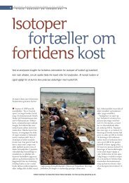
1 Paleoradiology: History and New Developments - Academia.dk
1 Paleoradiology: History and New Developments - Academia.dk
1 Paleoradiology: History and New Developments - Academia.dk

You also want an ePaper? Increase the reach of your titles
YUMPU automatically turns print PDFs into web optimized ePapers that Google loves.
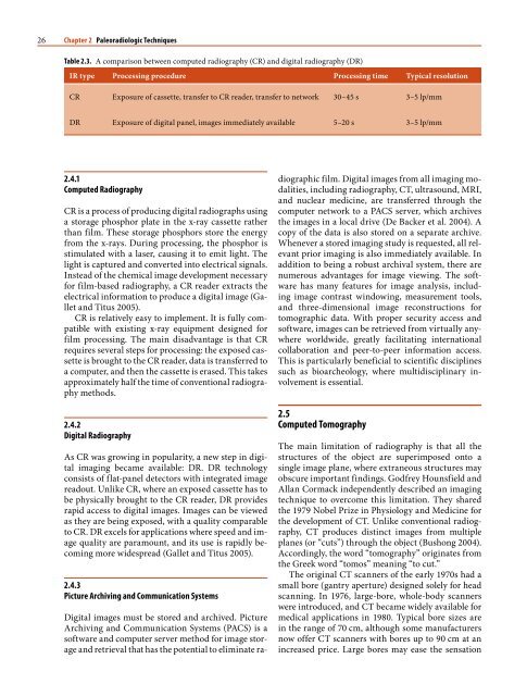
6 Chapter 1 <strong>Paleoradiology</strong>: <strong>History</strong> <strong>and</strong> <strong>New</strong> <strong>Developments</strong><br />
1.2.2<br />
1933 Amenophis I<br />
Amenophis I (also known as Amenhotep I) was the<br />
2nd Pharaoh of the 18th Dynasty, who is generally<br />
thought to have ruled for 20 years between 1526 <strong>and</strong><br />
1506 BC. His mummy was found by Victor Loret in<br />
1898 in the Deir el-Bahri cache in the mortuary temple<br />
of Queen Hatshepsut in the Valley of the Kings.<br />
An x-ray study was done at the Cairo Museum on Saturday<br />
January 30, Tuesday February 2nd, <strong>and</strong> Thursday<br />
February 4th, 1932, after removal of the mummy<br />
from the coffin <strong>and</strong> cartonnage. Dr. Douglas Derry<br />
used x-ray findings to assess the age of the mummy:<br />
“The body proved to be that of an adult man. It is not<br />
possible to assign an age, except to say that all epiphyses<br />
are completely united <strong>and</strong> he is therefore above<br />
25 years of age. So far as the teeth could be seen, they<br />
would were not unduly worn, nor are there any signs<br />
indicating advanced age such as loss of teeth or rarefaction<br />
of any of the bones, so that this king may have<br />
been about 40–50 years of age” (Derry 1933, p 47).<br />
In his further evaluation of the x-ray study, Derry<br />
reported the following findings. “The cranial cavity<br />
appears to contain a diffuse mass, but whether this<br />
is the remains of the brain <strong>and</strong> membranes or whether<br />
it represents linen packing introduced by way<br />
of the nose, cannot be definitely decided, as the photographs<br />
do not show the condition of the ethmoid”<br />
(Derry 1933, pp 46–47). “The body has suffered considerable<br />
damage probably at the h<strong>and</strong>s of the thieves.<br />
The right arm is bent at the elbow <strong>and</strong> the forearm is<br />
lying across the abdomen. There is a small amulet on<br />
the middle of the right arm, <strong>and</strong> towards the lower<br />
end of the arm there are two or three beads…”. “The<br />
body cavity both chest <strong>and</strong> abdomen probably contains<br />
linen package” (Derry 1933, p 48).<br />
1.2.3<br />
1965 Royal Mummy Collection<br />
In the spring of 1965, a team from the University of<br />
Michigan, in collaboration with Alex<strong>and</strong>ria University<br />
in Egypt, was invited to undertake a paleoradiological<br />
study of skulls of ancient Nubian populations<br />
who lived near the Nile River. The Michigan project<br />
focused mostly on craniofacial variation studies. The<br />
radiological equipment used included a portable xray<br />
cephalometer using ytterbium-169 isotope with a<br />
half-life of 32.5 days, which allowed the equipment to<br />
be totally independent of a power source.<br />
Following this first Nubian project, the Egyptian<br />
Department of Antiquities invited the same team, led<br />
by Dr. James E. Harris, Chairman of the Department<br />
of Orthodontics at the University of Michigan, to<br />
conduct an x-ray survey of the royal mummy collection<br />
of the Egyptian Museum. The project started in<br />
December 1967 with a radiographic study limited to<br />
the royal mummies’ skulls. At that time, x-ray images<br />
of mummies were taken while they were still lying<br />
within their glass cases, to prevent any possible damage.<br />
However, the glass was found to contain lead,<br />
which severely degraded the images. In 1968, permission<br />
was given to remove the glass cases so that the<br />
mummies could be x-rayed in their wooden coffins,<br />
which resulted in far fewer artifacts than those caused<br />
by the glass cases. At this time the ytterbium source<br />
was replaced by a conventional x-ray machine using<br />
90 kV, <strong>and</strong>, in addition to skull studies, a whole-body<br />
radiographic evaluation of the complete collection of<br />
royal mummies from the middle kingdom to the Roman<br />
period was performed.<br />
The st<strong>and</strong>ard x-ray protocol included lateral <strong>and</strong><br />
frontal views of the skull, the thorax, the pelvis, <strong>and</strong><br />
the lower limbs. The data obtained during those multiple<br />
expeditions to Egypt form the basis of the publication<br />
of the Atlas of Royal Mummies by Harris<br />
<strong>and</strong> Wente (1980). The Atlas focused primarily on<br />
craniofacial variations <strong>and</strong> dental malocclusion, underst<strong>and</strong>ably,<br />
as the analysis of the data was conducted<br />
by a team of academic dental surgeons. The main<br />
limitation of this study was the lack of a specific x-ray<br />
protocol designed to study specific skeletal regions,<br />
as whole-body radiography was obtained for the survey.<br />
In lieu of a thorough analysis of x-ray data, apart<br />
from the study of craniofacial variations, there was a<br />
limited radiological inventory made available to potential<br />
mummy scientists, which was described in the<br />
preface of the Atlas. The preface stated that the reader<br />
was provided with “copies of x-rays from which he<br />
may draw his own conclusions <strong>and</strong> interpretations”<br />
(Harris <strong>and</strong> Wente 1980). This approach, although<br />
laudable, did not offer appropriate x-ray data for a<br />
paleopathological study of any of the royal Egyptian<br />
mummies. In addition, these data were not validated<br />
in the peer-reviewed literature until 1988 when 12<br />
royal mummies were selected for paleopathological<br />
studies using x-rays as methods for disease detection<br />
(Braunstein et al. 1988). However, in 1973 the data<br />
were collated in a scholarly textbook, which became<br />
generally popular. Interestingly, this book entitled<br />
“X-raying the Pharaohs” (Harris <strong>and</strong> Weeks 1973)<br />
shed light on the context in which the radiological<br />
study of royal mummies was conducted, as revealed<br />
in the following quotes.<br />
“We arrived at the museum each morning at<br />
9 o’clock <strong>and</strong>, after signing the guard’s register, proceeded<br />
upstairs to Gallery 52 where the mummies<br />
were displayed. While some of the staff began the task<br />
of setting the x-ray unit on its tripod, adjusting the<br />
transformers, <strong>and</strong> loading the film cassettes, two of