1 Paleoradiology: History and New Developments - Academia.dk
1 Paleoradiology: History and New Developments - Academia.dk
1 Paleoradiology: History and New Developments - Academia.dk
Create successful ePaper yourself
Turn your PDF publications into a flip-book with our unique Google optimized e-Paper software.
were evaluated by CT in Toronto. The French team<br />
had missed a historical opportunity <strong>and</strong> the Toronto<br />
team became the first in the world to perform a CT<br />
scan of Egyptian mummies. The CT investigation included<br />
not only the study of a naturally mummified<br />
brain, but also whole-body imaging of the mummy.<br />
CT helps to assess not only the mummy’s anatomy<br />
without the need of unwrapping, but also in the detection<br />
of amulets or paleopathological lesions. As the<br />
Toronto team used a first-generation CT scanner, the<br />
image resolution was poor. The overall morphology of<br />
the cerebral hemispheres <strong>and</strong> the ventricular outline<br />
were identified, the demarcation between the white<br />
<strong>and</strong> grey matters was faint, <strong>and</strong> a few post-mortem<br />
lacunae were identified. The thickness of each slice<br />
was 12 mm (Harwood-Nash 1979).<br />
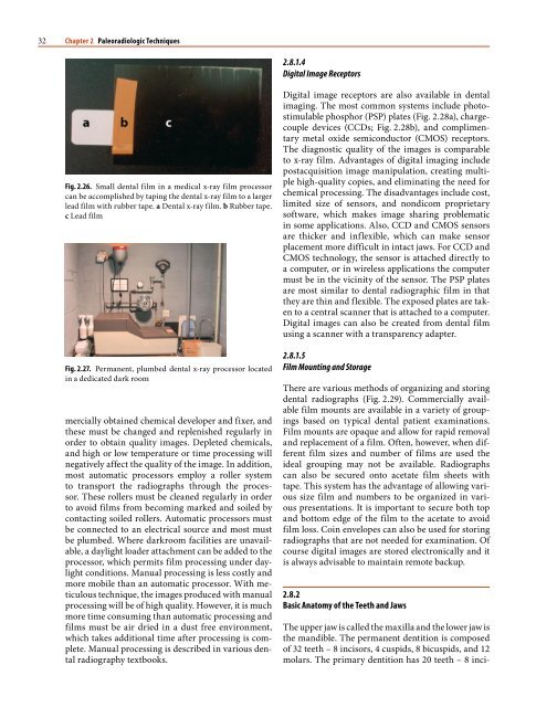
a<br />
a<br />
Fig. 1.9. a CT head. b CT abdomen. Both reprinted with permission<br />
from Lippincott, Williams, <strong>and</strong> Wilkins<br />
From this pioneering work to the current period,<br />
CT has been used to investigate many other mummies.<br />
Also, as CT technology has developed over<br />
time, the applications have exp<strong>and</strong>ed considerably<br />
(Table 1.3). More recently, micro-CT has been used to<br />
investigate mummy’s materials (Fig. 1.10) <strong>and</strong> fossils<br />
(Chhem 2006; McErlain et al. 2004). Note that CT<br />
<strong>and</strong> micro-CT have been used on many bioarcheological<br />
materials other than mummies <strong>and</strong> skeletal<br />
remains. Although this subject is beyond the scope<br />
of this chapter, there are other sources for this information<br />
(Hohenstein 2004; McErlaine et al. 2004; Van<br />
Kaik <strong>and</strong> Delorme 2005).<br />
1.4<br />
Paleo-MRI<br />
Magnetic resonance imaging (MRI) provides very<br />
detailed anatomical images of organs <strong>and</strong> tissues<br />
throughout the body without using x-rays, but MRI<br />
works by magnetizing the protons of the water mol-<br />
Table 1.3. A 30-year-history of advanced medical imaging of<br />
mummies: milestones<br />
Author Year Study subject<br />
Lewin <strong>and</strong><br />
Harwood-Nash<br />
1977 CT mummy’s brain<br />
(Nakht)-September 27,<br />
1976<br />
Harwood-Nash 1979 CT mummy’s brain <strong>and</strong><br />
whole-body<br />
Marx <strong>and</strong> D’Auria 1988 3D Skull/Face<br />
Magid et al. 1989 3D Entire skeleton<br />
Nedden et al. 1994 CT Guided Stereolithography<br />
Head<br />
Yardley <strong>and</strong> Rutka 1997 CT ENT (ear-nose)<br />
Melcher et al. 1997 CT dentition-3D<br />
Ciranni et al. 2002 CT skeleton/h<strong>and</strong>-<br />
tailored for arthritis<br />
Hoffman et al. 2002 3D/Virtual fly-through<br />
“endoscopy”<br />
Ruhli et al. 2002 CT-guided biopsy<br />
Cesareni et al. 2003 3D-Virtual removal<br />
of wrapping<br />
1.4 Paleo-MRI<br />
McErlain et al. 2007 Micro-CT of mummy’s<br />
brain (Nakht)<br />
Karlik et al. 2007 MR imaging <strong>and</strong> MR<br />
spectroscopy of<br />
mummy’s brain (Nakht)<br />
9


