1 Paleoradiology: History and New Developments - Academia.dk
1 Paleoradiology: History and New Developments - Academia.dk
1 Paleoradiology: History and New Developments - Academia.dk

You also want an ePaper? Increase the reach of your titles
YUMPU automatically turns print PDFs into web optimized ePapers that Google loves.
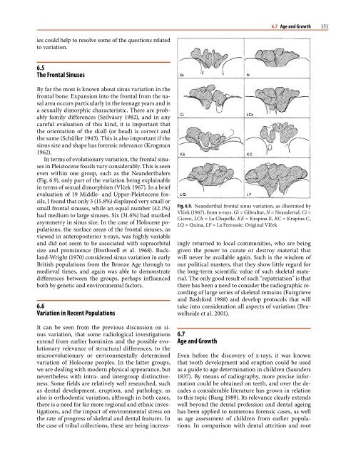
68 Chapter 3 The Taphonomic Process, Human Variation, <strong>and</strong> X-ray Studies<br />
stakes pressed into the Danish Huldremose bog body<br />
caused severe deformity of a forearm (Fig. 3.29) <strong>and</strong> a<br />
femur (Brothwell et al. 1990). Skulls can deform like<br />
deflated footballs.<br />
Fig. 3.28. Partly decalcified foot bones from the Huldremose<br />
bog body, showing poor bone density<br />
Fig. 3.29. X-ray of the thorax area of the Danish Huldremose<br />
bog body, displaying postmortem changes of the forearm<br />
3.10.5<br />
Abnormal Cavities in Bone<br />
Abnormal cavities within bone can develop for<br />
various reasons, <strong>and</strong> their interpretation will be influenced<br />
by their position in relation to the area of<br />
skeleton or dentition. They may not always be obvious<br />
on archeological bone since postmortem erosion<br />
can mask an osteolytic lesion. Radiographs can show<br />
whether or not a sclerotic lining exists, thus indicating<br />
a pathological condition that would otherwise<br />
have been missed. If there is the possibility that there<br />
is apical infection (an abscess) at one or more tooth<br />
positions – even without clear external evidence – it<br />
is advisable to radiograph the jaw. Cysts, neoplastic<br />
processes <strong>and</strong> trauma with infection may all lead to<br />
considerable bone destruction <strong>and</strong> remodeling. In an<br />
Iron Age pig m<strong>and</strong>ible from Danebury (Fig. 3.30),<br />
there is considerable bone destruction posterior to the<br />
canine, which in lateral view can be seen to extend<br />
under part of the posterior dentition. In the case of a<br />
Saxon phalanx from Southampton, an infection was<br />
obvious at the proximal joint, but only in x-ray can<br />
Fig. 3.30. Radiographic detail of a Danebury Iron Age pig,<br />
showing severe inflammatory changes within the jaw<br />
Fig. 3.31. Photocopy of a radiograph of a Saxon phalanx with a<br />
well-defined internal abscess