1 Paleoradiology: History and New Developments - Academia.dk
1 Paleoradiology: History and New Developments - Academia.dk
1 Paleoradiology: History and New Developments - Academia.dk

Create successful ePaper yourself
Turn your PDF publications into a flip-book with our unique Google optimized e-Paper software.
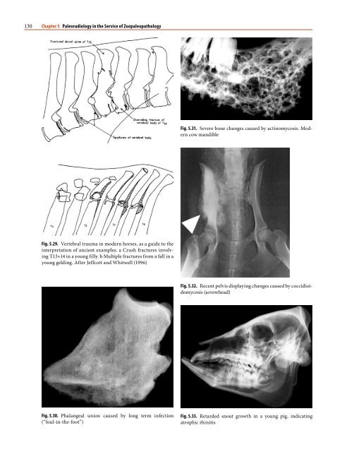
58 Chapter 3 The Taphonomic Process, Human Variation, <strong>and</strong> X-ray Studies<br />
Fig. 3.6. Xeroradiographic image of ancient Chinese lacquerware,<br />
with internal detail clearly visible. Courtesy of Jacqui<br />
Watson, English Heritage<br />
Fig. 3.7. Iron Age bread roll (burnt), x-rayed to show the inner<br />
granular detail<br />
ted plant debris, <strong>and</strong> x-rays provide an ideal method<br />
of scanning such material before any specimens are<br />
selected for further laboratory analysis.<br />
3.3<br />
Radiological Aspects of Zooarcheology<br />
Paleontologists have made use of radiographic techniques<br />
more enthusiastically than zooarcheologists<br />
in the past. Indeed, in some instances, it has been a<br />
highly successful tool in revealing the less commonly<br />
seen finer anatomy of organisms. For instance, from<br />
fine shale of the Ordovician <strong>and</strong> Devonian periods,<br />
the pyrite-preserved soft parts of trilobites within the<br />
shale were revealed by x-ray (Robison <strong>and</strong> Kaesler<br />
1987). An ideal x-ray of a fossil dem<strong>and</strong>s a balance<br />
Fig. 3.8. Ancient coprolites, containing some contrasting<br />
structures<br />
between sufficient penetration through the fossil <strong>and</strong><br />
matrix (needing high voltage) <strong>and</strong> sufficient light/<br />
dark contrasts (needing a low kV <strong>and</strong> a long exposure<br />
time). Depending on the lithology, fossil hydroids,<br />
graptolites, fish, <strong>and</strong> other vertebrate species have<br />
been revealed successfully by this technique (Harbersetzer<br />
1994; Longbottom 2005). Heavily fossilized<br />
skeletal material can present special problems with<br />
regard to obtaining sufficient detail of the internal<br />
structures, especially if the interior of the bone is invaded<br />
by siliceous deposits (Fig. 3.9).<br />
Although it is mainly vertebrate remains that have<br />
received radiographic study in archeology, it should<br />
be mentioned that there is clearly potential when considering<br />
invertebrate remains, for the extraction of<br />
information by means of x-rays. There is certainly a<br />
need for colleagues specifically working with invertebrates<br />
to consider the potential value of radiographic<br />
techniques, <strong>and</strong> perhaps undertake experimental<br />
investigations in order to evaluate what might be<br />
achieved with some kinds of archeological material.<br />
For instance, would it be possible, by means of digital<br />
x-rays, to detect not only insect damage, but also the<br />
actual animals buried within other organic remains?<br />
For instance, insect damage to bone might be confirmed<br />
radiographically by detecting the insect remains<br />
deep within the calcified tissue. However, experiments<br />
would be needed to establish that insect structures<br />
could be identified within bone or other tissue.<br />
Similarly, insect damage to mummified remains is