British Breeder Magazine May 2021

Create successful ePaper yourself
Turn your PDF publications into a flip-book with our unique Google optimized e-Paper software.
Corpus Luteum<br />
Following collapse of the dominant<br />
follicle and release of the oocyte/egg, a<br />
corpus luteum (CL) forms. The CL secretes<br />
progesterone, the dominant hormone of<br />
dioestrus (14 day period when the mare<br />
is not in season). If the mare becomes<br />
pregnant, progesterone secretion from<br />
the primary and secondary CLs will<br />
prevent the mare coming back into<br />
season and maintain the pregnancy until<br />
placental takeover at around 100-120<br />
days of gestation. Ultrasound<br />
examination provides visual and<br />
objective evaluation of both the structural<br />
and functional aspects of development<br />
of the CL from maturation to regression.<br />
Evaluating and ageing the CL gives us<br />
essential information regarding the stage<br />
of the mare’s cycle, and more recently<br />
has been used for selection of recipient<br />
mares for embryo transfer both of fresh<br />
and ICSI produced embryos, when<br />
knowing the exact age of the CL/<br />
number of days post ovulation is<br />
essential for a successful transfer.<br />
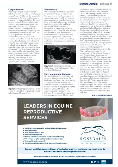
Figure 6: Ultrasonographic image of an<br />
ovary depicting two corpus luteum (CLs)<br />
following double ovulation.<br />
Uterine cysts<br />
Whilst the majority of uterine cysts are<br />
insignificant findings in older mares,<br />
their presence in the uterus may signify<br />
underlying issues. In addition, large or<br />
numerous cysts or those located at the<br />
base of the uterine horns may physically<br />
inhibit embryo mobility and fixation,<br />
resulting in failure of maternal<br />
recognition of pregnancy and early<br />
embryonic loss. Noting the size, number<br />
and location of cysts on ultrasound<br />
examination will help determine their<br />
potential significance; furthermore,<br />
mapping of cysts will also prevent any<br />
confusion over subsequent pregnancy<br />
diagnosis.<br />
Figure 7: Ultrasonographic image of a<br />
uterine cyst - note the irregular outline<br />
and fluid-filled centre.<br />
Early pregnancy diagnosis<br />
Ultrasound examination is pivotal for<br />
pregnancy diagnosis in mares. The<br />
equine embryo enters the uterus on day<br />
5.5-6 post ovulation and can be<br />
detected via ultrasound examination<br />
from as early as 10 days post ovulation.<br />
Since, however, at this stage the<br />
embryonic vesicle measures only<br />
between 3-5mm, most first pregnancy<br />
scans are performed at 14 days post<br />
Feature Article - Rossdales<br />
ovulation, at which stage the embryonic<br />
vesicle typically measures between<br />
16-20 mm, making it much easier to<br />
identify. Due to the unique equine<br />
embryonic capsule, the pregnancy will<br />
be perfectly spherical at this stage and<br />
ultrasonically generated specular<br />
reflections (bright white spots on the<br />
upper and lower surface of the embryo;<br />
see image) help differentiate embryos<br />
from uterine cysts or fluid, which are<br />
rarely perfectly spherical. Since mares<br />
are not able to carry twin pregnancies<br />
successfully to term, the diagnosis of twin<br />
pregnancies and subsequent reduction<br />
to a singleton pregnancy is performed<br />
most successfully at this early pregnancy<br />
scan, between 14-16 days post<br />
ovulation. Further pregnancy<br />
examinations are performed at regular<br />
intervals to monitor growth and<br />
development of the embryo/foetus. By<br />
day 21 the embryo proper can be<br />
visualised and at 28 days an embryonic<br />
heartbeat will be detectable. From 40<br />
days onwards, the pregnancy enters the<br />
foetal stage of development (the term<br />
embryo is used until day 39), at a 42<br />
day scan the start of the umbilical cord<br />
formation and the beginning of foetal<br />
activity can be detected. For those of<br />
you wanting to determine the gender of<br />
your foal, this can be reliably performed<br />
by transrectal ultrasound examination<br />
between 58-70 days post ovulation.<br />
Ultrasound detection of the genital<br />
tubercle, the precursor to the penis in the<br />
male and clitoris in the female is either<br />
detected under the tail in the case of a<br />
filly foal or behind the umbilicus with a<br />
colt foal.<br />
www.rossdales.com<br />
LEADERS IN EQUINE<br />
REPRODUCTIVE<br />
SERVICES<br />
✔ Artificial insemination with fresh, chilled and frozen semen<br />
✔ Embryo transfer<br />
✔ OPU and shipping for ICSI<br />
✔ Infertility Investigations<br />
✔ Semen collection, evaluation, distribution and freezing<br />
✔ World class hospital facilities and specialist expertise<br />
✔ Neonatal intensive care unit<br />
✔ State of the art laboratory, HBLB approved for CEM testing<br />
Contact our BEVA approved team of dedicated stud vets to discuss your requirements<br />
on 01638 663150, or practice@rossdales.com<br />
ROSSDALES VETERINARY SURGEONS, Beaufort Cottage Stables, High Street, Newmarket, CB8 8JS<br />
www.rossdales.com<br />
BRITISH BREEDER| 57