Entomology 311 Lab Manual - 1st Edition, 2019
Entomology 311 Lab Manual - 1st Edition, 2019
Entomology 311 Lab Manual - 1st Edition, 2019

You also want an ePaper? Increase the reach of your titles
YUMPU automatically turns print PDFs into web optimized ePapers that Google loves.
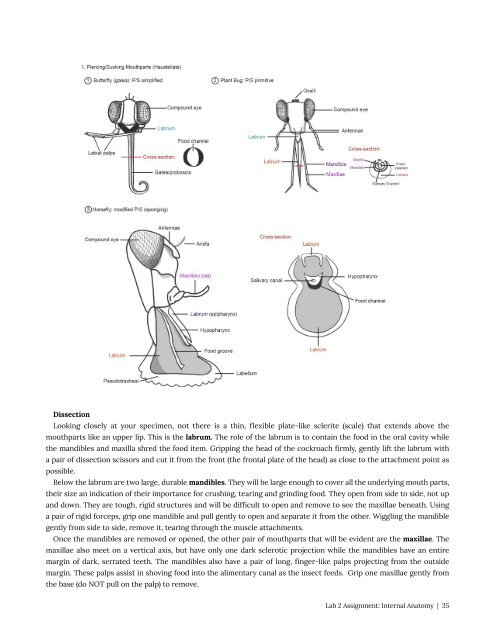
Dissection<br />
Looking closely at your specimen, not there is a thin, flexible plate-like sclerite (scale) that extends above the<br />
mouthparts like an upper lip. This is the labrum. The role of the labrum is to contain the food in the oral cavity while<br />
the mandibles and maxilla shred the food item. Gripping the head of the cockroach firmly, gently lift the labrum with<br />
a pair of dissection scissors and cut it from the front (the frontal plate of the head) as close to the attachment point as<br />
possible.<br />
Below the labrum are two large, durable mandibles. They will be large enough to cover all the underlying mouth parts,<br />
their size an indication of their importance for crushing, tearing and grinding food. They open from side to side, not up<br />
and down. They are tough, rigid structures and will be difficult to open and remove to see the maxillae beneath. Using<br />
a pair of rigid forceps, grip one mandible and pull gently to open and separate it from the other. Wiggling the mandible<br />
gently from side to side, remove it, tearing through the muscle attachments.<br />
Once the mandibles are removed or opened, the other pair of mouthparts that will be evident are the maxillae. The<br />
maxillae also meet on a vertical axis, but have only one dark sclerotic projection while the mandibles have an entire<br />
margin of dark, serrated teeth. The mandibles also have a pair of long, finger-like palps projecting from the outside<br />
margin. These palps assist in shoving food into the alimentary canal as the insect feeds. Grip one maxillae gently from<br />
the base (do NOT pull on the palp) to remove.<br />
<strong>Lab</strong> 2 Assignment: Internal Anatomy | 35