Dental Asia March/April 2023
For more than two decades, Dental Asia is the premium journal in linking dental innovators and manufacturers to its rightful audience. We devote ourselves in showcasing the latest dental technology and share evidence-based clinical philosophies to serve as an educational platform to dental professionals. Our combined portfolio of print and digital media also allows us to reach a wider market and secure our position as the leading dental media in the Asia Pacific region while facilitating global interactions among our readers.
For more than two decades, Dental Asia is the premium journal in linking dental innovators
and manufacturers to its rightful audience. We devote ourselves in showcasing the latest dental technology and share evidence-based clinical philosophies to serve as an educational platform to dental professionals. Our combined portfolio of print and digital media also allows us to reach a wider market and secure our position as the leading dental media in the Asia Pacific region while facilitating global interactions among our readers.
Create successful ePaper yourself
Turn your PDF publications into a flip-book with our unique Google optimized e-Paper software.
www.dentalasia.net<br />
MARCH / APRIL <strong>2023</strong><br />
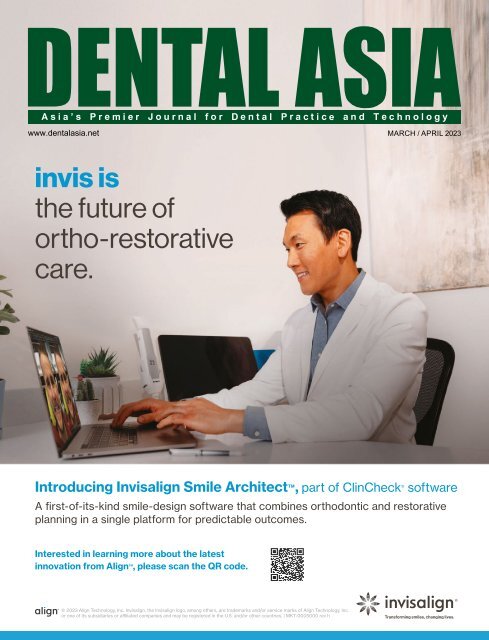
invis is<br />
the future of<br />
ortho-restorative<br />
care.<br />
Introducing Invisalign Smile Architect TM , part of ClinCheck ® software<br />
A first-of-its-kind smile-design software that combines orthodontic and restorative<br />
planning in a single platform for predictable outcomes.<br />
Interested in learning more about the latest<br />
innovation from Align TM , please scan the QR code.<br />
© <strong>2023</strong> Align Technology, Inc. Invisalign, the Invisalign logo, among others, are trademarks and/or service marks of Align Technology, Inc.<br />
or one of its subsidiaries or affiliated companies and may be registered in the U.S. and/or other countries. | MKT-0005000 rev h
TRIOS Share<br />
Digitize your entire clinic with just one<br />
TRIOS wireless scanner<br />
Scan every patient<br />
in any room<br />
Treatment plan<br />
on all PCs<br />
Maximize your<br />
investment<br />
Pass the scanner, share<br />
the power<br />
The first technology to enable you<br />
to use wireless TRIOS scanners on any<br />
PC in your practice via Wi-Fi.<br />
Discover real wireless freedom to scan<br />
and plan anywhere with award-winning<br />
3Shape TRIOS wireless intraoral scanners.<br />
Enable the TRIOS Share solution for<br />
free with every TRIOS wireless model!<br />
Learn more at: 3shape.com/share<br />
Scan to learn more<br />
about TRIOS Share
CONTENTS<br />
18<br />
22<br />
30<br />
TRENDS<br />
14 HKU Dentistry new jaw surgery concept<br />
is effective in treating moderate-tosevere<br />
obstructive sleep apnoea<br />
16 Artificial intelligence in dental practice<br />
UNDER THE SPOTLIGHT<br />
18 Under one roof: A multi-disciplinary,<br />
patient-centric model<br />
DENTAL PROFILE<br />
21 Optimising dental scanning process<br />
with Scanningspray<br />
22 Same-day dentistry made a reality by<br />
Ackuretta<br />
CLINICAL FEATURE<br />
26 Next generation synthetic graft<br />
materials: Clinical and scientific review<br />
30 Using occlusion technology to<br />
simplify and improve functional risk<br />
assessment<br />
34 Clinical management of root resorption<br />
and separated instruments<br />
48<br />
USER REPORT<br />
38 Mini Implants: Simplified protocol for<br />
stabilising removable dentures<br />
42 Innovative leap in glass ceramics<br />
BEHIND THE SCENES<br />
46 Heralding the era of digital dental<br />
technicians<br />
48 Inside story of press technology<br />
IN DEPTH WITH<br />
50 Choosing the best loupes<br />
52 Severe dental problems in children on the<br />
rise: Finnish innovation keeps tooth decay<br />
at bay<br />
EXCLUSIVE FEATURE<br />
58 100 Years of IDS: Shaping the dental future<br />
SHOW PREVIEW<br />
61 Advertorial: Seven reasons to attend the<br />
International Osteology Symposium<br />
62 Edentulism <strong>2023</strong>: Glimpse into the<br />
speakers’ presentations<br />
63 Implant Solutions World Summit:<br />
The latest innovations and science<br />
transforming implant dentistry<br />
64 Sino-<strong>Dental</strong> <strong>2023</strong>: All-in-one platform<br />
SHOW REVIEW<br />
66 AOSC celebrates successful first in-person<br />
edition back out of the pandemic<br />
REGULARS<br />
4 Editor’s note<br />
6 <strong>Dental</strong> Updates<br />
54 Product Highlights<br />
67 Events Calendar<br />
68 Advertisers’ Index<br />
46<br />
50<br />
62
HyFlex EDM<br />
STAYS ON TRACK<br />
Æ Safer use<br />
Æ Preparation following the anatomy<br />
Æ Regeneration for reuse<br />
006650 03.21<br />
www.coltene.com<br />
facebook.com/COLTENE.<strong>Asia</strong>Pacific/
EDITOR’S NOTE<br />
Era of dental<br />
revolution: Where<br />
past and future<br />
collides<br />
PABLO SINGAPORE<br />
Publisher<br />
Assistant Editor<br />
Graphic Designer<br />
William Pang<br />
williampang@pabloasia.com<br />
Czarmaine Masigla<br />
czarmaine@pabloasia.com<br />
Agatha Wong<br />
agatha@pabloasia.com<br />
Amira Yunos<br />
amira@pabloasia.com<br />
Cayla Ong<br />
cayla@pabloasia.com<br />
The dental industry is in a very exciting era —<br />
the once considered futuristic idea is now the<br />
reality. From 3D printing, software integrations<br />
and artificial intelligence (AI), the advancement<br />
in technology has enabled dental practices<br />
and laboratories to scale up their practices in<br />
multiple ways.<br />
For instance, the plug-and-play solutions of<br />
Ackuretta have made it possible for practitioners<br />
to adapt same-day dentistry. Thomas Wu,<br />
CTO of Ackuretta, shares how crucial it is to<br />
incorporate customers’ feedback in designing<br />
and building a product to make it more userfriendly.<br />
By tapping into AI technology, they<br />
aim to minimise the learning curve in using 3D<br />
printing technology, so more practitioners can<br />
use and benefit from it (p.22).<br />
As the dental sector continues to be more<br />
digitalised with integrated workflows, Prof Dr<br />
Sebastian Gell, co-founder of Scanningspray,<br />
said that scanning and 3D printing will play an<br />
important role for dental laboratories in the<br />
coming years. With their self-cleaning scanning<br />
spray, they aim to optimise the scanning<br />
process and improve the scan quality so dental<br />
laboratories can generate reproducible results<br />
in the 3D scan (p.21).<br />
Technological advancements have no doubt<br />
further enhanced treatment outcomes<br />
as exampled by how Dr Lee Ann Brady<br />
utilises dental occlusion technology to<br />
acquire a more complete picture of a<br />
patient’s temporomandibular joint and<br />
better understand how it contributes to<br />
temporomandibular disorders (p.30).<br />
In the past decade, we have seen a dramatic<br />
transformation in the workflow of dental<br />
clinics and laboratories. And with the<br />
return of in-person events and trade fairs —<br />
starting with the 40th edition of IDS — the<br />
digital revolution in dentistry is set to run at<br />
full throttle (p.58).<br />
Czarmaine Masigla<br />
Assistant Editor<br />
Scan for digital copy<br />
of <strong>Dental</strong> <strong>Asia</strong><br />
Circulation Manager<br />
Media Representative<br />
PABLO BEIJING<br />
General Manager<br />
PABLO SHANGHAI<br />
Senior Editor<br />
Shu Ai Ling<br />
circulation@pabloasia.com<br />
Jamie Tan<br />
jamietan@pabloasia.com<br />
Ellen Gao<br />
pablobeijing@163.com<br />
Daisy Wang<br />
pabloshanghai@163.net<br />
HEAD OFFICE<br />
PABLO PUBLISHING &<br />
EXHIBITION PTE LTD<br />
3 Ang Mo Kio Street 62 #01-23<br />
Link@AMK, Singapore 569139<br />
Tel: (65) 62665512<br />
Email: info@pabloasia.com<br />
Website: www.dentalasia.net<br />
Company Registration No.: 200001473N<br />
Singapore MICA (P) No. 079/12/2022<br />
Malaysia KDN: PPS1528/07/2013 (022978)<br />
REGIONAL OFFICES<br />
PABLO BEIJING<br />
Tel: +86-10-6509-7728<br />
Email: pablobeijing@163.com<br />
PABLO SHANGHAI<br />
Tel: +86-21-52389737<br />
Email: pabloshanghai@163.net<br />
ADVISORY BOARD<br />
Dr William Cheung<br />
Dr Choo Teck Chuan<br />
Dr Chung Kong Mun<br />
Dr George Freedman<br />
Dr Fay Goldstep<br />
Dr Clarence Tam<br />
Prof Nigel M. King<br />
Dr Anand Narvekar<br />
Dr Kevin Ng<br />
Dr William O’Reilly<br />
Dr Wong Li Beng<br />
Dr Adrian U J Yap<br />
Dr Christopher Ho<br />
Dr How Kim Chuan<br />
Dr Derek Mahony<br />
Prof Alex Mersel
#whdentalwerk<br />
wh.com<br />
Modularity for<br />
optimal flexibility<br />
Electric motor<br />
Particularly small and<br />
surprisingly light. It enables<br />
you to work with ease.<br />
Osstell Beacon<br />
Bluetooth connection<br />
to the Osstell Beacon<br />
for easy measurement<br />
of implant stability.<br />
Straight & Contra-angle<br />
Handpieces<br />
Provide optimal illumination<br />
of the treatment site and<br />
facilitate perfect, accurate<br />
treatment results.<br />
PDF<br />
XLS<br />
Documentation of<br />
osseointegration<br />
The documentation<br />
function conveniently<br />
saves all values of the<br />
implant insertion on a<br />
USB stick (.pdf and .xls).<br />
Piezomed Plus<br />
and Classic module<br />
Combine piezo surgery and<br />
implantology in one device.<br />
Wireless or wired foot control<br />
Control the Implantmed Plus<br />
and Piezomed module with<br />
just one foot control.
DENTAL UPDATES<br />
Ackuretta launches ALPHA AI<br />
Ackuretta, a global manufacturer of 3D printing<br />
solutions for dental professionals, has launched<br />
the new AI-assisted 3D slicing software —<br />
ALPHA AI. The new 3D printing slicing software<br />
is an evolution of Ackuretta’s ALPHA 3D<br />
and includes several improvements from its<br />
predecessor.<br />
Extensive research has driven Ackuretta<br />
towards artificial intelligence as the next<br />
logical step in their journey to make 3D dental<br />
printing accessible to everyone. Moreover, such<br />
technology provides predictable results every<br />
time — building upon Ackuretta’s signature<br />
reliability.<br />
ALPHA AI offers a streamlined process to<br />
guarantee success and eliminate human<br />
error: Import an STL file on the software, click<br />
the icon for the desired application and start<br />
printing in seconds.<br />
Ayush Bagla, CEO of Ackuretta, said: “Up<br />
to 75% of 3D printed applications result in<br />
failure because of poor orientation. Some<br />
clinicians and dental technicians have to<br />
go through a learning curve — which leads<br />
them to experience failed prints due to a few<br />
reasons, such as choosing the wrong printing<br />
parameters, not using the correct resin, lack of<br />
training, and dealing with complex dental 3D<br />
printing software.<br />
“ALPHA AI comes to solve that exact problem.<br />
It uses machine learning to analyse data from<br />
thousands of real designs and comes up with<br />
the optimal orientation for a given application,<br />
including adding the necessary supports. Users<br />
can achieve the best result with just one click.”<br />
Unlike most 3D printing slicing software in the<br />
market, ALPHA AI allows its users to orient<br />
an application, add the necessary supports,<br />
and save it not only as i3dp and ibf (Ackureta<br />
Solution proprietary file format) but also as an<br />
STL file.<br />
Ackuretta aims to empower its customers by<br />
offering them a truly open system — everything<br />
from 3D printing solutions to digital dentistry<br />
software. Users could configure the print they<br />
want on ALPHA AI (such as placing it with<br />
the right orientation and adding supports)<br />
and export the STL file to use on any other 3D<br />
printing CAM software specific to non-Ackuretta<br />
devices for printing.<br />
Ackuretta’s ALPHA AI is capable of orienting<br />
and creating supports for splints, crowns,<br />
bridges, and surgical guides. Moreover, the<br />
company releases constant updates every three<br />
months to add new features and functions to<br />
the dental 3D printing software. DA<br />
KaVo and MELAG agree on sales partnership<br />
With their cooperation, KaVo and MELAG are<br />
forming a new team that will additionally drive<br />
the future in instrument reprocessing with<br />
coordinated product and service offerings<br />
in order to continuously make daily work in<br />
dental practices safer, more reliable and more<br />
effective.<br />
The focus of the closer sales cooperation<br />
is, among other things, joint training for<br />
specialised trade partners as well as<br />
coordinated marketing and sales campaigns.<br />
Joint events for end customers are also<br />
planned worldwide. Stronger mutual support<br />
is also planned for the future in the area of<br />
research and development.<br />
Under the slogan “<strong>Dental</strong> Excellence”, KaVo<br />
<strong>Dental</strong> has been offering dental expertise and<br />
an almost complete premium product portfolio<br />
for dental practices and dental laboratories<br />
for over 100 years. KaVo has been a guarantor<br />
of outstanding premium products for over<br />
100 years and a leading supplier in the dental<br />
instrument segment with a large number of<br />
patents and utility models.<br />
As an owner-managed family business, MELAG<br />
has established itself as a market leader in dental<br />
hygiene with quality and innovative strength.<br />
Since the company was founded over 70 years<br />
ago, MELAG has been developing and producing<br />
innovative and technologically robust solutions<br />
for instrument reprocessing exclusively in Berlin,<br />
Germany. With its outstanding system solutions,<br />
including autoclaves, washer disinfectors<br />
and sealing devices, MELAG has been able to<br />
continuously develop and significantly shape<br />
instrument reprocessing.<br />
It is therefore not surprising that KaVo explicitly<br />
recommends products from MELAG for the<br />
reprocessing of its own premium instruments.<br />
“The high standards of our own products and<br />
the values of an innovative, traditional company<br />
make MELAG the perfect partner for KaVo in<br />
dental instrument reprocessing. As a reliable<br />
four-in-one solution, the Careclave from MELAG<br />
harmonises perfectly with our high-quality KaVo<br />
instruments and is therefore also our clear<br />
recommendation for reprocessing our KaVo<br />
instruments,” said Armin Imhof, CTO at KaVo.<br />
Dr Niklas Gebauer, managing partner of<br />
MELAG, added: “KaVo straight and contra-angle<br />
handpieces have always been the benchmark<br />
for us, not only in terms of quality and aboveaverage<br />
durability. Therefore, we have also<br />
tested intensively with KaVo instruments during<br />
the development of the Careclave.”<br />
At the International <strong>Dental</strong> Show in Cologne,<br />
which opens its doors on 14 Mar, the<br />
cooperation will also be visible to the general<br />
public: the MELAG Careclave will also be on<br />
the KaVo stand and on the MELAG stand<br />
the Careclaves will be equipped with KaVo<br />
instruments. DA<br />
6 DENTAL ASIA MARCH / APRIL <strong>2023</strong>
DENTAL UPDATES<br />
Carestream <strong>Dental</strong> introduces IO<br />
Scanner Link<br />
PERFECTION IN<br />
BONE SURGERY<br />
Carestream <strong>Dental</strong> is now making a seamlessly connected digital<br />
practice a reality with IO Scanner Link, its latest innovation that allows<br />
CS Imaging version 8 software to directly connect to third-party intraoral<br />
scanners’ acquisition software not developed by Carestream <strong>Dental</strong>.<br />
IO Scanner Link is more than a manual file import/export option.<br />
It leverages CS Imaging 8 — the imaging hub that centralises and<br />
displays all of a user’s Carestream <strong>Dental</strong> imaging data — as the core<br />
practice platform to drive efficiency and connectivity. By aggregating,<br />
storing and managing all images through a single software and patient<br />
database, a practitioner can optimise and simplify the workflow in<br />
their digital dental practice.<br />
→ YOUR SURGICAL<br />
APPROACH WILL CHANGE -<br />
THE PIEZOSURGERY® touch<br />
→ best cutting efficiency<br />
→ optimal intraoperative control<br />
→ perfect ergonomics<br />
→ made in Italy<br />
“In today’s digitally connected practice, it’s all about choice,” said<br />
Philippe Maillet, general manager, imaging equipment, Carestream<br />
<strong>Dental</strong>. “The choice to connect your existing technology with a<br />
multitude of partners or devices to create the workflow that’s right for<br />
your practice.”<br />
Within CS Imaging 8, an integrated button allows intraoral scans from<br />
supported third-party scanners, along with all the necessary patient<br />
data, to be launched with just one click. When the scan is complete, the<br />
intraoral scan is automatically imported back into CS Imaging 8 to be<br />
used for implant planning through Carestream <strong>Dental</strong>’s Prosthetic-driven<br />
Implant Planning (PDIP) module; surgical guide design with Smop<br />
software; sharing with labs; or exporting to other third-party software.<br />
“Making it easy for practitioners to use the intraoral scanner of their<br />
choice and giving them unrestricted access to their preferred clinical<br />
solutions aids them in optimising their daily treatments through an<br />
efficient workflow,” said Maillet.<br />
Medit is the latest intraoral scanner manufacturer to integrate with CS<br />
Imaging 8 through IO Scanner Link. This will allow users of Carestream<br />
<strong>Dental</strong> imaging equipment and Medit intraoral scanners to integrate<br />
their favourite devices with a single click of a button. Medit scanning<br />
solutions support both Windows and Mac computers.<br />
CS Imaging 8 also connects with Dexis intraoral scanners. In the<br />
coming months, Carestream <strong>Dental</strong> intends to expand the number of<br />
integrations to give practitioners even more choices. CS Imaging 8 is<br />
one of the most widely used dental imaging software on the market,<br />
thanks to its extremely user-friendly, modern interface. DA<br />
→ www.mectron.com<br />
DENTAL ASIA MARCH / APRIL <strong>2023</strong> 7<br />
ad_PStouch_dental_asia_95x250_en_211214.indd 1 14.12.21 15:38
DENTAL UPDATES<br />
Dürr <strong>Dental</strong> partners with 3Shape<br />
As a supplier of system solutions for the dental<br />
market, Dürr <strong>Dental</strong> relies on partners who also<br />
pursue the goal of supporting practices in their<br />
future-oriented development. In addition to<br />
manufacturing innovative 2D and 3D large x-ray<br />
units and imaging plate scanners, Dürr <strong>Dental</strong><br />
has also developed the VistaSoft imaging<br />
software.<br />
“Practices that use a TRIOS intraoral scanner<br />
from 3Shape can now work with 3Shape Unite<br />
from VistaSoft,” explained Frank Kiesele, head<br />
of Product Management Diagnostic Systems<br />
at Dürr <strong>Dental</strong>. “Users switch to 3Shape Unite<br />
with just one click.”<br />
The integration between Dürr <strong>Dental</strong> and<br />
3Shape Unite adds an intuitive workflow for<br />
patient management.<br />
The Danish 3Shape drive progress in digital<br />
dentistry with industry-recognised scanners<br />
for clinics and labs, and connect dentists and<br />
technicians with software workflows. 3Shape<br />
advances and coaches professionals on every<br />
step of their digital journey — helping to elevate<br />
the customer experience.<br />
For both companies, user-friendly software<br />
offerings are inseparably linked to the added<br />
value of their products for dental practices<br />
and clinics. That is why Dürr <strong>Dental</strong>’s VistaSoft<br />
software now has an interface to 3Shape Unite<br />
from version VistaSoft 3.0.20.<br />
“With the integration between VistaSoft and<br />
3Shape Unite, we are initiating a seamless<br />
digital workflow in the clinic. Together we<br />
are committed to deliver a more streamlined<br />
workflow, allowing the dentist to focus only on<br />
the patient,” said Rune Fisker, SVP of Product<br />
Strategy, 3Shape. DA<br />
Ivoclar celebrates centennial anniversary<br />
The year <strong>2023</strong> marks Ivoclar’s 100-year<br />
anniversary, which will be celebrated under<br />
the theme “A Century of Innovation”. Founded<br />
in Zurich, Switzerland, in 1923, the company<br />
relocated to the principality of Liechtenstein<br />
in 1933, where it established its present-day<br />
headquarters. Over the years, the former tooth<br />
factory evolved into a leading manufacturer<br />
of integrated solutions for high-quality dental<br />
applications. The family-owned business<br />
advanced to become a global market leader<br />
in various product segments, for example,<br />
ceramic furnaces (Programat series), by<br />
supplying a comprehensive portfolio of<br />
products and systems specially designed<br />
for dental practices and laboratories. The<br />
introduction of one of the most popular<br />
products of Ivoclar’s recent history — the highly<br />
aesthetic IPS e.max lithium disilicate glassceramic<br />
— ushered in the aesthetic revolution<br />
in 2005. The product went on to become a<br />
global market leader in its segment. Ivoclar<br />
also made a name for itself in the manufacture<br />
of highly aesthetic tooth replacements, which<br />
was the main business when the company was<br />
founded, and therefore links the company’s<br />
origins to its <strong>2023</strong> centenary.<br />
Diego Gabathuler, CEO of the Ivoclar Group,<br />
said: “We build our success on customerrelevant<br />
innovations paired with integrated<br />
system solutions, efficient applications,<br />
excellent quality and constant reliability as well<br />
as trust and respectful cooperation.<br />
“We focus on giving people a healthy and<br />
beautiful smile, resulting in a better quality of<br />
life. For this purpose, we supply our products to<br />
around 130 countries. We employ roughly 3500<br />
people worldwide, who continuously strive to<br />
optimise the company’s offerings of integrated<br />
solutions that feature intelligent systems,<br />
practice-oriented training and education as<br />
well as comprehensive after-sales support and<br />
customer-centric service.”<br />
In 2021, the group, which has more than 47<br />
subsidiaries and branch offices throughout<br />
the world, including production facilities in<br />
Liechtenstein, Germany, Austria, Italy, Sweden,<br />
the US and the Philippines, achieved a record<br />
increase in turnover of CHF 842 million in all of<br />
its markets. An additional growth in turnover<br />
has been achieved for the 2022 business year.<br />
“The 100th birthday of the Ivoclar Group takes<br />
place in dynamic times. Nevertheless, I am<br />
certain that the company will continue to be<br />
successful in the future,” said Helmut Schuster,<br />
chair of the Supervisory Board of the Ivoclar<br />
Group. “Why am I so confident? Because the<br />
corporate values of respect, smile, focus, act<br />
and grow are deeply rooted within the company,<br />
and they are upheld by the employees and<br />
managers of the company and above all by<br />
Christina and Christoph Zeller and the next<br />
generation of the entrepreneurial family.”<br />
The dental company maintains longstanding<br />
and trusting business partnerships<br />
with suppliers, customers and universities<br />
throughout the world, who — including the<br />
employees of the family-owned business — are<br />
all on a mission to “Making People Smile”.<br />
In the <strong>2023</strong> jubilee year, they can look forward<br />
to a series of special activities and events,<br />
such as trade fairs and further education and<br />
training courses as well as the inauguration of<br />
the new head office building at the company<br />
headquarters in Liechtenstein, which will<br />
also house the visitor and education centre.<br />
All these activities and events will highlight a<br />
century of innovation. DA<br />
8 DENTAL ASIA MARCH / APRIL <strong>2023</strong>
DENTAL UPDATES<br />
Neoss Group launches simplified<br />
digital workflow and a patented,<br />
single tooth PTFE implant-secured<br />
membrane<br />
For over 20 years, Neoss has developed smart solutions that allow<br />
dental professionals to provide reliable and cost-effective treatments<br />
to their patients with predictable long-term results. Now Neoss has<br />
created a digital workflow that all dental clinicians can implement. It<br />
consists of six steps that will help them save time, money and at the<br />
same time increase predictability that deliver the best functional and<br />
aesthetic outcomes for their patients.<br />
In this new workflow, clinicians will not only find the fast and precise<br />
impression machine NeoScan 1000, they also have the Neoss<br />
exclusive integrated aesthetic healing and scan abutment, Neoss<br />
Esthetic Healing Abutment with ScanPeg. This simple cost-effective<br />
solution is also patient-friendly, maintaining the biological seal and<br />
giving excellent aesthetics.<br />
“Building off the successful launch of NeoScan 1000, an intraoral<br />
scanner that changed the industry giving more dental professionals<br />
the possibility to afford to do digital impressions, we now have a<br />
simplified digital workflow that will help them save even more money<br />
and time,” said Dr Robert Gottlander, CEO and president of Neoss<br />
Group.<br />
In addition, Neoss has launched a new type of PTFE membrane<br />
exclusive to Neoss — the NeoGen Cape PTFE membrane. It is a new<br />
addition to the successful NeoGen PTFE family which is proven<br />
to support the regrowth of lost bone. This solution for predictable<br />
vertical bone growth helps with buccal bone deficiencies in the<br />
aesthetic zone and gives fewer treatment steps thanks to direct<br />
fixation to the implant.<br />
“The NeoGen PTFE Cape Membrane takes the GBR for the<br />
reconstruction of bone defects in the aesthetic area to a new level.<br />
The innovative membrane fixation method on the implant leads to<br />
a predictably undisturbed healing process and significantly reduces<br />
the complication rate. The excellent aesthetic results support this<br />
impressively,” said Dr Christian Schober, specialist in maxillofacial<br />
surgery in Vienna, Austria. DA<br />
VITAPAN EXCELL®<br />
Natural functionality,<br />
lifelike beauty.<br />
• brilliant play of shade and light<br />
• variety of natural tooth shapes<br />
• excellent abrasion durability<br />
Find out more!<br />
DENTAL ASIA MARCH / APRIL <strong>2023</strong> 9
DENTAL UPDATES<br />
Amann Girrbach presents a check over €17,000 to the Cleft Kinderhilfe<br />
A check to the amount of €17,000 was<br />
presented by Jörg Mayer, CFO of Amann<br />
Girrbach, to Stephanie Günther, representative<br />
of the Austrian Cleft Kinderhilfe.<br />
Amann Girrbach has been supporting the Cleft<br />
Kinderhilfe since 2018 in Germany as well as<br />
in Austria since 2019, and has already initiated<br />
several fundraising campaigns, in several cases<br />
together with dealers and customers. As part<br />
of a Christmas raffle by employees and with the<br />
support of the company’s top management, a<br />
further donation check has now been handed<br />
over: at the turn of the year 2022/23, sum of<br />
€17,000 was donated to the Austrian Cleft<br />
Kinderhilfe.<br />
“We are very pleased to be able to give<br />
something back to society in 2022/23 as<br />
well. It is important to us that we make our<br />
contribution and actively live commitment.<br />
Since we began our support for the German<br />
and Austrian Cleft Kinderhilfe, numerous<br />
surgical interventions and therapies have<br />
already been made possible,” said Mayer.<br />
Günther, who herself is also an employee<br />
at Amann Girrbach, was delighted with the<br />
generous donation.<br />
“I’m doubly happy, so to speak — I’m delighted<br />
to receive this fantastic donation check for<br />
something that is very close to my heart,<br />
and at the same time I’d like to thank my<br />
colleagues and our management for their<br />
great commitment. We can really make a<br />
big difference with this. To give an example<br />
— in the years 2020 to 2022, more than<br />
150 children were helped thanks to Amann<br />
Girrbach. After difficult and sometimes rather<br />
paralysing times due to the pandemic, it is<br />
wonderful to be able to get back on track<br />
and give as many children as possible the<br />
prospect of a future. After all, this is not a<br />
mere cosmetic correction, but often a matter<br />
of survival for the affected children.” DA<br />
Koite Health expands to Germany, signs Lumoral<br />
accord with white cross<br />
Finnish health technology company Koite<br />
Health has signed an agreement with German<br />
distribution and training company, white cross.<br />
Following the agreement white cross will<br />
focus their marketing efforts on over 55,000<br />
dentistry professionals in the German market<br />
to promote the use of Lumoral — the latest<br />
technology on the market for improved oral<br />
health self-care.<br />
With its training institute — praxisDienste<br />
Institut für Weiterbildung — white cross has<br />
been for the past 22 years market leader<br />
in Germany in the field of training dental<br />
prophylaxis specialists including prophylaxis<br />
assistants and dental hygienists. white cross<br />
is responsible for educating one third of<br />
all new prophylaxis assistants and dental<br />
hygienists in Germany. In addition, the CEO of<br />
white cross also runs a private university —<br />
Medical School 11 — in Heidelberg, Germany,<br />
which educates health professionals through<br />
its BSc <strong>Dental</strong> Hygiene study programme. The<br />
educational programme of the praxisDienste<br />
Institut für Weiterbildung is offered in 11 major<br />
cities in Germany with Heidelberg being the<br />
administrative headquarter.<br />
The agreement between Koite Health and white<br />
cross includes all activities necessary to launch<br />
a novel medical device and a new technology<br />
in the German market. Through training and<br />
education, the company seeks to ensure that<br />
dental professionals in the world’s second<br />
largest market are well informed about Lumoral<br />
so that they can recommend the treatment to<br />
patients.<br />
“We have about 55,000 active dentists in<br />
Germany in addition to around 25,000-30,000<br />
dental prophylaxis assistants and dental<br />
hygienists. Our mid-term goal is to see 10% of<br />
this target group becoming familiar with the<br />
Lumoral-treatment and actively recommending it<br />
to their patients based on the patients’ individual<br />
oral health risk profile,” said Prof Werner<br />
Birglechner, who leads both white cross and<br />
Medical School 11 private university.<br />
Prof Birglechner stressed that cutting-edge<br />
technologies are essential for the company’s<br />
training and distribution business. He said: “Our<br />
customers expect that they are trained with the<br />
latest scientifically proven technologies. We<br />
strongly believe that this is the case for Lumoral.”<br />
Prof Werner Birglechner, CEO of white cross and<br />
Medical School 11<br />
white cross strives to provide patients with the<br />
best possible modern treatment for oral diseases.<br />
The Lumoral-collaboration fits well with this goal.<br />
Sakari Nikinmaa, CEO of Koite Health, said:<br />
“Partnership with Prof Werner Birglechner and<br />
white cross is an important step for Lumoral’s<br />
geographical expansion from Nordics to Central<br />
Europe. Our strategy is to partner with industry<br />
leaders to raise awareness among professionals<br />
about the Lumoral product and its benefits and<br />
to build global sales structure to secure product<br />
availability globally.<br />
“Prof Birglechner and his team have excellent<br />
resources, and they are experts in educating new<br />
solutions to the dental segment. We see them<br />
as a perfect partner in building a presence in<br />
the second biggest dental market globally. By<br />
working together, we can take oral health to a new<br />
level and provide patients with the best possible<br />
treatment experience and outcomes.” DA<br />
10 DENTAL ASIA MARCH / APRIL <strong>2023</strong>
DENTAL UPDATES<br />
Nobel Biocare awards Dr Paulo Malo for 25 years of<br />
the All-on-4 treatment concept<br />
Dr Paulo Malo has been presented a Quarter<br />
Century Award from Nobel Biocare, honouring<br />
the 25th anniversary of the All-on-4 treatment<br />
concept. At a gathering in Nobel Biocare’s<br />
<strong>Dental</strong> Experience Centre in Zurich, Switzerland,<br />
Patrik Eriksson, company president, presented<br />
the trophy.<br />
Dr Malo led the development of this treatment<br />
protocol in the early 1990s, with much-valued<br />
support from the late Dr Bo Rangert of Nobel<br />
Biocare. In 1998, he treated the first patient<br />
documented in the first scientific study, with<br />
the final version of the concept that became<br />
commercially launched.<br />
The All-on-4 treatment concept is widely<br />
recognised as one of the most significant<br />
developments in implant dentistry. It brought<br />
a revolutionary way of providing patients with<br />
a cost-efficient, graftless solution for fixed full-<br />
arch prostheses on the day of surgery, on just<br />
four implants.<br />
Dr Malo said: “In the early years of the All-on-4<br />
treatment concept, very few people believed<br />
that it was possible. But I was seeing first-hand<br />
how successful the technique was for my<br />
patients and became completely dedicated to<br />
making it available for more people who suffer<br />
from edentulism. Twenty-five years ago, it was<br />
beyond my wildest dreams that it would reach<br />
the scale that it has today.<br />
“This level of impact is all thanks to Nobel<br />
Biocare. They could see its true potential<br />
despite scepticism in the industry, and they<br />
took it to a global level like no other company<br />
would.”<br />
Eriksson, said: “I’m delighted to award Dr Malo<br />
in recognition of this milestone. While many<br />
companies have tried to copy the All-on-4<br />
treatment concept, Dr Malo and Nobel Biocare<br />
have focused on pushing the boundaries to<br />
improve the products and further develop the<br />
end-to-end workflow. Together, we continue<br />
taking the lead for the future of the All-on-4<br />
treatment concept.”<br />
Throughout <strong>2023</strong>, there will be a series of<br />
events to mark the 25th anniversary, including<br />
training courses, webinars and the continuation<br />
of the All-on-4 Centre of Excellence programme,<br />
which awards practices that have distinguished<br />
experience in, and loyalty to, the genuine Allon-4<br />
treatment concept. DA<br />
DENTAL ASIA MARCH / APRIL <strong>2023</strong> 11
DENTAL UPDATES<br />
Roland DGA announces key changes to its leadership team<br />
Roland DGA Corporation, a provider of wideformat<br />
inkjet printers, vinyl cutters, 3D milling<br />
machines, and other digital imaging technology,<br />
has announced three key changes to its<br />
leadership team under President Amado Lara.<br />
Dan Johansen has joined Roland DGA as<br />
vice-president of Sales, replacing Lara, who<br />
previously held this position. Prior to joining<br />
Roland DGA, Johansen, a sign and graphics<br />
industry veteran of more than 22 years, served<br />
as US director of Sales and Marketing for<br />
Industrial Printing at Ricoh USA. In his new<br />
position with Roland DGA, Johansen will be<br />
responsible for driving sales and growth across<br />
multiple industries. In addition, as a member<br />
of the company’s senior management team,<br />
he will also collaborate with other members<br />
to create strategy and synergy across<br />
departments.<br />
Sid Lambert, who has been with Roland DGA<br />
for more than 18 years, serving most recently<br />
as US sales manager, has been promoted to<br />
director of Sales. In his expanded role, Lambert<br />
will be responsible for digital print division<br />
sales in North America and South America,<br />
excluding Brazil, overseeing Roland DGA’s<br />
current and growing reseller channel.<br />
Dan Wilson, who has held several positions<br />
at Roland DGA over the last 16 years, serving<br />
most recently as marketing director, has been<br />
promoted to vice-president of Marketing<br />
and Customer Development. In his new role,<br />
Wilson’s responsibilities centre around driving<br />
demand for the company’s product offerings<br />
— overseeing marketing, customer service and<br />
sales development functions to cultivate new<br />
customers and markets. Like Johansen and<br />
Lara, Dan Wilson also serves on Roland DGA’s<br />
senior management team.<br />
“We are extremely excited about these<br />
important organisational changes,” said Lara.<br />
“Johansen brings a wealth of talent and<br />
experience to our existing team, and his unique<br />
perspective will help generate new ideas that<br />
will allow us to expand outside of our current<br />
markets. We’re also thrilled about being able to<br />
promote from within. Both Lambert and Wilson<br />
have been instrumental in helping Roland DGA<br />
achieve its industry-leading position and record<br />
sales in recent years. We look forward to the<br />
future contributions they will make in their<br />
expanded roles.” DA<br />
exocad supports the nonprofit Mini Molars Cambodia<br />
exocad, an Align Technology company and<br />
dental CAD/CAM software provider, donated<br />
€10,000 to the Germany-based nonprofit<br />
Mini Molars Cambodia. A portion of the<br />
donation included proceeds collected during<br />
a charitable T-shirt sales campaign held at the<br />
exocad event Insights 2022 and donations<br />
from exocad employees worldwide.<br />
“This donation will help more than 600<br />
children onto a healthier and happier path.”<br />
Mini Molars began in 2015 with the goal<br />
to help develop a sustainable oral health<br />
programme in Cambodia, targeting<br />
underprivileged children who live in Phnom<br />
Penh and surrounding communities with the<br />
greatest need.<br />
exocad is proud to contribute to this endeavor.<br />
“Young people are among the most vulnerable<br />
populations suffering from oral health<br />
disease,” said Novica Savic, CCO and general<br />
manager of exocad. “We’re glad we can<br />
support Mini Molars’ outreach to those who<br />
have the greatest need for dental care.”<br />
Mini Molars provides dental care at a pagoda<br />
in Phnom Penh that hosts other aid projects<br />
and serves children from schools. Mini Molars<br />
workers also travel with mobile treatment<br />
units to communities throughout Cambodia to<br />
bring dental care to those in need. DA<br />
Mini Molars representatives said the donation<br />
will be used to support the organisation’s work<br />
providing dental care to children in Cambodia<br />
without regular access to dental care.<br />
“We see many children who have been in<br />
chronic pain and unable to attend school<br />
because of dental problems,” said Dr. Ulf<br />
Zuschlag, dentist and founder of Mini Molars.<br />
12 DENTAL ASIA MARCH / APRIL <strong>2023</strong>
Out of this world<br />
POWER steamer – Professional performance<br />
with built-in durability<br />
Superior steaming performance<br />
High cleaning performance, constant steam pressure<br />
Durability and reliability<br />
3-year guarantee, even on the heating element<br />
Easy to service<br />
Extra-large service opening for exceptionally simple cleaning<br />
POWER steamer ... the perfect choice!<br />
renfert.com/power-steamer<br />
Visit us at the IDS!<br />
Hall 10.1, booth B010-C019<br />
making work easy
TRENDS<br />
HKU Dentistry new jaw surgery<br />
concept is effective in treating<br />
moderate-to-severe obstructive<br />
sleep apnoea<br />
Obstructive sleep apnoea (OSA) is a condition<br />
in which the airway is blocked during sleep. It<br />
may cause multiple occurrences of hypopnea<br />
or apnoea during sleep. If left untreated,<br />
patients with OSA may experience reduced<br />
quality of life and health problems in more<br />
serious cases.<br />
Patients with moderate-to-severe OSA, i.e.,<br />
AHI (apnoea-hypopnoea index) of 15 events or<br />
above per hour, may require surgery to enlarge<br />
their airway if non-surgical treatment such as<br />
the use of continuous positive air pressure<br />
ventilator fails. Surgical treatment of OSA<br />
involves the removal or repositioning of soft<br />
tissues (e.g., tonsils) or advancing the jaw to<br />
expand the upper airway, since patients with<br />
OSA often have smaller or recessed jaws.<br />
Jaw advancement surgery is an effective<br />
treatment for OSA. However, it may change<br />
the facial appearance and the bite, especially<br />
among Eastern <strong>Asia</strong>ns who usually have more<br />
protrusive lips than Caucasians. This makes<br />
it harder to perform jaw advancement surgery<br />
without compromising on facial aesthetics.<br />
The research team in the Oral and Maxillofacial<br />
Surgery (OMFS) of the Faculty of Dentistry at<br />
the University of Hong Kong (HKU) recently<br />
conducted a pilot study to measure if a newly<br />
conceptualised jaw surgery technique could<br />
help improve moderate-to-severe level OSA.<br />
The findings of the paper entitled Segmental<br />
mandibular advancement for moderate-tosevere<br />
obstructive sleep apnoea: a pilot study,<br />
now published in the International Journal of<br />
Oral and Maxillofacial Surgery, indicate that<br />
3D surgical planning of the SMA as part of the jaw advancement surgery for patients with severe<br />
obstructive sleep apnoea<br />
this surgery can significantly alleviate sleep<br />
apnoea, and also maintain or even improve the<br />
patient’s appearance.<br />
All the patients involved in the study with<br />
moderate-to-severe OSA showed a 50% or<br />
more reduction in breathing disturbances at<br />
night after the surgery, and 58% of the patients<br />
were considered to be cured, showing no<br />
signs of sleep apnoea.<br />
The jaw surgery technique involves a multisegment<br />
osteotomy of the lower jaw called<br />
segmental mandibular advancement (SMA). It<br />
is a combination of a procedure to upright the<br />
anterior jaw segment to create space and a<br />
procedure to advance the whole lower jaw.<br />
The surgery is done to bring about significant<br />
enlargement of the skeletal airway at the<br />
base of the tongue, as well as an appealing<br />
aesthetic of the face and functional outcome<br />
in the bite.<br />
The lead researcher of the team, Dr Mike YY<br />
Leung, clinical associate professor in Oral and<br />
Maxillofacial Surgery, expressed that this multisegment<br />
jaw correction surgery as adopted<br />
in SMA has been used to correct facial<br />
deformities in Hong Kong for many years, but<br />
their study takes it several steps forward.<br />
“It was the first-ever study to prove that SMA<br />
could also effectively bring improvement in<br />
OSA. The uniqueness of facial features among<br />
the Eastern <strong>Asia</strong>n population was the reason to<br />
use this method, which takes into consideration<br />
the aesthetics and jaw function on top of the<br />
significant airway expansion,” he said.<br />
Twelve patients in Hong Kong with moderateto-severe<br />
OSA, referred by different dentists,<br />
14 DENTAL ASIA MARCH / APRIL <strong>2023</strong>
TRENDS<br />
general practitioners, and ENT specialists<br />
were evaluated for this study. They received<br />
SMA as a major part of their jaw advancement<br />
surgery.<br />
AHI, the diagnostic tool used for measuring<br />
the presence and severity of OSA, can be<br />
classified into three categories. Mild AHI<br />
usually entails five to 15 apnoeic or hypopneic<br />
events per hour. Moderate AHI sees 16 to<br />
30 events per hour, while severe AHI records<br />
more than 30 events per hour.<br />
The study found that the surgery helped<br />
improve pre-operative AHI from 42.4 events<br />
per hour to nine events per hour on average in<br />
one year post-operatively.<br />
Surgical success, as defined by a reduction of<br />
the initial AHI by 50% or more, was observed<br />
in 11 out of 12 patients. This implies that<br />
almost all the patients showed a 50% or more<br />
reduction in breathing disturbances at night.<br />
Surgical cure, as defined by an AHI of less<br />
than five events per hour — was also observed<br />
in seven out of 12 patients. Thus, 58% of the<br />
patients were cured after the surgery, showing<br />
no signs of sleep apnoea.<br />
On average, the airway volume was also found<br />
to have increased by 2.8 times after the surgery,<br />
allowing patients to breathe better. These<br />
figures remained constant during the one-year<br />
study period.<br />
There was no incidence of any major<br />
complications in the surgery, thus showing<br />
that SMA is potentially a safe and effective<br />
procedure for patients with severe OSA.<br />
Dr Joan CC Wan, co-investigator of the project,<br />
said the findings of the study are encouraging<br />
since they show significant improvement even<br />
in severe OSA cases as well as consistent<br />
results.<br />
“We believe the pilot study has set a<br />
cornerstone for a larger scale study that can<br />
observe the long-term effects of this technique<br />
and help us compare that with the other<br />
treatment methods for OSA,” said Dr Wan. DA<br />
Pre-operative<br />
Post-operative<br />
The pre-operative and post-operative airway<br />
images, showing a significant increase in the<br />
airway volume after SMA<br />
Research team members (from left) Dr Keira Chen, Dr Jasmine Chung, Dr Mike Leung, Dr Joan Wan and Dr Isla Fu<br />
DENTAL ASIA MARCH / APRIL <strong>2023</strong> 15
TRENDS<br />
Artificial intelligence<br />
in dental practice<br />
By Dr Kristina Bertl, PhD MSc MBA<br />
Consistent and flawless home oral hygiene<br />
is the most important element of periodontal<br />
therapy. However, it is also one of the most<br />
difficult aspects to effect positive change<br />
and improve our patients’ behaviour and<br />
attitude. With this in mind, why not try modern<br />
technology?<br />
A group of researchers from Taiwan has<br />
tested the use of artificial intelligence in<br />
periodontal therapy (Shen et al. 2022). As<br />
part of a randomised, controlled clinical trial,<br />
they recruited 53 stage III and IV periodontitis<br />
patients. All patients were given non-surgical<br />
periodontal treatment, the outcome of which<br />
was assessed after three months. The<br />
patients were divided into three groups:<br />
• Control group: No further/additional<br />
intervention<br />
• Artificial intelligence group: For this group,<br />
home oral hygiene was checked on a<br />
weekly basis using a smartphone app and<br />
standardised text messages sent to the<br />
patient based on the result<br />
• Artificial intelligence with personalised<br />
advice group: For this group, home oral<br />
hygiene was checked on a weekly basis<br />
using a smartphone app, but the patient<br />
was also given personalised tips from<br />
a dental hygienist in addition to the<br />
standardised text messages<br />
Three months after non-surgical periodontal<br />
therapy, there were significant differences<br />
between the three groups:<br />
• Both groups that used artificial intelligence<br />
showed better results than the control<br />
group for plaque values, probing depth and<br />
clinical attachment level<br />
• The personalised tips from a dental<br />
hygienist led to even greater improvements<br />
in probing depth and the clinical<br />
attachment level<br />
One weakness of this study was the high<br />
rate of participants who did not complete the<br />
study; only 38 of the 54 periodontitis patients<br />
attended all appointments up to the threemonth<br />
check. Nevertheless, these results<br />
are very interesting, particularly for a modern<br />
dental practice specialising in periodontology.<br />
On the one hand, they demonstrated that<br />
frequent (weekly) checks improves patient<br />
compliance in terms of home oral hygiene<br />
and that better clinical results could therefore<br />
be achieved, but also that the importance of<br />
personal commitment and personalised tips<br />
should not be underestimated. DA<br />
REFERENCE<br />
Shen, K.-L., Huang, C.-L., Lin, Y.-C., Du, J.-K., Chen,<br />
F.-L., Kabasawa, Y., Chen, C.-C., & Huang, H.-L.<br />
(2022). Effects of artificial intelligence-assisted<br />
dental monitoring intervention in patients with<br />
periodontitis: A randomised controlled trial. Journal<br />
of Clinical Periodontology, 49(10), 988–998.<br />
ABOUT THE AUTHOR<br />
Dr Kristina Bertl<br />
graduated from the<br />
Medical University of<br />
Vienna (MUV) in 2010<br />
and pursued MSc in<br />
Periodontology in 2012.<br />
She then achieved her<br />
PhD degree in 2015 and docent status<br />
in 2014. In 2018, she completed an MBA<br />
degree in Healthcare Management. Dr Bertl<br />
is a recognised specialist in periodontology<br />
from Sweden (2021), and published about<br />
80 papers in international, peer-reviewed<br />
journals.
STAY AHEAD<br />
OF THE GAME.<br />
+45<br />
NEW FEATURES<br />
<strong>Dental</strong>CAD 3.1 Rijeka<br />
More than 45 new and over 85 enhanced features level up your journey to beautiful results.<br />
Faster design of single-unit restorations, reuse of custom tooth setups, highly automated<br />
pre-op workflows, a more intuitive Model Creator and more flexible denture design awaits<br />
you in <strong>Dental</strong>CAD 3.1 Rijeka. Contact your reseller to upgrade.<br />
Visit us at IDS <strong>2023</strong> | Hall 1, Booth A040/C041
UNDER THE SPOTLIGHT<br />
Under one roof: A multidisciplinary,<br />
patientcentric<br />
model<br />
The dental profession can be more than<br />
simply providing treatment — it is also<br />
fertile ground for enhancing holistic care<br />
and wellness.<br />
By Agatha Wong<br />
One of Dr Samintharaj Kumar’s formative<br />
childhood memories stemmed from an<br />
emergency visit to a dental clinic when<br />
his sister needed a filling to be done. On<br />
watching the dentist single-handedly manage<br />
the procedure without the help of a dental<br />
assistant or clinic receptionist, the then-12-<br />
year-old Dr Kumar was inspired by the dentist’s<br />
skills and ability to ensure a safe and friendly<br />
environment for his patients.<br />
A few years later, whilst in his late teens, he<br />
was given the opportunity to visit KK Women’s<br />
and Children’s Hospital, where he saw firsthand<br />
children with craniofacial deformities<br />
such as cleft lips, palates, and misshapen<br />
heads. This visit further prompted Dr Kumar’s<br />
desire to make a difference in these patients’<br />
lives through surgery, giving them functional,<br />
normal lives.<br />
And while the doctor eventually attended<br />
dental school instead of medical school, he<br />
was touched by Dr Malcolm Harris, the visiting<br />
professor emeritus from University College<br />
London, to pursue a career in dentistry and,<br />
subsequently, a five-year medical degree at<br />
Royal Free, University College London Medical<br />
School.<br />
Altogether, these experiences formed a vital<br />
cornerstone in Dr Kumar’s path as a dental
UNDER THE SPOTLIGHT<br />
surgeon. According to him, the reconstruction<br />
and refinement of facial appearances provide<br />
him with a great sense of fulfilment:<br />
“I find that my greatest happiness is in<br />
managing patients who are unhappy with<br />
their appearance: they are unable to smile<br />
or function normally or have a regular social<br />
presence. They have poor self-confidence<br />
when they are, for example, at the workplace.<br />
By reconstructing smiles and teeth, I help them<br />
regain confidence and most importantly, give<br />
them a chance to look at themselves in the<br />
mirror, and become truly proud of who they are.”<br />
MORE THAN DELIVERING TREATMENT<br />
Nuffield <strong>Dental</strong>, where Dr Kumar serves as its<br />
founder and CEO, operates more than 10 clinics<br />
in Singapore. The practices are centred around<br />
delivering a holistic experience to patients,<br />
underscored by a philosophy to “pursue<br />
perfection with commitment to excellence”,<br />
something which stems from Dr Kumar’s<br />
personal work ethos.<br />
“I’m obsessed with accuracy,” the doctor<br />
admitted. “If there is any inaccuracy — whether<br />
by myself or others — I’d want to unpick and<br />
redo it until it becomes perfect. This may take<br />
a bit more time but it is the commitment I give<br />
to all my patients and they understand that my<br />
treatment for them is not over in a single visit.<br />
Until they are eating comfortably, or they are<br />
confidently wearing their smile, then only is my<br />
work considered done.”<br />
joint) disorder, it isn’t sufficient to just give<br />
patients a mouthguard. We seek to rehabilitate<br />
a patient to his regular routine and enable him<br />
to overcome social or psychological factors<br />
that contributed to TMJ pain. At Nuffield <strong>Dental</strong>,<br />
I have developed a holistic dental care regime<br />
that comprises of physiotherapists/massage<br />
therapists, cognitive behavioural therapists, and<br />
wellness consultants.”<br />
Another focal point of delivering a heightened<br />
patient experience is the adoption of specific<br />
technologies that enhance treatment<br />
outcomes. To that end, Dr Kumar shared his<br />
enthusiasm on using Osteopore on its own<br />
rather than with both membrane and particulate<br />
bone graft material — as part of a general trend<br />
for more non-antigenic, -animal, or -human<br />
materials that will reduce the possibility<br />
of rejection and inflammation.<br />
“I’ve used Osteopore in a patient’s<br />
maxilla for reconstruction of atrophic<br />
upper jaws. When we looked at<br />
the final result of the patient on<br />
the lateral profile view, it appeared<br />
that Osteomesh had increased<br />
the projection of the base of the<br />
nose — which gave more fullness<br />
to the upper lip. That was a very<br />
welcomed advantage of using<br />
Osteomesh in the reconstruction<br />
of the atrophic maxilla during our<br />
All-On-4 treatment, which strives to<br />
give patients teeth in a single day.”<br />
Indeed, for Dr Kumar, "good treatment" is<br />
defined as completing the works which the<br />
patient wants; "better", on the other hand, is<br />
providing something beyond the patient’s<br />
expectations. And the "best" is when the patient<br />
feels that their lives have changed as a result of<br />
the procedure.<br />
A core component of the latter aspect is<br />
ensuring patient well-being through a holistic<br />
and customised treatment experience. On<br />
Nuffield <strong>Dental</strong>’s website, a section for sedation<br />
and relaxation techniques are explained to<br />
patients who might struggle with pre-procedure<br />
anxiety.<br />
Beyond that, Dr Kumar also raised another<br />
example where patient wellness is key: “To<br />
manage pain due to TMJ (temporomandibular<br />
DENTAL ASIA MARCH / APRIL <strong>2023</strong> 19
and their brands through the services<br />
they provide and the products they use.<br />
Nuffield <strong>Dental</strong> is no exception. In this<br />
regard, it is perhaps more prudent to<br />
view the practice through a business<br />
lens, and how they have approached the<br />
needs of patients as consumers with<br />
demands.<br />
“I don’t just want to give a patient a<br />
crown — it would not be satisfactory.<br />
We only want to give our patients dental<br />
laboratory products that are made in<br />
Singapore. Hence, the crowns used are<br />
designed and produced right here in<br />
our own laboratory; it costs us a lot of<br />
money to maintain but gives us a high<br />
assurance of the quality of component<br />
products used,” shared Dr Kumar.<br />
AI AS AN ENHANCEMENT<br />
When it comes to enhancing treatment<br />
outcomes, artificial intelligence has<br />
become a hot topic of discussion. And<br />
on that front, Dr Kumar retains an open<br />
mind towards the use of AI in the future<br />
of dentistry. In his opinion, AI can be<br />
tapped into for the provision of better<br />
patient care, especially where dentists<br />
might be limited in making further<br />
diagnoses.<br />
“For example, if you observe a lesion,<br />
you take a photo and upload it to an<br />
AI programme that can suggest what<br />
type(s) of lesion it could be. Such packet<br />
recognition of a huge database can help<br />
clinicians derive a diagnosis. So yes, it is<br />
important that dentists are well-trained<br />
to make a spot diagnosis but if they<br />
cannot do so, AI can help.”<br />
He further added instances where AI<br />
can lend a helping hand: in radiographs<br />
and x-rays where hypocalcifications or<br />
potential areas of decay can be identified;<br />
to simple things like brushing, where<br />
the amplitude of the swing and the<br />
amount of pressure to be used can be<br />
ascertained for a better cleaning cycle.<br />
Further, with the advent of technology,<br />
which has enabled the common man<br />
access to an expansive library of<br />
information, patients may also come<br />
to the clinic with questions on esoteric<br />
conditions that dentists might have<br />
forgotten or not know about. The use<br />
of AI can thus broaden the dentist’s<br />
repertoire of knowledge, boosting<br />
patient confidence. This forms part and<br />
parcel of any dentist or clinician’s path<br />
towards success, especially if they are<br />
have just taken the first steps in their<br />
career.<br />
A STRONG PRESENCE BEYOND<br />
PRACTICE<br />
For the practice’s efforts, Nuffield <strong>Dental</strong><br />
has been ranked among Singapore’s<br />
fastest growing companies from<br />
2017 to 2020. This road to recognition<br />
had not been easy — with intense<br />
competition in the realm of cosmetic,<br />
aligner, and implant dentistry, practices<br />
are finding ways to define themselves<br />
In addition, Dr Kumar highlighted the<br />
importance of customer service. Though<br />
it was initially a challenge introducing<br />
hospitality to his staff, he stood firm<br />
alongside what his personal interest, and<br />
was able to shape a team that delivered<br />
a cohesive experience for the brand and<br />
its virtues.<br />
Understanding the anxieties and needs<br />
of patients due to the COVID-19<br />
pandemic, Dr Kumar also launched the<br />
ORASYL povidone-iodine mouthwash,<br />
which contains 1% of the eponymous<br />
chemical that reduces the viral burden in<br />
the oropharyngeal region. Subsequently,<br />
the SURGISYL line of surgical masks<br />
was also created — first for clinicians,<br />
then for their patients, and eventually for<br />
the public. These developments which<br />
tap into complementary markets have<br />
allowed Dr Kumar and Nuffield <strong>Dental</strong> to<br />
establish its presence in Singapore, and<br />
possibly further into the region.<br />
“In the next five to 10 years, I am looking<br />
forward to achieving greater growth for<br />
Nuffield <strong>Dental</strong>. We want to cater to<br />
patients who seek access to premium<br />
healthcare and technology; it will establish<br />
us as a luxury brand in dental care. We<br />
will take Nuffield <strong>Dental</strong> regionally and<br />
set up flagship clinics in major cities<br />
throughout South East <strong>Asia</strong>.” DA<br />
20 DENTAL ASIA MARCH / APRIL <strong>2023</strong>
DENTAL PROFILE<br />
Optimising dental scanning<br />
process with Scanningspray<br />
Specialised in scanning sprays for 3D dentistry, Scanningspray aims to<br />
contribute to higher scan quality and results with their products. But what<br />
makes their scanning sprays effective? <strong>Dental</strong> <strong>Asia</strong> finds out more from<br />
Prof Dr Sebastian Gell, co-founder of Scanningspray.<br />
By Amira Yunos<br />
The Scanningspray is a team of specialists with<br />
backgrounds in 3D scanning sector and aerosol<br />
development, aiming to simplify complex<br />
digitalisation process. They offer a range of<br />
solutions to make the scanning process easier<br />
and more accurate.<br />
objects or objects with deep pockets such as<br />
telescopes, brackets or occlusal and dental<br />
splints in the first place. Therefore, our selfcleaning<br />
scanning spray can contribute to the<br />
trend of digitalisation in the dental field in the<br />
future.”<br />
Bringing their specialisation to the dental<br />
industry, the company launched the SCANTIST<br />
3D product line developed for extraoral<br />
applications in dental laboratories.<br />
DENTAL SCANNING SPRAY SOLUTION<br />
The SCANTIST 3D dental spray comes in both<br />
vanishing and permanent sprays to address<br />
varying needs of the user. The coating of the<br />
spray improves the measurement properties<br />
of tooth models, plaster casts, preparations,<br />
stumps and impressions.<br />
It forms a homogeneous and fine, white matting<br />
layer on objects to avoid any reflection and<br />
precisely scan the model — without affecting<br />
the fine details in geometry and surface. Both<br />
the vanishing and permanent sprays are free<br />
Prof Dr<br />
Sebastian Gell,<br />
Scanningspray’s<br />
co-founder<br />
The Scantist 3D<br />
vanishing single can<br />
with nozzle<br />
The Scantist 3D<br />
permanent spray<br />
of titanium dioxide (TiO2), which can cause<br />
respiratory irritation.<br />
Prof Dr Gell said: “Unlike all other sprays used<br />
for matting glossy or reflective surfaces, our<br />
vanishing spray disappears completely by itself<br />
after application. This eliminates the need for<br />
subsequent cleaning and is a significant step<br />
towards a clean dental laboratory. We have<br />
minimised the layer thickness so that dental<br />
laboratories are able to generate reproducible<br />
results in the 3D scan.”<br />
In addition, SCANTIST 3D dental spray uses a<br />
unique valve with a fine nozzle that is beneficial<br />
for precise and efficient application to smallest<br />
parts.<br />
As the dental sector continues to be more<br />
digitalised with integrated workflows, 3D<br />
scanning and 3D printing will play an important<br />
role for dental laboratories in the coming years,<br />
according to Prof Dr Gell.<br />
He said: “Scan sprays improve the scan quality,<br />
and enable the scan of transparent, reflective<br />
TAPPING ON GROWTH POTENTIAL<br />
Scanningspray is poised to change and shape<br />
the dental scanning spray industry in the dental<br />
computer-aided design (CAD) / computer-aided<br />
manufacturing (CAM) field while significantly<br />
raising the quality level of scan sprays.<br />
The 3D printing market will continue to grow in the<br />
<strong>Asia</strong>-Pacific region. As Prof Dr Gell added: “The<br />
APAC market has an extreme growth potential<br />
and we want to participate in it. For us, trade<br />
shows are important, but also the cooperation<br />
with reliable, long-term oriented partners and<br />
resellers who serve the local market for us.”<br />
Today, the company has a network of 22<br />
distributors and 106 resellers in different parts of<br />
the world since its founding in 2019.<br />
“Customers were convinced of the unique value<br />
propositions of our products. This allowed us to<br />
grow quickly and expand our reseller network,”<br />
said Prof Dr Gell.<br />
Five to 10 years down the road, Prof Dr Gell<br />
hopes that their consumer products will become<br />
the standard products in dental laboratories<br />
worldwide. “SCANTIST 3D will have established<br />
itself as a brand and become the market leader<br />
for 3D scanning consumables in the dental field,”<br />
he said. DA<br />
DENTAL ASIA MARCH / APRIL <strong>2023</strong> 21
DENTAL PROFILE<br />
Same-day dentistry made<br />
a reality by Ackuretta<br />
With 3D printing, dental models, surgical guides, and even<br />
dentures can now be fabricated faster than ever. By ensuring<br />
consistency, accuracy, and quality in every print, dental<br />
professionals can rely on Ackuretta to provide premium yet<br />
accessible 3D printing solutions.<br />
The Taiwan-based company Ackuretta<br />
is a global manufacturer of 3D printing<br />
solutions for dental professionals. Its<br />
plug-and-play solutions bring the world<br />
of additive manufacturing and sameday<br />
dentistry to its global network.<br />
But how do these technologies and<br />
advancements become available on<br />
the market? What happens behind the<br />
scenes?<br />
Thomas Wu, chief technology officer<br />
(CTO) of Ackuretta, answers these<br />
questions and elaborates on how 3D<br />
printing has become what it is today.<br />
What does your role of CTO entail?<br />
Wu: My job is to understand and gather<br />
as much information as possible on the<br />
technology currently on the market, as<br />
well as on consumers and end users.<br />
Before we start to design a product,<br />
the first step is to know what we are<br />
going to deliver. For example, a cost<br />
target and a time target. I am given<br />
certain requirements, and then I have<br />
to find suitable technologies. I must<br />
then combine them and put all that<br />
technology into our machine. And I must<br />
make sure that the machine provides a<br />
solution to our customer’s problems.<br />
For the final product, the key is not<br />
the design but how to integrate all the<br />
necessary elements. The different<br />
engineers are in different fields —<br />
mechanical, firmware, and electrical.<br />
They are focused on their field and I<br />
have to connect them to give them a<br />
suitable target based on our products.<br />
If we need to add a motor to our printer,<br />
for example, I have to study how many<br />
kinds of motors are available, and what<br />
each one can do. We could look at<br />
performance, price, reliability, and more.<br />
There are a lot of criteria, and I have<br />
to make a decision based on certain<br />
conditions.<br />
Ackuretta is innovative — that is one<br />
of the company’s core values. How do<br />
you embody that from a technology<br />
perspective?<br />
Wu: With all products on the market,<br />
roughly 80% of the technology is<br />
comparable — all products have almost<br />
the same function. But we put different<br />
22 DENTAL ASIA MARCH / APRIL <strong>2023</strong>
DENTAL PROFILE<br />
details on it. Those details will make<br />
those machines unique and different<br />
from others. One of the things that set<br />
us apart is the overengineering of our<br />
products, for example with the z-axis.<br />
I focus on our machine’s reliability<br />
because it is for the dental industry.<br />
If they have an urgent task and their<br />
machine is broken, they will have trouble<br />
and will lose their case. Long life and<br />
reliability are unique features that set<br />
us apart. To build this kind of machine,<br />
we need to consider some technology<br />
or details more than others. It is more<br />
about engineering than technology —<br />
but that is still my responsibility. I need<br />
to ensure all the product quality is the<br />
same.<br />
Other than reliability, are there other<br />
areas of focus that set Ackuretta apart?<br />
Wu: We put a lot of effort into ensuring<br />
that the accuracy is as high as possible.<br />
With 3D technology, printing is the<br />
goal — but from a dental perspective,<br />
dimension accuracy is crucial. A<br />
patient’s mouth is very sensitive. For<br />
instance, if there is hair in your mouth,<br />
you can feel it. And the size of a hair is<br />
only 100 microns (0.1mm). So, we need<br />
to make sure our product accuracy is<br />
less than 100 microns. The size you<br />
design and the size you come up with<br />
must be very close — less than a 0.1mm<br />
difference.<br />
next products, we will consider those<br />
requests.<br />
How long does it usually take to<br />
develop a new product?<br />
Wu: Usually, from start to finish, we need<br />
one year. But the loading throughout<br />
the year varies greatly. The first three<br />
to six months are the most important.<br />
They are spent mostly on planning,<br />
evaluating, designing, simulating,<br />
coding, and more. In those first six<br />
months, there is almost no output — but<br />
the key here is not output, but rather<br />
to ensure that we are not going or<br />
choosing the wrong way; and come up<br />
with a low-risk plan.<br />
Part of the conceptualisation includes<br />
looking two to three years ahead and<br />
ensuring that our design will be suitable<br />
then. We don’t want it to be irrelevant<br />
to the market after a year. Hardware is<br />
difficult to upgrade, whereas software<br />
is much easier to upgrade. So, we must<br />
ensure that the hardware will still be<br />
suitable for the market. The last six<br />
months are to run reliability tests, then<br />
if we need to modify anything or make<br />
adjustments, we can do that, and then<br />
launch two to three months after that.<br />
What has changed or progressed the<br />
most in the past 10 years in terms of<br />
technology?<br />
Wu: The basic technology is no different<br />
than what has originally developed 30<br />
to 40 years ago when it was known as<br />
prototyping. It was owned by global<br />
companies that handled the basic<br />
patent — which meant that no one<br />
could make a 3D printer without being<br />
licensed by the company’s patent.<br />
Ten years ago, the patent expired,<br />
and everyone started making their 3D<br />
printers.<br />
How do you incorporate customers’<br />
feedback in designing and building a<br />
product?<br />
Wu: Our customer support team has<br />
a very close relationship with our<br />
customers. They help them if they have<br />
any questions, and give them solutions<br />
to their problems. Once a month,<br />
we have a meeting with the support<br />
team. They update a data sheet with<br />
all the problems that customers have<br />
had. Some problems are due to user<br />
error, and some are due to machine<br />
issues — they will discuss which<br />
issues are most urgent or serious.<br />
We will get new requests and new<br />
specifications, and then when<br />
we start to design or plan our<br />
DENTAL ASIA MARCH / APRIL <strong>2023</strong> 23
DENTAL PROFILE<br />
At this time, business people were<br />
very clever, they created a marketing<br />
word called “3D printing”. The idea was<br />
that people would easily understand<br />
this term — “3D” meant a solid, not<br />
just a flat shape; and “printer”, rather<br />
than “machine”, was a familiar term.<br />
Everyone knew how to set it up and<br />
how to use it. People would therefore<br />
think that 3D printing was something<br />
easy to use, and would be easy to<br />
create something. Since then, many<br />
new printers have been designed and<br />
sold. The most different element of 3D<br />
printing today is engineering. There is<br />
new technology and components for<br />
the manufacturer to come up with —<br />
they can make the components more<br />
efficient and reliable — but 3D printing<br />
remains, in essence, the same.<br />
What can you share about AI<br />
integration?<br />
Wu: In 3D printing, a lot of knowledge<br />
must be gained, such as model design,<br />
software, set-up and orientation, and<br />
post-processing. Users need to ensure<br />
that each detail runs smoothly to get a<br />
good result. Otherwise, they will have<br />
a problem with the final output. So,<br />
we try to minimise the learning curve<br />
by incorporating AI. For instance, the<br />
slicing software. If the users don’t<br />
know how to orient the model or build<br />
supports, the printer can still print, but<br />
the result will have problems. So rather<br />
than spend a lot of time educating them<br />
on using the technology, we integrate AI.<br />
For them, 3D printing is a tool. If it is too<br />
complicated, they don’t want to try it.<br />
What is your outlook on 3D printing in<br />
the coming years?<br />
Wu: I would say that 3D printing will<br />
not see remarkable changes in the next<br />
few years, but in the future, there will<br />
be more and more areas in which 3D<br />
printing can be used. And the key is<br />
the material. If more materials come<br />
out, which are at a lower cost and with<br />
better material performance that can<br />
compete with the current manufacturing<br />
process, then 3D printing can be applied<br />
in more fields. The resin is the key,<br />
especially in dental applications, where<br />
the final product goes into the patient’s<br />
mouth. You want to make sure it will<br />
not damage your health — there are lots<br />
of certifications to make sure that it<br />
will not harm the human body. The<br />
resin development is therefore more<br />
important than the machine.<br />
To have a suitable dental product<br />
from 3D printing, we have to handle<br />
three things: machine, material and<br />
software. Usually, the CEO or the<br />
CTO will think of something more<br />
high-end. But I feel that I cannot just<br />
develop a product that I want, because<br />
everything starts with the customer.<br />
You can develop an excellent product,<br />
but if many don’t need this kind of<br />
product and the market doesn’t accept<br />
it, then it’s nothing for the company.<br />
There is no best technology, only the<br />
most suitable. New technology does<br />
not always equal the best. DA<br />
24 DENTAL ASIA MARCH / APRIL <strong>2023</strong>
Simplifying the complex challenge<br />
Case Gallery<br />
As of today, iTero TM scans have been used in more than 12.6 million restorative case scans, including crown, bridge<br />
and custom implant cases*. iTero scanners have been helping to simplify the workflow of many complex cases.<br />
In this edition of the Simplifying the complex challenge, we are showcasing complex restorative cases by<br />
Dr Niraj Kinariwala (India).<br />
Full mouth rehabilitation: Implant supported upper denture<br />
Dr Niraj Kinnariwala<br />
White <strong>Dental</strong> Lounge, India<br />
Chief complaint<br />
Patient complained of loose upper teeth and difficulty in chewing<br />
Medical history<br />
The patient had a history of cardiac arrest 10 years back and has hypertension<br />
Patient was on anti-hypertensive drugs and blood thinners.<br />
Pre-treatment antibiotic coverage was given and with consent of his physician, blood thinners were skipped for 2<br />
days prior the treatment. <strong>Dental</strong> history- The lower denture was fabricated by his previous dentist 8 years ago and is<br />
implant supported fixed prosthesis<br />
Initial examinations and diagnosis<br />
Chronic periodontitis leading to periodontally compromised upper teeth owing to multifactorial reasons such as age,<br />
medication and traumatic occlusion owing to compromised teeth as a contributing factor<br />
Treatment plan<br />
Extraction of all upper periodontally compromised teeth and placement of 4<br />
implants followed by delivery of implant supported denture.<br />
Initial scan<br />
Frontal view Right lateral Left lateral<br />
Before restoration<br />
After restoration<br />
Treatment progress images<br />
Digital mock-up Scanbody image<br />
Scanbody scan<br />
Treatment workflow<br />
• [Initial consultation]<br />
Following consultation, initial scan was taken with iTero TM<br />
intraoral scanner and the posterior bite from the initial scan was<br />
used to establish tentative vertical dimension on exocad TM<br />
• [Extraction and implant placement]<br />
Extraction of upper periodontally compromised teeth from 17 to<br />
27 followed by immediate placement of 4 implants (All-on-4) with<br />
multi-unit abutments was carried out.<br />
• [Digital mock-up and Provisional]<br />
Exocad software was used with iTero scan to create the digital<br />
mock-up and it allowed patient to visualise the final outcome of<br />
the restoration. The provisional was 3D printed and delivered<br />
after 24 hours post minor adjustments, and the patient was<br />
asked to wear the denture for the next 6 months to ensure<br />
comfort before delivering the final denture.<br />
• [Post osseointegration]<br />
Post 6 months, the stability of the implant was ensured, and<br />
intraoral scan bodies were screwed on and iTero scan was taken<br />
for fabrication of implant-supported fixed denture<br />
• [Final restoration]<br />
Delivery of implant-supported fixed denture.<br />
View more complex<br />
restorative cases<br />
Contact us for<br />
more information<br />
Conclusion<br />
• Pre-treatment scan served as a communication starter during<br />
the initial consultation appointment and helped explain the<br />
patient the treatment plan.<br />
• Occlusogram tool helped explain the patient understand existing<br />
trauma from occlusion owing to upper compromised teeth acting<br />
as a contributing factor and the need to replace periodontally<br />
compromised teeth.<br />
*Data on file at Align Technology, as of June 30, 2022.<br />
Invisalign, the Invisalign logo, iTero, the iTero logo, among others, are trademarks and/or service marks of Align Technology, Inc.<br />
or one of its subsidiaries or affiliated companies and may be registered in the U.S. and/or other countries.<br />
© 2022 Align Technology, Inc. All rights reserved.
CLINICAL FEATURE<br />
Next generation synthetic<br />
graft materials: Clinical and<br />
scientific review<br />
Prof Dr Peter JM Fairbairn elaborates on the use of synthetic graft material in<br />
dentoalveolar bone regeneration.<br />
In the last 20 years, there has been a<br />
dramatic increase in the use and research in<br />
new synthetic or alloplastic particulate graft<br />
materials (Fig. A) as seen in the author’s own 2<br />
and other 1 research. Interest in synthetic<br />
graft materials all started in 1892 when<br />
Dreesman first used calcium sulphate (CS) in<br />
femur fracture repair. This is where the bulk<br />
of the work and research is still being done<br />
today by our medical colleagues.<br />
In this article, we will look at its dentoalveolar<br />
use where the author has done over 7500<br />
grafts in a 22-year period. There are many<br />
different synthetic materials used as<br />
particulates; initially CS was used but now<br />
the most popular material is beta-tricalcium<br />
phosphate (BTcP) with over 300 new papers<br />
per annum in research. Whilst there are<br />
many other materials such as hydroxyapatite<br />
(HA), silicates and polylactides, most of the<br />
work done by the author has been with the<br />
first two materials i.e., CS and BTcP, which we<br />
will concentrate on here.<br />
effect. The CS then resorbs at three to four<br />
weeks, and this creates space for further<br />
neovascular ingrowth resulting in improved<br />
angiogenesis. But the BTcP is the main<br />
reason for the up-regulated host healing. The<br />
shape size and porosity of the particles is of<br />
great importance as seen in the work of De<br />
Groot in 1985. This dramatic up-regulation<br />
of host healing is noted by measuring<br />
osteoprotegerin (OPG) in research 7 .<br />
Our studies, both animal 5 and clinical 6 have<br />
shown over 50% new bone at 10 weeks with<br />
only 10-12% residual graft material. It is also<br />
well documented that all calcium phosphate<br />
materials have an osteoinductive potential<br />
with BTcP showing greatest potential of<br />
them all.<br />
The stability given to the graft by the CS<br />
element changing from a hemihydrate<br />
to a dihydrate form enables it to be used<br />
without membrane hence the term “true<br />
bone regeneration”, optimising the immense<br />
benefits of the periosteum, as well as the<br />
blood supply it brings.<br />
The next thing was to develop predictable<br />
protocols for these materials to enable 99%<br />
success in outcomes and to assess the longerterm<br />
results which are again stable as the<br />
material has been turned over to the host<br />
bone. These materials are used in all aspects of<br />
dentoalveolar surgery, socket grafting, buccal<br />
defect repair, sinus augmentation, periodontal<br />
bone repair, peri-implantitis bone repair, cyst<br />
site repair and more.<br />
In 2012, work was done on the development<br />
of similar but improved material, EthOss<br />
(65% BTcP and 35% CS) (EthOss Regeneration<br />
Silsden, UK) which has better handling<br />
properties. A 10-year study of 600 grafts<br />
using these two materials was published in<br />
2015 4 .<br />
The key in both materials is the BTcP element<br />
as the CS only provides a short transitory<br />
role of stabilising the material and providing<br />
the barrier function as well as improving the<br />
soft tissue healing and having a bacteriostatic<br />
Fig. A<br />
26 DENTAL ASIA MARCH / APRIL <strong>2023</strong>
CLINICAL FEATURE<br />
CASE REPORT<br />
A 55-year-old male, non-smoker, nondiabetic<br />
who is a medico legal lawyer, was<br />
referred for implant placement at the upper<br />
left second premolar (25) site. The tooth was<br />
very mobile and had severe periodontal bone<br />
loss as well as suppuration on compression<br />
(Fig. 1). A radiograph (Fig. 2) of the site<br />
showed extensive bone loss and clearly the<br />
tooth needed to be removed, which was<br />
done easily under local anaesthetic (Figs. 3ab).<br />
At this visit, a 4.3mm wide drill (Anyridge,<br />
Busan Korea) was placed in the site to show<br />
the extent of the bone loss (Fig. 4). The site<br />
was then allowed to heal for four weeks<br />
following the authors published protocol 4 , to<br />
allow for soft tissue healing but prior to hard<br />
tissue modelling.<br />
Four weeks post extraction (Fig. 5), we now<br />
see the soft tissue healing. We then raised a<br />
flap, but as this case was done four years ago,<br />
the protocol now is to extend the incision<br />
to the adjacent papilla when bone is lost on<br />
adjacent teeth. This is more of an issue on<br />
tooth 26 as with bone loss on the molar, the<br />
tissue is mobile. But we now see extensive<br />
bone loss in both buccal and palatal, and the<br />
granulation tissue is moved out of the socket<br />
using a curette (Fig. 7). The granulation tissue<br />
was left attached at the palatal side (Fig. 8).<br />
The socket site was then further cleaned<br />
with degranulation burs (EthOsS, Silsden,<br />
UK) until clean and the bleeding bone is seen.<br />
Then, a shallow osteotomy was made in the<br />
optimal position for the future restoration,<br />
thus not too palatal as the site will be grafted<br />
(Fig. 9). This shallow osteotomy was dictated<br />
by the reduced bone in the site which is<br />
clearly seen on the radiograph (Fig. 10) which<br />
also shows the bone loss on the adjacent<br />
teeth.<br />
The palatal defect site was then grafted<br />
with a wetter mix (as per manufacturer’s<br />
instructions) of EthOss, which was mixed in<br />
the delivery syringe after half of the 0.5cc<br />
was removed and put in a sterile bowl to be<br />
Fig: 1: Periodontally involved tooth 25<br />
Fig. 2: Radiograph<br />
showing extensive<br />
bone loss<br />
Figs. 3a-b: Removal of the tooth and the socket site<br />
Fig. 4: Radiograph<br />
showing extent of the<br />
bone loss<br />
Fig. 5: Soft tissue healing four<br />
weeks post extraction<br />
Fig. 6: Flap raised exposing the buccal and<br />
palatal defects<br />
Fig. 7: Removal of granulation tissue by curette<br />
Fig. 8: Granulation tissue removed but left<br />
attached at palatal side<br />
Fig. 9: Approximately 1-2mm osteotomy made to<br />
floor of the sinus.<br />
Fig. 10: Osteotomy shown on radiograph<br />
DENTAL ASIA MARCH / APRIL <strong>2023</strong> 27
CLINICAL FEATURE<br />
used later. Into this wet mix, the Anyridge<br />
3.5x11.5mm implant (Fig. 11) was then placed<br />
with very low primary stability because<br />
of the shallow osteotomy. The remaining<br />
EthOss is then mixed with saline, but now<br />
a minimum was used to create a “drier” mix<br />
which is then applied over the site and a<br />
dry gauze is placed over the material. Thus,<br />
it was held by the assistant for 3-5 mins to<br />
allow for the setting of the graft.<br />
Once set, the granulation tissue was then<br />
placed over the graft to improve soft tissue<br />
healing (Fig. 12). The flap was sutured<br />
closed using 4.0 PTFE, Coreflon (Coreflon<br />
Poznan, Poland) suture. This procedure with<br />
the granulation tissue is not the routine<br />
protocol, but a one-off for this case and has<br />
been used occasionally over the last 15 years.<br />
Radiographically, the site looks well grafted<br />
(Fig. 13) but remember as the CS element<br />
resorbs, the x-ray will look less “white”.<br />
Fig. 11: Insertion of Anyridge implant 3.5x11.5mm<br />
into the “wet” EthOss<br />
Fig. 12: Drier mix of EthOss, placed over the<br />
implant and the granulation tissue placed over<br />
the site<br />
Ten weeks later and the site is now ready to<br />
be restored. We see a healthy site, but we<br />
have recession on tooth 26 (Fig. 14), possibly<br />
a better flap design would have reduced<br />
this. Radiographically, the material has<br />
now turned over to host bone (Fig. 15) and<br />
as stated, human bone does not show that<br />
well radiographically especially on the early<br />
phases as the organic component is not<br />
radiopaque. Thus, it appears that the area<br />
distal to the implant shows reduced bone<br />
but in fact this will improve. A stock healing<br />
cap was then fitted and a week later the site<br />
was ready for an open tray impression (Fig.<br />
16) with a restored profile.<br />
Fig. 13: Radiograph showing implant in EthOss<br />
grafted site<br />
Fig. 14: Ten weeks post-op<br />
One year later, we see the improved<br />
outcome radiographically (Fig. 17). The distal<br />
area is now looking well regenerated as the<br />
new bone and periodontal ligament on the<br />
first premolar. Clinically, the site is adequate<br />
(Fig. 18) and will continue to be so. By now,<br />
all the graft material would be resorbed, and<br />
the new host bone will ensure good attached<br />
host keratinised soft tissue. The site was<br />
then scanned using a CBCT to assess the<br />
area of teeth 26 and 27. At 18 months, we can<br />
see bone regeneration both on the palatal<br />
and buccal sides of the implant on tooth 25<br />
(Fig. 19).<br />
Fig. 15: Radiograph at 10 weeks post-op<br />
Fig. 17-18: One year follow up showing good signs of healing<br />
Fig. 16: Ready to load at 11 weeks<br />
Fig. 19: CBCT scan at<br />
18 months showing<br />
new bone regeneration<br />
28 DENTAL ASIA MARCH / APRIL <strong>2023</strong>
CLINICAL FEATURE<br />
Fig. 20: Three years follow up, tooth 26 was<br />
removed showing mature bone at tooth 25 site<br />
Fig. 21: Socket graft of the tooth 26 site<br />
Fig. 22: New bone<br />
At the three years follow-up, it was<br />
decided to remove tooth 26. A flap was<br />
raised showing the mature bone of the<br />
grafted 25 site from the prior procedure<br />
(Fig. 20). The site of tooth 26 was then<br />
socket grafted, again with EthOss (Fig. 21)<br />
for future placement of another implant.<br />
At 10 weeks, the site had regenerated new<br />
host bone (Fig. 22). An Anyridge implant<br />
was then placed with a small internal<br />
sinus augmentation (Figs. 23-24).<br />
CONCLUSION<br />
The ability to dispense the need of using<br />
a membrane as well as the up-regulatory<br />
effect of the synthetic particulate graft<br />
materials enable the host to regenerate<br />
host bone more effectively. This results<br />
in improved outcomes faster and patient<br />
pain reduced due to biocompatibility.<br />
After over 22 years of daily usage of<br />
the materials and surgical protocols,<br />
there appears to be potential although<br />
more research is needed to see how this<br />
potential can be further enhanced. DA<br />
REFERENCES:<br />
1<br />
Bohner M Bastien L et al B-Tricalcium<br />
Phosphate for Bone substitution: Synthesis and<br />
properties. Acta Biomateriala, 113 (2020) 23-41,<br />
Elsevier.<br />
2<br />
Cheah CW, RamanR, Fairbairn P, Ngeow WC et<br />
al., Synthetic Material for Bone m Periodontal<br />
and <strong>Dental</strong> Tissue regeneration; Where are we<br />
now and where are we heading next? Materials<br />
(Basel). 2021 Oct15;14(20):6123. Dos: 10.3390/<br />
ma14206123<br />
Fig. 23-24: Implant placement and graft<br />
3<br />
Leventis M, Fairbairn P, et al., Biological<br />
response to Beta-tricalcium Phosphate/<br />
Calcium Sulphate synthetic graft material: an<br />
Experimental study; Implant Dentistry, 2014 Feb<br />
;23(1037-43. Dos 10.109/id. 0000000000030<br />
PMID 24384743<br />
4<br />
Fairbairn P, Leventis M. Protocol for Bone<br />
Augmentation with Simultaneous Early Implant<br />
Placement: A Retrospective Multicenter Clinical<br />
Study. Int J Dent. 2015, 2015, 589135<br />
5<br />
Leventis M, Fairbairn P et al., Bone healing<br />
in Rabbit Calvaria Defects using a synthetic<br />
Bone substitute : A histological and Micro CT<br />
Comparative Study. Materials (Basel) 2018 Oct<br />
17;11(10): 2004. Dos 10.3390/ma11102004<br />
6<br />
Fairbairn P, Leventis M, Mangham C et al.,<br />
Aleolar ridge preservation using a Novel<br />
Synthetic grafting material: A case with 2 year<br />
follow up; Case Rep Dent.2018, 641280<br />
7<br />
Mohammed Ali Saleh et al., Biological Impact<br />
of Alloplastic Bone Graft vs Bovine Xenograft<br />
and allograft materials in Bone Healing; An<br />
Experimental study, J of Contemporary Dent<br />
Practice 2022-23(5) 482-491, 10.5005/jpjournals-10024-3337<br />
ABOUT THE AUTHOR<br />
Prof Dr Peter<br />
JM Fairbairn is a<br />
visiting professor<br />
at the University of<br />
Detroit Mercy school<br />
of Dentistry and<br />
has placed dental<br />
implants for 33 years at the Scarsdale<br />
<strong>Dental</strong> Clinic, Kensington, West<br />
London. He is an honorary life member<br />
and past director of the ADI (UK) as<br />
well as past president of the London<br />
<strong>Dental</strong> fellowship. He has worked<br />
in the development and research in<br />
synthetic graft particulates for over 20<br />
years, publishing many articles as well<br />
as chapters in three books and nine<br />
PubMed publications. Dr Fairbairn has<br />
lectured in over 50 countries and many<br />
national podiums.<br />
DENTAL ASIA MARCH / APRIL <strong>2023</strong> 29
CLINICAL FEATURE<br />
Using occlusion technology<br />
to simplify and improve<br />
functional risk assessment<br />
Dr Lee Ann Brady discusses how dental occlusion technology provides insights<br />
and data-driven results that can offer clinicians a more complete picture of a<br />
patient’s temporomandibular joint and how it contributes to temporomandibular<br />
disorders.<br />
Occlusion is subject to scrutiny and<br />
disagreement perhaps like no other facet of<br />
dentistry. Historically, dentists typically have<br />
not assessed occlusion risk at the front end<br />
of treatment. Occlusal symptoms largely are<br />
dealt with after patients have developed pain<br />
or dysfunction for one reason or another.<br />
Possible explanations for this can be<br />
complex such as a lack of emphasis in<br />
early training or dental school; difficulty in<br />
visualising or predicting occlusal disease<br />
and relationships to the temporomandibular<br />
joint (TMJ); inadequate education or scarce<br />
widespread acceptance in the dental<br />
community; difficulty applying occlusal<br />
tools; and determining which patients would<br />
benefit most from occlusal interventions.<br />
Currently, modern occlusal analysis<br />
technology may be leveraged that not only<br />
can transform patient care and improve<br />
long-term dental health, but also enhance<br />
the dentist’s professional satisfaction.<br />
Occlusal analysis is a valuable tool that can<br />
be implemented when dentistry does not<br />
work as expected. If restoration fails for<br />
no explicable reason, or a patient presents<br />
with increasing discomfort and jaw pain<br />
with no obvious contributors, the science<br />
of “how teeth touch” becomes the pathway<br />
to alleviating the patient’s pain and enabling<br />
lasting dental function. Modern-day dental<br />
occlusion technology includes innovative<br />
tools such as joint vibration analysis (JVA),<br />
digital articulating paper, digital articulators,<br />
digital jaw tracking, intraoral scanning,<br />
and others that are on the cutting edge of<br />
dental science. Predictable success is more<br />
achievable when dentists who are motivated<br />
to use their skill in occlusion leverage the<br />
right tools in the most appropriate situations<br />
to improve their patient’s quality of life.<br />
ANALYSIS OF TEMPOROMANDIBULAR<br />
JOINT DISORDERS<br />
Restorative dentistry allows dentists to<br />
simultaneously elevate a patient’s oral<br />
health along with aesthetics. This includes<br />
the somewhat complicated endeavour of<br />
evaluating and managing a patient’s occlusion.<br />
Patients who display heavy wear on past<br />
restorations may also feel emotional distress<br />
related to physical discomfort, their past<br />
experiences with dentistry, and how they<br />
think they are viewed as a dental patient.<br />
Patients may present having lived with<br />
long-term discomfort and pain associated<br />
with occlusion-related diseases and TMJ<br />
dysfunction and a sense that their issues are<br />
undetectable or untreatable. However, an<br />
array of occlusion tools is available to guide<br />
advanced patient care and improve outcomes.<br />
Temporomandibular disorders (TMDs) are<br />
characterised by reduced jaw movement<br />
and hyperalgesia or clicking noises<br />
regionalised to the jaw joint, specifically<br />
the area near the external part of the ear<br />
(Fig. 1). In evaluating the history and future<br />
of TMD diagnosis, Ohrbach and Dworkin<br />
noted the importance of considering the<br />
biopsychosocial aspect of TMD disorders,<br />
particularly how patients make sense of<br />
and seek treatment for chronic pain 1 . They<br />
also identified TMD as having a population<br />
prevalence of 10-15% and explained that<br />
it is often quite difficult to associate a<br />
physical finding with the source of the<br />
Fig. 1: Palpating the patient for joint sounds<br />
indicating TMJ changes<br />
30 DENTAL ASIA MARCH / APRIL <strong>2023</strong>
CLINICAL FEATURE<br />
pain. JVA is a tool used for the diagnosis of<br />
TMJs that has advanced in its efficacy and<br />
clinical reliability. JVA uses accelerometers<br />
and amplifiers to assess the motion and<br />
friction of joints and assumes that healthy<br />
biomechanical relationships demonstrate<br />
hardly any vibrations.<br />
Due to the variability of TMJ sounds,<br />
however, the validity of JVA has been called<br />
into question. A 2017 study by Sharma et al.<br />
analysed the reliability and diagnostic validity<br />
of JVA for bilateral disc displacement by<br />
comparing JVA output variables to magnetic<br />
resonance imaging (MRI) evaluation with a<br />
radiologist. From a group of 36 participants<br />
with a mean age of 39.03 +/- 13.6 years<br />
(21 with normal joints versus 15 having<br />
disc displacement with reduction), the<br />
researchers recorded maximum unassisted<br />
openings and lateral deflections, as well as<br />
the vibrations of the condyles as the mouth<br />
opened and closed. These JVA tracings<br />
were compared to previously recorded MRI<br />
images by blinded clinical examiners. The<br />
researchers concluded that a JVA composite<br />
score could be used with “acceptable levels<br />
of sensitivity and specificity” to differentiate<br />
between normal and displaced discs.<br />
JVA may soon face competition from<br />
incoming dental ultrasonography (known<br />
as “ultrasound”) technology, which was<br />
recently patented and given FDA market<br />
clearance as a class II medical device.<br />
Significant challenges of implementing<br />
ultrasound technology in the dental practice<br />
include dentists’ unfamiliarity with reading<br />
an ultrasound image and the digital interface<br />
being made more amenable to diagnostic<br />
interpretation. Ultrasound technology<br />
is based on the principle of how fast an<br />
ultrasound device emits high-frequency<br />
pulses that allow clinicians to visualise<br />
tissues safely and effectively. In dental<br />
research, ultrasound has been tested for<br />
purposes as wide ranging as the detection of<br />
carious and soft-tissue lesions, the diagnosis<br />
of TMDs, and assessment for implant<br />
dentistry.<br />
MRI is the current gold standard for<br />
visualisation of inflammation in the TMJ<br />
but is limited by factors such as cost, time,<br />
the need for radiologist expertise, and<br />
the inability to use it with patients who<br />
have metallic elements in their bodies. In a<br />
literature review, Marotti et al. determined<br />
that TMD diagnosis had ultrasonography<br />
accuracy of “54-100% for diagnosing disk<br />
displacement, 56-93% for osteoarthrosis,<br />
and 72-95% for joint effusion” with<br />
operator-dependent results. The authors<br />
indicated that operator training was the<br />
most significant shortcoming. They also<br />
noted that studies have analysed the use<br />
of ultrasound for TMD injection guidance,<br />
internal derangement detection, and<br />
assessment of increased capsular width.<br />
Greater frequencies may be necessary to<br />
improve the accuracy and sensitivity of<br />
ultrasound for dental purposes. Overall,<br />
however, oral cavity soft-tissue imaging is<br />
increasingly becoming a “simpler” reality.<br />
Devices are being introduced that provide<br />
ultrasound probes designed for the oral<br />
environment, and new visualisation systems<br />
are being used to guide new users in<br />
diagnostics.<br />
DIGITAL JAW TRACKING FOR<br />
VISUALISATION OF TMJ MOTION<br />
While occlusal diagnosis has benefited<br />
from advances in imaging technology, a<br />
related area of occlusion technology is also<br />
improving digital jaw tracking or jaw motion<br />
tracking (JMT). Applying information<br />
related to the movement of the mandible<br />
can simultaneously frustrate and delight<br />
dentists who seek to bring their patients<br />
into greater comfort through restorative<br />
dentistry. By bridging the initial occlusal<br />
diagnosis with the later application of<br />
occlusal analysis via an articulator, digital<br />
jaw tracking acts as the missing piece that<br />
enables dentists to translate more accurate<br />
information about the true hinge axis point<br />
location to a virtual articulator than what is<br />
usually possible with a traditional facebow<br />
(Fig. 2).<br />
In 2019, Kwon et al. described a digital<br />
approach to jaw tracking using a 3D optical<br />
scanner and target tracking system, which<br />
merged cone-beam computed tomography<br />
(CBCT) data for clearer simulation of the<br />
TMJ and mandible4. The researchers<br />
scanned the oral cavity in maximum<br />
intercuspation with a structured-light 3D<br />
facial scanner. The data captured from this<br />
scan was then placed in alignment with<br />
CBCT STL data and dental cast scan data<br />
to provide a more realistic recreation of<br />
the patient’s TMJ and mandible movement.<br />
With this data, they were then able to use a<br />
facial scanner again to track the movement<br />
of the mandible in real time. This technique<br />
was designed to use a target sticker placed<br />
on an anterior tooth’s labial surface. Novel<br />
digital methods like this one can vastly<br />
improve hinge axis recording.<br />
In their 2017 case report, Aslanidou et al.<br />
used dynamic jaw tracking to fabricate<br />
an occlusal splint for a patient with a<br />
TMD. JMT that could capture the range<br />
of mandibular movement was also<br />
combined with CBCT data in this case, as<br />
was information gained from an optical<br />
impression. The researchers used a JMT<br />
system that converts acoustic signals to<br />
spatial information. The combined system<br />
measures all mandibular movements,<br />
including chewing, left and right<br />
lateralisation, and more. This occlusal<br />
analysis solution enables clinicians to<br />
analyse limitations of dysfunction in the<br />
movement of jaw and teeth and joint space<br />
changes. Digital jaw tracking provides<br />
the guidance and insight necessary for<br />
clinicians to truly visualise a patient’s<br />
anatomy in action.<br />
Fig. 2: Measuring the maximum opening distance<br />
to assess patient’s jaw range of motion<br />
DENTAL ASIA MARCH / APRIL <strong>2023</strong> 31
CLINICAL FEATURE<br />
BRINGING DIGITAL ARTICULATOR<br />
TECHNOLOGY TO OCCLUSAL<br />
ANALYSIS<br />
Occlusal analysis and restorative<br />
dentistry are highly dependent on<br />
mechanical articulators (Figs. 3-4).<br />
But as digital dentistry becomes<br />
more prolific and dental scanning<br />
and milling easier to use, demand<br />
for digital articulation systems<br />
has grown. In a 2020 study, Yau<br />
and colleagues noted the need<br />
for improved virtual articulator<br />
mathematical modelling and<br />
verification of model accuracy. To<br />
verify digital articulator accuracy,<br />
they developed an optical tracking<br />
method and analysed their digital<br />
articulator system and found that<br />
their device had sub-millimetre<br />
control.<br />
A technique published by Kim et<br />
al. in 2020 used the patient’s CBCT<br />
data for occlusal plane reproduction<br />
alongside data gathered from a<br />
digital articulator. They developed<br />
this technique because of the<br />
difficulty, patient discomfort, and<br />
time-consuming nature involved in<br />
a facebow transfer and stone cast<br />
mounting to a mechanical articulator.<br />
The researchers used an industrial<br />
scanner to scan a mechanical<br />
articulator, obtained CBCT data,<br />
and then imported the two sets of<br />
data together to assess a spatial<br />
relationship between the articulator<br />
and CBCT.<br />
A 2014 review article on a virtual<br />
articulator described the advantages<br />
of the technology, including<br />
optimised communication between<br />
technician and dentist, static and<br />
dynamic occlusion analysis, and<br />
3D navigation, but also noted its<br />
limitations, such as poor costeffectiveness<br />
and inadequate<br />
technical knowledge of the user.<br />
These limitations result from the<br />
complexity of the technology, which<br />
requires navigation of scanners,<br />
sensors, and software, sometimes<br />
simultaneously. In their clinical<br />
study in 2020, Úry and colleagues<br />
tested the accuracy of virtual<br />
dentistry applied to the indirect<br />
digital workflow. Specifically, they<br />
examined the result of transferring<br />
analogue dental casts to a virtual<br />
articulator, which involved indirect<br />
scanning 18 mounted maxillary<br />
and mandibular gypsum casts. The<br />
authors measured the matching<br />
analogue and virtual contact points<br />
from an occlusal analysis and found<br />
the results indicated that the indirect<br />
digital method would be successful.<br />
Examples like this show the benefit<br />
of virtual articulators, which provide<br />
the integration necessary for dentists<br />
to fully digitise the occlusal diagnosis,<br />
analysis, and treatment process.<br />
THE AGE OF DIGITAL OCCLUSAL<br />
CONTACT REPORTING<br />
Digital occlusal contact reporting<br />
is another recent innovation that<br />
is transforming occlusal analysis.<br />
Occlusal analysis systems are now<br />
available that take what used to be<br />
done with paper, ink, and wax (Figs.<br />
5-6) and transform the technique<br />
Fig. 3: Gathering data using a dentofacial analyser to mount upper<br />
model on an analogue articulator<br />
Fig. 4: Mounting models on an analogue articulator<br />
Fig. 5: Equilibration<br />
using traditional<br />
articulating paper<br />
to mark occlusal<br />
contacts<br />
Fig. 6: Marking<br />
the incisal edge<br />
position for<br />
anterior guidance<br />
with articulating<br />
paper<br />
32 DENTAL ASIA MARCH / APRIL <strong>2023</strong>
CLINICAL FEATURE<br />
of marking and adjusting occlusal<br />
contacts into a precise estimation<br />
of applied force. A skilled clinician<br />
may require years of experience<br />
to develop the ability to adjust a<br />
patient’s restoration precisely so<br />
that the patient experiences an<br />
exceptionally comfortable bite and<br />
touching of maxillary and mandibular<br />
teeth. This is a truly impactful dental<br />
treatment. With digital occlusal<br />
analysis, dentists can go one step<br />
further and gain an even clearer<br />
picture of the patient’s bite.<br />
Occlusal pressure surfaces can be<br />
identified through digital occlusal<br />
analysis systems. In their 2015 in vitro<br />
study of one such system, Cerna et<br />
al. found a “good level of reliability”.<br />
The system provided relative bite<br />
force values, including the timing<br />
and distribution of the force. The<br />
study authors concluded that in<br />
comparing 18 sensors (including two<br />
different series of the same type of<br />
device), low validity was found in<br />
the system’s ability to approximate<br />
“total occlusal force under laboratory<br />
conditions”, but that the sensors had<br />
high reliability in the case of two<br />
consecutive measurements. The<br />
results of this study may indicate that<br />
occlusal analysis systems at that point<br />
in the technology’s design lacked<br />
accuracy but provided consistent<br />
data, which would make them useful<br />
at the individual patient level for realtime<br />
assessment of the location and<br />
relative force of occlusal contacts.<br />
More recently, a 2017 study of this<br />
occlusal analysis system for patients<br />
before and after orthognathic surgery<br />
also found high levels of reliability.<br />
Agbaje et al. tested how useful and<br />
consistent the occlusal analysis<br />
system was in recording information<br />
like maximum intercuspation and<br />
maximum bite force (preoperative<br />
and postoperative) for 40 patients<br />
in comparison to 30 patients with<br />
healthy occlusion. The technology is<br />
based on a “pressure sensitive bite<br />
transducer embedded in a dental<br />
arch-shaped recording sensor”. Twodimensional<br />
and 3D image analyses<br />
of patient ocular force distribution<br />
were gathered, and the data was<br />
analysed to assess the technology’s<br />
efficacy. The researchers concluded<br />
that the occlusal analysis system<br />
offered a comprehensive number<br />
of parameters for assessing<br />
occlusion and could even be used<br />
for faster detection of relapse.<br />
The landscape of occlusal contact<br />
analysis sensors and corresponding<br />
software continues to broaden to<br />
encompass complementary tools and<br />
even replacements for traditional<br />
articulating paper.<br />
CONCLUSION<br />
Occlusion is the lynchpin of a<br />
healthy masticatory system and<br />
crucial to a patient’s oral condition.<br />
Occlusal analysis and treatment<br />
tools are emerging like never before.<br />
The state-of-the-art in occlusion<br />
technology provides insights and<br />
data-driven results through tools<br />
such as joint vibration analysis,<br />
digital articulating paper and occlusal<br />
analysis systems, digital articulators,<br />
digital jaw tracking, intraoral<br />
scanning, and more. Although<br />
dentists may feel overwhelmed by the<br />
number and complexity of systems<br />
available, especially when more<br />
established techniques are already<br />
functional or adequate, once they<br />
have overcome an initial learning<br />
curve, they stand to gain a more<br />
thorough and clearer picture of a<br />
patient’s TMJ and how it contributes<br />
to TMDs. Knowing the benefits and<br />
drawbacks of occlusion technology<br />
can give dentists more options for<br />
providing high-quality restorative<br />
dentistry and pain management to<br />
patients in need. DA<br />
REFERENCES:<br />
1<br />
Ohrbach R, Dworkin SF. The evolution of<br />
TMD diagnosis: past, present, future. J<br />
Dent Res. 2016;95(10):1093-1101.<br />
2<br />
Sharma S, Crow HC, Kartha K, et al.<br />
Reliability and diagnostic validity of a joint<br />
vibration analysis device. BMC Oral Health.<br />
2017;17(1):56.<br />
3<br />
Marotti J, Heger S, Tinschert J, et al.<br />
Recent advances of ultrasound imaging<br />
in dentistry – a review of the literature.<br />
Oral Surg Oral Med Oral Pathol Oral Radiol.<br />
2013;115(6):819-832.<br />
4<br />
Kwon JH, Im S, Chang M, et al. a digital<br />
approach to dynamic jaw tracking using a<br />
target tracking system and a structuredlight<br />
three-dimensional scanner. J<br />
Prosthodont Res. 2019;63(1):115-119.<br />
5<br />
Aslanidou K, Kau CH, Vlachos C, Saleh<br />
TA. The fabrication of a customized<br />
occlusal splint based on the merging<br />
of dynamic jaw tracking records, cone<br />
beam computed tomography, and caDcaM<br />
digital impression. J Orthod Sci.<br />
2017;6(3):104-109.<br />
6<br />
Yau HT, Liao SW, Chang CH. Modeling of<br />
digital dental articulator and its accuracy<br />
verification using optical measurement.<br />
Comput Methods Programs Biomed.<br />
2020;196:105646.<br />
7<br />
Kim JE, Kim SJ, Kwon DH, et al. Mounting<br />
casts on a mechanical articulator<br />
by using digital multisource data: a<br />
dental technique. J Prosthet Dent.<br />
2021;125(1):41-45.<br />
8<br />
Koralakunte PR, Aljanakh M. The role<br />
of virtual articulator in prosthetic and<br />
restorative dentistry. J Clin Diagn Res.<br />
2014;8(7):ZE25-ZE28.<br />
9<br />
Úry E, Fornai C, Weber GW. Accuracy<br />
of transferring analog dental casts to<br />
a virtual articulator. J Prosthet Dent.<br />
2020;123 (2):305-313.<br />
10<br />
Cerna M, Ferreira R, Zaror C, et al.<br />
Validity and reliability of the T-scan® iii<br />
for measuring force under laboratory<br />
conditions. J Oral Rehabil. 2015;42(7):544-<br />
551.<br />
11<br />
Agbaje JO, de Casteele EV, Salem<br />
AS, et al. assessment of occlusion<br />
with the T-scan system in patients<br />
undergoing orthognathic surgery. Sci Rep.<br />
2017;7(1):5356.<br />
ABOUT THE AUTHOR<br />
Dr Lee Ann Brady<br />
began teaching<br />
dental education<br />
in 2005 when<br />
she joined the<br />
prestigious Pankey<br />
Institute as a<br />
member of the resident faculty and<br />
clinical director. Dr Brady has been<br />
recognised by <strong>Dental</strong> Products Report<br />
each of the last six years as a Leader<br />
in <strong>Dental</strong> Continuing Education.<br />
DENTAL ASIA MARCH / APRIL <strong>2023</strong> 33
CLINICAL FEATURE<br />
Clinical management<br />
of root resorption and<br />
separated instruments<br />
Despite advancements made in the metallurgy of Nickel titanium<br />
(NiTi) rotary instruments, instrument separation or fracture remains<br />
a major clinical concern as Dr Denzil Albuquerque, Dr Paras Kothari,<br />
and Dr Jojo Kottoor discuss more.<br />
Instrument separation or fracture is a<br />
frustrating and distressing experience for<br />
the clinician and the patient. A fractured<br />
instrument hinders biomechanical<br />
preparation of the root canal system,<br />
reducing the efficacy of disinfection and<br />
thus the ultimate success of endodontic<br />
treatment.<br />
The reported success rate for instrument<br />
retrieval ranges from 55-79%. Over<br />
the years, several techniques and kits<br />
have been designed for retrieval of the<br />
fractured instruments. The removal<br />
of separated instruments from root<br />
canals is complex, and requires specific<br />
instruments and equipment along<br />
with advanced operator skills.<br />
External inflammatory cervical root<br />
resorption (EICRR) is a pathologic<br />
process that occurs after a range<br />
of mechanical or chemical stimuli<br />
such as infection, pressure, trauma,<br />
or orthodontic tooth movement<br />
that cause inflammation and loss<br />
of the root protective barrier. It is<br />
radiographically characterised by<br />
radiolucent loss of tooth substance<br />
along with a radiolucency involving<br />
the adjacent periodontal ligament and<br />
bone.<br />
CASE REPORT<br />
A 35-year-old male patient presented<br />
with a complaint of persistent pain,<br />
swelling, and a draining sinus with his<br />
upper left front tooth for about five<br />
months. He had initiated root canal<br />
treatment for his upper left canine<br />
with a general dentist but treatment<br />
was incomplete with multiple<br />
instrument separations within the<br />
root canal. Another endodontist<br />
attempted root canal treatment<br />
unsuccessfully and a periodontist<br />
opted for localised curettage of the<br />
sinus. He was finally referred to the<br />
practice for a consult and treatment.<br />
Figs. 1a-b: Preoperative photo and radiograph<br />
Fig. 2: CBCT analysis<br />
34 DENTAL ASIA MARCH / APRIL <strong>2023</strong>
CLINICAL FEATURE<br />
Intraorally, a gingival sinus was<br />
noted on the buccal aspect between<br />
upper teeth 23 and 24, approximately<br />
6-8mm apical to the marginal gingiva.<br />
Tooth 23 was tender to percussion<br />
and had a palatal temporary filling.<br />
Radiographic periapical examination<br />
revealed a 16mm long broken<br />
instrument, extending from the<br />
coronal third to 0.5mm beyond the<br />
apex, along with an additional smaller<br />
piece of separated file in the middle<br />
third of the root canal (Fig. 2). A<br />
radiolucency in the distal aspect of<br />
the cervical third of the root with<br />
adjacent localised bone loss was<br />
noted — pathognomic of external<br />
cervical root resorption. Radiographic<br />
sinus tracing with gutta-percha<br />
directed to this site of cervical root<br />
resorption (Figs. 1a-b).<br />
A low volume CBCT scan of the<br />
area confirmed the clinical and<br />
radiographic diagnosis and provided<br />
an accurate assessment of the full<br />
extent of the cervical resorption<br />
defect and its boundaries. As the<br />
lesion was not extending<br />
circumferentially into the palatal<br />
aspect of the root, the treatment<br />
of the defect was planned to be<br />
approached directly by surgery. The<br />
scan also showed a single long canal<br />
with minimal curvature, presenting a<br />
favourable and predictable approach<br />
for orthograde retreatment of the<br />
root canal system. The prognosis<br />
for endodontic retreatment was<br />
favourable while long-term prognosis<br />
for cervical root resorption was<br />
guarded (Fig. 2).<br />
wire inserted through a needle. Once<br />
engaged under magnification using<br />
a dental operating microscope, the<br />
separated instrument was retrieved<br />
after multiple firm tugs in different<br />
directions. The second instrument<br />
was then retrieved using ultrasonics<br />
in a fluid-filled canal. Cleaning and<br />
shaping were completed followed by<br />
warm vertical compaction of guttapercha<br />
and composite restoration in<br />
a single visit (Figs. 3a-b).<br />
For the surgical management of the<br />
EICRR, a papilla preservation-full<br />
thickness flap was raised to expose<br />
the defect. The granulation tissue was<br />
curetted using hand instruments and<br />
ultrasonics with copious irrigation.<br />
The root dentin was then cleaned<br />
and prepared using ultrasonics<br />
and surgical burs, and filled with<br />
white mineral trioxide aggregate<br />
(MTA) (Figs. 4a-b). The flap was then<br />
repositioned and sutured using 6-0<br />
sutures.<br />
At the six months follow-up<br />
appointment, the patient was<br />
Figs. 3a-b: Instrument retrieval<br />
completely asymptomatic. Clinical<br />
and radiographic investigation<br />
showed good signs of healing (Figs.<br />
5a-b). The patient was recommended<br />
a night guard and was referred for an<br />
orthodontic consultation.<br />
DISCUSSION<br />
Factors attributed for instrument<br />
fracture include repeated usage of<br />
the instruments, instrument design,<br />
sharp canal curvatures and operator’s<br />
experience. When such multiple<br />
factors coincide, the chances of<br />
instrument separation increase<br />
manifold.<br />
Guidelines to minimise the risk of<br />
fracture of rotary instruments:<br />
• Completely de-roof the pulp<br />
chamber and ensure straight line<br />
access<br />
• Prepare a glide path and patency<br />
before rotary instrumentation<br />
• Prefer martensitic, heat-treated<br />
instruments or reciprocating files<br />
• Sequential, crown-down<br />
instrumentation without jumping<br />
sizes<br />
On the treatment aspect, after local<br />
anaesthesia and rubber dam isolation,<br />
the access cavity was modified to<br />
remove all tooth decay. Coronal<br />
radicular access was widened with<br />
gates glidden drills and ultrasonics<br />
to allow for the coronal end of the<br />
separated instrument to be visualised<br />
and engaged. A custom loop device<br />
was fabricated using a thin gauge<br />
Figs. 4a-b: Surgical MTA repair of EICRR<br />
Figs. 5a-b: Postoperative photo and radiograph at six months<br />
follow up<br />
DENTAL ASIA MARCH / APRIL <strong>2023</strong> 35
CLINICAL FEATURE<br />
• Pecking motion, light touch, stable finger<br />
rests<br />
• Wipe the instrument clean of debris after<br />
use in a canal<br />
• Examine flutes and the file tip, preferably<br />
under magnification.<br />
• Copious irrigation for flushing debris<br />
between instruments and the use of rotary<br />
instruments with an irrigant filled canal<br />
• Discard files regularly, especially after use<br />
in calcified, narrow or curved canals.<br />
• Familiarity and hands-on practice with the<br />
instrumentation technique<br />
Teeth presenting with concomitant periapical/<br />
periradicular lesions and a fractured<br />
instrument in the apical third are deemed<br />
complex endodontic cases. When a patient<br />
presents with a separated instrument, the<br />
advantages and disadvantages of attempting<br />
instrument retrieval should always be weighed.<br />
The clinician’s skillset, clinical experience and<br />
armamentarium available at his disposal play<br />
an important part in conservative instrument<br />
retrieval.<br />
Retrieval of the separated instruments with least<br />
removal of root dentin should be the highest<br />
priority. Critical prognostic determinants for<br />
instrument retrieval include the position of the<br />
fractured instrument, length of the separated<br />
instrument, canal curvature and visibility of<br />
the coronal end of the instrument. Moreover,<br />
canal anatomy, including the diameter, length<br />
and level of curvature of the canal, as well as<br />
the thickness of root dentin and the depth of<br />
external concavities will influence the safe<br />
removal of the separated instrument fragment.<br />
The dentinal engagement of the separated<br />
fragment at multiple sites cannot be efficiently<br />
overcome by ultrasonics alone, especially<br />
when the fragment length exceeds 3-4mm. A<br />
lasso tool or loop device is then necessary for<br />
predictable retrieval.<br />
Other relevant clinical findings for this case<br />
included heavy buttressing of alveolar bone<br />
including the entire upper anterior area,<br />
and an extremely deep anterior bite with<br />
retroclined incisors. The four upper incisors<br />
had a history of multiple repetitive nontraumatic<br />
fractures treated by endodontics<br />
and zirconia full coverage crowns pointing<br />
to the absence of a mutually protective<br />
occlusion. The patient had not received an<br />
orthodontic or restorative treatment for<br />
correction of his bite or occlusal overloads.<br />
In this scenario, the long-term impact on<br />
canines could account for localised trauma<br />
and a probable cause for cervical root<br />
resorption. The specific site of resorption,<br />
the disto-labial root surface at the cervical<br />
third could also correlate to this traumatic<br />
bite. The follow-up radiograph at six months<br />
showed complete healing but partial<br />
replacement or resorption of the repair<br />
material, MTA. This could be possibly caused<br />
by the persistence of excessive occlusal<br />
forces on the tooth, specific to the said area.<br />
The patient has not initiated any orthodontic<br />
treatment and is reluctant to do so. An<br />
occlusal appliance like a splint could be<br />
protective and thus useful in preventing or<br />
minimising diurnal non-functional forces.<br />
A single, localised and accessible site of<br />
EICRR allowed for surgical direct access and<br />
management. This allowed for immediate<br />
intervention as well as predictability in<br />
terms of direct visualisation and treatment.<br />
MTA is a bioactive, sterile, radiopaque and<br />
dimensionally stable material not sensitive<br />
to moisture and blood contamination. MTA<br />
provides an effective seal against dentin and<br />
cementum and promotes biologic repair and<br />
regeneration of the periodontal ligament and<br />
bone, cementum and dentin. Other alternate<br />
bioactive materials like bioceramics may also<br />
be used.<br />
Alternate treatment modalities for EICRR<br />
include long-term calcium hydroxide<br />
intracanal dressings alone or in conjunction<br />
with corticosteroids. Although they may<br />
attenuate the progression, they may not<br />
predictably arrest resorption. Regenerative<br />
endodontics and intentional replantation<br />
procedures have also been used for the<br />
management of EICRR.<br />
CONCLUSION<br />
Instrument retrieval and conservation of<br />
root dentin are critical for the long-term<br />
prognosis and predictable endodontic<br />
outcome. The present case had an added<br />
dimension of EICRR that was managed<br />
via a surgical approach and restored with<br />
a bioactive material, MTA. A six-month<br />
follow-up demonstrated adequate clinical<br />
and radiographic healing. The long-term<br />
prognosis of the tooth would ultimately<br />
depend on the removal or diminution of<br />
multiple etiologic factors. DA<br />
REFERENCES:<br />
Terauchi Y et al. Present status and future<br />
directions: Removal of fractured instruments. Int<br />
Endod J. 2022 May; Suppl 3:685-709.<br />
Abbott PV. Prevention and management of<br />
external inflammatory resorption following trauma<br />
to teeth. Aust Dent J. 2016 Mar; Suppl 1:82-94.<br />
Saoud TM et al. Regeneration and Repair in<br />
Endodontics-A Special Issue of the Regenerative<br />
Endodontics-A New Era in Clinical Endodontics.<br />
Dent J (Basel). 2016; 27:4(1):3.<br />
ABOUT THE AUTHORS<br />
Dr Denzil Albuquerque,<br />
MDS, is a clinician<br />
exclusive in microendodontics<br />
and<br />
advanced endodontics,<br />
restorative and<br />
cosmetic dentistry at his private practice<br />
<strong>Dental</strong> Expert, Mumbai, India. He is an<br />
invited speaker and conducts workshops,<br />
has authored multiple publications and is<br />
a peer-reviewer for distinguished journals.<br />
Dr Paras Kothari, MDS,<br />
is a micro-endodontist,<br />
speaker and educator<br />
associated with<br />
international companies<br />
and MUHS. He has a<br />
microscope assisted<br />
dental practice in Malad, Mumbai.<br />
Dr Jojo Kottoor,<br />
MDS, is a speaker,<br />
academician, teacher<br />
and professor at<br />
Royal <strong>Dental</strong> College,<br />
Kerala, India. He<br />
runs a distinguished<br />
private practice dedicated to microscope<br />
assisted endodontics, and restorative and<br />
cosmetic dentistry in Kochi, India.<br />
36 DENTAL ASIA MARCH / APRIL <strong>2023</strong>
USER REPORT<br />
Mini Implants: Simplified<br />
protocol for stabilising<br />
removable dentures<br />
Dr Francis Bailly shares his experience in using Anthogyr Mini<br />
Implant System to restore the smile and function of his patient with<br />
resorbed mandible.<br />
The 55-year-old male patient presents with a particularly<br />
degraded oral health status, requiring the extraction of all<br />
remaining teeth and the fitting of two complete dentures.<br />
Since retention of the mandibular denture is inadequate,<br />
four Mini Implants will be placed seven months later in<br />
order to stabilise it.<br />
Surgical protocol:<br />
Fig. 1: Initial dental status<br />
Fig. 2: The degraded condition of the<br />
patient’s teeth was indicated by his<br />
expression<br />
Fig. 3: The pre-operative panoramic x-ray shows terminal stage<br />
periodontal disease, as well as limited bone volume in the posterior<br />
region. Severe bone loss surrounding teeth 22 to 25 was noted<br />
Fig. 4: The remaining teeth were extracted<br />
38 DENTAL ASIA MARCH / APRIL <strong>2023</strong>
USER REPORT<br />
Fig. 6: Post-operative occlusal view. The sites were then filled and<br />
sutured<br />
Figs. 5a-d: Some teeth were cleaned, ground, and mixed with PRF to form a filling<br />
Fig. 7: Two and a half months after surgery, two complete dentures<br />
were made<br />
Fig. 8a: Pre-operative smile<br />
Fig. 8b: Smile with dentures. The patient’s<br />
physiognomy was transformed. The<br />
denture holds well to the maxilla, thanks to<br />
some preserved topography, but this is not<br />
the case with the mandible<br />
Figs. 9a-b: Seven months later, four Mini Implants were placed to<br />
stabilise the bottom denture. The diameter of the implants was<br />
particularly suitable given the absence of a floor of the mouth as a<br />
result of bone loss. The denture intrados was hollowed out in order<br />
not to put any stress on the implants for two months<br />
Figs. 10a-b: Six months after the implants were placed, gum and bone healing was very<br />
satisfactory<br />
DENTAL ASIA MARCH / APRIL <strong>2023</strong> 39
USER REPORT<br />
Fig. 11a: The new denture, created a few months beforehand, was customised<br />
to allow it to be adjusted to the Optiloc connections of the Mini Implants.<br />
A mounting ring was placed on each connection to protect the neck of the<br />
implant from any resin or glue residue. A denture cap with a retention insert<br />
was then put in position, in order to create compression between the ring<br />
and the denture cap<br />
Fig. 11b: The base of the denture was hollowed in the areas receiving the<br />
denture caps, using a contra angle and a resin drill. A silicone impression was<br />
then carried out in the mouth, in order to confirm that there is sufficient space<br />
between the denture caps and the base of the denture<br />
Fig. 12: Finished denture incorporating denture<br />
caps prior to being put into the patient’s mouth<br />
Fig. 11c: The denture recesses were prepared using<br />
a (resin) monomer. The denture caps were then<br />
polymerised in the denture using PMMA resin, and<br />
acrylic resin was applied. The denture can then<br />
be positioned in the mouth in occlusion while the<br />
acrylic resin is curing<br />
Fig. 14: Patient’s smile after the removable<br />
mandibular denture was fitted<br />
Fig. 11d. Following polymerisation of the<br />
impression material, the Optiloc mounting rings<br />
were removed. The denture was then cleaned and<br />
finalised for placement in the mouth<br />
CONCLUSION<br />
Minimally invasive surgery made it possible<br />
to restore the patient’s appearance and<br />
above all the comfort of stable, functional<br />
dentition. The strong retention of these<br />
Mini Implants and their compact design are<br />
the major advantages of the Anthogyr Mini<br />
Implant system. DA<br />
Fig. 13: X-ray taken after fitting the denture<br />
ABOUT THE AUTHOR<br />
Dr Francis Bailly<br />
graduated with a degree<br />
of doctor of dental<br />
surgery from the Lyon<br />
Faculty of Medicine<br />
in France. A former<br />
associate practitioner at Lyon hospitals,<br />
he holds a university diploma in oral and<br />
maxillofacial implantology and he is trained<br />
in advanced surgery and bone grafting with<br />
Pr Khoury in Schellenstein, Germany.<br />
40 DENTAL ASIA MARCH / APRIL <strong>2023</strong>
View details of the programme<br />
on our website and register now.<br />
world-dental-congress.org<br />
WORLD<br />
DENTAL<br />
CONGRESS<br />
SYDNEY <strong>2023</strong><br />
SUN 24 - WED 27 SEPTEMBER<br />
Register now for the premier<br />
dental event of <strong>2023</strong><br />
Enjoy a 4-day scientific programme with leading<br />
speakers from across the globe, a 25,000 sqm<br />
exhibition with the latest in dental products and<br />
services, and rich socialising opportunities right<br />
in the heart of Australia’s harbour city.
USER REPORT<br />
Innovative leap in glass<br />
ceramics<br />
Dr Ayupoon Opasakhun demonstrates the use of VITA SUPRINITY<br />
PC in a digital workflow and shows how robust and highly aesthetic<br />
posterior restorations can be achieved efficiently in just a single<br />
session.<br />
With the development of lithium<br />
disilicate, glass ceramics were long<br />
considered to have reached their limits<br />
in terms of function and aesthetics.<br />
This class of materials is essentially<br />
comprised of a glass and crystalline<br />
phase. For a long time, researchers<br />
attempted to improve the material<br />
properties of glass ceramics by<br />
increasing the crystalline phase. With<br />
the development of the formulation for<br />
VITA SUPRINITY PC (VITA Zahnfabrik,<br />
Bad Säckingen, Germany), scientists<br />
from the Fraunhofer Institute for<br />
Silicate Research ISC in Würzburg,<br />
Germany, were able to strengthen the<br />
glass phase with zirconia (10 wt.%).<br />
Improved material properties: The<br />
resulting lithium silicate that is<br />
reinforced with zirconia (ZLS) (with<br />
more than 500MPa), is considerably<br />
more fracture resistant 1 and robust 2<br />
than lithium disilicate (350MPa),<br />
and through crystallisation firing,<br />
has been proven to facilitate the<br />
repair of microcracks that occur<br />
during processing 3 . The fine-grained<br />
and homogenous microstructure<br />
also facilitates more precise milling<br />
results 4 , easier polishing 5,6 , and<br />
improved aesthetics, thanks to<br />
translucency 7 and opalescence 8<br />
similar to natural dentition and<br />
better true-to-shade masking 9 .<br />
More homogeneous microstructure:<br />
Compared to conventional lithium<br />
disilicate with its larger, more<br />
needle-shaped crystalline structure,<br />
the new glass ceramic formulation<br />
of VITA SUPRINITY PC showed<br />
significantly reduced abrasion 10 and<br />
was also gentler on antagonists 11<br />
in laboratory testing, due to its<br />
more homogenous microstructure.<br />
The zirconia-reinforced lithium<br />
silicate is bio-compatible 12 , results<br />
in less microbial biofilm 13 and offers<br />
greater shade stability than lithium<br />
disilicate 14 . After conditioning<br />
with hydrofluoric acid etching<br />
and silanisation, the fully adhesive<br />
bond with composite cement<br />
demonstrates proven reliability 15 .<br />
Clinically impressive: In a metaanalysis<br />
of several studies, the<br />
clinical performance of VITA<br />
SUPRINITY PC was shown to be<br />
highly promising, offering a genuine<br />
alternative to lithium disilicate 16 . In<br />
a prospective study, restorations<br />
Fig. 1: Condition of 15 and 16 after successful root canal treatment<br />
Fig. 2: Supragingival overlay preparation was performed on the<br />
enamel<br />
42 DENTAL ASIA MARCH / APRIL <strong>2023</strong>
USER REPORT<br />
fabricated from the zirconiareinforced<br />
lithium silicate achieved a<br />
survival rate of 100% after two years<br />
in situ 17 . The following clinical case<br />
study demonstrates the use of VITA<br />
SUPRINITY PC in a digital workflow.<br />
CASE STUDY<br />
A 38-year-old patient required root<br />
canal treatments due to advanced<br />
carious lesions on teeth 15 and 16.<br />
Both teeth were then scheduled<br />
to be prosthetically stabilised. The<br />
young patient wanted a restoration<br />
that would match the shape and<br />
shade of his remaining natural<br />
dentition. CAD/CAM-fabricated<br />
overlays were selected, made from<br />
the zirconia-reinforced lithium<br />
silicate VITA SUPRINITY PC. The<br />
robustness of the material was to<br />
reliably stabilise the weakened teeth,<br />
while the optical properties similar<br />
to natural dentition and true-toshade<br />
masking would meet the<br />
aesthetic demands of the patient.<br />
Clinical steps: Shoulder preparation<br />
was carried out on both teeth. The<br />
preparation margin was consistently<br />
supragingival in order to be as<br />
gentle on the tooth substance as<br />
possible and to ensure clean and<br />
safe, fully adhesive integration<br />
under the rubber dam in the enamel<br />
area. Preparation impressions were<br />
taken using A-silicone and used as<br />
a basis for fabricating the master<br />
model, which was digitalised using<br />
the Identica T500 (MEDIT, Seoul,<br />
South Korea) laboratory scanner.<br />
Both overlays were designed with<br />
VITA SUPRINITY HT 2M2 using<br />
exocad software (exocad, Darmstadt,<br />
Germany). The restorations were<br />
then machined using the DC1<br />
(<strong>Dental</strong> Concept Systems, Wesertal,<br />
Germany) milling unit. The block and<br />
the machined object were separated<br />
using fine diamond tools prior to<br />
crystallization firing.<br />
Finalisation and characterisation:<br />
Once the fit had been verified<br />
on the model, the overlays were<br />
finished using the VITA SUPRINITY<br />
Polishing Set. The fissures were<br />
characterised using rust-red VITA<br />
AKZENT Plus EFFECT STAINS 06<br />
(ES 06). Intensification was carried<br />
out towards the cervical area using<br />
cream-coloured ES 02. White accents<br />
were added using ES 01. After stain<br />
firing, final glazing was performed<br />
using VITA AKZENT Plus GLAZE LT.<br />
During clinical try-in, both overlays<br />
fitted precisely so that they were<br />
ready for fully adhesive seating.<br />
Conditioning and integration:<br />
Following hydrofluoric acid etching<br />
and silanisation of the restorations<br />
Fig. 3: Both overlay designs on the digitalised master model<br />
Fig. 4: The completed overlays following characterisation and<br />
glazing<br />
Fig. 5: The sandblasted preparations before fully adhesive seating<br />
of the restorations<br />
Fig. 6: Seating was performed in a completely dry environment<br />
using a rubber dam<br />
DENTAL ASIA MARCH / APRIL <strong>2023</strong> 43
USER REPORT<br />
on the lumen side, and after sandblasting<br />
the preparations, it was possible to seat the<br />
crowns fully adhesively using a rubber dam<br />
to ensure a completely dry environment.<br />
The Clearfil SE Bond adhesive system was<br />
used with heated CLEARFIL-AP-X universal<br />
composite in shade A2 (both from Kuraray<br />
Noritake, Tokyo, Japan). Following the<br />
removal of excess material and light curing,<br />
verification of the occlusion showed that<br />
the overlays had been fitted with absolute<br />
precision and no corrective milling was<br />
required.<br />
MORE THAN VIABLE ALTERNATIVE TO<br />
LITHIUM DISILICATE<br />
The zirconia reinforced lithium silicate<br />
allowed the dental treatment team to<br />
follow an efficient and streamlined<br />
workflow. Integration of the overlays<br />
with the remaining natural dentition<br />
was completely seamless. The patient<br />
was completely satisfied with the fast<br />
and aesthetic restoration that did not<br />
require any protracted adjustments or<br />
corrective milling. With its superior<br />
material properties in terms of function and<br />
aesthetics, VITA SUPRINITY PC is a very<br />
viable alternative to lithium disilicate in the<br />
digital workflow. DA<br />
REFERENCES<br />
1<br />
Zarone F, Ruggiero G, Leone R, Breschi L, Leuci S,<br />
Sorrentino R. Zirconia-reinforced lithium silicate<br />
(ZLS) mechanical and biological properties: A<br />
literature review. J Dent. 2021 Jun; 109: 103661.<br />
2<br />
Alves DM, Cadore-Rodrigues AC, Prochnow<br />
C, Burgo TAL, Spazzin AO, Bacchi A, Valandro<br />
LF, Rocha Pereira GK. Fatigue performance of<br />
adhesively luted glass or polycrystalline CAD-<br />
CAM monolithic crowns. J Prosthet Dent. 2021<br />
Jul;126(1):119-127.<br />
3<br />
Belli R, Lohbauer U, Goetz-Neunhoeffer F, Hurle K.<br />
Crack-healing during two-stage crystallization of<br />
biomedical lithium (di)silicate glass-ceramics. Dent<br />
Mater 14, 1 64-71 (1998) 2019 Aug; 35(8): 1130-1145.<br />
4<br />
Hasanzade M, Moharrami M, Alikhasi M. How<br />
adjustment could affect internal and marginal<br />
adaptation of CAD/CAM crowns made with<br />
different materials. J Adv Prosthodont. 2020 Dec;<br />
12(6): 344-350.<br />
5<br />
Abdalla MM, Ali IAA, Khan K, Mattheos N, Murbay<br />
S, Matinlinna JP, Neelakantan P. The Influence of<br />
Surface Roughening and Polishing on Microbial<br />
Biofilm Development on Different Ceramic<br />
Materials. J Prosthodont. 2021 Jun; 30(5): 447-<br />
453.<br />
6<br />
Vichi A, Fabian Fonzar R, Goracci C, Carrabba<br />
M, Ferrari M. Effect of Finishing and Polishing on<br />
Roughness and Gloss of Lithium Disilicate and<br />
Lithium Silicate Zirconia Reinforced Glass Ceramic<br />
for CAD/CAM Systems. Oper Dent. 2018 Jan/Feb;<br />
43(1): 90-100.<br />
7<br />
Sen N, Us YO. Mechanical and optical properties<br />
of monolithic CAD-CAM restorative materials. J<br />
Prosthet Dent. 2018 Apr; 119(4): 593-599.<br />
8<br />
Gunal B, Ulusoy MM. Optical properties of<br />
contemporary monolithic CAD-CAM restorative<br />
materials at different thicknesses. J Esthet Restor<br />
Dent. 2018 Sep; 30(5): 434-441.<br />
9<br />
Alayad AS, Alqhatani A, Alkatheeri MS, Alshehri<br />
M, AlQahtani MA, Osseil AEB, Almusallam RA.<br />
Effects of CAD/CAM ceramics and thicknesses<br />
on translucency and color masking of substrates.<br />
Saudi Dent J. 2021 Nov; 33(7): 761-768.<br />
10<br />
Yilmaz EÇ. Investigation of two-body wear<br />
behavior of zirconia-reinforced lithium silicate<br />
glass-ceramic for biomedical applications; in vitro<br />
chewing simulation. Comput Methods Biomech<br />
Biomed Engin. 2020 Nov 30: 1-19.<br />
11<br />
Wille S, Sieper K, Kern M. Wear resistance of<br />
crowns made from different CAM/CAD materials.<br />
Dent Mater 14, 1 64-71 (1998) 2021 Jul; 37(7):<br />
e407-e413.<br />
12<br />
Zarone F, Ruggiero G, Leone R, Breschi L, Leuci<br />
S, Sorrentino R. Zirconia-reinforced lithium silicate<br />
(ZLS) mechanical and biological properties: A<br />
literature review. J Dent. 2021 Jun; 109: 103661.<br />
e407-e413.<br />
13<br />
Abdalla MM, Ali IAA, Khan K, Mattheos N, Murbay<br />
S, Matinlinna JP, Neelakantan P. The Influence of<br />
Surface Roughening and Polishing on Microbial<br />
Biofilm Development on Different Ceramic<br />
Materials. J Prosthodont. 2021 Jun;30(5):447-453.<br />
14<br />
Ozen F, Demirkol N, Parlar Oz O. Effect of surface<br />
finishing treatments on the color stability of CAD/<br />
CAM materials. J Adv Prosthodont. 2020 Jun;<br />
12(3): 150-156.<br />
15<br />
Ustun S, Ayaz EA. Effect of different cement<br />
systems and aging on the bond strength of<br />
chairside CAD-CAM ceramics. J Prosthet Dent.<br />
2021 Feb; 125(2): -339.<br />
16<br />
Banh W, Hughes J, Sia A, Chien DCH,<br />
Tadakamadla SK, Figueredo CM, Ahmed KE.<br />
Longevity of Polymer-Infiltrated Ceramic<br />
Network and Zirconia-Reinforced Lithium Silicate<br />
Restorations: A Systematic Review and Meta-<br />
Analysis. Materials (Basel). 2021 Sep 3; 14(17):<br />
5058.<br />
17<br />
Rinke S, Brandt A, Hausdoerfer T, Leha A, Ziebolz<br />
D. Clinical Evaluation of Chairside-Fabricated<br />
Partial Crowns Made of Zirconia- Reinforced<br />
Lithium Silicate Ceramic - 2-Year-Results. Eur<br />
J Prosthodont Restor Dent. 2020 Feb 27; 28(1):<br />
36-42.<br />
Fig. 7: The final seated overlays fabricated using<br />
VITA SUPRINITY PC (lingual view)<br />
Fig. 9: No corrective milling was required following<br />
fully adhesive seating<br />
Fig. 8: The buccal view shows the harmonious<br />
shading between the tooth and the restoration<br />
Fig. 10: Both overlays were functionally and<br />
aesthetically integrated within the natural hard<br />
tooth substance<br />
ABOUT THE AUTHOR<br />
Dr Ayupoon Opasakhun<br />
graduated from the<br />
Faculty of Dentistry,<br />
Khon Kaen University,<br />
Thailand, in 2016. He<br />
specialises in restorative<br />
and biomimetic dentistry and was a course<br />
director for Logic and tips for restorative<br />
works (2020) and Biomimetic Dentistry<br />
Academy: Ceramic Overlay 101, 202.<br />
44 DENTAL ASIA MARCH / APRIL <strong>2023</strong>
BEHIND THE SCENES<br />
Heralding the era of digital<br />
dental technicians<br />
CAD/CAM dental technician Kristina Vaitelytė shares her influences in<br />
pursuing a career in dental technology and gives a millennial’s view on<br />
how digital workflows can attract more young people to the dental field.<br />
By Caitlan Reeg<br />
Kristina Vaitelyte is a firm believer that<br />
digital tools in dentistry are the present<br />
and the future — a way to connect<br />
patients, dentists and technicians together,<br />
ensuring a seamless workflow. Having<br />
seen the dental industry adopting digital<br />
workflows, the accuracy, predictability,<br />
and repeatability offered by these new<br />
techniques allow dentists, and technicians<br />
the confidence to deliver the perfect<br />
results, time after time.<br />
Inspired by the new technologies arriving<br />
on the dental scene, she strives to explore,<br />
trial, and adopt the latest workflows and<br />
techniques. She works daily with exocad’s<br />
<strong>Dental</strong>CAD software and believes it is<br />
the number one tool to deliver bespoke<br />
digital designs for aesthetic and restorative<br />
dentistry at Primo <strong>Dental</strong> and Primo <strong>Dental</strong><br />
Design Studio. Read on as she shares more.<br />
First, tell us about yourself.<br />
Vaitelyte: I am Lithuanian and currently live<br />
in Chester, UK. I am a digital dental technician<br />
at Primo <strong>Dental</strong> Studio who lives and breathes<br />
dental technology. I am from a dental family<br />
— my mom set a trend for everyone. She is<br />
a dentist, my dad is a dental technician, my<br />
brother is a prosthodontist and my sister is a<br />
ceramist. How lucky am I! For me, my normal<br />
childhood was playing with dental chairs,<br />
stamping patient cards with dentist details,<br />
and playing with impression plaster in the<br />
dental practice where my mom was working<br />
in the mid-1990s.<br />
My family members are massive dental<br />
fans. I grew up with dental catalogues on<br />
the kitchen table. Believe it or not, even<br />
our family holidays were always at the IDS<br />
in Cologne, Germany, around my birthday.<br />
It is weird how things change in life, I<br />
never actually thought I’d go into dental<br />
technology. When I looked at the analogue<br />
approach, it all looked messy, and I am not<br />
the biggest fan of the monomer smell either.<br />
However, going to IDS in 2013 changed my<br />
life. That year was quite a big dental CAD/<br />
CAM exhibition, and I was intrigued. Seeing<br />
new software and milling units looked like a<br />
winning combination of both technology and<br />
dentistry worlds colliding. It was a eureka<br />
moment for me.<br />
How did you get your start in CAD?<br />
Vaitelyte: I kind of fell into this by accident.<br />
My sister was a massive influence on me.<br />
She was working in a newly established<br />
digital dental laboratory way back in 2014,<br />
and they didn’t have anyone to work with<br />
the system. At that time, no roles like this<br />
existed. My sister and I persuaded the<br />
laboratory manager to give me a week. By the<br />
end of that week, I was producing zirconia<br />
copings, cutback crowns and bridges, and it<br />
snowballed from there.<br />
I applied to university to study dental<br />
technology, and from there I had<br />
opportunities to practice my studies first in<br />
Tenerife where I built my knowledge about<br />
immediate load cases, dental tourism, and,<br />
of course, enjoyed the sun. Next, to the UK<br />
where I originally planned to stay only for<br />
three months, but we all know how “plans’’<br />
work. I met amazing people, worked with<br />
some of the greatest dentists in the UK, and<br />
it grew from there exponentially. I have been<br />
here ever since.<br />
46 DENTAL ASIA MARCH / APRIL <strong>2023</strong>
BEHIND THE SCENES<br />
How challenging was the learning<br />
process for CAD/CAM design?<br />
Vaitelyte: That was a long time ago. Like<br />
most things, the more time you spend,<br />
the better you get. Every software is<br />
unique, and it takes patience to master. It<br />
is exciting to see new software releases<br />
and learn new features and techniques.<br />
I like the handy feature called exocad<br />
wiki. If you need help while designing,<br />
there is a trick. You can just press the<br />
question mark at the top right corner,<br />
and it takes you straight to relevant<br />
information.<br />
Do you like having more interaction<br />
with the patients and the dentists<br />
working in-house?<br />
Vaitelyte: I love it. No more guesswork!<br />
We can provide a much better patient<br />
experience, and I can get instant<br />
feedback from dentists. I know not<br />
everyone has this opportunity and not all<br />
our work is in-house.<br />
We also work with external dentists,<br />
and this is where exocad’s dentalshare<br />
and webview are fantastic tools to<br />
share, discuss and confirm designs with<br />
clinicians and patients, and they don’t<br />
need to be in the same building or city.<br />
Just think how much chair time this<br />
saves.<br />
Some want the “Hollywood smile”. What<br />
are your thoughts on this?<br />
Vaitelyte: I know “Hollywood smile” is not<br />
for everyone, but many who are not related<br />
to dentistry want straight, white and shiny<br />
smiles. Thanks to exocad’s Smile Creator,<br />
I don’t need to read patients’ minds. I can<br />
propose designs, overlay patients’ images<br />
and ask their opinion. It helps to see how<br />
digital wax-ups integrate with patients’<br />
faces. I can design patients’ mock-ups<br />
before appointments and confirm designs<br />
while they are in the dental chair.<br />
What do you like about exocad?<br />
Vaitelyte: Everything — stability, flexibility,<br />
and ease of use. I am obsessed with exocad’s<br />
sculpting tools, the libraries, and with how<br />
much better this software gets every year.<br />
I thought that it would be hard to improve<br />
on <strong>Dental</strong>CAD 3.0 Galway, but <strong>Dental</strong>CAD<br />
3.1 Rijeka is even better — much smoother<br />
model builds, a bigger variety of model<br />
articulators and attachments, and easy-toreuse<br />
digital wax-up set-ups. The cherry on<br />
top is that I can change the implant library<br />
without affecting the abutment or crown<br />
design.<br />
How does using exocad impact your job<br />
satisfaction?<br />
Vaitelyte: First of all, without exocad I<br />
would not be a dental CAD technician.<br />
It had a tremendous impact in my life. It<br />
pushed me out of my comfort zone, changed<br />
my career, and I moved to another country.<br />
It was so worth it! I met amazing people,<br />
created thousands of smiles and I found<br />
my passion. I am so grateful for the exocad<br />
community, and everyone that helped me on<br />
my journey.<br />
Do you think digital tools will help attract<br />
more women to the field?<br />
Vaitelyte: Of course! Dentistry is a maledominated<br />
industry, and we need more<br />
female professionals. It brings a different<br />
perspective, different approach and different<br />
skills. We have already begun to see the shift<br />
in new talent. It is already changing. Digital<br />
workflows remove the necessity of being<br />
in the laboratory, allowing the flexibility to<br />
juggle work-life balance.<br />
Any insights for other lab techs that you’d<br />
like to share?<br />
Vaitelyte: Play, don’t be afraid to experiment.<br />
Personally, what worked for me was dental<br />
congresses, shows and training. The best way<br />
to learn is from somebody else! The exocad<br />
team offers training sessions you can easily<br />
book them online and believe me it kickstarts<br />
your career at any stage whether you are<br />
a beginner or an expert. Last year was my<br />
first exocad Insights event. It was great to<br />
meet the exocad team, and other dental<br />
professionals, and to learn all about the new<br />
features. DA<br />
DENTAL ASIA MARCH / APRIL <strong>2023</strong> 47
BEHIND THE SCENES<br />
Inside<br />
story of press<br />
technology<br />
Thirty years ago, Nicola Pietrobon<br />
experienced the development of press<br />
technology. Here is his recollection of<br />
this memorable time.<br />
Nicola Pietrobon qualified as a dental<br />
technician in 1985. As a young technician,<br />
he participated in an advanced training<br />
programme at the University of Zurich<br />
between 1988 and 1990. This is where<br />
he met a very special person: Arnold<br />
Wohlwend.<br />
“Arnold Wohlwend was the chief dental<br />
technician at the University of Zurich.<br />
During my studies, I worked with him<br />
on a daily basis. He was busy developing<br />
a process in which a viscous ceramic<br />
material could be shaped as desired. Once<br />
the shape is attained, the ceramic would<br />
be hardened again. The idea as such<br />
was brilliant and I was impressed with<br />
how dedicated Arnold Wohlwend was in<br />
trying to achieve this goal. His innovative<br />
spirit was incredible as he undertook<br />
one experiment after the other,” shared<br />
Pietrobon.<br />
Reinventing how dental restorations<br />
are made also meant opening up<br />
new aesthetic design opportunities.<br />
Furthermore, at that time, the demand<br />
for minimally invasive, metal-free<br />
restorations, which would help conserve<br />
healthy tooth structure, started to grow.<br />
Pietrobon said: “It was a long journey,<br />
which required much patience. The<br />
equipment that was needed was as<br />
yet unheard of in the dental industry.<br />
Consequently, Arnold Wohlwend worked<br />
with injection moulding equipment<br />
and ceramic furnaces from other<br />
industries. Countless attempts were<br />
necessary before his vision of the press<br />
technique as we know it today became<br />
a reality with the introduction of the<br />
first prototype. It was an incredible<br />
experience to have shared in the<br />
enormous joy and satisfaction of this<br />
achievement.”<br />
The Programat press furnace has formed a part of the standard equipment of the Pietrobon &<br />
Michel <strong>Dental</strong> Laboratory for many years<br />
In order to develop a reliable and<br />
marketable product, the inventors<br />
decided to look for a partner in the<br />
dental industry who had the expertise<br />
that they needed. Ivoclar was prepared<br />
to provide the desired support and<br />
helped drive the further development of<br />
the product. The press technique was on<br />
its way.<br />
48 DENTAL ASIA MARCH / APRIL <strong>2023</strong>
BEHIND THE SCENES<br />
The breakthrough for this revolutionary<br />
technique came in 1991 as a result of the<br />
untiring innovation efforts of Arnold<br />
Wohlwend and the Ivoclar development<br />
team. From then on it was possible to<br />
press viscous leucite ceramic materials<br />
into a hollow mould using a specially<br />
developed furnace (Programat). The<br />
mould was made in the lost-wax<br />
technique.<br />
Thirty years have passed since the press<br />
technique reached market maturity.<br />
Pietrobon has been running his own<br />
dental laboratory with his business<br />
partner Reto Michel since 1998.<br />
Together with one other staff member,<br />
they specialise in dental aesthetics.<br />
WELL-THOUGHT-OUT SYSTEM<br />
Right from the beginning, Pietrobon said<br />
that they decided that their laboratory<br />
would focus on creating all-ceramic<br />
restorations. Hence, the press technique<br />
was indispensable to their work at that<br />
time and continues to be so to this day.<br />
“Naturally, every system has its pros and<br />
cons. However, this is what makes our<br />
work so exciting: Every case requires<br />
a different solution, and it is our job to<br />
find it. I have always enjoyed having a<br />
craft as a profession. I love to see all<br />
the individual steps come together and<br />
culminate in a tangible outcome,” he<br />
said.<br />
Press technique has been able to hold<br />
its own in modern dental laboratories,<br />
attributed to its exceptional efficiency<br />
and smart devices.<br />
“I welcome smart devices as long as<br />
they do not impede my work. I am not<br />
interested in time-consuming gadgets<br />
because time is always at a premium in<br />
our business. Smart devices have to work<br />
reliably, save time and they must sound<br />
the alarm in case of an emergency. I do<br />
not expect anything more or anything<br />
less from smart solutions.<br />
“Over the past few years, immense<br />
progress has been made in the<br />
development of the Programat ceramic<br />
and press furnaces. Initially, we were<br />
often faced with failures and we were<br />
unsure about whether or not the<br />
result would actually live up to our<br />
expectations. Today, using the hotpressing<br />
technique is a ‘no brainer’<br />
— due to the numerous intelligent<br />
features of the equipment, the excellent<br />
performance of the materials and the<br />
well-thought-out overall process,”<br />
shared Pietrobon.<br />
FUTURE PROOF<br />
The press technique from Ivoclar has<br />
established itself as the original press<br />
method on the market. Its development<br />
is regarded as a milestone in the history<br />
of dentistry. The success achieved<br />
by this technology over the past few<br />
decades shows that it has found its<br />
place in dental laboratories and firmly<br />
convinced dental lab technicians.<br />
Although practices are now going<br />
digital, the conventional workflow<br />
of press technique can be efficiently<br />
supplemented by digital processes, such<br />
as CAD/CAM milling and 3D printing.<br />
Sample restoration fabricated by Pietrobon in collaboration with Dr med dent Stefan Paul in Zurich. Crowns<br />
on canines for the re-establishment of proper canine guidance. Veneers on central and lateral incisors<br />
(Material: IPS e.max Press)<br />
Pietrobon in the training laboratory at the<br />
University of Zurich while participating in an<br />
advanced milling programme<br />
Pietrobon said: “Young dentists<br />
in particular make use of digital<br />
technologies as a matter of course<br />
and they expect us technicians to<br />
do so too. Nevertheless, I think it<br />
is important for dental technicians<br />
to understand the manual aspects<br />
of their craft first before they start<br />
to make use of digital possibilities.<br />
Digital workflows offer numerous<br />
advantages and they undoubtedly<br />
make our job easier. That said I like<br />
to use conventional techniques in<br />
certain cases — even though an<br />
equivalent digital option is available.<br />
We scan, mill and print — but at the<br />
same time, we have great confidence<br />
in the press technique.<br />
“Furthermore, I still prefer to make<br />
impressions by hand. I am quite<br />
certain that the press technique<br />
will continue to evolve. Now that<br />
this conventional technique can be<br />
combined with the printing process,<br />
it has taken the first major step<br />
towards digital transformation. The<br />
main aim will always be to find new<br />
ways of imitating natural teeth even<br />
more successfully. In all fairness,<br />
however, the quality and performance<br />
of the press technique are already<br />
extremely high.” DA<br />
DENTAL ASIA MARCH / APRIL <strong>2023</strong> 49
IN DEPTH WITH<br />
Choosing the best loupes<br />
Dr Jin Chang enumerates the wide range of loupes developed by SurgiTel to help<br />
clinicians determine the best magnification products for them.<br />
With the introduction of ErgoDeflection<br />
loupes, SurgiTel now has four main<br />
categories of ergonomic loupes:<br />
ErgoDeflection through-the-lens (TTL);<br />
Straight TTL; Frame mounted straight<br />
front-lens-mounted (FLM) and Headbandmounted<br />
straight FLM.<br />
SurgiTel Loupe Categories<br />
ErgoDeflection TTL Loupes<br />
The ErgoDeflection line of TTL loupes allows<br />
the most upright work posture. If you have<br />
had neck surgery or have a condition where<br />
you need to be in a very upright position,<br />
these are going to be the best choice for<br />
you. There are two key differences between<br />
SurgiTel ErgoDeflection loupes and others<br />
on the market.<br />
First, ErgoDeflection loupes use new patent<br />
pending prism which allow us to customise<br />
the viewing angle to give you the right<br />
amount of reach distance between you and<br />
the patient. The second key feature is that<br />
ErgoDeflection loupes use ergonomic frames<br />
and pantoscopic tilt, which give you better<br />
eye protection and more area below the<br />
oculars to see your working area as you bring<br />
a tool into your magnified vision.<br />
declination angles to support the safe neck<br />
posture of less than a 20-degree head/neck<br />
tilt. The advantage of straight FLM loupes<br />
on either a frame or headband is that you<br />
can incrementally change your declination<br />
angle to gradually go from your current<br />
posture to a more upright position.<br />
After years of working in a bad posture,<br />
your spine and body may not be able to go<br />
from a hunched-over posture directly to<br />
an upright ergonomic posture. If you are<br />
a clinician that is operating on patients at<br />
different heights, FLM loupes also give you<br />
the ability to adjust the vertical direction to<br />
align with your operating area. Combining<br />
this feature with working distance caps,<br />
you can also change the distance that is<br />
in focus. With customisable FLM loupes,<br />
you can have the most flexibility and<br />
customisation.<br />
Straight TTL loupes vs. Deflection TTL<br />
loupes<br />
The main advantage of TTL loupes for both<br />
straight and deflection is that they don’t<br />
need to be adjusted. And since the oculars<br />
go through the lens, they sit closer to your<br />
eyes to provide a wider field of view from a<br />
keyhole effect. If you have the capability to<br />
adjust your patients’ positioning to meet your<br />
optimal working posture, then TTL loupes<br />
are a great choice.<br />
The biggest difference between Straight<br />
TTL/FLM loupes and ErgoDeflection loupes<br />
is your line of sight to your operating area.<br />
Straight loupes give you a more natural<br />
direct line of sight to your hands. With<br />
your peripheral vision on the outside of the<br />
oculars, you are able to see the surrounding<br />
work area. With ErgoDeflection loupes, you<br />
look straight up to see the magnified area,<br />
and you look down below the oculars to see<br />
the work area and its surroundings with the<br />
naked eye. Deflection TTL loupes will require<br />
users to have a longer learning time.<br />
SCOPE STYLE/OPTICAL DESIGN OPTIONS<br />
SurgiTel offers three main optical styles:<br />
Galilean loupes, Compact Prism loupes, and<br />
Deflection loupes.<br />
Galilean loupes<br />
Galilean loupes use a simple design which<br />
helps keep the cost down. These loupes<br />
Straight FLM Loupes<br />
FLM loupes are available either mounted<br />
to a frame or headband. FLM loupes and<br />
most SurgiTel TTL loupes can deliver any<br />
Comparison of neck angles with competitor TTL Loupes vs SurgiTel Ergonomic Loupe Types<br />
50 DENTAL ASIA MARCH / APRIL <strong>2023</strong>
IN DEPTH WITH<br />
Galilean Loupe magnifications<br />
UltiView Lite 3.0 and UltiView Loupe magnifications<br />
UltiView Pro Loupe magnifications<br />
are ideal for low-power loupes (less than<br />
3.0x). If you are not looking to increase<br />
your magnification but to work in a more<br />
upright ergonomic posture, SurgiTel<br />
ergonomic Galilean loupes are the best<br />
loupes option. SurgiTel Galilean loupes are<br />
available in a standard size and a smaller<br />
micro size.<br />
Compact Prism loupes<br />
The new UltiView Lite 3.0x magnification<br />
prism loupes have been introduced as a<br />
new beginner loupe. With the increase of<br />
working distance for the average clinician,<br />
2.5x loupes are no longer an effective<br />
beginner magnification. The UltiView Lite<br />
has the same design and advantages as<br />
our compact prism loupes, but at a more<br />
compact size and lighter weight. The<br />
advantage over the 3.0x Galilean loupes<br />
is that it has a wider field of view, longer<br />
depth of field, and better edge-to-edge<br />
clarity.<br />
SurgiTel also offers the UltiView Pro line<br />
of loupes, which is a wider version of<br />
the UltiView loupes. With large objective<br />
lens and patented compact design,<br />
these loupes give you a wider field of<br />
view and good depth of field. Currently,<br />
magnifications go from 3.5x to 10.0x.<br />
Another key feature of our prism loupes<br />
is the patented working distance caps,<br />
which not only help you save on longterm<br />
maintenance costs from prescription<br />
changes, but also allow you to easily<br />
change your working distance for different<br />
procedures. If you like the size and weight<br />
of the UltiView Lite but are looking for an<br />
increased magnification, the UltiView 3.5x<br />
and 4.5x are right for you.<br />
Deflection loupes<br />
The newly released ErgoDeflection prism<br />
loupes are based off the UltiView Lite and<br />
UltiView in terms of lens sizes. They have the<br />
same compact prism design with all the same<br />
benefits but with the use of a patent pending<br />
deflection prism instead of a prism which<br />
rotates images. They are currently available<br />
in 3.5x, 4.5x, 5.5x and 6.5x magnifications.<br />
LOUPE MAGNIFICATION<br />
For some clinicians, it all comes down to<br />
loupe magnification and sharpness. SurgiTel<br />
currently offers a full line of ergonomic<br />
loupes with loupe magnifications of up to<br />
10x and uses laser alignment to maintain the<br />
binocular view of the object.<br />
The clarity and sharpness of an object are<br />
increased when both oculars and your eyes<br />
are perfectly aligned onto an object. This<br />
is especially important on higher-powered<br />
magnifications; the loupes need to be aligned<br />
precisely, otherwise the image and sharpness<br />
will degrade. Choosing sturdy, rigid frames<br />
is recommended to keep higher-power<br />
magnifications aligned.<br />
Choosing your ideal loupe magnification<br />
depends mostly on these three factors: The<br />
object you are working on; the age of your<br />
eyes and your working distance.<br />
Due to the distance effect, the longer your<br />
working distance is, the more magnification<br />
you need to see the same as those with a<br />
shorter working distance. As you get older,<br />
your eyes are less able to perceive details —<br />
this is when it is time to increase your loupe<br />
magnification. The more thoroughly you are<br />
able to see small objects, the quicker and<br />
more precisely you are able to work.<br />
For some procedures that you may have<br />
previously looked to use an operating<br />
microscope, many can now be done with<br />
10x-powered loupes. The advantages of loupes<br />
over a microscope are flexibility in movement,<br />
different points of view around the operating<br />
area, and a more natural coaxial alignment<br />
from your eyes to your hand. DA<br />
2.5x<br />
3.0x<br />
3.5x<br />
4.5x<br />
Actual Size<br />
5.5x 6.5x 8.0x 10.0x<br />
ErgoDeflection Loupe magnifications<br />
Loupe<br />
magnifications<br />
DENTAL ASIA MARCH / APRIL <strong>2023</strong> 51
IN DEPTH WITH<br />
Severe dental problems in children<br />
on the rise: Finnish innovation<br />
keeps tooth decay<br />
at bay<br />
Up to 20% of children have a virulent strain of<br />
Streptococcus mutans (S. mutans) in their mouths,<br />
which has a greater ability to cause severe tooth<br />
decay. Finnish health technology company Koite<br />
Health has launched Lumoral Junior, a method<br />
for targeted removal of dental plaque to improve<br />
children’s declining oral health.<br />
Lumoral Junior is an anti-bacterial oral cleaning<br />
method developed for children’s needs<br />
Lumoral Junior is a technology for improved<br />
oral health self-care developed to suit<br />
children’s needs. It aims to improve children’s<br />
oral care and provide a good starting point<br />
for oral health in adulthood. The method was<br />
developed by Finnish researchers with the<br />
aim of eliminating harmful bacteria in the<br />
mouth to treat and prevent oral diseases.<br />
Dr Heikki Alapulli, paediatric dentist at New<br />
Children’s Hospital of HUS<br />
Oral diseases differ from other diseases in<br />
that they are largely preventable. Despite<br />
this, caries and gingivitis are very common<br />
diseases in children. By influencing oral<br />
conditions through diet, oral hygiene and<br />
fluoride, children can be assured of good oral<br />
health through simple daily activities.<br />
Oral health is a balance between factors that<br />
protect against disease and those that make it<br />
possible to contract it. “Regular eating habits,<br />
thorough mechanical brushing of teeth to<br />
remove bacteria from the mouth and regular<br />
use of fluoride all protect against tooth decay,”<br />
said Dr Heikki Alapulli, paediatric dentist at<br />
HUS New Children’s Hospital.<br />
Frequent consumption of high-carbohydrate<br />
foods or sugary drinks combined with poor<br />
oral hygiene are factors that contribute to<br />
dental and oral diseases.<br />
KEEPING ORAL BACTERIA UNDER<br />
CONTROL<br />
S. mutans is one of the most common<br />
bacterial species causing tooth decay. Good<br />
oral hygiene is essential to prevent these<br />
bacteria from multiplying and damaging a<br />
child’s sensitive teeth. Careful mechanical<br />
brushing of teeth and the use of fluoride<br />
toothpaste are the foundation of children’s<br />
oral hygiene. But even the most effective<br />
mechanical brushing is not always enough.<br />
Lumoral Junior is suitable for children who<br />
are prone to caries, have incipient caries or<br />
have a high S. mutans bacteria population in<br />
the mouth.<br />
In a Swedish study published in 2017, up<br />
to 20% of children observed had a virulent<br />
strain of S. mutans in their mouths 1 . This<br />
increases the ability to attach to teeth<br />
surfaces, to tolerate acidic conditions in<br />
the mouth and cause more severe cases of<br />
caries, and thus challenges traditional caries<br />
methods.<br />
“Antibacterial oral care does not replace<br />
traditional oral self-care, such as brushing<br />
with fluoride toothpaste and flossing,<br />
but the approach can inspire and enable<br />
children and parents to take better care of<br />
their teeth,” said Dr Alapulli.<br />
52 DENTAL ASIA MARCH / APRIL <strong>2023</strong>
IN DEPTH WITH<br />
“<br />
Lumoral Junior improves children’s<br />
self-care of teeth when traditional<br />
methods are not enough or when an<br />
existing oral disease requires more<br />
than just mechanical brushing and<br />
flossing.”<br />
Dr Tommi Pätilä, Lumoral Junior’s developer<br />
The main responsibility for keeping children’s<br />
teeth clean always lies with the adults; at least<br />
until the third grade of primary school, he<br />
added.<br />
oral health in the future. Studies show that<br />
this protects the child also from developing<br />
many chronic illnesses such as cardiovascular<br />
disease in adulthood 2 .<br />
cleaned thoroughly enough,” he said. “Poor<br />
oral hygiene in childhood can be a step to<br />
serious gum disease in adulthood, known as<br />
periodontitis.”<br />
As children’s hand motor skills develop,<br />
the responsibility for cleaning their teeth<br />
can gradually be transferred to them. But<br />
the child’s oral hygiene must be monitored<br />
throughout primary school, right up to ages<br />
of 12-13.<br />
ANTIBACTERIAL TREATMENT HELPS<br />
Lumoral Junior reduces plaque formation<br />
and can be used to prevent oral bacterial<br />
diseases such as tooth decay and gingivitis. It<br />
is recommended to use the device to prevent<br />
tooth decay at least twice a week for children<br />
over three to 12 years of age.<br />
“Lumoral Junior improves children’s selfcare<br />
of teeth when traditional methods are<br />
not enough or when an existing oral disease<br />
requires more than just mechanical brushing<br />
and flossing,” said one of its developers,<br />
Dr Tommi Pätilä, who is also a HUS New<br />
Children’s Hospital surgeon.<br />
While mechanical teeth cleaning methods<br />
are key to good oral health, brushing only<br />
removes 50-60% of plaque at best. This<br />
antibacterial method is a gentle way of<br />
removing plaque and slowing down the<br />
formation of new plaque, he added.<br />
GOOD ORAL HEALTH PROTECTS THE<br />
HEART<br />
If permanent teeth of a child develop without<br />
having been infected by caries, it is likely<br />
that the child will continue to have good<br />
“The importance of good oral health in<br />
managing the risk of heart disease cannot be<br />
overemphasised,” said Dr Pätilä.<br />
Dr Alapulli also added that many long-term<br />
illnesses are associated with an increased risk<br />
of oral diseases, and that most oral diseases<br />
originate in dental biofilms. <strong>Dental</strong> biofilm<br />
is where the antibacterial effect of Lumoral<br />
Junior targets.<br />
“In addition to tooth decay, gingivitis is<br />
also common in children if teeth are not<br />
Lumoral Junior is intended to be used in<br />
combination with the light-activated Lumorinse<br />
mouth rinse that attaches to the plaque of the teeth<br />
Habits learnt early in life are more likely to<br />
be maintained during adulthood. It is vital<br />
to prevent dental diseases for children with<br />
underlying health conditions. Children<br />
with heart conditions are at a higher risk of<br />
developing caries. Good oral hygiene is also<br />
one of the most important steps in preventing<br />
endocarditis, an inflammation of the inner<br />
lining of the heart’s chambers and valves.<br />
One of the advantages of antibacterial<br />
treatment is that it is selective. Lumoral<br />
Junior targets hidden plaque on the teeth<br />
without disrupting normal oral flora<br />
which differs from oral anti-inflammatory<br />
antimicrobials such as chlorhexidine,<br />
according to Dr Alapulli.<br />
“Chemical mouthwashes also kill the good<br />
bacteria in the mouth, and with prolonged<br />
use, the mouth gets used to them,” he said.<br />
The use of Lumoral Junior facilitates the<br />
mechanical removal of biofilm in the child’s<br />
mouth. It is thus a key component of a<br />
thorough oral self-care routine to maintain<br />
children’s oral health. DA<br />
REFERENCES<br />
1.<br />
Streptococcus Mutans Adhesin biotypes that<br />
match and predict individual caries development.<br />
eBioMedicine. <br />
2.<br />
Association of childhood oral infections with<br />
cardiovascular risk factors and subclinical<br />
atherosclerosis in adulthood. JAMA Network.<br />
<br />
DENTAL ASIA MARCH / APRIL <strong>2023</strong> 53
PRODUCT HIGHLIGHTS<br />
Renfert<br />
New generation of steam cleaning unit<br />
With the POWER steamer from Renfert,<br />
dental laboratories can take steaming to<br />
a new level. The steam cleaning unit with<br />
its maintenance-free heating element and<br />
intelligent engineering enhances laboratory<br />
equipment with smart functions, attractive<br />
design, and reliable steaming performance.<br />
The heart of the unit is the boiler where the<br />
heating element is cast inside the aluminum<br />
floor. This is a real feat of engineering<br />
that makes the heating element virtually<br />
indestructible.<br />
Another detail is the extra-large service<br />
opening in the pressure tank ensuring<br />
convenient rinsing and descaling. Full steam<br />
ahead in daily laboratory routine as this<br />
unit maintains full power. Thanks to the<br />
robust heating element, only a short heating<br />
interval is required, with fast and continuous<br />
reheating too. What’s more, user-friendly<br />
handling and the services that users expect<br />
of Renfert have transformed a no-nonsense<br />
piece of everyday equipment into a champion<br />
performer that’s clever too.<br />
POWER steamer can be visualised as a 3D<br />
model in the dental laboratory by installing<br />
the AR-app. Moreover, on the Renfert<br />
website interested parties can find a virtual<br />
3D model with clear product descriptions<br />
and animations explaining how it works. DA<br />
Coltene<br />
Efficient dental dam placement<br />
COLTENE has launched a black dental dam<br />
with a printed tooth template for marking<br />
holes, allowing dentists to achieve their<br />
objective one step faster. The black colour<br />
of the dental dam provides the ultimate<br />
contrast for the working field and is therefore<br />
ideally suited for taking photographs during<br />
diagnosis and documentation.<br />
With a powder-free latex quality, COLTENE<br />
further optimises its globally popular range<br />
of dental dams. HySolate Latex <strong>Dental</strong><br />
Dam is made of pure natural rubber latex,<br />
is powder-free and low in protein. This<br />
greatly lowers the burden with allergenic<br />
proteins and reduces the risk of developing<br />
a latex allergy. Furthermore, the latex<br />
dental dam impresses with its customary<br />
high resistance, tear strength and varied<br />
selection of retraction thicknesses to suit the<br />
treatment situation.<br />
The new HySolate dental dam variants<br />
are available in a variety of colours, sizes,<br />
thicknesses and in the Fiesta variant with<br />
flavour in well-stocked dental retailers.<br />
“Our mission is to make the use of dental<br />
dams as simple and safe as possible,”<br />
said Gabriele Burkhardt, head of Product<br />
Segment Treatment Auxiliaries at COLTENE.<br />
“Reducing the number of work steps by using<br />
pre-existing markings is one of the many<br />
clever ideas that we have passed straight<br />
from dental practice to our research and<br />
development department. Quite literally, one<br />
can see the advantage in black and white;<br />
here we have anticipated at least two moves.”<br />
The practical product range is complemented<br />
by corresponding training material and the<br />
online further education modules of the<br />
COLTENE <strong>Dental</strong> Management Academy. DA<br />
54 DENTAL ASIA MARCH / APRIL <strong>2023</strong>
PRODUCT HIGHLIGHTS<br />
VOCO<br />
Durable and aesthetic material for splint<br />
and night guard fabrication<br />
VOCO has introduced V-Print Splint<br />
Comfort, their new FDA 510k-cleared<br />
biocompatible 3D printing resin for the<br />
production of clear, strong thermo-flexible<br />
therapeutic splints and night guards that<br />
are durable and aesthetic.<br />
BUSCH - if<br />
quality is what<br />
you are<br />
looking for<br />
V-Print Splint Comfort thermo-flexible<br />
resin allows for a precise fit to the teeth<br />
for optimal patient comfort. The high<br />
flexural strength and excellent wear<br />
resistance permits the fabrication of<br />
devices as thin as 1mm. The easy-to-polish and is engineered to work with, as well as<br />
V-Print Splint Comfort is clear, colourstable<br />
and doorless. All V-Print resins are source 3D printers that include Asiga,<br />
validated for, a growing number of open-<br />
sedimentation-free and therefore do not Ackuretta, <strong>Dental</strong> Wings, RapidShape,<br />
require shaking or “rolling” prior to use. Microlay, MiiCraft, Shera, Straumann<br />
and Solflex. And like all V-Print resins, it<br />
V-Print Splint is part of a large and growing is included in the design software from<br />
line of 3D printer materials from VOCO, 3Shape and exocad. DA<br />
Mectron<br />
Piezoelectric levers for third molar extraction<br />
Mectron has introduced the first<br />
piezoelectric lever to facilitate the luxation<br />
manoeuvre and even third molar root<br />
extraction, especially when ankylosed. This<br />
occurs when the manual force the operator<br />
exerts on the handpiece is added to the<br />
hammering action (typical of the Mectron<br />
PIEZOSURGERY) which propagates from<br />
the lever into the deep periodontium.<br />
Additionally, proper use of the piezoelectric<br />
lever can significantly reduce operating<br />
time. The efficiency of these levers was<br />
evaluated in a randomised, controlled study,<br />
comparing them to manual levers, where<br />
they showed the following benefits: better<br />
visibility, maximum intraoperative control,<br />
and faster third molar extraction. DA<br />
Since 1905 we produce rotary<br />
instruments for demanding<br />
users in dentistry and<br />
dental laboratory. We convince<br />
through first-class quality and<br />
a comprehensive product<br />
range.<br />
Made in Germany.<br />
Visit us:<br />
14.03.-18.03.23<br />
hall 10.2<br />
booth R010<br />
More information:<br />
www.busch.eu<br />
There is no substitute for quality<br />
<strong>Dental</strong> <strong>Asia</strong> EN 60x234 mm<br />
DENTAL ASIA MARCH / APRIL <strong>2023</strong> 55<br />
<strong>Dental</strong>_allgemein_export_60x234_EN.indd 1 30.01.23 16:59
PRODUCT HIGHLIGHTS<br />
Owandy Radiology<br />
Simplify orthodontic diagnosis, simulate treatment plans and deliver predictable<br />
patient outcomes<br />
Available as an optional module to Owandy’s<br />
I-Max Ceph panoramic digital radiography<br />
unit, the AI-powered Ceph Analysis<br />
Orthodontic Software is available in a<br />
standard or advanced feature package. Both<br />
packages include an artificial intelligence<br />
(AI) engine to automate orthodontic<br />
diagnosis and treatment planning for<br />
predictable results.<br />
Boris Loyez, general manager of Owandy<br />
Radiology, said: “Our Ceph Analysis<br />
Software was designed to simplify<br />
orthodontic diagnosis, simulate treatment<br />
plans and deliver predictable patient<br />
outcomes. This is all possible thanks to<br />
artificial intelligence.”<br />
Owandy’s Ceph Analysis Orthodontic<br />
Software includes the following features<br />
and benefits:<br />
• Fully automatic cephalometric tracing:<br />
The tracing feature automatically finds<br />
soft and hard tissue landmarks, planes, and<br />
silhouettes on lateral or PA radiographs<br />
within seconds<br />
• Radiograph analysis and image<br />
superimposition: Analysis and patient<br />
images can be superimposed automatically<br />
or manually. The operator can manage<br />
how analysis is displayed, generate report<br />
automatically and visualise growth and<br />
treatment progress<br />
• Visual growth projections: The skull<br />
growth projection feature will track skull<br />
tracing from current skeletal age to a<br />
chosen skeletal age. Projections up to<br />
maturity are possible<br />
• Treatment plan simulations: Ceph Analysis<br />
creates treatment plans by analysing<br />
different stages of tissue softness and<br />
rigidity to simulate orthodontic and/or<br />
surgical treatment plans DA<br />
3M<br />
An easier way to handle class II correction<br />
The Forsus Fatigue Resistant Device from<br />
3M is the optimal solution for class II<br />
correction — an automatic, hygienic and<br />
comfortable way to continuously apply light<br />
force.<br />
Class II correction is a common orthodontic<br />
case requirement. The options for class<br />
II treatment have traditionally presented<br />
a difficult decision. Choices were limited<br />
to fixed intraoral devices with known<br />
breakage concerns, or other appliances —<br />
including headgear — that require patient<br />
compliance.<br />
The Forsus Fatigue Resistant Device, now<br />
with more than 10 years of clinical use<br />
and over one million patients treated,<br />
overcomes these concerns and limitations.<br />
The left and right sides are universal. It<br />
is also used with the Forsus Wire Mount,<br />
permitting installation without a molar<br />
tube.<br />
Forsus Fatigue Resistant Device EZ2<br />
Module: The EZ2 module allows for more<br />
consistent installation and automatically<br />
prevents the spring from pivoting toward<br />
the cheek. The EZ2 module can be used<br />
only with an occlusal headgear tube and has<br />
permanently marked indicators for the left<br />
and right sides.<br />
Clinicians benefit from increased versatility<br />
with three installation options:<br />
Forsus L-pin Module: The L-pin module<br />
allows for more flexible installation options<br />
and movement in the mouth. It can be used<br />
with a gingival or occlusal headgear tube.<br />
Forsus Wire Mount: The Forsus Wire Mount<br />
slides onto the archwire and provides an<br />
attachment option for the L-pin Spring<br />
Module, thus eliminating the need for molar<br />
bands and making it easier to incorporate<br />
the Forsus Device mid-treatment. DA<br />
56 DENTAL ASIA MARCH / APRIL <strong>2023</strong>
PRODUCT HIGHLIGHTS<br />
BISCO<br />
Base and liner, the THERA way<br />
BISCO has launched TheraBase, a dual-cure, calcium and<br />
fluoride releasing, self-adhesive base/liner. Utilising the<br />
THERA technology, TheraBase chemically bonds to tooth<br />
structure, and releases and recharges calcium and fluoride<br />
ions 1 . TheraBase’s calcium release generates an alkaline pH<br />
which promotes pulp vitality 2 . It is a dual-cured material that<br />
will polymerise even in deep restorations where light cannot<br />
reach.<br />
TITANUS ® LED<br />
BRILLIANT<br />
PERFORMANCE<br />
TheraBase is stronger and more durable than other base<br />
materials, glass ionomers and resin-modified glass ionomers*.<br />
Additionally, it is radiopaque allowing for easy identification on<br />
radiographs, providing a quick and effective diagnosis.<br />
One 8g syringe of TheraBase provides approximately 27-29<br />
base applications. Clinicians can simply dispense TheraBase,<br />
and either light cure for 20 seconds or allow to self-cure, then<br />
they can immediately proceed with finishing the restoration.<br />
TheraBase has the strength, durability, and THERA technology<br />
to provide the best foundation for each restoration. It contains<br />
the adhesion promoting monomer MDP, ensuring a reliable and<br />
optimal bond to dentin 3 . DA<br />
REFERENCES:<br />
*Data on file, BISCO Inc.<br />
1<br />
Gleave CM, Chen L, Suh BI. Calcium & fluoride recharge of resin cements.<br />
Dent Mater. 2016 (32S):e26<br />
2<br />
T. Okabe, M. Sakamoto, H. Takeuchi, K. Matsushima. Effects of pH on<br />
Mineralization Ability of Human <strong>Dental</strong> Pulp Cells.Journal of Endodontics.<br />
Volume 32, Number 3, <strong>March</strong> 2006<br />
TITANUS ® ELED/SLED ULTRASONIC SCALERS<br />
SIMPLIFYING DENTAL MOTION<br />
info@teknedental.com<br />
www.teknedental.com<br />
Visit us at Hall 11.2, Stand N41<br />
DENTAL ASIA MARCH / APRIL <strong>2023</strong> 57<br />
Spazio pubblicitario <strong>Dental</strong> <strong>Asia</strong> 95x250_def.indd 1 30/01/23 11:45
100 YEARS OF IDS<br />
Shaping the dental future<br />
The International <strong>Dental</strong> Show (IDS) takes place in Cologne, Germany, every<br />
two years and is organised by the GFDI Gesellschaft zur Förderung der <strong>Dental</strong>-<br />
Industrie mbH, the commercial enterprise of the Association of German <strong>Dental</strong><br />
Manufacturers (VDDI), and staged by Koelnmesse GmbH, Cologne.<br />
The hot phase of the final preparations for IDS <strong>2023</strong> were heralded in<br />
a European Press Conference on 25 Jan <strong>2023</strong>. The conference, in the<br />
presence of around 50 trade journalists from Europe, gave an outlook of<br />
the forthcoming IDS from 14-18 Mar <strong>2023</strong>, which is also celebrating its<br />
100th anniversary this year.<br />
In his words of greeting, Mark Stephen Pace, chairman of the Executive<br />
Board of the Association of the German <strong>Dental</strong> Industry, highlighted the<br />
success story of IDS from its early days in Berlin in 1923 through the 40th<br />
edition.<br />
True to the motto, “100 Years IDS — Shaping the dental future“, attendees<br />
can look forward to numerous new products, live demonstrations and<br />
practical hands-on, from the key players in the industry. With around 1700<br />
exhibitors, IDS <strong>2023</strong> will be staged in Halls 1, 2, 3, 4, 5, 10 and 11 of the<br />
Cologne fair grounds.<br />
workflow. Visitors will be given the opportunity<br />
to experience this live on approximately<br />
640sqm of exhibition space at IDS <strong>2023</strong>. The<br />
complete digital workflow as well as individual<br />
product highlights and innovations will be made<br />
tangible on site.<br />
Veneering. They will also showcase variety of<br />
individual solutions for holistic workflow.<br />
3SHAPE<br />
Hall 4.2 | Stand J098/K099<br />
Visitors at the 3Shape booth will learn all<br />
about the latest in digital dentistry. 3shape will<br />
showcase their latest innovations and will hold<br />
insightful lectures from intraoral scanning to<br />
3Shape software and integrations. Visitors will<br />
discover more about the trends for digital dental<br />
labs in <strong>2023</strong>, and can look forward to other<br />
innovations that 3Shape will reveal at the event.<br />
Visitors will be able to experience how digital<br />
workflows can sustainably facilitate daily<br />
routines in dental laboratories and dental<br />
practices and thereby set new standards in<br />
patient treatment. An exciting showcase,<br />
together with the opportunity of experiencing<br />
products live in action or to learn from<br />
colleagues and product experts how the new<br />
technologies can be optimally integrated into<br />
the workflow, all make the abstract world of<br />
digitisation tangible.<br />
DENTSPLY SIRONA<br />
Hall 11.2 | Stands K010 N019, K011, K011a,<br />
K020 L029, K030 L049, K031, K050, K051<br />
Attendees are invited to experience the digital<br />
universe at Dentsply Sirona’s immersive<br />
1,900sqm booth. The booth will feature the<br />
company’s entire portfolio of products and<br />
solutions, which visitors will be able to see in<br />
action through product demonstrations and live<br />
presentations. A key highlight on display will<br />
be DS Core — an open, cloud-based platform<br />
powered by a collaboration between Dentsply<br />
Sirona and Google Cloud.<br />
AMANN GIRRBACH<br />
Hall 1.2 | Stands C040 D041, D040<br />
“Connect to the (Work-)Flow”— with this motto,<br />
Amann Girrbach summarises the essence of<br />
the company’s strategy in its IDS campaign:<br />
to connect treatment processes in dental<br />
laboratories and dental practices intelligently,<br />
simply and end-to-end in a perfect dental<br />
BEGO<br />
Hall 10.2 | Stands M-010 N-019, M020 N021<br />
This year, BEGO is celebrating 30 years of<br />
BEGO Implantology Solutions and visitors at<br />
their booth can look forward to birthday offers<br />
from their easy-to-use range of implants and<br />
implant prosthetics. BEGO will also present<br />
their Live Arena for the first time, where they<br />
will take the visitors into the world of SMART<br />
Dentsply Sirona will also implement a number of<br />
sustainability measures to reduce its footprint<br />
for the event, in line with the goals set out in<br />
its BEYOND sustainability strategy. The booth<br />
will be made mostly of reused furniture, while<br />
European-based employees will be encouraged<br />
to travel to IDS by train, local public transport or<br />
do carpooling. The company will also prioritise<br />
digital communications — adopting a paper-free<br />
approach wherever possible, alongside other<br />
measures and activities that customers can<br />
learn more about at the IDS.<br />
58 DENTAL ASIA MARCH / APRIL <strong>2023</strong>
100 YEARS OF IDS<br />
DR JEAN BAUSCH<br />
Hall 10.1 | Stands G010 H011, H006<br />
Dr Jean Bausch will celebrate their 70th anniversary<br />
this year at IDS. They are launching new design<br />
products, networking and uniting with their existing<br />
and new clients from all over the world. Attendees are<br />
invited to visit Bausch at the newly-designed booth<br />
with the new “Work Bench-Zone” for the OccluSense<br />
demonstrations.<br />
→ DISCOVER PERFECTION<br />
IN PROPHYLAXIS -<br />
THE NEW COMBI touch<br />
→ ultra-gentle prophylaxis<br />
→ ergonomic handling<br />
→ made in Italy<br />
→ 40 years experience<br />
DÜRR DENTAL<br />
Hall 10.1 | Stands E008, E020 F029, F011<br />
DÜRR DENTAL will present their highlights and<br />
innovations, focusing on their motto: “The best, by<br />
design — intelligent & sustainable solutions”. DÜRR<br />
DENTAL‘s vision is to support dental practices with<br />
intelligent and innovative solutions. At their stand,<br />
visitors will discover how they can make their<br />
everyday practice more efficient with their sustainable<br />
products and AI-supported solutions.<br />
exocad<br />
Hall 1.2 | Stands A040 C041, A048 C049<br />
exocad will showcase its newest series of 3.1 Rijeka<br />
software releases and demonstrate planning, design,<br />
and restoration workflows for dental technicians and<br />
dentists. exocad plans a diverse programme at IDS<br />
for attendees to experience and learn about its latest<br />
releases. With a focus on patient experience and case<br />
acceptance, exocad will run its live Smile Creator<br />
→ www.mectron.com<br />
DENTAL ASIA MARCH / APRIL <strong>2023</strong> 59
100 YEARS OF IDS<br />
Experience, a full smile design workshop that<br />
demonstrates how Smile Creator can help<br />
improve communication and predictability<br />
along the restoration workflow.<br />
More than two dozen top application<br />
specialists will run 11 demo stations to<br />
answer questions and present exocad’s most<br />
recent software releases. Renowned dental<br />
trailblazers Dr Gulshan Murgai and Prof Dr<br />
Guilherme Saavedra will offer first-hand<br />
experience on how to effectively implement<br />
exocad software solutions into daily practice.<br />
new product launches in all main product<br />
categories: dental units, imaging, CAD/CAM<br />
and software. Additionally, visitors at Planmeca<br />
booth have the chance to discover HeySmile,<br />
the recently launched clear aligner solution<br />
powered by Planmeca.<br />
In hands-on product demonstrations, visitors<br />
will learn how Planmeca all-digital workflows<br />
help dental professionals optimise their daily<br />
work, improve customer experience, and achieve<br />
predictable and precise treatment results every<br />
time. In the centre of each workflow stands the<br />
Planmeca Romexis all-in-one software platform,<br />
which includes a selection of easy-to-use tools<br />
and features for all needs and specialties. The<br />
Planmeca booth also includes dedicated areas<br />
for solutions specifically designed for the needs<br />
of large group practices, dental universities, and<br />
dental laboratories.<br />
<strong>Dental</strong> Scan Spray. The product specialist will<br />
present how the spray is optimised for minimal<br />
layer thickness. Free samples of the product<br />
will also be offered at the booth.<br />
VITA ZAHNFABRIK<br />
Hall 10.1 | Stands C008, C010 D019<br />
Throughout the entire trade fair, visitors<br />
to the VITA stand can look forward to<br />
presentations by key opinion leaders, live<br />
demonstrations and innovations from the<br />
field of tooth shade determination and<br />
digital prosthetics.<br />
KOITE HEALTH<br />
Hall 10.2 | Stand L071<br />
Koite Health will present its Lumoral treatment<br />
— a technology designed to enhance oral<br />
home care for the prevention and treatment<br />
of gum diseases and dental caries. Lumoral<br />
team members, including Tero Pasanen, COO,<br />
and the two scientists behind the Lumoral<br />
innovation Koite Health, Sakari Nikinmaa,<br />
CEO, and Dr Tommi Pätilä, researcher and<br />
heart surgeon at HUS New Children’s Hospital,<br />
will participate at the event. In February, the<br />
company signed a distribution agreement with<br />
white cross, a German distribution and training<br />
company specialising in oral health prevention.<br />
Visitors can also meet professionals from the<br />
white cross team at the company’s booth.<br />
RENFERT<br />
Hall 10.1 | Stand B010 C019, Hall 1.2 | Stand<br />
A055<br />
True to their motto “making work easy”, Renfert<br />
will be presenting new smart equipment,<br />
useful devices, and premium quality materials<br />
for dental laboratory and orthodontic clinics.<br />
Visitors to the trade fair will discover the<br />
SIMPLEX 3D printer, POWER steamer and many<br />
more products live at the Renfert booths.<br />
In addition, VITA will present the VITA<br />
EXCELLENCE AWARD at the IDS for the first<br />
time. The prize honours outstanding work<br />
with the VITAPAN EXCELL anterior set. A<br />
jury of four persons will use the following<br />
criteria to evaluate the patient cases<br />
that are submitted: aesthetics, function,<br />
documentation and preparation work.<br />
The award-winning patient case will be<br />
presented to the specialist audience at the<br />
VITA stand.<br />
PLANMECA<br />
Hall 1.2 | Stand A020 C029<br />
Planmeca will showcase its complete product<br />
portfolio. Product highlights will include<br />
SCANNINGSPRAY<br />
Hall 3.1 | Stand L071<br />
Visitors in the Scanningspray booth will witness<br />
the live product demonstration of SCANTIST 3D<br />
W&H<br />
Hall 10.1 | Stands A002, A020 B029<br />
Featuring a climate-neutral trade fair booth,<br />
visitors to the W&H booths will be able to<br />
experience the W&H ProService live for<br />
the first time, as well as numerous product<br />
innovations such as Piezomed module,<br />
Lara XL and Assistina One. W&H will also<br />
be showcasing attractive special offers for<br />
the trade fair, digital highlights, and product<br />
presentations. DA<br />
60 DENTAL ASIA MARCH / APRIL <strong>2023</strong>
SHOW PREVIEW<br />
Advertorial<br />
Seven reasons to<br />
attend the International<br />
Osteology Symposium<br />
Oral regeneration is your passion and<br />
investing in continuing education<br />
is important for you? Then you<br />
should consider the following seven<br />
reasons why you should attend the<br />
International Osteology Symposium in<br />
Barcelona on 27-29 Apr <strong>2023</strong>.<br />
1. Be up to date and learn from the<br />
best: In Barcelona, the world’s experts<br />
in oral tissue regeneration will meet,<br />
share knowledge and exchange<br />
ideas. Learn about the latest research<br />
results, new practices and emerging<br />
technologies and standards. Bringing<br />
this knowledge into your daily practice<br />
will definitely improve your clinical<br />
results.<br />
2. Sharpen surgical skills: A total of 14<br />
workshops will be offered alongside<br />
the symposium programme. Choose<br />
one of these hands-on workshops and<br />
refine your skills through practice and<br />
feedback. Learning the right surgical<br />
procedure step-by-step from an expert<br />
will improve your daily work.<br />
3. Meet experts face to face: Leading<br />
experts will gather in Barcelona on<br />
and off stage and will be accessible to<br />
participants. Take the opportunity for<br />
personal exchange and leave a lasting<br />
impression — careers, collaborations<br />
and friendships can emerge from<br />
chance encounters.<br />
4. Explore different career options:<br />
The promotion of the next generations<br />
is close to the hearts of many of<br />
the renowned experts — whether in<br />
research or clinical practice. Take the<br />
chance to connect with them and learn<br />
from their experience.<br />
5. Gain new perspectives: The<br />
symposium will cover all aspects of<br />
oral regeneration and also address<br />
perspectives on related disciplines.<br />
Become inspired by the whole<br />
conference package and maybe it will<br />
guide you to new horizons in the world<br />
of oral tissue regeneration.<br />
6. Osteology is the place to be in oral<br />
regeneration: Osteology Foundation is<br />
the only global specialist organisation<br />
in the field of oral-tissue regeneration.<br />
There’s nothing like being in a room<br />
“<br />
of likeminded people and taking that<br />
energy back to your workplace. Be part<br />
of the community and connect with<br />
your peers from near and far.<br />
7. Have fun: Conferences are fun! Live<br />
events with other people are fun! Enjoy<br />
three days of learning in a pleasant<br />
atmosphere with exciting formats, like<br />
live surgeries, clinical round-tables and<br />
battles of concepts. Don’t miss the<br />
opportunity to combine meeting your<br />
educational needs with inspiration and<br />
fun! DA<br />
There is nothing more inspiring than a<br />
great conference to bring that motivation<br />
back into your daily practice on a Monday<br />
morning.”<br />
Ronald E. Jung, member of the Osteology Foundation Board.<br />
DENTAL ASIA MARCH / APRIL <strong>2023</strong> 61
SHOW PREVIEW<br />
Edentulism<br />
<strong>2023</strong>: Glimpse<br />
into the<br />
speakers’<br />
presentations<br />
In the Canary Islands, Spain, on 4-6 May <strong>2023</strong>, world-leading dentists treating<br />
edentulous patients will disseminate their knowledge on treatment options<br />
during the conference, Edentulism<strong>2023</strong>. This conference will provide networking<br />
opportunities, spur research innovation, and bring old and new friends together.<br />
Being the first of its kind, the ZAGA Centers<br />
podcast series invited four of the upcoming<br />
speakers to discuss the presentations they<br />
prepared for this conference.<br />
Dr Ana Ferro, Dr Fadi Yassmin, Dr Costa<br />
Nicolopoulos and Dr Petros Yuvanoglu share<br />
their insights into the need for research on<br />
complications, the utility of having digital<br />
workflows as approaches to dentistry, and<br />
the usefulness of “smart concepts” that allow<br />
patients to achieve aesthetic and functional<br />
goals.<br />
The surgeons interviewed on the ZAGA Centers<br />
podcast, which can be found on Spotify and<br />
Apple Podcast, unanimously agreed they<br />
would attend Edentulism<strong>2023</strong> even if they<br />
had not been asked to share their experience.<br />
Because each dentist has a different viewpoint<br />
and role, there are differing preferences and<br />
protocols. However, the shared dedication<br />
to improving treatment options and clinical<br />
outcomes has led to the gradual harmonisation<br />
of knowledge in overcoming the challenges<br />
in dentistry — such as the issue of reducing<br />
long-term complications. The ZAGA network<br />
and conferences such as Edentulism<strong>2023</strong> are<br />
therefore crucial for disseminating scientific<br />
knowledge on rehabilitating edentulous patients.<br />
Dr Ana Ferro, practicing in Portugal, stated<br />
her focus in refining her method in treating<br />
edentulous patients. Referring to her process<br />
as “dynamic”, she continuously follows up<br />
on complications and follows a “strict oral<br />
hygiene regimen”. She said that throughout<br />
the treatment, all roles involved, including the<br />
He said: “That’s what I love with these cases<br />
because we design it from start to finish, and<br />
there is no compromise, to within the millimetre,<br />
where you want to reach your pink and white<br />
aesthetics, the shape, the vertical dimension, the<br />
AP distribution, lip support — we just have so<br />
many variables that we can control.”<br />
patient, are equal and important.<br />
Dr Costas Nikolopoulos and Dr Petros Yuvanoglu,<br />
“I think the surgery is just the starting point. I tell<br />
my patients that the moment I do the surgery it<br />
is the moment we marry,” said Dr Ferro.<br />
practicing in Dubai and Greece, gave credit to the<br />
ZAGA method, created by Carlos Aparicio, for<br />
helping them with issues of remote anchorage<br />
using zygomatic implants in their practice.<br />
Dr Fadi Yassmin, practicing in Sydney, Australia,<br />
emphasised the use of digital workflows. By<br />
refining a digital approach, he reassures his<br />
patients that he can treat them as “blank<br />
canvases”. By imagining what their smile would<br />
look like if they had not succumbed to an<br />
edentulous situation, he is then able to provide<br />
“ideal aesthetic results”.<br />
Given that many patients that are edentulous<br />
have unsuccessfully tried and tested other<br />
solutions, they said that it is important to educate<br />
patients — and that’s where the ZAGA concept<br />
really helped them a lot. In their presentation,<br />
they will discuss 10 guidelines on how to<br />
predictably and reproducibly be successful in<br />
full arch treatment. DA<br />
Dr Ana Ferro Dr Fadi Yassmin Dr Petros Yuvanoglu Dr Costa Nikolopoulos<br />
62 DENTAL ASIA MARCH / APRIL <strong>2023</strong>
Implant Solutions World<br />
Summit: The latest innovations<br />
and science transforming<br />
implant dentistry<br />
Registration is now open for Dentsply Sirona’s<br />
Implant Solutions World Summit <strong>2023</strong>, taking<br />
place in Athens, Greece, from 8-10 Jun <strong>2023</strong>.<br />
The congress will feature more than 40<br />
international speakers, with inspiring main<br />
stage presentations, hands-on workshops, and<br />
break-out tracks on topics such as aesthetics<br />
and the digital dentistry ecosystem.<br />
SHOW PREVIEW<br />
The Implant Solutions World Summit brings<br />
together professionals who are passionate<br />
about elevating the dental industry and<br />
improving the quality of implant treatments<br />
and care for patients. Attendees will be able<br />
to discover the latest innovations in implant<br />
solutions, including the EV Implant family,<br />
digital dentistry, and bone regeneration. The<br />
congress will take place at the InterContinental<br />
Athenaeum Athens hotel, close to Greece’s<br />
famous Acropolis.<br />
SCIENTIFIC PROGRAMME<br />
The Implant Solutions World Summit will<br />
feature presentations from more than 40<br />
experts in implant dentistry, who will share<br />
best practices, expertise, and insights.<br />
Attendees will learn about managing implant<br />
complications, the connections between<br />
systemic and oral health, maximising<br />
aesthetics, controlling risk factors, maintaining<br />
peri-implant health, and more.<br />
The programme is developed together with<br />
the scientific chairs: Dr Tara Aghaloo (US) and<br />
Dr Michael Norton (UK), and the programme<br />
chairs: Steve Campbell (UK), Dr Malene Hallund<br />
(DK), Dr Mark Ludlow (US), Dr Stijn Vervaeke,<br />
(Belgium), and Dr Martin Wanendeya, (UK).<br />
“Peer to peer education is vitally important<br />
for our implant solutions community and we<br />
are thrilled to bring implant professionals<br />
together from around the world to explore the<br />
latest innovations and science transforming<br />
implant dentistry,” said Tony Susino, group<br />
vice-president, Global Implant Solutions at<br />
Dentsply Sirona. “The event promises to be<br />
an inspirational opportunity for learning and<br />
networking as we glimpse into the future of<br />
implant dentistry and optimised patient care.”<br />
INNOVATIVE IMPLANT SOLUTIONS<br />
AND DIGITAL WORKFLOWS<br />
Attendees at the Implant Solutions World<br />
Summit <strong>2023</strong> will also be able to visit an<br />
interactive exhibition and sign up for masterclass<br />
workshops to learn more about Dentsply<br />
Sirona’s products, solutions, and workflows,<br />
including:<br />
• Dentsply Sirona’s implant portfolio: DS Prime-<br />
Taper Implant System, DS OmniTaper Implant<br />
System and Astra Tech Implant System<br />
• OSSIX regenerative solutions<br />
• DS Signature Workflows for single-tooth,<br />
partial and full-arch restorations<br />
• The cloud-based DS Core platform for<br />
improved practice efficiency<br />
• Practice building and social media<br />
Dr Marco Degidi, inventor of WeldOne and<br />
Conometric concepts, will also be leading a<br />
session on: Aesthetic Excellence Meets Digital<br />
Connectivity using the Ankylos implant system.<br />
“We are excited for the implant community<br />
to experience the latest innovations first-hand<br />
as we gather in Athens to learn from each<br />
other and be inspired by the latest research<br />
in implant dentistry,” said Dr Tara Aghaloo,<br />
scientific chair of the Implant Solutions<br />
World Summit and a professor in Oral and<br />
Maxillofacial Surgery at the UCLA School of<br />
Dentistry.<br />
“Dentsply Sirona continues to lead the way<br />
when it comes to implant dentistry, with<br />
state-of-the-art implant systems and seamless<br />
workflows,” added Dr Michael Norton, scientific<br />
chair of the Implant Solutions World Summit<br />
and adjunct clinical professor at the University<br />
of Pennsylvania <strong>Dental</strong> School.<br />
In addition to clinical education, attendees<br />
at the Implant Solutions World Summit also<br />
have the chance to network with peers for two<br />
evening events — an opening night welcome<br />
reception, as well as a networking dinner by the<br />
Aegean Sea on Friday evening. DA<br />
DENTAL ASIA MARCH / APRIL <strong>2023</strong> 63
SHOW PREVIEW<br />
Sino-<strong>Dental</strong> <strong>2023</strong>:<br />
All-in-one platform<br />
Happening on 9-12 Jun <strong>2023</strong>, this<br />
year marks the 27th anniversary<br />
of Sino-<strong>Dental</strong>. During 26 years of<br />
development, Sino-<strong>Dental</strong> has become<br />
one of the largest dental exhibitions in<br />
China and the most influential dental<br />
show in <strong>Asia</strong>-Pacific region.<br />
Sino-<strong>Dental</strong> has strived to introduce<br />
and promote advanced technologies<br />
and products; to provide a platform<br />
for national and international<br />
dental companies; and to provide<br />
opportunities for the exhibitors and<br />
professionals to communicate and<br />
exchange experiences.<br />
As the flagship dental event in <strong>Asia</strong>-<br />
Pacific region that presents one of the<br />
largest and most fast-growing dental<br />
market in the world, Sino-<strong>Dental</strong> invites<br />
participants to:<br />
• Establish business cooperation<br />
• Expand potential market and seek<br />
partners in the most dynamic dental<br />
market<br />
• Exchange of technology advances,<br />
latest research, and market information<br />
• Engage with peers, KOLs, leaders from<br />
home and abroad, public and private<br />
• Explore the excellent Chinese<br />
products with competitive price and<br />
quality<br />
• Experience with cutting-edge<br />
technology and innovation exhibition<br />
zone<br />
• Enhance professional skills via<br />
top-level academic seminars and<br />
workshop
SHOW PREVIEW<br />
FACTS AND FIGURES<br />
• Nearly 900 exhibitors are expected<br />
to participate with over 100 new<br />
products to debut in Chinese market<br />
• Five national pavilions: Germany,<br />
Switzerland, US, Japan, South Korea<br />
• More than 140,000 visitors are<br />
expected<br />
• Over 100 top-level academic<br />
seminars and workshops covering<br />
over 400 topics will be held during<br />
the exhibition period to introduce<br />
the most updated academic and<br />
technology development in the<br />
dental field<br />
SERVICE<br />
International dealers<br />
• International dealer’s lounge: private<br />
space, free access, complimentary<br />
snack and beverage<br />
• Free interpretation service<br />
(appointment requested)<br />
• Complimentary accommodation<br />
during exhibition period for whom<br />
invite 10 or more dealers to Sino-<br />
<strong>Dental</strong><br />
• Free information release service to<br />
facilitate the matchmaking with local<br />
partners and suppliers<br />
Visitor online pre-registration<br />
• No registration fee<br />
• On-site quick pass counter for badge<br />
claim<br />
• Free catalogue, drinking water and<br />
gift DA<br />
DENTAL ASIA MARCH / APRIL <strong>2023</strong> 65
AOSC celebrates<br />
successful first<br />
in-person edition<br />
back out of the<br />
pandemic<br />
The Association of Orthodontists (Singapore)<br />
Congress (AOSC) celebrated a successful<br />
return to in-person events with its first physical<br />
edition out of the pandemic. The congress<br />
took place from 17-19 Feb <strong>2023</strong>, followed by<br />
post-congress workshops on 20 Feb <strong>2023</strong>,<br />
at the Sands Expo and Convention Centre<br />
in Singapore. The congress drew a total of<br />
707 attendees from 37 countries, once again<br />
solidifying its reputation as the go-to event for<br />
orthodontists in the <strong>Asia</strong>-Pacific.<br />
Dr Koo Chieh Shen, chairperson of the<br />
Association of Orthodontists (Singapore)<br />
Congress <strong>2023</strong>, said: “We are delighted with<br />
the outcome of the AOSC <strong>2023</strong>. The congress<br />
was designed to provide delegates with<br />
opportunities to learn, explore and network in<br />
an enriching environment. As we look back at<br />
the event’s success, we extend our gratitude<br />
to all our attendees, speakers, sponsors,<br />
and exhibitors and look forward to providing<br />
more opportunities for dental professionals<br />
to reconnect in the future. We are pleased to<br />
know that the AOSC <strong>2023</strong> was an event worth<br />
smiling over.”<br />
The AOSC provided an excellent platform<br />
for orthodontists, researchers, and industry<br />
experts to learn from one another and to<br />
share their expertise. The event included a<br />
wide range of educational and enrichment<br />
opportunities, including keynote speeches,<br />
panel discussions, and scientific poster<br />
presentations. The scientific conference<br />
featured a total of 21 sessions including postcongress<br />
workshops.<br />
The AOSC scientific poster competition<br />
was another highlight of the event, with 38<br />
poster submissions accepted and 10 finalists<br />
shortlisted for poster judging. Dr Mei H’uah<br />
Chin was the research category winner<br />
with her poster titled Efficacy of Mandibular<br />
Advancement Appliances at Different<br />
Mandibular Protrusions for Obstructive Sleep<br />
Apnoea — A Randomised Controlled Trial. Dr<br />
Pongsakorn Warin won best poster in the<br />
case report category with his submission<br />
An Overcoming of Class III Skeletal Anterior<br />
Open Bite by Whole Arch Distalization and<br />
Intrusion Using an Indirect Palatal Mini-screw<br />
Anchorage and Distalization Appliance and<br />
Buccal Shelf Mini-screws With Tongue Posture<br />
Reprogramming Using Bonded Shark Tooth-Like<br />
Spurs.<br />
The trade exhibition was a thriving success,<br />
with 17 exhibitors and over 100 exhibiting<br />
brands showcasing the latest trends, clinical<br />
products, and innovations in the orthodontic<br />
industry. Attendees had the opportunity to<br />
explore the latest technologies and tap on<br />
existing knowledge, to update their clinical<br />
practice.<br />
“As an exhibitor at AOSC, we were thrilled<br />
to see the return of in-person events and<br />
the opportunity to connect with orthodontic<br />
professionals from around the world,” said Vida<br />
Lau, commercial director, SE <strong>Asia</strong>, Straumann<br />
Group and ClearCorrect Singapore. “The<br />
enthusiasm from attendees was infectious and<br />
we were able to make valuable connections<br />
while showcasing our latest products.”<br />
Attendees of the AOSC were not only able to<br />
attend informative sessions and engage with<br />
exhibitors, but they were also able to participate<br />
in fun and engaging social events. The AOSC<br />
Welcome Reception was held at Hopscotch<br />
Gardens by The Bay on the first night and the<br />
AOSC Gardens by The Bay Run on the second<br />
day morning were particularly popular, with all<br />
slots fully subscribed. These events provided<br />
ample opportunities for attendees to network<br />
and socialise, rounding up their conference<br />
experience with fond memories and new<br />
connections.<br />
Dr Bryce Lee, president of the AOSC,<br />
added: “The AOSC was an excellent event<br />
that provided a platform for the exchange<br />
of ideas and knowledge amongst dental<br />
professionals. We were thrilled to see the<br />
high level of engagement and participation<br />
from the delegates. This is a testament to the<br />
importance of continuing dental education.<br />
We also had a great opportunity to network<br />
amongst fellow professionals, after a lapse<br />
of four years. This event augurs well for our<br />
profession and we look forward to the next<br />
edition of the AOSC in 2025.” DA<br />
66 DENTAL ASIA MARCH / APRIL <strong>2023</strong>
EVENTS CALENDAR<br />
MARCH<br />
14 – 18 International <strong>Dental</strong> Show <strong>2023</strong><br />
(IDS) 40th edition<br />
Cologne, Germany<br />
Venue: Cologne Fair Grounds<br />
17 – 18 SIDO International Spring<br />
Meeting<br />
Rome, Italy<br />
Venue: Rome Cavalieri Waldorf<br />
Astoria<br />
24 – 25 BDIA <strong>Dental</strong> Showcase<br />
London, United Kingdom<br />
Venue: ExCeL Exhibition Centre<br />
APRIL<br />
20 – 22 ITI Congress Argentina &<br />
Uruguay<br />
Buenos Aires, Argentina<br />
Venue: Golden Center<br />
21 – 24 American Association of<br />
Orthodontists<br />
Annual Session <strong>2023</strong><br />
Chicago, IL, US<br />
Venue: McCormick Place<br />
Convention Center<br />
27 – 29 International Osteology<br />
Symposium<br />
Barcelona, Spain<br />
Venue: Barcelona International<br />
Convention Center<br />
MAY<br />
4 – 6 Edentulism <strong>2023</strong><br />
Canary Islands, Spain<br />
Venue: ExpoMeloneras<br />
4 – 7 IDENTEX <strong>2023</strong> - Oral and <strong>Dental</strong><br />
Health Exhibition<br />
Antalya, Turkey<br />
Venue: ANFAŞ Antalya<br />
Expo Center<br />
12 – 13 ITI Congress Germany & Austria<br />
Dresden, Germany<br />
Venue: Maritim Hotel &<br />
International Congress Center<br />
18 – 20 Expodental Meeting <strong>2023</strong><br />
Rimini, Italy<br />
Venue: Fiera di Rimini<br />
25 – 28 Istanbul <strong>Dental</strong> Equipment and<br />
Materials Exhibition (IDEX) <strong>2023</strong><br />
Istanbul, Turkey<br />
Venue: Istanbul Expo Center<br />
26 – 28 Seoul International <strong>Dental</strong><br />
Exhibition and<br />
Scientific Congress (SIDEX) <strong>2023</strong><br />
Seoul, South Korea<br />
Venue: COEX<br />
JUNE<br />
1 – 4 Morocco <strong>Dental</strong> Expo <strong>2023</strong><br />
El Jadida, Morocco<br />
Venue: Mohammed VI Exhibition<br />
Center<br />
2 – 4 FAMDENT Mumbai<br />
Mumbai, India<br />
Venue: Bombay Exhibition Center<br />
8 – 10 Implant Solutions World<br />
Summit <strong>2023</strong><br />
Athens, Greece<br />
Venue: InterContinental<br />
Athenaeum<br />
9 – 12 Sino<strong>Dental</strong> <strong>2023</strong><br />
Beijing, China<br />
Venue: China National<br />
Convention Center<br />
11 – 15 European Orthodontic<br />
Society Congress<br />
Oslo, Norway<br />
Venue: Oslo Spektrum<br />
AUGUST<br />
17 – 19 Vietnam International <strong>Dental</strong><br />
Exhibition & Congress<br />
(VIDEC) <strong>2023</strong><br />
Hanoi, Vietnam<br />
Venue: Cultural Palace<br />
24 – 26 ICDO <strong>2023</strong> - 8th Edition of<br />
International Conference<br />
on Dentistry and Oral Health<br />
(Hyrid)<br />
London, United Kingdom<br />
SEPTEMBER<br />
15 – 17 Indonesia <strong>Dental</strong> Exhibition &<br />
Conference (IDEC) <strong>2023</strong><br />
Jakarta, Indonesia<br />
Venue: Jakarta Convention Center<br />
21 – 23 CEDE <strong>2023</strong> Central European<br />
<strong>Dental</strong> Exhibition<br />
Łódź, Poland<br />
Venue: Expo-Lodz Lodz Poland<br />
24 – 27 FDI World <strong>Dental</strong> Congress<br />
Sydney, Australia<br />
Venue: International Convention<br />
Centre Sydney<br />
28 – 30 Berlin Reloaded: 30th EAO annual<br />
scientific meeting,<br />
37th DGI annual congress<br />
Berlin, Germany<br />
28 – 30 International Congress of Oral<br />
Implantologists (ICOI)<br />
World Congress <strong>2023</strong><br />
Dallas, TX, US<br />
OCTOBER<br />
12 – 14 <strong>Dental</strong> World <strong>2023</strong><br />
Budapest, Hungary<br />
Venue: HUNGEXPO Budapest<br />
Fair Center<br />
14 – 17 DenTech China <strong>2023</strong><br />
Shanghai, China<br />
Venue: Shanghai World Expo<br />
Exhibition and Convention Center<br />
18 – 20 Central <strong>Asia</strong> <strong>Dental</strong> Expo<br />
(CADEX) <strong>2023</strong> - 7th International<br />
<strong>Dental</strong> Exhibition<br />
Almaty, Kazakhstan<br />
Venue: ATAKENT Expo 11th Pavilion<br />
NOVEMBER<br />
1 – 4 American Academy of<br />
Implant Dentistry (AAID)<br />
Annual Meeting<br />
Las Vegas, NV, US<br />
Venue: Caesars Palace<br />
9 – 11 SIE National Congress <strong>2023</strong> -<br />
38th Edition<br />
Bologna, Italy<br />
13 – 14 32nd American <strong>Dental</strong> Congress<br />
San Francisco, US<br />
24 – 29 Greater New York <strong>Dental</strong> Meeting<br />
(GNYDM) <strong>2023</strong> - 99th Annual Session<br />
New York, US<br />
Venue: Jacob K. Javits<br />
Convention Center<br />
DENTAL ASIA MARCH / APRIL <strong>2023</strong> 67
ADVERTISERS’ INDEX<br />
COMPANY<br />
PAGE<br />
3Shape 1<br />
Align Technology 25<br />
Bausch 11<br />
Busch 55<br />
Carestream <strong>Dental</strong><br />
IFC<br />
Coltene 3<br />
<strong>Dental</strong> <strong>Asia</strong> House Ad 68<br />
exocad 17<br />
FDI 41<br />
FOTONA 37<br />
Invisalign<br />
FC<br />
Mectron 7, 59<br />
Osteology Foundation (Advertorial) 61<br />
Renfert 13<br />
Shofu Inc.<br />
0BC<br />
SIDEX 45<br />
TeKne <strong>Dental</strong> 57<br />
For information, visit us at www.dentalasia.net or<br />
contact us at sales@pabloasia.com<br />
VITA 9<br />
VOCO<br />
IBC<br />
@dentalasia<br />
W&H 5<br />
Company Registration No: 200001473N<br />
All rights reserved. Views of writers do not necessarily reflect the views of the Publisher. No part of this publication may be reproduced in any form or by any means, without<br />
prior permission in writing from the Publisher and copyright owner. Whilst every care is taken to ensure accuracy of the information in this publication, the Publisher accepts no<br />
liability for damages caused by misinterpretation of information, expressed or implied, within the pages of the magazine. All advertisements are accepted on the understanding<br />
that the Advertiser is authorised to publish the contents of the advertisements, and in this respect, the Advertiser shall indemnify the Publisher against all claims or suits for<br />
libel, violation of right of privacy and copyright infringements. <strong>Dental</strong> <strong>Asia</strong> is a controlled-circulation bi-monthly magazine. It is mailed free-of-charge to readers who meet a set<br />
of criteria. Paid subscription is available to those who do not fit our terms of control. Please refer to subscription form provided in the publication for more details.<br />
Printed by Times Printers Pte Ltd<br />
68 DENTAL ASIA MARCH / APRIL <strong>2023</strong>
• CHERRY •<br />
• BUBBLE GUM •<br />
• COLA LIME •<br />
• MINT •<br />
• PINA COLADA •<br />
• MELON •<br />
• CARAMEL •<br />
FIRST CLASS<br />
IN EFFECTIVENESS AND TASTE<br />
• Effective – quick desensitisation and fluoride release<br />
(5 % NaF ≙ 22.600 ppm)<br />
• Excellent handling – moisture tolerant<br />
• Aesthetic – tooth-shaded varnish<br />
• Universal – available in tube version, SingleDose and cartridge<br />
• Variety of flavours – mint, caramel, melon, cherry, bubble gum,<br />
cola lime and pina colada<br />
VOCO Profluorid ®<br />
Varnish<br />
Please visit us in Cologne<br />
14. − 18.03.<strong>2023</strong><br />
Hall 10.2: Stand N10/O19 + N20/O29<br />
Hall 5.2: Stand C40<br />
VOCO GmbH · Anton-Flettner-Straße 1-3 · 27472 Cuxhaven · Germany · Freecall 00 800 44 444 555 · www.voco.dental


