Inotrope and Vasopressor Therapy of Septic Shock
Inotrope and Vasopressor Therapy of Septic Shock
Inotrope and Vasopressor Therapy of Septic Shock

You also want an ePaper? Increase the reach of your titles
YUMPU automatically turns print PDFs into web optimized ePapers that Google loves.
<strong>Inotrope</strong> <strong>and</strong><br />
<strong>Vasopressor</strong> <strong>Therapy</strong><br />
<strong>of</strong> <strong>Septic</strong> <strong>Shock</strong><br />
Steven M. Hollenberg, MD a, *<br />
KEYWORDS<br />
Sepsis <strong>Septic</strong> shock <strong>Vasopressor</strong> <strong>Inotrope</strong> Dopamine<br />
Norepinephrine Epinephrine Vasopressin<br />
<strong>Septic</strong> shock results when infectious agents or mediators induced by these agents<br />
circulate in the bloodstream <strong>and</strong> produce hemodynamic decompensation. Its pathogenesis<br />
involves a complex interaction between pathologic vasodilation, relative <strong>and</strong><br />
absolute hypovolemia, myocardial dysfunction, <strong>and</strong> altered blood flow distribution<br />
caused by the inflammatory response to infection; even after the restoration <strong>of</strong> intravascular<br />
volume, microcirculatory abnormalities may persist <strong>and</strong> lead to maldistribution<br />
<strong>of</strong> cardiac output. 1,2 About half <strong>of</strong> the patients who succumb to septic shock die <strong>of</strong><br />
multiple organ system failure, <strong>and</strong> most other nonsurvivors have progressive hypotension<br />
with low systemic vascular resistance refractory to vasopressor agents. 1<br />
Although myocardial dysfunction is not uncommon, death from myocardial failure is<br />
rare. 3<br />
The initial priority in managing septic shock is to maintain a reasonable mean arterial<br />
pressure <strong>and</strong> cardiac output to keep the patient alive while the source <strong>of</strong> infection is<br />
identified <strong>and</strong> addressed, <strong>and</strong> measures to interrupt the pathogenic sequence leading<br />
to septic shock are undertaken. While these latter goals are being pursued, adequate<br />
organ system perfusion <strong>and</strong> function must be maintained, guided by cardiovascular<br />
monitoring.<br />
This article focuses on vasopressor <strong>and</strong> inotropic support for patients with septic<br />
shock. Hemodynamic therapy <strong>of</strong> sepsis can be conceptualized in three broad categories:<br />
(1) fluid resuscitation, (2) vasopressor therapy, <strong>and</strong> (3) inotropic therapy.<br />
Although many vasoactive agents have both vasopressor <strong>and</strong> inotropic actions, the<br />
distinction is made on the basis <strong>of</strong> the intended goals <strong>of</strong> therapy; vasopressor actions<br />
raise blood pressure, whereas inotropic actions raise cardiac output. This conceptualization<br />
is not intended to minimize the importance <strong>of</strong> assessing the effects <strong>of</strong> vasoactive<br />
agents on perfusion, as is made clear from the following discussion.<br />
The author has no conflicts <strong>of</strong> interest to declare in connection with this submission.<br />
a Divisions <strong>of</strong> Cardiovascular Disease <strong>and</strong> Critical Care Medicine, Coronary Care Unit, Cooper<br />
University Hospital, One Cooper Plaza, 366 Dorrance, Camden, NJ 08103, USA<br />
* Corresponding author.<br />
E-mail address: Hollenberg-Steven@cooperhealth.edu<br />
Crit Care Clin 25 (2009) 781–802<br />
doi:10.1016/j.ccc.2009.07.003 criticalcare.theclinics.com<br />
0749-0704/09/$ – see front matter ª 2009 Published by Elsevier Inc.
782<br />
Hollenberg<br />
GENERAL APPROACH<br />
<strong>Septic</strong> shock requires early, vigorous resuscitation. An integrated approach directed<br />
at rapidly restoring systemic oxygen delivery <strong>and</strong> improving tissue oxygenation has<br />
been shown to improve survival significantly in septic shock. 4 Although the specific<br />
approach that is used may vary, certain critical elements should be incorporated in<br />
any resuscitative effort. <strong>Therapy</strong> should be guided by parameters that reflect the<br />
adequacy <strong>of</strong> tissue <strong>and</strong> organ perfusion. Fluid infusion should be vigorous <strong>and</strong> titrated<br />
to clinical end points <strong>of</strong> volume repletion. Systemic oxygen delivery should be supported<br />
by ensuring arterial oxygen saturation, maintaining adequate levels <strong>of</strong> hemoglobin,<br />
<strong>and</strong> by using vasoactive agents directed to physiologic <strong>and</strong> clinical end points.<br />
In shock states, estimation <strong>of</strong> blood pressure using a cuff may be inaccurate; use <strong>of</strong><br />
an arterial cannula provides a more appropriate <strong>and</strong> reproducible measurement <strong>of</strong><br />
arterial pressure. 5–7 Arterial catheters also allow beat-to-beat analysis so that decisions<br />
regarding therapy can be based on immediate <strong>and</strong> reproducible blood pressure<br />
information, facilitating the administration <strong>of</strong> large quantities <strong>of</strong> fluids <strong>and</strong> potent vasopressor<br />
<strong>and</strong> inotropic agents to critically ill patients. 1<br />
Although patients with shock <strong>and</strong> mild hypovolemia may be treated successfully with<br />
rapid fluid replacement alone, hemodynamic monitoring may be useful to provide<br />
a diagnostic hemodynamic assessment in patients with moderate or severe shock. In<br />
addition, because hemodynamics can change rapidly in sepsis, <strong>and</strong> because noninvasive<br />
evaluation is frequently incorrect in estimating filling pressures <strong>and</strong> cardiac output,<br />
hemodynamic monitoring is <strong>of</strong>ten useful for monitoring the response to therapy.<br />
Changes in systolic arterial pressure or pulse arterial pressure caused by positive<br />
pressure ventilation can predict which patients respond to fluid loading (increased<br />
preload) with an increase in their cardiac output. 8 Echocardiography can also provide<br />
information on left ventricular size <strong>and</strong> systolic performance, right ventricular size <strong>and</strong><br />
function, <strong>and</strong> valvular <strong>and</strong> pericardial abnormalities; <strong>and</strong> echo Doppler allows for estimation<br />
<strong>of</strong> stroke volume, pulmonary systolic pressure, the severity <strong>of</strong> valvular stenosis<br />
or regurgitation, <strong>and</strong> diastolic function. This information can be useful to assess<br />
myocardial function, cardiac output, <strong>and</strong> fluid status in patients with septic shock,<br />
<strong>and</strong> is complementary to hemodynamic assessment.<br />
Goals <strong>and</strong> Monitoring <strong>of</strong> <strong>Vasopressor</strong> <strong>Therapy</strong><br />
When fluid administration fails to restore an adequate arterial pressure <strong>and</strong> organ perfusion,<br />
therapy with vasopressor agents should be initiated. 6 The ultimate goals <strong>of</strong> hemodynamic<br />
therapy in shock are to restore effective tissue perfusion <strong>and</strong> to normalize<br />
cellular metabolism. In septic shock, tissue hypoperfusion may result not only from<br />
decreased perfusion pressure attributable to hypotension but also from abnormal shunting<br />
<strong>of</strong> a normal or increased cardiac output. 1 Cellular alterations may also occur. Hemodynamic<br />
support <strong>of</strong> sepsis requires consideration <strong>of</strong> both global <strong>and</strong> regional perfusion.<br />
Arterial pressure is the end point <strong>of</strong> vasopressor therapy, <strong>and</strong> the restoration <strong>of</strong><br />
adequate pressure is the criterion <strong>of</strong> effectiveness. Blood pressure, however, does<br />
not always equate to blood flow, <strong>and</strong> the precise level <strong>of</strong> mean arterial pressure to<br />
aim for is not necessarily the same in all patients. Animal studies suggest that below<br />
a mean arterial pressure <strong>of</strong> 60 mm Hg, autoregulation in the coronary, renal, <strong>and</strong><br />
central nervous system vascular beds is compromised, <strong>and</strong> flow may become linearly<br />
dependent on pressure. 9,10 Loss <strong>of</strong> autoregulation can occur at different levels in<br />
different organs, however, <strong>and</strong> the degree to which septic patients retain intact autoregulation<br />
is uncertain. Some patients (especially those with pre-existing hypertension)<br />
may require higher blood pressures to maintain adequate perfusion.
The precise blood pressure goal to target in septic shock remains uncertain. Most<br />
experts agree, largely on the basis <strong>of</strong> the animal studies cited previously <strong>and</strong> on physiologic<br />
reasoning, that in septic patients with evidence <strong>of</strong> hypoperfusion, mean arterial<br />
pressure should be maintained above 60 6 or 65 mm Hg. 11 There are no data from<br />
r<strong>and</strong>omized clinical trials that demonstrate that failure to maintain blood pressure at<br />
this level worsens outcome, but it seems unlikely that such a clinical trial will be conducted<br />
soon. It should be recognized that individual patients may have blood pressures<br />
somewhat lower than these thresholds without hypoperfusion; it is clinical<br />
shock in the presence <strong>of</strong> hypotension that merits vasopressor support.<br />
Higher blood pressure targets may be warranted in some patients. The renal circulation<br />
may be especially sensitive to perfusion pressure, <strong>and</strong> vasopressor therapy to<br />
augment renal perfusion pressure has been shown to increase urine output or creatinine<br />
clearance in a number <strong>of</strong> open-label clinical series; the targeted mean blood<br />
pressure varied, but was as high as 75 mm Hg. 12–19 Improvements in renal function<br />
with increased perfusion pressure, however, have not been demonstrated in prospective,<br />
r<strong>and</strong>omized studies. R<strong>and</strong>omized trials comparing norepinephrine titrated to<br />
either 65 or 85 mm Hg in patients with septic shock have found no significant<br />
differences in metabolic variables or renal function. 20,21<br />
It is important to supplement end points, such as blood pressure, with assessment<br />
<strong>of</strong> regional <strong>and</strong> global perfusion. Bedside clinical assessment provides a good indication<br />
<strong>of</strong> global perfusion. Indications <strong>of</strong> decreased perfusion include oliguria, clouded<br />
sensorium, delayed capillary refill, <strong>and</strong> cool skin. Some caution is necessary in interpreting<br />
these signs in septic patients, however, because organ dysfunction can occur<br />
in the absence <strong>of</strong> global hypoperfusion. Clinical assessments can be supplemented by<br />
other measures, such as serum lactate levels <strong>and</strong> mixed venous oxygen saturation.<br />
Elevated lactate in sepsis may result from global hypoperfusion or from cellular metabolic<br />
alterations that may or may not represent tissue hypoxia, 22 but its prognostic<br />
value, particularly <strong>of</strong> the trend <strong>of</strong> lactate concentrations, has been well established<br />
in septic shock patients. 23–25 Mixed venous oxyhemoglobin saturation (SvO 2) reflects<br />
the balance between oxygen delivery <strong>and</strong> consumption, <strong>and</strong> can be elevated in septic<br />
patients because <strong>of</strong> maldistribution <strong>of</strong> blood flow, so values must be interpreted in the<br />
context <strong>of</strong> the wider hemodynamic picture. Low values, however, suggest increased<br />
oxygen extraction <strong>and</strong> potentially incomplete resuscitation. A recent study showed<br />
that monitoring <strong>of</strong> central venous oxygen saturation (ScvO2) can be a valuable guide<br />
to early resuscitation. 4 The correlation between ScvO 2 <strong>and</strong> SvO 2 is reasonable, 26 but<br />
may not always be reliable. 27<br />
Adequacy <strong>of</strong> regional perfusion is usually assessed clinically. 1 Methods <strong>of</strong><br />
measuring regional perfusion more directly have been under investigation, with a focus<br />
on the splanchnic circulation, which is especially susceptible to ischemia <strong>and</strong> may<br />
drive organ failure. 28 Measurements <strong>of</strong> oxygen saturation in the hepatic vein have<br />
revealed oxygen desaturation in a subset <strong>of</strong> septic patients, suggesting that hepatosplanchnic<br />
oxygen supply may be inadequate in some patients, even when more global<br />
parameters seem adequate. 29 Direct visualization <strong>of</strong> the sublingual circulation 30 or<br />
sublingual capnometry 31 may be useful to monitor the restoration <strong>of</strong> microvascular<br />
perfusion in patients with sepsis.<br />
ADRENERGIC AGENTS<br />
<strong>Inotrope</strong> <strong>and</strong> <strong>Vasopressor</strong> <strong>Therapy</strong> <strong>of</strong> <strong>Septic</strong> <strong>Shock</strong> 783<br />
There has been longst<strong>and</strong>ing debate about whether one catecholamine vasopressor<br />
agent is superior to another. These discussions may be enlightening in that they<br />
tend to highlight differences in pharmacology among the agents, but sometimes
784<br />
Hollenberg<br />
the arguments tend to focus on the agents themselves when the therapeutic<br />
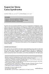
strategy is actually what differs. Different catecholamine agents have different<br />
effects on a- <strong>and</strong>b-adrenergic receptors, as shown in Fig. 1. The hemodynamic<br />
actions <strong>of</strong> these receptors are well known, with a-adrenergic receptors promoting<br />
vasoconstriction, b1-adrenergic receptors increasing heart rate <strong>and</strong> myocardial<br />
contractility, <strong>and</strong> b 2-adrenergic receptors causing peripheral vasodilation.<br />
The result <strong>of</strong> these differential effects on adrenergic receptors is that the different<br />
agents have different effects on pressure <strong>and</strong> flow, as shown in Fig. 2. Conceived in<br />
these terms, the argument about which catecholamine is best in a given situation is<br />
transformed into a discussion about which agent is best suited to implement the therapeutic<br />
strategy chosen. This may or may not make the choice easier, but it does<br />
emphasize the need to define the goals <strong>and</strong> end points <strong>of</strong> therapy <strong>and</strong> to identify<br />
how those end points will be monitored.<br />
INDIVIDUAL VASOPRESSOR AGENTS<br />
Dopamine<br />
Dopamine, the natural precursor <strong>of</strong> norepinephrine <strong>and</strong> epinephrine, has distinct<br />
dose-dependent pharmacologic effects. At doses less than 5 mg/kg/min, dopaminergic<br />
receptors are activated, leading to vasodilation in the renal <strong>and</strong> mesenteric<br />
beds. 32 At doses <strong>of</strong> 5 to 10 mg/kg/min, b1-adrenergic effects predominate, increasing<br />
cardiac contractility <strong>and</strong> heart rate. At doses above 10 mg/kg/min, a1-adrenergic effects predominate, leading to arterial vasoconstriction <strong>and</strong> an increase in blood<br />
pressure. There is a great deal <strong>of</strong> overlap in these effects, particularly in critically ill<br />
patients.<br />
Dopamine increases mean arterial pressure <strong>and</strong> cardiac output, primarily by an<br />
increase in stroke volume, <strong>and</strong> to a lesser extent by an increase in heart rate. 33–43 In<br />
open-label trials, dopamine (median dose, 15 mg/kg/min) increased mean arterial<br />
pressure by 24% in septic patients who remained hypotensive after optimal fluid<br />
resuscitation. 33–43 Dopamine has been shown to increase oxygen delivery, but its<br />
effects on calculated or measured oxygen consumption have been mixed, suggesting<br />
that tissue oxygenation may not always be improved, perhaps because <strong>of</strong> failure to<br />
Fig.1. a- <strong>and</strong> b-adrenergic effects <strong>of</strong> vasoactive catecholamines.
<strong>Inotrope</strong> <strong>and</strong> <strong>Vasopressor</strong> <strong>Therapy</strong> <strong>of</strong> <strong>Septic</strong> <strong>Shock</strong> 785<br />
Fig. 2. Effects <strong>of</strong> vasoactive catecholamines on pressure <strong>and</strong> blood flow.<br />
improve microcirculatory flow. 34,35,44,45 The effect <strong>of</strong> dopamine on splanchnic perfusion<br />
has also been mixed. Increases in splanchnic blood flow have been reported,<br />
but have not always been associated with increases in splanchnic oxygen consumption,<br />
beneficial effects on gastric intramucosal pH, or improvement in hepatosplanchnic<br />
energy balance. 33,34,36,46–48<br />
Low doses <strong>of</strong> dopamine increase renal blood flow <strong>and</strong> glomerular filtration rate in<br />
laboratory animals <strong>and</strong> healthy volunteers, supporting the idea that dopamine can<br />
reduce the risk <strong>of</strong> renal failure in critically ill patients by increasing renal blood flow.<br />
This notion has now been put to rest by a definitive clinical trial that r<strong>and</strong>omized<br />
328 critically ill patients with early renal dysfunction to low (‘‘renal’’) dose dopamine<br />
(2 mg/kg/min) or placebo. 49 No difference was found in either the primary outcome<br />
(peak serum creatinine); other renal outcomes (increase in creatinine, need for renal<br />
replacement, urine output); or secondary outcomes (survival to either ICU or hospital<br />
discharge, ICU or hospital stay, arrhythmias). 49<br />
Dopamine effectively increases mean arterial pressure in patients who remain hypotensive<br />
after optimal volume expansion, largely as a result <strong>of</strong> increasing cardiac index,<br />
so it may be chosen in patients with compromised cardiac function or cardiac reserve.<br />
Its major side effects are tachycardia <strong>and</strong> arrhythmogenesis, both <strong>of</strong> which are more<br />
prominent than with other vasopressor agents. Safety concerns have also been raised<br />
concerning extracardiac side effects. Dopamine has the potential to decrease<br />
prolactin release, favoring lymphocyte apoptosis with consequent<br />
immunosuppression. 50,51<br />
Dopamine use was associated with increased mortality in patients with shock in an<br />
observational cohort study <strong>of</strong> 198 European ICUs, <strong>and</strong> remained a significant<br />
predictor after multivariate analysis. 52 Another, similarly sized observational cohort<br />
<strong>of</strong> 17 Portuguese ICUs showed decreased mortality in septic shock patients treated<br />
with dopamine compared with norepinephrine, however, a finding that also persisted<br />
after multivariate analysis. 53 These observational studies have known limitations. A<br />
large prospective r<strong>and</strong>omized clinical trial comparing dopamine with norepinephrine<br />
in 1603 pressor-dependent patients with septic shock has recently been completed,<br />
<strong>and</strong> presented but not yet published. 54 No significant difference in mortality between<br />
use <strong>of</strong> dopamine <strong>and</strong> norepinephrine was observed, although there were more<br />
arrhythmias in the dopamine group. 54
786<br />
Hollenberg<br />
Norepinephrine<br />
Norepinephrine, the endogenous mediator <strong>of</strong> the sympathetic nervous system, is<br />
a potent a-adrenergic agonist with less pronounced b-adrenergic agonist effects.<br />
Norepinephrine increases mean arterial pressure by vasoconstriction, with a small<br />
(10%–15%) increase in cardiac output <strong>and</strong> stroke volume. 12–14,18,55,56 Filling pressures<br />
are either unchanged 12–14,18,57 or modestly increased (1–3 mm Hg). 17,19,34,36,38<br />
Norepinephrine is more potent than dopamine <strong>and</strong> may be more effective at<br />
reversing hypotension in septic shock patients. In open-label trials, norepinephrine<br />
at doses ranging from 0.01 to 3.3 mg/kg/min has been shown to increase mean arterial<br />
pressure in patients who remained hypotensive after fluid resuscitation <strong>and</strong> dopamine.<br />
13,14,18,19,36,56–59 The large doses <strong>of</strong> the drug required in some patients may be<br />
caused by a-receptor down-regulation in sepsis. 60<br />
In the only r<strong>and</strong>omized trial comparing vasopressor agents, 32 volume-resuscitated<br />
septic patients were given either dopamine or norepinephrine to achieve <strong>and</strong> maintain<br />
normal hemodynamic <strong>and</strong> oxygen transport parameters for at least 6 hours. Dopamine<br />
was successful in only 31% <strong>of</strong> patients, whereas norepinephrine administration (1.5<br />
1.2 mg/kg/min) was successful in 93% (P
<strong>Inotrope</strong> <strong>and</strong> <strong>Vasopressor</strong> <strong>Therapy</strong> <strong>of</strong> <strong>Septic</strong> <strong>Shock</strong> 787<br />
trial comparing dopamine with norepinephrine. 54 This trial showed no difference in<br />
mortality, but fewer arrhythmias with norepinephrine. 54 Because r<strong>and</strong>omized data<br />
do not suggest large differences in overall outcomes across broad populations,<br />
individualization <strong>of</strong> vasopressor agents based on clinical <strong>and</strong> hemodynamic factors<br />
still seems warranted.<br />
Phenylephrine<br />
Phenylephrine, a selective a1-adrenergic agonist, increases blood pressure by vasoconstriction.<br />
Its rapid onset, short duration, <strong>and</strong> primary vascular effects make it an<br />
attractive agent in the management <strong>of</strong> hypotension associated with sepsis, but there<br />
are concerns about its potential to reduce cardiac output in these patients.<br />
Data concerning the use <strong>of</strong> phenylephrine in hyperdynamic sepsis are sparse. Phenylephrine<br />
has been shown to increase blood pressure when given to normotensive hyperdynamic<br />
septic patients at doses <strong>of</strong> 0.5 to 8 mg/kg/min, with little change in cardiac<br />
output or stroke volume. 67,68 A small 13-patient study in hypotensive septic patients<br />
showed that phenylephrine added to either low-dose dopamine or dobutamine<br />
increased mean arterial pressure <strong>and</strong> cardiac index without a change in heart<br />
rate. 69 Recently, a crossover pilot study compared systemic hemodynamics, gastric<br />
tonometry, <strong>and</strong> renal function in 15 patients with septic shock changed from norepinephrine<br />
to phenylephrine titrated to maintain a similar blood pressure, <strong>and</strong> then<br />
back again. 70 Systemic hemodynamics were similar (although heart rate, as expected,<br />
was slightly lower), but indices <strong>of</strong> hepatosplanchnic perfusion <strong>and</strong> function were<br />
decreased with phenylephrine, as was renal function. 70 A 32-patient r<strong>and</strong>omized<br />
control trial comparing phenylephrine with norepinephrine for initial support <strong>of</strong> patients<br />
with septic shock by the same group, however, showed no significant difference in<br />
global or regional hemodynamics, or in renal function, which might suggest potential<br />
differences between delayed <strong>and</strong> early administration. 71<br />
The limited information available with phenylephrine suggests that this drug can<br />
increase blood pressure modestly in fluid-resuscitated septic shock patients, <strong>and</strong><br />
may be a good option when tachyarrhythmias limit therapy with other vasopressors. 6<br />
Epinephrine<br />
Epinephrine, which is synthesized, stored, <strong>and</strong> released from the chromaffin cells <strong>of</strong><br />
the adrenal medulla, is a potent a- <strong>and</strong> b-adrenergic agent that increases mean arterial<br />
pressure by increasing both cardiac index <strong>and</strong> peripheral vascular tone. 16,72–74<br />
Epinephrine increases oxygen delivery, but oxygen consumption also may be<br />
increased. 72–76 Lactate levels can be increased after use <strong>of</strong> epinephrine in sepsis,<br />
although whether this results from excess vasoconstriction <strong>and</strong> compromised<br />
perfusion or increased lactate production remains uncertain. 58,72,76<br />
The main concern with the use <strong>of</strong> epinephrine in sepsis is the potential to decrease<br />
regional blood flow, particularly in the splanchnic circulation. 58,77–79 In a recent study<br />
<strong>of</strong> patients with severe septic shock, epinephrine increased global oxygen delivery<br />
<strong>and</strong> consumption but caused a lower absolute <strong>and</strong> fractional splanchnic blood flow<br />
<strong>and</strong> lower indocyanine green clearance, validating the adverse effects <strong>of</strong> epinephrine<br />
alone on the splanchnic circulation. 65 Another group has reported improved gastric<br />
mucosal perfusion with epinephrine compared with norepinephrine-dobutamine<br />
combination, 80 but the same group subsequently reported superiority <strong>of</strong> a norepinephrine-dopexamine<br />
combination over epinephrine. 81<br />
A r<strong>and</strong>omized clinical trial comparing epinephrine with norepinephrine in 280 critically<br />
ill patients with shock found no difference in time to achieve arterial pressure<br />
goals, 28-day mortality, or 90-day mortality, although 13% <strong>of</strong> the patients in the
788<br />
Hollenberg<br />
epinephrine group were withdrawn from the study because <strong>of</strong> lactic acidosis or tachycardia.<br />
82 When a prespecified analysis <strong>of</strong> the 158 patients with septic shock was<br />
performed, results were similar, with no differences in hemodynamics or mortality. 82<br />
Another fairly large (N 5 330) r<strong>and</strong>omized clinical trial compared epinephrine with<br />
norepinephrine with or without dobutamine, with drugs titrated to maintain a mean<br />
arterial pressure above 70 <strong>and</strong> a cardiac index above 2.5 L/min, in patients with septic<br />
shock. 83 Metabolic abnormalities were transient in this trial, <strong>and</strong> no patients were withdrawn<br />
for this reason. There was no significant difference in time to hemodynamic<br />
success, vasopressor withdrawal, or mortality at 28 days in the ICU or in the hospital<br />
between epinephrine <strong>and</strong> norepinephrine with dobutamine. 83<br />
Epinephrine can increase blood pressure in patients unresponsive to traditional<br />
agents. It increases heart rate <strong>and</strong> has the potential to induce tachyarrhythmias,<br />
ischemia, <strong>and</strong> hypoglycemia. Because <strong>of</strong> its effects on gastric blood flow <strong>and</strong> its<br />
propensity to increase lactate concentrations, epinephrine has been considered<br />
a second-line agent whose use should be considered in patients failing to respond<br />
to traditional therapies. 6 Recent clinical trials, however, have cast some doubt on<br />
whether epinephrine is inferior to other agents.<br />
Vasopressin<br />
Vasopressin is a peptide hormone synthesized in the hypothalamus <strong>and</strong> then transported<br />
to <strong>and</strong> stored in the pituitary gl<strong>and</strong>. Released in response to decreases in blood<br />
volume, decreased intravascular volume, <strong>and</strong> increased plasma osmolality, vasopressin<br />
constricts vascular smooth muscle directly by V1 receptors <strong>and</strong> also increases<br />
responsiveness <strong>of</strong> the vasculature to catecholamines. 84,85 Vasopressin may also<br />
increase blood pressure by inhibition <strong>of</strong> vascular smooth muscle nitric oxide<br />
production 86 <strong>and</strong> K 1 -ATP channels. 85,87<br />
Normal levels <strong>of</strong> vasopressin have little effect on blood pressure in physiologic<br />
conditions 84 but vasopressin helps maintain blood pressure during hypovolemia, 88<br />
<strong>and</strong> seems to restore impaired hemodynamic mechanisms <strong>and</strong> also inhibit pathologic<br />
vascular responses in shock. 85 Increased levels <strong>of</strong> vasopressin have been documented<br />
in hemorrhagic shock, 89 but a growing body <strong>of</strong> evidence indicates that this<br />
response is abnormal or blunted in septic shock. One study found markedly increased<br />
levels <strong>of</strong> circulating vasopressin in 12 patients with cardiogenic shock, but much lower<br />
levels in 19 patients with septic shock, levels that were hypothesized to be inappropriately<br />
low. 90 One potential mechanism for this relative vasopressin deficiency is depletion<br />
<strong>of</strong> pituitary stores, possibly in conjunction with impaired synthesis. Depletion <strong>of</strong><br />
vasopressin stores in the neurohypophysis evaluated by MRI has been described in<br />
a small group <strong>of</strong> septic shock patients. 91 A recent prospective cohort study <strong>of</strong> patients<br />
with septic shock found that vasopressin levels were almost always elevated in the<br />
initial hours <strong>of</strong> septic shock <strong>and</strong> decreased afterward; one third <strong>of</strong> patients developed<br />
relative vasopressin deficiency as defined by the investigators. 92<br />
Given this theoretical rationale, observational studies demonstrated that the addition<br />
<strong>of</strong> a low dose <strong>of</strong> vasopressin (0.01–0.04 U/min) to catecholamines can raise blood pressure<br />
in patients with pressor-refractory septic shock. 93–95 Several small r<strong>and</strong>omized<br />
studies comparing vasopressin with norepinephrine have demonstrated that initiation<br />
<strong>of</strong> vasopressin decreases catecholamine requirements, 96,97 <strong>and</strong> one showed improved<br />
renal function. 96 Similar data are available for terlipressin, a synthetic vasopressin<br />
analog. 98 There is concern, however, that vasopressin infusion in septic patients may<br />
either decrease splanchnic perfusion or redistribute blood flow away from the<br />
splanchnic mucosa. 99,100 Vasopressin should be thought <strong>of</strong> as replacement therapy<br />
for relative deficiency rather than as a vasopressor agent to be titrated to effect.
<strong>Inotrope</strong> <strong>and</strong> <strong>Vasopressor</strong> <strong>Therapy</strong> <strong>of</strong> <strong>Septic</strong> <strong>Shock</strong> 789<br />
A large r<strong>and</strong>omized clinical trial (VASST) has now been completed comparing vasopressin<br />
with norepinephrine in 776 patients with pressor-dependent septic shock. 101<br />
Patients were r<strong>and</strong>omized to vasopressin (0.03 U/min) or 15 mg/min norepinephrine in<br />
addition to their original vasopressor infusion; the primary end point was 28-day<br />
mortality; a prespecified subgroup analysis was done on patients with less severe<br />
(NE 5–14 mg/min) <strong>and</strong> more severe (NE >15 mg/min) septic shock. For the group as<br />
a whole, there was no difference in mortality, but vasopressin seemed to be better<br />
in the less severe subgroup. 101<br />
Vasopressin (0.03 U/min) added to norepinephrine seems to be as safe <strong>and</strong> effective<br />
as norepinephrine in fluid-resuscitated patients with septic shock. Vasopressin<br />
may be more effective in patients on lower doses <strong>of</strong> norepinephrine than when started<br />
as rescue therapy, although what to do in patients with high vasopressor requirements<br />
despite vasopressin infusion remains uncertain.<br />
INOTROPIC THERAPY<br />
Background<br />
The broad outlines <strong>of</strong> myocardial dysfunction in patients with septic shock have been<br />
well defined. Despite the fact that cardiac output is usually normal or high, there is<br />
evidence that myocardial contractility may be impaired in a subgroup <strong>of</strong> septic<br />
patients. In the initial report, performed with serial radionuclide scans, left ventricular<br />
ejection fraction was decreased, <strong>and</strong> the left ventricle was dilated, so stroke volume<br />
was preserved. 102 Subsequent reports using echocardiography found a similarly<br />
decreased ejection fraction in a subset <strong>of</strong> septic patients, but less prominent ventricular<br />
dilation, <strong>and</strong> some <strong>of</strong> these patients were reported to have low stroke<br />
volumes. 103,104 In reports from both groups, myocardial depression developed 24 to<br />
48 hours after the onset <strong>of</strong> septic shock <strong>and</strong> was reversible in survivors. In addition<br />
to depressed left ventricular ejection fraction, some studies in septic patients have<br />
suggested abnormalities in ventricular responses to fluid loading, with lower increases<br />
in left ventricular performance (measured by left ventricular stroke work index)<br />
increased less in septic shock patients than in controls. 105<br />
The reversibility <strong>of</strong> myocardial dysfunction in sepsis suggests the involvement <strong>of</strong><br />
circulating mediators, but the precise mechanisms <strong>of</strong> myocardial dysfunction remain<br />
unclear. A role for inflammatory cytokines has been suggested by studies showing<br />
that tumor necrosis factor, 106 interleukin-1, 107 <strong>and</strong> other inflammatory cytokines,<br />
either alone or in combination, 108 depress contractility <strong>of</strong> isolated cardiac myocytes.<br />
The time course <strong>of</strong> myocardial depression in large animal models <strong>and</strong> in patients<br />
with sepsis, with onset between 24 <strong>and</strong> 48 hours, along with evidence for its induction<br />
by cytokines, suggests the possibility <strong>of</strong> cytokine-inducible nitric oxide synthase as<br />
a mediator. 108 Studies have implicated both nitric oxide production <strong>and</strong> reactive<br />
oxygen species in cytokine-induced myocardial depression, <strong>and</strong> have further suggested<br />
a role for peroxynitrite. 109 Other studies have implicated decreased myocyte<br />
my<strong>of</strong>ilament calcium responsiveness, possibly mediated by abnormal protein kinase<br />
A phosphorylation. 110 Regardless <strong>of</strong> the mechanism, the reversibility <strong>of</strong> myocardial<br />
depression in septic patients suggests the feasibility <strong>of</strong> a strategy <strong>of</strong> inotropic support<br />
while awaiting recovery.<br />
The challenge in interpreting myocardial dysfunction in sepsis is that the most<br />
important physiologic parameter is cardiac output, not ejection fraction. Some<br />
patients, especially those with pre-existing cardiac dysfunction, may have decreased<br />
cardiac output, <strong>and</strong> those patients are clearly c<strong>and</strong>idates for inotropic therapy to<br />
improve cardiac performance. For other patients, the clinical issue is not so much
790<br />
Hollenberg<br />
how to optimize cardiac systolic performance, but how to determine whether cardiac<br />
output is adequate to meet physiologic needs.<br />
Goals <strong>and</strong> Monitoring <strong>of</strong> Inotropic <strong>Therapy</strong><br />
Tissue perfusion is a function <strong>of</strong> both pressure <strong>and</strong> flow. The challenge in titrating<br />
therapy to a cardiac output is to determine when that output is adequate. Because<br />
<strong>of</strong> the complexity <strong>of</strong> assessment <strong>of</strong> clinical parameters in septic patients, direct<br />
measurement <strong>of</strong> cardiac output in patients receiving inotropic therapy is advisable,<br />
but other end points <strong>of</strong> global perfusion also should be followed. When global hypoperfusion<br />
is manifested by decreased mixed venous oxygen saturation, this measure may<br />
be used as a guide to the adequacy <strong>of</strong> inotropic therapy. Similarly, a fall in blood lactate<br />
concentrations concomitant with increased cardiac output is a good prognostic sign.<br />
Early <strong>and</strong> late inotropic therapy in sepsis may well be different. In the study by Rivers<br />
<strong>and</strong> coworkers4 <strong>of</strong> early goal-directed resuscitation for patients presenting with septic<br />
shock, if central venous oxygen saturation remained low despite resuscitation with<br />
fluids <strong>and</strong> vasopressors <strong>and</strong> packed red blood cells if necessary, patients received<br />
inotropic therapy; 13.7% <strong>of</strong> patients in the intervention group were given dobutamine,<br />
compared with 0.8% in the st<strong>and</strong>ard treatment group. In this setting, low central<br />
venous oxygen saturation provided presumptive evidence for inadequate oxygen<br />
delivery. Precisely which components <strong>of</strong> the resuscitation bundle were responsible<br />
for improved outcomes in this trial, however, remains uncertain, <strong>and</strong> is the subject<br />
<strong>of</strong> ongoing trials.<br />
After initial resuscitation, what to do is less clear. More is known about what not to<br />
do than what to do in this respect. Some critically ill septic patients are hypermetabolic<br />
<strong>and</strong> may require high levels <strong>of</strong> oxygen delivery to maintain oxidative metabolism. 111<br />
Accordingly, it has been hypothesized that such patients would benefit from therapeutic<br />
measures to increase oxygen delivery to ‘‘supranormal’’ levels. Retrospective<br />
analyses showed that achievement <strong>of</strong> cardiac index greater than 4.5 L/min/m2 ,<br />
oxygen delivery greater than 600 mL/min/m2 , <strong>and</strong> oxygen consumption greater than<br />
170 mL/min/m2 correlates with improved survival. 112 Two large r<strong>and</strong>omized studies<br />
to test the hypothesis that routinely increasing oxygen delivery to these predefined<br />
levels in all critically ill patients, however, did not show improved outcomes, 113,114<br />
<strong>and</strong> mortality in the treatment arm was higher than control in one <strong>of</strong> the studies, a trial<br />
that allowed very high doses <strong>of</strong> dobutamine in some patients. 114 A strategy <strong>of</strong> routinely<br />
increasing oxygen delivery to predetermined elevated end points <strong>of</strong> cardiac index <strong>and</strong><br />
oxygen delivery is not recommended in the guidelines. 6,115<br />
Nonetheless, some patients may have improved tissue perfusion with inotropic<br />
therapy aimed at increasing oxygen delivery. Because the goal <strong>of</strong> such therapy is to<br />
increase cardiac output, is seems logical that such therapy is best guided by monitoring<br />
<strong>of</strong> cardiac output, but this should be supplemented by clinical measures <strong>of</strong><br />
perfusion. When global hypoperfusion is manifest by decreased venous oxygen saturation,<br />
its monitoring can be helpful to guide response to therapy. Similarly, although<br />
lactate production in sepsis is complex, a fall in blood lactate levels during inotropic<br />
therapy is a good prognostic sign. 116 In addition, assessment <strong>of</strong> the adequacy <strong>of</strong><br />
regional or microcirculatory perfusion may also be useful in selected patients. 6<br />
INDIVIDUAL INOTROPIC AGENTS<br />
Dobutamine<br />
Dobutamine is a racemic mixture <strong>of</strong> two isomers, a D isomer with b1- <strong>and</strong> b2-adrenergic<br />
effects, <strong>and</strong> an L isomer with b1- <strong>and</strong> a1-adrenergic effects; its predominant
<strong>Inotrope</strong> <strong>and</strong> <strong>Vasopressor</strong> <strong>Therapy</strong> <strong>of</strong> <strong>Septic</strong> <strong>Shock</strong> 791<br />
effect is inotropic by stimulation <strong>of</strong> b1 receptors, with a variable effect on blood pressure.<br />
A number <strong>of</strong> studies have investigated the effect <strong>of</strong> dobutamine on cardiac function<br />
during sepsis or septic shock at doses ranging from 2 to 28 mg/kg/min. 56,117–121 In<br />
these studies, increases in cardiac index ranged from 12% to 61%. Heart rate<br />
increases, however, <strong>of</strong>ten significantly (9%–23%). Two studies reported that left<br />
ventricular stroke work index increased by 23% to 58% at mean dobutamine doses<br />
<strong>of</strong> 5 to 12 mg/kg/min. 117,119 Similar increases in right ventricular stroke work were<br />
also observed in these studies.<br />
Despite a paucity <strong>of</strong> r<strong>and</strong>omized data demonstrating its efficacy, dobutamine is the<br />
first-choice inotropic agent for patients with measured or suspected low cardiac<br />
output in the presence <strong>of</strong> adequate filling pressures. 6,115 Although dobutamine does<br />
not influence the distribution <strong>of</strong> blood flow, therapy is <strong>of</strong>ten aimed at increasing blood<br />
flow to organs, such as the gut or the kidneys.<br />
Dopamine<br />
Dopamine has b-adrenergic activity, usually at doses greater than 5 mg/kg/min. At<br />
doses <strong>of</strong> 5 to 10 mg/kg/min vasopressor effects caused by a-adrenergic stimulation<br />
occur <strong>and</strong> become predominant at higher doses. Dopamine also causes the release<br />
<strong>of</strong> norepinephrine from nerve terminals, contributing to its cardiac effects. The<br />
pharmacokinetics <strong>of</strong> dopamine in critically ill patients is highly variable.<br />
In patients with severe sepsis or septic shock, dopamine increases cardiac index,<br />
largely because <strong>of</strong> an increase in stroke volume, but to a lesser extent by increasing<br />
heart rate. 33–43 Patients receiving dopamine at rates greater than 20 mg/kg/min<br />
show increases in right heart pressures <strong>and</strong> in heart rate, <strong>and</strong> doses should not usually<br />
exceed 20 mg/kg/min, at least not without adequate hemodynamic monitoring. Dopamine<br />
may be used when both cardiac output <strong>and</strong> blood pressure are low, a setting in<br />
which its vasopressor effect may be desirable. Concerns about extracardiac immunosuppressive<br />
effects50,51 have limited enthusiasm for its use as a first-line inotropic<br />
agent.<br />
Epinephrine<br />
Epinephrine stimulates both a <strong>and</strong> b receptors. At low doses, the b-adrenergic effects<br />
predominate. Studies examining its hemodynamic effects in septic shock at doses<br />
ranging from 0.1 to 0.5 mg/kg/min have shown increases in cardiac index ranging<br />
from 24% to 54%. 73,74,76 This increase in oxygen delivery may be accompanied by<br />
metabolic effects, 58,72,76 the significance <strong>of</strong> which is uncertain in view <strong>of</strong> trials showing<br />
no difference in outcome between epinephrine <strong>and</strong> other catecholamines. 82,83<br />
Epinephrine does seem to be more arrhythmogenic than other catecholamines.<br />
Combination <strong>and</strong> Comparative Catecholamine Studies<br />
Most studies investigating catecholamine combinations have been limited by lack <strong>of</strong><br />
st<strong>and</strong>ardized infusion protocols, limiting the robustness <strong>of</strong> their conclusions. Patients<br />
who do not respond to dopamine with an increase in cardiac index may reach the<br />
desired end point with a dopamine-norepinephrine combination. 38 Epinephrine seems<br />
to be as good if not better at improving cardiac performance than dopamine or<br />
a dobutamine-norepinephrine combination. 58,76 In several studies, dopamine<br />
increased cardiac index <strong>and</strong> stroke volume index to a greater extent than norepinephrine<br />
but increases in left <strong>and</strong> right ventricular stroke volume index were about the same<br />
with the two agents, <strong>and</strong> tachycardia was less prominent with norepinephrine. 34,57
Table 1<br />
Consensus recommendations for vasopressor support in sepsis<br />
# ACCM Practice Parameters Level # Surviving Sepsis Campaign Level<br />
When fluid administration fails to restore an<br />
adequate arterial pressure <strong>and</strong> organ perfusion,<br />
therapy with vasopressor agents should be<br />
initiated. <strong>Vasopressor</strong> therapy may also be<br />
required transiently to maintain perfusion in the<br />
face <strong>of</strong> life-threatening hypotension, even when<br />
adequate cardiac filling pressures have not yet<br />
been attained.<br />
None (text) 1 — —<br />
Arterial cannulation should be performed in<br />
None (basic principle) 6 All patients requiring vasopressors should have an D<br />
patients with shock to provide a more accurate<br />
arterial catheter placed as soon as practical if<br />
measurement <strong>of</strong> intra-arterial pressure <strong>and</strong> to<br />
allow beat-to-beat analysis so that decisions<br />
regarding therapy can be based on immediate<br />
<strong>and</strong> reproducible blood pressure information.<br />
resources are available.<br />
<strong>Vasopressor</strong>s<br />
Mean arterial pressure (MAP) be maintained above<br />
65 mm Hg<br />
C<br />
1 Dopamine <strong>and</strong> norepinephrine are both effective C 1 Norepinephrine or dopamine as the first-choice C<br />
for increasing arterial blood pressure. It is<br />
vasopressor agent to correct hypotension in septic<br />
imperative to ensure that patients are adequately<br />
shock (administered through a central catheter as<br />
fluid resuscitated. Dopamine raises cardiac output<br />
more than norepinephrine, but its use may be<br />
limited by tachycardia. Norepinephrine may be<br />
a more effective vasopressor in some patients.<br />
soon as one is available).<br />
2 Phenylephrine is an alternative to increase blood D 2/3 Phenylephrine, epinephrine, or vasopressin not be C<br />
pressure, especially in the setting <strong>of</strong><br />
administered as the initial vasopressor in septic B<br />
tachyarrhythmias. Epinephrine can be considered<br />
shock. Epinephrine be the first alternative agent<br />
for refractory hypotension, although adverse<br />
in septic shock that is poorly responsive to<br />
effects are common, <strong>and</strong> epinephrine may<br />
potentially decrease mesenteric perfusion.<br />
norepinephrine or dopamine.<br />
792<br />
Hollenberg
3 Administration <strong>of</strong> low doses <strong>of</strong> dopamine to<br />
maintain renal function is not recommended.<br />
5 Low doses <strong>of</strong> vasopressin given after 24 hours as<br />
hormone replacement may be effective in raising<br />
blood pressure in patients refractory to other<br />
vasopressors, although no conclusive data are yet<br />
available regarding outcome.<br />
<strong>Inotrope</strong>s<br />
1 Dobutamine is the first choice for patients with low<br />
cardiac index or low mixed venous oxygen<br />
saturation <strong>and</strong> an adequate mean arterial<br />
pressure following fluid resuscitation.<br />
Dobutamine may cause hypotension or<br />
tachycardia in some patients, especially those<br />
with decreased filling pressures.<br />
2 In patients with evidence <strong>of</strong> tissue hypoperfusion,<br />
addition <strong>of</strong> dobutamine may be helpful to<br />
increase cardiac output <strong>and</strong> improve organ<br />
perfusion. A strategy <strong>of</strong> routinely increasing<br />
cardiac index to predefined ‘‘supranormal’’ levels<br />
(more than 4.5 L/min/m2 ) has not been shown to<br />
improve outcome.<br />
3 A vasopressor, such as norepinephrine, <strong>and</strong> an<br />
inotrope, such as dobutamine, can be titrated<br />
separately to maintain both mean arterial<br />
pressure <strong>and</strong> cardiac output.<br />
B 5 Low-dose dopamine not be used for renal<br />
protection.<br />
D 2 Vasopressin, 0.03 units, may be added to<br />
norepinephrine subsequently with anticipation <strong>of</strong><br />
an effect equivalent to norepinephrine alone.<br />
C 1 A dobutamine infusion be administered in the<br />
presence <strong>of</strong> myocardial dysfunction as suggested<br />
by elevated cardiac filling pressures <strong>and</strong> low<br />
cardiac output.<br />
B 2 Use <strong>of</strong> a strategy to increase cardiac index to<br />
predefined supranormal levels is not<br />
recommended.<br />
C — — —<br />
Strength <strong>of</strong> recommendation levels: A, supported by at least two level I investigations; B, supported by only one level I investigation; C, supported only by level II<br />
investigations; D, supported by at least one level III investigation; E, supported only by level IV or level V investigations.<br />
Strength <strong>of</strong> evidence: Level I, large, r<strong>and</strong>omized trials with clear-cut results, low risk <strong>of</strong> false-positive (a) error or false-negative (b) error; Level II, small, r<strong>and</strong>omized<br />
trials with uncertain results, moderate to high risk <strong>of</strong> false-positive (a) error or false-negative (b) error; Level III, nonr<strong>and</strong>omized, contemporaneous controls; Level<br />
IV, nonr<strong>and</strong>omized, historical controls <strong>and</strong> expert opinion; Level V, case series, uncontrolled studies, <strong>and</strong> expert opinion.<br />
A<br />
—<br />
C<br />
B<br />
<strong>Inotrope</strong> <strong>and</strong> <strong>Vasopressor</strong> <strong>Therapy</strong> <strong>of</strong> <strong>Septic</strong> <strong>Shock</strong> 793
794<br />
Hollenberg<br />
Phosphodiesterase Inhibitors<br />
Phosphodiesterase inhibitors increase intracellular cyclic AMP <strong>and</strong> have inotropic<br />
effects independent <strong>of</strong> b-adrenergic receptors. In view <strong>of</strong> recent data suggesting<br />
the potential for decreased myocardial adrenergic responsiveness in septic shock, 122<br />
their use might be considered in some settings. Most <strong>of</strong> the available case series are<br />
confounded by concomitant use <strong>of</strong> adrenergic agents, but one small r<strong>and</strong>omized trial<br />
<strong>of</strong> 12 pediatric patients was able to demonstrate increased cardiac output with milrinone<br />
in sepsis. 123 Phosphodiesterase inhibitors have vasodilatory effects that might<br />
exacerbate hypotension in sepsis, m<strong>and</strong>ating caution in their use, especially given<br />
their relatively long half-lives. The decision to use this drug in septic shock patients<br />
to increase cardiac output is expected to increase vasopressor requirements.<br />
Levosimendan<br />
Levosimendan is a novel agent that increases cardiac myocyte calcium responsiveness<br />
<strong>and</strong> also opens ATP-dependent potassium channels, giving the drug both<br />
inotropic <strong>and</strong> vasodilatory properties. Levosimendan has been most extensively<br />
studied in acute heart failure, but given the potential role for abnormal calcium<br />
h<strong>and</strong>ling in sepsis-induced myocardial depression, its use also has been proposed<br />
in sepsis. Studies in animal models <strong>of</strong> endotoxin infusion have suggested that levosimendan<br />
can improve myocardial performance with relatively modest decreases in<br />
arterial pressure. 124 One clinical trial r<strong>and</strong>omized 30 patients with septic shock <strong>and</strong><br />
ejection fraction less than 45% to dobutamine or levosimendan, with norepinephrine<br />
used to maintain blood pressure. 125 Levosimendan improved ejection fraction, stroke<br />
volume, <strong>and</strong> cardiac index <strong>and</strong> also improved urine output <strong>and</strong> gastric mucosal PO2<br />
compared with dobutamine. 125 Another trial by the same group r<strong>and</strong>omized 35<br />
patients with septic shock <strong>and</strong> acute respiratory distress syndrome to levosimendan<br />
or placebo. 126 Levosimendan improved right ventricular performance, <strong>and</strong> mixed<br />
venous oxygen saturation also was improved, suggesting that its effects on cardiac<br />
function translated into a systemic effect. 126<br />
Levosimendan is not currently approved for use in the United States. Despite<br />
a reasonable rationale for its use, <strong>and</strong> some experimental data suggesting some beneficial<br />
effects, larger r<strong>and</strong>omized trials with patient-centered end points, such as<br />
survival <strong>and</strong> length <strong>of</strong> stay, are needed before it can be considered for widespread<br />
use as an inotropic agent in sepsis.<br />
COMPLICATIONS OF VASOPRESSOR THERAPY<br />
All <strong>of</strong> the catecholamine agents can cause significant tachycardia, especially in<br />
patients who are inadequately volume resuscitated. In patients with significant coronary<br />
atherosclerosis, catecholamine-induced coronary artery constriction may precipitate<br />
myocardial ischemia <strong>and</strong> infarction; this is <strong>of</strong> particular concern in patients<br />
treated with vasopressin. In the presence <strong>of</strong> myocardial dysfunction, excessive vasoconstriction<br />
can decrease stroke volume, cardiac output, <strong>and</strong> oxygen delivery. Should<br />
this occur, the dose should be lowered, or the addition <strong>of</strong> an inotropic agent should be<br />
considered. 56 Excessive doses <strong>of</strong> vasopressors can also cause limb ischemia <strong>and</strong><br />
necrosis.<br />
Administration <strong>of</strong> vasopressor agents may potentially impair blood flow to the<br />
splanchnic system, <strong>and</strong> this can be manifested by stress ulceration, ileus, malabsorption,<br />
<strong>and</strong> even bowel infarction. 58,76 Gut mucosal integrity occupies a key position in<br />
the pathogenesis <strong>of</strong> multiple organ failure, <strong>and</strong> countercurrent flow in splanchnic<br />
microcirculation gives the gut a higher critical threshold for oxygen delivery than other
organs. It makes sense to avoid episodes <strong>of</strong> intramucosal acidosis, which might be<br />
detected either by a fall in gastric mucosal pHi or an increase in gastric mucosal<br />
PCO 2. Whether to monitor these parameters routinely is less certain, because pHi or<br />
gastric PCO2-directed care has not been shown to reduce mortality in patients with<br />
septic shock in prospective r<strong>and</strong>omized controlled trials.<br />
At inotropic doses, catecholamines can trigger tachyarrythmias, including supraventricular<br />
tachycardias, atrial fibrillation, <strong>and</strong> ventricular tachycardia. The phosphodiesterase<br />
inhibitors <strong>and</strong> levosimendan also have the potential to produce hypotension,<br />
especially in patients with inadequate fluid resuscitation. As such, monitoring stroke<br />
volume <strong>and</strong> cardiac output with these agents, so as to obtain the desired therapeutic<br />
effect at the minimal dosage, is advisable. Patients in septic shock may manifest severe<br />
clinical manifestations <strong>of</strong> disseminated intravascular coagulation including loss <strong>of</strong> digits<br />
<strong>and</strong> extremities. These patients may also be on significant doses <strong>of</strong> vasopressors,<br />
leading to a false conclusion that the limb loss is caused by the vasopressors.<br />
CONSENSUS RECOMMENDATIONS<br />
Consensus recommendations regarding vasopressor support in patients with septic<br />
shock have been put forth by the American College <strong>of</strong> Critical Care Medicine 6 <strong>and</strong><br />
the Surviving Sepsis campaign 11 ; these recommendations differ more in wording<br />
than in substance, <strong>and</strong> are compiled in Table 1. The Surviving Sepsis campaign will<br />
likely amend the vasopressin section to take the VASST trial results under<br />
consideration.<br />
SUMMARY<br />
The ultimate goals <strong>of</strong> hemodynamic therapy in shock are to restore effective tissue<br />
perfusion <strong>and</strong> to normalize cellular metabolism. In sepsis, both global <strong>and</strong> regional<br />
perfusion must be considered. In addition, mediators <strong>of</strong> sepsis can perturb cellular<br />
metabolism, leading to inadequate use <strong>of</strong> oxygen <strong>and</strong> other nutrients despite<br />
adequate perfusion; one would not expect organ dysfunction mediated by such<br />
abnormalities to be corrected by hemodynamic therapy.<br />
Despite the complex pathophysiology <strong>of</strong> sepsis, an underlying approach to its<br />
hemodynamic support can be formulated that is particularly pertinent with respect<br />
to vasoactive agents. Both arterial pressure <strong>and</strong> tissue perfusion must be taken into<br />
account when choosing therapeutic interventions <strong>and</strong> the efficacy <strong>of</strong> hemodynamic<br />
therapy should be assessed by monitoring a combination <strong>of</strong> clinical <strong>and</strong> hemodynamic<br />
parameters. It is relatively easy to raise blood pressure, but somewhat harder to raise<br />
cardiac output in septic patients. How to optimize regional blood <strong>and</strong> microcirculatory<br />
blood flow remains uncertain. Specific end points for therapy are debatable <strong>and</strong> are<br />
likely to evolve. Nonetheless, the idea that clinicians should define specific goals<br />
<strong>and</strong> end points, titrate therapies to those end points, <strong>and</strong> evaluate the results <strong>of</strong> their<br />
interventions on an ongoing basis remains a fundamental principle. The practice<br />
parameters were intended to emphasize the importance <strong>of</strong> such an approach so as<br />
to provide a foundation for the rational choice <strong>of</strong> vasoactive agents in the context <strong>of</strong><br />
evolving monitoring techniques <strong>and</strong> therapeutic approaches.<br />
REFERENCES<br />
<strong>Inotrope</strong> <strong>and</strong> <strong>Vasopressor</strong> <strong>Therapy</strong> <strong>of</strong> <strong>Septic</strong> <strong>Shock</strong> 795<br />
1. Hollenberg SM, Parrillo JE. <strong>Shock</strong>. In: Fauci AS, Braunwald E, Isselbacher KJ,<br />
et al, editors. Harrison’s principles <strong>of</strong> internal medicine. 14th edition. New York:<br />
McGraw-Hill; 1997. p. 214–22.
796<br />
Hollenberg<br />
2. Ince C, Sinaasappel M. Microcirculatory oxygenation <strong>and</strong> shunting in sepsis <strong>and</strong><br />
shock. Crit Care Med 1999;27:1369–77.<br />
3. Parrillo JE, Parker MM, Natanson C, et al. <strong>Septic</strong> shock in humans: advances in<br />
the underst<strong>and</strong>ing <strong>of</strong> pathogenesis, cardiovascular dysfunction, <strong>and</strong> therapy.<br />
Ann Intern Med 1990;113:227–42.<br />
4. Rivers E, Nguyen B, Havstad S, et al. Early goal-directed therapy in the treatment<br />
<strong>of</strong> severe sepsis <strong>and</strong> septic shock. N Engl J Med 2001;345:1368–77.<br />
5. Cohn JN. Blood pressure measurement in shock: mechanism <strong>of</strong> inaccuracy in<br />
ausculatory <strong>and</strong> palpatory methods. JAMA 1967;199:118–22.<br />
6. Hollenberg SM, Ahrens TS, Annane D, et al. Practice parameters for hemodynamic<br />
support <strong>of</strong> sepsis in adult patients: 2004 update. Crit Care Med 2004;<br />
32:1928–48.<br />
7. Task Force <strong>of</strong> the American College <strong>of</strong> Critical Care Medicine, Hollenberg SM,<br />
Ahrens TS, et al. Practice parameters for hemodynamic support <strong>of</strong> sepsis in<br />
adult patients. Crit Care Med 1999;27:639–60.<br />
8. Michard F, Teboul JL. Predicting fluid responsiveness in ICU patients: a critical<br />
analysis <strong>of</strong> the evidence. Chest 2002;121:2000–8.<br />
9. Bersten AD, Holt AW. Vasoactive drugs <strong>and</strong> the importance <strong>of</strong> renal perfusion<br />
pressure. New Horiz 1995;3:650–61.<br />
10. Kirchheim HR, Ehmke H, Hackenthal E, et al. Autoregulation <strong>of</strong> renal blood flow,<br />
glomerular filtration rate <strong>and</strong> renin release in conscious dogs. Pflugers Arch<br />
1987;410:441–9.<br />
11. Dellinger RP, Carlet JM, Masur H, et al. Surviving sepsis campaign guidelines for<br />
management <strong>of</strong> severe sepsis <strong>and</strong> septic shock. Crit Care Med 2004;32:858–73.<br />
12. Desjars P, Pinaud M, Bugnon D, et al. Norepinephrine therapy has no deleterious<br />
renal effects in human septic shock. Crit Care Med 1989;17:426–9.<br />
13. Desjars P, Pinaud M, Tasseau F, et al. A reappraisal <strong>of</strong> norepinephrine therapy in<br />
human septic shock. Crit Care Med 1987;15:134–7.<br />
14. Hesselvik JF, Brodin B. Low dose norepinephrine in patients with septic shock<br />
<strong>and</strong> oliguria: effects on afterload, urine flow, <strong>and</strong> oxygen transport. Crit Care<br />
Med 1989;17:179–80.<br />
15. Fukuoka T, Nishimura M, Imanaka H, et al. Effects <strong>of</strong> norepinephrine on renal<br />
function in septic patients with normal <strong>and</strong> elevated serum lactate levels. Crit<br />
Care Med 1989;17:1104–7.<br />
16. Lipman J, Roux A, Kraus P. Vasoconstrictor effects <strong>of</strong> adrenaline in human septic<br />
shock. Anaesth Intensive Care 1991;19:61–5.<br />
17. Martin C, Eon B, Saux P, et al. Renal effects <strong>of</strong> norepinephrine used to treat<br />
septic shock patients. Crit Care Med 1990;18:282–5.<br />
18. Meadows D, Edwards JD, Wilkins RG, et al. Reversal <strong>of</strong> intractable septic shock<br />
with norepinephrine therapy. Crit Care Med 1988;16:663–7.<br />
19. Redl-Wenzl EM, Armbruster C, Edelmann G, et al. The effects <strong>of</strong> norepinephrine<br />
on hemodynamics <strong>and</strong> renal function in severe septic shock states. Intensive<br />
Care Med 1993;19:151–4.<br />
20. LeDoux D, Astiz ME, Carpati CM, et al. Effects <strong>of</strong> perfusion pressure on tissue<br />
perfusion in septic shock. Crit Care Med 2000;28:2729–32.<br />
21. Bourgoin A, Leone M, Delmas A, et al. Increasing mean arterial pressure in<br />
patients with septic shock: effects on oxygen variables <strong>and</strong> renal function. Crit<br />
Care Med 2005;33:780–6.<br />
22. Levy B, Gibot S, Franck P, et al. Relation between muscle Na1K1 ATPase<br />
activity <strong>and</strong> raised lactate concentrations in septic shock: a prospective study.<br />
Lancet 2005;365:871–5.
<strong>Inotrope</strong> <strong>and</strong> <strong>Vasopressor</strong> <strong>Therapy</strong> <strong>of</strong> <strong>Septic</strong> <strong>Shock</strong> 797<br />
23. Friedman G, Berlot G, Kahn RJ. Combined measurements <strong>of</strong> blood lactate levels<br />
<strong>and</strong> gastric intramucosal pH in patients with severe sepsis. Crit Care Med 1995;<br />
23:1184–93.<br />
24. Vincent JL, Dufaye P, Berre J. Serial lactate determinations during circulatory<br />
shock. Crit Care Med 1983;11:449–51.<br />
25. Weil MH, Afifi AA. Experimental <strong>and</strong> clinical studies on lactate <strong>and</strong> pyruvate as<br />
indicators <strong>of</strong> the severity <strong>of</strong> acute circulatory failure. Circulation 1970;41:<br />
989–1001.<br />
26. Reinhart K, Kuhn HJ, Hartog C, et al. Continuous central venous <strong>and</strong> pulmonary<br />
artery oxygen saturation monitoring in the critically ill. Intensive Care Med 2004;<br />
30:1572–8.<br />
27. Varpula M, Karlsson S, Ruokonen E, et al. Mixed venous oxygen saturation<br />
cannot be estimated by central venous oxygen saturation in septic shock. Intensive<br />
Care Med 2006;32:1336–43.<br />
28. Nelson D, Beyer C, Samsel R, et al. Pathologic supply dependence <strong>of</strong> systemic <strong>and</strong><br />
intestinal O 2 uptake during bacteremia in the dog. J Appl Physiol 1987;63:1487–9.<br />
29. De Backer D, Creteur J, Noordally O, et al. Does hepato-splanchnic VO2/DO2<br />
dependency exist in critically ill septic patients? Am J Respir Crit Care Med<br />
1998;157:1219–25.<br />
30. Sakr Y, Dubois MJ, De Backer D, et al. Persistent microcirculatory alterations are<br />
associated with organ failure <strong>and</strong> death in patients with septic shock. Crit Care<br />
Med 2004;32:1825–31.<br />
31. Creteur J, De Backer D, Sakr Y, et al. Sublingual capnometry tracks microcirculatory<br />
changes in septic patients. Intensive Care Med 2006;32:516–23.<br />
32. Hoogenberg K, Smit AJ, Girbes ARJ. Effects <strong>of</strong> low-dose dopamine on renal <strong>and</strong><br />
systemic hemodynamics during incremental norepinephrine infusion in healthy<br />
volunteers. Crit Care Med 1998;26:260–5.<br />
33. Meier-Hellmann A, Bredle DL, Specht M, et al. The effects <strong>of</strong> low-dose dopamine<br />
on splanchnic blood flow <strong>and</strong> oxygen utilization in patients with septic shock.<br />
Intensive Care Med 1997;23:31–7.<br />
34. Marik PE, Mohedin M. The contrasting effects <strong>of</strong> dopamine <strong>and</strong> norepinephrine<br />
on systemic <strong>and</strong> splanchnic oxygen utilization in hyperdynamic sepsis. JAMA<br />
1994;272:1354–7.<br />
35. Hannemann L, Reinhart K, Grenzer O, et al. Comparison <strong>of</strong> dopamine to dobutamine<br />
<strong>and</strong> norepinephrine for oxygen delivery <strong>and</strong> uptake in septic shock. Crit<br />
Care Med 1995;23:1962–70.<br />
36. Ruokonen E, Takala J, Kari A, et al. Regional blood flow <strong>and</strong> oxygen transport in<br />
septic shock. Crit Care Med 1993;21:1296–303.<br />
37. Jardin F, Gurdjian F, Desfonds P, et al. Effect <strong>of</strong> dopamine on intrapulmonary<br />
shunt fraction <strong>and</strong> oxygen transport in severe sepsis with circulatory <strong>and</strong> respiratory<br />
failure. Crit Care Med 1979;7:273–7.<br />
38. Martin C, Papazian L, Perrin G, et al. Norepinephrine or dopamine for the treatment<br />
<strong>of</strong> hyperdynamic septic shock. Chest 1993;103:1826–31.<br />
39. Winslow EJ, Loeb HS, Rahimtoola SH, et al. Hemodynamic studies <strong>and</strong> results <strong>of</strong><br />
therapy in 50 patients with bacteremic shock. Am J Med 1973;54:421–32.<br />
40. Regnier B, Safran D, Carlet J, et al. Comparative haemodynamic effects <strong>of</strong> dopamine<br />
<strong>and</strong> dobutamine in septic shock. Intensive Care Med 1979;5:115–20.<br />
41. Samii K, Le Gall JR, Regnier B, et al. Hemodynamic effects <strong>of</strong> dopamine in<br />
septic shock with <strong>and</strong> without acute renal failure. Arch Surg 1978;113:1414–6.<br />
42. Regnier B, Rapin M, Gory G, et al. Haemodynamic effects <strong>of</strong> dopamine in septic<br />
shock. Intensive Care Med 1977;3:47–53.
798<br />
Hollenberg<br />
43. Wilson RF, Sibbald WJ, Jaanimagi JL. Hemodynamic effects <strong>of</strong> dopamine in critically<br />
ill septic patients. J Surg Res 1976;20:163–72.<br />
44. Meier-Hellmann A, Reinhart K. Effects <strong>of</strong> catecholamines on regional perfusion<br />
<strong>and</strong> oxygenation in critically ill patients. Acta Anaesthesiol Sc<strong>and</strong> Suppl 1995;<br />
107:239–48.<br />
45. Hiltebr<strong>and</strong> LB, Krejci V, Sigurdsson GH. Effects <strong>of</strong> dopamine, dobutamine, <strong>and</strong><br />
dopexamine on microcirculatory blood flow in the gastrointestinal tract during<br />
sepsis <strong>and</strong> anesthesia. Anesthesiology 2004;100:1188–97.<br />
46. Maynard ND, Bihari DJ, Dalton RN, et al. Increasing splanchnic blood flow in the<br />
critically ill. Chest 1995;108:1648–54.<br />
47. Neviere R, Chagnon JL, Vallet B, et al. Dobutamine improves gastrointestinal<br />
mucosal blood flow in a porcine model <strong>of</strong> endotoxic shock. Crit Care Med<br />
1997;25:1371–7.<br />
48. Guerin JP, Levraut J, Samat-Long C, et al. Effects <strong>of</strong> dopamine <strong>and</strong> norepinephrine<br />
on systemic <strong>and</strong> hepatosplanchnic hemodynamics, oxygen exchange, <strong>and</strong><br />
energy balance in vasoplegic septic patients. <strong>Shock</strong> 2005;23:18–24.<br />
49. Bellomo R, Chapman M, Finfer S, et al. Care Society (ANZICS) Clinical Trials<br />
Group. Low-dose dopamine in patients with early renal dysfunction:<br />
a placebo-controlled r<strong>and</strong>omised trial. Lancet 2000;356:2139–43.<br />
50. Van den Berghe G, de Zegher F. Anterior pituitary function during critical illness<br />
<strong>and</strong> dopamine treatment. Crit Care Med 1996;24:1580–90.<br />
51. Oberbeck R, Schmitz D, Wilsenack K, et al. Dopamine affects cellular immune<br />
functions during polymicrobial sepsis. Intensive Care Med 2006;32:731–9.<br />
52. Sakr Y, Reinhart K, Vincent JL, et al. Does dopamine administration in shock<br />
influence outcome? Results <strong>of</strong> the Sepsis Occurrence in Acutely Ill Patients<br />
(SOAP) Study. Crit Care Med 2006;34:589–97.<br />
53. Povoa PR, Carneiro AH, Ribeiro OS, et al. Influence <strong>of</strong> vasopressor agent in<br />
septic shock mortality: results from the Portuguese Community-Acquired Sepsis<br />
Study (SACiUCI study). Crit Care Med 2009;37:410–6.<br />
54. DeBacker D. Comparison <strong>of</strong> dopamine <strong>and</strong> norepinephrine as the first vasopressor<br />
agent in the management <strong>of</strong> shock. Presented, European Society <strong>of</strong><br />
Intensive Care Medicine 2008.<br />
55. Martin C, Perrin G, Saux P, et al. Effects <strong>of</strong> norepinephrine on right ventricular<br />
function in septic shock patients. Intensive Care Med 1994;20:444–7.<br />
56. Martin C, Saux P, Eon B, et al. <strong>Septic</strong> shock: a goal-directed therapy using<br />
volume loading, dobutamine <strong>and</strong>/or norepinephrine. Acta Anaesthesiol Sc<strong>and</strong><br />
1990;34:413–7.<br />
57. Schreuder WO, Schneider AJ, Groeneveld ABJ, et al. Effect <strong>of</strong> dopamine vs<br />
norepinephrine on hemodynamics in septic shock. Chest 1989;95:1282–8.<br />
58. Levy B, Bollaert PE, Charpentier C, et al. Comparison <strong>of</strong> norepinephrine <strong>and</strong> dobutamine<br />
to epinephrine for hemodynamics, lactate metabolism, <strong>and</strong> gastric tonometric<br />
variables in septic shock: a prospective, r<strong>and</strong>omized study. Intensive<br />
Care Med 1997;23:282–7.<br />
59. Martin C, Vivi<strong>and</strong> X, Arnaud S, et al. Effects <strong>of</strong> norepinephrine plus dobutamine<br />
or norepinephrine alone on left ventricular performance <strong>of</strong> septic shock patients.<br />
Crit Care Med 1999;27:1708–13.<br />
60. Chernow B, Roth BL. Pharmacologic manipulation <strong>of</strong> the peripheral vasculature<br />
in shock: clinical <strong>and</strong> experimental approaches. Circ <strong>Shock</strong> 1986;18:<br />
141–55.<br />
61. Murakawa K, Kobayashi A. Effects <strong>of</strong> vasopressors on renal tissue gas tensions<br />
during hemorrhagic shock in dogs. Crit Care Med 1988;16:789–92.
<strong>Inotrope</strong> <strong>and</strong> <strong>Vasopressor</strong> <strong>Therapy</strong> <strong>of</strong> <strong>Septic</strong> <strong>Shock</strong> 799<br />
62. Conger JD, Robinette JB, Guggenheim SJ. Effect <strong>of</strong> acetylcholine on the early<br />
phase <strong>of</strong> reversible norepinephrine-induced acute renal failure. Kidney Int<br />
1981;19:399–409.<br />
63. Schaer GL, Fink MP, Parrillo JE. Norepinephrine alone versus norepinephrine<br />
plus low-dose dopamine: enhanced renal blood flow with combination pressor<br />
therapy. Crit Care Med 1985;13:492–6.<br />
64. Albanese J, Leone M, Garnier F, et al. Renal effects <strong>of</strong> norepinephrine in septic<br />
<strong>and</strong> nonseptic patients. Chest 2004;126:534–9.<br />
65. De Backer D, Creteur J, Silva E, et al. Effects <strong>of</strong> dopamine, norepinephrine, <strong>and</strong><br />
epinephrine on the splanchnic circulation in septic shock: which is best? Crit<br />
Care Med 2003;31:1659–67.<br />
66. Martin C, Vivi<strong>and</strong> X, Leone M, et al. Effect <strong>of</strong> norepinephrine on the outcome <strong>of</strong><br />
septic shock. Crit Care Med 2000;28:2758–65.<br />
67. Yamazaki T, Shimada Y, Taenaka N, et al. Circulatory responses to afterloading<br />
with phenylephrine in hyperdynamic sepsis. Crit Care Med 1982;10:432–5.<br />
68. Flancbaum L, Dick M, Dasta J, et al. A dose-response study <strong>of</strong> phenylephrine in<br />
critically ill, septic surgical patients. Eur J Clin Pharmacol 1997;51:461–5.<br />
69. Gregory JS, Bonfiglio MF, Dasta JF, et al. Experience with phenylephrine as<br />
a component <strong>of</strong> the pharmacologic support <strong>of</strong> septic shock. Crit Care Med<br />
1991;19:1395–400.<br />
70. Morelli A, Lange M, Ertmer C, et al. Short-term effects <strong>of</strong> phenylephrine on<br />
systemic <strong>and</strong> regional hemodynamics in patients with septic shock: a crossover<br />
pilot study. <strong>Shock</strong> 2008;29:446–51.<br />
71. Morelli A, Ertmer C, Rehberg S, et al. Phenylephrine versus norepinephrine for<br />
initial hemodynamic support <strong>of</strong> patients with septic shock: a r<strong>and</strong>omized,<br />
controlled trial. Crit Care 2008;12:R143.<br />
72. Wilson W, Lipman J, Scribante J, et al. <strong>Septic</strong> shock: does adrenaline have a role<br />
as a first-line inotropic agent? Anesth Intens Care 1992;20:470–4.<br />
73. Moran JL, O’Fathartaigh MS, Peisach AR, et al. Epinephrine as an inotropic<br />
agent in septic shock: a dose-pr<strong>of</strong>ile analysis. Crit Care Med 1993;21:70–7.<br />
74. Mackenzie SJ, Kapadia F, Nimmo GR, et al. Adrenaline in treatment <strong>of</strong> septic<br />
shock: effects on haemodynamics <strong>and</strong> oxygen transport. Intensive Care Med<br />
1991;17:36–9.<br />
75. Le Tulzo Y, Seguin P, Gacouin A, et al. Effects <strong>of</strong> epinephrine on right ventricular<br />
function in patients with severe septic shock <strong>and</strong> right ventricular failure:<br />
a preliminary study. Intensive Care Med 1997;23:664–70.<br />
76. Day NP, Phu NH, Bethell DP, et al. The effects <strong>of</strong> dopamine <strong>and</strong> adrenaline infusions<br />
on acid-base balance <strong>and</strong> systemic haemodynamics in severe infection.<br />
Lancet 1996;348:219–23.<br />
77. Meier-Hellmann A, Reinhart K, Bredle DL, et al. Epinephrine impairs splanchnic<br />
perfusion in septic shock. Crit Care Med 1997;25:399–404.<br />
78. Zhou SX, Qiu HB, Huang YZ, et al. Effects <strong>of</strong> norepinephrine, epinephrine, <strong>and</strong><br />
norepinephrine-dobutamine on systemic <strong>and</strong> gastric mucosal oxygenation in<br />
septic shock. Acta Pharmacol Sin 2002;23:654–8.<br />
79. Martikainen TJ, Tenhunen JJ, Giovannini I, et al. Epinephrine induces tissue<br />
perfusion deficit in porcine endotoxin shock: evaluation by regional CO(2)<br />
content gradients <strong>and</strong> lactate-to-pyruvate ratios. Am J Physiol Gastrointest Liver<br />
Physiol 2005;288:G586–92.<br />
80. Seguin P, Bellissant E, Le Tulzo Y, et al. Effects <strong>of</strong> epinephrine compared with the<br />
combination <strong>of</strong> dobutamine <strong>and</strong> norepinephrine on gastric perfusion in septic<br />
shock. Clin Pharmacol Ther 2002;71:381–8.
800<br />
Hollenberg<br />
81. Seguin P, Laviolle B, Guinet P, et al. Dopexamine <strong>and</strong> norepinephrine versus<br />
epinephrine on gastric perfusion in patients with septic shock: a r<strong>and</strong>omized<br />
study [NCT00134212]. Crit Care 2006;10:R32.<br />
82. Myburgh JA, Higgins A, Jovanovska A, et al. A comparison <strong>of</strong> epinephrine <strong>and</strong><br />
norepinephrine in critically ill patients. Intensive Care Med 2008;34:2226–34.<br />
83. Annane D, Vignon P, Renault A, et al. Norepinephrine plus dobutamine versus<br />
epinephrine alone for management <strong>of</strong> septic shock: a r<strong>and</strong>omised trial. Lancet<br />
2007;370:676–84.<br />
84. Holmes CL, Patel BM, Russell JA, et al. Physiology <strong>of</strong> vasopressin relevant to<br />
management <strong>of</strong> septic shock. Chest 2001;120:989–1002.<br />
85. Barrett BJ, Parfrey PS. Clinical practice: preventing nephropathy induced by<br />
contrast medium. N Engl J Med 2006;354:379–86.<br />
86. Kusano E, Tian S, Umino T, et al. Arginine vasopressin inhibits interleukin-1 beta-stimulated<br />
nitric oxide <strong>and</strong> cyclic guanosine monophosphate production via the V1<br />
receptor in cultured rat vascular smooth muscle cells. J Hypertens 1997;15:627–32.<br />
87. Wakatsuki T, Nakaya Y, Inoue I. Vasopressin modulates K(1)-channel activities<br />
<strong>of</strong> cultured smooth muscle cells from porcine coronary artery. Am J Physiol<br />
1992;263:H491–6.<br />
88. Abboud FM, Floras JS, Aylward PE, et al. Role <strong>of</strong> vasopressin in cardiovascular<br />
<strong>and</strong> blood pressure regulation. Blood Vessels 1990;27:106–15.<br />
89. Wang BC, Flora-Ginter G, Leadley RJ Jr, et al. Ventricular receptors stimulate<br />
vasopressin release during hemorrhage. Am J Physiol 1988;254:R204–11.<br />
90. L<strong>and</strong>ry DW, Levin HR, Gallant EM, et al. Vasopressin deficiency contributes to<br />
the vasodilation <strong>of</strong> septic shock. Circulation 1997;95:1122–5.<br />
91. Sharshar T, Carlier R, Blanchard A, et al. Depletion <strong>of</strong> neurohypophyseal content<br />
<strong>of</strong> vasopressin in septic shock. Crit Care Med 2002;30:497–500.<br />
92. Sharshar T, Blanchard A, Paillard M, et al. Circulating vasopressin levels in<br />
septic shock. Crit Care Med 2003;31:1752–8.<br />
93. L<strong>and</strong>ry DW, Levin HR, Gallant EM, et al. Vasopressin pressor hypersensitivity in<br />
vasodilatory septic shock. Crit Care Med 1997;25:1279–82.<br />
94. Tsuneyoshi I, Yamada H, Kakihana Y, et al. Hemodynamic <strong>and</strong> metabolic effects<br />
<strong>of</strong> low-dose vasopressin infusions in vasodilatory septic shock. Crit Care Med<br />
2001;29:487–93.<br />
95. Holmes CL, Walley KR, Chittock DR, et al. The effects <strong>of</strong> vasopressin on hemodynamics<br />
<strong>and</strong> renal function in severe septic shock: a case series. Intensive<br />
Care Med 2001;27:1416–21.<br />
96. Patel BM, Chittock DR, Russell JA, et al. Beneficial effects <strong>of</strong> short-term vasopressin<br />
infusion during severe septic shock. Anesthesiology 2002;96:576–82.<br />
97. Dunser MW, Mayr AJ, Ulmer H, et al. Arginine vasopressin in advanced vasodilatory<br />
shock: a prospective, r<strong>and</strong>omized, controlled study. Circulation 2003;107:<br />
2313–9.<br />
98. Albanese J, Leone M, Delmas A, et al. Terlipressin or norepinephrine in hyperdynamic<br />
septic shock: a prospective, r<strong>and</strong>omized study. Crit Care Med 2005;<br />
33:1897–902.<br />
99. van Haren FM, Rozendaal FW, van der Hoeven JG. The effect <strong>of</strong> vasopressin on<br />
gastric perfusion in catecholamine-dependent patients in septic shock. Chest<br />
2003;124:2256–60.<br />
100. Klinzing S, Simon M, Reinhart K, et al. High-dose vasopressin is not superior to<br />
norepinephrine in septic shock. Crit Care Med 2003;31:2646–50.<br />
101. Russell JA, Walley KR, Singer J, et al. Vasopressin versus norepinephrine infusion<br />
in patients with septic shock. N Engl J Med 2008;358:877–87.
<strong>Inotrope</strong> <strong>and</strong> <strong>Vasopressor</strong> <strong>Therapy</strong> <strong>of</strong> <strong>Septic</strong> <strong>Shock</strong> 801<br />
102. Parker MM, Shelhamer JH, Bacharach SL, et al. Pr<strong>of</strong>ound but reversible myocardial<br />
depression in patients with septic shock. Ann Intern Med 1984;100:483–90.<br />
103. Jardin F, Fourme T, Page B, et al. Persistent preload defect in severe sepsis<br />
despite fluid loading: a longitudinal echocardiographic study in patients with<br />
septic shock. Chest 1999;116:1354–9.<br />
104. Vieillard-Baron A, Caille V, Charron C, et al. Actual incidence <strong>of</strong> global left<br />
ventricular hypokinesia in adult septic shock. Crit Care Med 2008;36:1701–6.<br />
105. Ognibene FP, Parker MM, Natanson C, et al. Depressed left ventricular performance.<br />
Response to volume infusion in patients with sepsis <strong>and</strong> septic shock.<br />
Chest 1988;93:903–10.<br />
106. Hollenberg SM, Cunnion RE, Lawrence M, et al. Tumor necrosis factor<br />
depresses myocardial cell function: results using an in vitro assay <strong>of</strong> myocyte<br />
performance [abstract]. Clin Res 1989;37:528A.<br />
107. Kumar A, Thota V, Dee L, et al. Tumor necrosis factor-a <strong>and</strong> interleukin-1b are<br />
responsible for in vitro myocardial cell depression induced by human septic<br />
shock serum. J Exp Med 1996;183:949–58.<br />
108. Ballig<strong>and</strong> J-L, Ungureanu-Longrois D, Simmons WW, et al. Cytokine-inducible<br />
nitric oxide synthase (iNOS) expression in cardiac myocytes. J Biol Chem<br />
1994;269:27580–8.<br />
109. Ferdin<strong>and</strong>y P, Danial H, Ambrus I, et al. Peroxynitrite is a major contributor to<br />
cytokine-induced myocardial contractile failure. Circ Res 2000;87:241–7.<br />
110. Layl<strong>and</strong> J, Cave AC, Warren C, et al. Protection against endotoxemia-induced<br />
contractile dysfunction in mice with cardiac-specific expression <strong>of</strong> slow skeletal<br />
troponin I. FASEB J 2005;19:1137–9.<br />
111. Astiz M, Rackow EC, Weil MH, et al. Early impairment <strong>of</strong> oxidative metabolism<br />
<strong>and</strong> energy production in severe sepsis. Circ <strong>Shock</strong> 1988;26:311–20.<br />
112. Tuchschmidt J, Fried J, Astiz M, et al. Elevation <strong>of</strong> cardiac output <strong>and</strong> oxygen<br />
delivery improves outcome in septic shock. Chest 1992;102:216–20.<br />
113. Gattinoni L, Brazzi L, Pelosi P, et al. A trial <strong>of</strong> goal-oriented hemodynamic<br />
therapy in critically ill patients. N Engl J Med 1995;333:1025–32.<br />
114. Hayes MA, Timmins AC, Yau EHS, et al. Elevation <strong>of</strong> systemic oxygen delivery in<br />
the treatment <strong>of</strong> critically ill patients. N Engl J Med 1994;330:1717–22.<br />
115. Dellinger RP, Levy MM, Carlet JM, et al. Surviving Sepsis Campaign: international<br />
guidelines for management <strong>of</strong> severe sepsis <strong>and</strong> septic shock: 2008.<br />
Crit Care Med 2008;36:296–327.<br />
116. Bakker J, C<strong>of</strong>femils M, Leon M, et al. Blood lactates are superior to oxygenderived<br />
variables in predicting outcome in human septic shock. Chest 1992;<br />
99:956–62.<br />
117. Jardin F, Sportiche M, Bazin M, et al. Dobutamine: a hemodynamic evaluation in<br />
human septic shock. Crit Care Med 1981;9:329–32.<br />
118. Vincent JL, Roman A, Kahn RJ. Dobutamine administration in septic shock:<br />
addition to a st<strong>and</strong>ard protocol. Crit Care Med 1990;18:689–93.<br />
119. De Backer D, Berre J, Zhang H, et al. Relationship between oxygen uptake <strong>and</strong><br />
oxygen delivery in septic patients: effects <strong>of</strong> prostacyclin versus dobutamine.<br />
Crit Care Med 1993;21:1658–64.<br />
120. Vallet B, Chopin C, Curtis SE, et al. Prognostic value <strong>of</strong> the dobutamine test in<br />
patients with sepsis syndrome <strong>and</strong> normal lactate values: a prospective, multicenter<br />
study. Crit Care Med 1993;21:1868–75.<br />
121. Gutierrez G, Clark C, Brown SD, et al. Effect <strong>of</strong> dobutamine on oxygen<br />
consumption <strong>and</strong> gastric mucosal pH in septic patients. Am J Respir Crit<br />
Care Med 1994;150:324–9.
802<br />
Hollenberg<br />
122. Cariou A, Pinsky MR, Monchi M, et al. Is myocardial adrenergic responsiveness<br />
depressed in human septic shock? Intensive Care Med 2008;34:917–22.<br />
123. Barton P, Garcia J, Kouatli A, et al. Hemodynamic effects <strong>of</strong> I.V. milrinone lactate<br />
in pediatric patients with septic shock: a prospective, double-blinded, r<strong>and</strong>omized,<br />
placebo- controlled, interventional study. Chest 1996;109:1302–12.<br />
124. Pinto BB, Rehberg S, Ertmer C, et al. Role <strong>of</strong> levosimendan in sepsis <strong>and</strong> septic<br />
shock. Curr Opin Anaesthesiol 2008;21:168–77.<br />
125. Morelli A, De Castro S, Teboul JL, et al. Effects <strong>of</strong> levosimendan on systemic <strong>and</strong><br />
regional hemodynamics in septic myocardial depression. Intensive Care Med<br />
2005;31:638–44.<br />
126. Morelli A, Teboul JL, Maggiore SM, et al. Effects <strong>of</strong> levosimendan on right<br />
ventricular afterload in patients with acute respiratory distress syndrome: a pilot<br />
study. Crit Care Med 2006;34:2287–93.