Macular Edema: Definition and Basic Concepts - Karger
Macular Edema: Definition and Basic Concepts - Karger
Macular Edema: Definition and Basic Concepts - Karger

Create successful ePaper yourself
Turn your PDF publications into a flip-book with our unique Google optimized e-Paper software.
a<br />
d<br />
determinant alterations occurring in the diabetic<br />
retina, retinal vein occlusion, <strong>and</strong> other retinal vasculopathies<br />
identifying the progression of retinopathy<br />
(Kohner et al., 1970; Coscas et al., 1978) 3,4 .<br />
Intravenous injection of sodium fluorescein is<br />
generally safe <strong>and</strong> easy to perform. It is routinely<br />
used in ophthalmological clinics despite severe<br />
anaphylactic reactions that may occur on rare occasions<br />
(1 in 200,000) (Yannuzzi et al., 1986) 5 .<br />
FA is an indispensable imaging tool in determining<br />
the definitive diagnosis of macular edema<br />
(Gass, 1997) 6 . The angiographic definition distinguishes<br />
between noncystoid <strong>and</strong> cystoid macular<br />
edema (CME) (Richard et al., 1998) 7 .<br />
The noncystoid form of macular edema is<br />
characterized by diffuse abnormal permeability<br />
of the retinal capillary bed with diffuse leakage<br />
<strong>and</strong> intraretinal fluid accumulation that has not<br />
b c<br />
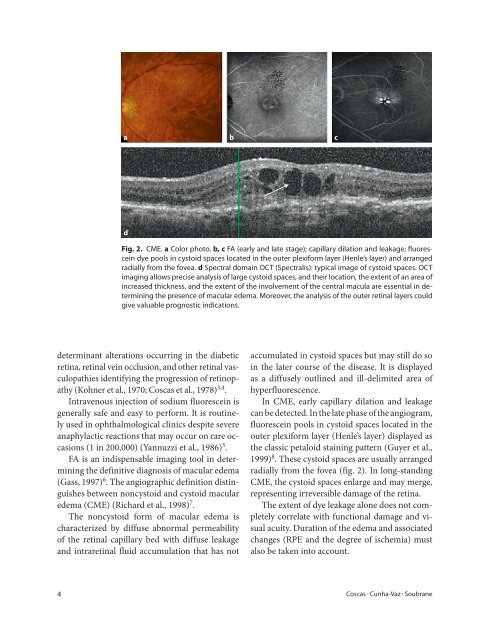
Fig. 2. CME. a Color photo. b, c FA (early <strong>and</strong> late stage); capillary dilation <strong>and</strong> leakage; fluorescein<br />
dye pools in cystoid spaces located in the outer plexiform layer (Henle’s layer) <strong>and</strong> arranged<br />
radially from the fovea. d Spectral domain OCT (Spectralis): typical image of cystoid spaces. OCT<br />
imaging allows precise analysis of large cystoid spaces, <strong>and</strong> their location, the extent of an area of<br />
increased thickness, <strong>and</strong> the extent of the involvement of the central macula are essential in determining<br />
the presence of macular edema. Moreover, the analysis of the outer retinal layers could<br />
give valuable prognostic indications.<br />
accumulated in cystoid spaces but may still do so<br />
in the later course of the disease. It is displayed<br />
as a diffusely outlined <strong>and</strong> ill-delimited area of<br />
hyperfluorescence.<br />
In CME, early capillary dilation <strong>and</strong> leakage<br />
can be detected. In the late phase of the angiogram,<br />
fluorescein pools in cystoid spaces located in the<br />
outer plexiform layer (Henle’s layer) displayed as<br />
the classic petaloid staining pattern (Guyer et al.,<br />
1999) 8 . These cystoid spaces are usually arranged<br />
radially from the fovea (fig. 2). In long-st<strong>and</strong>ing<br />
CME, the cystoid spaces enlarge <strong>and</strong> may merge,<br />
representing irreversible damage of the retina.<br />
The extent of dye leakage alone does not completely<br />
correlate with functional damage <strong>and</strong> visual<br />
acuity. Duration of the edema <strong>and</strong> associated<br />
changes (RPE <strong>and</strong> the degree of ischemia) must<br />
also be taken into account.<br />
4 Coscas � Cunha-Vaz � Soubrane