Macular Edema: Definition and Basic Concepts - Karger
Macular Edema: Definition and Basic Concepts - Karger
Macular Edema: Definition and Basic Concepts - Karger

You also want an ePaper? Increase the reach of your titles
YUMPU automatically turns print PDFs into web optimized ePapers that Google loves.
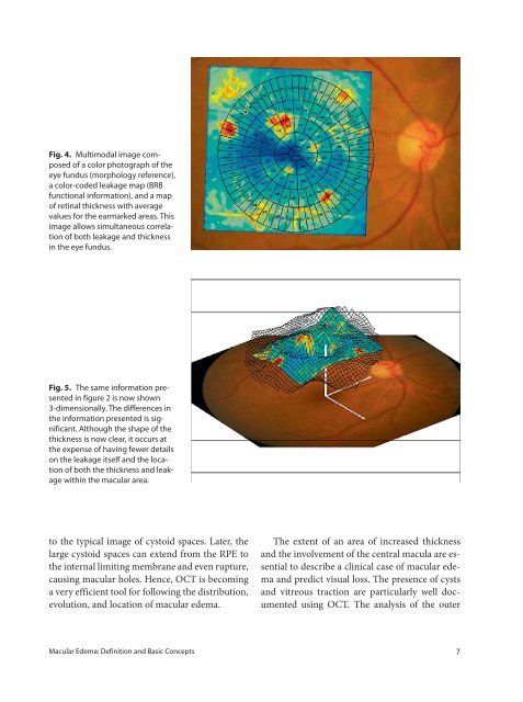
Fig. 4. Multimodal image composed<br />
of a color photograph of the<br />
eye fundus (morphology reference),<br />
a color-coded leakage map (BRB<br />
functional information), <strong>and</strong> a map<br />
of retinal thickness with average<br />
values for the earmarked areas. This<br />
image allows simultaneous correlation<br />
of both leakage <strong>and</strong> thickness<br />
in the eye fundus.<br />
Fig. 5. The same information presented<br />
in figure 2 is now shown<br />
3-dimensionally. The differences in<br />
the information presented is significant.<br />
Although the shape of the<br />
thickness is now clear, it occurs at<br />
the expense of having fewer details<br />
on the leakage itself <strong>and</strong> the location<br />
of both the thickness <strong>and</strong> leakage<br />
within the macular area.<br />
to the typical image of cystoid spaces. Later, the<br />
large cystoid spaces can extend from the RPE to<br />
the internal limiting membrane <strong>and</strong> even rupture,<br />
causing macular holes. Hence, OCT is becoming<br />
a very efficient tool for following the distribution,<br />
evolution, <strong>and</strong> location of macular edema.<br />
The extent of an area of increased thickness<br />
<strong>and</strong> the involvement of the central macula are essential<br />
to describe a clinical case of macular edema<br />
<strong>and</strong> predict visual loss. The presence of cysts<br />
<strong>and</strong> vitreous traction are particularly well documented<br />
using OCT. The analysis of the outer<br />
<strong>Macular</strong> <strong>Edema</strong>: <strong>Definition</strong> <strong>and</strong> <strong>Basic</strong> <strong>Concepts</strong> 7