Hydroquinone-induced exogenous ochronosis - instituto de ...
Hydroquinone-induced exogenous ochronosis - instituto de ...
Hydroquinone-induced exogenous ochronosis - instituto de ...

You also want an ePaper? Increase the reach of your titles
YUMPU automatically turns print PDFs into web optimized ePapers that Google loves.
Report<br />
Blackwell Oxford, IJD International 0011-9059 © XXX 2007 The UK Publishing International Journal Ltd of Dermatology Society of Dermatology<br />
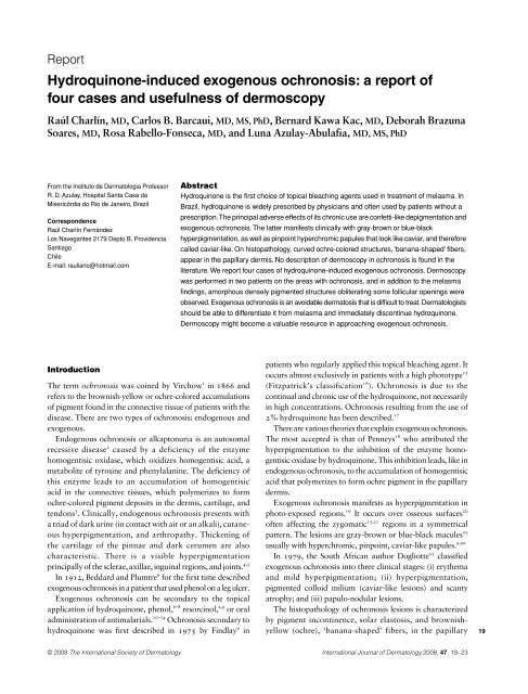
<strong>Hydroquinone</strong>-<strong>induced</strong> <strong>exogenous</strong> <strong>ochronosis</strong>: a report of<br />
four cases and usefulness of <strong>de</strong>rmoscopy<br />
Report<br />
Charlín Exogenous et al. <strong>ochronosis</strong> and <strong>de</strong>rmoscopy<br />
Raúl Charlín, MD,<br />
Carlos B. Barcaui, MD, MS, PhD,<br />
Bernard Kawa Kac, MD,<br />
Deborah Brazuna<br />
Soares, MD,<br />
Rosa Rabello-Fonseca, MD,<br />
and Luna Azulay-Abulafia, MD, MS, PhD<br />
From the Instituto <strong>de</strong> Dermatologia Professor<br />
R. D. Azulay, Hospital Santa Casa da<br />
Misericórdia do Rio <strong>de</strong> Janeiro, Brazil<br />
Correspon<strong>de</strong>nce<br />
Raúl Charlín Fernán<strong>de</strong>z<br />
Los Navegantes 2179 Depto B, Provi<strong>de</strong>ncia<br />
Santiago<br />
Chile<br />
E-mail: rauliano@hotmail.com<br />
Introduction<br />
1<br />
The term <strong>ochronosis</strong> was coined by Virchow in 1866 and<br />
refers to the brownish-yellow or ochre-colored accumulations<br />
of pigment found in the connective tissue of patients with the<br />
disease. There are two types of <strong>ochronosis</strong>: endogenous and<br />
<strong>exogenous</strong>.<br />
Endogenous <strong>ochronosis</strong> or alkaptonuria is an autosomal<br />
2<br />
recessive disease caused by a <strong>de</strong>ficiency of the enzyme<br />
homogentisic oxidase, which oxidizes homogentisic acid, a<br />
metabolite of tyrosine and phenylalanine. The <strong>de</strong>ficiency of<br />
this enzyme leads to an accumulation of homogentisic<br />
acid in the connective tissues, which polymerizes to form<br />
ochre-colored pigment <strong>de</strong>posits in the <strong>de</strong>rmis, cartilage, and<br />
3<br />
tendons . Clinically, endogenous <strong>ochronosis</strong> presents with<br />
a triad of dark urine (in contact with air or an alkali), cutaneous<br />
hyperpigmentation, and arthropathy. Thickening of<br />
the cartilage of the pinnae and dark cerumen are also<br />
characteristic. There is a visible hyperpigmentation<br />
4,5<br />
principally of the sclerae, axillae, inguinal regions, and joints.<br />
6<br />
In 1912, Beddard and Plumtre for the first time <strong>de</strong>scribed<br />
<strong>exogenous</strong> <strong>ochronosis</strong> in a patient that used phenol on a leg ulcer.<br />
Exogenous <strong>ochronosis</strong> can be secondary to the topical<br />
6–8<br />
2,9<br />
application of hydroquinone, phenol, resorcinol, or oral<br />
10–14<br />
administration of antimalarials. Ochronosis secondary to<br />
9<br />
hydroquinone was first <strong>de</strong>scribed in 1975 by Findlay in<br />
Abstract<br />
<strong>Hydroquinone</strong> is the first choice of topical bleaching agents used in treatment of melasma. In<br />
Brazil, hydroquinone is wi<strong>de</strong>ly prescribed by physicians and often used by patients without a<br />
prescription. The principal adverse effects of its chronic use are confetti-like <strong>de</strong>pigmentation and<br />
<strong>exogenous</strong> <strong>ochronosis</strong>. The latter manifests clinically with gray-brown or blue-black<br />
hyperpigmentation, as well as pinpoint hyperchromic papules that look like caviar, and therefore<br />
called caviar-like. On histopathology, curved ochre-colored structures, ‘banana-shaped’ fibers,<br />
appear in the papillary <strong>de</strong>rmis. No <strong>de</strong>scription of <strong>de</strong>rmoscopy in <strong>ochronosis</strong> is found in the<br />
literature. We report four cases of hydroquinone-<strong>induced</strong> <strong>exogenous</strong> <strong>ochronosis</strong>. Dermoscopy<br />
was performed in two patients on the areas with <strong>ochronosis</strong>, and in addition to the melasma<br />
findings, amorphous <strong>de</strong>nsely pigmented structures obliterating some follicular openings were<br />
observed. Exogenous <strong>ochronosis</strong> is an avoidable <strong>de</strong>rmatosis that is difficult to treat. Dermatologists<br />
should be able to differentiate it from melasma and immediately discontinue hydroquinone.<br />
Dermoscopy might become a valuable resource in approaching <strong>exogenous</strong> <strong>ochronosis</strong>.<br />
patients who regularly applied this topical bleaching agent. It<br />
15<br />
occurs almost exclusively in patients with a high phototype<br />
16<br />
(Fitzpatrick’s classification ). Ochronosis is due to the<br />
continual and chronic use of the hydroquinone, not necessarily<br />
in high concentrations. Ochronosis resulting from the use of<br />
17<br />
2% hydroquinone has been <strong>de</strong>scribed.<br />
There are various theories that explain <strong>exogenous</strong> <strong>ochronosis</strong>.<br />
18<br />
The most accepted is that of Penneys who attributed the<br />
hyperpigmentation to the inhibition of the enzyme homogentisic<br />
oxidase by hydroquinone. This inhibition leads, like in<br />
endogenous <strong>ochronosis</strong>, to the accumulation of homogentisic<br />
acid that polymerizes to form ochre pigment in the papillary<br />
<strong>de</strong>rmis.<br />
Exogenous <strong>ochronosis</strong> manifests as hyperpigmentation in<br />
19<br />
20<br />
photo-exposed regions. It occurs over osseous surfaces<br />
15,21<br />
often affecting the zygomatic regions in a symmetrical<br />
22<br />
pattern. The lesions are gray-brown or blue-black macules<br />
9,20<br />
usually with hyperchromic, pinpoint, caviar-like papules.<br />
23<br />
In 1979, the South African author Dogliotte classified<br />
<strong>exogenous</strong> <strong>ochronosis</strong> into three clinical stages: (i) erythema<br />
and mild hyperpigmentation; (ii) hyperpigmentation,<br />
pigmented colloid milium (caviar-like lesions) and scanty<br />
atrophy; and (iii) papulo-nodular lesions.<br />
The histopathology of <strong>ochronosis</strong> lesions is characterized<br />
by pigment incontinence, solar elastosis, and brownishyellow<br />
(ochre), ‘banana-shaped’ fibers, in the papillary<br />
© 2008 The International Society of Dermatology International Journal of Dermatology 2008, 47,<br />
19–23<br />
19
20 Report<br />
Exogenous <strong>ochronosis</strong> and <strong>de</strong>rmoscopy<br />
2,9,22,23<br />
<strong>de</strong>rmis, and eventually <strong>de</strong>generation of the collagen.<br />
9,22<br />
1<br />
Occasionally, colloid milium and/or granulomas are <strong>de</strong>tected.<br />
It is not difficult to differentiate <strong>ochronosis</strong> and melasma<br />
on histopathologic basis. The latter show a significant<br />
increase in the amount of melanin in all epi<strong>de</strong>rmal layers seen<br />
in Fontana-Masson. It is controversial whether melanocytes<br />
24<br />
are increase in number in melasma. Sanchez et al.<br />
and<br />
25<br />
Kang et al.<br />
reported an increase in the number of melano-<br />
26<br />
cytes in melasma. Recently, Grimes et al.<br />
were not able to<br />
confirm the quantitative increase in the number of epi<strong>de</strong>rmal<br />
melanocytes. Pigment incontinence and melanophages<br />
27<br />
might be present either in melasma and <strong>ochronosis</strong>. Solar<br />
25<br />
elastosis is seen both in melasma and in <strong>ochronosis</strong>. In fact,<br />
<strong>ochronosis</strong> is usually superimposed on the skin affected by<br />
melasma. There are no ochre fibers in melasma, the characteristic<br />
finding in <strong>ochronosis</strong>.<br />
Various treatments have been used for <strong>exogenous</strong> ochro-<br />
28<br />
nosis, usually with frustrating results. Satisfactory results<br />
29,30<br />
31,32<br />
have been <strong>de</strong>scribed with retinoic acid, <strong>de</strong>rmabrasion,<br />
21<br />
15<br />
33,34<br />
cryotherapy, CO2<br />
laser, Q-switched ruby laser, Q-<br />
22<br />
switched alexandrine 755 laser, among others.<br />
We report four patients with <strong>exogenous</strong> <strong>ochronosis</strong>, all<br />
classified as Dogliotte stage 2. All had used hydroquinone<br />
over an exten<strong>de</strong>d period of time for melasma treatment, in<br />
concentrations up to 6%.<br />
Case Reports<br />
Case 1<br />
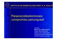
A 56-year-old woman (a teacher), phototype IV, presented<br />
with a history of long-term facial melasma, treated with 2–<br />
4% hydroquinone creams for 20 years. She complained of<br />
progressive worsening of the hyperpigmentation in the<br />
zygomatic region in the last years. No other oral or topical<br />
medications were used by the patient. On <strong>de</strong>rmatologic<br />
examination, she had hyperchromic, gray-brown macules,<br />
speckled with pinpoint, dark brown (caviar-like) papules in<br />
the zygomatic and infraorbital regions (Fig. 1). A brown<br />
macule on the dorsal of the nose was also observed. It was<br />
classified as stage 2 Dogliotte.<br />
Case 2<br />
A 44-year-old woman (a housewife), phototype IV, presented<br />
with a history of facial melasma over 10 years, starting with<br />
her last pregnancy. She was treated with 2–5% hydroquinone<br />
during this time, with initial improvement, but now presented<br />
with progressive darkening. She reported the occasional use<br />
of metamizole (dipyrone). On physical examination, she<br />
had bilateral blue-brown macules on the malar, submalar,<br />
and temporal regions, and blue-black pinpoint papules<br />
(caviar-like lesions) dispersed over the macules. She also had<br />
light brown macules on the frontal region, upper lip, and<br />
chin. It was classified as stage 2 Dogliotte.<br />
Charlín et al.<br />
Figure 1 Gray-brown macules in the zygomatic region, spotted<br />
with pinpoint, dark brown (caviar-like) papules<br />
Case 3<br />
A 56-year-old woman (a teacher), phototype V, with 25 years<br />
of melasma, treated with up to 6% hydroquinone, sought<br />
medical care for worsening of the hyperpigmentation. She<br />
<strong>de</strong>nied use of any other medication. On physical examination,<br />
she had a gray-brown pigmentation on the entire face,<br />
except for the upper lip and frontal regions. In addition, there<br />
were papules of the same color, or slightly more hyperchromic,<br />
of 2–3 mm in diameter, insi<strong>de</strong> the bor<strong>de</strong>rs of the hyperpigmentation.<br />
Confetti-like <strong>de</strong>pigmentation was observed on<br />
the cheeks. It was classified as stage 2 Dogliotte.<br />
Case 4<br />
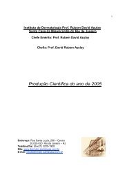
A 51-year-old woman (an ambulatory vendor), phototype V,<br />
with 10 years of up to 5% hydroquinone use, presented for<br />
treatment of melasma. She observed worsening of the hyperpigmentation<br />
in recent years. She used no other medications.<br />
On <strong>de</strong>rmatologic examination, bilateral brown macules, with<br />
confetti-like <strong>de</strong>pigmentation within the bor<strong>de</strong>rs of the lesion,<br />
were observed on the cheeks. She also presented with darker,<br />
gray-brown areas, with caviar-like pinpoint papules in the<br />
zygomatic region (Fig. 2). It was classified as stage 2 Dogliotte.<br />
No patient complained of arthralgia nor presented with<br />
altered urine color, hyperpigmentation of the sclerae, joints,<br />
axillae, or genitals. There was also no thickening of the pinnae<br />
or dark cerumen.<br />
Cutaneous biopsies from all patients were taken from the<br />
hyperchromic areas suspected of <strong>ochronosis</strong>. Besi<strong>de</strong>s the<br />
macular lesion, the pinpoint papules of the first, second, and<br />
International Journal of Dermatology 2008, 47,<br />
19–23 © 2008 The International Society of Dermatology
Charlín et al. Exogenous <strong>ochronosis</strong> and <strong>de</strong>rmoscopy<br />
Figure 2 Brown macules, with confetti-like <strong>de</strong>pigmentation on<br />
the cheek. Also with darker gray-brown areas, with caviar-like<br />
pinpoint papules in the zygomatic region<br />
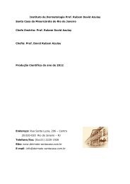
Figure 3 Histopathology of <strong>ochronosis</strong> lesion, showing normal<br />
epi<strong>de</strong>rmis, mild pigment incontinence, solar elastosis and<br />
brownish-yellow (ochre) “banana shaped” fibers in the<br />
papillary <strong>de</strong>rmis. H&E stain ×40<br />
fourth patients were also biopsied. On histopathologic exam,<br />
normal epi<strong>de</strong>rmis, pigment incontinence in the papillary<br />
<strong>de</strong>rmis, solar elastosis, and brownish-yellow (ochre) ‘bananashaped’<br />
fibers were found (Fig. 3). There was no variation in<br />
the findings, in<strong>de</strong>pen<strong>de</strong>nt of the type of lesion biopsied<br />
(macule or pinpoint papule).<br />
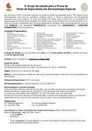
Figure 4 Dermoscopy of melasma lesion (×10). Showing<br />
accentuation of the normal pseudo-rete of the facial skin<br />
© 2008 The International Society of Dermatology International Journal of Dermatology 2008, 47,<br />
19–23<br />
Report<br />
Figure 5 Dermoscopy (×10). On the left si<strong>de</strong> of the picture<br />
(melasma), with accentuation of the normal pseudo-rete of facial<br />
skin. On the right si<strong>de</strong> (<strong>ochronosis</strong>), presenting blue-gray<br />
amorphous areas with obliteration of some follicular openings<br />
Patients one and four were subjected to <strong>de</strong>rmoscopy. There<br />
was no characteristic pigmentation on the normal skin. In the<br />
areas with melasma without <strong>ochronosis</strong>, an accentuation of<br />
the normal pseudo-rete of the facial skin was observed (Figs 4<br />
and 5). In the areas with <strong>ochronosis</strong>, besi<strong>de</strong>s the previous<br />
observation, blue-gray amorphous areas obliterating some<br />
follicular openings were observed (Figs 5 and 6).<br />
Instituted treatments varied. The first patient was treated<br />
with retinoic acid gel 0.05%, azelaic acid cream 20%, and<br />
kojic acid cream (Melani D®, La Roche-Posay), with partial<br />
improvement of the <strong>ochronosis</strong>. The second patient abandoned<br />
the medical follow-up and the third was subjected to<br />
21
22 Report<br />
Exogenous <strong>ochronosis</strong> and <strong>de</strong>rmoscopy<br />
Figure 6 Dermoscopy of <strong>ochronosis</strong> lesion (×10). Densely bluegray<br />
structures obliterating some follicular openings<br />
a test with the Nd-Yag laser Q-switched without improvement.<br />
Retinoic acid 0.05% and 20% azelaic acid were prescribed<br />
for the fourth patient. None of the therapeutic modalities<br />
achieved satisfactory results.<br />
Discussion<br />
The four patients have high phototypes – IV and V – similar<br />
to the cases of <strong>exogenous</strong> <strong>ochronosis</strong> <strong>de</strong>scribed in the literature.<br />
Melasma, the condition for which hydroquinone is used,<br />
is also more common in high phototypes. This might explain<br />
why <strong>exogenous</strong> <strong>ochronosis</strong> is found mostly in these patients.<br />
With respect to the concentrations of hydroquinone<br />
applied, the first, second, and fourth patients used relatively<br />
low concentrations, but for a long time. The third used higher<br />
concentrations in recent years, noticing intensified darkening<br />
of the skin after increasing the concentration of the medicine.<br />
Thus, we confirm that <strong>exogenous</strong> <strong>ochronosis</strong> can occur after<br />
use of different concentrations of hydroquinone, prolonged<br />
used being the principle factor.<br />
The four patients did not use other drugs related to<br />
<strong>exogenous</strong> <strong>ochronosis</strong> and no one presented with signs of<br />
endogenous <strong>ochronosis</strong>, i.e., arthralgia, altered urine color,<br />
hyperpigmentation of the sclerae, axillae, genitals, or skin<br />
over the joints. Neither thickening of the pinnae nor dark<br />
cerumen was observed. For the diagnosis of <strong>exogenous</strong><br />
<strong>ochronosis</strong>, physicians should, at least clinically, exclu<strong>de</strong> the<br />
possibility of endogenous <strong>ochronosis</strong>.<br />
The color of the patients’ lesions corresponds to that<br />
<strong>de</strong>scribed in the literature. In cases 1, 3, and 4, the macules<br />
were gray-brown and in case 2, predominantly blue-black.<br />
Cases 1, 2, and 4 presented with caviar-like, hyperchromic,<br />
pinpoint papules in the zygomatic regions. The third patient<br />
Charlín et al.<br />
presented with hyperpigmentation of almost the entire face,<br />
with 2–3 mm hyperchromic papules and small round<br />
<strong>de</strong>pigmented macules (confetti-like <strong>de</strong>pigmentation). Patients<br />
3 and 4 presented with two adverse effects of hydroquinone:<br />
<strong>exogenous</strong> <strong>ochronosis</strong> and confetti-like <strong>de</strong>pigmentation.<br />
All four cases correspon<strong>de</strong>d with the stage 2 classification<br />
for <strong>exogenous</strong> <strong>ochronosis</strong>, established by Dogliotte.<br />
The histopathologic exam of the four cases revealed solar<br />
elastosis and brownish-yellow (ochre), ‘banana-shaped’<br />
fibers in the papillary <strong>de</strong>rmis, characteristic of <strong>exogenous</strong><br />
<strong>ochronosis</strong>. The four patients did not <strong>de</strong>monstrate colloid<br />
milium or granulomas. There was not a significant difference<br />
among the histopathologic exams of the four patients, even<br />
though the third patient presented with a more intense clinical<br />
9<br />
35<br />
picture. Differing from Findlay’s and Jacyk <strong>de</strong>scriptions,<br />
we did not find a correlation between the caviar-like pinpoint<br />
papules of patients 1, 2, and 4, and the histopathologic<br />
presence of colloid milium.<br />
Dermoscopy of patients 1 and 4 showed obvious differences<br />
between normal skin, that with melasma, and that with<br />
<strong>ochronosis</strong>. In the literature there is little information about<br />
36<br />
the <strong>de</strong>rmoscopy of melasma. Stolz <strong>de</strong>scribed blue-gray,<br />
granular, annular structures around the follicles. However,<br />
<strong>de</strong>rmoscopy of melasma involved skin of patients 1 and 4,<br />
revealed just accentuation of the normal pseudo-rete of the<br />
face. We did not find reports in the literature of <strong>de</strong>rmoscopic<br />
examination of <strong>ochronosis</strong>. In the skin with <strong>ochronosis</strong> of<br />
patients 1 and 4, we observed bluish-gray amorphous areas<br />
obliterating the follicular structures rather than surrounding<br />
them, as Stolz <strong>de</strong>scribed for melasma. Of interest, the color of<br />
the ochronotic pigment observed clinically and by <strong>de</strong>rmoscopy<br />
was blue-gray, not ochre as in the histopathology. The<br />
blue color is due to the <strong>de</strong>pth at which the pigment is located<br />
37<br />
(Tyndall effect ).<br />
Concerning the therapy, only the first patient had a partial<br />
response, probably from the effect of retinoic acid on the<br />
<strong>ochronosis</strong> and the azelaic and kojic acid on the un<strong>de</strong>rlying<br />
melasma.<br />
Conclusion<br />
Dermatologists should be prepared to differentiate melasma<br />
from <strong>exogenous</strong> <strong>ochronosis</strong> <strong>induced</strong> by topical use of hydroquinone,<br />
employed to treat the first condition. An early<br />
diagnosis necessitates immediate discontinuation of hydroquinone,<br />
rather than increasing the concentration in attempt<br />
to clear the <strong>de</strong>rmatosis.<br />
The prescription of hydroquinone for any patient should<br />
be accompanied by orientation to the possible si<strong>de</strong>-effects,<br />
including information that the drug should be used for a<br />
limited period.<br />
There was no reference to the use of <strong>de</strong>rmoscopy on<br />
<strong>exogenous</strong> <strong>ochronosis</strong> in the researched literature. The<br />
International Journal of Dermatology 2008, 47,<br />
19–23 © 2008 The International Society of Dermatology
Charlín et al. Exogenous <strong>ochronosis</strong> and <strong>de</strong>rmoscopy<br />
<strong>de</strong>rmoscopic exam of the first and fourth patients revealed<br />
differences between the healthy skin, that affected by<br />
melasma and that of <strong>ochronosis</strong>. Dermoscopy might become<br />
a valuable complementary exam when approaching patients<br />
with <strong>exogenous</strong> <strong>ochronosis</strong>.<br />
References<br />
1 Virchow R. Ein Fall von allegemeiner ochronose <strong>de</strong>r knorpel<br />
aud knorpelahnlichen theile. Virchows Arch (Pathol Anat)<br />
1866; 37.<br />
2 Fisher AA. Exogenous <strong>ochronosis</strong> from hydroquinone<br />
bleaching cream. Cutis 1998; 62:<br />
11–12.<br />
3 La Du BN. Alkaptonuria. In: Scriver CR, Beau<strong>de</strong>t A,<br />
Sly W, et al.<br />
eds. The Metabolic and Molecular Bases of<br />
Inherited Disease,<br />
7th edn. New York: McGraw-Hill,<br />
2001, p. 1371.<br />
4 Van Offel JF, DeClerck LS, Francx LM, et al.<br />
The clinical<br />
manifestations of <strong>ochronosis</strong>: a review. Acta Clin Belg 1995;<br />
50:<br />
358–362.<br />
5 Goldsmith LA. Cutaneous changes in errors of amino acid<br />
metabolism: alkaptonuria. In: Fitzpatrick, TB, Eisen, AZ,<br />
Wolff, K, et al.<br />
eds. Dermatology in General Medicine,<br />
Chapter 48. New York: McGraw-Hill, 1993, pp. 1841–<br />
1845.<br />
6 Beddard AP, Plumtre CM. A further note on <strong>ochronosis</strong><br />
associated with carboluria. Q S Med 1912; 5:<br />
505–507.<br />
7 Berry JL, Peat S. Ochronosis: report of a case with<br />
carboluria. Lancet 1931; 2:<br />
124–126.<br />
8 Brogren N. Case of exogenetic <strong>ochronosis</strong> from carbolic acid<br />
compresses. Acta Derm Venereol (Stockholm) 1952; 32:<br />
258–260.<br />
9 Findlay GH, Morrison JG, Simson IW. Exogenous<br />
<strong>ochronosis</strong> and pigmented colloid milium from<br />
hydroquinone bleaching creams. Br J Dermatol 1975; 93:<br />
613–622.<br />
10 Sugar HS, Wad<strong>de</strong>ll WW. Ochronosis-like pigmentation<br />
associated with the use of atabrine. Ill Med J 1946; 89:<br />
234–<br />
239.<br />
11 Ludwig GD, Toole JF, Wood JC. Ochronosis from<br />
quinacrine (atabrine). Ann Intern Med 1963;<br />
59:<br />
378–384.<br />
12 Tuffanelli D, Abraham RK, Dubois EI. Pigmentation from<br />
antimalarial therapy. Its possible relationship to the ocular<br />
lesions. Arch Dermatol 1963; 88:<br />
419–426.<br />
13 Egorin MJ, Trump DL, Wainwright CW. Quinacrine<br />
<strong>ochronosis</strong> and rheumatoid arthritis. JAMA 1976; 236:<br />
385–386.<br />
14 Cullison D, Abele DC, O’Quinn JL. Localized<br />
<strong>exogenous</strong> <strong>ochronosis</strong>. J Am Acad Dermatol 1983<br />
June; 8:<br />
882–889.<br />
15 Diven DG, Smith EB, Pupo RA, et al.<br />
<strong>Hydroquinone</strong><strong>induced</strong><br />
localized <strong>exogenous</strong> <strong>ochronosis</strong> treated with<br />
<strong>de</strong>rmabrasion and CO2<br />
laser. J Dermatol Surg Oncol 1990;<br />
16:<br />
1018–1022.<br />
16 Fitzpatrick TB. Soleil et peau. J Med Esthet 1975; 2:<br />
33.<br />
© 2008 The International Society of Dermatology International Journal of Dermatology 2008, 47,<br />
19–23<br />
Report<br />
17 Hoshaw RA, Zimmerman KG, Menter A. Ochronosis-like<br />
pigmentation from hydroquinone bleaching creams in<br />
Americans blacks. Arch Dermatol 1985; 121:<br />
105–108.<br />
18 Penneys NS. Ochronosis-like pigmentation from bleaching<br />
creams. Arch Dermatol 1985; 121:<br />
1239–1240.<br />
19 O’Donoghue MN, Lynfield YL, Derbes V. Ochronosis due<br />
to hydroquinone. J Am Acad Dermatol 1983; 8:<br />
123.<br />
20 Jordaan HF, Van Niekerk DJ. Transepi<strong>de</strong>rmal elimination<br />
in <strong>exogenous</strong> <strong>ochronosis</strong>. A report of two cases. Am J<br />
Dermatopathol;<br />
13:<br />
418–424.<br />
21 Kramer KE, Lopez A, Stefanato CM, et al.<br />
Exogenous<br />
<strong>ochronosis</strong>. J Am Acad Dermatol 2000; 42 (5 Part 2): 869–<br />
871.<br />
22 Bellew SG, Alster TS. Treatment of <strong>exogenous</strong> <strong>ochronosis</strong><br />
with a Q-switched alexandrite (755 nm) laser. Dermatol<br />
Surg 2004; 30 (4 Part 1): 555–558.<br />
23 Dogliotte M, Leibowitz M. Granulomatous <strong>ochronosis</strong> – a<br />
cosmetic- <strong>induced</strong> skin disor<strong>de</strong>r in blacks. S Afr Med J 1979;<br />
56: 757–760.<br />
24 Sanchez NP, Pathak MA, Sato S, et al. Melasma: a clinical,<br />
light microscopic, ultrastructural, and immunofluorescence<br />
study. J Am Acad Dermatol 1981; 4: 698–710.<br />
25 Kang WH, Yoon KH, Lee ES, et al. Melasma:<br />
histopathological characteristics in 56 Korean patients. Br J<br />
Dermatol; 2002; 146: 228–237.<br />
26 Grimes PE, Yamada N, Bhawan J. Light microscopic,<br />
immunohistochemical, and ultrastructural alterations in<br />
patients with melasma. Am J Dermatopathol 2005; 27: 96–<br />
101.<br />
27 Victor FC, Gelber J, Rao B. Melasma: a review. J Cutan Med<br />
Surg 2004; 8: 97–102.<br />
28 Connor T, Braunstein B. Hyperpigmentation following the<br />
use of bleaching creams. Localized <strong>exogenous</strong> <strong>ochronosis</strong>.<br />
Arch Dermatol 1987; 123: 105–6, 108.<br />
29 Howard K, Furner B. Exogenous <strong>ochronosis</strong> in a Mexican-<br />
American woman. Cutis 1990; 45: 180–182.<br />
30 Schutz E, Summers B, Summers R. Inappropriate treatment<br />
of cosmetic <strong>ochronosis</strong> with hydroquinone. S Afr Med J<br />
1988; 73: 59–60.<br />
31 Sni<strong>de</strong>r RL, Thiers BH. Exogenous <strong>ochronosis</strong>. J Am Acad<br />
Dermatol 1993; 28: 662–664.<br />
32 Lang PG. Probable coexisting <strong>exogenous</strong> <strong>ochronosis</strong> and<br />
mercurial pigmentation managed by <strong>de</strong>rmabrasion. J Am<br />
Acad Dermatol 1988; 19: 942–946.<br />
33 Taylor CR, Gange RW, Dover JS et al. Treatment of tattoos<br />
by Q-switched ruby laser: a dose–response study. Arch<br />
Dermatol 1990; 126: 893–899.<br />
34 Hruza GJ, Dover JS, Flotte TJ et al. Q-switched ruby laser<br />
irradiation of normal human skin. Arch Dermatol 1991;<br />
127: 1799–1805.<br />
35 Jacyk WK. Annular granulomatous lesions in <strong>exogenous</strong><br />
<strong>ochronosis</strong> are manifestation of sarcoidosis. Am J<br />
Dermatopathol 1995; 17: 18–22.<br />
36 Stolz W. Color Atlas of Dermoscopy, 2nd edn. Berlin:<br />
Blackwell Wissenschafts-Verlag G 2002, pp. 121–131.<br />
37 Jeghers H. Pigmentation of the skin. N Engl J Med 1944;<br />
231: 88–100.<br />
23