Hydroquinone-induced exogenous ochronosis - instituto de ...
Hydroquinone-induced exogenous ochronosis - instituto de ...
Hydroquinone-induced exogenous ochronosis - instituto de ...

Create successful ePaper yourself
Turn your PDF publications into a flip-book with our unique Google optimized e-Paper software.
22 Report<br />
Exogenous <strong>ochronosis</strong> and <strong>de</strong>rmoscopy<br />
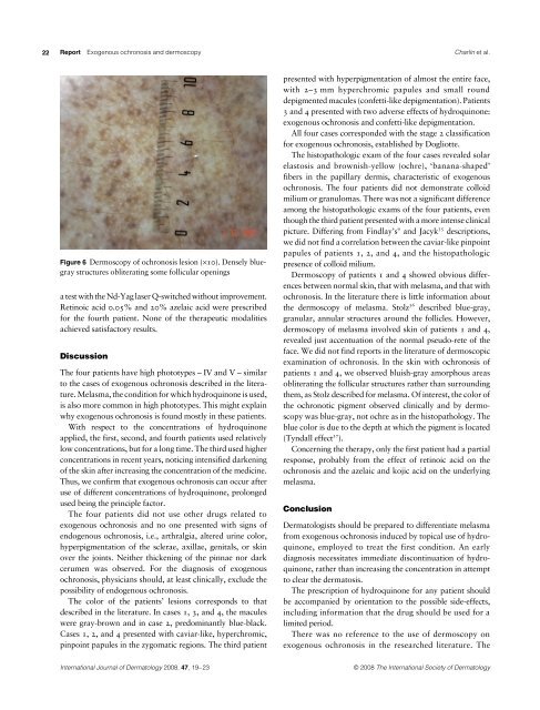
Figure 6 Dermoscopy of <strong>ochronosis</strong> lesion (×10). Densely bluegray<br />
structures obliterating some follicular openings<br />
a test with the Nd-Yag laser Q-switched without improvement.<br />
Retinoic acid 0.05% and 20% azelaic acid were prescribed<br />
for the fourth patient. None of the therapeutic modalities<br />
achieved satisfactory results.<br />
Discussion<br />
The four patients have high phototypes – IV and V – similar<br />
to the cases of <strong>exogenous</strong> <strong>ochronosis</strong> <strong>de</strong>scribed in the literature.<br />
Melasma, the condition for which hydroquinone is used,<br />
is also more common in high phototypes. This might explain<br />
why <strong>exogenous</strong> <strong>ochronosis</strong> is found mostly in these patients.<br />
With respect to the concentrations of hydroquinone<br />
applied, the first, second, and fourth patients used relatively<br />
low concentrations, but for a long time. The third used higher<br />
concentrations in recent years, noticing intensified darkening<br />
of the skin after increasing the concentration of the medicine.<br />
Thus, we confirm that <strong>exogenous</strong> <strong>ochronosis</strong> can occur after<br />
use of different concentrations of hydroquinone, prolonged<br />
used being the principle factor.<br />
The four patients did not use other drugs related to<br />
<strong>exogenous</strong> <strong>ochronosis</strong> and no one presented with signs of<br />
endogenous <strong>ochronosis</strong>, i.e., arthralgia, altered urine color,<br />
hyperpigmentation of the sclerae, axillae, genitals, or skin<br />
over the joints. Neither thickening of the pinnae nor dark<br />
cerumen was observed. For the diagnosis of <strong>exogenous</strong><br />
<strong>ochronosis</strong>, physicians should, at least clinically, exclu<strong>de</strong> the<br />
possibility of endogenous <strong>ochronosis</strong>.<br />
The color of the patients’ lesions corresponds to that<br />
<strong>de</strong>scribed in the literature. In cases 1, 3, and 4, the macules<br />
were gray-brown and in case 2, predominantly blue-black.<br />
Cases 1, 2, and 4 presented with caviar-like, hyperchromic,<br />
pinpoint papules in the zygomatic regions. The third patient<br />
Charlín et al.<br />
presented with hyperpigmentation of almost the entire face,<br />
with 2–3 mm hyperchromic papules and small round<br />
<strong>de</strong>pigmented macules (confetti-like <strong>de</strong>pigmentation). Patients<br />
3 and 4 presented with two adverse effects of hydroquinone:<br />
<strong>exogenous</strong> <strong>ochronosis</strong> and confetti-like <strong>de</strong>pigmentation.<br />
All four cases correspon<strong>de</strong>d with the stage 2 classification<br />
for <strong>exogenous</strong> <strong>ochronosis</strong>, established by Dogliotte.<br />
The histopathologic exam of the four cases revealed solar<br />
elastosis and brownish-yellow (ochre), ‘banana-shaped’<br />
fibers in the papillary <strong>de</strong>rmis, characteristic of <strong>exogenous</strong><br />
<strong>ochronosis</strong>. The four patients did not <strong>de</strong>monstrate colloid<br />
milium or granulomas. There was not a significant difference<br />
among the histopathologic exams of the four patients, even<br />
though the third patient presented with a more intense clinical<br />
9<br />
35<br />
picture. Differing from Findlay’s and Jacyk <strong>de</strong>scriptions,<br />
we did not find a correlation between the caviar-like pinpoint<br />
papules of patients 1, 2, and 4, and the histopathologic<br />
presence of colloid milium.<br />
Dermoscopy of patients 1 and 4 showed obvious differences<br />
between normal skin, that with melasma, and that with<br />
<strong>ochronosis</strong>. In the literature there is little information about<br />
36<br />
the <strong>de</strong>rmoscopy of melasma. Stolz <strong>de</strong>scribed blue-gray,<br />
granular, annular structures around the follicles. However,<br />
<strong>de</strong>rmoscopy of melasma involved skin of patients 1 and 4,<br />
revealed just accentuation of the normal pseudo-rete of the<br />
face. We did not find reports in the literature of <strong>de</strong>rmoscopic<br />
examination of <strong>ochronosis</strong>. In the skin with <strong>ochronosis</strong> of<br />
patients 1 and 4, we observed bluish-gray amorphous areas<br />
obliterating the follicular structures rather than surrounding<br />
them, as Stolz <strong>de</strong>scribed for melasma. Of interest, the color of<br />
the ochronotic pigment observed clinically and by <strong>de</strong>rmoscopy<br />
was blue-gray, not ochre as in the histopathology. The<br />
blue color is due to the <strong>de</strong>pth at which the pigment is located<br />
37<br />
(Tyndall effect ).<br />
Concerning the therapy, only the first patient had a partial<br />
response, probably from the effect of retinoic acid on the<br />
<strong>ochronosis</strong> and the azelaic and kojic acid on the un<strong>de</strong>rlying<br />
melasma.<br />
Conclusion<br />
Dermatologists should be prepared to differentiate melasma<br />
from <strong>exogenous</strong> <strong>ochronosis</strong> <strong>induced</strong> by topical use of hydroquinone,<br />
employed to treat the first condition. An early<br />
diagnosis necessitates immediate discontinuation of hydroquinone,<br />
rather than increasing the concentration in attempt<br />
to clear the <strong>de</strong>rmatosis.<br />
The prescription of hydroquinone for any patient should<br />
be accompanied by orientation to the possible si<strong>de</strong>-effects,<br />
including information that the drug should be used for a<br />
limited period.<br />
There was no reference to the use of <strong>de</strong>rmoscopy on<br />
<strong>exogenous</strong> <strong>ochronosis</strong> in the researched literature. The<br />
International Journal of Dermatology 2008, 47,<br />
19–23 © 2008 The International Society of Dermatology