Hydroquinone-induced exogenous ochronosis - instituto de ...
Hydroquinone-induced exogenous ochronosis - instituto de ...
Hydroquinone-induced exogenous ochronosis - instituto de ...

You also want an ePaper? Increase the reach of your titles
YUMPU automatically turns print PDFs into web optimized ePapers that Google loves.
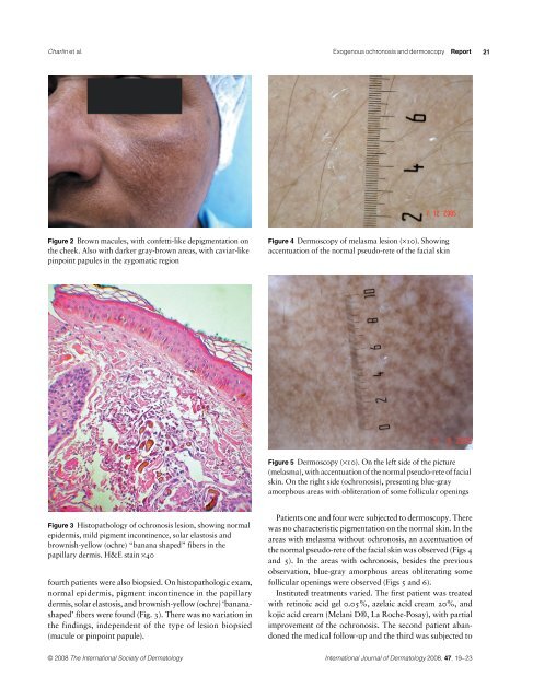
Charlín et al. Exogenous <strong>ochronosis</strong> and <strong>de</strong>rmoscopy<br />
Figure 2 Brown macules, with confetti-like <strong>de</strong>pigmentation on<br />
the cheek. Also with darker gray-brown areas, with caviar-like<br />
pinpoint papules in the zygomatic region<br />
Figure 3 Histopathology of <strong>ochronosis</strong> lesion, showing normal<br />
epi<strong>de</strong>rmis, mild pigment incontinence, solar elastosis and<br />
brownish-yellow (ochre) “banana shaped” fibers in the<br />
papillary <strong>de</strong>rmis. H&E stain ×40<br />
fourth patients were also biopsied. On histopathologic exam,<br />
normal epi<strong>de</strong>rmis, pigment incontinence in the papillary<br />
<strong>de</strong>rmis, solar elastosis, and brownish-yellow (ochre) ‘bananashaped’<br />
fibers were found (Fig. 3). There was no variation in<br />
the findings, in<strong>de</strong>pen<strong>de</strong>nt of the type of lesion biopsied<br />
(macule or pinpoint papule).<br />
Figure 4 Dermoscopy of melasma lesion (×10). Showing<br />
accentuation of the normal pseudo-rete of the facial skin<br />
© 2008 The International Society of Dermatology International Journal of Dermatology 2008, 47,<br />
19–23<br />
Report<br />
Figure 5 Dermoscopy (×10). On the left si<strong>de</strong> of the picture<br />
(melasma), with accentuation of the normal pseudo-rete of facial<br />
skin. On the right si<strong>de</strong> (<strong>ochronosis</strong>), presenting blue-gray<br />
amorphous areas with obliteration of some follicular openings<br />
Patients one and four were subjected to <strong>de</strong>rmoscopy. There<br />
was no characteristic pigmentation on the normal skin. In the<br />
areas with melasma without <strong>ochronosis</strong>, an accentuation of<br />
the normal pseudo-rete of the facial skin was observed (Figs 4<br />
and 5). In the areas with <strong>ochronosis</strong>, besi<strong>de</strong>s the previous<br />
observation, blue-gray amorphous areas obliterating some<br />
follicular openings were observed (Figs 5 and 6).<br />
Instituted treatments varied. The first patient was treated<br />
with retinoic acid gel 0.05%, azelaic acid cream 20%, and<br />
kojic acid cream (Melani D®, La Roche-Posay), with partial<br />
improvement of the <strong>ochronosis</strong>. The second patient abandoned<br />
the medical follow-up and the third was subjected to<br />
21