Hydroquinone-induced exogenous ochronosis - instituto de ...
Hydroquinone-induced exogenous ochronosis - instituto de ...
Hydroquinone-induced exogenous ochronosis - instituto de ...

You also want an ePaper? Increase the reach of your titles
YUMPU automatically turns print PDFs into web optimized ePapers that Google loves.
20 Report<br />
Exogenous <strong>ochronosis</strong> and <strong>de</strong>rmoscopy<br />
2,9,22,23<br />
<strong>de</strong>rmis, and eventually <strong>de</strong>generation of the collagen.<br />
9,22<br />
1<br />
Occasionally, colloid milium and/or granulomas are <strong>de</strong>tected.<br />
It is not difficult to differentiate <strong>ochronosis</strong> and melasma<br />
on histopathologic basis. The latter show a significant<br />
increase in the amount of melanin in all epi<strong>de</strong>rmal layers seen<br />
in Fontana-Masson. It is controversial whether melanocytes<br />
24<br />
are increase in number in melasma. Sanchez et al.<br />
and<br />
25<br />
Kang et al.<br />
reported an increase in the number of melano-<br />
26<br />
cytes in melasma. Recently, Grimes et al.<br />
were not able to<br />
confirm the quantitative increase in the number of epi<strong>de</strong>rmal<br />
melanocytes. Pigment incontinence and melanophages<br />
27<br />
might be present either in melasma and <strong>ochronosis</strong>. Solar<br />
25<br />
elastosis is seen both in melasma and in <strong>ochronosis</strong>. In fact,<br />
<strong>ochronosis</strong> is usually superimposed on the skin affected by<br />
melasma. There are no ochre fibers in melasma, the characteristic<br />
finding in <strong>ochronosis</strong>.<br />
Various treatments have been used for <strong>exogenous</strong> ochro-<br />
28<br />
nosis, usually with frustrating results. Satisfactory results<br />
29,30<br />
31,32<br />
have been <strong>de</strong>scribed with retinoic acid, <strong>de</strong>rmabrasion,<br />
21<br />
15<br />
33,34<br />
cryotherapy, CO2<br />
laser, Q-switched ruby laser, Q-<br />
22<br />
switched alexandrine 755 laser, among others.<br />
We report four patients with <strong>exogenous</strong> <strong>ochronosis</strong>, all<br />
classified as Dogliotte stage 2. All had used hydroquinone<br />
over an exten<strong>de</strong>d period of time for melasma treatment, in<br />
concentrations up to 6%.<br />
Case Reports<br />
Case 1<br />
A 56-year-old woman (a teacher), phototype IV, presented<br />
with a history of long-term facial melasma, treated with 2–<br />
4% hydroquinone creams for 20 years. She complained of<br />
progressive worsening of the hyperpigmentation in the<br />
zygomatic region in the last years. No other oral or topical<br />
medications were used by the patient. On <strong>de</strong>rmatologic<br />
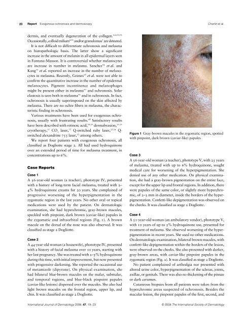
examination, she had hyperchromic, gray-brown macules,<br />
speckled with pinpoint, dark brown (caviar-like) papules in<br />
the zygomatic and infraorbital regions (Fig. 1). A brown<br />
macule on the dorsal of the nose was also observed. It was<br />
classified as stage 2 Dogliotte.<br />
Case 2<br />
A 44-year-old woman (a housewife), phototype IV, presented<br />
with a history of facial melasma over 10 years, starting with<br />
her last pregnancy. She was treated with 2–5% hydroquinone<br />
during this time, with initial improvement, but now presented<br />
with progressive darkening. She reported the occasional use<br />
of metamizole (dipyrone). On physical examination, she<br />
had bilateral blue-brown macules on the malar, submalar,<br />
and temporal regions, and blue-black pinpoint papules<br />
(caviar-like lesions) dispersed over the macules. She also had<br />
light brown macules on the frontal region, upper lip, and<br />
chin. It was classified as stage 2 Dogliotte.<br />
Charlín et al.<br />
Figure 1 Gray-brown macules in the zygomatic region, spotted<br />
with pinpoint, dark brown (caviar-like) papules<br />
Case 3<br />
A 56-year-old woman (a teacher), phototype V, with 25 years<br />
of melasma, treated with up to 6% hydroquinone, sought<br />
medical care for worsening of the hyperpigmentation. She<br />
<strong>de</strong>nied use of any other medication. On physical examination,<br />
she had a gray-brown pigmentation on the entire face,<br />
except for the upper lip and frontal regions. In addition, there<br />
were papules of the same color, or slightly more hyperchromic,<br />
of 2–3 mm in diameter, insi<strong>de</strong> the bor<strong>de</strong>rs of the hyperpigmentation.<br />
Confetti-like <strong>de</strong>pigmentation was observed on<br />
the cheeks. It was classified as stage 2 Dogliotte.<br />
Case 4<br />
A 51-year-old woman (an ambulatory vendor), phototype V,<br />
with 10 years of up to 5% hydroquinone use, presented for<br />
treatment of melasma. She observed worsening of the hyperpigmentation<br />
in recent years. She used no other medications.<br />
On <strong>de</strong>rmatologic examination, bilateral brown macules, with<br />
confetti-like <strong>de</strong>pigmentation within the bor<strong>de</strong>rs of the lesion,<br />
were observed on the cheeks. She also presented with darker,<br />
gray-brown areas, with caviar-like pinpoint papules in the<br />
zygomatic region (Fig. 2). It was classified as stage 2 Dogliotte.<br />
No patient complained of arthralgia nor presented with<br />
altered urine color, hyperpigmentation of the sclerae, joints,<br />
axillae, or genitals. There was also no thickening of the pinnae<br />
or dark cerumen.<br />
Cutaneous biopsies from all patients were taken from the<br />
hyperchromic areas suspected of <strong>ochronosis</strong>. Besi<strong>de</strong>s the<br />
macular lesion, the pinpoint papules of the first, second, and<br />
International Journal of Dermatology 2008, 47,<br />
19–23 © 2008 The International Society of Dermatology