- Page 1 and 2: A thesis submitted in partial fulfi
- Page 3 and 4: ABSTRACT The main aims and objectiv
- Page 5 and 6: ABBREVIATION LIST 1DE: 1-dimensiona
- Page 7 and 8: ml -1 : Per millilitre MgCl2: Magne
- Page 9 and 10: FIGURE AND TABLE LIST CHAPTER 1 - I
- Page 11 and 12: Figure 4.9 Incubation of T. cruzi e
- Page 13 and 14: Life cycle.........................
- Page 15 and 16: Effect of oil constituents on viabi
- Page 17 and 18: 4:3.3 Epimastigote culture analysis
- Page 19 and 20: 7:1 ABSTRACT.......................
- Page 21 and 22: CHAPTER 1 Introduction 1
- Page 23 and 24: ecome resistant to both chloroquine
- Page 25 and 26: Leptomonas colosoma, Crithidia fasc
- Page 27 and 28: viability of a population) determin
- Page 29 and 30: changes in phospholipid composition
- Page 31 and 32: killed by garlic (Zenner et al. 200
- Page 33 and 34: Table 1.2 Plant oils that demonstra
- Page 35 and 36: arouse, until the stomach was clear
- Page 37 and 38: Dermatological reactions Many plant
- Page 39: 2002). With industrialised nations,
- Page 43 and 44: the ER is the formation and folding
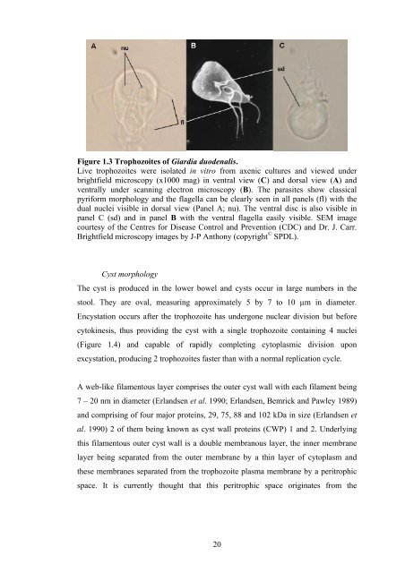
- Page 45 and 46: of the cysts through the stomach ex
- Page 47 and 48: Figure 1.5 Life-cycle of G. duodena
- Page 49 and 50: Cryptosporidium is an apicomplexan
- Page 51 and 52: Table 1.4 - Continued Species of Cr
- Page 53 and 54: Upon excystation, sporozoites attac
- Page 55 and 56: MacKenzie et al. 1994b). However, a
- Page 57 and 58: Figure 1.7 Life-cycle of C. parvum.
- Page 59 and 60: identified by the relative position
- Page 61 and 62: Amastigote These replicative stages
- Page 63 and 64: Life Cycle The sylvatic cycle start
- Page 65 and 66: Acute phase An intense and localise
- Page 67 and 68: inflammatory reactions induced by t
- Page 69 and 70: 1:8 Research Hypothesis Little scie
- Page 71 and 72: able to identify them molecularly a
- Page 73 and 74: CHAPTER 2 Giardia duodenalis The in
- Page 75 and 76: 2:2 INTRODUCTION The protozoan para
- Page 77 and 78: example, the understanding of antig
- Page 79 and 80: Jassim & Naji 2003) and more recent
- Page 81 and 82: 2:3 MATERIALS AND METHODS 2.3.1 Par
- Page 83 and 84: concentration equivalent to 0.02% v
- Page 85 and 86: whole oil respectively. Elemi oil c
- Page 87 and 88: Plus Protein standard; unstained; 1
- Page 89 and 90: 2:4 RESULTS Ethanol and DMSO titrat
- Page 91 and 92:
Figure 2.1 In vitro viability of G.
- Page 93 and 94:
and elemi was 0.005% although > 80%
- Page 95 and 96:
Trypan Stained / Non-viable Trophoz
- Page 97 and 98:
(10.90%). Whilst both the main cons
- Page 99 and 100:
Incubation of G. duodenalis trophoz
- Page 101 and 102:
Trophozoite protein profiling by SD
- Page 103 and 104:
used in this protein analysis exper
- Page 105 and 106:
MIC of 0.005%. This phenomenon was
- Page 107 and 108:
Effect of elemi oil on viability as
- Page 109 and 110:
0.05). This effect though, was less
- Page 111 and 112:
kDa 50- 37- 50- 37- 50- 37- MW G HK
- Page 113 and 114:
2:5 DISCUSSION These experiments we
- Page 115 and 116:
Using microscopic evaluation of par
- Page 117 and 118:
cell death may have been initiated,
- Page 119 and 120:
Using this information, investigati
- Page 121 and 122:
flavanones, anthocyanidins and isof
- Page 123 and 124:
protein was also abolished in elemi
- Page 125 and 126:
CHAPTER 3 Cryptosporidium parvum In
- Page 127 and 128:
3:2 INTRODUCTION The enteric parasi
- Page 129 and 130:
disease in extraintestinal location
- Page 131 and 132:
constituents and speed of activity.
- Page 133 and 134:
(empty), partially excysted and int
- Page 135 and 136:
and incubated for a further 1, 4 or
- Page 137 and 138:
3:4 RESULTS 3:4.1 Acidification exp
- Page 139 and 140:
When compared with MD, the IOWA iso
- Page 141 and 142:
with incubation at 5ºC (-0.5 ± 1.
- Page 143 and 144:
constituent in palmarosa oil which
- Page 145 and 146:
Spontaneous Excystation Rate (%) 10
- Page 147 and 148:
3:5 DISCUSSION Interventions in the
- Page 149 and 150:
When these acid treated oocysts wer
- Page 151 and 152:
PEOs augment the effect of temperat
- Page 153 and 154:
spontaneous excystation within 4 h.
- Page 155 and 156:
Trans-anethole is used as a flavour
- Page 157 and 158:
CHAPTER 4 Trypanosoma cruzi The act
- Page 159 and 160:
4:2 INTRODUCTION The disease caused
- Page 161 and 162:
It is clear that alternative therap
- Page 163 and 164:
analyse the activity of 12 differen
- Page 165 and 166:
µl of parasites. One 96 well plate
- Page 167 and 168:
Incubation of T. cruzi epimastigote
- Page 169 and 170:
4:4 RESULTS Comparison of incubatio
- Page 171 and 172:
MTS Absorbance (492nm) 1 0.9 0.8 0.
- Page 173 and 174:
Incubation of T. cruzi epimastigote
- Page 175 and 176:
A B Absorbance (492nm) Trypan Stain
- Page 177 and 178:
Titration of PEOs As all the PEOs a
- Page 179 and 180:
0.02% 0.01% 0.005% 0.0025% 0.00125%
- Page 181 and 182:
Incubation of T. cruzi epimastigote
- Page 183 and 184:
Trypan Stained / Non-viable Epimast
- Page 185 and 186:
published concerning the trypanocid
- Page 187 and 188:
using the MTS assay, where a lack o
- Page 189 and 190:
With myrtle, 2 constituents working
- Page 191 and 192:
mixed with inert carrier oils such
- Page 193 and 194:
5:1 ABSTRACT The protozoan parasite
- Page 195 and 196:
37ºC over a 24 h period, in the ab
- Page 197 and 198:
against Cryptosporidium with any su
- Page 199 and 200:
polyphenol content of PRBE was 920
- Page 201 and 202:
5:4 RESULTS Polyphenolic content of
- Page 203 and 204:
Figure 5.1 A Viable and dead G. duo
- Page 205 and 206:
Effect of FB, BB and PRBE on the sp
- Page 207 and 208:
5:5 DISCUSSION Here it is shown tha
- Page 209 and 210:
spontaneous excystation observed (F
- Page 211 and 212:
certain concentration is exceeded (
- Page 213 and 214:
6:1 ABSTRACT Current conventional t
- Page 215 and 216:
Flavonoids and phenolic acids form
- Page 217 and 218:
efore any dilutions in TYI-S-33 cou
- Page 219 and 220:
6:4 RESULTS Incubation of G. duoden
- Page 221 and 222:
Trypan Stained / Non-viable Trophoz
- Page 223 and 224:
6:5 DISCUSSION It was demonstrated
- Page 225 and 226:
vitro, is not due to the degradatio
- Page 227 and 228:
CHAPTER 7 The examination of latrin
- Page 229 and 230:
7:2 INTRODUCTION The examination of
- Page 231 and 232:
7:3 MATERIALS AND METHODS 7:3.1 Sou
- Page 233 and 234:
Figure 7.2 Excavation of Soutra Ais
- Page 235 and 236:
7:3.2 Sample preparations Formol et
- Page 237 and 238:
magnet. The tubes were rocked throu
- Page 239 and 240:
Using the methods of Smith et al. (
- Page 241 and 242:
incubated at 55°C for 3 h in a wat
- Page 243 and 244:
RNA Cryptosporidium gene to determi
- Page 245 and 246:
concentration recommended in publis
- Page 247 and 248:
7:4 RESULTS Microscopic examination
- Page 249 and 250:
Figure 7.6 Fluorescence staining of
- Page 251 and 252:
These slurries were subjected to IM
- Page 253 and 254:
Figure 7.10 PCR analysis for Crypto
- Page 255 and 256:
Pike 1967 & 1968; Jones 1982; Aspoc
- Page 257 and 258:
the digestion enzymes DraI and AseI
- Page 259 and 260:
CONCLUSIONS This series of experime
- Page 261 and 262:
distortion, blebbing and increased
- Page 263 and 264:
trophozoites and Trypanosoma epimas
- Page 265 and 266:
Reduviid bug is a means of entry fo
- Page 267 and 268:
anticryptosporidial activity may be
- Page 269 and 270:
Historically, many PEOs and extract
- Page 271 and 272:
REFERENCES Abdo, K.M., Cunningham,
- Page 273 and 274:
Anthony, J-P., Fyfe, L. Smith, H.V.
- Page 275 and 276:
Best, M., Sattar, S.A., Springthorp
- Page 277 and 278:
in the chronic phase of experimenta
- Page 279 and 280:
entidade morbida do homem. Memorias
- Page 281 and 282:
Current, W.L., Upton, S.J. and Hayn
- Page 283 and 284:
Elliot, B.C., Wisnewski, A.V., John
- Page 285 and 286:
Ferreira, L.F., Britto, C., Cardoso
- Page 287 and 288:
Gillin, F.D., Reiner, D.S. and Bouc
- Page 289 and 290:
Hashmey, R., Smith, N.H., Cron, S.,
- Page 291 and 292:
Hunter, P.R. and Syed, Q. 2001. Com
- Page 293 and 294:
Kim, H.C. & Healey, J.M. 2001. Effe
- Page 295 and 296:
Le Bourhis, B. 1968. Preliminary re
- Page 297 and 298:
ornidazole. Transactions of the Roy
- Page 299 and 300:
Moo-Puc, R.E., Mena-Rejon, G.J., Qu
- Page 301 and 302:
length polymorphism assay. Applied
- Page 303 and 304:
Pecevski, J., Savkovic, D., Radivoj
- Page 305 and 306:
Reduker, D.W. & Speer, C.A. 1985. F
- Page 307 and 308:
Rothhammer, F., Allison, M., Nuñez
- Page 309 and 310:
Sgambatti de Andrade, A.L., Zicker,
- Page 311 and 312:
Sosa Estani, S., Segura, E.L., Ruiz
- Page 313 and 314:
Tyler, K.M. and Engman, D.M. 2001.
- Page 315 and 316:
Wang, M., Kikuzaki, H., Lin, C.C.,
- Page 317 and 318:
Yasunaka, K., Abe, F., Nagayama, A.
- Page 319 and 320:
Family: Burseraceae, Commiphora myr
- Page 321 and 322:
Family: Lamiaceae, Pogostemon cabli
- Page 323 and 324:
Family: Rutaceae, Amyris balsamifer
- Page 325 and 326:
Family: Ericaceae, Vaccinium vitis-
- Page 327 and 328:
Family: Rosaceae, Rubus fruticosus
- Page 329 and 330:
Family: Rosaceae, Sorbus aucuparia
- Page 331 and 332:
311
- Page 333 and 334:
313
- Page 335 and 336:
315
- Page 337 and 338:
317
- Page 339 and 340:
319
- Page 341 and 342:
321
- Page 343 and 344:
323
- Page 345 and 346:
325
- Page 347 and 348:
327
- Page 349 and 350:
329
- Page 351 and 352:
Appendix 5. SDS-Polyacrylamide gel
- Page 353 and 354:
5. When the glass plates are in pla
- Page 355 and 356:
Appendix 6: Incubation of G. duoden
- Page 357 and 358:
Appendix 8. Agarose gel electrophor
- Page 359:
PUBLICATIONS Published papers: Anth