EMBO Conference on Protein Synthesis and Translational Control
EMBO Conference on Protein Synthesis and Translational Control
EMBO Conference on Protein Synthesis and Translational Control

Create successful ePaper yourself
Turn your PDF publications into a flip-book with our unique Google optimized e-Paper software.
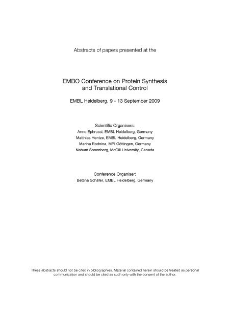
Abstracts of papers presented at the<br />
<str<strong>on</strong>g>EMBO</str<strong>on</strong>g> <str<strong>on</strong>g>C<strong>on</strong>ference</str<strong>on</strong>g> <strong>on</strong> <strong>Protein</strong> <strong>Synthesis</strong><br />
<strong>and</strong> Translati<strong>on</strong>al C<strong>on</strong>trol<br />
EMBL Heidelberg, 9 - 13 September 2009<br />
Scientific Organisers:<br />
Anne Ephrussi, EMBL Heidelberg, Germany<br />
Matthias Hentze, EMBL Heidelberg, Germany<br />
Marina Rodnina, MPI Göttingen, Germany<br />
Nahum S<strong>on</strong>enberg, McGill University, Canada<br />
<str<strong>on</strong>g>C<strong>on</strong>ference</str<strong>on</strong>g> Organiser:<br />
Bettina Schäfer, EMBL Heidelberg, Germany<br />
These abstracts should not be cited in bibliographies. Material c<strong>on</strong>tained herein should be treated as pers<strong>on</strong>al<br />
communicati<strong>on</strong> <strong>and</strong> should be cited as such <strong>on</strong>ly with the c<strong>on</strong>sent of the author.
C<strong>on</strong>tent<br />
Agenda i<br />
Posters A-Z xv<br />
Speaker Abstracts 1<br />
Poster Abstracts 68<br />
Authors Index 285<br />
List of Participants 321<br />
Useful Telef<strong>on</strong>numbers 362<br />
Map of EMBL Campus 363
15:00 - 18:00 Registrati<strong>on</strong><br />
18:00 - 19:00 Dinner<br />
19:00 - 19:15 Welcome<br />
19:15 - 20:00 Judith Kimble<br />
20:00 - 20:45 David Bartel<br />
DAY 1, 9 SEPTEMBER 2009<br />
i<br />
Agenda<br />
University of Wisc<strong>on</strong>sin-Madis<strong>on</strong>, United States of<br />
America<br />
Translati<strong>on</strong>al c<strong>on</strong>trol <strong>and</strong> stem cells:<br />
less<strong>on</strong>s from the C. elegans germline 1<br />
MIT/Whitehead Institute/HHMI, United States of<br />
America<br />
MicroRNAs 2<br />
20:45 - 21:15 Coffee Break<br />
21:15 - 22:00 Reinhard Luehrmann<br />
MPI fuer Biophysikalische Chemie, Germany<br />
At the heart of the spliceosome 3<br />
22:00 - 00:00 Get-together<br />
DAY 2, 10-SEPTEMBER 2009<br />
09:00 - 12:30 Sessi<strong>on</strong> 1: Translati<strong>on</strong> Initiati<strong>on</strong><br />
09:00 - 09:30 J<strong>on</strong> Lorsch<br />
Johns Hopkins University School of Medicine,<br />
United States of America<br />
Rec<strong>on</strong>stituti<strong>on</strong> <strong>and</strong> Analysis of<br />
Cap-stimulated mRNA Recruitment to the<br />
43S Pre-initiati<strong>on</strong> Complex 4
<str<strong>on</strong>g>EMBO</str<strong>on</strong>g> <str<strong>on</strong>g>C<strong>on</strong>ference</str<strong>on</strong>g> <strong>on</strong> <strong>Protein</strong> <strong>Synthesis</strong> <strong>and</strong> Translati<strong>on</strong>al C<strong>on</strong>trol<br />
EMBL Heidelberg, 9 – 13 September 2009<br />
09:30 - 09:45 Adesh Saini<br />
NICHD, NIH, United States of America<br />
Structural elements in eIF1A regulate AUG<br />
selecti<strong>on</strong> by c<strong>on</strong>trolling distinct modes of<br />
initiator binding to the preinitiati<strong>on</strong><br />
complex 5<br />
09:45 - 10:00 Martin Jennings<br />
10:00 - 10:15 Akira Fukao<br />
University of Manchester, United Kingdom<br />
eIF5 acts as a GDP dissociati<strong>on</strong> inhibitor in<br />
additi<strong>on</strong> to its GAP functi<strong>on</strong> 6<br />
Kobe University, Japan<br />
10:15 - 10:30 Franck Martin<br />
Enhancement of cap-dependent translati<strong>on</strong><br />
by the ELAV protein HuD: A novel functi<strong>on</strong><br />
of HuD which is eIF4A- <strong>and</strong><br />
poly(A)-dependent 7<br />
Université de Strasbourg, France<br />
10:30 - 11:00 Coffee Break<br />
11:00 - 11:15 Rivka Dikstein<br />
Tethering of ribosomes downstream of<br />
translati<strong>on</strong>al start cod<strong>on</strong> drives translati<strong>on</strong><br />
of hist<strong>on</strong>e H4 8<br />
Weizmann Institute of Science, Israel<br />
Characterizati<strong>on</strong> of TISU, a translati<strong>on</strong><br />
initiator specific to mRNAs with extremely<br />
short 5’UTR 9<br />
11:15 - 11:30 Christine Luttermann<br />
Friedrich-Loeffler-Institut, Germany<br />
The importance of inter- <strong>and</strong><br />
intramolecular base pairing for translati<strong>on</strong><br />
reinitiati<strong>on</strong> <strong>on</strong> a eukaryotic bicistr<strong>on</strong>ic<br />
mRNA 10<br />
ii
11:30 - 11:45 Anna Maria Giuliodori<br />
11:45 - 12:00 Dieter Wolf<br />
12:00 - 12:15 Helena Firczuk<br />
Laboratory of Genetics, Department of Biology<br />
MCA, University of Camerino,, Italy<br />
iii<br />
Agenda<br />
Translati<strong>on</strong>al regulati<strong>on</strong> of cold-shock<br />
gene expressi<strong>on</strong> 11<br />
Burnham Institute for Medical Research, United<br />
States of America<br />
The eIF3 interactome reveals the<br />
translasome, a supercomplex linking<br />
protein synthesis <strong>and</strong> degradati<strong>on</strong><br />
machineries 12<br />
Manchester Interdisciplinary Biocentre, The<br />
University of Manchester, United Kingdom<br />
Comprehensive rate c<strong>on</strong>trol analysis of the<br />
eukaryotic translati<strong>on</strong> pathway 13<br />
12:15 - 12:30 Nicholas Ingolia<br />
12:30 - 14:00 Lunch<br />
University of California, San Francisco, United<br />
States of America<br />
Genome-wide analysis of in vivo<br />
translati<strong>on</strong> with single-nucleotide<br />
resoluti<strong>on</strong> 14<br />
14:00 - 16:30 Sessi<strong>on</strong> 2: Translati<strong>on</strong> el<strong>on</strong>gati<strong>on</strong> <strong>and</strong><br />
terminati<strong>on</strong><br />
14:00 - 14:30 Terri Kinzy<br />
UMDNJ Robert Wood Johns<strong>on</strong> Medical School,<br />
United States of America<br />
ADP-ribosylati<strong>on</strong> of eukaryotic el<strong>on</strong>gati<strong>on</strong><br />
factor 2 by bacterial toxins <strong>and</strong> its effects<br />
<strong>on</strong> translati<strong>on</strong> el<strong>on</strong>gati<strong>on</strong> in vivo <strong>and</strong> in<br />
vitro 15
<str<strong>on</strong>g>EMBO</str<strong>on</strong>g> <str<strong>on</strong>g>C<strong>on</strong>ference</str<strong>on</strong>g> <strong>on</strong> <strong>Protein</strong> <strong>Synthesis</strong> <strong>and</strong> Translati<strong>on</strong>al C<strong>on</strong>trol<br />
EMBL Heidelberg, 9 – 13 September 2009<br />
14:30 - 14:45 Alena Paleskava<br />
14:45 - 15:00 James Munro<br />
Max Planck Institute for Biophysical Chemistry,<br />
Germany<br />
Unusually tight binding of Sec-tRNASec to<br />
the el<strong>on</strong>gati<strong>on</strong> factor SelB due to the<br />
specific recogniti<strong>on</strong> of the selenocysteyl<br />
group by the GTP-bound form 16<br />
Weill Cornell Medical College, United States of<br />
America<br />
Single-molecule observati<strong>on</strong>s of<br />
rate-limiting c<strong>on</strong>formati<strong>on</strong>al events during<br />
ribosomal translocati<strong>on</strong> 17<br />
15:00 - 15:15 Shashi Bhushan<br />
15:15 - 15:30 C. Axel Innis<br />
15:30 - 15:45 Kristen Bartoli<br />
15:45 - 16:00 Erik Böttger<br />
16:00 - 16:30 Coffee Break<br />
Gene Cemter, LMU Munich, Germany<br />
Visualizati<strong>on</strong> of nascent chains in the<br />
ribosomal exit tunnel: Implicati<strong>on</strong> for<br />
sec<strong>on</strong>dary structure formati<strong>on</strong> 18<br />
Yale University, United States of America<br />
Shedding Light Onto Nascent<br />
Chain-Mediated Translati<strong>on</strong>al Stalling 19<br />
University of Pittsburgh, School of Medicine, United<br />
States of America<br />
Novel Role for Mitotic Microtubule Motor<br />
<strong>Protein</strong> Eg5 in <strong>Protein</strong> Translati<strong>on</strong> 20<br />
Institute of Medical Microbiology, Switzerl<strong>and</strong><br />
Aminoglycoside Ototoxicity <strong>and</strong> <strong>Synthesis</strong><br />
of New Compounds with Altered<br />
Drug-Target Interacti<strong>on</strong> 21<br />
iv
16:30 - 18:30 Sessi<strong>on</strong> 3: N<strong>on</strong>-coding RNAs in translati<strong>on</strong><br />
16:30 - 17:00 Witold Filipowicz<br />
Friedrich Miescher Institute for Biomedical<br />
Research, Germany<br />
v<br />
Agenda<br />
Mechanism of the HuR-mediated reversal<br />
of miRNA repressi<strong>on</strong> in human cells 22<br />
17:00 - 17:15 Elisa Izaurralde<br />
17:15 - 17:30 Thomas Preiss<br />
Max Planck Institute for Developmental Biology,<br />
Germany<br />
A C-terminal silencing domain in GW182<br />
family proteins is essential for miRNA<br />
functi<strong>on</strong> in animal cells 23<br />
VCCRI, Australia<br />
17:30 - 17:45 Incheol Ryu<br />
Identifying miRNA targets through<br />
changes in mRNA poly(A) tail length 24<br />
POSTECH, Republic of Korea<br />
Eukaryotic Translati<strong>on</strong> Initiati<strong>on</strong> Factor 4G<br />
Mediates MicroRNA-Regulated<br />
Translati<strong>on</strong>al Gene Silencing 25<br />
17:45 - 18:00 Ania Wilczynska<br />
Univerisity of Cambridge, United Kingdom<br />
Investigating the microRNA pathway in<br />
Xenopus oocytes 26<br />
18:00 - 18:15 Anne-Catherine Prats<br />
Inserm U858, France<br />
FGF1 inducti<strong>on</strong> in myogenesis depends <strong>on</strong><br />
novel cross-talks between IRES, promoter<br />
<strong>and</strong> 3’UTR 27
<str<strong>on</strong>g>EMBO</str<strong>on</strong>g> <str<strong>on</strong>g>C<strong>on</strong>ference</str<strong>on</strong>g> <strong>on</strong> <strong>Protein</strong> <strong>Synthesis</strong> <strong>and</strong> Translati<strong>on</strong>al C<strong>on</strong>trol<br />
EMBL Heidelberg, 9 – 13 September 2009<br />
18:15 - 18:30 William Merrick<br />
Case Western Reserve University, United States of<br />
America<br />
Possible mechanism of regulati<strong>on</strong> of<br />
IRES-mediated expressi<strong>on</strong> by eIF2A 28<br />
18:30 - 20:00 Dinner<br />
20:00 - 22:00 Poster Sessi<strong>on</strong> I<br />
22:00 - 00:00 Wine & Cheese<br />
DAY 3, 11-SEPTEMBER 2009<br />
09:00 - 12:30 Sessi<strong>on</strong> 4: Translati<strong>on</strong> in development <strong>and</strong> the<br />
CNS<br />
09:00 - 09:30 Christine Holt<br />
09:30 - 09:45 Raúl Méndez<br />
University of Cambridge, United Kingdom<br />
Sub-cellular profiling reveals distinct <strong>and</strong><br />
dynamic repertoire of growth c<strong>on</strong>e mRNAs 29<br />
Centre for Genomic Regulati<strong>on</strong> (CRG), Spain<br />
CPEB1 regulates the expressi<strong>on</strong> of CPEB4<br />
to complete meiosis 30<br />
09:45 - 10:00 Howard Lipshitz<br />
University of Tor<strong>on</strong>to, Canada<br />
10:00 - 10:15 Maria Barna<br />
C<strong>on</strong>trol of mRNA translati<strong>on</strong> <strong>and</strong> stability<br />
during early Drosophila development 31<br />
University of California, San Francisco, United<br />
States of America<br />
Translati<strong>on</strong>al specificity of ribosomal<br />
proteins regulates vertebrate embry<strong>on</strong>ic<br />
development 32<br />
vi
10:15 - 10:30 Michael Sheets<br />
Univ. of Wisc<strong>on</strong>sin Dept. of Biomol. Chem., United<br />
States of America<br />
vii<br />
Agenda<br />
Spatially regulated translati<strong>on</strong> of the xCR1<br />
mRNA in xenopus embryos by poly (A)<br />
independent mechanism 33<br />
10:30 - 11:00 Coffee Break<br />
11:00 - 11:15 Sebastian Baumann<br />
Max Planck Institute for Terrestrial Microbiology,<br />
Germany<br />
Microtubule-dependent mRNA transport<br />
during pathogenic development in Ustilago<br />
maydis 34<br />
11:15 - 11:30 Peter Lukavsky<br />
MRC LMB, United Kingdom<br />
A’-form RNA helices drive<br />
microtubule-based mRNA transport in<br />
Drosophila 35<br />
11:30 - 11:45 Greco Hern<strong>and</strong>ez<br />
McGill University, Canada<br />
11:45 - 12:00 Ana Villlalba<br />
Mextli is a novel eIF4E-binding protein<br />
from Drosophila 36<br />
FUNDACIO PRIVADA CENTRE DE REGULACIO<br />
GENOMICA, Spain<br />
A novel, n<strong>on</strong>-can<strong>on</strong>ical mechanism of<br />
cytoplasmic polyadenylati<strong>on</strong> in Drosophila 37<br />
12:00 - 12:15 Kobi Rosenblum<br />
Haifa University, Israel<br />
Translati<strong>on</strong>al C<strong>on</strong>trol in the Gustatory<br />
Cortex Determines the Stability of a Taste<br />
Memory 38
<str<strong>on</strong>g>EMBO</str<strong>on</strong>g> <str<strong>on</strong>g>C<strong>on</strong>ference</str<strong>on</strong>g> <strong>on</strong> <strong>Protein</strong> <strong>Synthesis</strong> <strong>and</strong> Translati<strong>on</strong>al C<strong>on</strong>trol<br />
EMBL Heidelberg, 9 – 13 September 2009<br />
12:15 - 12:30 Kent Duncan<br />
12:30 - 14:00 Lunch<br />
EMBL, Germany<br />
The SXL-UNR Corepressor Complex Uses<br />
a Novel PABP-Mediated Mechanism to<br />
Inhibit Ribosome Recruitment to msl-2<br />
mRNA 39<br />
14:00 - 17:30 Sessi<strong>on</strong> 5: Translati<strong>on</strong> factors & complexes, <strong>and</strong><br />
their functi<strong>on</strong> in health <strong>and</strong> disease<br />
14:00 - 14:30 Fatima Gebauer<br />
Fundacio Privada Centre de Regulaciao Genomica,<br />
Spain<br />
Regulatory networks c<strong>on</strong>trolled by<br />
Drosophila UNR 40<br />
14:30 - 14:45 Shelt<strong>on</strong> Bradrick<br />
14:45 - 15:00 Michal Shapira<br />
15:00 - 15:15 Gregory Boel<br />
Duke University, United States of America<br />
Identificati<strong>on</strong> of gemin5 as a novel<br />
cap-binding protein that associates with<br />
coding <strong>and</strong> n<strong>on</strong>-coding RNAs 41<br />
Ben Guri<strong>on</strong> University of the Negev, Israel<br />
Evoluti<strong>on</strong>ary diversity of the<br />
trypanosomatid cap4-binding complex – a<br />
potential drug target against<br />
trypanosomatids? 42<br />
Columbia University, United States of America<br />
YjjK, a member of the ATP binding<br />
cassette superfamily is a novel<br />
transcripti<strong>on</strong>al factor 43<br />
viii
15:15 - 15:30 Lyubov Ryabova<br />
Institut de Biologie Moléculaire des Plantes (IBMP),<br />
UPR CNRS 2357, France<br />
ix<br />
Agenda<br />
Functi<strong>on</strong> of RISP in virus-induced<br />
translati<strong>on</strong> reinitiati<strong>on</strong> 44<br />
15:30 - 15:45 Philip Farabaugh<br />
University of Maryl<strong>and</strong> Baltimore County, United<br />
States of America<br />
Effects of haploinsufficiency of ribosomal<br />
protein <strong>and</strong> assembly factor genes <strong>on</strong><br />
cellular physiology mediated by RACK1 45<br />
15:45 - 16:15 Coffee Break<br />
16:15 - 16:30 Vitaly Polunovsky<br />
University of Minnesota, United States of America<br />
Translati<strong>on</strong>al C<strong>on</strong>trol of Malignancy in the<br />
Murine Models Epithelial Carcinogenesis:<br />
The Role of the Translati<strong>on</strong>al Repressors<br />
4E-BPs 46<br />
16:30 - 16:45 Robert Rhoads<br />
LSU Health Sciences Center, United States of<br />
America<br />
Effects <strong>on</strong> translati<strong>on</strong> initiati<strong>on</strong> <strong>and</strong><br />
malignant transformati<strong>on</strong> of MMTV<br />
inserti<strong>on</strong> into the eIF3e gene 47<br />
16:45 - 17:00 Robert Schneider<br />
NYU School of Medicine, United States of America<br />
AUF1 links the chr<strong>on</strong>ic inflammatory<br />
resp<strong>on</strong>se to telomere maintenance,<br />
premature aging <strong>and</strong> tumorigenesis<br />
through c<strong>on</strong>trol of short-lived mRNA<br />
stability 48
<str<strong>on</strong>g>EMBO</str<strong>on</strong>g> <str<strong>on</strong>g>C<strong>on</strong>ference</str<strong>on</strong>g> <strong>on</strong> <strong>Protein</strong> <strong>Synthesis</strong> <strong>and</strong> Translati<strong>on</strong>al C<strong>on</strong>trol<br />
EMBL Heidelberg, 9 – 13 September 2009<br />
17:00 - 17:15 Harald König<br />
Forschungszentrum Karlsruhe GmbH, Germany<br />
Interfering with translati<strong>on</strong> of transcripts<br />
enhances their splicing 49<br />
17:15 - 17:30 Michael Kiebler<br />
Medical University of Vienna, Austria<br />
Functi<strong>on</strong>al Characterizati<strong>on</strong> of Neur<strong>on</strong>al<br />
RNA granules <strong>and</strong> their role in dendritic<br />
RNA localizati<strong>on</strong> 50<br />
17:30 - 20:00 Dinner<br />
20:00 - 22:00 Poster Sessi<strong>on</strong> II<br />
22:00 - 00:00 Wine & Cheese<br />
DAY 4, 12 SEPTEMBER 2009<br />
09:00 - 11:15 Sessi<strong>on</strong> 6: mRNA stability <strong>and</strong><br />
n<strong>on</strong>sense-mediated decay<br />
09:00 - 09:30 Elena C<strong>on</strong>ti<br />
Max Planck Institute of Biochemistry, Germany<br />
Structural studies of n<strong>on</strong>sense mediated<br />
mRNA decay 51<br />
09:30 - 09:45 Oliver Mühlemann<br />
09:45 - 10:00 Fulvia B<strong>on</strong>o<br />
University of Bern, Switzerl<strong>and</strong><br />
SMG6-mediated end<strong>on</strong>ucleolytic cleavage<br />
of n<strong>on</strong>sense mRNA in human cells 52<br />
Max Planck of Dev. Biology, Germany<br />
Structural analysis of Mago-Y14 import<br />
receptor 53<br />
x
10:00 - 10:15 Gabriele Neu-Yilik<br />
MMPU EMBL/University of Heidelberg, Germany<br />
xi<br />
Agenda<br />
Premature terminati<strong>on</strong> cod<strong>on</strong>s within ex<strong>on</strong><br />
1 of the human beta-globin mRNA create<br />
short ORFs permissive for reinitiati<strong>on</strong> 54<br />
10:15 - 10:30 J<strong>on</strong>athan Dinman<br />
10:30 - 10:45 Sevim Ozgur<br />
10:45 - 11:00 Felix Tritschler<br />
University of Maryl<strong>and</strong>, United States of America<br />
mRNA destabilizati<strong>on</strong> by programmed<br />
ribosomal frameshifting <strong>and</strong> its effects <strong>on</strong><br />
telomere maintenance <strong>and</strong> aging in yeast 55<br />
German Cancer Research Center, Germany<br />
Human Pat1 C<strong>on</strong>trols the Assembly of<br />
Processing-Bodies <strong>and</strong> Promotes mRNA<br />
Degradati<strong>on</strong> 56<br />
Max-Planck-Institute for Developmental Biology,<br />
Germany<br />
A metazoan-specific DCP1 C-terminal<br />
extensi<strong>on</strong> is required for the assembly of<br />
active mRNA decapping complexes in<br />
human cells 57<br />
11:00 - 11:15 Markus L<strong>and</strong>thaler<br />
BIMSB, Germany<br />
PURE-CLIP - Transcriptome-wide<br />
identificati<strong>on</strong> of RNA targets <strong>and</strong> binding<br />
sites of RNA-binding proteins 58<br />
11:15 - 13:00 Poster Sessi<strong>on</strong> III<br />
13:00 - 18:30 Lunch & Free Time<br />
18:30 - 19:30 Drinks<br />
19:30 - 01:00 Banquett Dinner & Party
<str<strong>on</strong>g>EMBO</str<strong>on</strong>g> <str<strong>on</strong>g>C<strong>on</strong>ference</str<strong>on</strong>g> <strong>on</strong> <strong>Protein</strong> <strong>Synthesis</strong> <strong>and</strong> Translati<strong>on</strong>al C<strong>on</strong>trol<br />
EMBL Heidelberg, 9 – 13 September 2009<br />
DAY 5, 13 SEPTEMBER 2009<br />
09:15 - 12:15 Sessi<strong>on</strong> 7: Structure <strong>and</strong> functi<strong>on</strong> of the<br />
ribosome<br />
09:15 - 09:45 Jamie Cate<br />
09:45 - 10:00 Stefano Marzi<br />
UC Berkeley, United States of America<br />
Insights into protein synthesis from<br />
structures of the E. coli ribosome 59<br />
IBMC Strasbourg, France<br />
A dynamic view of translati<strong>on</strong> initiati<strong>on</strong> <strong>and</strong><br />
its c<strong>on</strong>trol by structured mRNAs in<br />
bacteria 60<br />
10:00 - 10:15 Claudio Gualerzi<br />
University of Camerino, Italy<br />
10:15 - 10:30 Joachim Frank<br />
10:30 - 11:00 Coffee Break<br />
11:00 - 11:15 Lasse Jenner<br />
Mechanism of ribosomal recruitment of<br />
fMet-tRNA by bacterial translati<strong>on</strong> initiati<strong>on</strong><br />
factor IF2 61<br />
HHMI, Columbia University, United States of<br />
America<br />
Structural Discriminati<strong>on</strong> Between Cognate<br />
<strong>and</strong> Near-cognate Ternary Complexes By<br />
the Ribosome 62<br />
CERBM-GIE / IGBMC, France<br />
Crystal structure of the ribosome<br />
c<strong>on</strong>taining three tRNAs 63<br />
xii
11:15 - 11:30 Rebecca Voorhees<br />
11:30 - 11:45 Niels Fischer<br />
Medical Research Council: Laboratory of Molecular<br />
Biology, United Kingdom<br />
xiii<br />
Agenda<br />
Insights into substrate stabilizati<strong>on</strong> from<br />
structural studies of the peptidyl<br />
transferase center of the intact 70S<br />
ribosome 64<br />
Max Planck Institute for Biophysical Chemistry,<br />
Germany<br />
The trajectory of tRNA movement through<br />
the ribosome visualized by time-resolved<br />
electr<strong>on</strong> cryomicroscopy 65<br />
11:45 - 12:00 Christian Spahn<br />
Charite - Universitätsmedizin Berlin, Germany<br />
Visualizati<strong>on</strong> of c<strong>on</strong>formati<strong>on</strong>al modes of<br />
ribosomal complexes by multi-particle cryo-EM 66<br />
12:00 - 12:15 Rebecca Kohler<br />
ETH Zuerich, Switzerl<strong>and</strong><br />
YidC <strong>and</strong> Oxa1 Form Dimeric Inserti<strong>on</strong><br />
Pores <strong>on</strong> the Translating Ribosome 67<br />
12:15 - 12:30 Lunch & Departure
xv<br />
Posters A-Z<br />
Achsel, Tilmann<br />
Identificati<strong>on</strong> <strong>and</strong> characterisati<strong>on</strong> of the human Pat1: a novel deadenylati<strong>on</strong> factor 68<br />
Akbergenov, Rashid Z.<br />
rRNA Sequence Polymorphism within the Bacterial Domain <strong>and</strong> Susceptibility to Drugs<br />
Targeting <strong>Protein</strong> <strong>Synthesis</strong> 69<br />
Anders<strong>on</strong>, Ross C.<br />
Investigating the Expressi<strong>on</strong> <strong>and</strong> Functi<strong>on</strong>s of the Poly(A)-Binding <strong>Protein</strong> Family Within<br />
Mammalian G<strong>on</strong>ads 70<br />
Andreev, Dmitry E.<br />
Translati<strong>on</strong> machinery can efficiently scan through the highly structured<br />
5’ UTR of Apaf-1 mRNA c<strong>on</strong>taining putative IRES 71<br />
Arribere, Joshua A.<br />
A comprehensive analysis of envir<strong>on</strong>mentally regulated yeast<br />
5'UTR variants: annotati<strong>on</strong> <strong>and</strong> insights into functi<strong>on</strong>ality in translati<strong>on</strong> regulati<strong>on</strong> 72<br />
Badura, Michelle<br />
DNA damage mediates a novel c<strong>on</strong>trol of translati<strong>on</strong> by signalling through the DNA<br />
damage resp<strong>on</strong>se complex to c<strong>on</strong>trol mTOR activity <strong>and</strong> 4E-BP1 stability 73<br />
Balagopal, Vidya<br />
Stm1 modulates translati<strong>on</strong> <strong>and</strong> mRNA decay in Saccharomyces cerevisiae 74<br />
Bastide, Am<strong>and</strong>ine<br />
Investigati<strong>on</strong> of Translati<strong>on</strong>al Regulati<strong>on</strong> during Cold-Shock 75<br />
Belsham, Graham J.<br />
Themes <strong>and</strong> variati<strong>on</strong>s in HCV-like IRES elements 76<br />
Benyumov, Alexey<br />
Translati<strong>on</strong>al regulati<strong>on</strong> of the epithelial -TO-mesenchymal transiti<strong>on</strong>: insights from<br />
mesoderm restricti<strong>on</strong> in zebrafish gastrulati<strong>on</strong> 77<br />
Bhattacharya, Rumpa<br />
Regulati<strong>on</strong> of mRNA transalti<strong>on</strong> during recovery from heat shock 78<br />
Boehringer, Daniel<br />
Co-translati<strong>on</strong>al folding <strong>and</strong> membrane inserti<strong>on</strong> of newly synthesized polypeptides 79<br />
Bottley, Andrew<br />
Translati<strong>on</strong>al profiling reveals an important role for eIF4A in the regulati<strong>on</strong> of specific<br />
mRNAs 80<br />
Brogna, Saverio<br />
Visualizati<strong>on</strong> of ribosome subunits interacti<strong>on</strong> in cells 81<br />
Burgess, Hannah M.<br />
PABP1 <strong>and</strong> PABP4 relocalise to the nucleus following UV irradiati<strong>on</strong> 82
<str<strong>on</strong>g>EMBO</str<strong>on</strong>g> <str<strong>on</strong>g>C<strong>on</strong>ference</str<strong>on</strong>g> <strong>on</strong> <strong>Protein</strong> <strong>Synthesis</strong> <strong>and</strong> Translati<strong>on</strong>al C<strong>on</strong>trol<br />
EMBL Heidelberg, 9 – 13 September 2009<br />
Bushell, Martin D.<br />
Localisati<strong>on</strong> of MicroRNA-repressed MRNAS to P-Bodies prevents an initiati<strong>on</strong><br />
independent viral IRES from overcomingMicroRNA repressi<strong>on</strong> 83<br />
Byström, Anders S.<br />
Wobble uridine modificati<strong>on</strong>s in yeast 84<br />
Cajigas, Iván J.<br />
Regulati<strong>on</strong> of mRNA stability in the hippocampus 85<br />
Calkhoven, Cornelis F.<br />
Discovery of Translati<strong>on</strong>ally Active Small Molecules that Inhibit Proliferati<strong>on</strong> of Cancer<br />
Cells Using Translati<strong>on</strong> Re-initiati<strong>on</strong> Index (TRI) Determinati<strong>on</strong> 86<br />
Casanova, Claudia M.<br />
High-throughput siRNA screen to identify regulators of VEGF IRES translati<strong>on</strong> 87<br />
Cencic, Regina<br />
Antitumor Activity <strong>and</strong> Mechanism of Acti<strong>on</strong> of the Cyclopenta[b]benzofuran, Silvestrol 88<br />
Chavatte, Laurent<br />
Structural elements from the SECIS that determine UGA/selenocysteine recoding<br />
efficiency 89<br />
Chen, Changchun<br />
Defects in tRNA Modificati<strong>on</strong> Associate with Neurological <strong>and</strong> Developmental<br />
Dysfuncti<strong>on</strong>s in Caenorhabditis elegans El<strong>on</strong>gator Mutants 90<br />
CHIEN, Wei Wen<br />
p16INK4a inhibits CDK1 expressi<strong>on</strong> in MCF7 cells via the microRNA pathway 91<br />
Chirkova, Anna<br />
How does 23S rRNA c<strong>on</strong>tribute to tRNA movement through the ribosome? 92<br />
Chung, Betty Y.<br />
A small slip back for the ribosome reveals more than just moving forward 93<br />
Clarks<strong>on</strong>, Bryan K.<br />
Examining Functi<strong>on</strong>s of eIF4G Isoforms in Saccharomyces cerevisiae 94<br />
Cobbold, Laura C.<br />
A mutant form of the c-myc IRES has increased interacti<strong>on</strong>s with the ITAFs PTB <strong>and</strong><br />
YB-1 95<br />
Coordes, Britta<br />
Translati<strong>on</strong>al regulati<strong>on</strong> by phosphorylati<strong>on</strong> of ribosomal proteins 96<br />
Cuchalova, Lucie<br />
Yeast eIF3g promotes resumpti<strong>on</strong> of scanning of post-terminati<strong>on</strong> ribosomes as a part of<br />
the GCN4 reinitiati<strong>on</strong> mechanism whereas eIF3i stimulates processivity of scanning 97<br />
xvi
xvii<br />
Posters A-Z<br />
Curran, Joseph<br />
Impact of alternative 5'UTRs <strong>on</strong> the translati<strong>on</strong>al expressi<strong>on</strong> of the human<br />
mdm2 (hdm2) <strong>and</strong> elk-1 mRNAs 98<br />
Czech, Andreas<br />
Influence of stress c<strong>on</strong>diti<strong>on</strong>s <strong>on</strong> tRNA compositi<strong>on</strong> <strong>and</strong> translati<strong>on</strong><br />
efficiency 99<br />
Darzynkiewicz, Edward B.<br />
Enzymatically Stable <strong>and</strong> Translati<strong>on</strong>ally Highly Effective<br />
Phoshorothioate Cap Analogs 100<br />
Dauger<strong>on</strong>, Marie-Claire<br />
The yeast GTPases Rbg1 <strong>and</strong> Rbg2 are implicated in translati<strong>on</strong> 101<br />
Dave, Richa<br />
Direct observati<strong>on</strong>s of the tetrameric ribosomal stalk protein L12 in the<br />
multistep process of tRNA selecti<strong>on</strong> 102<br />
David, Alex<strong>and</strong>re<br />
Aminoacyl synthetases Reveal Compartmentalizati<strong>on</strong> of <strong>Protein</strong> Translati<strong>on</strong> 103<br />
De Colibus, Luigi<br />
Structural studies of Nhm1, a key enzyme in the nuclear <strong>and</strong> cytoplasmic<br />
metabolism of RNA 104<br />
de Melo Neto, Osvaldo P.<br />
Two eIF4G Homologues from Trypanosomatids Display Functi<strong>on</strong>al<br />
Properties Compatible With Roles In Two Diverged eIF4F Complexes 105<br />
de Moor, Cornelia H.<br />
The polyadenylati<strong>on</strong> inhibitor cordycepin disrupts mTOR signalling 106<br />
De Rubeis, Silvia<br />
CYFIP1, a neur<strong>on</strong>al eIF4E-BP, links local translati<strong>on</strong>al regulati<strong>on</strong> to spine remodeling:<br />
insights into the Fragile X Syndrome 107<br />
de Vries, Sebastian<br />
The role of 3’UTR binding factors in VEGF IRES mediated mRNA translati<strong>on</strong> 108<br />
Deborah, Silvera<br />
Translati<strong>on</strong>al regulati<strong>on</strong> of the epithelial to mesenchymal transiti<strong>on</strong> in<br />
inflammatory breast cancer 109<br />
Desterro, Joana P.<br />
Auto-regulati<strong>on</strong> of the heterodimeric splicing factor U2AF 110<br />
Dever, Thomas E.<br />
Requirement for Kinase-induced C<strong>on</strong>formati<strong>on</strong>al Change in eIF2a Restricts<br />
Phosphorylati<strong>on</strong> of Ser51 111
<str<strong>on</strong>g>EMBO</str<strong>on</strong>g> <str<strong>on</strong>g>C<strong>on</strong>ference</str<strong>on</strong>g> <strong>on</strong> <strong>Protein</strong> <strong>Synthesis</strong> <strong>and</strong> Translati<strong>on</strong>al C<strong>on</strong>trol<br />
EMBL Heidelberg, 9 – 13 September 2009<br />
Dobrikova, Elena<br />
Associati<strong>on</strong> of herpes simplex virus (HSV-1) proteins ICP27 <strong>and</strong> UL47 with<br />
polyA-binding protein (PABP) 112<br />
Dobs<strong>on</strong>, Tara<br />
Identifying Mechanisms C<strong>on</strong>tributing to (Over)Expressi<strong>on</strong> of<br />
Aurora A Kinase 113<br />
Doller, Anke<br />
PKC delta coordinates RNA-binding <strong>and</strong> export of nuclear HuR via a dual<br />
phosphorylati<strong>on</strong> <strong>on</strong> serine 221 <strong>and</strong> 318 114<br />
Dor<strong>on</strong>ina, Victoria A.<br />
Dissecting ‘stop – carry <strong>on</strong>’ translating recoding 115<br />
Dougherty, J<strong>on</strong>athan D.<br />
Disrupti<strong>on</strong> of processing body (PB) formati<strong>on</strong> by a plus-str<strong>and</strong> RNA virus 116<br />
Dunkle, Jack A.<br />
Structures of the ribosome in an intermediate state of translocati<strong>on</strong> 117<br />
Duss, Olivier<br />
NMR Study of the 60kDa Complex between the ncRNA RsmZ <strong>and</strong> the Bacterial Global<br />
Regulatory <strong>Protein</strong> RsmE 118<br />
Eldad, Naama<br />
RNA Polymerase II subunits link transcripti<strong>on</strong> <strong>and</strong> mRNA decay<br />
to translati<strong>on</strong> 119<br />
Eliseeva, Irina<br />
On the mechanism of YB-1 mRNA translati<strong>on</strong> inhibiti<strong>on</strong> by polyadenylati<strong>on</strong> 120<br />
Endo, Kei<br />
Post-transcripti<strong>on</strong>al gene repressi<strong>on</strong> induced by an artificial cis<br />
element of RNA aptamer to mammalian initiati<strong>on</strong> factor eIF4AIII 121<br />
Fedyunin, Ivan<br />
Impact of altered translati<strong>on</strong> by modified tRNA-profile <strong>on</strong> the co-translati<strong>on</strong>al folding 122<br />
Filbin, Megan E.<br />
Importance of L<strong>on</strong>g-Range Communicati<strong>on</strong> Between Two Domains<br />
in the HCV IRES for Formati<strong>on</strong> <strong>and</strong> Fidelity of 80S Ribosomes 123<br />
Fischer, Jeffrey<br />
Mechanistic insight into the functi<strong>on</strong> of the universally c<strong>on</strong>served GTPASE HFLX from<br />
ESCHERICHIA COLI 124<br />
Flanagan, John<br />
Observati<strong>on</strong> of distinct A/P hybrid-state tRNAs in translocating ribosomes 125<br />
xviii
xix<br />
Posters A-Z<br />
Fox, Paul L.<br />
Cdk5- <strong>and</strong> p70 S6K-mediated, 2-site phosphorylati<strong>on</strong> of a tRNA<br />
synthetase induces transcript-selective inhibiti<strong>on</strong> of translati<strong>on</strong> 126<br />
Frugier, Magali<br />
Particularities of Plasmodial translati<strong>on</strong>al machinery: Structural <strong>and</strong> functi<strong>on</strong>al<br />
analysis of specific inserti<strong>on</strong>s in Plasmodium falciparum Aspartyl-tRNA synthetase 127<br />
Gamberi, Chiara<br />
Bicaudal-C <strong>and</strong> Ccr4 repress nanos expressi<strong>on</strong> during oogenesis 128<br />
Gehring, Niels H.<br />
Disassembly of ex<strong>on</strong> juncti<strong>on</strong> complexes by the ribosome-associated<br />
protein PYM 129<br />
Gilbert, Robert J.<br />
Eukaryotic ribosome structure in the light of cellular differentiati<strong>on</strong> 130<br />
Goetz, Christian<br />
eIF4E Status C<strong>on</strong>trols cap-Independent Translati<strong>on</strong> <strong>and</strong> Oncolysis of a Poliovirus<br />
Recombinant 131<br />
Golovina, Anna<br />
The yfiC gene of E. coli encodes an adenine-N6 methyltransferase that specifically<br />
modifies A37 of tRNA1Val (cmo5UAC) 132<br />
Gorg<strong>on</strong>i, Barbara<br />
Multiple Poly(A)-Binding <strong>Protein</strong>s are essential for<br />
Xenopus laevis development 133<br />
Grosso, Stefano<br />
mTORc1 inhibiti<strong>on</strong> does not repress initiati<strong>on</strong> of translati<strong>on</strong> in cancer cell models 134<br />
Guarneros, Gabriel<br />
Translati<strong>on</strong> terminati<strong>on</strong> factors (RF1, RF2, <strong>and</strong> RF3), ribosome recycling factor (RRF)<br />
<strong>and</strong> el<strong>on</strong>gati<strong>on</strong> factor (EF-G) rescue ribosomes stalled at sense cod<strong>on</strong>s 135<br />
Guo, Huili<br />
Investigating the effect of microRNAs <strong>on</strong> ribosome occupancy 136<br />
Haas, Gabrielle<br />
Analysis of the interacti<strong>on</strong>s <strong>and</strong> P-body localizati<strong>on</strong> of decapping activators in metazoan137<br />
Hauryliuk, Vasili V.<br />
Interacti<strong>on</strong> framework am<strong>on</strong>g eRF1 / eRF3 / PABP <strong>and</strong> G nucleotides: complete<br />
thermodynamical analysis 138<br />
Heurgué-Hamard, Valérie<br />
Functi<strong>on</strong> of eRF1 methyltransferase subunits Mtq2p <strong>and</strong> Trm112p<br />
in ribosome biogenesis in S. cerevisiae 139
<str<strong>on</strong>g>EMBO</str<strong>on</strong>g> <str<strong>on</strong>g>C<strong>on</strong>ference</str<strong>on</strong>g> <strong>on</strong> <strong>Protein</strong> <strong>Synthesis</strong> <strong>and</strong> Translati<strong>on</strong>al C<strong>on</strong>trol<br />
EMBL Heidelberg, 9 – 13 September 2009<br />
Hirnet, Juliane<br />
Cap-independent translati<strong>on</strong> of poliovirus is affected by a<br />
neur<strong>on</strong>-specific microRNA 140<br />
Howe, Philip H.<br />
Transforming Growth Factor-ß (TGFß)-mediated Transcript Selective Translati<strong>on</strong>al<br />
Activati<strong>on</strong> of EMT Inducer mRNAs 141<br />
Igreja, Cátia<br />
Multiple roles for CUP in translati<strong>on</strong>al repressi<strong>on</strong> 142<br />
Inada, Toshifumi<br />
40S ribosome-bound RACK1 functi<strong>on</strong>s in nascent peptide-dependent<br />
Translati<strong>on</strong> arrest 143<br />
Jang, JC<br />
The Arabidopsis t<strong>and</strong>em zinc finger protein AtTZF1 traffics between the nucleus<br />
<strong>and</strong> cytoplasmic foci <strong>and</strong> affects development <strong>and</strong> horm<strong>on</strong>e resp<strong>on</strong>se 144<br />
Jaroszynski, Lukasz<br />
Translati<strong>on</strong>al c<strong>on</strong>trol of DDX3Y gene transcripts by miRNA binding<br />
in 3’ UTR 145<br />
Jemielity, Jacek<br />
Tetraphosphate mRNA Cap Analogues with high affinity for eIF4E <strong>and</strong> increased stability<br />
toward decapping enzymes 146<br />
Jinek, Martin<br />
Biochemical <strong>and</strong> structural studies of the microRNA-mediated translati<strong>on</strong>al repressi<strong>on</strong> 147<br />
Jopling, Catherine L.<br />
Regulati<strong>on</strong> of translati<strong>on</strong> by microRNA-122 binding to the hepatitis C virus 5' untranslated<br />
regi<strong>on</strong> 148<br />
Jovanovic, Marko<br />
A Quantitative Targeted Proteomics Approach to Identify microRNA<br />
Targets in C. elegans 149<br />
Kafasla, Panagiota<br />
Mapping the orientati<strong>on</strong> of PTB binding to picornavirus IRESs 150<br />
Karim, Zhala<br />
The EF4 (LepA) Effect <strong>on</strong> Reverse Translocati<strong>on</strong> 151<br />
Karimian Pour, Navaz<br />
Translati<strong>on</strong>al C<strong>on</strong>trol of ApoB mRNA: Insulin Modulati<strong>on</strong> via Localizati<strong>on</strong> in<br />
Cytoplasmic P Bodies 152<br />
Kelleher, Ray<br />
The functi<strong>on</strong>al roles of microRNAs in the developing <strong>and</strong> adult brain 153<br />
xx
xxi<br />
Posters A-Z<br />
Khoshnevis, Sohail<br />
biophysical studies of interacti<strong>on</strong>s of eIF3i within the eIF3 complex 154<br />
Knutsen, J<strong>on</strong> Halvor J.<br />
A possible link between cell growth <strong>and</strong> the cell cycle: eIF2 alpha phosphorylati<strong>on</strong> 155<br />
Komarova, Anastassia V.<br />
Measles virus rib<strong>on</strong>ucleoprotein core <strong>and</strong> cellular translati<strong>on</strong>al machinery 156<br />
Kowalska, Joanna<br />
Boranophosphate Analogs of mRNA 5' end 157<br />
Kramer, Günter<br />
The functi<strong>on</strong> of the chaper<strong>on</strong>e trigger factor in co-translati<strong>on</strong>al folding of proteins 158<br />
Krebber, Heike<br />
The yeast mRNA export factor Npl3p functi<strong>on</strong>s in translati<strong>on</strong> initiati<strong>on</strong> 159<br />
Krjuchkova, Polina N.<br />
Stop cod<strong>on</strong> recogniti<strong>on</strong> sites in eRF1 160<br />
Kropiwnicka, Anna<br />
Binding affinities of eIF4E <strong>and</strong> eIF(iso)4E from Arabidopsis thaliana for mRNA cap<br />
analogues 161<br />
Kubacka, Dorota<br />
Investigating the cap-binding ability of the ovary-specific Xenopus eIF4E1b 162<br />
Lammich, Sven<br />
The Expressi<strong>on</strong> of the alpha-secretase ADAM10 is regulated via its 5'UTR 163<br />
Lawrence, Marl<strong>on</strong><br />
The Extended Loops of Ribosomal <strong>Protein</strong>s L4 <strong>and</strong> L22 are Not Essential for<br />
Ribosome Functi<strong>on</strong>, Cell Survival, or Peptide-Mediated Translati<strong>on</strong>al Arrest 164<br />
Leichter, Michael<br />
A potential role for the survival of motorneur<strong>on</strong> complex <strong>and</strong> methylosome in the<br />
assembly of selenoprotein mRNAs 165<br />
Lerner, Rachel S.<br />
SLIP1 Plays a Role in Transport, Translati<strong>on</strong>, <strong>and</strong> Degradati<strong>on</strong> of Hist<strong>on</strong>e mRNA 166<br />
Lescure, Alain<br />
Homozygous mutati<strong>on</strong>s in the SEPN1 gene affecting inserti<strong>on</strong> of selenocysteine <strong>and</strong><br />
causing rigid spine muscular dystrophy 167<br />
Leung, Kin-Mei<br />
Live imaging of ß-actin mRNA transport in retinal ax<strong>on</strong>s <strong>and</strong> growth c<strong>on</strong>es 168
<str<strong>on</strong>g>EMBO</str<strong>on</strong>g> <str<strong>on</strong>g>C<strong>on</strong>ference</str<strong>on</strong>g> <strong>on</strong> <strong>Protein</strong> <strong>Synthesis</strong> <strong>and</strong> Translati<strong>on</strong>al C<strong>on</strong>trol<br />
EMBL Heidelberg, 9 – 13 September 2009<br />
Leutz, Achim<br />
Physiological relevance of the C/EBPbeta uORF 169<br />
Liberman, Noa<br />
DAP5 - A Translati<strong>on</strong> Initiati<strong>on</strong> Factor that mediates Cap-Independent Translati<strong>on</strong> 170<br />
Lin, Chien-Ling<br />
The Nuclear Experience of CPEB: Implicati<strong>on</strong>s for RNA Processing <strong>and</strong> Translati<strong>on</strong>al<br />
C<strong>on</strong>trol 171<br />
Lin, Jing-Yi<br />
Mechanism of negative regulati<strong>on</strong> by Far upstream element<br />
binding protein 2 interacts with enterovirus 71 internal ribosomal entry site 172<br />
Lin, Zhaoru<br />
Probing the Mysteries of Ribosomal Frameshifting using Antisense Olig<strong>on</strong>ucleotides 173<br />
Liu, Weizhi<br />
Structural Insights into Parasite eIF4E Dual Binding Specificity<br />
for M<strong>on</strong>omethyl <strong>and</strong> Trimethylguanosine mRNA Caps 174<br />
Lopez-Lastra, Marcelo<br />
The 5’UTR of the MMTV mRNA exhibits cap-independent<br />
translati<strong>on</strong> initiati<strong>on</strong> 175<br />
Loreni, Fabrizio<br />
PIM1 <strong>on</strong>coprotein is destabilized by ribosomal stress <strong>and</strong> inhibits cell cycle progressi<strong>on</strong> 176<br />
Luft, Eugenie<br />
Translati<strong>on</strong>al C<strong>on</strong>trol of Inducible Nitric Oxide Synthase<br />
by Arginine Availability <strong>and</strong> Arginase in vitro <strong>and</strong> in vivo 177<br />
Lukaszewicz, Maciej R.<br />
Phosphorothioate analogs of m7GTP are enzymatically stable inhibitors of<br />
cap-dependent translati<strong>on</strong> 178<br />
Lukoszek, Radoslaw<br />
tRNA levels vary in Arabidopsis thaliana cells: C<strong>on</strong>sequences for protein biosynthesis 179<br />
Manickam, N<strong>and</strong>ini<br />
Genetic analysis of translati<strong>on</strong>al accuracy 180<br />
MARIS, Christophe<br />
Structural investigati<strong>on</strong> of IRES RNA stemloop H of EMCV virus in complex with PTB<br />
RRM1 181<br />
Marnef, Aline<br />
Pat1 <strong>and</strong> Pat2 proteins are RNA-binding proteins that repress translati<strong>on</strong> in Xenopus<br />
laevis oocytes 182<br />
xxii
xxiii<br />
Posters A-Z<br />
Martinez-Salas, Encarna<br />
Identificati<strong>on</strong> of Gemin5 as a novel IRES transacting factor 183<br />
Mateus, Denisa<br />
Molecular rec<strong>on</strong>structi<strong>on</strong> of a C<strong>and</strong>ida genetic code alterati<strong>on</strong> in Saccharomyces<br />
cerevisiae 184<br />
Mauxi<strong>on</strong>, Fabienne H.<br />
C<strong>on</strong>trol of mRNA deadenylati<strong>on</strong> by BTG/Tob factors 185<br />
Mc Mah<strong>on</strong>, Robert<br />
The role of host cell signaling <strong>and</strong> eIF4F in reactivati<strong>on</strong> of quiescent herpes<br />
simplex virus type 1 (HSV-1) 186<br />
Mestel, Celine<br />
Overexpressi<strong>on</strong> of eIF4E regulates tumor cell invasi<strong>on</strong> largely through translati<strong>on</strong>al<br />
c<strong>on</strong>trol of ß1 integrin mRNA 187<br />
Mil<strong>on</strong>, Pohl<br />
Kinetic mechanisms <strong>on</strong> mRNA selecti<strong>on</strong> by the ribosome 188<br />
Miska, Eric A.<br />
LIN-28 <strong>and</strong> the poly(U) polymerase PUP-2 regulate<br />
let-7 microRNA processing in Caenorhabditis elegans 189<br />
Mohammad-Qureshi, Sarah<br />
Characterisati<strong>on</strong> of Phosphoresidues within the Catalytic Subcomplex of eIF2B 190<br />
Mokrejš, Martin<br />
Deciphering the transcriptome of all eIF4E class I, II <strong>and</strong> III genes from full-length<br />
cDNA, EST, HTC data from most organisms 191<br />
Moretti, Francesca<br />
Positi<strong>on</strong>al effects of microRNA-mediated translati<strong>on</strong>al regulati<strong>on</strong> <strong>and</strong> their<br />
mechanistic basis 192<br />
Müller, Christine<br />
Nucleolar retenti<strong>on</strong> of a translati<strong>on</strong>al C/EBPalpha isoform stimulates rDNA transcripti<strong>on</strong><br />
<strong>and</strong> cell growth 193<br />
Munzarova, V<strong>and</strong>a<br />
Mutati<strong>on</strong>al analysis of the interacti<strong>on</strong> between the N-terminal domain of eIF3a <strong>and</strong> the 5’<br />
enhancer of uORF1 from the GCN4 mRNA leader that is critically required for efficient<br />
reinitiati<strong>on</strong> 194<br />
Musner, Nicolo'<br />
Analysis of the stress transducer, PERK, in sciatic nerves of the CMT 1B neuropathy<br />
mouse 195
<str<strong>on</strong>g>EMBO</str<strong>on</strong>g> <str<strong>on</strong>g>C<strong>on</strong>ference</str<strong>on</strong>g> <strong>on</strong> <strong>Protein</strong> <strong>Synthesis</strong> <strong>and</strong> Translati<strong>on</strong>al C<strong>on</strong>trol<br />
EMBL Heidelberg, 9 – 13 September 2009<br />
Musnier, Astrid<br />
Developmental regulati<strong>on</strong> of p70 S6 kinase by a G protein-coupled<br />
receptor dynamically modelized in primary cells 196<br />
Naarmann, Isabel S.<br />
DDX6 is a novel regulator of reticulocyte 15-lipoxgenase mRNA translati<strong>on</strong> 197<br />
Nadera, Ainaoui<br />
FGF1 inducti<strong>on</strong> in myogenesis depends <strong>on</strong> IRES novel cross-talks with promoter <strong>and</strong><br />
3’UTR elements 198<br />
N<strong>and</strong>a, Jagpreet S.<br />
eIF1 c<strong>on</strong>trols multiple steps in start cod<strong>on</strong> recogniti<strong>on</strong> during eukaryotic translati<strong>on</strong><br />
initiati<strong>on</strong> 199<br />
Napthine, Sawsan<br />
Translati<strong>on</strong> terminati<strong>on</strong> – reinitiati<strong>on</strong> in murine norovirus 200<br />
Naveau, Marie<br />
Role of e/aIF2 subunits in initiator tRNA binding 201<br />
Niedzwiecka, Anna<br />
Molecular mechanism of the mRNA 5’ cap binding by poly(A)-specific 3’ rib<strong>on</strong>uclease<br />
(PARN) 202<br />
Niepmann, Michael<br />
Influence of the Hepatitis C Virus 3´-untranslated regi<strong>on</strong> <strong>on</strong> IRES-dependent <strong>and</strong><br />
IRES-independent translati<strong>on</strong> initiati<strong>on</strong> 203<br />
Nieradka, Andrzej<br />
Interacti<strong>on</strong> between Grsf1 <strong>and</strong> elements within alternative ex<strong>on</strong> of SCF sensitive gene –<br />
Use1 affects translati<strong>on</strong> 204<br />
Niessing, Dierk<br />
An Extended RNA-binding Surface of She2p Oligomers is Required<br />
for mRNP Assembly <strong>and</strong> Localizati<strong>on</strong> 205<br />
Nikolic, Emily<br />
Programmed ribosomal frameshifting: a structural approach 206<br />
Ott, Martin<br />
Mrpl36 is important for generati<strong>on</strong> of assembly competent proteins during<br />
mitoch<strong>on</strong>drial translati<strong>on</strong> 207<br />
Perez-Martinez, Xochitl<br />
Pet309 is a multidomain protein involved in translati<strong>on</strong>al activati<strong>on</strong><br />
of the COX1 mRNA in Saccharomyces cerevisiae mitoch<strong>on</strong>dria 208<br />
Phillips, Nicola M.<br />
Single Molecule Investigati<strong>on</strong>s of eukaryotic Initiati<strong>on</strong> Factor 4A 209<br />
xxiv
xxv<br />
Posters A-Z<br />
Pierre, Philippe D.<br />
Investigating the role of translati<strong>on</strong> regulati<strong>on</strong> in dendritic cells 210<br />
Pilotte, Julie<br />
Expressi<strong>on</strong> levels of the cold-stress inducible RNA-binding motif<br />
3 protein, RBM3, alter the outcome of microRNA processing 211<br />
Pinheiro, Hugo<br />
Allele Specific CDH1 Down-Regulati<strong>on</strong> Increases Susceptibility to Diffuse Gastric Cancer212<br />
Plank, Terra-Dawn<br />
Identificati<strong>on</strong> of RNA binding proteins to the HIV-1 5’ leader: insights into mechanisms of<br />
translati<strong>on</strong> initiati<strong>on</strong> 213<br />
Powell, Michael L.<br />
Further characterisati<strong>on</strong> of the translati<strong>on</strong>al terminati<strong>on</strong>-reinitiati<strong>on</strong><br />
signal of influenza B segment 7 214<br />
Poyry, Tuija A.<br />
The mechanism of internal ribosome entry <strong>and</strong> initiati<strong>on</strong> site selecti<strong>on</strong><br />
<strong>on</strong> the FMDV IRES 215<br />
Prestele, Martin<br />
Mrpl36 is important for generati<strong>on</strong> of assembly competent proteins during<br />
mitoch<strong>on</strong>drial translati<strong>on</strong> 216<br />
Prizant, Maya<br />
Cytoskeletal C<strong>on</strong>trol of c-Jun Translati<strong>on</strong> 217<br />
Quattr<strong>on</strong>e, Aless<strong>and</strong>ro<br />
Widespread uncoupling between transcripti<strong>on</strong>al <strong>and</strong> translati<strong>on</strong>al<br />
c<strong>on</strong>trol of gene expressi<strong>on</strong> in mammalian cells 218<br />
Ramalho, Anabela S.<br />
A cystic fibrosis mutati<strong>on</strong> that unravels alternative translati<strong>on</strong> initiati<strong>on</strong>:structural,<br />
functi<strong>on</strong>al <strong>and</strong> clinical implicati<strong>on</strong>s 219<br />
Remme, Jaanus<br />
Ribosome reactivati<strong>on</strong> by replacement of damaged proteins 220<br />
Ribeiro, Luís<br />
A new functi<strong>on</strong> for the C<strong>on</strong>tactin associated protein 1, Caspr 1, in the regulati<strong>on</strong><br />
of GluR1 mRNA stability 221<br />
Ricci, Emiliano P.<br />
Untreated rabbit – reticulocyte lysate as an in vitro system to recapitulate translati<strong>on</strong><br />
inhibiti<strong>on</strong> driven by endogenous miRNAs as well as pre-miRNA processing 222<br />
Ricciardi, Adele S.<br />
Identificati<strong>on</strong> of SLIP1 Binding <strong>Protein</strong>s 223
<str<strong>on</strong>g>EMBO</str<strong>on</strong>g> <str<strong>on</strong>g>C<strong>on</strong>ference</str<strong>on</strong>g> <strong>on</strong> <strong>Protein</strong> <strong>Synthesis</strong> <strong>and</strong> Translati<strong>on</strong>al C<strong>on</strong>trol<br />
EMBL Heidelberg, 9 – 13 September 2009<br />
Ruggeri, Valentina<br />
eIF6 haploinsufficiency suppresses Myc <strong>on</strong>cogenic activity 224<br />
Ruggero, Davide<br />
Genetic dissecti<strong>on</strong> of <strong>on</strong>cogenic PI3K-Akt-mTOR pathway reveals deregulati<strong>on</strong>s in<br />
translati<strong>on</strong>al c<strong>on</strong>trol via 4EBP1-eIF4E as a<br />
key determinant of cancer 225<br />
S<strong>and</strong>ikci, Arzu<br />
Coordinati<strong>on</strong> of N-terminal enzymatic processing with<br />
co-translati<strong>on</strong>al folding <strong>and</strong> targeting of nascent polypeptides 226<br />
Sargueil, Bruno<br />
Molecular mechanisms for HIV genomic RNA translati<strong>on</strong> initiati<strong>on</strong> 227<br />
Sattlegger, Evelyn<br />
Gcn1 <strong>and</strong> actin binding to the central regi<strong>on</strong> of Yih1: Implicati<strong>on</strong>s <strong>on</strong><br />
Gcn2 activati<strong>on</strong> 228<br />
Scheckel, Claudia<br />
The STAR-domain protein GLD-1 stabilizes mRNAs in the C. elegans germ line 229<br />
Schenk, Luca d.<br />
Global Identificati<strong>on</strong> of Potential RNA Targets for the<br />
Paralogous La-related RNA binding <strong>Protein</strong>s Slf1p <strong>and</strong> Sro9p 230<br />
Scheper, Gert C.<br />
Mutati<strong>on</strong>s in the genes encoding the eIF2B subunits lead to abnormal maturati<strong>on</strong> <strong>and</strong><br />
functi<strong>on</strong> of glial cells in the white matter of the brain 231<br />
Schmeing, T. Martin<br />
Structural Studies of Translati<strong>on</strong> Initiati<strong>on</strong> 232<br />
Schreiner, Eduard<br />
Mechanistic insights into quality c<strong>on</strong>trol by the ribosome 233<br />
Shchepetilnikov, Mikhail<br />
The role of TOR kinase in regulati<strong>on</strong> of translati<strong>on</strong> reinitiati<strong>on</strong> events<br />
during CaMV infecti<strong>on</strong> 234<br />
Shin, Nara<br />
Premature Terminati<strong>on</strong> Cod<strong>on</strong> C<strong>on</strong>taining mRNAs Are Translati<strong>on</strong>ally Repressed<br />
NMD-inhiniti<strong>on</strong> 235<br />
Shirokikh, Nikolay E.<br />
Quantitative analysis of ribosome-mRNA complexes at different translati<strong>on</strong> stages 236<br />
Simm<strong>on</strong>ds, Rachel E.<br />
Gene specific translati<strong>on</strong>al c<strong>on</strong>trol by an immunosuppressive mycobacterial virulence<br />
factor 237<br />
xxvi
xxvii<br />
Posters A-Z<br />
Sim<strong>on</strong>etti, Angelita<br />
Structural analysis of the translati<strong>on</strong> initiati<strong>on</strong> process 238<br />
Sokabe, Masaaki<br />
Formati<strong>on</strong> of human Multi-Factor Complex with purified comp<strong>on</strong>ents<br />
for EM structural analysis 239<br />
Soppa, Joerg<br />
Initiati<strong>on</strong> <strong>and</strong> regulati<strong>on</strong> of translati<strong>on</strong> in halophilic Archaea 240<br />
Soto Rifo, Ricardo<br />
A deep comparis<strong>on</strong> between HIV-1 <strong>and</strong> HIV-2 reveals str<strong>on</strong>g<br />
differences in genomic RNA localizati<strong>on</strong> <strong>and</strong> their translati<strong>on</strong>al properties 241<br />
Squires, Jeffrey<br />
Mapping 5-methylcytosine in RNA using bisulfite sequencing 242<br />
Stevens<strong>on</strong>, Abigail L.<br />
Intra-ribosome FRET <strong>and</strong> cryo-EM reveal c<strong>on</strong>formati<strong>on</strong>al changes<br />
in the yeast 43S ribosomal complex 243<br />
Stöhr, Nadine<br />
ZBP1 c<strong>on</strong>trols mRNA turnover during stress independent of<br />
stress granules 244<br />
Susmitha, Suresh<br />
Characterizati<strong>on</strong> of ribosomal/ribosomal associated proteins<br />
in S.cerevisiae 245<br />
Szczepaniak, Sylwia A.<br />
Cap analog modified enzymatically stable affinity resins –<br />
a new tool for the analysis of cap binding proteins 246<br />
Tahiri-Alaoui, Abdessamad<br />
The 5' Leader of an immediate-early transcript from Marek's<br />
Disease Virus c<strong>on</strong>tains intr<strong>on</strong>ic IRES with allosteric properties 247<br />
Tellam, Judy T.<br />
Regulati<strong>on</strong> of <strong>Protein</strong> Translati<strong>on</strong> through mRNA Structure Influences<br />
MHC Class I Loading <strong>and</strong> T cell Recogniti<strong>on</strong> 248<br />
Temme, Claudia<br />
The Drosophila CCR4 NOT deadenylase: compositi<strong>on</strong> <strong>and</strong> functi<strong>on</strong> 249<br />
Terenin, Ilya M.<br />
Delay in eIF5-mediated hydrolysis of eIF2-bound GTP regulates<br />
start cod<strong>on</strong> selecti<strong>on</strong> during translati<strong>on</strong> initiati<strong>on</strong> in mammals 250<br />
Thermann, Rolf<br />
Drosophila miR2 primarily targets the m7GpppN cap structure<br />
for translati<strong>on</strong>al repressi<strong>on</strong> 251
<str<strong>on</strong>g>EMBO</str<strong>on</strong>g> <str<strong>on</strong>g>C<strong>on</strong>ference</str<strong>on</strong>g> <strong>on</strong> <strong>Protein</strong> <strong>Synthesis</strong> <strong>and</strong> Translati<strong>on</strong>al C<strong>on</strong>trol<br />
EMBL Heidelberg, 9 – 13 September 2009<br />
Thomps<strong>on</strong>, Mary K.<br />
Ribosome-profiling of a RACK1 ribosome-binding defective mutant<br />
in resp<strong>on</strong>se to Calcofluor-white induced stress 252<br />
Thomps<strong>on</strong>, Sunnie R.<br />
A ribosomal protein that is essential for IRES-mediated translati<strong>on</strong> 253<br />
Toledo, Juliano S.<br />
Leishmania mutants overexpressing the Spliced Leader RNA present an altered pattern<br />
of gene expressi<strong>on</strong> <strong>and</strong> are unable to<br />
cause infecti<strong>on</strong> in vivo 254<br />
Trabuco, Le<strong>on</strong>ardo G.<br />
Computati<strong>on</strong>al studies of regulatory nascent chain recogniti<strong>on</strong><br />
by the ribosome 255<br />
Tsai, Becky p.<br />
Identifying LEF1 IRES <strong>Protein</strong> Complexes by Mass Spectrometry 256<br />
Tseng, Joseph Ta-Chien<br />
Translati<strong>on</strong>al up-regulati<strong>on</strong> of Aurora-A in EGFR-overexpressed cancer 257<br />
Tuller, Tamir<br />
A universal translati<strong>on</strong> efficiency profile of proteins 258<br />
Tumer, Nilgun E.<br />
Ricin A chain interacts with isolated ribosomal stalk in a single<br />
step binding model 259<br />
Umesh, Varshney<br />
Impact of rRNA methylati<strong>on</strong>s <strong>on</strong> ribosome recycling <strong>and</strong> fidelity of<br />
initiati<strong>on</strong> in Escherichia coli 260<br />
Valasek, Leos<br />
The indispensable N-terminal half of eIF3j co-operates with its structurally c<strong>on</strong>served<br />
binding partner eIF3b-RRM <strong>and</strong> eIF1A in stringent AUG selecti<strong>on</strong> 261<br />
Vazquez-Laslop, Nora<br />
Regulati<strong>on</strong> of translati<strong>on</strong> by the nascent peptide 262<br />
Veo, Bethany L.<br />
Identificati<strong>on</strong> of potential ITAFS that regulate the TAU IRES 263<br />
Viero, Gabriella<br />
The shapes of native mammalian polysomes 264<br />
Volta, Viviana<br />
Just a Minute? 265<br />
xxviii
xxix<br />
Posters A-Z<br />
Vopalensky, Vaclav<br />
Capping enzyme encoded by Kluyveromyces lactis linear plasmids doesn’t support<br />
cap-dependent translati<strong>on</strong> of their mRNAs 266<br />
Wallace, Adam<br />
Nematode Trans-Spliced Leader Sequence <strong>and</strong> Structure Required for Translati<strong>on</strong> of<br />
TMG-Capped mRNAs 267<br />
Weil, Dominique<br />
Unravelling the ultrastructure of stress granules <strong>and</strong> associated P-bodies<br />
in human cells 268<br />
Wie, Sten m.<br />
Translati<strong>on</strong>al Regulati<strong>on</strong> of the Thymidylate Synthase mRNA 269<br />
Wieden, Hans-Joachim<br />
Emerging roles of the universally c<strong>on</strong>served GTPases HflX <strong>and</strong> YchF during protein<br />
synthesis 270<br />
Wilhelm, Jim<br />
The eIF4E binding portein Hubcap defines a novel class of localized RNPs 271<br />
Willett, Mark<br />
Translati<strong>on</strong>al C<strong>on</strong>trol during Cell Spreading, Adhesi<strong>on</strong> <strong>and</strong> Migrati<strong>on</strong> 272<br />
Willis, Anne E.<br />
Polypyrimidine tract binding protein is a regulator of cytoskeletal organisati<strong>on</strong><br />
<strong>and</strong> cell migrati<strong>on</strong> 273<br />
Wolf, Annika<br />
Plakophilin 1 stimulates cell proliferati<strong>on</strong> <strong>and</strong> growth by promoting eIF4A activity 274<br />
Woo, C<strong>on</strong>nie Wai H<strong>on</strong>g<br />
Prol<strong>on</strong>ged Physiologic ER Stress Triggers Adaptive Suppressi<strong>on</strong> of ATF4-CHOP by a<br />
Mechanism that Appears to "Compensate" for the <strong>Protein</strong> Translati<strong>on</strong>al Effects of P-eIF2<br />
alpha 275<br />
Wypijewska, Anna<br />
Substrate specificity of C. elegans scavenger decapping enzyme DcpS for m7GpppG,<br />
m2,2,7GpppG <strong>and</strong> chemically modified dinucleotide cap analogues 276<br />
Yanagiya, Akiko<br />
Charactarizati<strong>on</strong> of the role of PABP interacting protein 2 in the late stage of<br />
spermatogenesis in PAIP2 knockout mouse model 277<br />
Yángüez, Emilio<br />
Influenza virus requirements for eIF4F comp<strong>on</strong>ents: hijacking cellular translati<strong>on</strong><br />
machinery 278<br />
Yatime, Laure<br />
Structure of the RACK1 dimer from Saccharomyces cerevisiae 279
<str<strong>on</strong>g>EMBO</str<strong>on</strong>g> <str<strong>on</strong>g>C<strong>on</strong>ference</str<strong>on</strong>g> <strong>on</strong> <strong>Protein</strong> <strong>Synthesis</strong> <strong>and</strong> Translati<strong>on</strong>al C<strong>on</strong>trol<br />
EMBL Heidelberg, 9 – 13 September 2009<br />
Young, Lucy A.<br />
Translati<strong>on</strong>al reprogramming following DNA damage 280<br />
Zaborowska, Izabela<br />
A role for Vaccinia Virus I3L in the redistributi<strong>on</strong> of host translati<strong>on</strong> factors to cytosolic<br />
viral replicati<strong>on</strong> compartments 281<br />
Zhang, Y<strong>on</strong>gl<strong>on</strong>g<br />
Characterizati<strong>on</strong> of YafO, an Escherichia coli toxin 282<br />
Zilow, S<strong>on</strong>ja<br />
Upstream of N-Ras (Unr) is involved in Translati<strong>on</strong>al C<strong>on</strong>trol of ADAM10 283<br />
Zuberek, Joanna<br />
Binding specificities of multiple Drosophila eIF4E family members to cap structure 284<br />
xxx
JUDITH KIMBLE<br />
1<br />
Speaker Abstracts<br />
Translati<strong>on</strong>al c<strong>on</strong>trol <strong>and</strong> stem cells: less<strong>on</strong>s from the C. elegans germline<br />
University of Wisc<strong>on</strong>sin-Madis<strong>on</strong>, United States of America<br />
Stem cells maintain themselves by self-renewal <strong>and</strong> also produce differentiated cells. My<br />
laboratory investigates stem cell c<strong>on</strong>trols in the experimentally tractable nematode<br />
Caenorhabditis elegans with its cellular simplicity <strong>and</strong> powerful genetics/genomics. In this<br />
small worm, a mesenchymal stem cell niche employs Notch signaling <strong>and</strong> a downstream RNA<br />
regulatory network to c<strong>on</strong>trol the germline decisi<strong>on</strong> between self-renewal <strong>and</strong> differentiati<strong>on</strong>.<br />
Two key nodes in this network are FBF <strong>and</strong> GLD-2. FBF is a PUF (for Pumilio <strong>and</strong> FBF)<br />
RNA-binding protein that is required for stem cell maintenance; GLD-2 is the catalytic subunit<br />
of a cytoplasmic poly(A) polymerase that activates differentiati<strong>on</strong>. I will discuss recent findings<br />
that identify c<strong>on</strong>served PUF target mRNAs, advance our underst<strong>and</strong>ing of FBF c<strong>on</strong>trol<br />
mechanisms <strong>and</strong> reveal distinct GLD-2 partners for antag<strong>on</strong>istic fates. Our findings exp<strong>and</strong> the<br />
C. elegans RNA regulatory network c<strong>on</strong>trolling stem cells <strong>and</strong> their progeny, <strong>and</strong> have<br />
important implicati<strong>on</strong>s for underst<strong>and</strong>ing RNA c<strong>on</strong>trols more broadly.
<str<strong>on</strong>g>EMBO</str<strong>on</strong>g> <str<strong>on</strong>g>C<strong>on</strong>ference</str<strong>on</strong>g> <strong>on</strong> <strong>Protein</strong> <strong>Synthesis</strong> <strong>and</strong> Translati<strong>on</strong>al C<strong>on</strong>trol<br />
EMBL Heidelberg, 9-13 September 2009<br />
DAVID BARTEL<br />
MicroRNAs<br />
MIT/Whitehead Institute/HHMI, United States of America<br />
MicroRNAs are small endogenous RNAs that can guide the posttranscripti<strong>on</strong>al repressi<strong>on</strong> of<br />
protein-coding genes. We have been using molecular <strong>and</strong> computati<strong>on</strong>al approaches to find<br />
microRNAs in plants <strong>and</strong> animals, identify the messages that they repress, <strong>and</strong> then investigate<br />
their functi<strong>on</strong>s during development, <strong>on</strong>cogenesis, <strong>and</strong> other processes. This talk will focus <strong>on</strong><br />
recent results shedding light <strong>on</strong> the genomics of metazoan microRNAs <strong>and</strong> their regulatory<br />
targets, including analyses of high-throughput sequencing of small RNAs, computati<strong>on</strong>al <strong>and</strong><br />
experimental results revealing the widespread impact of miRNAs <strong>on</strong> mRNA evoluti<strong>on</strong> <strong>and</strong><br />
protein output, <strong>and</strong> methods for identifying mRNAs that are most effectively repressed by<br />
miRNAs.<br />
2
REINHARD LUEHRMANN<br />
At the heart of the spliceosome<br />
MPI fuer Biophysikalische Chemie, Germany<br />
3<br />
Speaker Abstracts<br />
The spliceosome is a multi-MDa RNP machine that c<strong>on</strong>sists of the small nuclear (sn)RNPs U1,<br />
U2, U4/U6 <strong>and</strong> U5, <strong>and</strong> numerous n<strong>on</strong>-snRNP proteins. The spliceosome exhibits unique<br />
design principles that are unprecedented am<strong>on</strong>g RNP machines. For example, the stepwise<br />
interacti<strong>on</strong> of the snRNPs with the pre-mRNA during spliceosome assembly culminates with<br />
the formati<strong>on</strong> of the so-called B complex which still lacks an active site. During the subsequent<br />
catalytic activati<strong>on</strong> step major RNA-RNA <strong>and</strong> RNP remodelling events occur, generating the<br />
activated B* complex, which then catalyses the first step of splicing to yield the C complex. To<br />
investigate biochemically the transiti<strong>on</strong>s from the pre-catalytic B complex to B* <strong>and</strong> C<br />
complexes, we affinity purified B, B* <strong>and</strong> C complexes under native c<strong>on</strong>diti<strong>on</strong>s from human, D.<br />
melanogaster <strong>and</strong> S. cerevisiae. Proteomic analyses of these complexes revealed that the<br />
dramatic compositi<strong>on</strong>al dynamics during catalytic activati<strong>on</strong> are c<strong>on</strong>served across evoluti<strong>on</strong>,<br />
but that the yeast spliceosome has a radically lower number of proteins compared to<br />
metazoans, <strong>and</strong> thus its proteome represents the c<strong>on</strong>served core set of proteins required for<br />
splicing. We purified human C complexes that exhibit catalytic activity <strong>on</strong> their own <strong>and</strong> defined<br />
their stable RNP core. Moreover, we established an in vitro splicing complementati<strong>on</strong> system<br />
that allows to rec<strong>on</strong>stitute both steps of yeast splicing with purified comp<strong>on</strong>ents. I will also<br />
report <strong>on</strong> recent advances in 3D structural studies of human <strong>and</strong> yeast spliceosomes using<br />
electr<strong>on</strong> cryomicroscopy <strong>and</strong> in localizing important proteins <strong>and</strong> pre-mRNA sequences within<br />
spliceosomes by immuno-EM. Finally, I will present crystal structures of several functi<strong>on</strong>ally<br />
important proteins of the U4/U6.U5 tri-snRNP including Prp8 <strong>and</strong> Brr2.
<str<strong>on</strong>g>EMBO</str<strong>on</strong>g> <str<strong>on</strong>g>C<strong>on</strong>ference</str<strong>on</strong>g> <strong>on</strong> <strong>Protein</strong> <strong>Synthesis</strong> <strong>and</strong> Translati<strong>on</strong>al C<strong>on</strong>trol<br />
EMBL Heidelberg, 9-13 September 2009<br />
JON LORSCH<br />
Rec<strong>on</strong>stituti<strong>on</strong> <strong>and</strong> Analysis of Cap-stimulated mRNA Recruitment to the 43S<br />
Pre-initiati<strong>on</strong> Complex<br />
Johns Hopkins University School of Medicine, United States of America<br />
To provide a bridge between in vivo <strong>and</strong> in vitro studies of eukaryotic translati<strong>on</strong> initiati<strong>on</strong>, we<br />
have developed a rec<strong>on</strong>stituted translati<strong>on</strong> initiati<strong>on</strong> system using S. cerevisiae comp<strong>on</strong>ents.<br />
This system initially used an unstructured, model mRNA to remove the need for factors<br />
involved in mRNA recruitment. We have now exp<strong>and</strong>ed the system to include 5'-cap<br />
stimulated mRNA loading <strong>and</strong> analyzed the factor requirements of this important step. Our<br />
data indicate that eIF3 al<strong>on</strong>e is sufficient for maximal recruitment of unstructured, model mRNA<br />
to the pre-initiati<strong>on</strong> complex. In c<strong>on</strong>trast, natural mRNA requires eIF3 as well as eIF4F (eIF4F =<br />
eIF4E, eIF4G, eIF4A) <strong>and</strong>, surprisingly, eIF4B. eIF4B, a ssRNA binding protein, has generally<br />
been c<strong>on</strong>sidered an accessory factor to the DEAD-box helicase eIF4A rather than a central<br />
player in its own right. Yeast eIF4B c<strong>on</strong>sists of an N-terminal RRM domain <strong>and</strong> a C-terminal<br />
domain (CTD) of 7 sequence repeats. We have dem<strong>on</strong>strated that eIF4B binds specifically to<br />
the 40S subunit in additi<strong>on</strong> to ssRNA. Point mutati<strong>on</strong>s in the RRM have identified it as being of<br />
primary importance in ssRNA binding, while CTD deleti<strong>on</strong>s have implicated the C-terminal<br />
regi<strong>on</strong> in 40S subunit binding. Deleti<strong>on</strong> of 6 of the 7 repeat sequences in the CTD severely<br />
inhibited the activity of eIF4B in mRNA recruitment to the pre-initiati<strong>on</strong> complex in vitro, while<br />
mutati<strong>on</strong>s in the RRM domain that abrogate ssRNA binding had no effect. Additi<strong>on</strong> of a<br />
sec<strong>on</strong>d repeat restores 40S binding <strong>and</strong> increases mRNA recruitment activity. These data are<br />
c<strong>on</strong>sistent with in vivo work that dem<strong>on</strong>strated the importance of the C-terminal regi<strong>on</strong> of yeast<br />
eIF4B in complementati<strong>on</strong> of the slow growth phenotype of the eIF4B deleti<strong>on</strong> strain1. Our<br />
results suggest that eIF4B does not serve solely as an accessory ssRNA binding protein but<br />
instead plays a central role in mRNA recruitment that involves its interacti<strong>on</strong> with the 40S<br />
ribosomal subunit.<br />
[1] Niederberger et al., (1998) RNA, 4, 1259-1267.<br />
4
ADESH SAINI<br />
5<br />
Speaker Abstracts<br />
Structural elements in eIF1A regulate AUG selecti<strong>on</strong> by c<strong>on</strong>trolling distinct<br />
modes of initiator binding to the preiniti<strong>on</strong> complex<br />
Jagpreet N<strong>and</strong>a 1, J<strong>on</strong> Lorsch 1, Alan Hinnebusch 2, Adesh Saini 2<br />
1 Johns Hopkins School of Medicine, United States of America<br />
2 Nati<strong>on</strong>al Institute of Health, United States of America<br />
eIF1A is the eukaryotic ortholog of bacterial initiati<strong>on</strong> factor 1(IF1), but c<strong>on</strong>tains an additi<strong>on</strong>al<br />
helical domain <strong>and</strong> l<strong>on</strong>g unstructured N- <strong>and</strong> C-terminal tails (NTT <strong>and</strong> CTT). We identified a<br />
repeated motif in the CTT (SE-1 <strong>and</strong> SE-2) that promotes recruitment of the<br />
eIF2·GTP·Met-tRNAiMet ternary complex (TC), <strong>and</strong> also suppresses initiati<strong>on</strong> at n<strong>on</strong>-AUG<br />
cod<strong>on</strong>s. Compound mutati<strong>on</strong>s affecting both SEs produce str<strong>on</strong>ger defects in TC recruitment<br />
in vitro <strong>and</strong> in vivo (Gcd- phenotype), greater UUG initiati<strong>on</strong> in vivo (Sui- phenotype), <strong>and</strong><br />
str<strong>on</strong>ger growth defects compared to mutating SEs individually, <strong>and</strong> the complete eliminati<strong>on</strong><br />
of both SEs is lethal. Remarkably, the elevated UUG initiati<strong>on</strong> <strong>and</strong> growth phenotypes of SE<br />
mutati<strong>on</strong>s are suppressed by overexpressing eIF1, a putative scanning enhancer, <strong>and</strong> by<br />
mutati<strong>on</strong>s in three other segments of eIF1A: the NTT (SI-1) <strong>and</strong> the structured N- <strong>and</strong> C-<br />
str<strong>and</strong>s that pack against α2 in the helical domain (SI-2N <strong>and</strong> SI-2C). Strikingly, SI mutati<strong>on</strong>s<br />
also rescue the TC binding/Gcd- defects c<strong>on</strong>ferred by SE mutati<strong>on</strong>s <strong>and</strong> a Sui-/Gcd- mutati<strong>on</strong><br />
in eIF2β. These results indicate that SE <strong>and</strong> SI elements regulate start cod<strong>on</strong> selecti<strong>on</strong> through<br />
opposing effects <strong>on</strong> TC binding. We envisi<strong>on</strong> that TC binds to the scanning-c<strong>on</strong>ducive<br />
c<strong>on</strong>formati<strong>on</strong> of the preinitiati<strong>on</strong> complex in a manner that prevents base-pairing between<br />
initiator <strong>and</strong> P-site triplets (P[out] state), <strong>and</strong> that the transiti<strong>on</strong> to a scanning-arrested<br />
c<strong>on</strong>formati<strong>on</strong> is promoted by perfect cod<strong>on</strong>-anticod<strong>on</strong> pairing at AUG (P[in] state). SE<br />
elements appear to stabilize TC binding in the P[out] state <strong>and</strong> thereby promote scanning <strong>and</strong><br />
suppress n<strong>on</strong>-AUG initiati<strong>on</strong>, whereas SI elements antag<strong>on</strong>ize TC binding in the P[out] state to<br />
enhance P[in] binding <strong>and</strong> start cod<strong>on</strong> recogniti<strong>on</strong>. Biochemical analysis indicates that SE<br />
elements also stimulate eIF1 dissociati<strong>on</strong> specifically at AUG cod<strong>on</strong>s, <strong>and</strong> loss of this functi<strong>on</strong><br />
likely further enhances n<strong>on</strong>-AUG initiati<strong>on</strong> in SE mutants.
<str<strong>on</strong>g>EMBO</str<strong>on</strong>g> <str<strong>on</strong>g>C<strong>on</strong>ference</str<strong>on</strong>g> <strong>on</strong> <strong>Protein</strong> <strong>Synthesis</strong> <strong>and</strong> Translati<strong>on</strong>al C<strong>on</strong>trol<br />
EMBL Heidelberg, 9-13 September 2009<br />
MARTIN JENNINGS<br />
eIF5 acts as a GDP dissociati<strong>on</strong> inhibitor in additi<strong>on</strong> to its GAP functi<strong>on</strong><br />
Martin Jennings, Graham Pavitt<br />
University of Manchester, United Kingdom<br />
During protein translati<strong>on</strong>, the G-protein eIF2 is activated by interacti<strong>on</strong> with GTP. In this GTP<br />
bound state, eIF2 interacts with tRNAiMet to form ternary complex before interacting with the<br />
40S ribosomal subunit. During AUG start cod<strong>on</strong> recogniti<strong>on</strong>, eIF2-bound GTP hydrolysis is<br />
activated by the GTPase activating protein (GAP) eIF5. eIF2·GDP is then reactivated by the<br />
guanine nucleotide exchange factor (GEF) eIF2B to facilitate c<strong>on</strong>tinuous translati<strong>on</strong>.<br />
Recent work characterised a large cellular pool of an eIF5/eIF2 complex lacking tRNAiMet <strong>and</strong><br />
genetic evidence suggested this complex may antag<strong>on</strong>ise nucleotide exchange by eIF2B. eIF5<br />
has a c<strong>on</strong>served N-terminal domain (NTD) providing GAP activity, <strong>and</strong> C-terminal domain<br />
(CTD) critical for interacti<strong>on</strong>s with several initiati<strong>on</strong> factors. These are joined by a linker regi<strong>on</strong><br />
(LR). We investigated the functi<strong>on</strong> of this eIF2/eIF5 complex. By m<strong>on</strong>itoring [3H]-GDP<br />
dissociati<strong>on</strong> from eIF2 we have dem<strong>on</strong>strated that eIF5 acts as a guanine nucleotide<br />
dissociati<strong>on</strong> inhibitor (GDI), i.e. eIF5 inhibits the sp<strong>on</strong>taneous release of GDP from eIF2.<br />
A range of eIF5 mutants were used to further characterise this novel activity. These<br />
experiments dem<strong>on</strong>strate that the GDI <strong>and</strong> GAP functi<strong>on</strong>s are separate <strong>and</strong> c<strong>on</strong>firm the CTD is<br />
important for interacti<strong>on</strong> with eIF2. Furthermore, the LR is dem<strong>on</strong>strated to interact with eIF2g<br />
through evoluti<strong>on</strong>ary c<strong>on</strong>served residues <strong>and</strong> this interacti<strong>on</strong> is shown to be required for GDI<br />
activity<br />
We propose that eIF2•GDP is released from the ribosome in complex with eIF5, preventing<br />
premature/sp<strong>on</strong>taneous release of GDP from eIF2. This would ensure that eIF2B GEF activity<br />
is rate limiting, enabling tight c<strong>on</strong>trol of translati<strong>on</strong> initiati<strong>on</strong>. In support of this, our in vivo<br />
evidence shows both eIF5 CTD & LR are important for sensitivity to eIF2(aP) <strong>and</strong> inducti<strong>on</strong> of<br />
GCN4.<br />
6
AKIRA FUKAO<br />
Enhancement of cap-dependent translati<strong>on</strong> by the ELAV protein HuD:<br />
A novel functi<strong>on</strong> of HuD which is eIF4A- <strong>and</strong> poly(A)-dependent<br />
7<br />
Speaker Abstracts<br />
Akira Fukao 1, Kunio Inoue 1, Hiroshi Sakamoto 1, Yumi Sasano 1, Toshinobu Fujiwara 1,<br />
Nahum S<strong>on</strong>enberg 2, Christian Thoma 3, Hiroaki Imataka 4<br />
1 Kobe University, Japan<br />
2 McGill University, Canada<br />
3 University Hospital of Freiburg, Germany<br />
4 University of Hyogo, Japan<br />
RNA-binding proteins have emerged as specific regulators of gene expressi<strong>on</strong>. The<br />
RNA-binding protein HuD, <strong>on</strong>e member of the neur<strong>on</strong>al Hu family of proteins promotes<br />
neur<strong>on</strong>al differentiati<strong>on</strong> by an unknown mechanism. Here we show that HuD enhances<br />
translati<strong>on</strong>. Translati<strong>on</strong> stimulati<strong>on</strong> by HuD requires both a poly(A) tail at the 3´ end of mRNAs<br />
<strong>and</strong> a 5´ m7G cap structure. We also show that HuD associates with the cap-binding complex<br />
via direct binding to eIF4A <strong>and</strong> poly(A) <strong>and</strong> that the poly(A) <strong>and</strong> eIF4A binding activity of HuD<br />
are critical for its translati<strong>on</strong>al enhancer functi<strong>on</strong> because HuD-eIF4A- <strong>and</strong> HuD-poly(A) binding<br />
mutants fail to stimulate translati<strong>on</strong>. We also find that these molecular interacti<strong>on</strong>s are<br />
important for HuD-induced neurite outgrowth in PC12 cells. Our results reveal a novel functi<strong>on</strong><br />
of HuD in translati<strong>on</strong>, which might explain at least in part how HuD promotes the inducti<strong>on</strong> of<br />
neur<strong>on</strong>al differentiati<strong>on</strong>.
<str<strong>on</strong>g>EMBO</str<strong>on</strong>g> <str<strong>on</strong>g>C<strong>on</strong>ference</str<strong>on</strong>g> <strong>on</strong> <strong>Protein</strong> <strong>Synthesis</strong> <strong>and</strong> Translati<strong>on</strong>al C<strong>on</strong>trol<br />
EMBL Heidelberg, 9-13 September 2009<br />
FRANCK MARTIN<br />
Tethering of ribosomes downstream of translati<strong>on</strong>al start cod<strong>on</strong> drives<br />
translati<strong>on</strong> of hist<strong>on</strong>e H4<br />
Franck Martin 1, Sophie Jaeger 1, Laure Schaeffer 1, Lydia Pr<strong>on</strong>gidi-Fix 1, Gilbert Eriani 1,<br />
Sharief Barends 2<br />
1 “Architecture et Réactivité de l’ARN” UPR 9002 du CNRS, Université de Strasbourg, IBMC,<br />
15, rue René Descartes, F-67084 Strasbourg cedex, France<br />
2 ProteoNic, Niels Bohrweg 11-13, 2333 CA Leiden, Netherl<strong>and</strong>s<br />
In metazoans, hist<strong>on</strong>es are produced exclusively during the S-phase of the cell-cycle in order<br />
to provide the large amounts of proteins needed for genome packing. These proteins are<br />
translated from n<strong>on</strong>-c<strong>on</strong>venti<strong>on</strong>al mRNA in that they are not polyadenylated at their 3’ end but<br />
instead end in a highly c<strong>on</strong>served hairpin structure. In additi<strong>on</strong>, 5’ <strong>and</strong> 3’ UTRs are usually<br />
rather short. In eukaryotes, <strong>on</strong>e crucial step of translati<strong>on</strong> initiati<strong>on</strong> c<strong>on</strong>sists in the binding of the<br />
multifactor complex eIF4F to the 5’ end of the mRNA. The cap-binding factor eIF4E binds the<br />
m7G cap structure, prerequisite to the subsequent recruitment of the activated small ribosomal<br />
43S particle. Here we show that the mRNA of hist<strong>on</strong>e H4 c<strong>on</strong>tains structural elements in the<br />
coding sequence that, dictate an alternative translati<strong>on</strong> initiati<strong>on</strong> process. The mRNA is folded<br />
in order to bind eIF4E without the need of the cap. From this internal binding site, ribosomal<br />
43S particles are recruited <strong>and</strong> directly loaded <strong>on</strong> the AUG by a tethering mechanism. In<br />
additi<strong>on</strong>, H4 mRNA also c<strong>on</strong>tains a tight RNA structure that sequesters the cap <strong>and</strong> prevents<br />
can<strong>on</strong>ical binding of eIF4E to the 5’-end of the mRNA. This unc<strong>on</strong>venti<strong>on</strong>al translati<strong>on</strong> initiati<strong>on</strong><br />
is c<strong>on</strong>sistent with the small sized 5’ UTR, seemingly too short to account for the c<strong>on</strong>venti<strong>on</strong>al<br />
scanning-initiati<strong>on</strong> model.<br />
8
RIVKA DIKSTEIN<br />
Characterizati<strong>on</strong> of TISU, a translati<strong>on</strong> initiator specific to mRNAs with<br />
extremely short 5’UTR<br />
Weizmann Institute of Science, Israel<br />
9<br />
Speaker Abstracts<br />
Regulati<strong>on</strong> of expressi<strong>on</strong> of protein-encoding gene occurs primarily at the initiati<strong>on</strong> stage of<br />
transcripti<strong>on</strong> <strong>and</strong> translati<strong>on</strong>. Transcripti<strong>on</strong> is c<strong>on</strong>trolled by regulatory elements, proximal <strong>and</strong><br />
distal to the transcripti<strong>on</strong> start site (TSS). Some proximal elements are localized downstream to<br />
the TSS, <strong>and</strong> are present in the mRNA as well, often in the 5’UTR, where they could also<br />
influence translati<strong>on</strong>. However there is little evidence for the existence of such comm<strong>on</strong><br />
transcripti<strong>on</strong> <strong>and</strong> translati<strong>on</strong> regulatory elements. We have identified an element downstream<br />
to the TSS up to positi<strong>on</strong> +30, which, remarkably, c<strong>on</strong>trols the initiati<strong>on</strong> stages of both<br />
transcripti<strong>on</strong> <strong>and</strong> translati<strong>on</strong>. This composite element is present in genes encoding for proteins<br />
involved in basic cellular functi<strong>on</strong>s such as respirati<strong>on</strong>, protein metabolism <strong>and</strong> RNA synthesis.<br />
We showed this element to be essential for transcripti<strong>on</strong> initiati<strong>on</strong> <strong>and</strong> promoter strength. The<br />
core of the motif has an invariable ATG sequence, which serves also as the translati<strong>on</strong> initiati<strong>on</strong><br />
cod<strong>on</strong> in the majority of the genes. In these genes the initiating AUG is preceded by an<br />
unusually short 5’UTR. We dem<strong>on</strong>strated that translati<strong>on</strong> in vitro <strong>and</strong> in vivo is initiated<br />
exclusively from the AUG of this motif, <strong>and</strong> that the AUG flanking sequences create a str<strong>on</strong>g<br />
translati<strong>on</strong> initiati<strong>on</strong> c<strong>on</strong>text that is distinguished from the well-known Kozak in its unique ability<br />
to direct efficient <strong>and</strong> accurate translati<strong>on</strong> initiati<strong>on</strong> from mRNAs with a very short 5’UTR<br />
without ribosome scanning. We therefore named it TISU for Translati<strong>on</strong> Initiator of Short<br />
5’UTR. Our findings suggest that the transcripti<strong>on</strong> <strong>and</strong> translati<strong>on</strong> processes, <strong>on</strong>ce c<strong>on</strong>sidered<br />
to be independent of each other due to cellular compartmentalizati<strong>on</strong>, are linked through a<br />
comm<strong>on</strong> regulatory element.
<str<strong>on</strong>g>EMBO</str<strong>on</strong>g> <str<strong>on</strong>g>C<strong>on</strong>ference</str<strong>on</strong>g> <strong>on</strong> <strong>Protein</strong> <strong>Synthesis</strong> <strong>and</strong> Translati<strong>on</strong>al C<strong>on</strong>trol<br />
EMBL Heidelberg, 9-13 September 2009<br />
CHRISTINE LUTTERMANN<br />
The importance of inter- <strong>and</strong> intramolecular base pairing for translati<strong>on</strong><br />
reinitiati<strong>on</strong> <strong>on</strong> a eukaryotic bicistr<strong>on</strong>ic mRNA<br />
Christine Luttermann, Gregor Meyers<br />
Friedrich-Loeffler-Institut, Germany<br />
Calicivirus structure proteins are expressed from a subgenomic mRNA with two overlapping<br />
cistr<strong>on</strong>s. The first open reading frame of this RNA codes for the viral major capsid protein VP1<br />
<strong>and</strong> the sec<strong>on</strong>d for the minor capsid protein VP2. Translati<strong>on</strong> of VP2 is mediated by a<br />
terminati<strong>on</strong>/reinitiati<strong>on</strong> mechanism, which depends <strong>on</strong> an upstream sequence element of<br />
about 70 nucleotides denoted “terminati<strong>on</strong> upstream ribosomal binding site” (TURBS). Two<br />
short sequence motifs within the TURBS were found to be essential for reinitiati<strong>on</strong>. Motif 1 is<br />
c<strong>on</strong>served am<strong>on</strong>g caliciviruses <strong>and</strong> is complementary to a regi<strong>on</strong> within helix 26 of mammalian<br />
18S rRNA. By a whole set of single site mutati<strong>on</strong>s <strong>and</strong> reciprocal base exchanges we<br />
dem<strong>on</strong>strate for the first time c<strong>on</strong>clusive evidence for the necessity of mRNA/18S rRNA<br />
hybridizati<strong>on</strong> for translati<strong>on</strong> reinitiati<strong>on</strong> in a eukaryotic system. Moreover, we show that motif 2<br />
exhibits intramolecular hybridizati<strong>on</strong> with a complementary regi<strong>on</strong> upstream of motif 1, thus<br />
forming a sec<strong>on</strong>dary structure that positi<strong>on</strong>s post-terminati<strong>on</strong> ribosomes in an optimal distance<br />
to the VP2 start cod<strong>on</strong>. Analysis of the essential elements of the TURBS led to a better<br />
underst<strong>and</strong>ing of the requirements for translati<strong>on</strong> terminati<strong>on</strong>/reinitiati<strong>on</strong> in eukaryotes.<br />
10
ANNA MARIA GIULIODORI<br />
Translati<strong>on</strong>al regulati<strong>on</strong> of cold-shock gene expressi<strong>on</strong><br />
11<br />
Speaker Abstracts<br />
Cynthia P<strong>on</strong>, Mara Giangrossi, Anna Br<strong>and</strong>i, Anna Maria Giuliodori, Claudio Gualerzi<br />
University of Camerino, Italy<br />
All living organisms have evolved mechanisms to cope with the effects caused by sudden<br />
temperature downshifts (cold-shock). Following cold-shock Escherichia coli enters an<br />
acclimati<strong>on</strong> phase during which cell growth stops for 3-6 hours, bulk gene expressi<strong>on</strong> is<br />
drastically reduced <strong>and</strong> the expressi<strong>on</strong> of a set of cold-shock genes is selectively <strong>and</strong><br />
transiently enhanced.<br />
Cold-shock gene expressi<strong>on</strong> is mainly subject to translati<strong>on</strong>al c<strong>on</strong>trol, promoted by both<br />
cis-acting elements of the transcripts <strong>and</strong> trans-acting factors, whose levels increase during<br />
cold-shock. To elucidate the nature of these cis-acting elements, we determined the<br />
sec<strong>on</strong>dary structure of cspA mRNA, encoding for CspA, the best studied cs protein in E. coli.<br />
This allowed us to dem<strong>on</strong>strate that cspA mRNA undergoes a temperature-dependent<br />
structural rearrangement, likely resulting from stabilizati<strong>on</strong> in the cold of an otherwise<br />
thermodynamically unstable transcripti<strong>on</strong>al intermediate, thereby acting as a thermosensor.<br />
Unlike the 37°C-structure, the cold–shock structure of cspA mRNA is endowed with a rather<br />
open c<strong>on</strong>formati<strong>on</strong> of the translati<strong>on</strong> initiati<strong>on</strong> regi<strong>on</strong>, which favours its translati<strong>on</strong>. In turn, the<br />
accumulati<strong>on</strong> of CspA stimulates translati<strong>on</strong> of its own as well as of other mRNAs at low<br />
temperature. Other important trans-acting factors involved in the cold-shock translati<strong>on</strong>al<br />
regulati<strong>on</strong> are IF3 <strong>and</strong> IF1, whose levels with respect to ribosomes increase c<strong>on</strong>siderably<br />
during cold shock. The increased IF3-IF1/ribosome ratio is essential to overcome the higher<br />
stability of 70S m<strong>on</strong>omers at low temperature so as to provide a sufficient pool of dissociated<br />
30S subunits capable of ‘‘70S initiati<strong>on</strong> complex’’ formati<strong>on</strong>. Furthermore, IF3 is able to<br />
stimulate selectively, at low temperature <strong>and</strong> in a dose-dependent manner, translati<strong>on</strong> of<br />
cold-shock mRNAs, while discriminating against n<strong>on</strong>-cold-shock mRNAs causing them to form<br />
n<strong>on</strong>-productive 70S initiati<strong>on</strong> complexes.
<str<strong>on</strong>g>EMBO</str<strong>on</strong>g> <str<strong>on</strong>g>C<strong>on</strong>ference</str<strong>on</strong>g> <strong>on</strong> <strong>Protein</strong> <strong>Synthesis</strong> <strong>and</strong> Translati<strong>on</strong>al C<strong>on</strong>trol<br />
EMBL Heidelberg, 9-13 September 2009<br />
DIETER WOLF<br />
The eIF3 interactome reveals the translasome, a supercomplex linking protein<br />
synthesis <strong>and</strong> degradati<strong>on</strong> machineries<br />
Zhe Sha 1, Eric C. Chang 1, Rodrigo Cabrera 1, Dieter Wolf 2, Laurence M. Brill 2, Michael H.<br />
Glickman 3, Oded Kleifeld 3<br />
1 Baylor College of Medicine, United States of America<br />
2 Burnham Institute for Medical Research, United States of America<br />
3 Techni<strong>on</strong> - Israel Institute of Technology, Israel<br />
eIF3 promotes initiati<strong>on</strong> of translati<strong>on</strong> in vitro, but relatively little is known about how it<br />
accomplishes its functi<strong>on</strong> in vivo. Here, we employed affinity purificati<strong>on</strong> <strong>and</strong> highly sensitive<br />
LC-MS/MS to decipher the fissi<strong>on</strong> yeast eIF3 interactome, which was found to c<strong>on</strong>tain 230<br />
proteins. eIF3 assembles into a large supercomplex, the translasome, which c<strong>on</strong>tains<br />
el<strong>on</strong>gati<strong>on</strong> factors, tRNA-synthetases, 80S ribosomes, chaper<strong>on</strong>es, <strong>and</strong> the proteasome. eIF3<br />
also associates with ribosome biogenesis factors <strong>and</strong> the b-importins Kap123p <strong>and</strong> Sal3p,<br />
suggesting that it has nuclear functi<strong>on</strong>s. Genetic data indicated that the binding of<br />
beta-importins is essential for cell growth, <strong>and</strong> photobleaching experiments revealed a critical<br />
role for Sal3p in the nuclear import of <strong>on</strong>e of the translasome c<strong>on</strong>stituents, the proteasome.<br />
Our data revealed an unexpected breadth of the eIF3 interactome <strong>and</strong> suggest that molecules<br />
involved in translati<strong>on</strong> initiati<strong>on</strong>, ribosome biogenesis, translati<strong>on</strong> el<strong>on</strong>gati<strong>on</strong>, quality c<strong>on</strong>trol,<br />
<strong>and</strong> transport are physically linked to allow for efficient protein synthesis.<br />
12
HELENA FIRCZUK<br />
13<br />
Speaker Abstracts<br />
Comprehensive rate c<strong>on</strong>trol analysis of the eukaryotic translati<strong>on</strong> pathway<br />
John E.G. McCarthy, Helena Firczuk, Shichina Kannambath, Xi Wang, Helen Bryant, Hans<br />
Westerhoff, Pedro Mendes<br />
Manchester Interdisciplinary Biocentre, United Kingdom<br />
Elucidati<strong>on</strong> of the c<strong>on</strong>trol principles governing a complex pathway like translati<strong>on</strong> can <strong>on</strong>ly be<br />
achieved via quantitative systems analysis. We have performed the first comprehensive in vivo<br />
rate c<strong>on</strong>trol analysis of the yeast translati<strong>on</strong> machinery. Using the tet07 operator we have<br />
progressively suppressed transcripti<strong>on</strong> of each translati<strong>on</strong> factor gene <strong>and</strong> have determined<br />
how this affects yeast growth rate <strong>and</strong> in vivo protein synthesis rate. The relati<strong>on</strong>ship between<br />
the protein synthesis rate <strong>and</strong> cellular c<strong>on</strong>centrati<strong>on</strong> of each factor enables us to estimate the<br />
sensitivity coefficient CJ. The latter reflects how each individual translati<strong>on</strong> factor c<strong>on</strong>tributes to<br />
the steady state translati<strong>on</strong> rate. For most of the proteins we also explored the CJ values at<br />
cellular c<strong>on</strong>centrati<strong>on</strong>s above wt levels (>100%). This in vivo analysis has been extended by<br />
parallel in vitro experiments in a yeast cell-free system, allowing us to assess the effects of<br />
ribosome:mRNA saturati<strong>on</strong> <strong>on</strong> c<strong>on</strong>trol. All three stages of translati<strong>on</strong> (initiati<strong>on</strong>, el<strong>on</strong>gati<strong>on</strong> <strong>and</strong><br />
terminati<strong>on</strong>) have been represented as a set of ordinary (<strong>and</strong>/or stochastic) differential<br />
equati<strong>on</strong>s <strong>and</strong> simulated using the software COPASI.<br />
Translati<strong>on</strong>al c<strong>on</strong>trol is distributed over multiple comp<strong>on</strong>ents of the translati<strong>on</strong> machinery.<br />
Some comp<strong>on</strong>ents, for example eIF1, eIF3a <strong>and</strong> eIF4A, show str<strong>on</strong>g c<strong>on</strong>trol (large CJ). Others,<br />
for example Pab1 <strong>and</strong> eIF5, show surprisingly low CJ values – indeed, these factors appear to<br />
be present at levels in excess (~50%) to the requirements of translati<strong>on</strong>. Generally, the value of<br />
CJ shows no simple correlati<strong>on</strong> with a factor’s intracellular c<strong>on</strong>centrati<strong>on</strong>. These, <strong>and</strong> other,<br />
results reveal many unexpected features of the system that could not have been predicted <strong>on</strong><br />
the basis of n<strong>on</strong>-quantitative data.
<str<strong>on</strong>g>EMBO</str<strong>on</strong>g> <str<strong>on</strong>g>C<strong>on</strong>ference</str<strong>on</strong>g> <strong>on</strong> <strong>Protein</strong> <strong>Synthesis</strong> <strong>and</strong> Translati<strong>on</strong>al C<strong>on</strong>trol<br />
EMBL Heidelberg, 9-13 September 2009<br />
NICHOLAS INGOLIA<br />
Genome-wide analysis of in vivo translati<strong>on</strong> with single-nucleotide resoluti<strong>on</strong><br />
Nicholas Ingolia, J<strong>on</strong>athan Weissman<br />
University of California, San Francisco, United States of America<br />
We have developed a technique, called ribosome profiling, that enables genome-wide<br />
investigati<strong>on</strong> of translati<strong>on</strong> with single-nucleotide resoluti<strong>on</strong>. Our approach is based <strong>on</strong><br />
nuclease footprinting of in vivo derived translating ribosomes followed by deep sequencing of<br />
ribosome-protected mRNA fragments. In the course of developing this technique, we<br />
optimized a broadly applicable protocol for generating deep sequencing libraries from small<br />
RNA fragments.<br />
We used ribosome profiling to comprehensively m<strong>on</strong>itor translati<strong>on</strong> in budding yeast. Our<br />
studies defined the exact protein sequences being translated, including both their positi<strong>on</strong> <strong>on</strong><br />
the mRNA <strong>and</strong> the reading frame being decoded. We also quantified translati<strong>on</strong> with high<br />
precisi<strong>on</strong> <strong>and</strong> measured differences in translati<strong>on</strong>al efficiency between different messages that<br />
play a major role in determining absolute protein abundance. We further measured translati<strong>on</strong>al<br />
regulati<strong>on</strong> by using ribosome profiling <strong>on</strong> cells subject to amino acid starvati<strong>on</strong>, an<br />
envir<strong>on</strong>mental stress which is known to affect translati<strong>on</strong>.<br />
Ribosome profiling revealed novel features of translati<strong>on</strong>, including the existence of distinct<br />
phases characterized by a large decrease in ribosome density going from early to later peptide<br />
el<strong>on</strong>gati<strong>on</strong>. In additi<strong>on</strong> to translati<strong>on</strong> of protein-coding genes, we observed translati<strong>on</strong> of<br />
upstream open reading frames as well as wide-spread initiati<strong>on</strong> at n<strong>on</strong>-AUG cod<strong>on</strong>s. Upstream<br />
n<strong>on</strong>-AUG initiati<strong>on</strong> changed in resp<strong>on</strong>se to starvati<strong>on</strong> <strong>and</strong> may play a role in regulating the<br />
translati<strong>on</strong> of downstream protein-coding genes.<br />
We have adapted ribosome profiling to mammalian cells <strong>and</strong> present preliminary<br />
measurements of in vivo translati<strong>on</strong> in higher eukaryotes.<br />
14
TERRI KINZY<br />
15<br />
Speaker Abstracts<br />
ADP-ribosylati<strong>on</strong> of eukaryotic el<strong>on</strong>gati<strong>on</strong> factor 2 by bacterial toxins <strong>and</strong> its<br />
effects <strong>on</strong> translati<strong>on</strong> el<strong>on</strong>gati<strong>on</strong> in vivo <strong>and</strong> in vitro<br />
Maria M. Mateyak 1, Pedro Ortiz 1, Eric Jan 2, Terri Kinzy 3<br />
1 Department of Molecular Genetics, Microbiology, <strong>and</strong> Immunology, Robert Wood Johns<strong>on</strong><br />
Medical School, UMDNJ, Piscataway, NJ 08854, United States of America<br />
2 Department of Biochemistry <strong>and</strong> Molecular Biology, University of British Columbia,<br />
Vancouver, BC V6T 1Z3, United States of America<br />
3 UMDNJ Robert Wood Johns<strong>on</strong> Medical School, United States of America<br />
ADP-ribosylati<strong>on</strong> of eukaryotic el<strong>on</strong>gati<strong>on</strong> factor 2 (eEF2) by bacterial toxins leads to inhibiti<strong>on</strong><br />
of its functi<strong>on</strong> in translati<strong>on</strong> el<strong>on</strong>gati<strong>on</strong>. Diphtheria toxin, produced by Corynebacterium<br />
diphtheria, <strong>and</strong> exotoxin A, produced by Pseudom<strong>on</strong>as aeruginosa, ADP-ribosylate eEF2 <strong>on</strong> a<br />
modified histidine residue at positi<strong>on</strong> 699 called diphthamide, that is highly c<strong>on</strong>served<br />
throughout eukaryotes. However, mechanistically how ADP-ribosylati<strong>on</strong> of eEF2 inhibits its<br />
functi<strong>on</strong> is poorly understood. We have developed a novel Saccharomyces cerevisiae system<br />
to directly address the effects of ADP-ribosylati<strong>on</strong> <strong>on</strong> eEF2’s role in translati<strong>on</strong> el<strong>on</strong>gati<strong>on</strong> in<br />
vivo. This system employs eEF2 mutati<strong>on</strong>s identified within the anticod<strong>on</strong> mimicry domain that<br />
display dominant resistance to galactose-induced diphtheria toxin expressi<strong>on</strong> in yeast. These<br />
eEF2 mutants lack the diphthamide modificati<strong>on</strong> <strong>and</strong> are not a target of diphtheria toxin;<br />
however, their expressi<strong>on</strong> allows growth in the presence of ADP-ribosylated wild type eEF2,<br />
thus providing an excellent system to study the in vivo effects of eEF2 ADP-ribosylati<strong>on</strong>. For<br />
example, yeast expressing both mutant <strong>and</strong> wild type eEF2 display an increase in parmomycin<br />
sensitivity specifically up<strong>on</strong> diphtheria toxin inducti<strong>on</strong> dem<strong>on</strong>strating a link between<br />
ADP-ribosylati<strong>on</strong> of eEF2 <strong>and</strong> translati<strong>on</strong> defects in vivo. These effects are due to the<br />
presence of the ADP-ribosylated form of eEF2, as determined by MS analysis. Interestingly,<br />
the level of ADP ribosylati<strong>on</strong> <strong>and</strong> the presence of a pool of eEF2 <strong>on</strong>ly partially modified at<br />
histidine 699 to diphthamide <strong>and</strong> thus resistant to ADP ribosylati<strong>on</strong> in vivo is not c<strong>on</strong>sistent<br />
with a complete inhibiti<strong>on</strong> of all eEF2 activity. However, in vitro analysis indicates alterati<strong>on</strong>s in<br />
the anticod<strong>on</strong> mimicry domain, which includes the locati<strong>on</strong> of the ADP ribosylati<strong>on</strong> target<br />
diphthamide 699, clearly affect translocati<strong>on</strong>. In summary, the establishment of this novel yeast<br />
system will provide insight into this fundamental <strong>and</strong> l<strong>on</strong>g-st<strong>and</strong>ing questi<strong>on</strong> of the role of the<br />
ADP-ribosylati<strong>on</strong> <strong>on</strong> eEF2
<str<strong>on</strong>g>EMBO</str<strong>on</strong>g> <str<strong>on</strong>g>C<strong>on</strong>ference</str<strong>on</strong>g> <strong>on</strong> <strong>Protein</strong> <strong>Synthesis</strong> <strong>and</strong> Translati<strong>on</strong>al C<strong>on</strong>trol<br />
EMBL Heidelberg, 9-13 September 2009<br />
ALENA PALESKAVA<br />
Unusually tight binding of Sec-tRNASec to the el<strong>on</strong>gati<strong>on</strong> factor SelB due to<br />
the specific recogniti<strong>on</strong> of the selenocysteyl group by the GTP-bound form of<br />
SelB<br />
Alena Paleskava, Andrey L. K<strong>on</strong>evega, Marina Rodnina<br />
Max Planck Institute for Biophysical Chemistry, Germany<br />
SelB is a specialized translati<strong>on</strong> el<strong>on</strong>gati<strong>on</strong> factor which binds selenocysteyl-tRNA<br />
(Sec-tRNASec) <strong>and</strong> a SECIS element of the mRNA <strong>and</strong> promotes recoding of UGA stop<br />
cod<strong>on</strong>s as selenocysteine. The aim of the present work was to underst<strong>and</strong> the mechanism by<br />
which SelB specifically recognizes Sec-tRNASec. We show that the affinity of Sec-tRNASec<br />
binding to SelB•GTP is extremely high (Kd ≈ 0.2 pM) <strong>and</strong> is more than a milli<strong>on</strong>-fold higher<br />
than that to the GDP-bound or the apo forms of the factor, despite the similar structures of<br />
SelB in the GTP-, GDP-bound <strong>and</strong> apo forms. The tight binding of Sec-tRNASec to SelB•GTP<br />
correlates with the net formati<strong>on</strong> of four i<strong>on</strong> pairs, three of which seem to involve the Sec<br />
residue. The high selectivity for SelB•GTP is restricted to Sec-tRNASec, whereas Ser-tRNASec<br />
<strong>and</strong> deacylated tRNASec bind to all forms of SelB with equally low affinity (Kd ≈ 0.2 μM).<br />
Furthermore, while the SelB•GTP•Sec-tRNASec complex is very stable kinetically, with a<br />
half-life time in the hours range, GTP hydrolysis increases the dissociati<strong>on</strong> rate c<strong>on</strong>stant by six<br />
orders of magnitude, to about 200 s-1. The destabilizati<strong>on</strong> of Sec-tRNASec interacti<strong>on</strong>s with<br />
SelB explains why GTP hydrolysis is required for the release of Sec-tRNASec from SelB up<strong>on</strong><br />
delivery of Sec-tRNASec to the ribosome. The tRNA-binding properties of SelB are reminiscent<br />
of those of another specialized factor, eukaryotic translati<strong>on</strong> initiati<strong>on</strong> factor eIF2, rather than<br />
those of EF-Tu, the comm<strong>on</strong> delivery factor for all other aminoacyl-tRNAs, supporting the idea<br />
of a comm<strong>on</strong> evoluti<strong>on</strong>ary origin of SelB <strong>and</strong> eIF2. In c<strong>on</strong>trast to EF-Tu, which binds all<br />
el<strong>on</strong>gator aminoacyl-tRNAs with uniform affinity, recogniti<strong>on</strong> by SelB <strong>and</strong> eIF2 of their<br />
respective aminoacyl-tRNAs has evolved to sense the amino acid moiety of aa-tRNA.<br />
16
JAMES MUNRO<br />
17<br />
Speaker Abstracts<br />
Single-molecule observati<strong>on</strong>s of rate-limiting c<strong>on</strong>formati<strong>on</strong>al events during<br />
ribosomal translocati<strong>on</strong><br />
Kevin Sanb<strong>on</strong>matsu 1, Chang-Shung Tung 1, Jamie Cate 2, James Munro 3, Scott Blanchard 3,<br />
Roger Altman 3, Michael Wasserman 3<br />
1 Los Alamos Nati<strong>on</strong>al Laboratory, United States of America<br />
2 UC Berkeley, United States of America<br />
3 Weill Cornell Medical College, United States of America<br />
The mechanism by which tRNA <strong>and</strong> mRNA translocate with respect to the ribosome is critical<br />
to gene expressi<strong>on</strong>. C<strong>on</strong>temporary models posit a multi-step, kinetically-driven translocati<strong>on</strong><br />
mechanism, rate-limited by c<strong>on</strong>formati<strong>on</strong>al processes in the ribosome <strong>and</strong> el<strong>on</strong>gati<strong>on</strong> factor G<br />
(EF-G). However, the precise order <strong>and</strong> timing <strong>and</strong> specific roles of distinct c<strong>on</strong>formati<strong>on</strong>al<br />
degrees of freedom in the ribosome complex remain open questi<strong>on</strong>s that have thus far been<br />
difficult or impossible to delineate. Here, using single-molecule fluorescence res<strong>on</strong>ance energy<br />
transfer (smFRET), we have investigated the order <strong>and</strong> timing of c<strong>on</strong>formati<strong>on</strong>al changes in the<br />
ribosome complex critical to tRNA-mRNA movements. By obtaining data from multiple<br />
structural perspectives we have directly m<strong>on</strong>itored the events leading up to formati<strong>on</strong> of the<br />
unlocked ribosome c<strong>on</strong>formati<strong>on</strong>, in which the P/E hybrid state is formed, the small ribosomal<br />
subunit is ratcheted, <strong>and</strong> the L1 stalk has adopted a closed c<strong>on</strong>formati<strong>on</strong>. These results reveal<br />
that c<strong>on</strong>formati<strong>on</strong>al processes <strong>on</strong> path to the unlocked state do not occur in a c<strong>on</strong>certed<br />
fashi<strong>on</strong>, but are instead loosely coupled. Under equilibrium c<strong>on</strong>diti<strong>on</strong>s, tRNAs exchange<br />
between hybrid <strong>and</strong> classical states rapidly, subunit ratcheting-unratcheting exchange occurs<br />
relatively slowly, <strong>and</strong> unlocked state formati<strong>on</strong> is rate-limited by L1 stalk closure. The loosely<br />
coupled nature of these events specifies that multiple structural pathways may be transited<br />
during translocati<strong>on</strong>. Direct observati<strong>on</strong>s of EF-G binding <strong>and</strong> translocati<strong>on</strong> events suggest that<br />
EF-G captures the unlocked state of the ribosome, inhibiting its return to the locked<br />
c<strong>on</strong>figurati<strong>on</strong> <strong>and</strong> promoting a fast dynamic mode of the L1 stalk. In this model, opening of the<br />
L1 stalk <strong>on</strong> the ratcheted/P/E hybrid state ribosome c<strong>on</strong>tributes directly to movement of P-site<br />
tRNA to the E site.
<str<strong>on</strong>g>EMBO</str<strong>on</strong>g> <str<strong>on</strong>g>C<strong>on</strong>ference</str<strong>on</strong>g> <strong>on</strong> <strong>Protein</strong> <strong>Synthesis</strong> <strong>and</strong> Translati<strong>on</strong>al C<strong>on</strong>trol<br />
EMBL Heidelberg, 9-13 September 2009<br />
SHASHI BHUSHAN<br />
Visualizati<strong>on</strong> of nascent chains in the ribosomal exit tunnel: Implicati<strong>on</strong> for<br />
sec<strong>on</strong>dary structure formati<strong>on</strong><br />
Shashi Bhushan 1,2 , Marco Gartmann 1,2 , Mario Halic 1,2 , Jean-Paul Armache 1,2 , Alex<strong>and</strong>er<br />
Jarasch 1,2 , Thorsten Mielke 3 , Daniel N. Wils<strong>on</strong> 1,2 , Rol<strong>and</strong> Beckmann 1,2<br />
1 Gene Center <strong>and</strong> Department for Chemistry <strong>and</strong> Biochemistry, University of Munich,<br />
Feodor-Lynen-Str. 25, 81377 Munich, Germany<br />
2 Center for integrated <strong>Protein</strong> Science Munich (CiPSM), University of Munich, 81377 Munich,<br />
Germany<br />
3 UltraStrukturNetzwerk, Max Planck Institute for Molecular Genetics, Ihnestr. 73, 14195-Berlin,<br />
Germany<br />
As translati<strong>on</strong> proceeds, the nascent polypeptide chain passes through a tunnel in the large<br />
ribosomal subunit. Once thought <strong>on</strong>ly to be a passive c<strong>on</strong>duit for the growing nascent chain,<br />
accumulating evidence suggests that the ribosomal exit tunnel may in fact play a more active<br />
role in regulating translati<strong>on</strong> <strong>and</strong> initial protein folding events. In order to study the possibility of<br />
sec<strong>on</strong>dary structure formati<strong>on</strong> within the tunnel, three different nascent chains c<strong>on</strong>taining a<br />
either str<strong>on</strong>g helix forming sequence of five EAAAK repeats, 19 c<strong>on</strong>secutive alanine residues or<br />
a hydrophobic transmembrane helix segment (7 Leu/12 Ala) were used as a substrate.<br />
Ribosome nascent chain complexes (RNCs) were generated using an in vitro wheat germ<br />
translati<strong>on</strong> system <strong>and</strong> purified using affinity tags in the nascent chains. Cryo-EM <strong>and</strong><br />
single-particle rec<strong>on</strong>structi<strong>on</strong> of these RNCs resulted in the direct visualizati<strong>on</strong> of the substrate<br />
nascent chains, both in helical as well as in extended c<strong>on</strong>formati<strong>on</strong>s. In additi<strong>on</strong> to the direct<br />
visualizati<strong>on</strong> of density for the nascent chains, the high quality of the maps enables the sites of<br />
interacti<strong>on</strong> with the tunnel wall comp<strong>on</strong>ents to be elucidated. In particular regi<strong>on</strong>s of the tunnel,<br />
the nascent chain is seen to adopt distinct c<strong>on</strong>formati<strong>on</strong>s <strong>and</strong> establish specific c<strong>on</strong>tacts with<br />
tunnel comp<strong>on</strong>ents - both ribosomal RNA <strong>and</strong> proteins.<br />
18
C. AXEL INNIS<br />
Shedding Light Onto Nascent Chain-Mediated Translati<strong>on</strong>al Stalling<br />
19<br />
Speaker Abstracts<br />
Birgit Seidelt 1, Daniel Wils<strong>on</strong> 1, Marco Gartmann 1, Jean-Paul Armache 1, Thomas Becker 1,<br />
Rol<strong>and</strong> Beckmann 1, Thomas Steitz 2, Thorsten Mielke 3, Le<strong>on</strong>ardo Trabuco 4,<br />
Klaus Schulten 4,C. Axel Innis 5<br />
1 Gene Center <strong>and</strong> Department of Chemistry <strong>and</strong> Biochemistry / CiPSM, University of<br />
Munich, Germany<br />
2 HHMI / Yale University, United States of America<br />
3 Max Planck Institute for Molecular Genetics, Berlin, Germany<br />
4 University of Illinois at Urbana-Champaign, United States of America<br />
5 Yale University, United States of America<br />
Although most nascent polypeptide chains are thought to transit passively through the exit<br />
tunnel of the ribosome during translati<strong>on</strong>, a number of regulatory peptide sequences have<br />
been proposed to interact with the exit tunnel specifically, causing ribosomes to stall. One of<br />
the best studied examples of nascent chain-mediated translati<strong>on</strong>al stalling is the TnaC system,<br />
in which the expressi<strong>on</strong> of genes involved in the catabolism of soluble tryptophan is switched<br />
<strong>on</strong> or off in resp<strong>on</strong>se to intracellular levels of this amino acid. This process has been shown to<br />
be dependent up<strong>on</strong> ribosome stalling during translati<strong>on</strong> of the 24-residue TnaC leader peptide<br />
<strong>and</strong> the interacti<strong>on</strong> between the resulting nascent chain <strong>and</strong> the ribosomal tunnel are critical for<br />
efficient stalling. Here, we present the 5.8 Å resoluti<strong>on</strong> cryo-EM <strong>and</strong> single particle<br />
rec<strong>on</strong>structi<strong>on</strong> of an E. coli 70S ribosome stalled during translati<strong>on</strong> of the tnaC leader gene.<br />
The high quality of the experimental map reveals how an ordered TnaC nascent chain makes<br />
discrete c<strong>on</strong>tacts with the walls of the ribosomal exit tunnel. Moreover, the universally<br />
c<strong>on</strong>served A2602 <strong>and</strong> U2585 at the peptidyl transferase center adopt c<strong>on</strong>formati<strong>on</strong>s that are<br />
incompatible with co-habitati<strong>on</strong> of the terminati<strong>on</strong> release factors. Put together, these<br />
observati<strong>on</strong>s allow us to propose a general model by which structural changes produced<br />
within the ribosome by TnaC are relayed to <strong>and</strong> inactivate the peptidyl transferase center. In<br />
additi<strong>on</strong>, our study provides compelling evidence that nascent chains can adopt distinct<br />
c<strong>on</strong>formati<strong>on</strong>s within the exit tunnel.
<str<strong>on</strong>g>EMBO</str<strong>on</strong>g> <str<strong>on</strong>g>C<strong>on</strong>ference</str<strong>on</strong>g> <strong>on</strong> <strong>Protein</strong> <strong>Synthesis</strong> <strong>and</strong> Translati<strong>on</strong>al C<strong>on</strong>trol<br />
EMBL Heidelberg, 9-13 September 2009<br />
KRISTEN BARTOLI<br />
Novel Role for Mitotic Microtubule Motor <strong>Protein</strong> Eg5 in <strong>Protein</strong> Translati<strong>on</strong><br />
Jelena Jakovljevic 1, William Saunders 2, John Woolford 1, Kristen Bartoli 3<br />
1 Department of Biological Sciences, Carnegie Mell<strong>on</strong> University, United States of America<br />
2 Department of Biological Sciences, University of Pittsburgh, Germany<br />
3 University of Pittsburgh, School of Medicine, United States of America<br />
Kinesin motor proteins are important for the transport of specific intracellular cargo al<strong>on</strong>g<br />
microtubules. In additi<strong>on</strong> to this transport functi<strong>on</strong>, some kinesins have been implicated in<br />
mitosis <strong>and</strong> are critical for proper cell divisi<strong>on</strong>. Eg5 is a plus-end directed microtubule motor<br />
that localizes to spindles during mitosis. To date the characterizati<strong>on</strong> of Eg5 has almost<br />
exclusively been limited to its role in mitosis. Although recently, we have determined through<br />
ribosomal subunit fracti<strong>on</strong>ati<strong>on</strong> studies <strong>and</strong> immunoprecipitati<strong>on</strong>, that during interphase Eg5<br />
predominantly associates with ribosomes. In additi<strong>on</strong>, inhibiti<strong>on</strong> of Eg5 functi<strong>on</strong>, either by a<br />
knockdown or a small molecule inhibitor, resulted in a marked decrease of ~50% in protein<br />
translati<strong>on</strong>. Through polysome profiling studies, we have determined that after knockdown or<br />
inhibiti<strong>on</strong> of Eg5, the decrease in protein translati<strong>on</strong> is accompanied by an increase in the 80S<br />
complex, which suggests a defect in translati<strong>on</strong> el<strong>on</strong>gati<strong>on</strong>. Furthermore, through in vitro<br />
microtubule binding assays, we observed that the 80S ribosome binds microtubules via Eg5 in<br />
mammalian cells. Collectively, our data proposes that Eg5s major functi<strong>on</strong> in a cell is to aid<br />
protein translati<strong>on</strong> <strong>and</strong> suggests a novel role for mitotic microtubule motor Eg5 in translati<strong>on</strong>.<br />
20
ERIK BÖTTGER<br />
21<br />
Speaker Abstracts<br />
Aminoglycoside Ototoxicity <strong>and</strong> <strong>Synthesis</strong> of New Compounds with Altered<br />
Drug-Target Interacti<strong>on</strong><br />
D. Fern<strong>and</strong>ez 1, S.R. Dubbaka 1, A. Vasella 1, D. Sherbakov 2, S. Kalapala 2, A. Subramanian 2,<br />
T. Matt 2, M. Kulstrunk 2, Erik Böttger 2, Rashid Akbergenov 2<br />
1 ETH Zürich, Laboratorium für Organische Chemie, Switzerl<strong>and</strong><br />
2 Institut für Medizinische Mikrobiologie, Universität Zürich, Switzerl<strong>and</strong><br />
Aminoglycoside antibiotics interfere with ribosomal decoding by binding to the small subunit’s<br />
A-site. While these antibiotics preferentially target prokaryotic ribosomes, they are associated<br />
with significant toxicity. Recent data have identified the mitoribosome as a comp<strong>on</strong>ent<br />
important in aminoglycoside toxicity. The link between toxicity <strong>and</strong> aminoglycoside-induced<br />
malfuncti<strong>on</strong> of the mitoch<strong>on</strong>drial ribosome prompted us to address the questi<strong>on</strong> whether it is<br />
possible to redirect drug-target interacti<strong>on</strong>, so as to develop more selective aminoglycoside<br />
derivatives, i.e. compounds, which have lost the affinity for the mitoribosome while retaining the<br />
antibacterial activity. Key ribosomal structures relevant for drug binding involve the<br />
phylogenetically polymorphic 16S rRNA positi<strong>on</strong>s 1408, 1409 <strong>and</strong> 1491. We have used<br />
site-directed mutagenesis to generate a set of mutants characterized by single point mutati<strong>on</strong>s<br />
in the drug binding site. We used this set of mutants to guide a step-by-step synthesis of novel<br />
aminoglycoside compounds. Initially, we focussed our efforts <strong>on</strong> modificati<strong>on</strong> of residues C(4’),<br />
C(6’) <strong>and</strong> C(5’’) as we wished to specifically modify A1408-ring I <strong>and</strong> G1491-ring III<br />
interacti<strong>on</strong>s.<br />
After having synthesized various aminoglycoside derivatives with specific modificati<strong>on</strong>s<br />
introduced at the residues chosen, we were eventually able to synthesize a series of<br />
compounds which maximally exploit the phylogenetically diverse rRNA positi<strong>on</strong>s present in the<br />
drug binding pocket. Compared to available aminoglycosides the series of aminoglycosides<br />
synthesized have lost their affinity for the mitoch<strong>on</strong>drial ribosome, while retaining their activity<br />
for the bacterial ribosome. These results testify to the feasibility of a combined genetic<br />
engineering/chemical synthesis approach to successfully alter drug-target interacti<strong>on</strong> towards<br />
increased selectivity of aminoglycoside compounds.
<str<strong>on</strong>g>EMBO</str<strong>on</strong>g> <str<strong>on</strong>g>C<strong>on</strong>ference</str<strong>on</strong>g> <strong>on</strong> <strong>Protein</strong> <strong>Synthesis</strong> <strong>and</strong> Translati<strong>on</strong>al C<strong>on</strong>trol<br />
EMBL Heidelberg, 9-13 September 2009<br />
WITOLD FILIPOWICZ<br />
Mechanism of the HuR-mediated reversal of miRNA repressi<strong>on</strong> in human cells<br />
Ellen I. Closs 1, Witold Filipowicz 2, Pradipta Kundu 2, Regula Dueggeli 2,<br />
Caroline Artus-Revel 2, Suvendra Bhattacharyya 2<br />
1 Department of Pharmacology, Johannes Gutenberg University, Germany<br />
2 Friedrich Miescher Institute for Biomedical Research, Switzerl<strong>and</strong><br />
MicroRNAs (miRNAs) are 21- to 24-nt-l<strong>on</strong>g n<strong>on</strong>-coding regulatory RNAs expressed in<br />
metazoan animals <strong>and</strong> plants. In animals, miRNAs c<strong>on</strong>trol gene expressi<strong>on</strong><br />
post-transcripti<strong>on</strong>ally by regulating translati<strong>on</strong> or stability of mRNA in the cytoplasm.<br />
MiRNA-mediated translati<strong>on</strong>al repressi<strong>on</strong> is a reversible process in mammalian cells. As shown<br />
previously by us, target mRNAs with AU-rich regulatory elements (AREs) in the 3’UTR can be<br />
relieved from miRNA repressi<strong>on</strong> in resp<strong>on</strong>se to different forms of cellular stress applied to<br />
human hepatoma Huh7 or HeLa cells. The ELAV family protein HuR, an ARE-binding protein,<br />
translocates from the nucleus to the cytoplasm in resp<strong>on</strong>se to stress <strong>and</strong> its binding to target<br />
mRNA is essential for the relief of miRNA-mediated inhibiti<strong>on</strong> (Bhattacharyya et al., Cell 125,<br />
111-1124, 2006).<br />
We are investigating the mechanism of HuR-mediated effect <strong>on</strong> miRNA repressi<strong>on</strong> in human<br />
cells. Using mutants of HuR accumulating in the cytoplasm in the absence of stress <strong>and</strong> tumor<br />
cell lines c<strong>on</strong>stitutively accumulating endogenous HuR in the cytoplasm we were able to<br />
uncouple the HuR effect <strong>on</strong> miRNA repressi<strong>on</strong> from stress. We also found that Ago2 <strong>and</strong> HuR<br />
do not interact with each other <strong>and</strong> that their binding to target mRNA appears to me mutually<br />
exclusive. We are using recombinant wild-type <strong>and</strong> mutant HuR proteins <strong>and</strong> in vitro RISC<br />
assay to find out whether HuR has an effect <strong>on</strong> binding <strong>and</strong> activity of mi-RISC. Current status<br />
of these experiments will be discussed.<br />
22
ELISA IZAURRALDE<br />
23<br />
Speaker Abstracts<br />
A C-terminal silencing domain in GW182 family proteins is essential for miRNA<br />
functi<strong>on</strong> in animal cells<br />
Elisa Izaurralde, Ana Eulalio, Felix Tritschler, Vincent Truffault, Isabelle Tournier, Daniela<br />
Lazzaretti, Latifa Zekri<br />
Max Planck Institute for Developmental Biology, Germany<br />
<strong>Protein</strong>s of the GW182 family are essential for miRNA-mediated gene silencing in animal cells;<br />
they interact with Arg<strong>on</strong>aute proteins (AGOs) <strong>and</strong> are required for both the translati<strong>on</strong>al<br />
repressi<strong>on</strong> <strong>and</strong> mRNA degradati<strong>on</strong> mediated by miRNAs. To gain insight into the role of the<br />
GW182–AGO interacti<strong>on</strong> in silencing, we generated protein mutants that do not interact <strong>and</strong><br />
tested them in complementati<strong>on</strong> assays. We show silencing of miRNA targets requires the<br />
N-terminal domain of GW182, which interacts with AGOs through multiple glycine-tryptophan<br />
(GW)-repeats. Indeed, a GW182 mutant that does not interact with AGO1 cannot rescue<br />
silencing in D. melanogaster cells depleted of endogenous GW182. C<strong>on</strong>versely, silencing is<br />
impaired by mutati<strong>on</strong>s in AGO1 that str<strong>on</strong>gly reduce the interacti<strong>on</strong> with GW182 but not with<br />
miRNAs. We further show that a GW182 mutant that does not localize to P-bodies, but<br />
interacts with AGO1 rescues silencing in GW182-depleted cells, even though in these cells<br />
AGO1 also fails to localize to P-bodies. Finally, we show that in additi<strong>on</strong> to the N-terminal<br />
AGO1-binding domain, the middle <strong>and</strong> C-terminal regi<strong>on</strong>s of GW182 (referred to as bipartite<br />
silencing regi<strong>on</strong>) are essential for silencing. Similarly, the three human GW182 paralogs (termed<br />
TNRC6A, TNRC6B <strong>and</strong> TNRC6C) interact with AGOs via their N-terminal domains <strong>and</strong><br />
promote translati<strong>on</strong>al repressi<strong>on</strong> <strong>and</strong>/or degradati<strong>on</strong> of miRNA targets through their C-terminal<br />
silencing domains. Together our results indicate that miRNA silencing in animal cells is<br />
mediated by AGOs in complex with GW182, <strong>and</strong> that P-body localizati<strong>on</strong> is not required for<br />
silencing.
<str<strong>on</strong>g>EMBO</str<strong>on</strong>g> <str<strong>on</strong>g>C<strong>on</strong>ference</str<strong>on</strong>g> <strong>on</strong> <strong>Protein</strong> <strong>Synthesis</strong> <strong>and</strong> Translati<strong>on</strong>al C<strong>on</strong>trol<br />
EMBL Heidelberg, 9-13 September 2009<br />
THOMAS PREISS<br />
Identifying miRNA targets through changes in mRNA poly(A) tail length<br />
Thomas Preiss, Traude Beilharz, Jennifer Clancy, David Humphreys<br />
VCCRI, Australia<br />
It has been observed that miRNAs can trigger target mRNA deadenylati<strong>on</strong> in fruit fly, zebrafish<br />
<strong>and</strong> mammalian systems. These studies further indicate that accumulati<strong>on</strong> of an<br />
oligoadenylated intermediate is a widespread feature of the microRNA mechanism, whether<br />
the miRNA is markedly destabilizing the target or not. We, <strong>and</strong> others are pursuing implicati<strong>on</strong>s<br />
of these findings for the miRNA mechanism. A further challenge in the field remains to develop<br />
facile experimental techniques to identify miRNA targets. Here we explore the potential of<br />
transcriptome-wide measurements of mRNA polyadenylati<strong>on</strong> state as the basis for such<br />
screens. Using either northern blotting or the ligati<strong>on</strong>-mediated poly(A) test (LM-PAT), we<br />
showed in HeLa cells or translati<strong>on</strong> extracts that administrati<strong>on</strong> of an anti-miR against let-7<br />
causes a shift of a let-7 targeted reporter as well as endogenous HMGA2 mRNA from short<br />
(~20 adenosines) to markedly l<strong>on</strong>ger poly(A) tails. Based <strong>on</strong> these observati<strong>on</strong>s we posit that<br />
targets for a given miRNA can be identified by virtue of their change from a short-tailed state<br />
when the miRNA is active, to a l<strong>on</strong>g-tailed state when it is inactive. To measure<br />
transcriptome-wide changes in mRNA polyadenylati<strong>on</strong> state between cellular c<strong>on</strong>diti<strong>on</strong>s we<br />
have optimised c<strong>on</strong>diti<strong>on</strong>s for tail length-based separati<strong>on</strong> of mammalian mRNA <strong>on</strong> poly(U)<br />
sepharose beads, <strong>and</strong> are establishing procedures to analyse the resulting fracti<strong>on</strong>s by<br />
RNA-seq using SOLiD 3 technology. We will apply this methodology to situati<strong>on</strong>s where<br />
specific miRNAs are inhibited using anti-miRs, as well as to disrupti<strong>on</strong>s of the<br />
miRNA-processing pathway. Our approach may have several advantages over transcriptome<br />
analyses simply measuring changes in mRNA abundance. It is more likely to enrich for primary<br />
miRNA targets <strong>and</strong> it should also detect targets that are (near) exclusively repressed at the<br />
translati<strong>on</strong>al level.<br />
24
INCHEOL RYU<br />
25<br />
Speaker Abstracts<br />
Eukaryotic Translati<strong>on</strong> Initiati<strong>on</strong> Factor 4G Mediates MicroRNA-Regulated<br />
Translati<strong>on</strong>al Gene Silencing<br />
Incheol Ryu, Ji Ho<strong>on</strong> Park, Oh Sung Kw<strong>on</strong>, Ki Young Paek, Sung Key Jang<br />
POSTECH, Korea<br />
MicroRNAs (miRNAs) are 21~23-nucleotide small n<strong>on</strong>coding RNAs <strong>and</strong> mediate<br />
post-transcripti<strong>on</strong>al gene silencing by binding to their target mRNAs according to the<br />
sequence complementarity <strong>and</strong> recruiting miRNP complex to the target mRNAs. However, the<br />
molecular basis of the silencing is unclear. Here, we show that Ago2 interacts with eukaryotic<br />
translati<strong>on</strong> initiati<strong>on</strong> factors including eIF3, eIF4A, eIF4E, eIF4G <strong>and</strong> PABP. Am<strong>on</strong>g them,<br />
knockdown of eIF4G abrogates miRNA-mediated translati<strong>on</strong>al gene silencing <strong>and</strong> decreases<br />
the binding of Ago2 to the cap structure. The N-terminal <strong>and</strong> the middle domains of eIF4GI<br />
were resp<strong>on</strong>sible for the interacti<strong>on</strong> with Ago2. Moreover, overexpressi<strong>on</strong> of eIF4GI<br />
dramatically increases the cap-binding affinity of Ago2. Taken together, the data suggest that<br />
eIF4G mediates miRNA-mediated gene silencing by promoting interacti<strong>on</strong> of Ago2 with the cap<br />
structure.
<str<strong>on</strong>g>EMBO</str<strong>on</strong>g> <str<strong>on</strong>g>C<strong>on</strong>ference</str<strong>on</strong>g> <strong>on</strong> <strong>Protein</strong> <strong>Synthesis</strong> <strong>and</strong> Translati<strong>on</strong>al C<strong>on</strong>trol<br />
EMBL Heidelberg, 9-13 September 2009<br />
ANIA WILCZYNSKA<br />
Investigating the microRNA pathway in Xenopus oocytes<br />
Ania Wilczynska1, Nancy St<strong>and</strong>art, Javier Armisen, Eric Miska<br />
Univerisity of Cambridge, United Kingdom<br />
Translati<strong>on</strong>al c<strong>on</strong>trol mediated by specific RNA-binding proteins such as CPEB is critical in<br />
early development. Here we examine the possible role of the microRNA pathway in gene<br />
expressi<strong>on</strong> c<strong>on</strong>trol in Xenopus oocytes.<br />
Firstly, reporter mRNAs injected into the cytoplasm are repressed ~ two-fold when tethered to<br />
the C-terminal domain of human GW1821; the cap- <strong>and</strong> poly(A)-dependence of this effect is<br />
being investigated. Sec<strong>on</strong>dly, deep sequencing of X. tropicalis small RNAs throughout<br />
oogenesis, verified in both X. tropicalis <strong>and</strong> laevis by Northern analysis, identified several<br />
moderately abundant <strong>and</strong> fully processed microRNAs (in additi<strong>on</strong> to many piRNAs <strong>and</strong><br />
endogenous siRNAs2,3), including miR-202-5p, highly enriched in ovary <strong>and</strong> testis in frog <strong>and</strong><br />
mouse4,5. Injecti<strong>on</strong> of 202-5p AS LNA oligos, but not c<strong>on</strong>trol oligos, accelerates meiotic<br />
maturati<strong>on</strong>, which is delayed by injecti<strong>on</strong> of 202-5p mimic duplexes. Furthermore, the MI-MII<br />
transiti<strong>on</strong> is impaired by AS 202-5p LNA, but not c<strong>on</strong>trol oligos. Our observati<strong>on</strong>s support the<br />
existence of an active microRNA pathway in oocytes. We are testing potential targets of<br />
202-5p, <strong>and</strong> other maternal microRNAs which may be involved in the resumpti<strong>on</strong> <strong>and</strong><br />
completi<strong>on</strong> of meiosis.<br />
1. Zipprich J, et al. 2009. Importance of the C-terminal domain of the human GW182 protein<br />
TNRC6C for translati<strong>on</strong>al repressi<strong>on</strong>. RNA 15, 781<br />
2. Armisen J, et al. Abundant <strong>and</strong> dynamically expressed miRNAs, endo-siRNAs <strong>and</strong> piRNAs<br />
in the African clawed frog Xenopus tropicalis; submitted<br />
3. Wilczynska A, et al. 2009. Two Piwi proteins, Xiwi <strong>and</strong> Xili, are expressed in the Xenopus<br />
female germline. RNA 15, 337<br />
4. Ro S, et al. 2007. Cl<strong>on</strong>ing <strong>and</strong> expressi<strong>on</strong> profiling of small RNAs expressed in the mouse<br />
ovary. RNA, 13, 2366<br />
5. Michalak P <strong>and</strong> Mal<strong>on</strong>e JH. 2008. Testis-derived microRNA profiles of African clawed frogs<br />
(Xenopus) <strong>and</strong> their sterile hybrids. Genomics. 91, 158<br />
26
ANNE-CATHERINE PRATS<br />
27<br />
Speaker Abstracts<br />
FGF1 inducti<strong>on</strong> in myogenesis depends <strong>on</strong> novel cross-talks between IRES,<br />
promoter <strong>and</strong> 3’UTR<br />
Frederic Lopez 1, Anne-Catherine Prats 2, Nadera Ainaoui 2, Caroline C<strong>on</strong>te 2, Aurelie<br />
Delluc-Clavieres 2, Marie Khoury 2, Rania Azar 2, Yvan Martineau 2, Stephane Pyr<strong>on</strong>net 2<br />
1 IFR150, France<br />
2 Inserm U858, France<br />
Fibroblast growth factor 1 (FGF1) is involved in muscle development <strong>and</strong> regenerati<strong>on</strong>, <strong>and</strong><br />
required for muscle fiber formati<strong>on</strong>. The FGF1 gene structure is complex as it c<strong>on</strong>tains four<br />
tissue-specific promoters allowing, by a process of alternative splicing, synthesis of four<br />
transcripts with distinct leader regi<strong>on</strong>s. Two of them c<strong>on</strong>tain internal ribosome entry sites<br />
(IRESs), which are RNA elements allowing mRNA translati<strong>on</strong> to occur in c<strong>on</strong>diti<strong>on</strong>s of blockade<br />
of the classical cap-dependent mechanism.<br />
We have investigated the molecular mechanisms regulating FGF1 expressi<strong>on</strong> during myoblast<br />
differentiati<strong>on</strong>. We show that FGF1 is induced in differentiating myoblasts <strong>and</strong> regenerating<br />
mouse muscle, <strong>and</strong> that such inducti<strong>on</strong> is both transcripti<strong>on</strong>al <strong>and</strong> translati<strong>on</strong>al, involving<br />
specific <strong>and</strong> simultaneous activati<strong>on</strong> of FGF1 promoter A <strong>and</strong> IRES A at day 2 of differentiati<strong>on</strong>,<br />
when cap-dependent translati<strong>on</strong> is down-regulated. Furthermore, IRES activati<strong>on</strong> is drastically<br />
increased by the presence of a mRNA 3’UTR element.<br />
Strikingly, we show that transcripti<strong>on</strong>al <strong>and</strong> translati<strong>on</strong>al levels of FGF1 inducti<strong>on</strong> are<br />
molecularly coupled, as IRES-driven translati<strong>on</strong> is clearly activated by a promoter cis-acting<br />
element. The mechanisms c<strong>on</strong>trolling these cross-talks of FGF1 IRES with promoter <strong>and</strong><br />
3’UTR have been addressed by the biacore leading edge technology coupled with mass<br />
spectrometry, in order to identify protein complexes bound to FGF1 promoter DNA, as well as<br />
to FGF1 IRES <strong>and</strong> 3’UTR RNA during myoblast differentiati<strong>on</strong>. Biacore technology turned out<br />
to be successful for protein recovery <strong>and</strong> identificati<strong>on</strong>. New data about identificati<strong>on</strong> of<br />
potential ITAFs regulating the FGF1 IRES activity will be presented.<br />
These resultas reveal a novel mechanism of regulati<strong>on</strong> of IRES-dependent translati<strong>on</strong>, involving<br />
both promoter <strong>and</strong> 3’UTR, which has dramatic c<strong>on</strong>sequences <strong>on</strong> a physiological event, that is,<br />
muscle development.
<str<strong>on</strong>g>EMBO</str<strong>on</strong>g> <str<strong>on</strong>g>C<strong>on</strong>ference</str<strong>on</strong>g> <strong>on</strong> <strong>Protein</strong> <strong>Synthesis</strong> <strong>and</strong> Translati<strong>on</strong>al C<strong>on</strong>trol<br />
EMBL Heidelberg, 9-13 September 2009<br />
WILLIAM MERRICK<br />
Possible mechanism of regulati<strong>on</strong> of IRES-mediated expressi<strong>on</strong> by eIF2A<br />
William Merrick, Yu Cao, Lucas Reineke, Diane Baus<br />
Case Western Reserve University, United States of America<br />
We have previously shown that eIF2A has the capability of repressing expressi<strong>on</strong> from the<br />
URE2 IRES in vivo by use of a knock out strain of yeast.1 Current studies have been<br />
addressing how this down regulati<strong>on</strong> might be achieved. There are now five pieces of evidence<br />
that allow for the postulati<strong>on</strong> of a model for this regulati<strong>on</strong>: 1. the eIF2A mRNA disappears<br />
within 2-3 minutes with essentially any stress 2. eIF2A protein may or may not show enhanced<br />
degradati<strong>on</strong> depending <strong>on</strong> the stress 3. eEF1A binds to the URE2 IRES element 4. by pull<br />
down experiments, eEF1A <strong>and</strong> eIF2A reciprocally bind each other (although each also pulls<br />
down several other proteins) 5. it appears that the interacti<strong>on</strong> between eEF1A <strong>and</strong> eIF2A is<br />
through the C-terminal regi<strong>on</strong>s of each protein In the model to be presented, it is anticipated<br />
that the binding of eEF1A to the URE2 IRES-element facilitates the binding of eIF2A (<strong>and</strong><br />
Met-tRNA) <strong>and</strong> that the resulting complex at the level of the 80S ribosome is slow to c<strong>on</strong>vert to<br />
an el<strong>on</strong>gating ribosome With the down-regulati<strong>on</strong> of eIF2A protein <strong>and</strong>/or activity, eIF2 is<br />
presumed to direct Met-tRNA binding in a manner that is much more efficient allowing for a<br />
10-fold increase in expressi<strong>on</strong> from this IRES element. Finally, we have preliminary evidence<br />
that the regulati<strong>on</strong> of IRES-mediated expressi<strong>on</strong> is not exclusive to the URE2 IRES element but<br />
that eIF2A down-regulates expressi<strong>on</strong> from several other IRES elements as well. However, for<br />
most of the IRES elements with poly(A) stretches 5’ of the initiating AUG (identified by Gilbert et<br />
al.2), there appears to be no regulati<strong>on</strong> by eIF2A. 1. Komar, A. A., Gross, S. R., Barth-Baus,<br />
D., Strachan, R., Hensold, J. O., Kinzy, T. G. <strong>and</strong> Merrick, W. C., J. Biol. Chem. 280,<br />
15601-15611, 2005. 2. Gilbert, W. V., Zhou, K., Butler, T. K. <strong>and</strong> Doudna, J. A., Science 317,<br />
1224-1227, 2007.<br />
28
CHRISTINE HOLT<br />
29<br />
Speaker Abstracts<br />
Sub-cellular profiling reveals distinct <strong>and</strong> dynamic repertoire of growth c<strong>on</strong>e<br />
mRNAs<br />
KH Zivrai, YCL Tung, M Piper, J Fawcett, GSH Yeo, Christine Holt<br />
Dept. of Physiology, Development <strong>and</strong> Neuroscience, University of Cambridge, United<br />
Kingdom<br />
Growing ax<strong>on</strong>s c<strong>on</strong>tain multiple mRNAs <strong>and</strong> cue-induced local translati<strong>on</strong> in Xenopus retinal<br />
growth c<strong>on</strong>es plays a key role in directed migrati<strong>on</strong>. The growth c<strong>on</strong>e, at the leading tip of the<br />
ax<strong>on</strong>, is a functi<strong>on</strong>ally distinct sub-cellular compartment that steers ax<strong>on</strong> growth yet little is<br />
known about the repertoire of mRNAs <strong>and</strong> their developmental regulati<strong>on</strong>. Here, we have used<br />
laser microdissecti<strong>on</strong> to capture the growth c<strong>on</strong>es of embry<strong>on</strong>ic retinal gangli<strong>on</strong> cells (RGCs) in<br />
two vertebrate species, Xenopus <strong>and</strong> mouse, <strong>and</strong> have performed unbiased genome-wide<br />
microarray profiling. We identify a surprisingly large number of growth c<strong>on</strong>e mRNAs, Xenopus<br />
(n=958) <strong>and</strong> mouse (n=2187), representing 4.8-6% of the total number of transcripts <strong>on</strong> the<br />
arrays, <strong>and</strong> encoding proteins of diverse functi<strong>on</strong>s. Crucially, there is an overlap of 238 mRNAs<br />
present in both the Xenopus <strong>and</strong> mouse growth c<strong>on</strong>es indicating a fair degree of c<strong>on</strong>servati<strong>on</strong>.<br />
Comparative profiling of mRNAs from laser-captured ax<strong>on</strong> shafts versus growth c<strong>on</strong>es<br />
revealed about 50 transcripts that are enriched specifically in the growth c<strong>on</strong>e. The data were<br />
validated by both quantitative PCR <strong>and</strong> fluorescent in situ hybridizati<strong>on</strong>. Since retinal growth<br />
c<strong>on</strong>es alter their resp<strong>on</strong>siveness to guidance cues as they advance through the visual pathway<br />
through an age-dependent mechanism, we asked whether the mRNA pool changes<br />
dynamically with age. Comparative profiling of laser-captured ‘young’ (stage 24) versus ‘old’<br />
(stage 32) Xenopus growth c<strong>on</strong>es (approximately 12-18 hours difference) revealed<br />
unexpectedly large differences in the size <strong>and</strong> compositi<strong>on</strong> of mRNAs at the two ages. Thus,<br />
our studies reveal a dynamic repertoire of growth c<strong>on</strong>e mRNAs that may c<strong>on</strong>tribute to steering,<br />
target selecti<strong>on</strong> <strong>and</strong> synapse formati<strong>on</strong> in this specialized pre-synaptic compartment.
<str<strong>on</strong>g>EMBO</str<strong>on</strong>g> <str<strong>on</strong>g>C<strong>on</strong>ference</str<strong>on</strong>g> <strong>on</strong> <strong>Protein</strong> <strong>Synthesis</strong> <strong>and</strong> Translati<strong>on</strong>al C<strong>on</strong>trol<br />
EMBL Heidelberg, 9-13 September 2009<br />
RAÚL MÉNDEZ<br />
CPEB1 regulates the expressi<strong>on</strong> of CPEB4 to complete meiosis<br />
Ana Igea, Raúl Méndez<br />
1 Center for Genomic Regulati<strong>on</strong> (CRG), Spain<br />
The Maternal mRNAs that drive meiotic progressi<strong>on</strong> in oocytes c<strong>on</strong>tain short poly(A) tails <strong>and</strong> it<br />
is <strong>on</strong>ly when these tails are el<strong>on</strong>gated that translati<strong>on</strong> takes place. Cytoplasmic polyadenylati<strong>on</strong><br />
requires two elements in the 3’-UTR, the hexanucleotide AAUAAA <strong>and</strong> the cytoplasmic<br />
polyadenylati<strong>on</strong> element (CPE), which recruits the CPE-Binding protein (CPEB1). However, not<br />
all CPE-c<strong>on</strong>taining mRNAs are activated at the same time during cell cycle, <strong>and</strong><br />
polyadenylati<strong>on</strong> is temporally <strong>and</strong> spatially regulated during meiosis. We have recently<br />
deciphered a combinatorial code that can be used to qualitatively <strong>and</strong> quantitatively predict the<br />
translati<strong>on</strong>al behavior of CPE-c<strong>on</strong>taining mRNAs (Piqué et al. 2008). This code defines positive<br />
<strong>and</strong> negative feed-back loops that generate waves of polyadenylati<strong>on</strong> <strong>and</strong> deadenylati<strong>on</strong>,<br />
creating a circuit of mRNA specific translati<strong>on</strong>al regulati<strong>on</strong> that drives meiotic progressi<strong>on</strong><br />
(Belloc <strong>and</strong> Méndez 2008). Using this code we have identified the mRNA coding for CPEB4, a<br />
member of the CPEB-familly of RNA-binding proteins, as a maternal mRNA regulated by<br />
cytoplasmic changes of its poly(A) tail length. We show that CPEB4 mRNA is a maternal<br />
mRNA transiently activated during metaphase I generating a pulse of CPEB4 synthesis that is<br />
required for the transiti<strong>on</strong> between the first <strong>and</strong> the sec<strong>on</strong>d meiotic divisi<strong>on</strong>s. A model of<br />
sequential CPEB1 <strong>and</strong> CPEB4 activities in meiotic progressi<strong>on</strong>, where CPEB4 replaces CPEB1<br />
for the sec<strong>on</strong>d meiotic divisi<strong>on</strong> will be presented. REFERENCES: Piqué M, López JM, Foissac<br />
S, Guigó R, Méndez R. “A combinatorial code for CPE-mediated translati<strong>on</strong>al c<strong>on</strong>trol”. Cell<br />
132:434-448 (2008). Eulàlia Belloc <strong>and</strong> Raúl Méndez. “A deadenylati<strong>on</strong> negative feedback<br />
mechanism governs meiotic metaphase arrest”. Nature, 452(7190):1017-21 (2008)<br />
30
HOWARD LIPSHITZ<br />
31<br />
Speaker Abstracts<br />
C<strong>on</strong>trol of mRNA translati<strong>on</strong> <strong>and</strong> stability during early Drosophila development<br />
University of Tor<strong>on</strong>to, Canada<br />
Drosophila Smaug (SMG) <strong>and</strong> Pumilio (PUM) are posttranscripti<strong>on</strong>al regulators that regulate<br />
mRNA translati<strong>on</strong> <strong>and</strong>/or stability. We previously reported that eliminati<strong>on</strong> of the majority of<br />
unstable maternal mRNAs in the early embryo requires SMG. RNP-IP microarray (‘RIP-chip’)<br />
experiments have now identified several hundred mRNAs that are associated with<br />
SMG-c<strong>on</strong>taining mRNPs, including a significant subset of the aforementi<strong>on</strong>ed stabilized<br />
mRNAs, c<strong>on</strong>sistent with a direct role for SMG in binding to, <strong>and</strong> eliminati<strong>on</strong> of, these<br />
transcripts. Similar methods are being used to assess PUM’s role in regulati<strong>on</strong> of transcript<br />
stability <strong>and</strong> to identify direct targets. In additi<strong>on</strong>, mass spectrometry of purified<br />
PUM-c<strong>on</strong>taining complexes, will identify proteins that participate in PUM-mediated<br />
posttranscripti<strong>on</strong>al regulati<strong>on</strong>.<br />
When Drosophila primordial germ cells (PGCs) bud, some germ plasm is incorporated into the<br />
underlying somatic cells but it is unclear how these cells avoid taking <strong>on</strong> germ-cell<br />
characteristics. SMG is enriched in novel mRNPs (‘S bodies’) apical to the nuclei of these<br />
posterior somatic cells. Live imaging of Vasa (VAS-GFP) simultaneous with Cherry-SMG, has<br />
shown that VAS is initially present in S bodies but then disappears. In c<strong>on</strong>trast, several other<br />
germ-plasm comp<strong>on</strong>ents are stably maintained in S bodies, including nos mRNA <strong>and</strong> Oskar,<br />
Tudor <strong>and</strong> Germ cell-less proteins. Translati<strong>on</strong>al repressi<strong>on</strong> of nos mRNA by SMG in the S<br />
bodies is required to prevent posterior somatic cells from adopting germ-cell characteristics.<br />
Expressi<strong>on</strong> of NOS in these cells causes delaminati<strong>on</strong> from the posterior midgut epithelium,<br />
extrusi<strong>on</strong> of lamellipodia, <strong>and</strong> co-migrati<strong>on</strong> with the PGCs. Thus S bodies functi<strong>on</strong> as sites of<br />
translati<strong>on</strong>al repressi<strong>on</strong> of the left-over germ plasm to distinguish somatic from germ-cell fates.
<str<strong>on</strong>g>EMBO</str<strong>on</strong>g> <str<strong>on</strong>g>C<strong>on</strong>ference</str<strong>on</strong>g> <strong>on</strong> <strong>Protein</strong> <strong>Synthesis</strong> <strong>and</strong> Translati<strong>on</strong>al C<strong>on</strong>trol<br />
EMBL Heidelberg, 9-13 September 2009<br />
MARIA BARNA<br />
Translati<strong>on</strong>al specificity of ribosomal proteins regulates vertebrate embry<strong>on</strong>ic<br />
development<br />
Toshihiko Shiroishi 1, Maria Barna 2, Nadya K<strong>on</strong>drashov 2, Aya Pusic 2<br />
1 Nati<strong>on</strong>al Institute of Genetics, Japan<br />
2 University of California, San Francisco, United States of America<br />
Loss of functi<strong>on</strong> of distinct ribosomal proteins result in surprisingly specific phenotypic<br />
c<strong>on</strong>sequences <strong>and</strong> this effect is evoluti<strong>on</strong>ary c<strong>on</strong>served in Plants, Drosophila, Zebrafish,<br />
Mouse <strong>and</strong> Humans. How these phenotypes arise remains poorly understood <strong>and</strong> has not<br />
been directly linked to changes in gene expressi<strong>on</strong> at the level of translati<strong>on</strong>al c<strong>on</strong>trol. Here we<br />
show that in three mouse mutants Tail-short (Ts), Tail-short shi<strong>on</strong>ogi (Tss) <strong>and</strong> Rabo torcido<br />
(Rbt) the Rpl38 gene, which encodes a small 8.1kD comp<strong>on</strong>ent of the 60S large ribosome<br />
subunit is found mutated. All three mouse mutants exhibit similar <strong>and</strong> dramatic<br />
tissue-patterning defects, including axial skeletal homeotic transformati<strong>on</strong>s. By establishing an<br />
unbiased polysome profiling approach, optimized for small embryos, we show that Rpl38<br />
selectively promotes the translati<strong>on</strong> of a subset of homeobox (Hox) genes required to establish<br />
the mammalian body plan. We further show that the ability of Rpl38 to regulate gene<br />
expressi<strong>on</strong> is dictated by unique signatures present in the 3’Untranslated Regi<strong>on</strong> of target<br />
mRNAs. The tissue specific phenotypes <strong>and</strong> changes in Hox gene expressi<strong>on</strong> observed in Ts<br />
mutant embryos can be explained, at least in part, by enriched expressi<strong>on</strong> of Rpl38 in affected<br />
tissues such as somites, the precursors of vertebrae. Further quantitative analysis of all 79<br />
ribosomal proteins associated with both large <strong>and</strong> small ribosomal subunits reveals an<br />
unexpectedly dynamic <strong>and</strong> specific expressi<strong>on</strong> pattern during organogenesis. Collectively,<br />
these findings support the existence of a “ribosomal protein code” in which translati<strong>on</strong>al<br />
specificity of ribosomal proteins that show distinct patterns of expressi<strong>on</strong> may provide a new<br />
level of regulati<strong>on</strong> in gene expressi<strong>on</strong>.<br />
32
MICHAEL SHEETS<br />
33<br />
Speaker Abstracts<br />
Spatially regulated translati<strong>on</strong> of the xCR1 mRNA in xenopus embryos by poly<br />
(A) independent mechanisms<br />
Amy Cook 1, Marvin Wickens 1, Michael Sheets 2, Yan Zhang 2, Kara Forinash 2, Jered<br />
McGivern 2, Brian Fritz 2<br />
1 University of Wisc<strong>on</strong>sin Department of Biochemistry, United States of America<br />
2 University of Wisc<strong>on</strong>sin Department of Biomolecular Chemistry, United States of America<br />
In vertebrate embryos the cripto protein xCR1 is a maternal cell-surface receptor required for<br />
nodal signaling. In Xenopus embryos the xCR1 protein is synthesized specifically by the animal<br />
cells <strong>and</strong> this differential accumulati<strong>on</strong> of the xCR1 protein is due to vegetal cell-specific<br />
repressi<strong>on</strong> of xCR1 mRNA translati<strong>on</strong> (Zhang et al MCB 2009). Surprisingly differential<br />
polyadenylati<strong>on</strong> is not the primary determinant of this regulated translati<strong>on</strong>. Vegetal cell-specific<br />
translati<strong>on</strong>al repressi<strong>on</strong> is mediated by a regi<strong>on</strong> of the xCR1 mRNA 3’UTR that c<strong>on</strong>tains both<br />
pumilio binding elements (PBEs) <strong>and</strong> binding elements for CUG-BP1. Mutati<strong>on</strong> of either the<br />
PBEs or the CUG-BP1 sites in the xCR1 3’UTR abolishes binding by each respective protein<br />
<strong>and</strong> also eliminates translati<strong>on</strong>al repressi<strong>on</strong> of an mRNA reporter within vegetal cells.<br />
Immunoprecipitati<strong>on</strong> RT-PCR experiments reveal that both pumilio <strong>and</strong> CUG-BP1 bind the<br />
xCR1 mRNA in embryos. Pumilio proteins often functi<strong>on</strong> in deadenylase complexes to<br />
negatively regulate mRNAs either by inhibiting translati<strong>on</strong> or directing mRNA degradati<strong>on</strong>. Our<br />
results suggest that pumilio <strong>and</strong> CUG-BP1 functi<strong>on</strong> together in a unique deadenylase complex<br />
that forms <strong>on</strong> the xCR1 mRNA 3’UTR. This complex is specialized for repressing the<br />
translati<strong>on</strong> of maternal mRNAs in vegetal cells of Xenopus embryos independent of poly(A). In<br />
additi<strong>on</strong>, we have found that translati<strong>on</strong> of the maternal mRNA encoding another receptor for<br />
nodal signaling (the activin type-II-A receptor) is regulated similarly to the xCR1 mRNA. Thus<br />
the mechanisms that regulate xCR1 mRNA translati<strong>on</strong> may also spatially regulate the<br />
translati<strong>on</strong> of other maternal mRNAs encoding proteins important for nodal signaling. Our<br />
findings represent the first example of spatially regulated translati<strong>on</strong> in c<strong>on</strong>trolling the<br />
asymmetric distributi<strong>on</strong> of a maternal determinant in vertebrate embryos.
<str<strong>on</strong>g>EMBO</str<strong>on</strong>g> <str<strong>on</strong>g>C<strong>on</strong>ference</str<strong>on</strong>g> <strong>on</strong> <strong>Protein</strong> <strong>Synthesis</strong> <strong>and</strong> Translati<strong>on</strong>al C<strong>on</strong>trol<br />
EMBL Heidelberg, 9-13 September 2009<br />
SEBASTIAN BAUMANN<br />
Microtubule-dependent mRNA transport during pathogenic development in<br />
Ustilago maydis<br />
Sebastian Baumann 1, Koepke Janine 1, Thomas Pohlmann 1, Kathi Zarnack 1, Michael<br />
Feldbrügge 1, Julian König 2<br />
1 Max Planck Institute for Terrestrial Microbiology, Germany<br />
2 MRC Laboratory of Molecular Biology, United Kingdom<br />
In the plant pathogen Ustilago maydis the formati<strong>on</strong> of polar-growing filaments is essential for<br />
infecti<strong>on</strong>. This filamentati<strong>on</strong> depends <strong>on</strong> the RNA-binding protein Rrm4 that assembles into<br />
particles shuttling bi-directi<strong>on</strong>ally al<strong>on</strong>g cytoplasmic microtubules. Particle formati<strong>on</strong> <strong>and</strong> active<br />
transport by molecular motors is important for the functi<strong>on</strong> of the protein. Combinati<strong>on</strong> of in<br />
vivo UV crosslinking (CLIP) <strong>and</strong> λN-Gfp RNA live imaging revealed that Rrm4 mediates<br />
microtubule-dependent transport of distinct sets of transcripts encoding, e.g., the ubiquitin<br />
fusi<strong>on</strong> protein Ubi1 or the small G protein Rho3. These mRNAs accumulate in directi<strong>on</strong>ally<br />
moving messenger rib<strong>on</strong>ucleoprotein particles (mRNPs) that co-localise with Rrm4.<br />
Importantly, the CA-rich 3’ UTR of ubi1 identified by CLIP functi<strong>on</strong>s as zipcode during mRNA<br />
transport, as c<strong>on</strong>firmed by λN-Gfp RNA live imaging. Furthermore, motile mRNPs are not<br />
formed when the RNA-binding domain of Rrm4 is deleted, although the protein is still shuttling.<br />
Thus, Rrm4 c<strong>on</strong>stitutes an integral comp<strong>on</strong>ent of the transport machinery. Loss of mRNA<br />
transport correlates with defects in polar growth indicating that Rrm4-mediated mRNP<br />
trafficking is required for polarity. This is the first example for l<strong>on</strong>g-distance mRNA transport in<br />
fungi <strong>and</strong> our data suggest that the fundamental principles are c<strong>on</strong>served am<strong>on</strong>g fungi, plants<br />
<strong>and</strong> animals.<br />
34
PETER LUKAVSKY<br />
35<br />
Speaker Abstracts<br />
A’-form RNA helices drive microtubule-based mRNA transport in Drosophila<br />
Inbal Ringel 1, David Ish-Horowicz 1, Peter Lukavsky 2, Sim<strong>on</strong> Bullock 2<br />
1 Cancer Research, United Kingdom<br />
2 MRC LMB, United Kingdom<br />
Microtubule-based mRNA transport is widely used to restrict protein expressi<strong>on</strong> to selected<br />
regi<strong>on</strong>s in the cell, <strong>and</strong> has important roles in defining cell polarity, axis determinati<strong>on</strong> <strong>and</strong> for<br />
neur<strong>on</strong>al functi<strong>on</strong>. However, the molecular basis of recogniti<strong>on</strong> of cis-acting mRNA localizati<strong>on</strong><br />
signals by motor complexes is poorly understood. We have used NMR spectroscopy to<br />
describe the structure of the 44 nt RNA element resp<strong>on</strong>sible for dynein-mediated localizati<strong>on</strong> of<br />
Drosophila fs(1)K10 (K10) transcripts. The K10 signal adopts a stem-loop with unusual<br />
structural features. Stacking interacti<strong>on</strong>s of purine bases within can<strong>on</strong>ical, double-str<strong>and</strong>ed<br />
RNA helices give rise to base pair inclinati<strong>on</strong>s <strong>and</strong> widened major grooves c<strong>on</strong>sistent with<br />
A’-form c<strong>on</strong>formati<strong>on</strong>. We investigate the nature <strong>and</strong> functi<strong>on</strong>al importance of these features<br />
by structure determinati<strong>on</strong> of four mutant K10 elements <strong>and</strong> by m<strong>on</strong>itoring in vivo the transport<br />
of a large series of mutant RNAs. Our data suggest that two spatially registered widened<br />
major grooves represent the binding sites for the transport machinery, the occupancy of which<br />
determines the RNA distributi<strong>on</strong> within the cell. This study also dem<strong>on</strong>strates that<br />
double-str<strong>and</strong>ed RNA with regular base pairs has unappreciated structural complexity capable<br />
of mediating selective recogniti<strong>on</strong> <strong>and</strong> thereby assigns a key biological functi<strong>on</strong> to the A’-form<br />
RNA c<strong>on</strong>formati<strong>on</strong>.
<str<strong>on</strong>g>EMBO</str<strong>on</strong>g> <str<strong>on</strong>g>C<strong>on</strong>ference</str<strong>on</strong>g> <strong>on</strong> <strong>Protein</strong> <strong>Synthesis</strong> <strong>and</strong> Translati<strong>on</strong>al C<strong>on</strong>trol<br />
EMBL Heidelberg, 9-13 September 2009<br />
GRECO HERNANDEZ<br />
Mextli is a novel eIF4E-binding protein from Drosophila<br />
Greco Hern<strong>and</strong>ez, Mathieu Mir<strong>on</strong>, Gritta Tettweiler, Nahum S<strong>on</strong>enberg, Paul Lasko<br />
McGill University, Canada<br />
eIF4E is a key regulator of translati<strong>on</strong>. eIF4E-binding proteins (4E-BPs) play a critical role in the<br />
regulati<strong>on</strong> of eIF4E. They bind the dorsal c<strong>on</strong>vex surface of eIF4E to form a translati<strong>on</strong>ally<br />
inactive eIF4E/4E-BP complex. Up<strong>on</strong> stimulati<strong>on</strong> of cells with horm<strong>on</strong>es or growth factors,<br />
4E-BPs become phosphorylated <strong>and</strong> dissociate from eIF4E, rendering eIF4E available to<br />
interact with eIF4G to form the translati<strong>on</strong>ally active eIF4G/eIF4E complex. Recently, the<br />
interacti<strong>on</strong> of eIF4E with other proteins, including 4E-transporter (4E-T) <strong>and</strong> maskin, has been<br />
shown to regulate specific cellular or developmental processes. For these reas<strong>on</strong>s, we are<br />
searching for additi<strong>on</strong>al eIF4E binding proteins involved in Drosophila development. From a<br />
far-western genomic screen <strong>on</strong> a embry<strong>on</strong>ic cDNA library, using 32P-labeled<br />
FLAG-HMK-eIF4E1 as a probe, we identified a novel eIF4E-binding protein with no similarity to<br />
other eIF4E-binding proteins such as eIF4G, 4E-T <strong>and</strong> the 4E-BPs. It possesses a can<strong>on</strong>ical<br />
eIF4E binding site YXXXXLL that, when mutated to AXXXXAA, the interacti<strong>on</strong> with different<br />
Drosophila eIF4Es is abrogated. The novel eIF4E binding protein has a predicted molecular<br />
weight of 70.1 kDa <strong>and</strong> is encoded by three alternatively-spliced mRNAs encoded by a single<br />
gene. The functi<strong>on</strong>al features of this protein in the ovary <strong>and</strong> embryos will be presented<br />
36
ANA VILLLALBA<br />
A novel, n<strong>on</strong>-can<strong>on</strong>ical mechanism of cytoplasmic polyadenylati<strong>on</strong> in<br />
Drosophila<br />
Olga Coll, Fátima Gebauer, Ana Villlalba<br />
Center for Genomic Regulati<strong>on</strong>, Spain<br />
37<br />
Speaker Abstracts<br />
Cytoplasmic polyadenylati<strong>on</strong> is a widespread mechanism to regulate mRNA translati<strong>on</strong>. This<br />
process has been thoroughly studied during vertebrate oocyte maturati<strong>on</strong>, where two<br />
sequences in the 3’UTR are required: the c<strong>on</strong>served AAUAAAA hexanucleotide <strong>and</strong> the U-rich<br />
cytoplasmic polyadenylati<strong>on</strong> element (CPE). Cytoplasmic polyadenylati<strong>on</strong> also occurs during<br />
early embryogenesis, but the sequences <strong>and</strong> factors that functi<strong>on</strong> at this time of development<br />
are not well understood. Using an in vitro system derived from Drosophila embryos, we show<br />
that the vertebrate cytoplasmic polyadenylati<strong>on</strong> signals functi<strong>on</strong> in Drosophila. Surprisingly,<br />
deleti<strong>on</strong> of these sequences in a known Drosophila substrate, toll mRNA, did not affect<br />
polyadenylati<strong>on</strong>. Extensive mutati<strong>on</strong>al analysis showed that a proximal regi<strong>on</strong> of toll 3’ UTR,<br />
referred to as PR, is critical for polyadenylati<strong>on</strong>. Competiti<strong>on</strong> experiments indicated that PR<br />
interferes with cytoplasmic polyadenylati<strong>on</strong> of toll in vitro <strong>and</strong> in vivo, leading to reduced viability<br />
of early embryos. Importantly, PR did not affect polyadenylati<strong>on</strong> of a typical CPE- <strong>and</strong><br />
hexanucleotide- dependent substrate. These data indicate that toll mRNA is polyadenylated by<br />
a n<strong>on</strong>-can<strong>on</strong>ical mechanism, <strong>and</strong> suggest that a novel machinery functi<strong>on</strong>s for cytoplasmic<br />
polyadenylati<strong>on</strong> during Drosophila embryogenesis.
<str<strong>on</strong>g>EMBO</str<strong>on</strong>g> <str<strong>on</strong>g>C<strong>on</strong>ference</str<strong>on</strong>g> <strong>on</strong> <strong>Protein</strong> <strong>Synthesis</strong> <strong>and</strong> Translati<strong>on</strong>al C<strong>on</strong>trol<br />
EMBL Heidelberg, 9-13 September 2009<br />
KOBI ROSENBLUM<br />
Translati<strong>on</strong>al C<strong>on</strong>trol in the Gustatory Cortex Determines the Stability of a<br />
Taste Memory<br />
Haifa University, Israel<br />
The off-line processing of acquired sensory informati<strong>on</strong> in the mammalian cortex is an example<br />
for the unique way biology creates to compute <strong>and</strong> store informati<strong>on</strong> which guides behavior.<br />
The relatively short temporal phase in the process (i.e. hours following acquisiti<strong>on</strong>) is defined<br />
biochemically by its sensitivity to protein synthesis inhibitors. Until recently this negative<br />
definiti<strong>on</strong> of molecular c<strong>on</strong>solidati<strong>on</strong> did not reveal the details of the endogenous processes<br />
taking place, minutes to hours, in the neur<strong>on</strong>s <strong>and</strong> circuit underlying a given memory. We use<br />
taste learning paradigms in order to study this process of molecular c<strong>on</strong>solidati<strong>on</strong> in the<br />
gustatory cortex.<br />
Recent results, from our laboratory, obtained from genetic, pharmacological, biochemical,<br />
electrophysiological <strong>and</strong> behavioral studies dem<strong>on</strong>strate that translati<strong>on</strong>al c<strong>on</strong>trol, at the<br />
initiati<strong>on</strong> <strong>and</strong> el<strong>on</strong>gati<strong>on</strong> phases of translati<strong>on</strong>, plays a key role in the process of molecular<br />
c<strong>on</strong>solidati<strong>on</strong>. Moreover, this spatially <strong>and</strong> temporally regulated translati<strong>on</strong> c<strong>on</strong>trol modifies<br />
both general <strong>and</strong> synaptic protein expressi<strong>on</strong> that is crucial for memory stabilizati<strong>on</strong>. We<br />
propose a model to explain the interplay between regulati<strong>on</strong> of initiati<strong>on</strong> <strong>and</strong> el<strong>on</strong>gati<strong>on</strong> phases<br />
of translati<strong>on</strong> <strong>and</strong> dem<strong>on</strong>strate that in certain situati<strong>on</strong>s cognitive enhancement can be<br />
achieved.<br />
38
KENT DUNCAN<br />
The SXL-UNR Corepressor Complex Uses a Novel PABP-Mediated<br />
Mechanism to Inhibit Ribosome Recruitment to msl-2 mRNA<br />
Kent Duncan, Claudia Strein, Matthias W Hentze<br />
EMBL, Germany<br />
39<br />
Speaker Abstracts<br />
Modulati<strong>on</strong> of translati<strong>on</strong> initiati<strong>on</strong> by proteins bound to specific sequence elements in mRNA<br />
3' untranslated regi<strong>on</strong>s (UTRs) is a comm<strong>on</strong> mode for c<strong>on</strong>trol of eukaryotic gene expressi<strong>on</strong>.<br />
Nevertheless, molecular mechanisms by which 3'UTR regulators influence initiati<strong>on</strong> events <strong>on</strong><br />
the mRNA 5' end remain largely unclear. Regulati<strong>on</strong> of Drosophila MSL-2 protein synthesis<br />
provides a powerful model system to address this key issue in translati<strong>on</strong>al c<strong>on</strong>trol. Inhibiti<strong>on</strong> of<br />
MSL-2 synthesis is crucial for female viability <strong>and</strong> requires interacti<strong>on</strong> of the female-specific<br />
RNA binding protein sex-lethal (SXL) with specific sites in the msl-2 mRNA 3'UTR. SXL recruits<br />
the protein UNR to adjacent sites, <strong>and</strong> this 3'UTR repressor complex inhibits recruitment of the<br />
small ribosomal subunit to the 5' end of the mRNA by an unknown mechanism. We have<br />
investigated this mechanism using functi<strong>on</strong>al assays in Drosophila embryo cell-free translati<strong>on</strong><br />
extracts. Our results reveal a key role for the poly(A) binding protein (PABP), a translati<strong>on</strong>al<br />
activator, in this inhibitory mechanism. Efficient msl-2 mRNA silencing via the 3'UTR requires<br />
both a poly(A) tail <strong>and</strong> PABP functi<strong>on</strong>, <strong>and</strong> we find that UNR directly interacts with PABP. To<br />
investigate how the SXL-UNR repressor complex <strong>and</strong> PABP affect RNP compositi<strong>on</strong> during<br />
early steps in translati<strong>on</strong> initiati<strong>on</strong>, we established two general assays based <strong>on</strong> the GRNA<br />
affinity chromatography approach: (1) a direct biochemical assay for synergistic recruitment of<br />
eIF4F by the cap <strong>and</strong> poly(A) tail <strong>and</strong> (2) a novel method to m<strong>on</strong>itor specific mRNA associati<strong>on</strong><br />
of both the small <strong>and</strong> large ribosomal subunits via quantitative real-time RT-PCR analysis. We<br />
find that the repressor complex targets small subunit recruitment after poly(A)-mediated<br />
recruitment of eIF4E/G. Our results uncover an important regulatory mechanism of Drosophila<br />
dosage compensati<strong>on</strong> <strong>and</strong> highlight a new mode of PABP-dependent translati<strong>on</strong>al c<strong>on</strong>trol by<br />
3'UTR-bound regulatory proteins.
<str<strong>on</strong>g>EMBO</str<strong>on</strong>g> <str<strong>on</strong>g>C<strong>on</strong>ference</str<strong>on</strong>g> <strong>on</strong> <strong>Protein</strong> <strong>Synthesis</strong> <strong>and</strong> Translati<strong>on</strong>al C<strong>on</strong>trol<br />
EMBL Heidelberg, 9-13 September 2009<br />
FATIMA GEBAUER<br />
Regulatory networks c<strong>on</strong>trolled by Drosophila UNR<br />
Marija Mihailovich 1, Irina Abaza 1, Federico Zambelli 2, Giulio Pavesi 2, Fatima Gebauer 1<br />
1 Center for Genomic Regulati<strong>on</strong>, Spain<br />
2 Department of Biomolecular Science <strong>and</strong> Biotechnology, University of Milano, Italy<br />
UNR is a c<strong>on</strong>served, cytoplasmic protein c<strong>on</strong>taining five cold shock domains that serve to bind<br />
single str<strong>and</strong>ed nucleic acids. In Drosophila, UNR has been identified as a major regulator of<br />
X-chromosome dosage compensati<strong>on</strong>, the process by which expressi<strong>on</strong> of X-linked genes is<br />
equalized in males <strong>and</strong> females. In this c<strong>on</strong>text, UNR performs sex-specific opposite functi<strong>on</strong>s:<br />
in female flies, UNR inhibits the translati<strong>on</strong> of msl-2 mRNA, which encodes a comp<strong>on</strong>ent of the<br />
dosage compensati<strong>on</strong> complex (DCC), <strong>and</strong> hence blocks dosage compensati<strong>on</strong>. In males,<br />
UNR promotes DCC assembly <strong>on</strong> the X chromosome by a mechanism that is independent of<br />
msl-2 translati<strong>on</strong>al regulati<strong>on</strong>.<br />
To gain insight into the sex-specific roles of UNR, we set to identify UNR regulatory networks.<br />
We performed high scale immunoprecipitati<strong>on</strong> of UNR from either male or female adult<br />
extracts, <strong>and</strong> identified UNR-bound RNAs by microarray analysis <strong>and</strong> Solexa deep sequencing.<br />
Curiously, UNR binds to n<strong>on</strong>-overlapping set of transcripts in males <strong>and</strong> females, suggesting<br />
that UNR largely c<strong>on</strong>tributes to sex-specific gene expressi<strong>on</strong>. <strong>Protein</strong>s encoded by UNR<br />
mRNA targets fall into a variety of functi<strong>on</strong>al groups, including important transcripti<strong>on</strong>al <strong>and</strong><br />
post-transcripti<strong>on</strong>al regulators. Interestingly, mRNAs for four other factors involved in dosage<br />
compensati<strong>on</strong> (mle, su(var)3-7, msl-1 <strong>and</strong> Mtor) were selected specifically in females.<br />
Knock-down assays in female Kc cells c<strong>on</strong>firmed that UNR regulates the expressi<strong>on</strong> of at least<br />
some of these transcripts. Our data suggest that UNR regulates a post-transcripti<strong>on</strong>al oper<strong>on</strong><br />
to repress dosage compensati<strong>on</strong> in female flies.<br />
40
SHELTON BRADRICK<br />
41<br />
Speaker Abstracts<br />
Identificati<strong>on</strong> of gemin5 as a novel cap-binding protein that associates with<br />
coding <strong>and</strong> n<strong>on</strong>-coding RNAs<br />
Shelt<strong>on</strong> Bradrick, Matthias Gromeier<br />
Duke University, United States of America<br />
RNA molecules synthesized by RNA polymerase II bear a 7-methylguanosine (m7G) cap<br />
structure added co-transcripti<strong>on</strong>ally to the 5’ end. Through associati<strong>on</strong> with trans-acting<br />
effector proteins, the m7G cap participates in multiple aspects of RNA metabolism including<br />
localizati<strong>on</strong>, translati<strong>on</strong> <strong>and</strong> decay. Although m7G mediates critical roles in gene regulati<strong>on</strong>, it is<br />
presently unclear whether the full complement of cellular proteins capable of binding the cap<br />
structure has been defined. Utilizing an unbiased cap-affinity chromatography approach, we<br />
identified the n<strong>on</strong>-can<strong>on</strong>ical RNA-binding protein gemin5, a predominantly cytoplasmic<br />
comp<strong>on</strong>ent of the survival of motor neur<strong>on</strong> (SMN) complex, as a factor capable of direct <strong>and</strong><br />
specific interacti<strong>on</strong> with the m7G cap. Gemin5 bound m7G-linked resin in the absence of<br />
detectable eIF4E or the full SMN complex. Moreover, gemin5 specifically cross-linked to<br />
radiolabeled cap structure in a manner that depended up<strong>on</strong> integrity of WD40 motifs located<br />
within its N-terminal regi<strong>on</strong>. Molecular characterizati<strong>on</strong> of endogenous gemin5 RNP complexes<br />
by microarray <strong>and</strong> RT-PCR analyses unexpectedly revealed the presence of mRNAs in additi<strong>on</strong><br />
to the n<strong>on</strong>-coding U1 snRNA. Further, cell-free translati<strong>on</strong> assays dem<strong>on</strong>strated that gemin5 is<br />
capable of specifically inhibiting cap-dependent mRNA translati<strong>on</strong> through its affinity for the 5’<br />
cap. In summary, these findings identify a new m7G cap-binding protein <strong>and</strong> implicate gemin5<br />
as a post-transcripti<strong>on</strong>al regulator that participates in diverse RNP complexes c<strong>on</strong>taining<br />
coding <strong>and</strong> n<strong>on</strong>-coding RNAs.
<str<strong>on</strong>g>EMBO</str<strong>on</strong>g> <str<strong>on</strong>g>C<strong>on</strong>ference</str<strong>on</strong>g> <strong>on</strong> <strong>Protein</strong> <strong>Synthesis</strong> <strong>and</strong> Translati<strong>on</strong>al C<strong>on</strong>trol<br />
EMBL Heidelberg, 9-13 September 2009<br />
MICHAL SHAPIRA<br />
Evoluti<strong>on</strong>ary diversity of the trypanosomatid cap4-binding complex – a<br />
potential drug target against trypanosomatids?<br />
Michal Shapira 1, Yael Yoffe 1, Alex<strong>and</strong>ra Zinoviev 1, Melissa Leger 2, Gerhard Wagner 2,<br />
Joanna Zuberek 3, Edward Darzynkiewicz 3<br />
1 Ben Guri<strong>on</strong> University of the Negev, Israel<br />
2 Harvard Medical School, United States of America<br />
3 Warsaw University, Pol<strong>and</strong><br />
All trypanosomatids possess a highly modified cap-4 structure at the 5’ ends of their mRNAs,<br />
d<strong>on</strong>ated by the spliced leader RNA during trans-splicing. We have been analyzing the<br />
cap-binding complex of Leishmania. Subunits of the trypanosomatid cap binding complex<br />
have evolved to bind the unique cap-4 structure <strong>and</strong> are therefore structurally diverged from<br />
their higher eukaryote orthologues. We identified four eIF4E homologues in the TriTryp genome<br />
database <strong>and</strong> at least five eIF4G c<strong>and</strong>idates. The evoluti<strong>on</strong>ary diversity of the trypanosomatid<br />
eIF4E proteins is emphasized by their inability to functi<strong>on</strong>ally complement the mutated yeast<br />
protein, unlike the Drosophila <strong>and</strong> Arabidopsis eIF4Es. In the absence of a functi<strong>on</strong>al system<br />
for in vitro translati<strong>on</strong> in trypanosomatids, their functi<strong>on</strong>al annotati<strong>on</strong> is complex <strong>and</strong> was based<br />
<strong>on</strong> m<strong>on</strong>itoring the binding kinetics to a synthetic cap-4 analogue, migrati<strong>on</strong> <strong>on</strong> sucrose<br />
gradients <strong>and</strong> their ability to generate eIF4E-eIF4G complexes. LeishIF4E-1 <strong>and</strong> LeishIF4E-4<br />
are therefore most probably the basal translati<strong>on</strong> initiati<strong>on</strong> factors; LeishIF4E-1 seems to also<br />
functi<strong>on</strong> under stress c<strong>on</strong>diti<strong>on</strong>s. The eIF4G homologue of Leishmania was identified by pull<br />
down assays (LeishIF4G-3) <strong>and</strong> it too reflects evoluti<strong>on</strong>ary changes. It c<strong>on</strong>tains a c<strong>on</strong>served<br />
MIF4G domain, but has a short N-terminus that is resp<strong>on</strong>sible for binding the parasite eIF4E,<br />
but not the human protein. In higher eukaryotes the eIF4E-eIF4G interacti<strong>on</strong> is based <strong>on</strong> a<br />
c<strong>on</strong>served peptide signature [Y(X4)LΦ]. A parallel, but extensively variable eIF4E binding<br />
peptide was identified in LeishIF4G-3 (20-YPGFSLDE-27). In additi<strong>on</strong> to Y20 <strong>and</strong> L25, a strict<br />
requirement for F23 was m<strong>on</strong>itored, whereas the hydrophobic amino acid (Φ) is dispensable.<br />
The eIF4E-eIF4G interacti<strong>on</strong> in Leishmania was also c<strong>on</strong>firmed by NMR studies. In view of<br />
these diversities, the characterizati<strong>on</strong> of the parasite eIF4E-eIF4G interacti<strong>on</strong> may serve as a<br />
novel target for inhibiting Leishmaniasis.<br />
42
GREGORY BOEL<br />
YjjK, a member of the ATP binding cassette superfamily is a novel<br />
transcripti<strong>on</strong>al factor<br />
43<br />
Speaker Abstracts<br />
Gregory Boel, Magali Cottevielle, Joachim Frank, John F. Hunt, Samuel K. H<strong>and</strong>elman,<br />
Paul C. Smith 1<br />
Columbia University, United States of America<br />
The soluble t<strong>and</strong>em ATP-Binding Cassette (stABC or ABC-F) family c<strong>on</strong>tains soluble<br />
cytoplasmic proteins that c<strong>on</strong>sist of two fused ABC domains. Unlike most ABC superfamily<br />
members, they are not involved in transport across membranes. The biochemical functi<strong>on</strong> of<br />
the proteins in this family is unknown, although they are most closely related to the<br />
fungal-specific el<strong>on</strong>gati<strong>on</strong> factor 3 (EF3). The X-ray crystal structure of YjjK, an ABC-F family<br />
member from Escherichia coli, shows a head-to-tail dimer. This molecular arrangement<br />
suggests that c<strong>on</strong>certed binding of four ATP molecules triggers a large mechanical<br />
rearrangement in this complex. Furthermore, each ABC domain features an “Arm” that extends<br />
from the protein core. Investigati<strong>on</strong> into the cellular functi<strong>on</strong> of YjjK shows that this protein is<br />
associated with the ribosome during translati<strong>on</strong>. The crystal structure of YjjK was used to<br />
design biochemical experiments, which dem<strong>on</strong>strated that the protein is functi<strong>on</strong>ally<br />
associated with the ribosome. An engineered variant of YjjK, which str<strong>on</strong>gly binds to the 70S<br />
ribosome, was used to purify the YjjK-70S ribosome complex. Cryo-EM image rec<strong>on</strong>structi<strong>on</strong><br />
studies of this complex show that a m<strong>on</strong>omer of YjjK binds between the interface of the large<br />
<strong>and</strong> small ribosomal subunits blocking the binding site for the st<strong>and</strong>ard translati<strong>on</strong>al el<strong>on</strong>gati<strong>on</strong><br />
factors. An E. coli strain inactivated for the yjjK gene shows different phenotypes including<br />
motility deficiencies <strong>and</strong> gain of growth for <strong>on</strong>e carb<strong>on</strong> source. We are now using high<br />
throughput phenotypic profiling of a YjjK knockout strain to establish the physiological<br />
c<strong>on</strong>sequences of YjjK functi<strong>on</strong>. In additi<strong>on</strong>, microarray profiling <strong>and</strong> whole-cell proteomics will<br />
be used to establish the underlying changes in mRNA transcript <strong>and</strong> protein levels which<br />
should be readily correlated with the observed phenotypic properties.
<str<strong>on</strong>g>EMBO</str<strong>on</strong>g> <str<strong>on</strong>g>C<strong>on</strong>ference</str<strong>on</strong>g> <strong>on</strong> <strong>Protein</strong> <strong>Synthesis</strong> <strong>and</strong> Translati<strong>on</strong>al C<strong>on</strong>trol<br />
EMBL Heidelberg, 9-13 September 2009<br />
LYUBOV RYABOVA<br />
Functi<strong>on</strong> of RISP in virus-induced translati<strong>on</strong> reinitiati<strong>on</strong><br />
Lyubov Ryabova, Od<strong>on</strong> Thiébeauld, Mikhail Schepetilnikov, Angèle Geldreich, Mario Keller<br />
Institut de Biologie Moléculaire des Plantes (IBMP), UPR CNRS 2357, France<br />
Eukaryotic ribosomes usually do not translate the downstream ORF of a polycistr<strong>on</strong>ic RNA, if<br />
the first ORF is l<strong>on</strong>g. The excepti<strong>on</strong>al case was discovered in the Cauliflower mosaic virus,<br />
where transactivator/viroplasmin (TAV) transactivates translati<strong>on</strong> of viral <strong>and</strong> artificial<br />
polycistr<strong>on</strong>ic mRNAs. TAV participates in multiple interacti<strong>on</strong>s with host factors: eIF3 via<br />
subunit g, the 60S ribosomal subunit <strong>and</strong> a plant protein -reinitiati<strong>on</strong>-supporting protein (RISP).<br />
Interacti<strong>on</strong> of TAV with eIF3 <strong>and</strong> ribosomal protein L24 is required for its functi<strong>on</strong> in<br />
transactivati<strong>on</strong>, where TAV might prevent loss of eIF3 from the translating ribosome after the<br />
first initiati<strong>on</strong> event <strong>and</strong> participate in recruitment of the ternary complex <strong>and</strong> 60S for the<br />
reinitiati<strong>on</strong> event. RISP can interact with TAV via its central domain that was proven to be<br />
essential for transactivati<strong>on</strong>. RISP may physically interact with eIF3 via subunits a/c, <strong>and</strong> with<br />
60S via ribosomal protein L24 in vitro <strong>and</strong> in vivo. TAV-c<strong>on</strong>taining polysomes are specifically<br />
enriched with RISP <strong>and</strong> eIF3 in infected plants. Overexpressi<strong>on</strong> of RISP enhances<br />
TAV-mediated transactivati<strong>on</strong> activity in plant protoplasts. TAV mutant that can not bind RISP<br />
is not active in transactivati<strong>on</strong>. The GST-pull down <strong>and</strong> IP assays were used to analyze the<br />
interacti<strong>on</strong> network between TAV, RISP, eIF3, L24 or 40S. The RISP/eIF3 complex interacts<br />
with 40S; eIF3/RISP/L24 <strong>and</strong> eIF3/TAV/RISP/L24 complex formati<strong>on</strong> can be dem<strong>on</strong>strated in<br />
vitro. We speculate that RISP acting together with TAV bridges interacti<strong>on</strong>s between eIF3 <strong>and</strong><br />
60S to achieve the reinitiati<strong>on</strong> event. We suggest that RISP can be c<strong>on</strong>sidered as a novel<br />
comp<strong>on</strong>ent of the translati<strong>on</strong> machinery. Recently, we discovered a link between TAV <strong>and</strong><br />
protein kinase TOR, which is required for TAV-mediated transactivati<strong>on</strong> <strong>and</strong> assists virus<br />
propagati<strong>on</strong>.<br />
44
PHILIP FARABAUGH<br />
45<br />
Speaker Abstracts<br />
Effects of haploinsufficiency of ribosomal protein <strong>and</strong> assembly factor genes<br />
<strong>on</strong> cellular physiology mediated by RACK1<br />
David Garfinkel 1, Arun Dakshinamurthy 1, Philip Farabaugh 2, Susmitha Suresh 2<br />
1 Nati<strong>on</strong>al Cancer Institute, United States of America<br />
2 University of Maryl<strong>and</strong> Baltimore County, United States of America<br />
Haploinsufficiency for ribosomal proteins occurs in human genetic diseases including<br />
Diam<strong>on</strong>d-blackfan anemia, Schwachman-Bodian-Diam<strong>on</strong>d syndrome <strong>and</strong><br />
Hoyerall-Hreidarss<strong>on</strong> syndrome. This haploinsufficiency causes developmental malformati<strong>on</strong>,<br />
immunodeficiency, premature aging <strong>and</strong> susceptibility to cancers. Though the etiology of these<br />
symtoms is unknown although they appear to result from defects in ribosome biogenesis or<br />
functi<strong>on</strong>.<br />
Deleti<strong>on</strong>s of n<strong>on</strong>-essential or duplicated genes encoding ribosomal proteins <strong>and</strong> ribosome<br />
assembly factors alter Ty1 retrotranspos<strong>on</strong> transpositi<strong>on</strong> in the yeast Saccharomyces<br />
cerevisiae. Ty transpositi<strong>on</strong> requires programmed +1 translati<strong>on</strong>al frameshifting between the Ty<br />
gag <strong>and</strong> pol genes. Frameshifting is reduced or increased ~2 fold in many of the mutants but<br />
not universally, implying that it is not the proximal cause of the defects.<br />
A clue to the origin of the phenotype came from our finding that the few mutati<strong>on</strong>s causing<br />
increased transpositi<strong>on</strong> include <strong>on</strong>e targeting the ribosomal protein RACK1 (ASC1 in yeast).<br />
RACK1 is a 40S ribosomal protein that is a receptor for a variety of protein kinases; it binds<br />
near the RNA helicase center at the opening of the mRNA entrance channel. In yeast, RACK1<br />
is also the Gβ protein that represses the activity of the Gpa2 Gα. Gpa2 activates the Cyr1<br />
adenyl cyclase <strong>and</strong>, through it, the Tpk2 isoform of protein kinase A. Cyr1 <strong>and</strong> Tpk2 are also<br />
activated by Ras2. Significantly, null mutati<strong>on</strong>s of Gpa2, Ras2 <strong>and</strong> Tpk2 all show reduced Ty<br />
transpositi<strong>on</strong>, opposite that of asc1Δ.<br />
We are testing if RACK1 m<strong>on</strong>itors the state of ribosome biogenesis <strong>and</strong> signals through its<br />
interacti<strong>on</strong> with Gpa2, Cyr1 <strong>and</strong> Tpk2 to regulate Ty transpositi<strong>on</strong> <strong>and</strong> perhaps other aspects<br />
of cell physiology. We believe that a similar signalling system may be the cause of the disease<br />
effects of ribosome protein deleti<strong>on</strong>s in humans.
<str<strong>on</strong>g>EMBO</str<strong>on</strong>g> <str<strong>on</strong>g>C<strong>on</strong>ference</str<strong>on</strong>g> <strong>on</strong> <strong>Protein</strong> <strong>Synthesis</strong> <strong>and</strong> Translati<strong>on</strong>al C<strong>on</strong>trol<br />
EMBL Heidelberg, 9-13 September 2009<br />
VITALY POLUNOVSKY<br />
Translati<strong>on</strong>al C<strong>on</strong>trol of Malignancy in the Murine Models Epithelial<br />
Carcinogenesis: The Role of the Translati<strong>on</strong>al Repressors 4E-BPs<br />
Ola Larss<strong>on</strong> 1, Vitaly Polunovsky 2, Y<strong>on</strong>g Kim 2, Linda V<strong>on</strong> Weymarn 2, Danhau Fan 2, J<strong>on</strong><br />
Underwood 2, Stiven Hecht 2, Svetlana Avdulov 2, Peter Bitterman 2<br />
1 McGill University, Canada<br />
2 University of Minnesota, United States of America<br />
Translati<strong>on</strong>al c<strong>on</strong>trol plays a key role in cancer genesis <strong>and</strong> progressi<strong>on</strong>. Whereas the<br />
<strong>on</strong>cogenic functi<strong>on</strong> of the initiati<strong>on</strong> complex eIF4F has been firmly established, the role of the<br />
eIF4F antag<strong>on</strong>ist’s 4E-BPs remains disputable. To address this issue in the c<strong>on</strong>text of the<br />
whole mammalian organism, we developed two complementary models of 4E-BP1-regulated<br />
tumorigenesis by using mice genetically engineered to:(a) luck of 4E-BP1 <strong>and</strong> 4E-BP2<br />
(4ebp1-/-/4ebp2-/-); (b) express the hypophosphorylated versi<strong>on</strong> of 4E-BP1 (4E-BP1A37/A46)<br />
in mammary gl<strong>and</strong>s. (a) The 4E-BP deficient state per se leads to pro-<strong>on</strong>cogenic,<br />
genome-wide skewing of the molecular l<strong>and</strong>scape - with translati<strong>on</strong>al activati<strong>on</strong> of genes<br />
governing angiogenesis, growth <strong>and</strong> proliferati<strong>on</strong>; <strong>and</strong> translati<strong>on</strong>al activati<strong>on</strong> of the precise<br />
cytochrome p450 enzyme isoform (CYP2A5) that bioactivates the tobacco carcinogen NNK<br />
into mutagenic metabolites. When challenged with NNK, 4ebp1-/-/4ebp2-/- mice develop<br />
5-fold more tumors than their wild type counterparts. (b) Mice expressing 4E-BP1A37/A46<br />
under c<strong>on</strong>trol of the mammary specific promoter were bred to a transgenic strain pr<strong>on</strong>e to<br />
breast cancer by the ErbB2 <strong>on</strong>cogene. Bitransgenic F1 animals developed tumors with kinetics<br />
similar to those of m<strong>on</strong>otransgenic erbb2/neu mice. Strikingly, expressi<strong>on</strong> of the<br />
4E-BP1A37/A46 transgene was gradually down regulated in the course of ErbB2-induced<br />
tumors formati<strong>on</strong> in a manner similar to silencing of known tumor suppressor genes during<br />
breast tumorigenesis. Taken together, these data provide in vivo proof that 4E-BPs operate at<br />
the translati<strong>on</strong>al c<strong>on</strong>trol checkpoint in tumor defense.<br />
46
ROBERT RHOADS<br />
47<br />
Speaker Abstracts<br />
Effects <strong>on</strong> translati<strong>on</strong> initiati<strong>on</strong> <strong>and</strong> malignant transformati<strong>on</strong> of MMTV inserti<strong>on</strong><br />
into the eIF3e gene<br />
Robert Rhoads 1, David Chiluiza 2, Robert Callahan 3<br />
1 LSU Health Sciences Center, United States of America<br />
2 LSUHSC-S, Shreveport, LA, United States of America<br />
3 Nati<strong>on</strong>al Cancer Institute, Bethesda, MD, United States of America<br />
An “extrachromosomal influence” <strong>on</strong> mammary tumor development first observed in 1936 was<br />
later attributed to mouse mammary tumor virus (MMTV), but insight into the mechanism was<br />
not obtained until it was shown that MMTV integrates into a small number of genomic sites.<br />
One comm<strong>on</strong> integrati<strong>on</strong> site, Int-6, encodes the “e” subunit of eIF3. Our laboratory recently<br />
found that eIF3e forms part of the binding surface for eIF4G. We speculated that truncated<br />
eIF3e protein (eIF3e-sh) interferes with associati<strong>on</strong> of eIF3-eIF4G <strong>and</strong> the resulting<br />
dysregulati<strong>on</strong> of cap-dependent translati<strong>on</strong> is resp<strong>on</strong>sible for tumorigenesis. We found that<br />
HC11 expressing eIF3e-sh exhibited reduced protein synthesis, growth in soft agar, resistance<br />
to apoptosis, <strong>and</strong> formati<strong>on</strong> of nodular growths in nude mice. With an exogenous bicistr<strong>on</strong>ic<br />
mRNA we showed that cap-dependent firefly luciferase was reduced compared to HCV<br />
IRES-dependent renilla luciferase. To more closely model MMTV integrati<strong>on</strong>, we inserted<br />
sequences encoding eIF3e-sh at a single-copy Cre recombinase site in mouse 3T3 cells.<br />
Expressi<strong>on</strong> of eIF3e-sh diminished protein synthesis <strong>and</strong> decreased cap-dependent versus<br />
HCV-IRES-dependent translati<strong>on</strong>. Exogenously introduced mRNAs do not undergo a “nuclear<br />
experience” <strong>and</strong> may not behave like endogenous mRNAs, so we measured the polysome<br />
distributi<strong>on</strong> of endogenous mRNAs. We found that GAPDH mRNA (cap-dependent) shifted to<br />
lower polysomes in eIF3e-sh-expressing cells, indicating a decrease in initiati<strong>on</strong> rate relative to<br />
el<strong>on</strong>gati<strong>on</strong>/terminati<strong>on</strong>, while the cellular IRES-c<strong>on</strong>taining mRNAs for c-Myc <strong>and</strong> the X-linked<br />
inhibitor of apoptosis (XIAP) were not affected. We speculate that expressi<strong>on</strong> of eIF3e-sh tips<br />
the balance toward cap-independent translati<strong>on</strong>, thereby causing upregulati<strong>on</strong> of known<br />
<strong>on</strong>coproteins such as c-Myc <strong>and</strong> inhibitors of apoptosis such as XIAP. (Supported by NIH<br />
Grant R01GM20818)
<str<strong>on</strong>g>EMBO</str<strong>on</strong>g> <str<strong>on</strong>g>C<strong>on</strong>ference</str<strong>on</strong>g> <strong>on</strong> <strong>Protein</strong> <strong>Synthesis</strong> <strong>and</strong> Translati<strong>on</strong>al C<strong>on</strong>trol<br />
EMBL Heidelberg, 9-13 September 2009<br />
ROBERT SCHNEIDER<br />
AUF1 links the chr<strong>on</strong>ic inflammatory resp<strong>on</strong>se to telomere maintenance,<br />
premature aging <strong>and</strong> tumorigenesis through c<strong>on</strong>trol of short-lived mRNA<br />
stability<br />
Robert Schneider, Sadri Navid, P<strong>on</strong>t Adam<br />
NYU School of Medicine, United States of America<br />
Regulati<strong>on</strong> of telomere length plays an essential role in preventing premature senescence <strong>and</strong><br />
maintaining genetic stability. Several heterogeneous rib<strong>on</strong>ucleoproteins have been implicated in<br />
telomere length regulati<strong>on</strong> based <strong>on</strong> in vitro studies. Here we show that AUF1-deficient<br />
(knockout) mice exhibit a striking increase in premature ageing phenotypes, cellular<br />
senescence <strong>and</strong> tumorigenesis. Litters from late generati<strong>on</strong> heterozygous crossings show a<br />
sharp decrease in the frequency of AUF1-/- offspring, which exhibit premature senescence<br />
<strong>and</strong> mortality in the first m<strong>on</strong>th of life. Late generati<strong>on</strong> AUF1-/- mice dem<strong>on</strong>strate shorter<br />
telomere lengths, an increase in the frequency of chromosomes without detectable telomere<br />
signals <strong>and</strong> numerous chromosomal abnormalities. AUF1-/- embry<strong>on</strong>ic fibroblasts (MEFs)<br />
exhibit increased senescence, DNA damage foci at telomeres <strong>and</strong> activati<strong>on</strong> of the DNA<br />
damage resp<strong>on</strong>se. Premature aging, increased tumorigenic properties of AUF1-/- MEFs <strong>and</strong><br />
increased tumor incidence in AUF1-/- mice are associated with extensive genetic alterati<strong>on</strong>s,<br />
particularly the loss of p16ink4a <strong>and</strong> p21CIP mRNA destabilizati<strong>on</strong>, key cell cycle regulators.<br />
AUF1 is also shown to regulate telomerase activity through the abundance of its associated<br />
RNA. Backcrossing late generati<strong>on</strong> AUF1-/- mice for <strong>on</strong>e generati<strong>on</strong> with wild type mice<br />
rescues these defects, indicating the central role for AUF1 in maintenance of normal aging <strong>and</strong><br />
telomere stability.<br />
48
HARALD KÖNIG<br />
Interfering with translati<strong>on</strong> of transcripts enhances their splicing<br />
Harald König, Nico Braunegger, Helga Olinger<br />
Forschungszentrum Karlsruhe GmbH, Germany<br />
49<br />
Speaker Abstracts<br />
The cell nucleus <strong>and</strong> nucleus-cytosol compartmentalizati<strong>on</strong> are defining hallmarks of eukaryotic<br />
cells. Although their origins are unclear, they are thought to have evolved under the pressure to<br />
segregate intr<strong>on</strong>-c<strong>on</strong>taining transcripts <strong>and</strong> emerging pre-mRNA splicing from translati<strong>on</strong> by<br />
ribosomes. Whether such interference between translati<strong>on</strong> <strong>and</strong> splicing has existed is,<br />
however, not known. If so, its remnants may be still detectable in present-day eukaryotic cells,<br />
<strong>and</strong> might give hints <strong>on</strong> how str<strong>on</strong>g a driving force for cell evoluti<strong>on</strong> this interference could have<br />
been. We report here that interfering with translati<strong>on</strong> of endogenous transcripts enhances their<br />
splicing in vivo. Transcript-specific inhibiti<strong>on</strong> experiments <strong>and</strong> in-vivo analysis of<br />
rib<strong>on</strong>ucleoprotein complexes argue in favor of a cis-acting mechanism underlying this effect.<br />
Our results point to interfering forces <strong>on</strong> transcripts between translati<strong>on</strong> <strong>and</strong> the pre-mRNA<br />
splicing machinery, that can apparently not be completely prevented by current eukaryotic cell<br />
architecture <strong>and</strong> functi<strong>on</strong>.
<str<strong>on</strong>g>EMBO</str<strong>on</strong>g> <str<strong>on</strong>g>C<strong>on</strong>ference</str<strong>on</strong>g> <strong>on</strong> <strong>Protein</strong> <strong>Synthesis</strong> <strong>and</strong> Translati<strong>on</strong>al C<strong>on</strong>trol<br />
EMBL Heidelberg, 9-13 September 2009<br />
MICHAEL KIEBLER<br />
Functi<strong>on</strong>al Characterizati<strong>on</strong> of Neur<strong>on</strong>al RNA granules <strong>and</strong> their role in<br />
dendritic RNA localizati<strong>on</strong><br />
Keiryn Bennett 1, Giulio Superti-Furga 1, Michael Kiebler 2, Daniela Karra 3, Georgia Vendra 3,<br />
Martin Mikl 3<br />
1 CeMM, OEAW, Austria<br />
2 Medical University of Vienna, Austria<br />
3 MUW, Austria<br />
In mature hippocampal neur<strong>on</strong>s, a small subset of RNAs localizes to dendrites in the form of<br />
rib<strong>on</strong>ucleoprotein particles (RNPs). Using the RNA-binding proteins Staufen2 <strong>and</strong> Barentsz<br />
(MLN51) as neur<strong>on</strong>al RNA granule markers, we isolated RNPs from rat brain via affinity<br />
purificati<strong>on</strong>. This allowed us to identify – besides factors that have been implicated in RNA<br />
localizati<strong>on</strong> – protein comp<strong>on</strong>ents with known roles in mRNA stability, mRNA decay,<br />
translati<strong>on</strong>al c<strong>on</strong>trol <strong>and</strong> small RNA-mediated translati<strong>on</strong>al silencing <strong>and</strong> RNA degradati<strong>on</strong>.<br />
Using known P-body markers, e.g. DDX6/p54, Rck or Ago2, we have started to characterize<br />
their relati<strong>on</strong>ships to the neur<strong>on</strong>al RNA granules involved in RNA transport. In parallel, we are<br />
currently analyzing the RNA compositi<strong>on</strong> of these Stau2 <strong>and</strong> Btz granules in detail. In order to<br />
address whether different dendritic transcripts reside in the same or distinct neur<strong>on</strong>al RNPs,<br />
we are performing double in situ hybridizati<strong>on</strong> <strong>and</strong> microinjecti<strong>on</strong> of fluorescently labeled RNA<br />
in two colors, in primary neur<strong>on</strong>s in culture. This allowed us to assess RNA sorting in individual<br />
dendritic RNPs. Our data supports the hypothesis that individual transcripts localize as<br />
oligomers, rather than large complexes, in the dendrites of mammalian neur<strong>on</strong>s.<br />
50
ELENA CONTI<br />
Structural studies of n<strong>on</strong>sense mediated mRNA decay<br />
Max Planck Institute of Biochemistry, Germany<br />
51<br />
Speaker Abstracts<br />
N<strong>on</strong>sense-mediated mRNA decay (NMD) is a eukaryotic quality c<strong>on</strong>trol process that subjects<br />
mRNAs with premature translati<strong>on</strong> terminati<strong>on</strong> cod<strong>on</strong>s (PTCs) to rapid degradati<strong>on</strong>. From a<br />
mechanistic st<strong>and</strong>point, NMD factors are recruited to an mRNA when translating ribosomes<br />
recognize a stop cod<strong>on</strong> as premature by sensing the presence of a sec<strong>on</strong>d signal<br />
downstream. While the three central NMD effectors (the proteins UPF1, UPF2 <strong>and</strong> UPF3) are<br />
c<strong>on</strong>served across species, the nature of the downstream signal varies. The most comm<strong>on</strong> <strong>and</strong><br />
best-studied signal that is required for efficient NMD in mammals is the ex<strong>on</strong> juncti<strong>on</strong> complex<br />
(EJC). We have been addressing different steps of the NMD pathway by structural studies. The<br />
talk will focus <strong>on</strong> recent results, in particular <strong>on</strong> the mechanism by which the UPF3b<br />
comp<strong>on</strong>ent of the surveillance complex bridges the interacti<strong>on</strong> to the EJC.
<str<strong>on</strong>g>EMBO</str<strong>on</strong>g> <str<strong>on</strong>g>C<strong>on</strong>ference</str<strong>on</strong>g> <strong>on</strong> <strong>Protein</strong> <strong>Synthesis</strong> <strong>and</strong> Translati<strong>on</strong>al C<strong>on</strong>trol<br />
EMBL Heidelberg, 9-13 September 2009<br />
OLIVER MÜHLEMANN<br />
SMG6-mediated end<strong>on</strong>ucleolytic cleavage of n<strong>on</strong>sense mRNA in human cells<br />
Søren Lykke-Andersen 1, Torben Heick Jensen 1, Oliver Mühlemann 2, Andrea Eberle 2, Lukas<br />
Stalder 2<br />
1 Aarhus University, Denmark<br />
2 University of Bern, Switzerl<strong>and</strong><br />
Messenger RNAs <strong>on</strong> which translati<strong>on</strong> terminates aberrantly, at premature terminati<strong>on</strong> cod<strong>on</strong>s<br />
(PTCs) or at physiological terminati<strong>on</strong> cod<strong>on</strong>s too far away from the poly(A) tail, are recognized<br />
<strong>and</strong> rapidly degraded by a process called n<strong>on</strong>sense-mediated mRNA decay (NMD). Whereas a<br />
unified model for identificati<strong>on</strong> of NMD substrates emerged over the last few years,<br />
degradati<strong>on</strong> mechanisms of NMD have been suggested to differ between species. In<br />
Drosophila, NMD is initiated by end<strong>on</strong>ucleolysis near the PTC, whereas the current view posits<br />
that NMD in yeast <strong>and</strong> human cells occurs by ex<strong>on</strong>ucleolysis from <strong>on</strong>e or both RNA termini.<br />
However, we found that degradati<strong>on</strong> of human n<strong>on</strong>sense mRNAs can also be initiated by<br />
PTC-proximal end<strong>on</strong>ucleolytic cleavage. In additi<strong>on</strong>, we identified the metazoan-specific NMD<br />
factor SMG6 as the resp<strong>on</strong>sible end<strong>on</strong>uclease by dem<strong>on</strong>strating that mutati<strong>on</strong> of c<strong>on</strong>served<br />
residues in its nuclease domain - the C-terminal PIN motif – abolishes end<strong>on</strong>ucleolysis in vivo<br />
<strong>and</strong> in vitro. These data reveal a new degradati<strong>on</strong> pathway for n<strong>on</strong>sense mRNA in human cells<br />
<strong>and</strong> suggest that end<strong>on</strong>ucleolytic cleavage is a c<strong>on</strong>served feature in metazoan NMD.<br />
Furthermore, SMG6 does not reside in P-bodies <strong>and</strong> c<strong>on</strong>sistent with the evidence for<br />
decapping-independent SMG6-mediated end<strong>on</strong>ucleolytic decay of human n<strong>on</strong>sense mRNAs,<br />
we showed that microscopically detectable P-bodies are not required for NMD in HeLa cells.<br />
52
FULVIA BONO<br />
Structural analysis of Mago-Y14 import receptor<br />
53<br />
Speaker Abstracts<br />
Judith Ebert 1, Esben Lorentzen 1, Elena C<strong>on</strong>ti 1, Marlene Gruenwald 2, Fulvia B<strong>on</strong>o 2, Cátia<br />
Igreja 2<br />
1 Max Planck Institute for Biochemistry, Germany<br />
2 Max Planck Institute for Developmental Biology, Germany<br />
The bulk of protein <strong>and</strong> RNA transport in <strong>and</strong> out of the nucleus is performed by karyopherins.<br />
These proteins are generally subdivided into importins <strong>and</strong> exportins depending <strong>on</strong> the<br />
directi<strong>on</strong> of their transporting movement across the nuclear pores. The directi<strong>on</strong>ality is<br />
achieved through a RanGTP gradient. High RanGTP levels in the nucleus dissociate import<br />
cargo complexes <strong>and</strong> promote the binding of export cargos to exportins. Most karyopherins<br />
are unidirecti<strong>on</strong>al transporters, with the excepti<strong>on</strong> of Exportin 5 <strong>and</strong> Importin 13, which mediate<br />
bidirecti<strong>on</strong>al transport. Importin 13 imports several cargos into the nucleus, including the<br />
Mago-Y14 complex, a core comp<strong>on</strong>ent of the Ex<strong>on</strong> Juncti<strong>on</strong> Complex (EJC), <strong>and</strong> it also<br />
exports the translati<strong>on</strong> initiati<strong>on</strong> factor eIF1A in a trimeric complex with RanGTP. Even though<br />
there is ample structural informati<strong>on</strong> <strong>on</strong> unidirecti<strong>on</strong>al karyopherins, an unsolved structural<br />
problem has been so far the mechanism of acti<strong>on</strong> of bidirecti<strong>on</strong>al transport receptors. We<br />
present the structure of Importin13 in complex with Mago-Y14, showing how the heterodimeric<br />
cargo is specifically recognized. We also present the structure of Importin13 in complex with<br />
RanGTP, which gives insight into how the dissociati<strong>on</strong> of the Mago-Y14 cargo in the nucleus is<br />
achieved. In general, Importin 13 shows mixed features between importins <strong>and</strong> exportins in<br />
RanGTP <strong>and</strong> cargo binding.
<str<strong>on</strong>g>EMBO</str<strong>on</strong>g> <str<strong>on</strong>g>C<strong>on</strong>ference</str<strong>on</strong>g> <strong>on</strong> <strong>Protein</strong> <strong>Synthesis</strong> <strong>and</strong> Translati<strong>on</strong>al C<strong>on</strong>trol<br />
EMBL Heidelberg, 9-13 September 2009<br />
GABRIELE NEU-YILIK<br />
Premature terminati<strong>on</strong> cod<strong>on</strong>s within ex<strong>on</strong> 1 of the human beta-globin mRNA<br />
create short ORFs permissive for reinitiati<strong>on</strong><br />
Niels Gehring 1, Gabriele Neu-Yilik 2, Beate Amthor 2, Matthias W. Hentze 2, Andreas E.<br />
Kulozik 2<br />
1 EMBL, Germany<br />
2 MMPU EMBL/University of Heidelberg, Germany<br />
The analysis of n<strong>on</strong>sense mutated β-globin transcripts has revealed important functi<strong>on</strong>al<br />
aspects of NMD - such as the necessity of splicing <strong>and</strong> translati<strong>on</strong>, the 50 nt rule, <strong>and</strong> the<br />
NMD resistance of last ex<strong>on</strong> n<strong>on</strong>sense mutati<strong>on</strong>s - but also interesting examples of NMD<br />
insensitive transcripts that cannot be explained by current models of NMD. Specifically,<br />
experiments in transfected MEL <strong>and</strong> HeLa cells indicated that β-globin transcripts with<br />
n<strong>on</strong>sense mutati<strong>on</strong>s in the first of three ex<strong>on</strong>s are NMD insensitive (Romão et.al., Blood, 2000;<br />
Inácio et al., JBC, 2004). This observati<strong>on</strong> challenged the general view that a splice event 3’ to<br />
the premature terminati<strong>on</strong> cod<strong>on</strong> (PTC) suffices to trigger NMD.<br />
We have systematically analyzed a series of PTCs in ex<strong>on</strong> 1 of the human β-globin gene <strong>and</strong><br />
identify a sharp border between cod<strong>on</strong>s 23 <strong>and</strong> 26 in the first ex<strong>on</strong>, 3’ to which PTC mutati<strong>on</strong>s<br />
trigger NMD whereas they do not if positi<strong>on</strong>ed 5’ to this border. Using venus-tagged β-globin<br />
genes, we show by immunoblotting <strong>and</strong> by flurorescence microscopy, that translati<strong>on</strong><br />
terminati<strong>on</strong> at PTCs within this border induces reinitiati<strong>on</strong> at cod<strong>on</strong> 55Met in ex<strong>on</strong> 2. Mutati<strong>on</strong><br />
of this methi<strong>on</strong>ine cod<strong>on</strong> both abrogates reinitiati<strong>on</strong> <strong>and</strong> directs previously NMD-insensitive<br />
transcripts to NMD. Likewise, extensi<strong>on</strong>s or truncati<strong>on</strong>s of the ORFs specified by the normal<br />
AUG <strong>and</strong> a PTC in ex<strong>on</strong> 1 by 10 cod<strong>on</strong>s can render a NMD-resistant transcript NMD-sensitive<br />
<strong>and</strong> vice versa. Surprisingly, the positi<strong>on</strong> of these inserti<strong>on</strong>s or deleti<strong>on</strong>s displays differential<br />
effects <strong>on</strong> both reinitiati<strong>on</strong>- <strong>and</strong> NMD-efficiency of not <strong>on</strong>ly ex<strong>on</strong> 1 n<strong>on</strong>sense mutati<strong>on</strong>s, but<br />
also of a normally reinitiati<strong>on</strong>-incompetent <strong>and</strong> NMD-sensitive n<strong>on</strong>sense mutati<strong>on</strong> in ex<strong>on</strong> 2.<br />
54
JONATHAN DINMAN<br />
55<br />
Speaker Abstracts<br />
mRNA destabilizati<strong>on</strong> by programmed ribosomal frameshifting <strong>and</strong> its effects<br />
<strong>on</strong> telomere maintenance <strong>and</strong> aging in yeast<br />
J<strong>on</strong>athan Dinman 1, Asht<strong>on</strong> Belew 1, Rasa Rakauskaite 1, Olaf Haubenreisser 2, Jia Zhou 2,<br />
Lore Breitenbach-Koller 2<br />
1 University of Maryl<strong>and</strong>, United States of America<br />
2 University of Salzburg, Austria<br />
Cis-acting mRNA elements that program ribosomes to shift translati<strong>on</strong>al reading frame were<br />
first discovered in viruses. Typical programmed -1 ribosomal frameshift (-1 PRF) signals are<br />
composed of a heptameric "slippery site" followed by an mRNA pseudoknot sec<strong>on</strong>dary<br />
structure. We previously developed a computati<strong>on</strong>al method to identify putative -1 PRF signals<br />
in eukaryotic genomic sequences (Belew et al., 2008), <strong>and</strong> c<strong>and</strong>idate sequences were shown<br />
to stimulate significant -1 PRF (Jacobs et al., 2007). Analysis of these signals shows that<br />
ribosomes are directed to translate premature terminati<strong>on</strong> cod<strong>on</strong>s following a frameshift event,<br />
suggesting that -1 PRF signals can functi<strong>on</strong> as mRNA destabilizing elements via the<br />
n<strong>on</strong>sense-mediated mRNA decay (NMD) pathway. In additi<strong>on</strong>, ribosome stalling induced by<br />
str<strong>on</strong>g mRNA structures can promote mRNA degradati<strong>on</strong> via 'No-go decay’ (NGD). Northern<br />
blot analysis using PGK1-based reporters c<strong>on</strong>taining -1 PRF signals from 4 genes showed that<br />
-1 PRF signals can promote significant mRNA destabilizati<strong>on</strong> through both pathways in yeast.<br />
Followup experiments <strong>on</strong> EST2 (telomerase) showed that NMD is the dominant pathway<br />
destabilizing this mRNA. Ablati<strong>on</strong> of -1 PRF signals stabilizes the full length EST2 mRNA,<br />
affects telomere length, increases cell lifespan, <strong>and</strong> results in a mitotic delay.
<str<strong>on</strong>g>EMBO</str<strong>on</strong>g> <str<strong>on</strong>g>C<strong>on</strong>ference</str<strong>on</strong>g> <strong>on</strong> <strong>Protein</strong> <strong>Synthesis</strong> <strong>and</strong> Translati<strong>on</strong>al C<strong>on</strong>trol<br />
EMBL Heidelberg, 9-13 September 2009<br />
SEVIM OZGUR<br />
Human Pat1 C<strong>on</strong>trols the Assembly of Processing-Bodies <strong>and</strong> Promotes<br />
mRNA Degradati<strong>on</strong><br />
Sevim Ozgur, Georg Stoecklin<br />
German Cancer Research Center, Germany<br />
Processing (P)-bodies are small cytoplasmic foci that c<strong>on</strong>tain many enzymes required for<br />
mRNA deadenylati<strong>on</strong>, decapping <strong>and</strong> decay. mRNAs destined for degradati<strong>on</strong> are recruited to<br />
P-bodies where decay is thought to happen. In additi<strong>on</strong>, P-bodies c<strong>on</strong>tain comp<strong>on</strong>ents of the<br />
RNAi-induced silencing complex such as arg<strong>on</strong>aute proteins, miRNAs <strong>and</strong> translati<strong>on</strong>ally<br />
repressed mRNAs. Thus, P-bodies appear to serve a dual functi<strong>on</strong>: to transiently store mRNAs<br />
that are translati<strong>on</strong>ally silenced, <strong>and</strong> to target mRNAs for degradati<strong>on</strong> by the decapping/5'-3'<br />
decay pathway. In yeast, the Pat1 protein was previously identified as an enhancer of<br />
decapping <strong>and</strong> mRNA decay. In additi<strong>on</strong>, yeast Pat1 together with the helicase Dhh1/Rck<br />
localizes to P-bodies <strong>and</strong> acts as an inhibitor of translati<strong>on</strong> during glucose starvati<strong>on</strong>. We<br />
identified two possible human orthologs, hPat1 <strong>and</strong> hPatR, which share a recognizably<br />
c<strong>on</strong>served regi<strong>on</strong> of <strong>on</strong>ly 36 amino acid with yeast Pat1. We found that hPat1 localizes to<br />
P-bodies, whereas hPatR does not. Overexpressed hPat1 str<strong>on</strong>gly increases the number of<br />
P-bodies, while its knock down suppresses P-bodies. In agreement with its localizati<strong>on</strong>, we<br />
found that hPat1 interacts with several P-body proteins. An acidic domain within hPat1<br />
interacts with the RNA helicase Rck, <strong>and</strong> the C-terminal regi<strong>on</strong> interacts with the<br />
exorib<strong>on</strong>uclease Xrn1 <strong>and</strong> the decapping enhancers Hedls, Lsm1 <strong>and</strong> Lsm4. We further show<br />
that an N-terminal regi<strong>on</strong> c<strong>on</strong>tains an aggregati<strong>on</strong>-pr<strong>on</strong>e domain, whereas the acidic domain<br />
c<strong>on</strong>trols the step by which small aggregates grow into larger P-bodies. Using a tethering<br />
assay, we dem<strong>on</strong>strate that human Pat1 promotes the rapid degradati<strong>on</strong> of mRNAs, whereas<br />
it does not appear to affect translati<strong>on</strong>. Taken together, our data suggest that hPat1 plays an<br />
important role in c<strong>on</strong>trolling the assembly of P-bodies. In additi<strong>on</strong>, hPat1 may promote rapid<br />
mRNA degradati<strong>on</strong> by bridging subcomplexes that are important for the different steps of<br />
mRNA decay.<br />
56
FELIX TRITSCHLER<br />
57<br />
Speaker Abstracts<br />
A metazoan-specific DCP1 C-terminal extensi<strong>on</strong> is required for the assembly<br />
of active mRNA decapping complexes in human cells<br />
Jörg Braun, Carina Motz, Gabrielle Haas, Vincent Truffault, Oliver Weichenrieder, Elisa<br />
Izaurralde, Felix Tritschler, Cátia Igreja<br />
Max Planck Institute for Developmental Biology, Germany<br />
The removal of the 5′ cap structure by the decapping enzyme DCP2 irreversibly commits<br />
eukaryotic mRNAs to degradati<strong>on</strong>. DCP2 activity is stimulated by DCP1, <strong>and</strong> in multicellular<br />
eukaryotes this requires additi<strong>on</strong>al proteins including EDC4 (enhancer of decapping 4), which<br />
stabilizes the DCP1-DCP2 interacti<strong>on</strong>. DCP2 is c<strong>on</strong>served, but its activati<strong>on</strong> has mainly been<br />
studied in S. cerevisiae. However, in fungi EDC4 is absent <strong>and</strong> other decapping activators lack<br />
the extensi<strong>on</strong>s <strong>and</strong> additi<strong>on</strong>al domains present in metazoan proteins. In particular, DCP1<br />
c<strong>on</strong>sists of an N-terminal EVH1 domain c<strong>on</strong>served in all eukaryotes <strong>and</strong> a C-terminal extensi<strong>on</strong><br />
that is <strong>on</strong>ly present in orthologs from metazoa <strong>and</strong> plants. Here, we show that this C-terminal<br />
extensi<strong>on</strong> is required for human DCP1a to be incorporated into active decapping complexes.<br />
These minimally c<strong>on</strong>tain DCP2 <strong>and</strong> EDC4 in additi<strong>on</strong> to DCP1 <strong>and</strong> can assemble<br />
independently from alternative complexes comprising DCP1, EDC3 <strong>and</strong> the DEAD-box protein<br />
DDX6. Our results suggest the decapping network in multicellular eukaryotes has evolved an<br />
increased complexity that may provide additi<strong>on</strong>al opportunities for regulating mRNA<br />
degradati<strong>on</strong> in these organisms.
<str<strong>on</strong>g>EMBO</str<strong>on</strong>g> <str<strong>on</strong>g>C<strong>on</strong>ference</str<strong>on</strong>g> <strong>on</strong> <strong>Protein</strong> <strong>Synthesis</strong> <strong>and</strong> Translati<strong>on</strong>al C<strong>on</strong>trol<br />
EMBL Heidelberg, 9-13 September 2009<br />
MARKUS LANDTHALER<br />
PURE-CLIP - Transcriptome-wide identificati<strong>on</strong> of RNA targets <strong>and</strong> binding<br />
sites of RNA-binding proteins<br />
Mohsen Khorshid 1, Markus L<strong>and</strong>thaler 2, Lukas Burger 1, Mihaela Zavolan 1, Markus Hafner 3,<br />
Andrea Rothballer 3, Manuel Ascano 3, Anna-Carina Jungkamp 3, Mathias Munschauer 3,<br />
Alex<strong>and</strong>er Ulrich 3, Scott Dewell 3, Thomas Tuschl 3<br />
1 Biozentrum der Universität Basel, Switzerl<strong>and</strong><br />
2 BIMSB, Germany<br />
3 Rockefeller University, United States of America<br />
RNA transcripts are subject to posttranscripti<strong>on</strong>al gene regulati<strong>on</strong> involving RNAbinding<br />
proteins (RBPs) that modulate stability, transport, editing <strong>and</strong> translati<strong>on</strong>. A prerequisite for the<br />
underst<strong>and</strong>ing of these regulatory mechanisms is the generati<strong>on</strong> of high-resoluti<strong>on</strong> maps of<br />
cellular protein-RNA interacti<strong>on</strong>s. To define protein-RNA networks, we developed a new<br />
method that allows for transcriptome-wide identificati<strong>on</strong> of the precise RNA recogniti<strong>on</strong> sites of<br />
regulatory RNA-binding proteins, which we refer to as Photoreactive-Uridine-Enhanced<br />
Crosslinking <strong>and</strong> Immunoprecipitati<strong>on</strong> or PURE-CLIP. 4-Thiouridine, when provided to cultured<br />
cells, is incorporated into nascent RNAs <strong>and</strong> crosslinked to RNA-interacting proteins. In<br />
c<strong>on</strong>trast to other methods, the sites of crosslinking are identified by mapping T to C transiti<strong>on</strong>s<br />
residing in the cDNA of libraries prepared from RNAs crosslinked to a specific RNA-binding<br />
protein. In human embry<strong>on</strong>ic kidney cells, we mapped thous<strong>and</strong>s of binding sites for the RBPs,<br />
Pumilio 2 (PUM2), Quaking (QKI), <strong>and</strong> the three members of the IGF2 mRNA-binding protein<br />
family (IGF2BP1, 2, 3), <strong>and</strong> deduced RNA motifs required for protein recogniti<strong>on</strong>. Knockdowns<br />
of the endogenously expressed RBPs indicate that QKI destabilizes its bound transcripts while<br />
IGF2BP1-3 stabilize their mRNA targets, indicating that our versatile approach to map<br />
RNA-protein interacti<strong>on</strong>s identified functi<strong>on</strong>al binding sites.<br />
58
JAMIE CATE<br />
Insights into protein synthesis from structures of the E. coli ribosome<br />
Jamie Cate, Jack Dunkle, Wen Zhang<br />
UC Berkeley, United States of America<br />
59<br />
Speaker Abstracts<br />
<strong>Protein</strong> biosynthesis requires many large-scale rearrangements in the ribosome as each amino<br />
acid is added to a growing polypeptide chain. A key c<strong>on</strong>formati<strong>on</strong>al change in the ribosome<br />
that is essential for translati<strong>on</strong> is rotati<strong>on</strong> of the small ribosomal subunit relative to the large<br />
subunit. Rotati<strong>on</strong> of the ribosomal subunits occurs in all stages of translati<strong>on</strong>–initiati<strong>on</strong>,<br />
el<strong>on</strong>gati<strong>on</strong>, terminati<strong>on</strong>, <strong>and</strong> ribosome recycling–<strong>and</strong> is targeted by clinically useful antibiotics.<br />
In additi<strong>on</strong> to rotati<strong>on</strong> of the two ribosomal subunits, large-scale movements of the head<br />
domain of the small ribosomal subunit are thought to c<strong>on</strong>trol the positi<strong>on</strong> of tRNAs as they<br />
traverse the three tRNA binding sites within the ribosome. As with subunit rotati<strong>on</strong>, movement<br />
of the head domain is a target for clinically useful antibiotics. I will present new structures of the<br />
70S ribosome from Escherichia coli that reveal new aspects of the molecular basis for subunit<br />
rotati<strong>on</strong> <strong>and</strong> movement of the head domain of the small subunit. In additi<strong>on</strong>, I will present<br />
structures of the ribosome with antibiotics that shed light <strong>on</strong> how these compounds inhibit<br />
specific steps of the translocati<strong>on</strong> mechanism.
<str<strong>on</strong>g>EMBO</str<strong>on</strong>g> <str<strong>on</strong>g>C<strong>on</strong>ference</str<strong>on</strong>g> <strong>on</strong> <strong>Protein</strong> <strong>Synthesis</strong> <strong>and</strong> Translati<strong>on</strong>al C<strong>on</strong>trol<br />
EMBL Heidelberg, 9-13 September 2009<br />
STEFANO MARZI<br />
A dynamic view of translati<strong>on</strong> initiati<strong>on</strong> <strong>and</strong> its c<strong>on</strong>trol by structured mRNAs in<br />
bacteria<br />
Stefano Marzi 1, Pierre Fetcher 1, Pascale Romby 1, Mathias Springer 2, Bruno Klaholz 3<br />
1 IBMC Strasbourg, France<br />
2 IBPC Paris, France<br />
3 IGBMC Illkirch, France<br />
Binding of the mRNA to the 30S subunit is <strong>on</strong>e of the most critical steps of the translati<strong>on</strong><br />
initiati<strong>on</strong> process in bacteria. mRNA structures directly influence this process <strong>and</strong> modulate<br />
protein synthesis. It is now admit that structured mRNAs bind to the 30S into two defined<br />
steps: docking of the mRNA followed by an accommodati<strong>on</strong> step to promote the correct<br />
cod<strong>on</strong>-anticod<strong>on</strong> interacti<strong>on</strong> into the P site. The docking step, comm<strong>on</strong>ly referred to as the<br />
pre-initiati<strong>on</strong> step, is rapid <strong>and</strong> transient while the accommodati<strong>on</strong> step can be influenced by<br />
the stability of the structures that very often need to be melted. Recently, we have shown by<br />
cryo-EM, how mRNA folded elements are recognized by the platform of the 30S before the<br />
mRNA is adapted into the decoding channel1. These cryo-EM structures have shown that the<br />
pre-accommodated mRNA interacts with several ribosomal proteins located at the platform<br />
(S2, S7, S11, S18 <strong>and</strong> presumably S1). A systematic structure <strong>and</strong> sequence analysis revealed<br />
that c<strong>on</strong>served residues of these proteins form patches of positive charges <strong>on</strong> the surface of<br />
the platform suggesting their implicati<strong>on</strong> in binding structured mRNA <strong>on</strong> the ribosome. We<br />
have recently performed mutati<strong>on</strong>s at strategic positi<strong>on</strong>s of these r- proteins to analyze their<br />
effect in vivo <strong>on</strong> translati<strong>on</strong> efficiency <strong>and</strong> its c<strong>on</strong>trol, <strong>and</strong> in vitro <strong>on</strong> the formati<strong>on</strong> of the<br />
initiati<strong>on</strong> complexes involving structured mRNAs. These experiments will address the<br />
c<strong>on</strong>tributi<strong>on</strong> of the c<strong>on</strong>served residues of the ribosomal proteins in docking folded mRNAs or in<br />
promoting mRNA adaptati<strong>on</strong> <strong>and</strong> the intrinsic properties of initiating 30S to melt these<br />
structures. Our main objective is to get a dynamic view of the different steps leading to a<br />
productive initiati<strong>on</strong> complex involving structured mRNAs.<br />
1. Marzi, S. et al. (2007) Cell, 130:1019-31.<br />
60
CLAUDIO GUALERZI<br />
61<br />
Speaker Abstracts<br />
Mechanism of ribosomal recruitment of fMet-tRNA by bacterial translati<strong>on</strong><br />
initiati<strong>on</strong> factor IF2<br />
Marcello Carotti 2 Andrey K<strong>on</strong>ewega 1, Pohl Mil<strong>on</strong> 1, Wolfgang Wintermeyer 1,<br />
Marina Rodnina 1, Claudio Gualerzi 2<br />
1 MPI for Biophysical Chemistry, Germany<br />
2 University of Camerino, Italy<br />
The recogniti<strong>on</strong> of fMet-tRNA by IF2 <strong>and</strong> a potential analogy between this factor <strong>and</strong> e/aIF2<br />
<strong>and</strong> EF-Tu <strong>and</strong> EF1 have c<strong>on</strong>tributed to attributing the role of “fMet-tRNA carrier” to IF2. In the<br />
absence of the 30S subunit, IF2 can form a fairly stable (Kd ~ 1 μM) ternary complex (TC) with<br />
fMet-tRNA <strong>and</strong> GTP. Thus, the c<strong>on</strong>centrati<strong>on</strong>s of these lig<strong>and</strong>s would theoretically allow the<br />
formati<strong>on</strong> of such a TC in vivo. To test if fMet-tRNA is indeed carried to the 30S by IF2, the<br />
interacti<strong>on</strong>s between fMet-tRNA, IF2 <strong>and</strong> 30S subunit have been analyzed by fast filtrati<strong>on</strong> <strong>and</strong><br />
FRET-based rapid kinetic methods. We found that IF2 binds to the 30S through a fast <strong>and</strong> a<br />
slow step. The first step is both guanosine nucleotide- <strong>and</strong> IF1-independent while the sec<strong>on</strong>d<br />
step, which does not display a linear dependence <strong>on</strong> the IF2 c<strong>on</strong>centrati<strong>on</strong>, depends up<strong>on</strong> the<br />
presence of IF1. Based <strong>on</strong> what is known c<strong>on</strong>cerning the structure of IF2 <strong>and</strong> the functi<strong>on</strong>al<br />
properties of its domains, it is likely that the fast step is due to the interacti<strong>on</strong> of the 30S with<br />
the N-terminal “ribosomal anchor” of IF2 while the slower step reflects the interacti<strong>on</strong> of its<br />
“functi<strong>on</strong>al core” (subdomains G2 <strong>and</strong> G3) <strong>and</strong> may entail an IF1-promoted accommodati<strong>on</strong> of<br />
IF2 <strong>on</strong> the 30S surface. Compared to IF2, fMet-tRNA binds to the 30S complex ~ 20 times<br />
slower <strong>and</strong> its apparent binding rate depends linearly <strong>on</strong> its c<strong>on</strong>centrati<strong>on</strong>, indicating the<br />
occurrence of a two-comp<strong>on</strong>ents binding reacti<strong>on</strong>. Furthermore, fMet-tRNA binding is not<br />
faster when the initiator tRNA is pre-incubated with IF2 <strong>and</strong> GTP to form a TC. Instead, under<br />
these c<strong>on</strong>diti<strong>on</strong>s fMet-tRNA binding is rate-limited by the accommodati<strong>on</strong> of IF2 <strong>on</strong> the 30S,<br />
while IF2 binding is also slowed down <strong>and</strong> its binding may be rate-limited by the dissociati<strong>on</strong> of<br />
the preformed TC. These observati<strong>on</strong>s indicate that, even though an IF2-GTP-fMet-tRNA TC<br />
may form, this is not productive <strong>and</strong> may result in a slower formati<strong>on</strong> of the 30S initiati<strong>on</strong><br />
complex.
<str<strong>on</strong>g>EMBO</str<strong>on</strong>g> <str<strong>on</strong>g>C<strong>on</strong>ference</str<strong>on</strong>g> <strong>on</strong> <strong>Protein</strong> <strong>Synthesis</strong> <strong>and</strong> Translati<strong>on</strong>al C<strong>on</strong>trol<br />
EMBL Heidelberg, 9-13 September 2009<br />
JOACHIM FRANK<br />
Structural Discriminati<strong>on</strong> Between Cognate <strong>and</strong> Near-cognate Ternary<br />
Complexes By the Ribosome<br />
Le<strong>on</strong>ardo Trabuco 1, Klaus Schulten 1, Xabier Agirrezabala 2, Jianlin Lei 2, Joachim Frank 3,<br />
Rodrigo F. Ortiz-Meoz 4, Rachel Green 4<br />
1 Beckman Institute, United States of America<br />
2 Columbia University, United States of America<br />
3 HHMI, Columbia University, United States of America<br />
4 HHMI, Johns Hopkins University, United States of America<br />
In this work, we investigate the structural basis of the induced-fit mechanism of the decoding<br />
process. We prepared cryo-EM maps from ribosomes programmed with near-cognate<br />
(UGA/stop) cod<strong>on</strong>s, loaded with initiati<strong>on</strong> fMet-tRNAfMet in the P site, that were incubated with<br />
ternary Trp-tRNATrp•EF-Tu•GTP complexes in the presence of kirromycin. Using<br />
classificati<strong>on</strong> of 350,000 images, 3D rec<strong>on</strong>structi<strong>on</strong> <strong>and</strong> molecular dynamics flexible fitting<br />
(MDFF), we find the majority (92%) of the ribosomes unbound, but a subpopulati<strong>on</strong> (~8%) )<br />
with the ternary complex bound. Here the near-cognate aa-tRNA is kinked as previously<br />
observed (Valle et al., 2003, Nature Struct. Biol. 10 , 899-906) but there are striking<br />
differences: (a) the distorti<strong>on</strong> is distinct from the distorti<strong>on</strong> seen in the cognate case, (b) the<br />
interacti<strong>on</strong> between aa-tRNA <strong>and</strong> EF-Tu is changed, <strong>and</strong> (c) the switch I regi<strong>on</strong> of EF-Tu is<br />
apparently remodeled. Our results show that in the absence of the cognate aa-tRNA species,<br />
the cycling through the near-cognate species creates a steady state reflected by a small<br />
sub-populati<strong>on</strong> in which the ternary complex is arrested <strong>on</strong> the ribosome. Analysis of the<br />
interacti<strong>on</strong> between aa-tRNA <strong>and</strong> EF-Tu <strong>and</strong> comparis<strong>on</strong> with the cognate case may offer<br />
clues <strong>on</strong> how the c<strong>on</strong>formati<strong>on</strong>al signals c<strong>on</strong>veyed by the aa-tRNA to EF-Tu differ in the two<br />
cases.<br />
62
LASSE JENNER<br />
Crystal structure of the ribosome c<strong>on</strong>taining three tRNAs<br />
Lasse Jenner 1, Natalia Demeshkina 2, Gulnara Yusupova 2, Marat Yusupov 2<br />
1 CERBM-GIE / IGBMC, France<br />
2 IGBMC, Strasbourg, France<br />
63<br />
Speaker Abstracts<br />
We have determined the X-ray crystal structure of a 70S ribosome functi<strong>on</strong>al complex at the<br />
state of el<strong>on</strong>gati<strong>on</strong> at 3.1A of resoluti<strong>on</strong>. This complex c<strong>on</strong>tains a 66 nucleotide mRNA <strong>and</strong><br />
tRNAPhe in all three binding sites. The structure shows how the hypermodified nucleotide at<br />
positi<strong>on</strong> 37 of tRNAPhe augments P-site tRNA / mRNA interacti<strong>on</strong>s to prevent peptidyl-tRNA<br />
slippage <strong>and</strong> thereby frameshifting. Furthermore structures of vacant ribosomes <strong>and</strong> a<br />
ribosome initiati<strong>on</strong> complex have been determined at high resoluti<strong>on</strong>. A detailed comparis<strong>on</strong> of<br />
these structures will be presented in terms of c<strong>on</strong>formati<strong>on</strong>al differences, <strong>and</strong> the importance<br />
of a novel intersubunit bridge will be discussed.
<str<strong>on</strong>g>EMBO</str<strong>on</strong>g> <str<strong>on</strong>g>C<strong>on</strong>ference</str<strong>on</strong>g> <strong>on</strong> <strong>Protein</strong> <strong>Synthesis</strong> <strong>and</strong> Translati<strong>on</strong>al C<strong>on</strong>trol<br />
EMBL Heidelberg, 9-13 September 2009<br />
REBECCA VOORHEES<br />
Insights into substrate stabilizati<strong>on</strong> from structural studies of the peptidyl<br />
transferase center of the intact 70S ribosome<br />
Rebecca Voorhees, Weixlbaumer Albert, David Loakes, Ann Kelley, V Ramakrishnan<br />
Medical Research Council: Laboratory of Molecular Biology, United Kingdom<br />
No high-resoluti<strong>on</strong> structure of the intact ribosome has c<strong>on</strong>tained a complete active site<br />
including both A- <strong>and</strong> P-site tRNAs. There has also been disagreement as to the validity of<br />
studies performed <strong>on</strong> the 50S subunit al<strong>on</strong>e, as well as the relevance of the small<br />
olig<strong>on</strong>ucleotide tRNA-mimics used in structural studies of the PTC. Additi<strong>on</strong>ally, though<br />
structures of the 50S subunit found no ordered proteins at the PTC, biochemical evidence<br />
suggests specific ribosomal proteins are capable of interacting with the 3' ends of the tRNA<br />
lig<strong>and</strong>s. In particular, data suggests that ribosomal proteins L16 <strong>and</strong> L27 may be closer to the<br />
active site than previously reported <strong>and</strong> may therefore play a supporting role in catalysis by the<br />
ribosome. However no structural data have yet elucidated the possible role of these proteins<br />
in peptidyl transfer.<br />
Here we present structures at 3.6 A <strong>and</strong> 3.5 A resoluti<strong>on</strong> of the 70S ribosome in complex with<br />
A- <strong>and</strong> P-site tRNAs that mimic pre- <strong>and</strong> post-peptidyl transfer states. Both structures c<strong>on</strong>tain<br />
a complete peptidyl transferase center including an A-site tRNA for which the CCA <strong>and</strong> amino<br />
acid at its 3' end are ordered within the PTC. Together, these structures provide a more<br />
complete descripti<strong>on</strong> of the lig<strong>and</strong>-bound ribosome <strong>and</strong> dem<strong>on</strong>strate that the PTC is very<br />
similar between the 50S subunit <strong>and</strong> the intact ribosome. Furthermore, density for the<br />
N-terminal tail of L27 as well as that for protein L16 were clearly resolved, thus revealing their<br />
interacti<strong>on</strong>s with the ribosomal substrates. These interacti<strong>on</strong>s provide insights into the role of<br />
these proteins in stabilizing the tRNA substrates <strong>and</strong> facilitating peptidyl transfer by the<br />
ribosome. Finally, the structures should dispel c<strong>on</strong>cerns about the validity of previous<br />
structural work <strong>on</strong> the PTC using the 50S subunit. Further work <strong>on</strong> other functi<strong>on</strong>al complexes<br />
will also be discussed.<br />
64
NIELS FISCHER<br />
The trajectory of tRNA movement through the ribosome visualized by<br />
time-resolved electr<strong>on</strong> cryomicroscopy<br />
65<br />
Speaker Abstracts<br />
Niels Fischer, Wolfgang Wintermeyer, Holger Stark, Andrey L. K<strong>on</strong>evega, Marina Rodnina<br />
Max Planck Institute for Biophysical Chemistry, Germany<br />
During the translocati<strong>on</strong> step of protein synthesis, a complex of two tRNA molecules bound to<br />
mRNA moves through the ribosome. The reacti<strong>on</strong> is intrinsically sp<strong>on</strong>taneous <strong>and</strong> reversible<br />
but is greatly accelerated <strong>and</strong> driven in the forward directi<strong>on</strong> by the el<strong>on</strong>gati<strong>on</strong> factor G at the<br />
cost of GTP hydrolysis. Here we report the trajectory of the sp<strong>on</strong>taneous tRNA movement<br />
through the ribosome derived from a set of ~50 cryo-EM rec<strong>on</strong>structi<strong>on</strong>s. Image datasets were<br />
collected at different time points of tRNA movement <strong>and</strong> computati<strong>on</strong>ally separated into<br />
homogeneous sub-populati<strong>on</strong>s of ribosome complexes. We identify a number of pre- <strong>and</strong><br />
posttranslocati<strong>on</strong> substates of the ribosome that differ in the positi<strong>on</strong>s of the tRNAs, relative<br />
arrangement of the 30S <strong>and</strong> 50S subunits <strong>and</strong> the c<strong>on</strong>formati<strong>on</strong> of the 30S subunit.<br />
Comparing the distributi<strong>on</strong> of the substates at the early, intermediate <strong>and</strong> late states of the<br />
reacti<strong>on</strong> allowed us to simulate the trajectory of tRNA moti<strong>on</strong> through the ribosome <strong>and</strong> reveal<br />
the coupling between the tRNA movement <strong>and</strong> the c<strong>on</strong>formati<strong>on</strong> changes of the ribosome.
<str<strong>on</strong>g>EMBO</str<strong>on</strong>g> <str<strong>on</strong>g>C<strong>on</strong>ference</str<strong>on</strong>g> <strong>on</strong> <strong>Protein</strong> <strong>Synthesis</strong> <strong>and</strong> Translati<strong>on</strong>al C<strong>on</strong>trol<br />
EMBL Heidelberg, 9-13 September 2009<br />
CHRISTIAN SPAHN<br />
Visualizati<strong>on</strong> of c<strong>on</strong>formati<strong>on</strong>al modes of ribosomal complexes by multi-particle<br />
cryo-EM<br />
Charite - Universitätsmedizin Berlin, Germany<br />
The ribosome is a dynamic macromolecular machine capable of undergoing large-scale<br />
c<strong>on</strong>formati<strong>on</strong>al changes. Throughout the four functi<strong>on</strong>al phases of translati<strong>on</strong>, i.e. initiati<strong>on</strong>,<br />
el<strong>on</strong>gati<strong>on</strong>, terminati<strong>on</strong> <strong>and</strong> recycling the ribosome is c<strong>on</strong>trolled <strong>and</strong> guided by translati<strong>on</strong><br />
factors <strong>and</strong> binding of such a factor can induce c<strong>on</strong>formati<strong>on</strong>al movements within the<br />
ribosomal machinery. However, the ribosome appears to be also capable of sp<strong>on</strong>taneous<br />
c<strong>on</strong>formati<strong>on</strong>al changes that occur in the absence of factor binding. Recent structural work<br />
has shown that pre-translocati<strong>on</strong>al (PRE) complexes from prokaryotes are c<strong>on</strong>formati<strong>on</strong>al<br />
heterogeneous <strong>and</strong> that the ratchet-like subunit rearrangement <strong>and</strong> tRNA hybrid state<br />
formati<strong>on</strong> occurs in a subset of the complexes [1, 2]. This behaviour has been previously<br />
proposed by single molecule FRET experiments [3, 4]. By using cryo-EM <strong>and</strong> multi particle<br />
methods we can directly visualize various c<strong>on</strong>formati<strong>on</strong>al modes of the ribosome in a variety of<br />
other ribosomal complexes as well, including a PRE complex from eukaryotes, a decoding<br />
complex [5], a 80S-IRES complex <strong>and</strong> complexes between the 70S ribosome <strong>and</strong> EF-G. Thus,<br />
the ribosome is even more dynamic than previously anticipated. Our results support the<br />
emerging theory of the energy l<strong>and</strong>scape for the ribosome [4].<br />
[1] X. Agirrezabala et al., (2008) Mol. Cell 32: 190-197<br />
[2] P. Julián et al., (2008) PNAS 105: 16924-16927<br />
[3] R.A. Marshall et al., (2008) Annu. Rev. Biochem. 77:177-203<br />
[4] J.P. Munro et al., (2009) Trends Biochem. Sci., in press<br />
[5] J.C. Schuette (2009) <str<strong>on</strong>g>EMBO</str<strong>on</strong>g> J. 28:755-765<br />
[6] M. Schüler et al., (2006) Nature Struct. Mol. Biol. 13:1092-1096<br />
66
REBECCA KOHLER<br />
67<br />
Speaker Abstracts<br />
YidC <strong>and</strong> Oxa1 Form Dimeric Inserti<strong>on</strong> Pores <strong>on</strong> the Translating Ribosome<br />
Christiane Schaffitzel 1, Rebecca Kohler 2, Daniel Boehringer 2, Basil Greber 2, Nenad Ban 2,<br />
Rouven Bingel-Erlenmeyer 3, Ian Collins<strong>on</strong> 4<br />
1 EMBL Grenoble, France<br />
2 ETH Zuerich, Switzerl<strong>and</strong><br />
3 Swiss Light Source, Switzerl<strong>and</strong><br />
4 University of Bristol, United Kingdom<br />
The YidC/Oxa1/Alb3 family of membrane proteins facilitates the inserti<strong>on</strong> <strong>and</strong> assembly of<br />
membrane proteins in bacteria, mitoch<strong>on</strong>dria <strong>and</strong> chloroplasts. The importance of the<br />
YidC/Oxa1/Alb3 proteins is reflected by their extraordinary functi<strong>on</strong>al complementarity, which<br />
spans large evoluti<strong>on</strong>ary distances. Here, we show that Escherichia coli YidC <strong>and</strong><br />
Saccharomyces cerevisiae Oxa1 interact directly with the bacterial ribosome. We also present<br />
the structures of both YidC <strong>and</strong> Oxa1 bound to E. coli ribosome nascent chain complexes<br />
determined by cryo-electr<strong>on</strong> microscopy. Dimers of YidC <strong>and</strong> Oxa1 are localized above the exit<br />
of the ribosomal tunnel. Crosslinking experiments show that the ribosome specifically stabilizes<br />
the dimeric state of the two “insertases”. Functi<strong>on</strong>ally important <strong>and</strong> c<strong>on</strong>served<br />
transmembrane helices of YidC <strong>and</strong> Oxa1 were localized at the dimer interface by cysteine<br />
crosslinking. Both Oxa1 <strong>and</strong> YidC dimers c<strong>on</strong>tact the ribosome at ribosomal protein L23 <strong>and</strong><br />
c<strong>on</strong>served rRNA helices 59 <strong>and</strong> 24 similarly to what was observed for the n<strong>on</strong>-homologous<br />
SecYEG transloc<strong>on</strong>. We suggest that dimers of the YidC <strong>and</strong> Oxa1 proteins form inserti<strong>on</strong><br />
pores for nascent transmembrane proteins <strong>and</strong> share a comm<strong>on</strong> overall architecture with the<br />
n<strong>on</strong>-homologous SecY m<strong>on</strong>omer.<br />
Kohler, R., Boehringer, D., Greber, B., Bingel-Erlenmeyer, R., Collins<strong>on</strong>, C., Schaffitzel, C.,<br />
Ban, N. (2009). YidC <strong>and</strong> Oxa1 form dimeric inserti<strong>on</strong> pores <strong>on</strong> the ribosome. Molecular Cell<br />
34, 344-53.
<str<strong>on</strong>g>EMBO</str<strong>on</strong>g> <str<strong>on</strong>g>C<strong>on</strong>ference</str<strong>on</strong>g> <strong>on</strong> <strong>Protein</strong> <strong>Synthesis</strong> <strong>and</strong> Translati<strong>on</strong>al C<strong>on</strong>trol<br />
EMBL Heidelberg, 9 – 13 September 2009<br />
TILMANN ACHSEL<br />
Identificati<strong>on</strong> <strong>and</strong> characterisati<strong>on</strong> of the human Pat1: a novel deadenylati<strong>on</strong><br />
factor<br />
Ant<strong>on</strong>io Totaro 1, Claudia Mattioli 1, M<strong>on</strong>ika Raabe 2, Henning Urlaub 2, Tilmann Achsel 3,<br />
Giorgio La Fata 3<br />
1 F<strong>on</strong>dazi<strong>on</strong>e Santa Lucia, Rome, Italy<br />
2 Max Planck-Institut Göttingen, Germany<br />
3 VIB - K.U. Leuven, Belgium<br />
The LSm–Pat1p complex has an important role in yeast mRNA degradati<strong>on</strong>, as it is required<br />
for translati<strong>on</strong>al shutdown after deadenylati<strong>on</strong>. While the LSm proteins are highly c<strong>on</strong>served<br />
throughout evoluti<strong>on</strong>, there is no obvious Pat1 homologue in vertebrates. We developed a<br />
novel immunoprecipitati<strong>on</strong> method that yields virtually pure complexes <strong>and</strong> thus limits, after<br />
mass spec analysis, the tedious screening of many potentially false positives. In HeLa<br />
cytoplasmic extracts, the method identified just <strong>on</strong>e n<strong>on</strong>-LSm protein that specifically<br />
co-purifies with LSm1. The interacti<strong>on</strong> with LSm1 was verified by c<strong>on</strong>venti<strong>on</strong>al<br />
immunoprecipitati<strong>on</strong>. As the protein shares homology with the central domain of yPat1p, we<br />
name it hPat1. As expected, the protein localizes to the P bodies, <strong>and</strong> its N-terminus c<strong>on</strong>tains<br />
an aggregati<strong>on</strong> domain typical of the nucleators of P bodies. When tethered to a reporter<br />
mRNA, hPat1 reduces protein expressi<strong>on</strong>, c<strong>on</strong>sistent with being a functi<strong>on</strong>al orthologue of<br />
yPat1p. Surprisingly, however, the reducti<strong>on</strong> is owed primarily not to translati<strong>on</strong>al repressi<strong>on</strong><br />
but to deadenylati<strong>on</strong>, indicating that hPat1 acts further upstream in the mRNA decay cascade<br />
than its yeast counterpart. Depleti<strong>on</strong> of endogenous hPat1 stabilises <strong>on</strong>ly <strong>on</strong>e of the three<br />
ARE-c<strong>on</strong>taining mRNAs that we tested by reducing the porti<strong>on</strong> of the transcript with critically<br />
short poly(A) tails. hPat1 thus is a novel deadenylati<strong>on</strong> factor that specifically regulates a subset<br />
of the mRNAs.<br />
68
RASHID AKBERGENOV<br />
69<br />
Poster Abstracts<br />
rRNA Sequence Polymorphism within the Bacterial Domain <strong>and</strong> Susceptibility<br />
to Drugs Targeting <strong>Protein</strong> <strong>Synthesis</strong><br />
A. Subramanian, S. Kalapala, M. Bertea, M. Kulstrunk, Erik Böttger, Rashid Akbergenov<br />
Institut für Medizinische Mikrobiologie, Universität Zürich, Switzerl<strong>and</strong><br />
The ribosome is target for many antibacterial agents, e.g. aminoglycosides, tetracyclines,<br />
macrolides, ketolides, lincosamides, oxazolidin<strong>on</strong>es. It is unclear whether the in part significant<br />
polymorphism present in a drug’s binding site affects antibiotic susceptibility or resistance<br />
development. To address this questi<strong>on</strong> we have investigated the drug binding pocket of<br />
aminoglycosides <strong>and</strong> macrolide/ketolides. The drug binding site of aminoglycosides maps to<br />
helix 44 of the 16S rRNA (A-site), while that of macrolides/ketolides localizes to domain V of<br />
23S rRNA (peptidyl-transferase center). Sequence polymorphism within the aminoglycoside<br />
binding pocket involves 16S rRNA residues 1409-1491 <strong>and</strong> 1410-1490, polymorphism of the<br />
macrolide binding site involves 23S rRNA residues 2057-2611. Sequence alterati<strong>on</strong>s<br />
corresp<strong>on</strong>ding to the polymorphic residues identified were introduced by genetic means into a<br />
single rRNA allelic model to result in isogenic recombinants carrying homogenous populati<strong>on</strong>s<br />
of the mutated ribosomes under study. Subsequent drug susceptibility studies allowed for the<br />
following c<strong>on</strong>clusi<strong>on</strong>s. 1. Natural sequence variati<strong>on</strong>s in the small subunit’s A-site of bacteria in<br />
part influence aminoglycoside susceptibility, but do not affect the resistance phenotype of the<br />
A1408G mutati<strong>on</strong>. 2. Natural sequence variati<strong>on</strong>s in the bacterial large subunit’s<br />
peptidyl-transfer center do not affect macrolide/ketolide susceptibility, but significantly impact<br />
<strong>on</strong> the resistance phenotype of the A2058G mutati<strong>on</strong>.
<str<strong>on</strong>g>EMBO</str<strong>on</strong>g> <str<strong>on</strong>g>C<strong>on</strong>ference</str<strong>on</strong>g> <strong>on</strong> <strong>Protein</strong> <strong>Synthesis</strong> <strong>and</strong> Translati<strong>on</strong>al C<strong>on</strong>trol<br />
EMBL Heidelberg, 9 – 13 September 2009<br />
ROSS ANDERSON<br />
Investigating the Expressi<strong>on</strong> <strong>and</strong> Functi<strong>on</strong>s of the Poly(A)-Binding <strong>Protein</strong><br />
Family Within Mammalian G<strong>on</strong>ads<br />
Nicola Gray , Lora McCracken, Ross Anders<strong>on</strong>, Matthew Brook<br />
MRC Human Reproductive Sciences Unit, University of Edinburgh, United Kingdom<br />
Translati<strong>on</strong>al regulati<strong>on</strong> during gametogenesis frequently depends <strong>on</strong> changes in poly(A) tail<br />
length which occur in the cytoplasm. The functi<strong>on</strong> of these poly(A) tails is mediated by a family<br />
of poly(A)-binding proteins (PABP's). PABP's are multi-functi<strong>on</strong>al translati<strong>on</strong> initiati<strong>on</strong> factors<br />
that also have important roles in mRNA stability <strong>and</strong> n<strong>on</strong>-sense mediated decay(1).<br />
To date, <strong>on</strong>ly the functi<strong>on</strong>s of the prototypical member of this family (PABP1) have been<br />
described in much detail. However, studies in Xenopus oocytes have established that other<br />
PABP's (e.g. ePABP) share the ability to promote translati<strong>on</strong>(2). Thus, the presence of multiple<br />
structurally similar PABP's within the same cell(1) raises questi<strong>on</strong>s pertaining to their individual<br />
functi<strong>on</strong>s.<br />
Mammals encode four structurally related PABP proteins; PABP1, PABP4, tPABP, ePABP,<br />
<strong>and</strong> a structurally distinct protein, PABP5. The expressi<strong>on</strong> patterns of these proteins have not<br />
been studied in any detail in mammals, however, PABP1 <strong>and</strong> PABP4 are generally c<strong>on</strong>sidered<br />
to be ubiquitously expressed. tPABP expressi<strong>on</strong> is limited to a subset of male germ cells,<br />
while ePABP appears to be present in germ cells <strong>and</strong> early embryos.<br />
Here, we undertake a detailed analysis of the expressi<strong>on</strong> patterns of the PABP family within the<br />
mammalian g<strong>on</strong>ad <strong>and</strong> find that PABP proteins display complex overlapping yet distinct<br />
patterns of expressi<strong>on</strong> at both the RNA <strong>and</strong> protein level. A variety of biochemical <strong>and</strong><br />
molecular assays have been employed to dissect the functi<strong>on</strong>s of different PABP's to explore<br />
the extent to which they have distinct molecular roles.<br />
1. Gorg<strong>on</strong>i B., Gray N.K. The roles of cytoplasmic poly(A)-binding proteins in regulating gene<br />
expressi<strong>on</strong>: a developmental perspective. Brief Funct Genomic Proteomic. 3(2):125-41 (2004)<br />
2. Wilkie G.S., Gautier P., Laws<strong>on</strong> D., Gray N.K. Embry<strong>on</strong>ic poly(A)-binding protein stimulates<br />
translati<strong>on</strong> in germ cells. Mol Cell Biol. 25(5):2060-71 (2005)<br />
70
DMITRY ANDREEV<br />
71<br />
Poster Abstracts<br />
Translati<strong>on</strong> machinery can efficiently scan through the highly structured 5’<br />
UTR of Apaf-1 mRNA c<strong>on</strong>taining putative IRES<br />
Dmitry Andreev, Sergey Dmitriev, Ilya Terenin, Ivan Shatsky<br />
Moscow State University, Russian Federati<strong>on</strong><br />
It is widely assumed that the sec<strong>on</strong>dary structure of 5’ UTR is a key feature which affects the<br />
efficiency of cap-dependent translati<strong>on</strong>. Thus for l<strong>on</strong>g <strong>and</strong> highly structured 5’ UTRs alternative<br />
mechanisms, such as internal ribosomal entry, have been proposed.<br />
Here we dem<strong>on</strong>strate that the m7G-capped reporter mRNA with the 577 nt l<strong>on</strong>g 5’ UTR of<br />
Apaf-1 mRNA can be translated both in vivo <strong>and</strong> in vitro (in nuclease-untreated cytoplasmic<br />
extract) with efficiency comparable to that of 53 nt l<strong>on</strong>g 5’ UTR of β-globin, in spite of the fact<br />
that this leader c<strong>on</strong>tains several highly structured domains which are believed to c<strong>on</strong>stitute an<br />
IRES. It should be noted that placement of the Apaf-1 5’ UTR in the intercistr<strong>on</strong>ic positi<strong>on</strong> of a<br />
bicistr<strong>on</strong>ic mRNA results in an extremely weak translati<strong>on</strong> of the sec<strong>on</strong>d cistr<strong>on</strong> as compared<br />
with the respective m<strong>on</strong>ocistr<strong>on</strong>ic c<strong>on</strong>struct. When m7G-cap is replaced by n<strong>on</strong>-functi<strong>on</strong>al<br />
A-cap for m<strong>on</strong>ocistr<strong>on</strong>ic mRNAs, the translati<strong>on</strong> driven by the Apaf-1 5’ UTR is str<strong>on</strong>gly<br />
reduced. Inserti<strong>on</strong> of uAUG cod<strong>on</strong>s in the 5’ UTR of Apaf-1 results in a dramatic reducti<strong>on</strong> of<br />
translati<strong>on</strong> not <strong>on</strong>ly for m7G-capped, but also for A-capped m<strong>on</strong>ocistr<strong>on</strong>ic mRNAs.<br />
C<strong>on</strong>secutive deleti<strong>on</strong>s of the structural domains in the Apaf-1 leader shows <strong>on</strong>ly modest<br />
changes in translati<strong>on</strong>, in case of both m7G- or A-capped mRNAs. Thus, these data str<strong>on</strong>gly<br />
suggest that the 40S ribosome is capable of efficient scanning the 5’ UTR of Apaf-1 over its<br />
entire length <strong>and</strong> this is the case for both capped <strong>and</strong> uncapped RNAs. However, the intriguing<br />
fact found is that in some cell lines the Apaf-1 5’ UTR is nevertheless more resistant (5-10 fold)<br />
to omissi<strong>on</strong> of the cap than other cellular 5’ UTRs used in this study. The mechanism which<br />
may determine the different cap-dependence of translati<strong>on</strong> for various 5’ UTRs <strong>and</strong> which is<br />
alternative to the c<strong>on</strong>cept of cellular IRESs will be discussed.
<str<strong>on</strong>g>EMBO</str<strong>on</strong>g> <str<strong>on</strong>g>C<strong>on</strong>ference</str<strong>on</strong>g> <strong>on</strong> <strong>Protein</strong> <strong>Synthesis</strong> <strong>and</strong> Translati<strong>on</strong>al C<strong>on</strong>trol<br />
EMBL Heidelberg, 9 – 13 September 2009<br />
JOSHUA ARRIBERE<br />
A comprehensive analysis of envir<strong>on</strong>mentally regulated yeast 5'UTR variants:<br />
annotati<strong>on</strong> <strong>and</strong> insights into functi<strong>on</strong>ality in translati<strong>on</strong> regulati<strong>on</strong><br />
Joshua Arribere, Maria F. Rojas Duran, Wendy V. Gilbert<br />
MIT - Gilbert Lab, United States of America<br />
5’UTR features can have large, c<strong>on</strong>diti<strong>on</strong>-dependent effects <strong>on</strong> the translati<strong>on</strong>al efficiency of<br />
mRNAs. For example, uORFs in the 5’UTR of GCN4 allow selective translati<strong>on</strong>al activati<strong>on</strong> of<br />
GCN4 when global translati<strong>on</strong> is down-regulated by reducti<strong>on</strong> of ternary complex via increased<br />
eIF2alpha phosphorylati<strong>on</strong>. Similarly, IRES elements in some 5’UTRs permit selective<br />
cap-independent translati<strong>on</strong> in glucose-starved cells when global translati<strong>on</strong> is repressed by<br />
the decapping machinery. Alternative 5’UTR producti<strong>on</strong> is ubiquitous in eukaryotes <strong>and</strong> has<br />
enormous though under-appreciated regulatory potential. Although current evidence from<br />
cDNA sequencing suggests widespread 5’UTR heterogeneity in yeast, the full extent of<br />
variability of 5’UTRs within genes <strong>and</strong> across the genome is not known. We are employing a<br />
novel “cap-trap” protocol to sequence a comprehensive library of yeast 5’UTR variants<br />
expressed under three different growth c<strong>on</strong>diti<strong>on</strong>s: rich media, amino-acid starvati<strong>on</strong>, <strong>and</strong><br />
glucose-starvati<strong>on</strong>. Recent advances in deep-sequencing technology will allow us to<br />
c<strong>on</strong>fidently measure 5’UTR isoform distributi<strong>on</strong>s of even lowly expressed regulatory genes. The<br />
precise definiti<strong>on</strong> of yeast 5’UTRs is a critical step in the study of translati<strong>on</strong>, particularly for the<br />
identificati<strong>on</strong> of cis-regulatory elements involved in ‘global’ <strong>and</strong> selective translati<strong>on</strong>al c<strong>on</strong>trol.<br />
Since eukaryotic translati<strong>on</strong> mechanisms are highly c<strong>on</strong>served, our work will likely provide<br />
insight into comm<strong>on</strong> regulatory themes attributable to 5’UTR heterogeneity.<br />
72
MICHELLE BADURA<br />
73<br />
Poster Abstracts<br />
DNA damage mediates a novel c<strong>on</strong>trol of translati<strong>on</strong> by signaling through the<br />
DNA damage resp<strong>on</strong>se complex to c<strong>on</strong>trol mTOR activity <strong>and</strong> 4E - BP1<br />
stability<br />
NYU School of Medicine, United States of America<br />
I<strong>on</strong>izing radiati<strong>on</strong> (IR) is a physiologically important stress to which cells resp<strong>on</strong>d by activati<strong>on</strong><br />
of multiple signaling pathways. Using immortalized <strong>and</strong> transformed breast epithelial cells, we<br />
dem<strong>on</strong>strate that IR regulati<strong>on</strong> of translati<strong>on</strong> occurs in n<strong>on</strong>-transformed cells <strong>and</strong> is lost with<br />
transformati<strong>on</strong>. In n<strong>on</strong>-transformed cells, IR rapidly activates the MAP kinases ERK-1/2,<br />
resulting in an early transient increase in cap-dependent mRNA translati<strong>on</strong> involving mTOR.<br />
This increase is radio-protective, enhancing translati<strong>on</strong> of a subset of mRNAs encoding<br />
proteins involved in DNA repair <strong>and</strong> cell survival. Following the increase in translati<strong>on</strong>,<br />
IR-sensitive (n<strong>on</strong>-transformed) cells inhibit cap-dependent protein synthesis through a<br />
mechanism involving activati<strong>on</strong> of p53, inducti<strong>on</strong> of Sestrin1 <strong>and</strong> 2, <strong>and</strong> stimulati<strong>on</strong> of AMP<br />
kinase, resulting in inhibiti<strong>on</strong> of mTOR <strong>and</strong> hypo-phosphorylati<strong>on</strong> of 4E-BP1. IR, but not other<br />
stresses such as hypoxia, is shown to inhibit proteasome-mediated decay of 4E-BP1,<br />
increasing its abundance <strong>and</strong> sequestrati<strong>on</strong> of eIF4E. In additi<strong>on</strong>, over-expressi<strong>on</strong> of 4E-BP1 in<br />
transformed cells restores their ability to inhibit translati<strong>on</strong> following IR, indicating the<br />
importance of translati<strong>on</strong>al c<strong>on</strong>trol in resp<strong>on</strong>se to DNA damage. The IR-resp<strong>on</strong>se signal that<br />
c<strong>on</strong>trols mTOR-dependent translati<strong>on</strong> is shown to be assembly of the DNA damage resp<strong>on</strong>se<br />
machinery, c<strong>on</strong>sisting of Mre11, Rad50 <strong>and</strong> NBS1 (MRN), activati<strong>on</strong> of ATM, <strong>and</strong> activati<strong>on</strong> of<br />
p53. These results link genotoxic signaling from the DNA damage resp<strong>on</strong>se complex to the<br />
c<strong>on</strong>trol of protein synthesis.
<str<strong>on</strong>g>EMBO</str<strong>on</strong>g> <str<strong>on</strong>g>C<strong>on</strong>ference</str<strong>on</strong>g> <strong>on</strong> <strong>Protein</strong> <strong>Synthesis</strong> <strong>and</strong> Translati<strong>on</strong>al C<strong>on</strong>trol<br />
EMBL Heidelberg, 9 – 13 September 2009<br />
VIDYA BALAGOPAL<br />
Stm1 modulates translati<strong>on</strong> <strong>and</strong> mRNA decay in Saccharomyces cerevisiae<br />
University of Ariz<strong>on</strong>a, United States of America<br />
The c<strong>on</strong>trol of mRNA degradati<strong>on</strong> <strong>and</strong> translati<strong>on</strong> are important for the regulati<strong>on</strong> of gene<br />
expressi<strong>on</strong>. mRNA degradati<strong>on</strong> is often initiated by deadenylati<strong>on</strong>, which leads to decapping<br />
<strong>and</strong> 5' to 3' decay. In the budding yeast Saccharomyces cerevisae, decapping is promoted by<br />
Dhh1 <strong>and</strong> Pat1 which both inhibit translati<strong>on</strong> initiati<strong>on</strong> <strong>and</strong> promote decapping. To underst<strong>and</strong><br />
the functi<strong>on</strong> of these factors, we identified the ribosome binding protein Stm1, as a multicopy<br />
suppressor of the temperature sensitivity of the pat1Δ strain. Stm1 loss of functi<strong>on</strong> alleles <strong>and</strong><br />
over-expressi<strong>on</strong> strains show several genetic interacti<strong>on</strong>s with Pat1 <strong>and</strong> Dhh1 alleles in a<br />
manner c<strong>on</strong>sistent with Stm1 working upstream of Dhh1 to promote Dhh1 functi<strong>on</strong>. C<strong>on</strong>sistent<br />
with Stm1 affecting Dhh1 functi<strong>on</strong>, stm1Δ strains are defective in the degradati<strong>on</strong> of the EDC1<br />
<strong>and</strong> COX17 mRNAs, whose decay is str<strong>on</strong>gly affected by the loss of Dhh1. In vitro,<br />
recombinant Stm1 inhibits translati<strong>on</strong> <strong>and</strong> leads to the accumulati<strong>on</strong> of a novel mRNP<br />
suggesting Stm1 inhibits a specific step in translati<strong>on</strong>. We interpret these results to suggest<br />
that Stm1 induced translati<strong>on</strong>aly stalled mRNPs are remodeled by the DEAD-box helicase,<br />
Dhh1, to allow the mRNA to be targeted for translati<strong>on</strong> repressi<strong>on</strong>, decapping, <strong>and</strong>/or<br />
potentially to re-enter translati<strong>on</strong>. This could be a novel functi<strong>on</strong> for repressi<strong>on</strong> proteins/<br />
granules as a quality c<strong>on</strong>trol mechanism for translati<strong>on</strong>.<br />
74
AMANDINE BASTIDE<br />
Investigati<strong>on</strong> of Translati<strong>on</strong>al Regulati<strong>on</strong> during Cold-Shock<br />
Anne Willis 1, Mark Smales 2, Am<strong>and</strong>ine Bastide 1, Ruth Spriggs 1, Martin Bushell 1<br />
1 Nottingham University, United Kingdom<br />
2 University of Kent, United Kingdom<br />
75<br />
Poster Abstracts<br />
Many industrial fermentati<strong>on</strong>s for biotherapeutics producti<strong>on</strong> operate a biphasic temperature<br />
strategy: cells are initially cultured at 37°C to accumulate biomass <strong>and</strong> then the temperature is<br />
shifted to sub-physiological temperatures (typically 32°C) to enhance protein producti<strong>on</strong> <strong>and</strong><br />
prol<strong>on</strong>g culture viability in the stati<strong>on</strong>ary growth/producti<strong>on</strong> phase. However, as cells approach<br />
maximum c<strong>on</strong>centrati<strong>on</strong>s <strong>and</strong> as a direct c<strong>on</strong>sequence of cold stress there is suppressi<strong>on</strong> of<br />
global translati<strong>on</strong> initiati<strong>on</strong> by the normal cap-dependent mechanism due to an increase in<br />
phosphorylati<strong>on</strong> of the alpha subunit of eIF2. To identify those mRNA preferentially<br />
up-regulated at the translati<strong>on</strong>al level during cold-shock, we have performed polysomal mRNA<br />
profiling. This technique couples sucrose density gradient separati<strong>on</strong> of polysomally<br />
associated mRNAs with cDNA microarray <strong>and</strong> allows a ‘translati<strong>on</strong>al profile’ to be obtained.<br />
This technique was used following culture of human embry<strong>on</strong>ic kidney (HEK) cells at 32°C <strong>and</strong><br />
the data obtained compared to cells cultured at normal temperature (37°C). mRNAs<br />
translati<strong>on</strong>ally up-/down-regulated during cold-shock were identified: those showing increased<br />
polysomal associati<strong>on</strong> following cold-shock encoded proteins with functi<strong>on</strong>s in cell cycle<br />
regulati<strong>on</strong> <strong>and</strong> those associated with ER stress. Changes in micro RNA expressi<strong>on</strong> following<br />
cold-shock were also determined since these are known to permit selective up-regulati<strong>on</strong> of<br />
mRNAs under other cell stresses. Data obtained from both screens were analysed to identify<br />
putative sequence elements in the 5’ <strong>and</strong> 3’ UTRs of the mRNAs translati<strong>on</strong>ally up-regulated<br />
following cold-shock. The role of these elements in translati<strong>on</strong>al regulati<strong>on</strong> is under<br />
investigati<strong>on</strong>. In the l<strong>on</strong>ger term these data will be used to design vectors with UTRs that<br />
c<strong>on</strong>fer <strong>and</strong> aid selective translati<strong>on</strong> during cold-shock that are able to override the global<br />
shutdown of mRNA translati<strong>on</strong>.
<str<strong>on</strong>g>EMBO</str<strong>on</strong>g> <str<strong>on</strong>g>C<strong>on</strong>ference</str<strong>on</strong>g> <strong>on</strong> <strong>Protein</strong> <strong>Synthesis</strong> <strong>and</strong> Translati<strong>on</strong>al C<strong>on</strong>trol<br />
EMBL Heidelberg, 9 – 13 September 2009<br />
GRAHAM BELSHAM<br />
Themes <strong>and</strong> variati<strong>on</strong>s in HCV-like IRES elements<br />
Chanti Polacek 1, Thomas Bruun Rasmussen 1, Lisa Roberts 2, Margaret Willcocks 2, Graham<br />
Belsham 3<br />
1 DTU, Denmark<br />
2 Surrey University, United Kingdom<br />
3 Technical University of Denmark, Denmark<br />
The positive sense RNA genomes from all members of the picornaviridae (e.g. poliovirus,<br />
foot-<strong>and</strong>-mouth disease virus) <strong>and</strong> from certain flaviviridae (hepatitis C virus (HCV), classical<br />
swine fever virus, CSFV) c<strong>on</strong>tain IRES elements. There are now recognized to be 4 different<br />
classes of IRES element present within the picornavirus family. Surprisingly, <strong>on</strong>e of these is<br />
closely related to the IRES present within HCV <strong>and</strong> the pestiviruses such as CSFV. These<br />
elements c<strong>on</strong>tain two major domains, termed domain II <strong>and</strong> domain III, the latter includes an<br />
essential pseudoknot (IIIf). Although the overall structure of the HCV <strong>and</strong> CSFV IRES elements<br />
is very similar there are key differences, for example the presence of domain IIId2 <strong>and</strong> the<br />
bipartite stem 1 of the pseudoknot in CSFV. The IRES elements from the picornaviruses PTV,<br />
AEV, PEV-8 <strong>and</strong> SV2 are most closely related to the HCV IRES. However, Seneca Valley virus<br />
(SVV), a recently identified picornavirus, has an IRES which is more closely related to the CSFV<br />
IRES <strong>and</strong> shares with it a requirement for about 50nt of coding sequence. Mutati<strong>on</strong>al analysis<br />
of the CSFV <strong>and</strong> SVV IRES elements has been performed. While modificati<strong>on</strong> of the<br />
pseudoknot regi<strong>on</strong>s is very deleterious for the activity of each of these IRES elements, the IIId2<br />
domain is rather insensitive to modificati<strong>on</strong>. Furthermore, although complete removal of<br />
domain II has a major effect <strong>on</strong> IRES functi<strong>on</strong> (80-90% loss of activity), the domain II<br />
sequences can be extensively modified <strong>and</strong> even completely replaced without dramatic loss of<br />
IRES activity when assayed in the c<strong>on</strong>text of dicistr<strong>on</strong>ic mRNAs within cells.<br />
76
ALEXEY BENYUMOV<br />
77<br />
Poster Abstracts<br />
Translati<strong>on</strong>al regulati<strong>on</strong> of the epithelial – to - mesenchymal transiti<strong>on</strong>insights<br />
form mesoderm restricti<strong>on</strong> in zebrafish gastrulati<strong>on</strong><br />
Alexey Benyumov, Mark Peters<strong>on</strong>, Vitaly Polunovsky, Peter Bitterman<br />
Department of Medicine, University of Minnesota, United States of America<br />
The epithelial-to-mesenchymal transiti<strong>on</strong> (EMT) is an essential differentiati<strong>on</strong> program in early<br />
embry<strong>on</strong>ic development when germ layers <strong>and</strong> organ topography are established¹. The<br />
process involves loss of both cell polarity <strong>and</strong> tight inter-cellular juncti<strong>on</strong>s as ectodermal<br />
epithelial cells acquire a n<strong>on</strong>-polarized, migratory mesenchymal phenotype. However, in some<br />
of the most prevalent <strong>and</strong> morbid human diseases, the EMT is usurped to mediate<br />
pathological changes. In cancer the EMT enables epithelial cells to transit the cancer pathway<br />
acquiring the capacity to migrate, invade tissue planes <strong>and</strong> metastasize; in fibroproliferative<br />
diseases of the lung, liver <strong>and</strong> kidney, EMT is a source of pathological fibroblasts that deposit<br />
c<strong>on</strong>nective tissue distorting normal anatomy thus leading to organ failure. Zebrafish, a<br />
genetically tractable vertebrate species amenable to drug screening <strong>and</strong> cancer research,<br />
provides several models of EMT. We employed <strong>on</strong>e such model – EMT in mesoderm formati<strong>on</strong><br />
– to study translati<strong>on</strong>al c<strong>on</strong>trol of the EMT in vivo using polyribosome preparati<strong>on</strong>s to stratify<br />
mRNA according to the number of ribosomes bound. Tissue samples c<strong>on</strong>taining embry<strong>on</strong>ic<br />
shield (destined to undergo EMT) <strong>and</strong> flanking tissue samples (no EMT) were obtained by<br />
microdissecti<strong>on</strong> from mid-gastrula blastoderms at 50-65% of epiboly. Polyribosome-stratified<br />
RNA was fracti<strong>on</strong>ated as previously described², <strong>and</strong> RNA from translati<strong>on</strong>ally active <strong>and</strong><br />
inactive fracti<strong>on</strong>s was analyzed by Q-PCR for stage <strong>and</strong> linage-specific marker expressi<strong>on</strong>:<br />
EMT-relevant transcripts, ectoderm markers <strong>and</strong> housekeepers. Here we present pilot<br />
experimental data showing a pair-wise comparis<strong>on</strong> of the translati<strong>on</strong>al activity of selected<br />
messages in shield (“EMT”) vs. flanking (“no-EMT”) samples. ¹ - Thiery JP <strong>and</strong> Sleeman JP.<br />
2006. Nat Rev Mol Cell Biol, 7, 131-142. ² - Larss<strong>on</strong> O, Perlman DM, Fan D. et al. 2006.<br />
Nucleic Acids Res, 34, 4375–86.
<str<strong>on</strong>g>EMBO</str<strong>on</strong>g> <str<strong>on</strong>g>C<strong>on</strong>ference</str<strong>on</strong>g> <strong>on</strong> <strong>Protein</strong> <strong>Synthesis</strong> <strong>and</strong> Translati<strong>on</strong>al C<strong>on</strong>trol<br />
EMBL Heidelberg, 9 – 13 September 2009<br />
RUMPA BHATTACHARYA<br />
Regulati<strong>on</strong> of mRNA transalti<strong>on</strong> during recovery from heat shock<br />
Rumpa Bhattacharya, Jnanankur Bag, Thangima Zannat<br />
University of Guelph, Canada<br />
The Poly (A) binding protein (PABP) plays a crucial role in regulating the translati<strong>on</strong> <strong>and</strong> stability<br />
of eukaryotic mRNA. PABP behaves like an early resp<strong>on</strong>se gene as its expressi<strong>on</strong> is c<strong>on</strong>trolled<br />
by growth stimulati<strong>on</strong>. During exposure of cells to heat shock cap dependent translati<strong>on</strong> of<br />
normal cellular mRNAs is repressed <strong>and</strong> IRES mediated translati<strong>on</strong> of mRNAs encoding HSPs<br />
is induced. However, when cells are transferred to an ambient temperature recovery begins<br />
with translati<strong>on</strong> of capped cellular mRNAs while expressi<strong>on</strong> of heat shock proteins c<strong>on</strong>tinues.<br />
We found that in the heat-shocked cells PABP1 <strong>and</strong> eIF4G complex dissociates, <strong>and</strong> both<br />
polypeptides translocate with the HSP27 to the nucleus as detergent insoluble granules.<br />
During recovery after heat shock, PABP1 <strong>and</strong> eIF4G are redistributed into the cytoplasm <strong>and</strong><br />
co-localized with each other. In additi<strong>on</strong>, PABP1 expressi<strong>on</strong> is up-regulated by approximately<br />
2-3 folds <strong>and</strong> its translati<strong>on</strong> efficiency increased during the recovery period. Results of our<br />
studies show that the terminal oligopyrimidine (TOP) cis-element of PABP1 mRNA is<br />
resp<strong>on</strong>sible for the preferential increase of PABP1 mRNA translati<strong>on</strong> in cells undergoing<br />
recovery from heat shock. Since both HSP 70 <strong>and</strong> HSP 27 accumulate in large quantities in<br />
cells during the recovery period we investigated the role of HSP 70 in regulating TOP <strong>and</strong> IRES<br />
mediated translati<strong>on</strong>. Cells stably transfected with a doxycycline inducible dicistr<strong>on</strong>ic HSP 70<br />
<strong>and</strong> GFP c<strong>on</strong>struct where the GFP expressi<strong>on</strong> was under the c<strong>on</strong>trol of an IRES cis element<br />
showed that HSP 70 expressi<strong>on</strong> al<strong>on</strong>e was sufficient for IRES mediated translati<strong>on</strong> but<br />
inducti<strong>on</strong> of PABP expressi<strong>on</strong> required exposure of cells to heat shock. Translati<strong>on</strong> of both<br />
cistr<strong>on</strong>s c<strong>on</strong>tinued for at least 48 hours at 37˚C. Our results suggest that cap <strong>and</strong> IRES<br />
dependent mRNA translati<strong>on</strong> are not mutually exclusive processes. [Supported by NSERC]<br />
78
DANIEL BOEHRINGER<br />
Co-translati<strong>on</strong>al folding <strong>and</strong> membrane inserti<strong>on</strong> of newly synthesized<br />
polypeptides<br />
Christiane Schaffitzel 1, Daniel Boehringer 2, Rebecca Kohler 2, Nenad Ban 2<br />
1 EMBL Grenoble, France<br />
2 ETH, Switzerl<strong>and</strong><br />
79<br />
Poster Abstracts<br />
Newly synthesized polypeptides are subjected to enzymatic processing, chaper<strong>on</strong>e-assisted<br />
folding <strong>and</strong> targeting to translocati<strong>on</strong> pores at membranes c<strong>on</strong>currently with their synthesis by<br />
the ribosome. The ribosome itself plays a key role in governing the interplay between the<br />
various factors involved. We have studied the interacti<strong>on</strong> between ribosomes <strong>and</strong> various<br />
enzymes <strong>and</strong> factors involved in nascent chain processing, folding, membrane targeting <strong>and</strong><br />
inserti<strong>on</strong>.<br />
Trigger factor, the first protein to interact with the nascent chains, provides a protective cradle<br />
for folding of the newly synthesized polypeptide. We suggest that the growing nascent chain<br />
follows a defined path inside trigger factor <strong>and</strong> thereby the interacti<strong>on</strong> with targeting factors,<br />
signal recogniti<strong>on</strong> factor, <strong>and</strong> processing factors, peptide deformylase <strong>and</strong> methi<strong>on</strong>ine<br />
aminopeptidase, could be coordinated.<br />
For the co-translati<strong>on</strong>al inserti<strong>on</strong> of membrane proteins two different systems exists, the Sec<br />
system <strong>and</strong> the YidC system of membrane protein insertases. These insertase systems can act<br />
al<strong>on</strong>e or together depending <strong>on</strong> the substrate protein. In the Sec dependent inserti<strong>on</strong> nascent<br />
transmembrane segments are laterally released from SecY into the lipid bilayer. In this case<br />
YidC can interact with these nascent transmembrane segments acting like a membrane<br />
chaper<strong>on</strong>e. In cases when YidC acts al<strong>on</strong>e, we propose that it forms dimeric pore that can<br />
insert proteins mechanistically analogous to SecY. This requires different modes of interacti<strong>on</strong><br />
of YidC with the ribosome suggesting a dynamic model for the interplay of both insertase<br />
systems <strong>on</strong> the ribosome.
<str<strong>on</strong>g>EMBO</str<strong>on</strong>g> <str<strong>on</strong>g>C<strong>on</strong>ference</str<strong>on</strong>g> <strong>on</strong> <strong>Protein</strong> <strong>Synthesis</strong> <strong>and</strong> Translati<strong>on</strong>al C<strong>on</strong>trol<br />
EMBL Heidelberg, 9 – 13 September 2009<br />
ANDREW BOTTLEY<br />
Translati<strong>on</strong>al profiling reveals an important role for eIF4A in the regulati<strong>on</strong> of<br />
specific mRNAs<br />
Nicola Phillips, Thomas Webb, Keith Spriggs, Andrew Bottley<br />
Nottingham University, United Kingdom<br />
In eukaryotes, most translati<strong>on</strong> initiati<strong>on</strong> is coordinated by recruitment of the translati<strong>on</strong>al<br />
machinery to a cap structure at the 5' terminus of the message <strong>and</strong> represents a major target<br />
for direct, rapid c<strong>on</strong>trol of the rate of translati<strong>on</strong>.This initiati<strong>on</strong> complex then scans through the<br />
5' UTR of the mRNA until it recognises an AUG initiati<strong>on</strong> cod<strong>on</strong>, at which point protein<br />
synthesis can begin.<br />
Ribosome scanning is an active process driven by the ATP dependent RNA helicase eIF4A, the<br />
prototypical DEAD-box helicase <strong>and</strong> most abundant translati<strong>on</strong> initiati<strong>on</strong> factor. The 5' UTR of<br />
many messages is l<strong>on</strong>g <strong>and</strong> structured <strong>and</strong> presents a significant physical barrier to scanning<br />
ribosomes. It has been proposed that the requirement of an mRNA for eIF4A in vitro is<br />
proporti<strong>on</strong>al to the degree of 5' UTR sec<strong>on</strong>dary structure. Inhibiting eIF4A may therefore not<br />
<strong>on</strong>ly reduce the level of proteins encoded by transcripts with l<strong>on</strong>g structured 5’UTRs, but may<br />
also result in an elevati<strong>on</strong> of the level of proteins encoded by transcripts possessing shorter<br />
less complex UTRs.<br />
It has not yet been determined <strong>on</strong> a large scale which mRNAs are regulated by eIF4A activity.<br />
Therefore we have used a translati<strong>on</strong>al profiling approach to identify mRNAs that are<br />
particularly sensitive to eIF4A. By treating cells with the eIF4A inhibitor hippuristanol, isolating<br />
differentially labelling polysomal <strong>and</strong> subpolysomal pools of mRNA, <strong>and</strong> then hybridising<br />
probes derived from these pools to cDNA microarray chips, we have identified putative targets<br />
for regulati<strong>on</strong> by translati<strong>on</strong>al c<strong>on</strong>trol through eIF4A inhibiti<strong>on</strong>. Some eIF4A sensitive transcripts<br />
are associated with disease when elevated, while c<strong>on</strong>versely some transcripts insensitive to<br />
eIF4A inhibiti<strong>on</strong> are cytoprotective. This suggests a mechanism by which a subset of mRNAs<br />
encoding regulatory proteins may be co-ordinately regulated <strong>and</strong> by extensi<strong>on</strong> may provide an<br />
attractive model for therapeutic interventi<strong>on</strong>.<br />
80
SAVERIO BROGNA<br />
Visualizati<strong>on</strong> of ribosome subunits interacti<strong>on</strong> in cells<br />
Saverio Brogna, Khalid Al-Jubran, Preethi Ramanathan<br />
University of Birmingham, United Kingdom<br />
81<br />
Poster Abstracts<br />
The 60S <strong>and</strong> 40S ribosome subunits are mostly assembled in the nucleolus but mRNA binding<br />
<strong>and</strong> 80S formati<strong>on</strong> are believed to occur <strong>on</strong>ly in the cytoplasm during translati<strong>on</strong>. To track<br />
ribosome subunits associati<strong>on</strong> we have tagged ribosomal proteins located near the subunits<br />
interface, with bimolecular fluorescence complementati<strong>on</strong> (BiFC) fragments expected to fold<br />
into a functi<strong>on</strong>al fluorescent protein up<strong>on</strong> ribosome subunits associati<strong>on</strong>. Using this approach<br />
we were able to visualize ribosome subunits joining in living Drosophila cells, both in cell culture<br />
<strong>and</strong> in flies. As expected, most of the interacti<strong>on</strong> occurs in the cytoplasm <strong>and</strong> is translati<strong>on</strong><br />
dependent. However, we also detected interacti<strong>on</strong> in the nucleolus <strong>and</strong>, when blocking nuclear<br />
export, also in the nucleoplasm: suggesting that ribosome subunits can interact prior to<br />
nuclear export.
<str<strong>on</strong>g>EMBO</str<strong>on</strong>g> <str<strong>on</strong>g>C<strong>on</strong>ference</str<strong>on</strong>g> <strong>on</strong> <strong>Protein</strong> <strong>Synthesis</strong> <strong>and</strong> Translati<strong>on</strong>al C<strong>on</strong>trol<br />
EMBL Heidelberg, 9 – 13 September 2009<br />
HANNAH BURGESS<br />
PABP1 <strong>and</strong> PABP4 relocalise to the nucleus following UV irradiati<strong>on</strong><br />
Nicola Gray 1, Hannah Burgess 2, Matthew Brook 2, Christine Salaun 3, Sheila V. Graham 3,<br />
Lyndsay Howard 3<br />
1 Medical Research Council, United Kingdom<br />
2 MRC HRSU / University of Edinburgh, United Kingdom<br />
3 University of Glasgow, United Kingdom<br />
Poly(A)-binding proteins (PABPs) are important regulators of mRNA translati<strong>on</strong> <strong>and</strong> stability. In<br />
mice <strong>and</strong> humans five cytoplasmic PABPs with a comm<strong>on</strong> domain structure have been<br />
described - PABP1, tPABP, PABP4, PABP5 <strong>and</strong> ePABP. Work from our lab <strong>and</strong> others has<br />
dem<strong>on</strong>strated that a key difference between mammalian PABP proteins is their tissue <strong>and</strong><br />
cellular distributi<strong>on</strong>. The vast majority of research <strong>on</strong> PABP mechanism, functi<strong>on</strong> <strong>and</strong><br />
sub-cellular localisati<strong>on</strong> is however limited to PABP1.<br />
PABP1 is known to localise to cytoplasmic stress granules, sites of mRNA sorting <strong>and</strong> storage,<br />
following certain cellular stresses e.g. arsenite, osmotic shock. PABP1 has also been shown to<br />
localise to the nucleus during rotavirus infecti<strong>on</strong>, after heatshock <strong>and</strong> following treatment with<br />
transcripti<strong>on</strong>al inhibitors such as actinomycin D. Here we show that PABP4 is also a<br />
comp<strong>on</strong>ent of arsenite-induced stress granules. Further, we show that both PABP1 <strong>and</strong><br />
PABP4 relocalise to the nucleus following UV-C irradiati<strong>on</strong> <strong>and</strong> a small number of additi<strong>on</strong>al<br />
stresses. We show that nuclear export of PABP1 <strong>and</strong> PABP4 is not dependent <strong>on</strong> the CRM-1<br />
pathway as PABP1 <strong>and</strong> PABP4 remain cytoplasmic up<strong>on</strong> treatment with leptomycin B, in<br />
c<strong>on</strong>trast to paxillin, a classical NES c<strong>on</strong>taining protein which has been suggested to be<br />
resp<strong>on</strong>sible for the nuclear export of PABP1. We find that accumulati<strong>on</strong> of PABP proteins in<br />
the nucleus is not coincident with transcripti<strong>on</strong>al shut-off, suggesting that the transient<br />
transcripti<strong>on</strong>al block which occurs during UV-stress is not linked to nuclear PABP<br />
accumulati<strong>on</strong>. We are further investigating the mechanism of PABP nuclear accumulati<strong>on</strong>,<br />
focusing <strong>on</strong> the links between poly(A) mRNA <strong>and</strong> PABP localisati<strong>on</strong>.<br />
82
MARTIN BUSHELL<br />
Localisati<strong>on</strong> of microRNA – repressed mRNAs to p-bodies prevents an<br />
initiati<strong>on</strong> independent viral IRES from overcoming microRNA repressi<strong>on</strong><br />
Anne Willis 1, Eric Jan 2, Martin Bushell 1, YiWen K<strong>on</strong>g 1, Ian Cannell 1<br />
1 Nottingham University, United Kingdom<br />
2 University of British Columbia, Canada<br />
83<br />
Poster Abstracts<br />
MicroRNA (miRNA) repressi<strong>on</strong> is mediated by a number of mechanisms, <strong>on</strong>e of which is the<br />
direct inhibiti<strong>on</strong> of protein synthesis. Recently we have shown that transcripti<strong>on</strong> driven by<br />
specific promoters of the miRNA-targeted mRNA can dictate translati<strong>on</strong>al repressi<strong>on</strong> at either<br />
the initiati<strong>on</strong> step or the post-initiati<strong>on</strong> step. Specifically, SV40- <strong>and</strong> TK-initiated transcripti<strong>on</strong><br />
leads to miRNA-dependent repressi<strong>on</strong> at initiati<strong>on</strong> <strong>and</strong> post-initiati<strong>on</strong> steps, respectively. To<br />
gain further mechanistic insight into this process, we have utilised the intergenic internal<br />
ribosome entry segment (IRES) of the Cricket Paralysis virus (CrPV), which engages translati<strong>on</strong><br />
in a factor-independent manner. We postulated that CrPV IRES-dependent translati<strong>on</strong> should<br />
overcome the initiati<strong>on</strong> block mediated by a miRNA-targeted mRNA transcribed by the SV40<br />
promoter. Rather surprisingly, miRNA-targeted reporter RNAs c<strong>on</strong>taining the CrPV IRES was<br />
unable to overcome this initiati<strong>on</strong> block after transfecti<strong>on</strong> of the plasmid into cells. However,<br />
the CrPV IRES could overcome this repressi<strong>on</strong> when the reporter RNA was in vitro transcribed<br />
<strong>and</strong> transfected into cells, suggesting that the biogenesis of the CrPV IRES-c<strong>on</strong>taining<br />
transcript dictates whether miRNA-targeted RNAs are translati<strong>on</strong>ally repressed. These data<br />
suggests that SV40-derived transcripts may be compartmentalised away from ribosomes,<br />
possibly in P-bodies. In agreement with this, deleti<strong>on</strong> of LSM family members (LSM1 <strong>and</strong> 3,<br />
essential for P-body formati<strong>on</strong>), enabled the CrPV IRES to overcome microRNA-mediated<br />
repressi<strong>on</strong>. Our data therefore suggest that while P-bodies are not essential for translati<strong>on</strong>al<br />
repressi<strong>on</strong> by mRNAs, they have a subsequent role in preventing ribosomes from accessing<br />
repressed mRNAs. This raises interesting possibilities for the role of P-bodies in the<br />
microRNA-repressi<strong>on</strong> mechanism.
<str<strong>on</strong>g>EMBO</str<strong>on</strong>g> <str<strong>on</strong>g>C<strong>on</strong>ference</str<strong>on</strong>g> <strong>on</strong> <strong>Protein</strong> <strong>Synthesis</strong> <strong>and</strong> Translati<strong>on</strong>al C<strong>on</strong>trol<br />
EMBL Heidelberg, 9 – 13 September 2009<br />
ANDERS BYSTRÖM<br />
Wobble uridine modificati<strong>on</strong>s in yeast<br />
Bo Huang 1, Marcus Johanss<strong>on</strong> 1, Jian Lu 1, Anders Esberg 1, Anders Byström 2<br />
1 Molecular Biology, Sweden<br />
2 Umea University, Sweden<br />
El<strong>on</strong>gator complex c<strong>on</strong>sisting of the six Elp1-Elp6 proteins is c<strong>on</strong>served from yeast to humans.<br />
In yeast, El<strong>on</strong>gator has been proposed to participate in three distinct cellular processes;<br />
transcripti<strong>on</strong>al el<strong>on</strong>gati<strong>on</strong>, polarized exocytosis, <strong>and</strong> formati<strong>on</strong> of 5-methoxycarb<strong>on</strong>ylmethyl<br />
(mcm5) <strong>and</strong> 5-carbamoylmethyl (ncm5) side-chains of wobble uridines in tRNA. We have<br />
shown that the phenotypes of El<strong>on</strong>gator deficient cells linking the complex to transcripti<strong>on</strong> <strong>and</strong><br />
exocytosis are suppressed by increased expressi<strong>on</strong> of two tRNA species. El<strong>on</strong>gator is required<br />
for formati<strong>on</strong> of the mcm5 group of the modified wobble nucleoside<br />
5-methoxycarb<strong>on</strong>ylmethyl-2-thiouridine (mcm5s2U) in these tRNAs. Our results indicate that in<br />
cells with normal levels of these tRNAs, presence of mcm5s2U is crucial for<br />
post-transcripti<strong>on</strong>al expressi<strong>on</strong> of gene products important in transcripti<strong>on</strong> <strong>and</strong> exocytosis.<br />
Furthermore we have shown that presence of mcm5 <strong>and</strong> ncm5 side-chains promote decoding<br />
of G-ending cod<strong>on</strong>s.<br />
84
IVÁN CAJIGAS<br />
Regulati<strong>on</strong> of mRNA stability in the hippocampus<br />
Iván Cajigas 1, Erin Schuman 2<br />
1 California Institute of Technology, United States of America<br />
2 California Institute of Technology/ HHMI, United States of America<br />
85<br />
Poster Abstracts<br />
It is clear that de novo protein synthesis plays an important role in synaptic transmissi<strong>on</strong> <strong>and</strong><br />
plasticity. A substantial amount of work has dem<strong>on</strong>strated that mRNA translati<strong>on</strong> in the<br />
hippocampus is spatially c<strong>on</strong>trolled <strong>and</strong> that dendritic protein synthesis is required for different<br />
forms of l<strong>on</strong>g-term synaptic plasticity. Despite many recent observati<strong>on</strong>s that emphasize the<br />
importance of local protein synthesis in memory formati<strong>on</strong>, little is known about the<br />
determinants that aid dendritic mRNA translati<strong>on</strong>. In this work we are testing the hypothesis<br />
that neur<strong>on</strong>al activity regulates mRNA stability in order to facilitate local protein synthesis. Using<br />
c<strong>on</strong>focal microscopy <strong>and</strong> immunofluorescence experiments we have gathered evidence<br />
showing that factors involved in modulating the half-life of mRNAs are present in dendrites <strong>and</strong><br />
that their localizati<strong>on</strong> is regulated by neur<strong>on</strong>al activity. In order to obtain additi<strong>on</strong>al insights <strong>on</strong><br />
the role of mRNA stabilizati<strong>on</strong> in the hippocampus, we are currently developing techniques to<br />
identify <strong>and</strong> track the localizati<strong>on</strong> of newly synthesized RNAs in neur<strong>on</strong>s. Altogether these<br />
results suggest that mRNA stability can c<strong>on</strong>tribute to synaptic plasticity <strong>and</strong> memory<br />
processing.
<str<strong>on</strong>g>EMBO</str<strong>on</strong>g> <str<strong>on</strong>g>C<strong>on</strong>ference</str<strong>on</strong>g> <strong>on</strong> <strong>Protein</strong> <strong>Synthesis</strong> <strong>and</strong> Translati<strong>on</strong>al C<strong>on</strong>trol<br />
EMBL Heidelberg, 9 – 13 September 2009<br />
CORNELIS CALKHOVEN<br />
Discovery of Translati<strong>on</strong>ally Active Small Molecules that Inhibit Proliferati<strong>on</strong> of<br />
Cancer Cells Using Translati<strong>on</strong> Re-initiati<strong>on</strong> Index (TRI) Determinati<strong>on</strong><br />
Cornelis Calkhoven, Anna Bremer, Christine Kamperdick, Matthias Görlach<br />
Leibniz Institute for Age Research - Fritz Lipmann Institute, Germany<br />
mRNAs coding for proliferati<strong>on</strong>-promoting or survival factors often harbor uORFs as<br />
cis-regulatory elements that restrain their translati<strong>on</strong> to c<strong>on</strong>diti<strong>on</strong>s were re-initiati<strong>on</strong> is favored.<br />
Interference with aberrantly enhanced translati<strong>on</strong> re-initiati<strong>on</strong> may suppress the expressi<strong>on</strong> of<br />
those factors, <strong>and</strong> could reduce the proliferati<strong>on</strong> <strong>and</strong> survival potential of cancer cells.<br />
However, <strong>on</strong>ly few anti-cancer drugs are known to interfere with translati<strong>on</strong> (re-)initiati<strong>on</strong>,<br />
leaving its therapeutic potential largely unexplored. We developed cell lines c<strong>on</strong>taining<br />
uORF-regulated dual luciferase reporters that are designed for high-throughput screening<br />
strategies to facilitate the search for compounds that specifically inhibit or enhance translati<strong>on</strong><br />
re-initiati<strong>on</strong>. Using these reporter cell lines we identified small molecules from a library of low<br />
molecular weight scaffolds (SHAPES library) that either decrease or increase the translati<strong>on</strong><br />
re-initiati<strong>on</strong> indexed against regular translati<strong>on</strong> initiati<strong>on</strong>. In a follow-up screen we identified<br />
further modified small molecules with a low translati<strong>on</strong> re-initiati<strong>on</strong> index (TRI). These small<br />
molecules inhibit proliferati<strong>on</strong> of anaplastic large cell lymphoma cell lines Karpas 299 (ALK+)<br />
<strong>and</strong> FE-PD (ALK-) that represent lymphomas with poor prognosis, where novel therapeutic<br />
opti<strong>on</strong>s are needed. We show that a key event in the anti-proliferative acti<strong>on</strong> of the small<br />
molecules is the downregulati<strong>on</strong> of the proto-<strong>on</strong>cogene C/EBPbeta-LIP that is generated by<br />
translati<strong>on</strong> re-initiati<strong>on</strong>. Moreover, the small molecules similarly suppress proliferati<strong>on</strong> of breast<br />
carcinoma cells with c<strong>on</strong>comitant decrease in C/EBPbeta-LIP expressi<strong>on</strong>. We anticipate that<br />
our reporter system will facilitate the search for new therapeutic drugs against cancer <strong>and</strong><br />
other diseases where translati<strong>on</strong>ally c<strong>on</strong>trolled genes are casually implicated.<br />
86
CLAUDIA CASANOVA<br />
87<br />
Poster Abstracts<br />
High-throughput siRNA screen to identify regulators of VEGF IRES translati<strong>on</strong><br />
Peter Sehr 1, Beate Neumann 1, Petra Binninger 2, Claudia Casanova 3, Christian Thoma 3<br />
1 EMBL Heidelberg, Germany<br />
2 University Hospital of Freiburg, Germany<br />
3 University Hospital of Freiburg/ EMBL Heidelberg, Germany<br />
Hypoxia activates a switch from cap-dependent to IRES-dependent translati<strong>on</strong> that promotes<br />
tumor angiogenesis <strong>and</strong> growth (1). Interestingly, the VEGF mRNA can be translated by such<br />
an IRES mechanism which is stimulated by hypoxia (1). However, the molecular mechanisms<br />
resp<strong>on</strong>sible for VEGF IRES translati<strong>on</strong> are poorly understood. In particular, it has not been<br />
addressed whether specific factors are necessary for VEGF IRES functi<strong>on</strong>. To identify specific<br />
functi<strong>on</strong>al modulators that c<strong>on</strong>trol, positively or negatively, VEGF IRES-mediated translati<strong>on</strong><br />
under hypoxic c<strong>on</strong>diti<strong>on</strong>s we performed a high-throughput siRNA screen using a previously<br />
established in vivo assay based <strong>on</strong> RNA transfecti<strong>on</strong>s (2). Our objective is to underst<strong>and</strong> the<br />
molecular <strong>and</strong> cellular mechanisms that enable selective translati<strong>on</strong> of VEGF mRNA in cancer<br />
cells. This could ultimately provide novel in vivo-validated targets for therapeutic interventi<strong>on</strong>.<br />
We identify seven genes that negatively regulate VEGF IRES translati<strong>on</strong> <strong>and</strong> two genes that<br />
positively regulate VEGF IRES functi<strong>on</strong>. The hits were identified with at least two independent<br />
siRNA sequences <strong>and</strong> validated by using reporter mRNAs for cap-dependent translati<strong>on</strong> as a<br />
c<strong>on</strong>trol. The results from the high-throughput siRNA screen <strong>and</strong> the validati<strong>on</strong> experiments will<br />
be presented. References (1) Braunstein et al., Mol Cell (2007) (2) Thoma et al., Mol Cell (2004)
<str<strong>on</strong>g>EMBO</str<strong>on</strong>g> <str<strong>on</strong>g>C<strong>on</strong>ference</str<strong>on</strong>g> <strong>on</strong> <strong>Protein</strong> <strong>Synthesis</strong> <strong>and</strong> Translati<strong>on</strong>al C<strong>on</strong>trol<br />
EMBL Heidelberg, 9 – 13 September 2009<br />
REGINA CENCIC<br />
Antitumor Activity <strong>and</strong> Mechanism of Acti<strong>on</strong> of the Cyclopenta[b]benzofuran,<br />
Silvestrol<br />
John A Porco Jr 1, Regina Cencic 2, Marilyn Carrier 2, Gabriela Galicia-Vazquéz 2, Rami<br />
Sukarieh 2, Jerry Pelletier 2<br />
1 Bost<strong>on</strong> University, United States of America<br />
2 McGill University, Canada<br />
Translati<strong>on</strong> initiati<strong>on</strong> is regulated by eIF4F at the level of the ribosome recruitment step. Levels<br />
of cellular eIF4F are regulated by the target of rapamycin, mTOR. The extent to which<br />
translati<strong>on</strong> of specific mRNAs is altered in resp<strong>on</strong>se to changes in mTOR activity <strong>and</strong> eIF4F<br />
levels varies substantially am<strong>on</strong>g different transcripts <strong>and</strong> is largely dependent up<strong>on</strong> sequence<br />
elements within each mRNA, such as the presence of discrete hairpin structures in the 5’<br />
untranslated regi<strong>on</strong>s. The deregulati<strong>on</strong> of the PI3K/Akt/mTOR signaling axis in human cancers,<br />
the finding that ectopic expressi<strong>on</strong> of eIF4E is <strong>on</strong>cogenic <strong>and</strong> the dem<strong>on</strong>strati<strong>on</strong> that targeted<br />
down-regulati<strong>on</strong> of eIF4E displays therapeutic benefit in xenograft models suggest that the<br />
process of translati<strong>on</strong> initiati<strong>on</strong> is a potential anti-cancer target. We have previously shown that<br />
the natural product silvestrol can re-sensitize tumor cells to st<strong>and</strong>ard-of-care agents in the<br />
Eμ-myc lymphoma model. Silvestrol inhibits translati<strong>on</strong> initiati<strong>on</strong> by targeting the RNA helicase,<br />
eukaryotic initiati<strong>on</strong> factor (eIF) 4A subunit of the eIF4F complex, <strong>and</strong> prevents ribosome<br />
loading <strong>on</strong>to mRNA templates. Data will be presented to further elucidate the mode of acti<strong>on</strong><br />
of silvestrol as an inhibitor of eIF4A, as well as its anticancer activity in human breast <strong>and</strong><br />
prostate cancer xenograft models.<br />
88
LAURENT CHAVATTE<br />
Structural elements from the SECIS that determine UGA/selenocysteine<br />
recoding efficiency<br />
D<strong>on</strong>na Driscoll 1, Laurent Chavatte 2, Lynda Latrèche 2, Olivier Jean-Jean 2<br />
1 Clevel<strong>and</strong> Clinic, United States of America<br />
2 CNRS, France<br />
89<br />
Poster Abstracts<br />
In humans, the synthesis of twenty-five selenoproteins involves a remarkable mechanism of<br />
translati<strong>on</strong>al recoding of the UGA stop cod<strong>on</strong> into a selenocysteine (Sec), also known as the<br />
21st amino acid. To do so, the 3'UTR of the selenoprotein mRNAs harbors a Sec Inserti<strong>on</strong><br />
Sequence (SECIS). It is composed of two essential motifs: an AAA/G motif in the apical loop<br />
<strong>and</strong> a SECIS core c<strong>on</strong>taining two sheared t<strong>and</strong>em GA base pairs resp<strong>on</strong>sible for a kink-turn<br />
folding of the RNA. SBP2 <strong>and</strong> L30 bind the t<strong>and</strong>em GA base pair motif of the SECIS, which<br />
has been suggested to act as a molecular switch characterized by a c<strong>on</strong>formati<strong>on</strong>al transiti<strong>on</strong><br />
up<strong>on</strong> protein binding. Our data indicate that the nature of the SECIS element acts at the<br />
translati<strong>on</strong>al level to modulate selenoprotein expressi<strong>on</strong>, which is highly tuned in mammalian<br />
cells. To define the precise determinants of the SECIS element, we have used a<br />
luciferase-based reporter gene system. The firefly luciferase coding regi<strong>on</strong> (in which Cys258 is<br />
mutated to UGA) is linked to various human SECIS elements in an expressi<strong>on</strong> vector to assay<br />
the UGA/Sec recoding activity in transfected cells <strong>and</strong> in in vitro translati<strong>on</strong>. Using this system,<br />
we have verified that the selenium status of the culture media modulates the UGA/Sec<br />
recoding efficiency in cells to the same extent as endogenous selenoproteins. Surprisingly, we<br />
found that the SECIS elements displayed a wide range of UGA recoding activities, spanning<br />
several thous<strong>and</strong>-fold in vivo <strong>and</strong> several hundred-fold in vitro. We have generated chimeric<br />
c<strong>on</strong>structs between <strong>on</strong>e weak <strong>and</strong> <strong>on</strong>e str<strong>on</strong>g element that we cl<strong>on</strong>ed downstream of our<br />
luciferase c<strong>on</strong>struct. Within the SECIS elements, we have identified the structural determinants<br />
resp<strong>on</strong>sible for positive <strong>and</strong> negative regulati<strong>on</strong> of UGA recoding efficiency in cells <strong>and</strong> in vitro.<br />
Functi<strong>on</strong>al implicati<strong>on</strong>s of these structural determinants will be analysed <strong>and</strong> discussed.
<str<strong>on</strong>g>EMBO</str<strong>on</strong>g> <str<strong>on</strong>g>C<strong>on</strong>ference</str<strong>on</strong>g> <strong>on</strong> <strong>Protein</strong> <strong>Synthesis</strong> <strong>and</strong> Translati<strong>on</strong>al C<strong>on</strong>trol<br />
EMBL Heidelberg, 9 – 13 September 2009<br />
CHANGCHUN CHEN<br />
Defects in tRNA Modificati<strong>on</strong> Associate with Neurological <strong>and</strong> Developmental<br />
Dysfuncti<strong>on</strong>s in Caenorhabditis elegans El<strong>on</strong>gator Mutants<br />
Sim<strong>on</strong> Tuck 1, Changchun Chen 2, Anders Byström 2<br />
1 Umea Centre of Molecular Medicine, Sweden<br />
2 Umeå University, Sweden<br />
El<strong>on</strong>gator is a six subunit protein complex, c<strong>on</strong>served from yeast to humans. Mutati<strong>on</strong>s in the<br />
human El<strong>on</strong>gator homologue, hELP1, are associated with the neurological disease Familial<br />
Dysaut<strong>on</strong>omia. However, how El<strong>on</strong>gator functi<strong>on</strong>s in metazoans, <strong>and</strong> how the human<br />
mutati<strong>on</strong>s affect neural functi<strong>on</strong>s is incompletely understood. Here we show that in<br />
Caenorhabditis elegans, ELPC-1 <strong>and</strong> ELPC-3, comp<strong>on</strong>ents of the El<strong>on</strong>gator complex, are<br />
required for the formati<strong>on</strong> of the 5-carbamoylmethyl <strong>and</strong> 5-methylcarboxymethyl side chains of<br />
wobble uridines in tRNA. The lack of these modificati<strong>on</strong>s leads to defects in translati<strong>on</strong> in C.<br />
elegans. ELPC-1::GFP <strong>and</strong> ELPC-3::GFP reporters are str<strong>on</strong>gly expressed in a subset of<br />
chemosensory neur<strong>on</strong>s required for salt chemotaxis learning. elpc-1 <strong>and</strong> elpc-3 gene<br />
inactivati<strong>on</strong> cause a defect in this process, associated with a posttranscripti<strong>on</strong>al reducti<strong>on</strong> of<br />
neuropeptide <strong>and</strong> a decreased accumulati<strong>on</strong> of acetylcholine in the synaptic cleft. elpc-1 <strong>and</strong><br />
elpc-3 mutati<strong>on</strong>s are synthetic lethal together with those in tuc-1, which is required for<br />
thiolati<strong>on</strong> of tRNAs having the 5´methylcarboxymethyl side chain. elpc-1; tuc-1 <strong>and</strong> elpc-3;<br />
tuc-1 double mutants display developmental defects. Our results suggest that by its effect <strong>on</strong><br />
tRNA modificati<strong>on</strong>, El<strong>on</strong>gator promotes both neural functi<strong>on</strong> <strong>and</strong> development.<br />
90
WEI WEN CHIEN<br />
91<br />
Poster Abstracts<br />
p16INK4a inhibits CDK1 expressi<strong>on</strong> in MCF7 cells via the microRNA pathway<br />
Wei Wen Chien, Régine Catallo<br />
Université Claude Bernard Ly<strong>on</strong> 1, CNRS UMR 5239, France<br />
The p16INK4a protein regulates cell cycle progressi<strong>on</strong> mainly by inhibiting the activity of<br />
cyclin-dependent kinases (CDKs) 4 <strong>and</strong> 6, the subsequent phosphorylati<strong>on</strong> of retinoblastoma<br />
protein (pRb) <strong>and</strong> the release of transcripti<strong>on</strong> factor E2F. In additi<strong>on</strong>, p16INK4a represses the<br />
activity of other transcripti<strong>on</strong> factors such as c-myc, NF-κB <strong>and</strong> c-jun/AP1. Here, we report<br />
that the ectopic expressi<strong>on</strong> of p16INK4a in MCF7 cells provoked a G1 phase cell accumulati<strong>on</strong><br />
as well as a lengthening of S phase, suggesting the involvement of downstream cell cycle<br />
regulatory proteins. The expressi<strong>on</strong> of CDK1, an essential cell cycle gene, was down-regulated<br />
by p16INK4a at the post-transcripti<strong>on</strong>al level. This down-regulati<strong>on</strong> was mediated by the<br />
3’-untranslated regi<strong>on</strong> of CDK1 mRNA <strong>and</strong> was associated with a modified expressi<strong>on</strong> balance<br />
of microRNAs that potentially regulate CDK1. We provide evidence that p16INK4a<br />
up-regulated miR-410 <strong>and</strong> miR-650 expressi<strong>on</strong> <strong>and</strong> that these two miRNAs targeted CDK1. In<br />
additi<strong>on</strong>, we dem<strong>on</strong>strate that the inducti<strong>on</strong> of miR-410, but not miR-650, was related to the<br />
inhibitory functi<strong>on</strong> of p16INK4a <strong>on</strong> the pRb/E2F pathway. We thus propose that p16INK4a<br />
may regulate gene expressi<strong>on</strong> at different levels by modifying the functi<strong>on</strong>al equilibrium of<br />
transcripti<strong>on</strong> factors <strong>and</strong> c<strong>on</strong>sequently the expressi<strong>on</strong> balance of miRNAs.
<str<strong>on</strong>g>EMBO</str<strong>on</strong>g> <str<strong>on</strong>g>C<strong>on</strong>ference</str<strong>on</strong>g> <strong>on</strong> <strong>Protein</strong> <strong>Synthesis</strong> <strong>and</strong> Translati<strong>on</strong>al C<strong>on</strong>trol<br />
EMBL Heidelberg, 9 – 13 September 2009<br />
ANNA CHIRKOVA<br />
How does 23S rRNA c<strong>on</strong>tribute to tRNA movement through the ribosome?<br />
Anna Chirkova, Matthias Erlacher, Nina Clementi Norbert Polacek<br />
Innsbruck Medical University, Biocenter, Austria<br />
Years of ribosome explorati<strong>on</strong>s made it clear that the main role in the catalytic activity of the<br />
ribosome is provided by the ribosomal RNA. Chemical engineering <strong>and</strong> site-directed<br />
mutagenesis of the 23S rRNA are combined here together to precisely study the functi<strong>on</strong> of<br />
selected residues in the peptidyl transferase center (PTC). The principle <strong>and</strong> the main<br />
advantage of this “atomic mutagenesis” approach (so called gapped-cp-rec<strong>on</strong>stituti<strong>on</strong> of 50S<br />
ribosomal subunits) c<strong>on</strong>sist in the precise incorporati<strong>on</strong> of diverse n<strong>on</strong>-natural nucleoside<br />
analogues at 23S rRNA sites of interest.<br />
Comparis<strong>on</strong> of the atomic mutagenesis effects <strong>on</strong> the in vitro translati<strong>on</strong> system with the<br />
effects obtained in single turnover reacti<strong>on</strong>s of the el<strong>on</strong>gati<strong>on</strong> cycle gives us additi<strong>on</strong>al<br />
informati<strong>on</strong> about the functi<strong>on</strong>s of PTC nucleosides. Removal of single nucleobases from the<br />
PTC nucleosides predicted to be involved in peptide b<strong>on</strong>d formati<strong>on</strong> or tRNA translocati<strong>on</strong><br />
(A2602, U2585 or A2451), did not significantly affect polyU-directed synthesis of (Phe)n<br />
peptides. However, ribosome mutati<strong>on</strong>s causing disrupti<strong>on</strong> of universally c<strong>on</strong>served pair<br />
C2063-A2450, str<strong>on</strong>gly decreased product formati<strong>on</strong> in the polyU-directed in vitro translati<strong>on</strong><br />
system, while having little effect <strong>on</strong> peptide b<strong>on</strong>d formati<strong>on</strong> as well as <strong>on</strong> EF-G binding <strong>and</strong><br />
GTPase activati<strong>on</strong>. Further analysis of the (Phe)n chain lengths revealed that producti<strong>on</strong> of<br />
peptides l<strong>on</strong>ger than (Phe)2 is impaired <strong>on</strong> the ribosomes with the broken C2063-A2450 pair.<br />
This evidence indicates that the integrity of the C2063-A2450 base pair in the PTC is essential<br />
for effective tRNA translocati<strong>on</strong> during protein synthesis.<br />
92
BETTY CHUNG<br />
93<br />
Poster Abstracts<br />
A small slip back for the ribosome reveals more than just moving forward<br />
Betty Chung, Andrew Firth, John Atkins<br />
Recoding Lab, BioSciences Institute, University College Cork, Irel<strong>and</strong><br />
Programmed ribosomal frameshifting is a mechanism that allows the synthesis of alternative,<br />
N-terminally coincident, C-terminally distinct, proteins from the same RNA. Many viruses utilize<br />
frameshifting to optimize the coding potential of compact genomes, circumvent the host cell’s<br />
can<strong>on</strong>ical rule of <strong>on</strong>e functi<strong>on</strong>al protein per mRNA, or to express alternative proteins in a fixed<br />
ratio. Programmed frameshifting is also used in a small number of cellular genes. Recently we<br />
reported a new example of ribosomal -1 frameshifting. It occurs with an estimated efficiency of<br />
~5 - 50%, depending <strong>on</strong> species, at a c<strong>on</strong>served UUUUUUA motif within the sequence<br />
encoding the alphavirus 6K protein. The frameshifting results in the synthesis of an additi<strong>on</strong>al<br />
small protein, termed ‘TF’ (TransFrame protein, ~8 kDa) [1]. This new case of frameshifting was<br />
unusual in that the -1 frame ORF was very short, <strong>and</strong> completely embedded within the<br />
sequence encoding the overlapping polyprotein.<br />
Here, we report an analysis of the remarkable diversity am<strong>on</strong>g the 3’signals predicted to<br />
stimulate efficient frameshifting at the UUUUUUA motif. While many alphavirus species utilizes<br />
a 3’ RNA structure such as a hairpin or pseudoknot, some species apparently lack any 3’<br />
stimulatory structure. Instead, up to 10% frameshifting can be achieved through stimulati<strong>on</strong> by<br />
just the 20 nucleotides 3’-adjacent to the shift site. In additi<strong>on</strong>, the identity of the shift site itself<br />
is important for efficient frameshifting within the c<strong>on</strong>text of Semliki Forest virus. These<br />
experimental <strong>and</strong> bioinformatics analyses have exp<strong>and</strong>ed the repertoire of -1 frameshifting<br />
stimulators in mammalian systems.<br />
[1] Firth, A., Chung, B., Fleet<strong>on</strong>, M., & Atkins, J. (2008). Discovery of frameshifting in alphavirus<br />
6k resolves a 20-year enigma. Virol J, 5(1), 108.
<str<strong>on</strong>g>EMBO</str<strong>on</strong>g> <str<strong>on</strong>g>C<strong>on</strong>ference</str<strong>on</strong>g> <strong>on</strong> <strong>Protein</strong> <strong>Synthesis</strong> <strong>and</strong> Translati<strong>on</strong>al C<strong>on</strong>trol<br />
EMBL Heidelberg, 9 – 13 September 2009<br />
BRYAN CLARKSON<br />
Examining Functi<strong>on</strong>s of eIF4G Isoforms in Saccharomyces cerevisiae<br />
Bryan Clarks<strong>on</strong>, Wendy Gilbert, Jennifer Doudna<br />
University of California, Berkeley, United States of America<br />
Eukaryotic initiati<strong>on</strong> factor 4G (eIF4G) plays a central role in translati<strong>on</strong>, serving as a scaffold to<br />
nucleate a number of molecules whose positi<strong>on</strong>ing is critical for efficient initiati<strong>on</strong>. Most<br />
eukaryotic genomes encode multiple eIF4G gene variants. Although previous work has<br />
revealed functi<strong>on</strong>al differences between eIF4G isoforms, their specific roles in translati<strong>on</strong><br />
remain a mystery. Using genomic, genetic, <strong>and</strong> biochemical approaches, we are exploring the<br />
functi<strong>on</strong> of two eIF4G variants, encoded by TIF4631 <strong>and</strong> TIF4632, in Saccharomyces<br />
cerevisiae.<br />
Under st<strong>and</strong>ard laboratory c<strong>on</strong>diti<strong>on</strong>s deleti<strong>on</strong> of TIF4631 causes a growth defect <strong>and</strong><br />
decreased levels of translati<strong>on</strong> initiati<strong>on</strong>, whereas TIF4632Δ cells exhibit wild-type growth <strong>and</strong><br />
translati<strong>on</strong> profiles. Yeast express three times more Tif4631 than Tif4632. Therefore, the<br />
TIF4631Δ-associated phenotypes may result from the removal of a larger fracti<strong>on</strong> of total<br />
eIF4G <strong>and</strong> not the absence of an isoform-specific functi<strong>on</strong>ality.<br />
To investigate this, we c<strong>on</strong>structed “homogenic” strains, in which the open reading frame of a<br />
single isoform was placed at both eIF4G loci, resulting in cells with similar overall levels, but<br />
distinct types, of eIF4G. Normalizati<strong>on</strong> of eIF4G amounts restored growth <strong>and</strong> translati<strong>on</strong><br />
initiati<strong>on</strong> rates to wild type levels. Higher resoluti<strong>on</strong> analysis using polysome microarrays<br />
revealed that no mRNAs rely exclusively <strong>on</strong> a specific eIF4G isoform for their translati<strong>on</strong>.<br />
Therefore, under st<strong>and</strong>ard laboratory c<strong>on</strong>diti<strong>on</strong>s, there exists a large functi<strong>on</strong>al overlap<br />
between eIF4G isoforms.<br />
To further explore eIF4G functi<strong>on</strong>ality, we are systematically investigating epistatic interacti<strong>on</strong>s<br />
between each isoform <strong>and</strong> other yeast genes <strong>and</strong> examining the fitness of the homogeneic<br />
strains under a variety of envir<strong>on</strong>mental c<strong>on</strong>diti<strong>on</strong>s. These experiments will further define the<br />
roles of eIF4G variants <strong>and</strong> have the potential to identify novel mechanisms of translati<strong>on</strong>al<br />
c<strong>on</strong>trol.<br />
94
LAURA COBBOLD<br />
95<br />
Poster Abstracts<br />
A mutant form of the c-myc IRES has increased interacti<strong>on</strong>s with the ITAFs<br />
PTB <strong>and</strong> YB-1<br />
Anne Willis, Laura Cobbold, Lindsay Wils<strong>on</strong>, Alex<strong>and</strong>ra K<strong>on</strong>drashov, Keith Spriggs, Martin<br />
Bushell<br />
Nottingham University, United Kingdom<br />
It has been shown previously that there is 10- to 25-fold increase in c-Myc protein expressi<strong>on</strong><br />
in Mutiple Myeloma (MM) both in material directly obtained from patients <strong>and</strong> in derived cell<br />
lines. This increase is not accompanied by a corresp<strong>on</strong>ding increase in the overall level of<br />
c-myc mRNA suggesting that de-regulated expressi<strong>on</strong> of c-Myc occurs at the level of<br />
translati<strong>on</strong>. In agreement with these data a mutant versi<strong>on</strong> of the c-myc IRES, c<strong>on</strong>taining a<br />
single C-T nucleotide change is present in 42% of cells obtained from MM patients <strong>and</strong> this<br />
IRES has greater activity in vivo than the wildtype IRES. Recently, we have identified the<br />
proteins YB-1, PTB-associated splicing factor (PSF), p54nrb, GRSF1 <strong>and</strong> PTB as c-myc IRES<br />
trans-acting factors. The interacti<strong>on</strong> of these proteins with the wild-type <strong>and</strong> mutant versi<strong>on</strong>s<br />
of the c-myc IRES have been investigated <strong>and</strong> we show that YB-1 <strong>and</strong> PTB both interact with<br />
the regi<strong>on</strong> of the mutant IRES c<strong>on</strong>taining the nucleotide base change, <strong>and</strong> bind to the mutant<br />
versi<strong>on</strong> x <strong>and</strong> y fold more tightly. Moreover these protein <strong>and</strong> stimulatory the activity to the<br />
mutant form of the IRES x times than the wildtype IRES. Interestingly, these proteins are<br />
shown to form a native complex both in vivo <strong>and</strong> in vitro. Finally we show the levels of these<br />
two ITAFs are altered in MM derived cell lines. These data suggest that the deregulated<br />
expressi<strong>on</strong> of c-Myc in MM results from enhanced IRES activity due to increased ITAF binding<br />
to a mutant form of the c-myc IRES.
<str<strong>on</strong>g>EMBO</str<strong>on</strong>g> <str<strong>on</strong>g>C<strong>on</strong>ference</str<strong>on</strong>g> <strong>on</strong> <strong>Protein</strong> <strong>Synthesis</strong> <strong>and</strong> Translati<strong>on</strong>al C<strong>on</strong>trol<br />
EMBL Heidelberg, 9 – 13 September 2009<br />
BRITTA COORDES<br />
Translati<strong>on</strong>al regulati<strong>on</strong> by phosphorylati<strong>on</strong> of ribosomal proteins<br />
Katja Straesser 1, Britta Coordes 2, Katharina Brünger 2, Ivan Matic 3, Jesper Olsen 3, Matthias<br />
Mann 3<br />
1 Gene Center, LMU Munich, Germany<br />
2 LMU Munich, Germany<br />
3 MPI Biochemistry Martinsried, Germany<br />
Translati<strong>on</strong>al regulati<strong>on</strong> has been extensively studied <strong>and</strong> was found to be mainly mediated at<br />
the step of initiati<strong>on</strong>. There, the phosphorylati<strong>on</strong> of initiati<strong>on</strong> factors is the most prominent<br />
means of regulati<strong>on</strong>. In c<strong>on</strong>trast, little is known about the regulati<strong>on</strong> of translati<strong>on</strong> by the<br />
phosphorylati<strong>on</strong> of ribosomal proteins.<br />
Our lab identified the cyclin dependent kinase Ctk1 as a novel regulator in translati<strong>on</strong>. Ctk1 is<br />
the kinase comp<strong>on</strong>ent of the C-terminal-domain kinase I (CTDK-I) complex, which is important<br />
for transcripti<strong>on</strong> el<strong>on</strong>gati<strong>on</strong> by phosphorylating the C-terminal-domain (CTD) of RNA<br />
polymerase II (PolII). Interestingly, our group showed that Ctk1 also functi<strong>on</strong>s in translati<strong>on</strong> by<br />
phosphorylating the ribosomal protein Rps2, thereby increasing translati<strong>on</strong>al fidelity during<br />
el<strong>on</strong>gati<strong>on</strong>. Recently, we discovered an additi<strong>on</strong>al role of Ctk1 in translati<strong>on</strong> initiati<strong>on</strong>. In the<br />
absence of Ctk1 the formati<strong>on</strong> of 80S ribosomes during translati<strong>on</strong> initiati<strong>on</strong> is impaired, while<br />
48S initiati<strong>on</strong> complexes accumulate.<br />
As a global approach we aim to identify new, biologically relevant phosphorylati<strong>on</strong> sites <strong>on</strong><br />
ribosomal proteins. We applied phosphoproteomic methods to analyse the phosphorylati<strong>on</strong><br />
status of different ribosomal populati<strong>on</strong>s yielding a comprehensive catalogue of ribosomal<br />
phosphorylati<strong>on</strong> sites in S. cerevisiae. In order to identify biologically important phosphosites in<br />
stress c<strong>on</strong>diti<strong>on</strong>s, we investigated changes in the phosphorylati<strong>on</strong> pattern of ribosomal<br />
proteins in resp<strong>on</strong>se to stress using Stable Isotope Labelling by Amino Acids (SILAC).<br />
The investigati<strong>on</strong> of the biological functi<strong>on</strong> of the newly identified phosphorylati<strong>on</strong> sites by<br />
biochemical means as well as the elucidati<strong>on</strong> of the molecular functi<strong>on</strong> of Ctk1 in translati<strong>on</strong><br />
initiati<strong>on</strong> might provide new aspects of translati<strong>on</strong>al c<strong>on</strong>trol.<br />
96
LUCIE CUCHALOVA<br />
97<br />
Poster Abstracts<br />
Yeast eIF3g promotes resumpti<strong>on</strong> of scanning of post-terminati<strong>on</strong> ribosomes<br />
as a part of the GCN4 reinitiati<strong>on</strong> mechanism whereas eIF3i stimulates<br />
processivity of scanning<br />
Lucie Cuchalova, Leos Valasek, Tomas Kouba<br />
Academy of Sciences of the Czech Republic, Institute of Microbiology, Czech Republic<br />
Eukaryotic translati<strong>on</strong> initiati<strong>on</strong> factor 3 (eIF3) is the most complex yet, <strong>on</strong> the molecular level,<br />
the least understood factor implicated in stimulati<strong>on</strong> of virtually all initiati<strong>on</strong> steps including<br />
recruitment of Met-tRNAiMet <strong>and</strong> mRNA to the 40S ribosome. Here we describe functi<strong>on</strong>al<br />
characterizati<strong>on</strong> of two small essential core subunits eIF3i <strong>and</strong> eIF3g from Saccharomyces<br />
cerevisiae thought to be dispensable for the latter key eIF3 roles both in yeast <strong>and</strong> mammals.<br />
Specific mutati<strong>on</strong>s targeting the RRM of yeIF3g or the WD40 repeat #6 of yeIF3i both impair<br />
the inducti<strong>on</strong> of GCN4 translati<strong>on</strong> that occurs via reinitiati<strong>on</strong>. However, whereas the yeIF3i<br />
mutant prevents full GCN4 derepressi<strong>on</strong> by decreasing the rate of scanning <strong>and</strong> increasing the<br />
rate of leaky scanning, the yeIF3g-RRM mutant impedes 40S ribosomes terminating at the first<br />
uORF1 of the GCN4 leader to resume scanning for reinitiati<strong>on</strong> downstream. Remarkably,<br />
detailed genetic analysis <strong>and</strong> localizati<strong>on</strong> of the yeIF3g positi<strong>on</strong> <strong>on</strong> the 40S ribosomes revealed<br />
that the yeIF3g-RRM promotes resumpti<strong>on</strong> of scanning by a distinct mechanism than that that<br />
was recently described for the N-terminal domain of yeIF3a. Together, our results provide the<br />
first in vivo evidence that both yeIF3g <strong>and</strong> eIF3i subunits stimulate translati<strong>on</strong>al steps following<br />
formati<strong>on</strong> of the 48S pre-initiati<strong>on</strong> complex. The study was supported by HHMI <strong>and</strong> the<br />
Wellcome Trust.
<str<strong>on</strong>g>EMBO</str<strong>on</strong>g> <str<strong>on</strong>g>C<strong>on</strong>ference</str<strong>on</strong>g> <strong>on</strong> <strong>Protein</strong> <strong>Synthesis</strong> <strong>and</strong> Translati<strong>on</strong>al C<strong>on</strong>trol<br />
EMBL Heidelberg, 9 – 13 September 2009<br />
JOSEPH CURRAN<br />
Impact of alternative 5'UTRs <strong>on</strong> the translati<strong>on</strong>al expressi<strong>on</strong> of the human<br />
mdm2 (hdm2) <strong>and</strong> elk-1 mRNAs<br />
Joseph Curran, Tanguy Araud, Raphael Genolet, Pascale Jaquier-Gubler<br />
University of Geneva Medical School, Switzerl<strong>and</strong><br />
The lab is interested in underst<strong>and</strong>ing how changes in the organisati<strong>on</strong> of the 5’ UTR impacts<br />
<strong>on</strong> the translati<strong>on</strong> read-out, <strong>and</strong> how such changes are exploited in the regulati<strong>on</strong> of cell<br />
growth <strong>and</strong> differentiati<strong>on</strong>. Work focuses <strong>on</strong> two cellular <strong>on</strong>cogenes, elk-1 <strong>and</strong> hdm2. The<br />
former is a transcripti<strong>on</strong> factor that plays a key role in the IEG resp<strong>on</strong>se during proliferati<strong>on</strong>. We<br />
have characterised two transcripts that arise as a result of alternative splicing within the 5’<br />
UTR. Both l<strong>on</strong>g <strong>and</strong> short c<strong>on</strong>tain features that play a role in ribosomal access to the ELK-1<br />
AUG including structure <strong>and</strong> uORFs. The sec<strong>on</strong>d uORF c<strong>on</strong>tains two cod<strong>on</strong>s <strong>and</strong> directs<br />
re-initiati<strong>on</strong> at downstream start sites. The rat elk-1 mRNA expresses a sec<strong>on</strong>d protein<br />
(sELK-1) by de-novo initiati<strong>on</strong> <strong>on</strong> the seventh AUG. This protein is expressed in rat neur<strong>on</strong>al<br />
cells <strong>and</strong> in PC12 cells during NGF driven differentiati<strong>on</strong>. However, we have been unable to<br />
mimic this translati<strong>on</strong>al switch using a human cDNA. Recent results have shown that the<br />
alternatively spliced ex<strong>on</strong>II in humans <strong>and</strong> rat is different opening the possibility that changes in<br />
the organisati<strong>on</strong> of the 5’UTR may be modulating access to the sELK-1 AUG. This is currently<br />
being tested. HDM2 is an E3 ubiquitin ligase that serves to regulate the intracellular levels of<br />
the P53 tumour suppressor. Its mRNA also has two 5’ UTRs arising by the use of alternative<br />
promoters. The l<strong>on</strong>g comes from the c<strong>on</strong>stitutive P1 promoter whereas the short is expressed<br />
from the P53 activated P2 promoter. The hdm-2 mRNA was fished-out of a high-throughput<br />
translati<strong>on</strong>al profiling screen whose aim was to identify transcripts exhibiting rapamycin<br />
resistance. Results indicate that neither 5’ UTR possesses IRES activity although both c<strong>on</strong>fer<br />
rapamycin resistance to a reporter c<strong>on</strong>struct. Our recent work c<strong>on</strong>firms that this phenomen<strong>on</strong><br />
is coupled to mTOR signalling.<br />
98
ANDREAS CZECH<br />
99<br />
Poster Abstracts<br />
Influence of stress c<strong>on</strong>diti<strong>on</strong>s <strong>on</strong> tRNA compositi<strong>on</strong> <strong>and</strong> translati<strong>on</strong> efficiency<br />
Andreas Czech, Zoya Ignatova<br />
University Potsdam, Germany<br />
<strong>Protein</strong> pattern differs in cells comprising various organs <strong>and</strong> at different developmental stage<br />
(mitotic <strong>and</strong> postmitotic) <strong>and</strong> c<strong>on</strong>centrati<strong>on</strong>s of tRNA species are directly proporti<strong>on</strong>al to the<br />
amino acid dem<strong>and</strong> <strong>and</strong> compositi<strong>on</strong> of proteins in these c<strong>on</strong>diti<strong>on</strong>s. In additi<strong>on</strong>, cells rapidly<br />
reorganize their protein expressi<strong>on</strong> pattern in resp<strong>on</strong>se to stress stimuli, e.g., oxidative or<br />
osmotic stress. Thereby the mRNA level remains mostly unaltered suggesting a c<strong>on</strong>trol point at<br />
the translati<strong>on</strong>al level. Modulating tRNA levels is a key to regulate protein synthesis at the level<br />
of translati<strong>on</strong> el<strong>on</strong>gati<strong>on</strong>; proteins up-regulated under stress may include slow-translating<br />
cod<strong>on</strong>s that limit protein abundance in normal c<strong>on</strong>diti<strong>on</strong>s. A stress-regulated increase of the<br />
c<strong>on</strong>centrati<strong>on</strong> of some tRNAs will accelerate the translati<strong>on</strong> <strong>and</strong> increase the abundance of<br />
stress-related proteins. Currently we are determining variati<strong>on</strong>s in the tRNAs c<strong>on</strong>centrati<strong>on</strong> in<br />
mammalian neuroblastoma cells (murine N2a, human SH-SY5Y cells) under osmotic <strong>and</strong><br />
oxidative stress. The rate of translati<strong>on</strong> of each cod<strong>on</strong> is dependent <strong>on</strong> the recharging of the<br />
cognate tRNA <strong>and</strong> <strong>on</strong> the delivery of the ternary complex to the ribosome driven by passive<br />
diffusi<strong>on</strong>, which in turn is str<strong>on</strong>gly proporti<strong>on</strong>al to the tRNA c<strong>on</strong>centrati<strong>on</strong>. Determining the<br />
accurate tRNA c<strong>on</strong>centrati<strong>on</strong> in normal growth und under stress c<strong>on</strong>diti<strong>on</strong>s will enable us to<br />
accurately determine translati<strong>on</strong>al kinetics of the proteome under these c<strong>on</strong>diti<strong>on</strong>s.<br />
Additi<strong>on</strong>ally, it will shed light <strong>on</strong> the translati<strong>on</strong>al regulati<strong>on</strong> of protein expressi<strong>on</strong> under stress<br />
c<strong>on</strong>diti<strong>on</strong>s.
<str<strong>on</strong>g>EMBO</str<strong>on</strong>g> <str<strong>on</strong>g>C<strong>on</strong>ference</str<strong>on</strong>g> <strong>on</strong> <strong>Protein</strong> <strong>Synthesis</strong> <strong>and</strong> Translati<strong>on</strong>al C<strong>on</strong>trol<br />
EMBL Heidelberg, 9 – 13 September 2009<br />
EDWARD DARZYNKIEWICZ<br />
Enzymatically Stable <strong>and</strong> Translati<strong>on</strong>ally Highly Effective Phoshorothioate Cap<br />
Analogs<br />
Warsaw University, Pol<strong>and</strong><br />
E. Darzynkiewicz 1 , J. Kowalska, J. Jemielity 1 , J. Zuberek 1 , M. Lukaszewicz 1 ,<br />
E. Grudzien-Noglaska, M. Lewdorowicz, E. Bojarska, M. Ziemniak, J. Stepinski, R.E. Rhoads 2 ,<br />
R.E. Davis 3<br />
1 University of Warsaw, Pol<strong>and</strong><br />
2 Louisiana State University, USA<br />
3 University of Colorado, USA<br />
The synthesis <strong>and</strong> properties of dinucleotide phoshorothioate cap analogs will be presented.<br />
This serie covers six dinucleotide cap analogs bearing a single phosphorothioate modificati<strong>on</strong><br />
at either the α, β, or γ positi<strong>on</strong> of the 5’,5’-triphosphate chain. Three of them were also<br />
modified with methyl groups at the 2’-O positi<strong>on</strong> of 7-methylguanosine to produce<br />
anti-reverse cap analogs (ARCAs). Due to the presence of stereogenic P centers in the<br />
phosphorothioate moieties, each analog was obtained as a mixture of two diastereomers, D1<br />
<strong>and</strong> D2. The mixtures were resolved by RP HPLC, providing 12 different compounds. It has<br />
been found that phosphorothioate modificati<strong>on</strong>s generally stabilized the complex between<br />
eIF4E <strong>and</strong> the cap analog. The most str<strong>on</strong>gly bound phosphorothioate analog (the D1 isomer<br />
of the β-substituted analog m7GppSpG) was characterized by a KAS that was more than<br />
4-fold higher than unmodified m7GpppG. Analogs modified in the γ positi<strong>on</strong> were resistant to<br />
hydrolysis by the scavenger decapping pyrophosphatase DcpS from both human <strong>and</strong> C.<br />
elegans sources. The analogs resistant to DcpS act as potent inhibitors of in vitro protein<br />
synthesis in rabbit reticulocyte lysates. Luciferase mRNA capped with m27,2’-OGppSpG (D2)<br />
was translated 5-fold more efficiently in HC11 cells than its counterpart with m7GpppG. In<br />
summary, the newly synthesized phosphothioate cap analogs, incorporated into mRNAs, can<br />
be used as highly effective “translators”.<br />
100
MARIE-CLAIRE DAUGERON<br />
The yeast GTPases Rbg1 <strong>and</strong> Rbg2 are implicated in translati<strong>on</strong><br />
101<br />
Poster Abstracts<br />
Manoel Prouteau 1, François Lacroute 1, Bertr<strong>and</strong> Séraphin 1, Marie-Claire Dauger<strong>on</strong> 2<br />
1 CNRS, France<br />
2 Equipe labellisée La Ligue, CGM – CNRS, avenue de la Terrasse, 91198 Gif sur Yvette<br />
Cedex, France <strong>and</strong> Université Paris Sud XI, France<br />
Rbg1 <strong>and</strong> Rbg2 from Saccharomyces cerevisiae bel<strong>on</strong>g to a highly c<strong>on</strong>served subfamily of<br />
GTP binding proteins found in a wide variety of archaeal <strong>and</strong> eukaryotic species. The exact<br />
functi<strong>on</strong> of these factors is still unknown. We present here a set of data str<strong>on</strong>gly indicating that<br />
Rbg factors play a role in translati<strong>on</strong>. We first have identified Rbg1 <strong>and</strong> Rbg2 associated<br />
proteins by TAP purificati<strong>on</strong> <strong>and</strong> mass spectrometry. These data dem<strong>on</strong>strate that Rbg1 <strong>and</strong><br />
Rbg2 are part of two distinct protein complexes. Accurate measurement of synthetic<br />
interacti<strong>on</strong>s at the genomic scale (Decourty et al., Proc Natl Acad Sci USA. (2008) 105,<br />
5821-5826) revealed a functi<strong>on</strong>al redundancy of the Rbg's protein complexes. Thus, we<br />
performed a genetic screen for synthetic slow growth or lethal mutants with a Δrbg1Δrbg2<br />
strain. This identified a novel factor functi<strong>on</strong>ally related to the Rbg complexes. Interestingly,<br />
mutati<strong>on</strong> of this factor produced severe growth defects <strong>and</strong> altered polysome profiles when<br />
associated with various combinati<strong>on</strong>s of mutants of the various subunits of the Rbg<br />
complexes. We also observed that Rbg1, Rbg2 <strong>and</strong> their partners are present in polysomes<br />
after fracti<strong>on</strong>ati<strong>on</strong> in sucrose gradient. Finally, we also identified physical <strong>and</strong> genetic<br />
interacti<strong>on</strong>s between Rbg's <strong>and</strong> known regulators of translati<strong>on</strong>. Altogether, these data<br />
indicate that the Rbg1 <strong>and</strong> Rbg2 functi<strong>on</strong> is linked to translati<strong>on</strong>. This suggests that more<br />
GTPases are implicated in eukaryotic translati<strong>on</strong> than assumed by current models.
<str<strong>on</strong>g>EMBO</str<strong>on</strong>g> <str<strong>on</strong>g>C<strong>on</strong>ference</str<strong>on</strong>g> <strong>on</strong> <strong>Protein</strong> <strong>Synthesis</strong> <strong>and</strong> Translati<strong>on</strong>al C<strong>on</strong>trol<br />
EMBL Heidelberg, 9 – 13 September 2009<br />
RICHA DAVE<br />
Direct observati<strong>on</strong>s of the tetrameric ribosomal stalk protein L12 in the<br />
multistep process of tRNA selecti<strong>on</strong><br />
Richa Dave, Roger Altman, Peter Geggier, Daniel Terry, Scott Blanchard<br />
Weill Cornell Medical College, United States of America<br />
The c<strong>on</strong>served GTPases El<strong>on</strong>gati<strong>on</strong> Factor-Tu (EF-Tu) <strong>and</strong> EF-G bind n<strong>on</strong>-competitively to the<br />
ribosomal A site via interacti<strong>on</strong>s with the highly mobile, large (50S) ribosomal subunit protein<br />
L12. Through direct interacti<strong>on</strong>s with L12, as well as the neighboring GTPase activating center<br />
(GAC) of the 50S subunit, EF-Tu <strong>and</strong> EF-G increase the rate <strong>and</strong> fidelity of tRNA selecti<strong>on</strong> <strong>and</strong><br />
translocati<strong>on</strong>, respectively. Mutati<strong>on</strong>al studies as well as depleti<strong>on</strong> of L12 from the ribosome<br />
suggest that L12 plays a direct role in factor recruitment, GTP hydrolysis <strong>and</strong> inorganic<br />
phosphate release. Detailed structural informati<strong>on</strong> regarding L12’s role during el<strong>on</strong>gati<strong>on</strong><br />
factor-mediated processes has thus far been hampered by its absence from high-resoluti<strong>on</strong><br />
structures of the ribosome. Here we report direct observati<strong>on</strong>s of L12’s participati<strong>on</strong> in the<br />
multistep process of tRNA selecti<strong>on</strong> through the use of high- spatial <strong>and</strong> –time resoluti<strong>on</strong><br />
single-molecule fluorescence res<strong>on</strong>ance energy transfer (smFRET) imaging. In order to directly<br />
m<strong>on</strong>itor time-dependent, nanometer-scale c<strong>on</strong>formati<strong>on</strong>al changes of the L12 protein during<br />
the process of tRNA selecti<strong>on</strong>, ribosome complexes were site-specifically labeled <strong>on</strong> the<br />
C-terminal domain (CTD) of L12, where FRET could be detected with respect to comp<strong>on</strong>ents<br />
of the ternary complex (EF-Tu <strong>and</strong> aa-tRNA) as well as the GAC (L11). Using this system we<br />
show that a single CTD of L12 assists in the initial binding of the ternary complex to the<br />
ribosome, directly facilitating EF-Tu’s interacti<strong>on</strong> with GAC to stabilize the state where GTP<br />
hydrolysis can occur. These data suggests a model in which following EF-Tu-catalyzed GTP<br />
hydrolysis, the L12 CTD dissociates from EF-Tu to precipitate inorganic phosphate release <strong>and</strong><br />
c<strong>on</strong>formati<strong>on</strong>al changes in EF-Tu that allow aa-tRNA accommodati<strong>on</strong> into the A-site.<br />
102
ALEXANDRE DAVID<br />
103<br />
Poster Abstracts<br />
Aminoacyl synthetases Reveal Compartmentalizati<strong>on</strong> of <strong>Protein</strong> Translati<strong>on</strong><br />
Alex<strong>and</strong>re David 1, Michael Brad Strader 1, Nir Netzer 2, Suman Das 2, J<strong>on</strong>athan W. Yewdell 2,<br />
Jack R. Bennink 2, Jeffrey Goodenbour 3, Tao Pan 3<br />
1 NIH, United States of America<br />
2 NIH / NIAID, United States of America<br />
3 University of Chicago, United States of America<br />
Transfer RNAs (tRNAs) are informati<strong>on</strong> adaptor molecules, decoding RNA informati<strong>on</strong> into<br />
amino-acid sequence. The faithful translati<strong>on</strong> of mRNA into protein requires highly accurate<br />
coupling of amino acids to their cognate tRNAs. This is accomplished by twenty different<br />
tRNA aminoacyl synthetases (aaRSs); <strong>on</strong>e for each of the comm<strong>on</strong> amino acids. In<br />
collaborati<strong>on</strong> with Tao Pan <strong>and</strong> colleagues, we recently discovered that the fidelity of<br />
attachment of Met to its cognate tRNAs is altered by infectious <strong>and</strong> chemical stressors, <strong>and</strong><br />
that this misacylati<strong>on</strong> leads to Met substituti<strong>on</strong>s in proteins. To elucidate the mechanism<br />
behind this remarkable stress-mediated Met-misacylati<strong>on</strong>, we focused our attenti<strong>on</strong> <strong>on</strong> the<br />
cellular biology of the aaRSs. We were particularly interested in the nine synthetases that<br />
comprise the multi-aaRS complex (MSC), which is directly implicated in the Met misacylati<strong>on</strong><br />
phenomen<strong>on</strong>. Using a novel immunofluorescence-based method to identify actively translating<br />
ribosomes, we found that the MSC associates with translating ribosomes <strong>and</strong> dissociates<br />
rapidly up<strong>on</strong> blocking translati<strong>on</strong> via multiple modalities. Parallel findings were made by<br />
biochemical characterizati<strong>on</strong> of ribosomes <strong>and</strong> MSC following cellular fracti<strong>on</strong>ati<strong>on</strong>. The close<br />
associati<strong>on</strong> of translating ribosomes <strong>and</strong> MSC was clear following infecti<strong>on</strong> of cells with either a<br />
poxvirus or an alphavirus, where the MSC localized to focal z<strong>on</strong>es of intense translati<strong>on</strong> of viral<br />
gene products. These findings dem<strong>on</strong>strate that translati<strong>on</strong> is a highly organized process that<br />
entails the formati<strong>on</strong> of translati<strong>on</strong> compartments c<strong>on</strong>sisting of translating ribosomes <strong>and</strong><br />
aaRSs. Moreover, we show that some viruses exploit this compartmentalizati<strong>on</strong> to selectively<br />
shut down host translati<strong>on</strong> <strong>and</strong> enhance the synthesis of their own proteins. Finally, we provide<br />
a foundati<strong>on</strong> for underst<strong>and</strong>ing the modulati<strong>on</strong> of aaRS fidelity under cell stress c<strong>on</strong>diti<strong>on</strong>s.
<str<strong>on</strong>g>EMBO</str<strong>on</strong>g> <str<strong>on</strong>g>C<strong>on</strong>ference</str<strong>on</strong>g> <strong>on</strong> <strong>Protein</strong> <strong>Synthesis</strong> <strong>and</strong> Translati<strong>on</strong>al C<strong>on</strong>trol<br />
EMBL Heidelberg, 9 – 13 September 2009<br />
LUIGI DE COLIBUS<br />
Structural studies of Nhm1, a key enzyme in the nuclear <strong>and</strong> cytoplasmic<br />
metabolism of RNA<br />
Lijie Sun 1, John E.G. McCarthy 1, Luigi De Colibus 2, Robert Gilbert 2<br />
1 Interdisciplinary Biocentre, University of Manchester, United Kingdom<br />
2 Oxford University, United Kingdom<br />
mRNA turnover plays a key role in c<strong>on</strong>trol of gene expressi<strong>on</strong> both by setting the basal level of<br />
gene expressi<strong>on</strong> <strong>and</strong> as a site of regulatory resp<strong>on</strong>ses. Two general paths of mRNA decay<br />
have been identified downstream of the deadenylati<strong>on</strong> event in eukaryotic cells. -a decapping<br />
enzyme c<strong>on</strong>sisting of two subunits, Dcp1p <strong>and</strong> Dcp2p, removes the 5' cap structure, exposing<br />
the transcript to digesti<strong>on</strong> by a 5'→ 3' ex<strong>on</strong>uclease, <strong>and</strong> generating m7GDP. -mRNAs can be<br />
degraded in a 3'→5' directi<strong>on</strong> by the cytoplasmic exosome. This generates a range of<br />
m7Gppp-olig<strong>on</strong>ucleotides. While heterodimeric Dcp1-Dcp2 decaps full-length mRNAs, the<br />
DcpS type of “scavenging” enzyme typically hydrolyses the m7GDP <strong>and</strong> m7Gppp-<br />
olig<strong>on</strong>ucleotides generated by the two pathways above. Recently, we have identified <strong>and</strong><br />
biochemically characterized a Schizosaccharomyces pombe DcpS ortholog, called Nhm1.<br />
Unlike mammalian DcpS <strong>and</strong> yeast Dcs1, this enzyme is able to catalyse the decapping of<br />
capped mRNAs c<strong>on</strong>siderably l<strong>on</strong>ger than 10 nucleotides. Fluorescence <strong>and</strong><br />
immunofluorescence microscopy show that Nhm1 is located both in the nucleus <strong>and</strong> in the<br />
cytoplasm. We have solved crystal structures for Nhm1 in the Apo form, in complex with a<br />
n<strong>on</strong>-hydrolysable GTP analogue, <strong>and</strong> in complex with m7GpppG as an H244N mutant. This<br />
mutati<strong>on</strong> prevents enzyme turnover <strong>and</strong> has allowed us to view atomic details of Nhm1’s<br />
characteristic catalytic histidine triad motif, <strong>and</strong> shed light <strong>on</strong> the catalytic mechanism of the<br />
enzyme. Catalysis involves the generati<strong>on</strong> of an asymmetric dimer in which <strong>on</strong>e m<strong>on</strong>omer is in<br />
an open c<strong>on</strong>formati<strong>on</strong> <strong>and</strong> inactive <strong>and</strong> the other in a closed c<strong>on</strong>formati<strong>on</strong> <strong>and</strong> active. Each<br />
m<strong>on</strong>omer alternatively swaps between c<strong>on</strong>formers via a 30 Å swing. Altogether these data<br />
provide new insight into the mechanism of acti<strong>on</strong> of an enzyme that is involved in both<br />
cytoplasmic <strong>and</strong> nuclear RNA metabolism.<br />
104
OSVALDO DE MELO NETO<br />
105<br />
Poster Abstracts<br />
Two eIF4G Homologues from Trypanosomatids Display Functi<strong>on</strong>al Properties<br />
Compatible With Roles In Two Diverged eIF4F Complexes<br />
Osvaldo de Melo Neto, Danielle M. N. Moura, Christian R. S. Reis, Rodrigo P. Lima, Regina<br />
C. B. Q. Figueiredo<br />
Centro de Pesquisas Aggeu Magalhães / Fundação Oswaldo Cruz, Brazil<br />
The trypanosomatids are pathogenic protozoan which include species bel<strong>on</strong>ging to the genera<br />
Trypanosoma <strong>and</strong> Leishmania <strong>and</strong> are characterized by unique molecular mechanisms<br />
associated with their gene expressi<strong>on</strong>. Translati<strong>on</strong> initiati<strong>on</strong> is not well known but may include<br />
potentially novel processes not yet observed in other organisms. In higher eukaryotes, the<br />
scaffolding protein eIF4G, part of the eIF4F complex, performs many functi<strong>on</strong>s in translati<strong>on</strong><br />
initiati<strong>on</strong> related to ribosome recruitment to the mRNA. Here we describe work aimed at the<br />
functi<strong>on</strong>al characterizati<strong>on</strong> of the trypanosomatid eIF4G homologues, five of which have been<br />
described (LmEIF4G1-5 in L. major). Through pull-down assays, two of these (LmEIF4G3-4)<br />
were found to bind to Leishmania homologues of the remaining eIF4F subunits, eIF4A <strong>and</strong><br />
eIF4E, <strong>and</strong> were selected for further studies. The two proteins differ in the binding to eIF4A <strong>and</strong><br />
bind to different eIF4E homologues. The eIF4E binding sequences were located to their short<br />
N-terminal regi<strong>on</strong>s, but site directed mutagenesis of selected residues uncovered binding<br />
motifs which differ for each protein <strong>and</strong> which are also distinct from the c<strong>on</strong>sensus described<br />
from other eukaryotes. To study these proteins in vivo, we opted to use T. brucei <strong>and</strong> selected<br />
their orthologues (TbEIF4G3-4), plus TbEIF4G5, for further analysis. The three proteins were<br />
found to be cytoplasmic <strong>and</strong> all were essential for cellular viability after knock-down through<br />
RNAi. Depleti<strong>on</strong> of TbEIF4G3 led to a quick reducti<strong>on</strong> in translati<strong>on</strong> with subsequent cellular<br />
death whilst depleti<strong>on</strong> of TbEIF4G4-5 produced a more delayed growth retardati<strong>on</strong> effect prior<br />
to cell death. Cell lacking TbEIF4G4 also exhibited changes in morphology but no substantial<br />
inhibiti<strong>on</strong> in protein synthesis. In all our results are c<strong>on</strong>sistent with the existence of at least two<br />
distinct eIF4F complexes, with the <strong>on</strong>e c<strong>on</strong>taining TbEIF4G3 having a major role in translati<strong>on</strong>.
<str<strong>on</strong>g>EMBO</str<strong>on</strong>g> <str<strong>on</strong>g>C<strong>on</strong>ference</str<strong>on</strong>g> <strong>on</strong> <strong>Protein</strong> <strong>Synthesis</strong> <strong>and</strong> Translati<strong>on</strong>al C<strong>on</strong>trol<br />
EMBL Heidelberg, 9 – 13 September 2009<br />
CORNELIA DE MOOR<br />
The polyadenylati<strong>on</strong> inhibitor cordycepin disrupts mTOR signalling<br />
Cornelia de Moor 1, Ying Ying W<strong>on</strong>g 1, Alice Mo<strong>on</strong> 1, Ruth Duffin 1, Adeline Barthet-Barateig 1,<br />
Hedda Meijer 1, Michael Clemens 2<br />
1 University of Nottingham, United Kingdom<br />
2 University of Sussex, United Kingdom<br />
Cordycepin (3’ deoxyadenosine) is found in the parasitic fungus Cordyceps miltaris <strong>and</strong> has<br />
been proposed as the active comp<strong>on</strong>ent of traditi<strong>on</strong>al medicati<strong>on</strong> that is reputed to alleviate a<br />
large variety of ailments. It is a known polyadenylati<strong>on</strong> inhibitor with a large spectrum of<br />
biological activities, including anti-proliferative, pro-apoptotic <strong>and</strong> anti-inflammatory effects. In<br />
this study, we c<strong>on</strong>firm that cordycepin causes a decrease in the poly(A) tail size of specific<br />
mRNAs with some mRNAs being much more sensitive to cordycepin than others. Low doses<br />
of cordycepin cause a decrease in cell proliferati<strong>on</strong>. At high doses, however, cordycepin<br />
prominently affects cell adhesi<strong>on</strong> <strong>and</strong> indirectly reduces protein synthesis to very low levels. It<br />
shuts down a signal transducti<strong>on</strong> pathway, the mTOR pathway, which is known to c<strong>on</strong>trol<br />
proliferati<strong>on</strong>, cell adhesi<strong>on</strong> <strong>and</strong> protein synthesis. In c<strong>on</strong>trast to rapamycin, cordycepin inhibits<br />
the activities of both the mTORC1 <strong>and</strong> the mTORC2 complexes, affecting the activity of the<br />
protein kinase Akt. Adenosine is a cordycepin antag<strong>on</strong>ist <strong>and</strong> inhibitors of adenosine import<br />
<strong>and</strong> phosphorylati<strong>on</strong> prevent the effect of cordycepin <strong>on</strong> protein synthesis, indicating that this<br />
drug is acting intracellularly <strong>and</strong> needs to be c<strong>on</strong>verted to cordycepin m<strong>on</strong>ophospate.<br />
Cordycepin was also shown to functi<strong>on</strong> as an activator of the AMPK pathway. An inhibitor of<br />
AMPK blocked cordycepin mediated inhibiti<strong>on</strong> of translati<strong>on</strong> <strong>and</strong> Akt dephosphorylati<strong>on</strong>,<br />
indicating the effects of cordycepin <strong>on</strong> translati<strong>on</strong> <strong>and</strong> mTOR signalling are mediated by its<br />
activati<strong>on</strong> of AMPK. These effects of cordycepin explain most of the observati<strong>on</strong>s reported in<br />
tissue culture experiments <strong>and</strong> provide a mechanistic explanati<strong>on</strong> for the acti<strong>on</strong> of this agent as<br />
an anti-proliferative <strong>and</strong> anti-inflammatory drug.<br />
106
SILVIA DE RUBEIS<br />
107<br />
Poster Abstracts<br />
CYFIP1, a neur<strong>on</strong>al eIF4E-BP, links local translati<strong>on</strong>al regulati<strong>on</strong> to spine<br />
remodeling: insights into the Fragile X Syndrome<br />
Claudia Bagni 1, Silvia De Rubeis 2, Ka Wan Li 3, August B. Smit 3<br />
1 Center for Hum Genet, KU Leuven, Belgium; Dept Mol <strong>and</strong> Dev Genet, VIB, Leuven,<br />
Belgium; Dept Exp Medicine Biochem Sci, Univ Tor Vergata, Roma, Italy<br />
2 Dept Biology, Univ "Tor Vergata", Roma, Italy; 2Center Hum Genet, KU Leuven, Belgium;<br />
Dept of Mol <strong>and</strong> Dev Genet, VIB, Leuven, Belgium<br />
3 Dept of Mol <strong>and</strong> Cell Neurobiology, Center for Neurogenomics <strong>and</strong> Cognitive Research, VU<br />
University Amsterdam, Amsterdam, The Netherl<strong>and</strong>s<br />
Fine regulati<strong>on</strong> of mRNA transport <strong>and</strong> translati<strong>on</strong> at synapses underlies synaptic plasticity <strong>and</strong><br />
brain development. One of the key molecules implicated in this process is the Fragile X Mental<br />
Retardati<strong>on</strong> <strong>Protein</strong> (FMRP), the protein lost in the mental retardati<strong>on</strong> form called Fragile X<br />
Syndrome (FXS). We have recently dem<strong>on</strong>strated that FMRP represses translati<strong>on</strong> initiati<strong>on</strong> via<br />
its cytoplasmic interacting protein CYFIP1/Sra1, known as a regulator of the actin<br />
cytoskelet<strong>on</strong>. FMRP tethers a specific subset of neur<strong>on</strong>al mRNAs to CYFIP1, which can in turn<br />
bind the translati<strong>on</strong> initiati<strong>on</strong> factor eIF4E, <strong>and</strong> thus blocks the access of eIF4G. After neur<strong>on</strong>al<br />
stimulati<strong>on</strong>, the CYFIP1-eIF4E complex is released <strong>and</strong> protein synthesis ensues.<br />
By using CYFIP1 immunoprecipitati<strong>on</strong> from different subcellular compartments of the neur<strong>on</strong><br />
<strong>and</strong> protein identificati<strong>on</strong> by mass spectrometry, we found new interactors of the<br />
CYFIP1-FMRP particle assembled in specific molecular complexes according to their<br />
subcellular locati<strong>on</strong>. Some of these factors are specifically involved in mRNP transport, others<br />
in translati<strong>on</strong>al regulati<strong>on</strong> <strong>and</strong> a third class seems to be mainly involved in cytoskelet<strong>on</strong><br />
remodeling <strong>and</strong> anchoring to membrane receptors. In particular, we were able to (a) identify<br />
new molecular partners of CYFIP1 that link protein synthesis to the cytoskelet<strong>on</strong>, (b)<br />
dem<strong>on</strong>strate that actin-remodelling factors have an effect <strong>on</strong> the CYFIP1-FMRP-eIF4E<br />
complex, <strong>and</strong> (c) show that the translati<strong>on</strong>al machinery physically associates with membrane<br />
receptors <strong>and</strong> scaffolding proteins.<br />
These findings provide novel evidence for the interplay of local translati<strong>on</strong>al regulati<strong>on</strong> <strong>and</strong><br />
cytoskelet<strong>on</strong> remodeling. Neur<strong>on</strong>al activity through receptor activati<strong>on</strong> would c<strong>on</strong>trol this<br />
process, leading to synaptic macromolecular changes <strong>and</strong> enabling synaptic plasticity.
<str<strong>on</strong>g>EMBO</str<strong>on</strong>g> <str<strong>on</strong>g>C<strong>on</strong>ference</str<strong>on</strong>g> <strong>on</strong> <strong>Protein</strong> <strong>Synthesis</strong> <strong>and</strong> Translati<strong>on</strong>al C<strong>on</strong>trol<br />
EMBL Heidelberg, 9 – 13 September 2009<br />
SEBASTIAN DE VRIES<br />
The role of 3’UTR binding factors in VEGF IRES mediated mRNA translati<strong>on</strong><br />
Sebastian de Vries 1, Susan Weinlich 1, Nadine Flach 1, Dirk H. Ostareck 2, Antje<br />
Ostareck-Lederer 2<br />
1 Martin-Luther-University Halle-Wittenberg, Germany<br />
2 University Hospital, RWTH Aachen, Germany<br />
The vascular endothelial growth factor (VEGF) is <strong>on</strong>e of the most important players in both<br />
physiological <strong>and</strong> pathological angiogenesis. Many cancer types (e.g. breast cancer) were<br />
shown to be associated with high VEGF expressi<strong>on</strong>. Low oxygen levels (hypoxia) in growing<br />
solid tumors lead to an increase of VEGF expressi<strong>on</strong> resulting in tumor vascularisati<strong>on</strong>. Insight<br />
in the molecular mechanisms, which c<strong>on</strong>trol VEGF expressi<strong>on</strong> might help to develop tools for<br />
alternative VEGF-targeting cancer therapies. While the expressi<strong>on</strong> of VEGF is well studied <strong>on</strong><br />
the transcripti<strong>on</strong>al level, there are still open questi<strong>on</strong>s c<strong>on</strong>cerning its post-transcripti<strong>on</strong>al<br />
c<strong>on</strong>trol. Translati<strong>on</strong> initiati<strong>on</strong> of VEGF mRNA is mediated 5’cap independent by two internal<br />
ribosomal entry sites (IRES) A <strong>and</strong> B. Regulatory functi<strong>on</strong>s of the VEGF mRNA 3’UTR in<br />
translati<strong>on</strong> c<strong>on</strong>trol are not investigated in detail.<br />
Our work focuses <strong>on</strong> the identificati<strong>on</strong> <strong>and</strong> functi<strong>on</strong>al characterisati<strong>on</strong> of cellular factors that are<br />
involved in regulati<strong>on</strong> of VEGF IRES driven translati<strong>on</strong> <strong>and</strong> the role of the VEGF mRNA 3’UTR.<br />
As VEGF levels are increased in breast cancer tissues we developed an in vitro translati<strong>on</strong><br />
system based <strong>on</strong> cytoplasmic extract of the human breast cancer cell line MCF-7. This extract<br />
is a valuable tool that recapitulates 5’cap-dependent translati<strong>on</strong>, as well as 5’cap-independent<br />
translati<strong>on</strong> of VEGF 5’UTR bearing reporter mRNAs.<br />
To isolate potential regulating factors we employ RNA affinity purificati<strong>on</strong> approaches.<br />
C<strong>and</strong>idates identified by mass spectrometry will be validated in vivo <strong>and</strong> in vitro.<br />
108
SILVERA DEBORAH<br />
Translati<strong>on</strong>al regulati<strong>on</strong> of the epithelia to mesenchymal transiti<strong>on</strong> in<br />
inflammatory breast cancer<br />
109<br />
Poster Abstracts<br />
Ladan Zolfaghari 1, Silvera Deborah 2, Robert Schneider 2, Rezina Arju 3, Farbod Darvishian 3,<br />
Judith Goldberg 3, Tsivia Hochman 3, Silvia C Formenti 3, Paul H Levine 1<br />
1 The George Washingt<strong>on</strong> University School of Public Health <strong>and</strong> Health Services, United<br />
States of America<br />
2 NYU School of Medicine, United States of America<br />
3 NYU School of Medicine <strong>and</strong> Cancer Institute, United States of America<br />
Inflammatory breast cancer (IBC), the most lethal form of breast cancer, presents as an<br />
unusual disease that generates highly metastatic cell clusters (emboli). A hallmark of IBC is<br />
overexpressi<strong>on</strong> of E-cadherin, which is localized at the cell surface in the adherens juncti<strong>on</strong> (AJ)<br />
complex with p120- <strong>and</strong> β-catenins. Loss of E-cadherin, a marker for the epithelial to<br />
mesenchymal transiti<strong>on</strong> (EMT) can be induced by a variety of signaling pathways including Wnt<br />
signaling through β-catenin. E-cadherin promotes IBC metastasis by supporting str<strong>on</strong>g<br />
interacti<strong>on</strong>s within the tumor emboli, leading to passive metastasis of the emboli, or an active<br />
process termed collective invasi<strong>on</strong>.<br />
We previously reported that many of the pathological disease characteristics of IBC, such as<br />
E-cadherin overexpressi<strong>on</strong>, result in large part from the overexpressi<strong>on</strong> of eIF4GI <strong>and</strong> its ability<br />
to drive a high level of translati<strong>on</strong> of IRES-c<strong>on</strong>taining mRNAs including that of p120 (Nature Cell<br />
Bio, 2009). Silencing eIF4GI by shRNA in IBC cells slightly reduced global protein synthesis,<br />
but selectively inhibited IBC tumor growth <strong>and</strong> angiogenesis in animal models. eIF4GI silencing<br />
selectively decreased translati<strong>on</strong> from IRES-c<strong>on</strong>taining p120 mRNAs, leading to destabilizati<strong>on</strong><br />
of E-cadherin, dissoluti<strong>on</strong> of tumor emboli <strong>and</strong> reducti<strong>on</strong> of invasiveness. eIF4GI silencing<br />
promoted an increase in the levels of several markers for EMT, likely through signaling by<br />
unanchored β-catenin, which is released into the cytoplasm after AJ dissoluti<strong>on</strong> <strong>and</strong> partially<br />
relocalized to the nucleus. Thus, we show that availability of a major transcripti<strong>on</strong> factor<br />
(β-catenin) required for metastasis of breast cancer cells is c<strong>on</strong>trolled through eIF4GI<br />
IRES-dependent translati<strong>on</strong> of p120. Results describing the eIF4GI dependent EMT in IBC as<br />
well as its possible role in modulating EMT in other breast cancer cell lines will be presented.
<str<strong>on</strong>g>EMBO</str<strong>on</strong>g> <str<strong>on</strong>g>C<strong>on</strong>ference</str<strong>on</strong>g> <strong>on</strong> <strong>Protein</strong> <strong>Synthesis</strong> <strong>and</strong> Translati<strong>on</strong>al C<strong>on</strong>trol<br />
EMBL Heidelberg, 9 – 13 September 2009<br />
JOANA DESTERRO<br />
Auto-regulati<strong>on</strong> of the heterodimeric splicing factor U2AF<br />
Angela Kramer 1, Nicolas Antih 1, Hansruedi Mathys 2, Witold Filipowicz 2, Joana Desterro 3,<br />
Teresa R Pacheco 3, Maria Carmo-F<strong>on</strong>seca 3<br />
1 Department Cell Biology, University of Geneve, Switzerl<strong>and</strong><br />
2 Friedrich Miescher Institute, Switzerl<strong>and</strong><br />
3 Institute of Molecular Medicine, Lisb<strong>on</strong>, Portugal<br />
The U2 snRNP auxiliary factor (U2AF) is an essential splicing factor composed of two subunits,<br />
a large, 65-kDa subunit (U2AF65) <strong>and</strong> a small subunit, U2AF35. Our lab has recently shown<br />
that specific U2AF65-targeting siRNA caused a significant knockdown of both U2AF65 <strong>and</strong><br />
U2AF35 protein levels. This result reveals a feedback mechanism by which depleti<strong>on</strong> of the<br />
U2AF large subunit triggers the down-regulati<strong>on</strong> of the small subunit. Such a feedback loop<br />
can be c<strong>on</strong>sistent with regulati<strong>on</strong> of U2AF35 by U2AF65 at either transcripti<strong>on</strong> or<br />
post-transcripti<strong>on</strong>al levels. An expected role of U2AF65 <strong>on</strong> transcripti<strong>on</strong> <strong>and</strong>/or processing of<br />
U2AF35 mRNA were initially postulated however quantitative RT-PCR results show that<br />
U2AF65 down-regulati<strong>on</strong> does not affect U2AF35 mRNA levels. We have been focused <strong>on</strong><br />
trying to underst<strong>and</strong> the mechanism underlying this feed-back loop modulated by U2AF65 <strong>and</strong><br />
the results suggest that U2AF65 can bind to the 3ÚTR of U2AF35 mRNA <strong>and</strong> act as a positive<br />
modulator for its expressi<strong>on</strong>. U2AF65 shuttles between the nucleus <strong>and</strong> cytoplasm <strong>and</strong> these<br />
data is unveiling a new role for this splicing factor in c<strong>on</strong>trolling gene expressi<strong>on</strong> independently<br />
of its well known role in splicing.<br />
110
THOMAS DEVER<br />
111<br />
Poster Abstracts<br />
Requirement for Kinase-induced C<strong>on</strong>formati<strong>on</strong>al Change in eIF2a Restricts<br />
Phosphorylati<strong>on</strong> of Ser51<br />
Thomas Dever 1, Madhusudan Dey 1, Frank Sicheri 2<br />
1 NIH, United States of America<br />
2 Samuel Lunenfeld Research Institute - University of Tor<strong>on</strong>to, Canada<br />
Phosphorylati<strong>on</strong> of eIF2α <strong>on</strong> Ser51 is a c<strong>on</strong>served mechanism to down-regulate cellular protein<br />
synthesis. Docking of the OB-fold domain of eIF2α, principally around the residue Asp83, <strong>on</strong><br />
helix αG in the PKR C-terminal lobe positi<strong>on</strong>s Ser51 in the vicinity of the kinase active site.<br />
Comparis<strong>on</strong> of the structure of free eIF2α versus eIF2α in complex with PKR revealed that<br />
Ser51 must be re-positi<strong>on</strong>ed ≈20 angstroms to access the phospho-transfer site. We set up a<br />
genetic screen to identify eIF2α mutati<strong>on</strong>s that bypass the kinase-substrate recogniti<strong>on</strong><br />
mediated by the helix αG-OB fold interacti<strong>on</strong>. The PKR helix αG mutant T487A is n<strong>on</strong>-toxic in<br />
yeast due to poor eIF2α phosphorylati<strong>on</strong>. An L47I mutati<strong>on</strong> in eIF2α restored PKR-T487A<br />
toxicity in yeast. Interestingly, the residue Leu47 is <strong>on</strong>e comp<strong>on</strong>ent of a hydrophobic network<br />
(Leu47, Leu50, Ile58, Ile62) that restricts the positi<strong>on</strong> of Ser51 in free eIF2α. Mutati<strong>on</strong>s that<br />
alter residues of the hydrophobic network enable phosphorylati<strong>on</strong> of Ser51 by PKR helix αG<br />
mutants <strong>and</strong> by the unrelated kinase PKCα.<br />
Based <strong>on</strong> the structure of the PKR-eIF2α complex, we proposed that the docking interacti<strong>on</strong><br />
between eIF2α str<strong>and</strong> β5 <strong>and</strong> PKR helix αG induces a c<strong>on</strong>formati<strong>on</strong>al change that is<br />
transmitted to the hydrophobic network via the linking residue Ser85. C<strong>on</strong>sistently, while<br />
mutati<strong>on</strong> of Asp83 in eIF2α, like mutati<strong>on</strong> of helix αG in PKR, impaired Ser51 phosphorylati<strong>on</strong>,<br />
mutati<strong>on</strong> of the hydrophobic network or of Ser85, which links the Asp83-helix αG c<strong>on</strong>tact with<br />
the Ser51 regi<strong>on</strong> of eIF2α, restored phosphorylati<strong>on</strong>. We propose that the protected state of<br />
Ser51 in free eIF2α prevents phosphorylati<strong>on</strong> by heterologous kinases <strong>and</strong> that docking of<br />
eIF2α <strong>on</strong> PKR helix αG induces a c<strong>on</strong>formati<strong>on</strong>al change that exposes Ser51 <strong>and</strong> thus restricts<br />
phosphorylati<strong>on</strong> to the proper kinases.
<str<strong>on</strong>g>EMBO</str<strong>on</strong>g> <str<strong>on</strong>g>C<strong>on</strong>ference</str<strong>on</strong>g> <strong>on</strong> <strong>Protein</strong> <strong>Synthesis</strong> <strong>and</strong> Translati<strong>on</strong>al C<strong>on</strong>trol<br />
EMBL Heidelberg, 9 – 13 September 2009<br />
ELENA DOBRIKOVA<br />
Associati<strong>on</strong> of herpes simplex virus (HSV-1) proteins ICP27 <strong>and</strong> UL47 with<br />
polyA-binding protein (PABP)<br />
Elena Dobrikova, Matthias Gromeier, Mayya Shveygert<br />
Duke University Medical Center, United States of America<br />
All human pathogenic viruses manipulate the cellular translati<strong>on</strong>al machinery to ensure efficient<br />
translati<strong>on</strong> of viral genes <strong>and</strong> compete with host protein synthesis. Am<strong>on</strong>g the strategies<br />
viruses use are cleavage of translati<strong>on</strong> initiati<strong>on</strong> factors <strong>and</strong> associated proteins, manipulating<br />
translati<strong>on</strong> factors abundance <strong>and</strong> recruitment into translati<strong>on</strong> initiati<strong>on</strong> complex or expressi<strong>on</strong><br />
of viral translati<strong>on</strong> factor analogs. Analyzing possible alterati<strong>on</strong>s in associati<strong>on</strong> of translati<strong>on</strong><br />
initiati<strong>on</strong> factors in herpes simplex virus (HSV-1) infected HeLa cells we found that less PABP is<br />
associated with the cap-binding complex up<strong>on</strong> infecti<strong>on</strong>. Although total PABP amounts were<br />
unchanged, we observed accumulati<strong>on</strong> of this protein in the nucleus of infected cells in<br />
c<strong>on</strong>trast with predominantly cytoplasmic localizati<strong>on</strong> in mock infected cells. Using GST-PABP<br />
pull-down <strong>and</strong> proteomic analyses, we identified several viral proteins interacting with PABP<br />
including tegument protein UL47 <strong>and</strong> infected cell protein ICP27. Moreover, we observed<br />
reduced PABP associati<strong>on</strong> with its binding partner PABP interacting protein 2 (Paip2) in HSV-1<br />
infected cells. Transient expressi<strong>on</strong> of ICP27 <strong>and</strong> UL47 in HeLa cells suggested that UL47<br />
might play a role in Paip2 displacement from PABP. Co-expressi<strong>on</strong> of Renilla luciferase<br />
reporters with ICP27 <strong>and</strong> UL47 dem<strong>on</strong>strated that these viral proteins may affect translati<strong>on</strong> by<br />
interfering with mRNA nuclear export <strong>and</strong>/or by activating host cell stress resp<strong>on</strong>ses.<br />
112
TARA DOBSON<br />
113<br />
Poster Abstracts<br />
Identifying Mechanisms C<strong>on</strong>tributing to (Over)Expressi<strong>on</strong> of Aurora A Kinase<br />
Les Krushel, Tara Dobs<strong>on</strong><br />
University of Colorado Denver, School of Medicine, United States of America<br />
Aurora A kinase activity is essential for mitosis, requiring tight regulati<strong>on</strong> of protein expressi<strong>on</strong><br />
during the G2/M phase of the cell cycle. Indeed, misregulating Aurora protein levels is<br />
detrimental to the cell. For example, induced overexpressi<strong>on</strong> of Aurora A leads to<br />
tumorigenesis, while reduced Aurora A kinase expressi<strong>on</strong> inhibits cell divisi<strong>on</strong> <strong>and</strong> initiates cell<br />
death. C<strong>on</strong>sequently, agents targeting Aurora A kinase expressi<strong>on</strong> could be effective<br />
chemotherapeutic tools.<br />
To determine the mechanism by which Aurora A protein levels are enhanced in cancer we<br />
identified five immortalized cell lines whose protein levels were higher than normal diploid<br />
fibroblasts yet the mRNA levels were equivalent. This result discounted a transcripti<strong>on</strong>al<br />
c<strong>on</strong>tributi<strong>on</strong> to the overexpressi<strong>on</strong>. Moreover the increased Aurora A expressi<strong>on</strong> was not due<br />
to protein stability, indicating that enhanced protein synthesis was resp<strong>on</strong>sible. However, the<br />
proteins that regulate cap-dependent translati<strong>on</strong> eIF-4E <strong>and</strong> 4E-BP1 were equally expressed<br />
between the immortalized cells <strong>and</strong> normal fibroblasts while the amount of 4E-BP1 that was<br />
hyperphosphorylated was actually higher in the immortalized cells suggesting that there was<br />
no global increase in cap-dependent translati<strong>on</strong>. Moreover, inhibiting cap-dependent<br />
translati<strong>on</strong> by knocking down eIF-4E expressi<strong>on</strong> or overexpressing a hypophosphorylated<br />
mutant of 4E-BP1 did not alter Aurora A expressi<strong>on</strong>. On the other h<strong>and</strong>, assays utilizing<br />
m<strong>on</strong>ocistr<strong>on</strong>ic <strong>and</strong> dicistr<strong>on</strong>ic RNA c<strong>on</strong>structs revealed that not <strong>on</strong>ly does the Aurora A 5’<br />
leader c<strong>on</strong>tain an IRES but that Aurora A IRES activity is increased in the immortalized cells.<br />
Taken together, the results suggest that IRES-dependent translati<strong>on</strong> initiati<strong>on</strong> may be a major<br />
c<strong>on</strong>tributor to the normal synthesis of Aurora A <strong>and</strong> we hypothesize that misregulati<strong>on</strong> of this<br />
mechanism c<strong>on</strong>tributes to the enhanced expressi<strong>on</strong> of Aurora A leading to tumorgenesis.
<str<strong>on</strong>g>EMBO</str<strong>on</strong>g> <str<strong>on</strong>g>C<strong>on</strong>ference</str<strong>on</strong>g> <strong>on</strong> <strong>Protein</strong> <strong>Synthesis</strong> <strong>and</strong> Translati<strong>on</strong>al C<strong>on</strong>trol<br />
EMBL Heidelberg, 9 – 13 September 2009<br />
ANKE DOLLER<br />
PKC delta coordinates RNA-binding <strong>and</strong> export of nuclear HuR via a dual<br />
phosphorylati<strong>on</strong> <strong>on</strong> serine 221 <strong>and</strong> 318<br />
Anke Doller 1, Josef Pfeilschifter 2, Wolfgang Eberhardt 2<br />
1 Hospital of the Johann-Wolfgang-Goethe-University, Germany<br />
2 Pharmazentrum Frankfurt, Germany<br />
HuR is a ubiquitous RNA-binding protein that associates with AU-rich element (ARE) bearing<br />
mRNAs encoding many proinflammatory <strong>and</strong> proliferative proteins. Although predominantly<br />
nuclear, HuR translocati<strong>on</strong> to the cytoplasm (HuR-shuttling) is linked to its ability to stabilize<br />
target mRNAs <strong>and</strong> modulate their translati<strong>on</strong>. By employing RNA interference technology <strong>and</strong><br />
actinomycin-D experiments, we dem<strong>on</strong>strate that in human mesangial cells the amplificati<strong>on</strong> of<br />
cytokine-induced COX-2 by Angiotensin II (AngII) occurs via a HuR-mediated increase of<br />
mRNA stability. Using RNA pulldown assay <strong>and</strong> EMSA experiments, we furthermore<br />
dem<strong>on</strong>strate that the AngII- dependent increase in mRNA stability is preceded by at least two<br />
well mediated processes including the binding to ARE-bearing target mRNA <strong>and</strong> the export of<br />
nuclear HuR-mRNA complex to free <strong>and</strong> cytoskeletal-bound polysomes indicative for an active<br />
rib<strong>on</strong>ucleoprotein complex. Mapping of PKC-phosphorylati<strong>on</strong> sites by an in vitro kinase assay<br />
identified serines 221 <strong>and</strong> 318 as critical target sites for PKCδ-triggered HuR phosphorylati<strong>on</strong>.<br />
Mutati<strong>on</strong>al analysis revealed that phosphorylati<strong>on</strong> of serine 221 did not affect ARE binding, but<br />
is indispensable for nuclear- cytoplasmic-shuttling of the HuR-mRNA complex. In c<strong>on</strong>trast,<br />
mutati<strong>on</strong> of serine 318 although destroying the AngII- induced binding of HuR to the target<br />
mRNA but did not affect the stimulus-induced nucleo-cytoplasmic HuR-shuttling. Functi<strong>on</strong>ally,<br />
HuR <strong>and</strong> PKCδ, both are indispensably involved in the AngII-triggered expressi<strong>on</strong> of cyclin A,<br />
D1 <strong>and</strong> COX-2 <strong>and</strong> cell migrati<strong>on</strong> by AngII. Our data implicate that serine phosphorylati<strong>on</strong> at<br />
different HuR domains by nuclear PKCδ couples mRNA-binding with nuclear export of HuR.<br />
Posttranslati<strong>on</strong>al modificati<strong>on</strong> of HuR by PKCδ represents a novel mode of HuR activati<strong>on</strong><br />
implied in renal gene regulati<strong>on</strong>.<br />
114
VICTORIA DORONINA<br />
Dissecting ‘stop - carry <strong>on</strong> translati<strong>on</strong>al recoding<br />
Victoria Dor<strong>on</strong>ina, Pamila Sharma, Fu Yan, Anna Tang, Jeremy Brown<br />
Newcastle University, United Kingdom<br />
115<br />
Poster Abstracts<br />
The 19 amino acid “2A” peptide from foot <strong>and</strong> mouth disease virus has a unique property of<br />
directing in cis, co-translati<strong>on</strong>al separati<strong>on</strong> of nascent polypeptide chains. Previous data<br />
indicate that the first part of the 2A reacti<strong>on</strong> is an unusual terminati<strong>on</strong> catalysed by release<br />
factors with the final glycine <strong>and</strong> proline cod<strong>on</strong>s of 2A in the ribosomal P <strong>and</strong> A sites. The<br />
current model proposes that the N-terminal part of the peptide forms an alpha helix, <strong>and</strong> that<br />
interacti<strong>on</strong>s of this <strong>and</strong> the C-terminal c<strong>on</strong>served motif with the ribosome a) sterically hinder<br />
formati<strong>on</strong> of the glycine-proline b<strong>on</strong>d encoded by the mRNA <strong>and</strong> b) direct the ribosome into a<br />
c<strong>on</strong>formati<strong>on</strong> compatible with RF binding leading to release the peptide from glycyl-tRNA. To<br />
probe sequence requirements within 2A we have carried out mutagenesis. C<strong>on</strong>sistent with the<br />
model, mutati<strong>on</strong>s that reduce the propensity of the N-terminal porti<strong>on</strong> of the peptide to form an<br />
alpha helix are generally detrimental. In additi<strong>on</strong>, some mutati<strong>on</strong>s that significantly reduce 2A<br />
activity <strong>on</strong> their own can be suppressed by further mutati<strong>on</strong>s within the peptide, suggesting<br />
that the c<strong>on</strong>formati<strong>on</strong>/interacti<strong>on</strong>s of the peptide as a whole are critical for its functi<strong>on</strong>. In<br />
further mutagenesis experiments cod<strong>on</strong>s within the c<strong>on</strong>served motif were replaced by<br />
stop-cod<strong>on</strong>s. At most positi<strong>on</strong>s this led to normal terminati<strong>on</strong> events, but translati<strong>on</strong> of mRNAs<br />
c<strong>on</strong>taining stop cod<strong>on</strong>s at the final posti<strong>on</strong>s of 2A yielded stable peptidyl(2A)-tRNA adducts.<br />
This has similarity to the acti<strong>on</strong> of a number of uORF peptides that stall ribosomes <strong>and</strong><br />
suggests that c<strong>on</strong>formati<strong>on</strong>al changes driven by 2A <strong>and</strong> uORF peptides lead to incompatibility<br />
between release factors engaging productively with both decoding <strong>and</strong> peptidyl-transferase<br />
centres.
<str<strong>on</strong>g>EMBO</str<strong>on</strong>g> <str<strong>on</strong>g>C<strong>on</strong>ference</str<strong>on</strong>g> <strong>on</strong> <strong>Protein</strong> <strong>Synthesis</strong> <strong>and</strong> Translati<strong>on</strong>al C<strong>on</strong>trol<br />
EMBL Heidelberg, 9 – 13 September 2009<br />
JONATHAN DOUGHERTY<br />
Disrupti<strong>on</strong> of processing body (PB) formati<strong>on</strong> by a plus-str<strong>and</strong> RNA virus<br />
J<strong>on</strong>athan Dougherty, Richard Lloyd<br />
Baylor College of Medicine, United States of America<br />
Cytoplasmic mRNA granules such as stress granules (SG) <strong>and</strong> processing bodies (PB) are<br />
proposed to be sites of aggregati<strong>on</strong> of translati<strong>on</strong>ally silenced mRNA <strong>and</strong> mRNA degradati<strong>on</strong><br />
in eukaryotic cells. We have investigated whether poliovirus (PV), which is a plus str<strong>and</strong> RNA<br />
virus c<strong>on</strong>taining a genome that is a functi<strong>on</strong>al mRNA, may antag<strong>on</strong>ize the processes that lead<br />
to formati<strong>on</strong> of these structures. Thus, investigati<strong>on</strong> of viral interference in cellular processes<br />
can provide informati<strong>on</strong> about key mechanisms that c<strong>on</strong>trol SG <strong>and</strong> PB formati<strong>on</strong>. We have<br />
previously shown that PV infecti<strong>on</strong> inhibits the ability of cells to form stress granules by cleaving<br />
RasGAP-SH3-binding protein (G3BP) but not Tia-1 or TIAR, implicating G3BP as a key factor<br />
required for SG formati<strong>on</strong>. Here we show that PB are also disrupted during PV infecti<strong>on</strong> in cells<br />
by 4 hours post infecti<strong>on</strong>. The kinetics of PB disrupti<strong>on</strong> correlate with producti<strong>on</strong> of viral<br />
proteases. The organizing principle that forms PB foci in cells is unknown, however potential<br />
scaffolding, aggregating or other types of organizing proteins may be targets of viral proteases.<br />
We examined the fate of several proteins known to be integral comp<strong>on</strong>ents of PBs <strong>and</strong><br />
determined that two factors involved in 5' end decapping <strong>and</strong> RNA degradati<strong>on</strong>, Xrn1 <strong>and</strong><br />
Dcp1a, undergo cleavage or accelerated degradati<strong>on</strong> during virus infecti<strong>on</strong>. In additi<strong>on</strong>, <strong>on</strong>e<br />
comp<strong>on</strong>ent of 3' deadenylase complexes, Pan3, was also degraded during infecti<strong>on</strong>. Other<br />
deadenylase comp<strong>on</strong>ents Pan2, Ccr4 <strong>and</strong> Caf1 were not degraded during infecti<strong>on</strong>, as well as<br />
Rck/p54 helicase. Further investigati<strong>on</strong> revealed that Dcp1 may be a direct substrate of<br />
poliovirus 3C proteinase. Since it has been shown that deadenylati<strong>on</strong> activity is required for PB<br />
formati<strong>on</strong>, viral inhibiti<strong>on</strong> of deadenylati<strong>on</strong>, through Pan3 degradati<strong>on</strong>, is a potential mechanism<br />
of P-body disrupti<strong>on</strong>. Further experiments are required to determine if loss of scaffolding or<br />
deadenylati<strong>on</strong> activity are key to disrupti<strong>on</strong> of PBs.<br />
116
JACK DUNKLE<br />
Structures of the ribosome in an intermediate state of translocati<strong>on</strong><br />
Jamie Cate, Jack Dunkle, Wen Zhang<br />
UC Berkeley, United States of America<br />
117<br />
Poster Abstracts<br />
After each cod<strong>on</strong> of mRNA is translated, the ribosome must move the mRNA <strong>and</strong> tRNAs, so<br />
that a new aminoacylated tRNA can enter the ribosome. The process of moving mRNA <strong>and</strong><br />
tRNAs by <strong>on</strong>e cod<strong>on</strong> through the ribosome, termed translocati<strong>on</strong>, is a mechanically<br />
challenging process for the ribosome. The mRNA <strong>and</strong> tRNA must be moved by tens of<br />
Ångstroms in space without the reading frame of the message being disrupted. A necessary<br />
feature of translocati<strong>on</strong> (Horan, 2007) is rotati<strong>on</strong> of the ribosomal 30S <strong>and</strong> 50S subunits with<br />
respect to each other (Frank, 2007). Electr<strong>on</strong> microscopy has revealed some details regarding<br />
how the ribosome accomplishes this task, but an atomic resoluti<strong>on</strong> x-ray crystal structure<br />
would reveal many more details. We have now solved x-ray crystal structures of the ribosome<br />
that reveal the structural rearrangements at the ribosomal interface that likely occur during the<br />
subunit rotati<strong>on</strong> process. Based <strong>on</strong> the structures, we hypothesize that some intersubunit<br />
c<strong>on</strong>tacts, or bridges, rearrange before others, i.e. the bridges migrate in a sequential, not<br />
c<strong>on</strong>certed, fashi<strong>on</strong> during mRNA <strong>and</strong> tRNA translocati<strong>on</strong>. These structures help to explain how<br />
the ribosome accomplishes translocati<strong>on</strong> in an orderly way during protein synthesis.
<str<strong>on</strong>g>EMBO</str<strong>on</strong>g> <str<strong>on</strong>g>C<strong>on</strong>ference</str<strong>on</strong>g> <strong>on</strong> <strong>Protein</strong> <strong>Synthesis</strong> <strong>and</strong> Translati<strong>on</strong>al C<strong>on</strong>trol<br />
EMBL Heidelberg, 9 – 13 September 2009<br />
OLIVIER DUSS<br />
NMR Study of the 60kDa Complex between the ncRNA RsmZ <strong>and</strong> the<br />
Bacterial Global Regulatory <strong>Protein</strong> RsmE<br />
Olivier Duss, Mario Schubert, Frédéric Allain<br />
ETH Zurich, Switzerl<strong>and</strong><br />
In bacteria a class of n<strong>on</strong>-coding RNA (CsrB/RsmZ) regulates translati<strong>on</strong> by sequestering the<br />
global translati<strong>on</strong>al repressor CsrA/RsmE (Babitzke et al., Curr Opin Microbiol., 2007). The 15<br />
kDa homo-dimeric global bacterial regulatory protein RsmE repress translati<strong>on</strong> in binding to the<br />
Ribosome Binding Site (RBS) of a subset of mRNAs c<strong>on</strong>taining an ANGGA binding c<strong>on</strong>sensus<br />
sequence. The soluti<strong>on</strong> structure of the complex between the Shine-Dalgarno sequence (RBS)<br />
in the 5’UTR of the hcnA mRNA <strong>and</strong> RsmE has been solved recently in our group <strong>and</strong> revealed<br />
the molecular basis of RNA recogniti<strong>on</strong> by RsmE (Schubert et al., Nat Struct Mol Biol., 2007).<br />
However not much is known <strong>on</strong> how a n<strong>on</strong>-coding RNA c<strong>on</strong>taining several ANGGA motifs is<br />
able to remove the regulatory protein RsmE from the RBS of the mRNA de-repressing<br />
translati<strong>on</strong>. We therefore started to investigate a complex between the n<strong>on</strong>-coding RNA RsmZ<br />
(1-89) in complex with <strong>on</strong>e to several RsmE protein dimers by NMR. The n<strong>on</strong>-coding RNA<br />
RsmZ has four predicted stem-loops (SL1-4) c<strong>on</strong>taining an ANGGA motif.<br />
Based <strong>on</strong> our current results, we can propose a first model <strong>on</strong> how RsmE binds the ncRNA<br />
RsmZ. In a first step, SL2 <strong>and</strong> SL3 bind RsmE yielding a stable intermediate. In a sec<strong>on</strong>d step<br />
SL1 <strong>and</strong> SL4 bind another RsmE dimer yielding a 60kDa complex. We now aim at determining<br />
the structure of this complex following a modular approach: first in studying the single<br />
stem-loops of this RNA in complex with the protein dimer <strong>and</strong> sec<strong>on</strong>d in c<strong>on</strong>structing a model<br />
of the whole complex using RDCs, PRE <strong>and</strong> segmental isotope labeling of the RNA (Lukavsky<br />
et al., Methods Enzymol., 2005). We will report here <strong>on</strong> the solved NMR structures of SL1, SL2<br />
<strong>and</strong> SL3 of RsmZ in complex with the RsmE dimer. The structures explain the fine-tuning of<br />
the binding affinity to RsmE depending <strong>on</strong> the specific RNA sequence of the stem-loop.<br />
118
NAAMA ELDAD<br />
119<br />
Poster Abstracts<br />
RNA Polymerase II subunits link transcripti<strong>on</strong> <strong>and</strong> mRNA decay to translati<strong>on</strong><br />
Techni<strong>on</strong>, Israel<br />
Little is known about cross talk between transcripti<strong>on</strong> <strong>and</strong> translati<strong>on</strong> machineries that operate<br />
in the different eukaryotic compartments. The yeast Rpb4/7 heterodimer shuttles between the<br />
nucleus, where it functi<strong>on</strong>s in transcripti<strong>on</strong>, as a part of RNA Polymerase II (Pol II), <strong>and</strong> the<br />
cytoplasm, where it functi<strong>on</strong>s in the major mRNA decay pathways. Here we show that Rpb4/7<br />
interacts physically <strong>and</strong> functi<strong>on</strong>ally with comp<strong>on</strong>ents of the translati<strong>on</strong> initiati<strong>on</strong> factor 3 (eIF3)<br />
in RNA independent manner. Using various approaches (drug sensitivity test, methi<strong>on</strong>ine<br />
incorporati<strong>on</strong> rate <strong>and</strong> polysomal separati<strong>on</strong> through sucrose gradient), we show that Rpb4/7<br />
is required for efficient translati<strong>on</strong> initiati<strong>on</strong> <strong>and</strong> for transport of translati<strong>on</strong>ally repressed mRNPs<br />
from processing bodies (P-bodies) to polysomes. Remarkably, efficient translati<strong>on</strong> depends <strong>on</strong><br />
associati<strong>on</strong> of Rpb4/7 with Pol II in the nucleus. Thus, Pol II c<strong>on</strong>trols translati<strong>on</strong> in the<br />
cytoplasm, suggesting the first time in eukaryotes, a coupling between transcripti<strong>on</strong> <strong>and</strong><br />
translati<strong>on</strong> events.
<str<strong>on</strong>g>EMBO</str<strong>on</strong>g> <str<strong>on</strong>g>C<strong>on</strong>ference</str<strong>on</strong>g> <strong>on</strong> <strong>Protein</strong> <strong>Synthesis</strong> <strong>and</strong> Translati<strong>on</strong>al C<strong>on</strong>trol<br />
EMBL Heidelberg, 9 – 13 September 2009<br />
IRINA ELISEEVA<br />
On the mechanism of YB-1 mRNA translati<strong>on</strong> inhibiti<strong>on</strong> by polyadenylati<strong>on</strong><br />
Irina Eliseeva, Dmitry Lyabin, Lev Ovchinnikov<br />
Institute of <strong>Protein</strong> Research, Russian Federati<strong>on</strong><br />
The eukaryotic multifuncti<strong>on</strong>al protein YB-1 is involved in almost all DNA- <strong>and</strong><br />
mRNA-dependent events in the cell. As we showed previously, YB-1 synthesis in vitro is under<br />
negative c<strong>on</strong>trol of YB-1 <strong>and</strong> positive c<strong>on</strong>trol of PABP that compete with each other for the<br />
overlapping specific binding sites in a regulatory sequence of the 3′ UTR of YB-1 mRNA [1,2].<br />
Also, it was found that polyadenylati<strong>on</strong> of YB-1 mRNA decreases its translati<strong>on</strong>al activity<br />
presumably due to formati<strong>on</strong> of a cyclic structure at its 3′ terminus that comprises the poly(A)<br />
tail, some proteins of reticulocyte lysate, <strong>and</strong> the regulatory sequence.<br />
Here, we report that the inhibitory effect of the poly(A) tail <strong>on</strong> YB-1 mRNA translati<strong>on</strong> is<br />
dependent <strong>on</strong> the length of the spacer between the regulatory sequence <strong>and</strong> the poly(A) tail,<br />
<strong>and</strong> independent of its nucleotide sequence, <strong>and</strong> that this effect is observed for capped mRNA<br />
<strong>on</strong>ly. Unlike in c<strong>on</strong>trols where luciferase poly(А)+ mRNA binds much more eIF4G than its<br />
poly(А)- form, the both forms of YB-1 mRNA bind the same amount of eIF4G. Additi<strong>on</strong>ally, we<br />
showed that the 3′ UTR fragment of YB-1 mRNA specifically binds eIF4G from reticulocyte<br />
lysate in a PABP-independent manner.<br />
These experimental results will be helpful in describing the mechanism of regulati<strong>on</strong> of YB-1<br />
mRNA translati<strong>on</strong>.<br />
This study was supported by RFBR (#07-04-00403-а) <strong>and</strong> by the RAS Programs <strong>on</strong><br />
"Molecular <strong>and</strong> Cellular Biology" <strong>and</strong> "Basic Science - to Medicine".<br />
Skabkina, OV et al. (2003) J Biol Chem 278:18191-18198.<br />
Skabkina, OV et al. (2005) Mol Cell Biol 25:3317-3323.<br />
120
KEI ENDO<br />
121<br />
Poster Abstracts<br />
Post-transcripti<strong>on</strong>al gene repressi<strong>on</strong> induced by an artificial cis element of RNA<br />
aptamer to mammalian initiati<strong>on</strong> factor eIF4AIII<br />
Kei Endo, Yoshikazu Nakamura<br />
Institute of Medical Science, University of Tokyo, Japan<br />
To c<strong>on</strong>trol transgene expressi<strong>on</strong> at will is <strong>on</strong>e of the great challenges in biotechnology <strong>and</strong><br />
biomedical engineering, <strong>and</strong> has become increasingly important. Transcripti<strong>on</strong> has<br />
fundamental effects <strong>on</strong> gene expressi<strong>on</strong>, but recent studies revealed the importance of<br />
post-transcripti<strong>on</strong>al regulati<strong>on</strong>. In c<strong>on</strong>trast to transcripti<strong>on</strong>al c<strong>on</strong>trol, however, tools for<br />
post-transcripti<strong>on</strong>al c<strong>on</strong>trol remain largely undeveloped. From this viewpoint, we expect RNA<br />
aptamers, which are capable of capturing their targets, can become powerful tools as cis<br />
regulatory elements <strong>on</strong> mRNA. In this study we developed RNA aptamer to mammalian<br />
initiati<strong>on</strong> factor eIF4AIII, <strong>and</strong> examined for its capacity to c<strong>on</strong>trol transgene expressi<strong>on</strong> as a<br />
regulatory cis-element. eIF4AIII is known as a core factor of the Ex<strong>on</strong> Juncti<strong>on</strong> Complex (EJC),<br />
which is recruited to mRNA in a splicing dependent manner <strong>and</strong> acts as an intermediary<br />
between post-transcripti<strong>on</strong>al processes. The EJC in the 3' UTR down-regulates the gene<br />
expressi<strong>on</strong> by a process called n<strong>on</strong>sense mediated mRNA decay (NMD). RNA aptamer to<br />
eIF4AIII was selected by SELEX procedure. It binds specifically to eIF4AIII, but not to eIF4AI,<br />
<strong>and</strong> does not interfere with the known interacti<strong>on</strong> of eIF4AIII with other cellular comp<strong>on</strong>ents<br />
such as eIF4G, Y14, Magoh, <strong>and</strong> MLN51, nor the ATP hydrolysis activity of eIF4AIII.<br />
Nevertheless, when the aptamer sequence was inserted in the 3' UTR of a reporter gene, it<br />
greatly reduced the reporter expressi<strong>on</strong> in cultured mammalian cells. These findings suggest a<br />
splicing-independent rec<strong>on</strong>stituti<strong>on</strong> of the EJC leading to NMD, or a novel translati<strong>on</strong>al<br />
repressi<strong>on</strong> independent of the EJC. These <strong>and</strong> other attempts of c<strong>on</strong>structing cis-acting<br />
regulatory elements with RNA aptamers to translati<strong>on</strong> initiati<strong>on</strong> factors will be presented.
<str<strong>on</strong>g>EMBO</str<strong>on</strong>g> <str<strong>on</strong>g>C<strong>on</strong>ference</str<strong>on</strong>g> <strong>on</strong> <strong>Protein</strong> <strong>Synthesis</strong> <strong>and</strong> Translati<strong>on</strong>al C<strong>on</strong>trol<br />
EMBL Heidelberg, 9 – 13 September 2009<br />
IVAN FEDYUNIN<br />
Impact of altered translati<strong>on</strong> by modified tRNA-profile <strong>on</strong> the co-translati<strong>on</strong>al<br />
folding<br />
Ivan Fedyunin, G<strong>on</strong>g Zhang, Zoya Ignatova<br />
Potsdam University, Germany<br />
The nucleotide sequence (mRNA) is not an inert structure <strong>and</strong> can also provide an additi<strong>on</strong>al<br />
structural informati<strong>on</strong> to kinetically fine-tune protein biogenesis (i.e., protein abundance, folding<br />
fidelity). The speed of translati<strong>on</strong> al<strong>on</strong>g mRNA is n<strong>on</strong>-uniform <strong>and</strong> is determined by the<br />
c<strong>on</strong>centrati<strong>on</strong> of tRNAs that pair to the corresp<strong>on</strong>ding cod<strong>on</strong>s, determining thus the speed<br />
each cod<strong>on</strong> is read. Analysis of proteome-wide scale in E. coli revealed that local accelerati<strong>on</strong><br />
of the protein el<strong>on</strong>gati<strong>on</strong> by up-regulati<strong>on</strong> of some low-abundant tRNAs increased the<br />
aggregati<strong>on</strong> propensity of proteins with a rough translati<strong>on</strong> profile undergoing through several<br />
local slow-translating minima. The level of up-regulated tRNAs was quantified with both, 2D-gel<br />
electrophoresis <strong>and</strong> RT-PCR. In additi<strong>on</strong>, the cell growth is significantly slowed down when<br />
low-abundant tRNAs are up-regulated, whereas there is no difference in cell growth at 240C<br />
when global rate of translati<strong>on</strong> is slowed down. These results indicate the significance of the<br />
n<strong>on</strong>-uniform translati<strong>on</strong>al kinetics for co-translati<strong>on</strong>al protein folding <strong>on</strong> a proteome-wide scale<br />
<strong>and</strong> the importance of fine-tuning tRNA-profile to maintain cell viability.<br />
122
MEGAN FILBIN<br />
123<br />
Poster Abstracts<br />
Importance of L<strong>on</strong>g-Range Communicati<strong>on</strong> Between Two Domains in the HCV<br />
IRES for Formati<strong>on</strong> <strong>and</strong> Fidelity of 80S Ribosomes<br />
Megan Filbin, Bradley Nels<strong>on</strong>, Jeffrey Kieft<br />
University of Colorado Denver, Anschutz Medical Campus, United States of America<br />
During the first major step in infecti<strong>on</strong>, Hepatitis C virus (HCV) uses a highly structured internal<br />
ribosome entry site (IRES) to translate its genome. The IRES RNA c<strong>on</strong>sists of three major<br />
domains (II-IV) spanning approximately 340 nucleotides that specifically bind eukaryotic<br />
initiati<strong>on</strong> factor (eIF) 3 <strong>and</strong> the 40S ribosomal subunit. Domain II of the IRES RNA lies near the<br />
E-site of the 40S ribosomal subunit <strong>and</strong> is important for the release of eIF2 <strong>and</strong> 80S formati<strong>on</strong>,<br />
however the mechanism by which it does this is unknown. We hypothesize domain II plays a<br />
specific role via intramolecular communicati<strong>on</strong> with domain IV in orienting the AUG start cod<strong>on</strong><br />
in the P-site of the 40S ribosomal subunit, a necessary step for 80S formati<strong>on</strong> that normally<br />
requires scanning <strong>and</strong> the presence of the Kozak sequence. To test this idea, we have used<br />
selective 2’ hydroxyl acylati<strong>on</strong> analyzed by primer extensi<strong>on</strong> (SHAPE) in a novel way to probe<br />
the structure of the IRES within preinitiati<strong>on</strong> complexes. SHAPE reveals that domain II might<br />
have a role in the unwinding of the AUG start cod<strong>on</strong>-c<strong>on</strong>taining domain IV stem-loop <strong>and</strong><br />
maintaining the AUG in the decoding groove of the 40S subunit. Mutati<strong>on</strong>s made in the apex<br />
of domain II can form 80S complexes initially, however these mutants are defective in their<br />
ability to initiate translati<strong>on</strong>. Hence, 80S formati<strong>on</strong> by the HCV IRES is decoupled from<br />
successful progressi<strong>on</strong> to el<strong>on</strong>gati<strong>on</strong>. Toeprinting analysis reveals that the inability to translate<br />
a downstream reporter may be because the AUG is not properly docked into the 40S<br />
decoding groove, even though it can form 80S ribosomes. Therefore, domain II appears to be<br />
specifically important for not <strong>on</strong>ly proper 40S-IRES interacti<strong>on</strong> resulting in eIF2 release <strong>and</strong> 80S<br />
ribosome formati<strong>on</strong>, but also for events that occur between 80S formati<strong>on</strong> <strong>and</strong> the start of<br />
el<strong>on</strong>gati<strong>on</strong>.
<str<strong>on</strong>g>EMBO</str<strong>on</strong>g> <str<strong>on</strong>g>C<strong>on</strong>ference</str<strong>on</strong>g> <strong>on</strong> <strong>Protein</strong> <strong>Synthesis</strong> <strong>and</strong> Translati<strong>on</strong>al C<strong>on</strong>trol<br />
EMBL Heidelberg, 9 – 13 September 2009<br />
JEFFREY FISCHER<br />
Mechanistic insights into the functi<strong>on</strong> of the universally c<strong>on</strong>served GTPASE<br />
HFLX from escherichia coli<br />
Jeffrey Fischer, Michael Shields, Hans-Joachim Wieden<br />
University of Lethbridge, Canada<br />
<strong>Protein</strong> synthesis is a highly c<strong>on</strong>served process in all living cells involving several members of<br />
the TRAFAC class of P-loop GTPases, which play essential roles during translati<strong>on</strong>. The<br />
universally c<strong>on</strong>served GTPase HflX has previously been shown to associate with the 50S<br />
ribosomal subunit, as well as to bind <strong>and</strong> hydrolyze both GTP <strong>and</strong> ATP. In an effort to elucidate<br />
the cellular functi<strong>on</strong> of HflX, we have determined the kinetic parameters governing the<br />
interacti<strong>on</strong> between HflX <strong>and</strong> these two purine nucleotides using fluorescence based<br />
steady-state <strong>and</strong> pre-steady state techniques. Based <strong>on</strong> the obtained kinetic parameters, we<br />
dem<strong>on</strong>strate that the GTPase activity of HflX is stimulated by 50S ribosomal subunits, as well<br />
as by empty <strong>and</strong> poly(U) programmed 70S ribosomes. Interestingly, the 70S stimulated<br />
GTPase activity is specifically inhibited by the antibiotic chloramphenicol, which binds to the<br />
large ribosomal subunit, but not by kanamycin, an aminoglycoside targeting the small<br />
ribosomal subunit.<br />
124
JOHN FLANAGAN<br />
125<br />
Poster Abstracts<br />
Observati<strong>on</strong> of distinct A/P hybrid-state tRNAs in translocating ribosomes<br />
Olivier Namy 1, John Flanagan 2<br />
1 Institut de Genetique et Microbiologie, France<br />
2 University of Oxford, United Kingdom<br />
During translocati<strong>on</strong>, tRNAs move between ribosomal subunits from aminoacyl (A) site to<br />
peptidyl (P) site, <strong>and</strong> from P-site to exit (E) site, a series of ‘hybrid’ intermediate states. Some<br />
intermediate states occur sp<strong>on</strong>taneously <strong>and</strong> some appear to require presence of the<br />
translocase enzyme EF2 (in eukaryotes, EF-G in prokaryotes). The translocase is essential for<br />
productive protein synthesis, but some tRNA movement can be fostered by the ribosome<br />
itself, or occurs sp<strong>on</strong>taneously. We are studying ribosome translocati<strong>on</strong> by determining<br />
structures of intermediate states purified from translati<strong>on</strong> reacti<strong>on</strong>s <strong>and</strong> stalled <strong>on</strong> mRNA<br />
sec<strong>on</strong>dary structures. We have shown that a populati<strong>on</strong> of mammalian ribosomes stalled at an<br />
mRNA pseudoknot structure c<strong>on</strong>tains structurally-distorted tRNAs in two different A/P hybrid<br />
states. In <strong>on</strong>e, which we denote A/P’, the tRNA is in c<strong>on</strong>tact with the translocase EF-2 which<br />
induces it. In the other, called A/P’’, the translocase is absent. The existence of these<br />
alternative A/P intermediate states has relevance to our underst<strong>and</strong>ing of the mechanics <strong>and</strong><br />
kinetics of translocati<strong>on</strong>, <strong>and</strong> provides for a structural underst<strong>and</strong>ing of single molecule kinetic<br />
studies of ribosome movement.
<str<strong>on</strong>g>EMBO</str<strong>on</strong>g> <str<strong>on</strong>g>C<strong>on</strong>ference</str<strong>on</strong>g> <strong>on</strong> <strong>Protein</strong> <strong>Synthesis</strong> <strong>and</strong> Translati<strong>on</strong>al C<strong>on</strong>trol<br />
EMBL Heidelberg, 9 – 13 September 2009<br />
PAUL FOX<br />
Cdk5- <strong>and</strong> p70 S6K-mediated, 2-site phosphorylati<strong>on</strong> of a tRNA synthetase<br />
induces transcript-selective inhibiti<strong>on</strong> of translati<strong>on</strong><br />
Paul Fox, Abul Arif<br />
Clevel<strong>and</strong> Clinic, United States of America<br />
Interfer<strong>on</strong> (IFN)-gamma induces assembly of the heterotetrameric GAIT (Gamma-interfer<strong>on</strong><br />
Activated Inhibitor of Translati<strong>on</strong>) complex c<strong>on</strong>sisting of the bifuncti<strong>on</strong>al glutamyl-prolyl tRNA<br />
synthetase (EPRS), ribosomal protein L13a, NS1-associated protein-1 (NSAP1), <strong>and</strong> GAPDH.<br />
The complex binds a structural element in the 3’UTR of multiple inflammatory transcripts <strong>and</strong><br />
silences their translati<strong>on</strong> in myeloid cells. Mass spectrometric analysis revealed IFN-gamma<br />
induces phosphorylati<strong>on</strong> at Ser886 <strong>and</strong> Ser999 in the n<strong>on</strong>-catalytic linker joining the synthetase<br />
domains of human EPRS. These phosphorylati<strong>on</strong> events orchestrate the stepwise release of<br />
EPRS from the parental multi-aminoacyl tRNA synthetase complex, assembly of the GAIT<br />
complex, mRNA binding, <strong>and</strong> translati<strong>on</strong>al silencing activity. Taking advantage of bioinformatic,<br />
biochemical, <strong>and</strong> genetic approaches, we have identified a network of IFN-gamma-activated<br />
kinases that coordinates the two-step phosphorylati<strong>on</strong> <strong>and</strong> c<strong>on</strong>sequent activati<strong>on</strong> of EPRS.<br />
Cyclin-dependent kinase 5 (Cdk5), in c<strong>on</strong>juncti<strong>on</strong> with its regulatory protein Cdk5R1 (p35),<br />
induces the initial phosphorylati<strong>on</strong> of EPRS at Ser886. Subsequently Cdk5/p35, in coordinati<strong>on</strong><br />
with mTORC1, phosphorylates <strong>and</strong> activates 70 kDa ribosomal protein S6 kinase (S6K), which<br />
in turn phosphorylates EPRS at Ser999 to generate functi<strong>on</strong>al, translati<strong>on</strong> silencing-competent<br />
EPRS. In previous reports, anabolic signals caused S6K-mediated activati<strong>on</strong> of the protein<br />
synthetic apparatus to stimulate translati<strong>on</strong> globally. In c<strong>on</strong>trast, we find that an inflammatory<br />
stimulus induces S6K-mediated phosphorylati<strong>on</strong> of an mRNA-binding protein for<br />
transcript-selective inhibiti<strong>on</strong> of translati<strong>on</strong>.<br />
126
MAGALI FRUGIER<br />
127<br />
Poster Abstracts<br />
Particularities of Plasmodial translati<strong>on</strong>al machinery: Structural <strong>and</strong> functi<strong>on</strong>al<br />
analysis of specific inserti<strong>on</strong>s in Plasmodium falciparum Aspartyl-tRNA<br />
synthetase<br />
Magali Frugier, Tania Bour, Bernard Lorber, Richard Giegé<br />
UPR ARN du CNRS, France<br />
Distinctive features of aminoacyl-transfer RNA (tRNA) synthetases (aaRS) from the protozoan<br />
Plasmodium genus are described. We have identified 36 aaRS genes in the P. falciparum<br />
genome. Encoded proteins are dedicated to both cytosolic <strong>and</strong> apicoplastic translati<strong>on</strong>s.<br />
Sequence alignments of Plasmodium aaRSs with their analogues (Homo sapiens <strong>and</strong><br />
Saccharomyces cerevisiae) reveal str<strong>on</strong>g differences. Indeed, the presence of many extensi<strong>on</strong>s<br />
<strong>and</strong> inserti<strong>on</strong>s characterizes the parasite enzymes. In general these additi<strong>on</strong>al domains are<br />
c<strong>on</strong>served in all Plasmodia, their presence does not correlate with aaRS classificati<strong>on</strong>,<br />
oligomerizati<strong>on</strong> or with their putative cellular localizati<strong>on</strong>s.<br />
The plasmodial AspRSs display an inserti<strong>on</strong> in their anticod<strong>on</strong>-binding domains <strong>and</strong> a<br />
remarkably l<strong>on</strong>g N-terminal appendix that varies in size from 110 to 165 aa, which c<strong>on</strong>tains<br />
two c<strong>on</strong>served potential initiati<strong>on</strong> cod<strong>on</strong>s. We focused <strong>on</strong> the protein from Plasmodium<br />
falciparum, the causative parasite of human malaria. In this specie, AspRS is 626 or 577 amino<br />
acid-l<strong>on</strong>g, depending <strong>on</strong> whether initiati<strong>on</strong> starts <strong>on</strong> the first or sec<strong>on</strong>d in-frame initiati<strong>on</strong><br />
cod<strong>on</strong>. The l<strong>on</strong>ger protein has poor solubility <strong>and</strong> propensity to aggregate. The recombinant<br />
short versi<strong>on</strong> migrates with endogenous AspRS <strong>and</strong> thus was priviledged in this study.<br />
Comparis<strong>on</strong> of the tRNA aminoacylati<strong>on</strong> activity of wild-type <strong>and</strong> mutant P. falciparum<br />
AspRSs with functi<strong>on</strong>ality of the yeast <strong>and</strong> human AspRSs revealed unique properties. The<br />
N-terminal extensi<strong>on</strong> c<strong>on</strong>tains a motif that provides unexpectedly str<strong>on</strong>g RNA binding capacity<br />
to the plasmodial AspRS. Further, experiments dem<strong>on</strong>strate the requirement of the plasmodial<br />
inserti<strong>on</strong> for AspRS dimerizati<strong>on</strong>, hence for tRNA aminoacylati<strong>on</strong> <strong>and</strong> putative other functi<strong>on</strong>s.<br />
These data provide a robust background for unraveling the precise functi<strong>on</strong>ing of the parasite<br />
translati<strong>on</strong> <strong>and</strong> for developing novel lead compounds against malaria targeting its idiosyncratic<br />
domains.
<str<strong>on</strong>g>EMBO</str<strong>on</strong>g> <str<strong>on</strong>g>C<strong>on</strong>ference</str<strong>on</strong>g> <strong>on</strong> <strong>Protein</strong> <strong>Synthesis</strong> <strong>and</strong> Translati<strong>on</strong>al C<strong>on</strong>trol<br />
EMBL Heidelberg, 9 – 13 September 2009<br />
CHIARA GAMBERI<br />
Bicaudal-C <strong>and</strong> Ccr4 repress nanos expressi<strong>on</strong> during oogenesis<br />
Chiara Gamberi, Paul Lasko<br />
McGill University, Canada<br />
Bicaudal-C (Bic-C) functi<strong>on</strong>s to establish anterior-posterior polarity in the oocyte via repressi<strong>on</strong><br />
of mRNA translati<strong>on</strong>. In wild-type oocytes, Nanos (Nos) protein is restricted to the pole plasm<br />
through multiple levels of translati<strong>on</strong>al repressi<strong>on</strong>. We found that Bic-C binds to nos mRNA<br />
both in vitro <strong>and</strong> in vivo <strong>and</strong> participates in regulating nos translati<strong>on</strong>. Unlike wild-type ovaries,<br />
where Nos is <strong>on</strong>ly detectable in the germarium <strong>and</strong> at the end of oogenesis, in Bic-C<br />
homozygous mutants Nos protein accumulates in the oocyte, starting as early as stage 4, <strong>and</strong><br />
is also detectable in the nurse cells. In mid-oogenesis, the average polyA tail length of nos<br />
mRNA in Bic-C ovaries is greater than in wild-type c<strong>on</strong>trols. We also observe this in twin (ccr4)<br />
mutant ovaries, suggesting that Bic-C <strong>and</strong> CCR4 may regulate nos expressi<strong>on</strong> via<br />
deadenylati<strong>on</strong>, as has been previously established for Bic-C mRNA. Bic-C<br />
co-immunoprecipitates with Glorund (Glo), another nos repressor, <strong>and</strong> this interacti<strong>on</strong> is almost<br />
completely dependent <strong>on</strong> the presence of RNA, suggesting that they co-exist in the same nos<br />
rib<strong>on</strong>ucleoprotein complexes. Embryos produced by Bic-C/+ heterozygous mothers exhibit<br />
ectopic Nos, which may interfere with anterior morphogenesis. Therefore, Bic-C dependent<br />
Nos derepressi<strong>on</strong> may underlie the observed bicaudal phenotype.<br />
128
NIELS GEHRING<br />
129<br />
Poster Abstracts<br />
Disassembly of ex<strong>on</strong> juncti<strong>on</strong> complexes by the ribosome-associated protein<br />
PYM<br />
Niels Gehring 1, Styliani Lamprinaki 1, Matthias Hentze 1, Andreas E. Kulozik 2<br />
1 EMBL, Germany<br />
2 MMPU EMBL/University of Heidelberg, Germany<br />
During processing of newly transcribed RNAs in the nucleus, the spliceosome loads<br />
ex<strong>on</strong>-juncti<strong>on</strong> complexes (EJC) <strong>on</strong>to spliced mRNAs. These EJCs play a decisive role in<br />
n<strong>on</strong>sense-mediated mRNA decay (NMD) <strong>and</strong> enhance translati<strong>on</strong> <strong>and</strong> polysome associati<strong>on</strong> in<br />
mammalian cells.<br />
The removal <strong>and</strong> recycling of EJCs from mRNAs have been attributed to ribosomal passage<br />
during translati<strong>on</strong>. We have identified the protein PYM as an EJC disassembly factor. EJCs fully<br />
assembled by in vitro splicing are efficiently disassembled by PYM, a functi<strong>on</strong> that is abolished<br />
when the direct interacti<strong>on</strong> between PYM <strong>and</strong> the MAGOH-Y14 heterodimer, a structural<br />
comp<strong>on</strong>ent of EJCs, is impaired.<br />
PYM overexpressi<strong>on</strong> in cells decreases the associati<strong>on</strong> of EJCs with spliced mRNA <strong>and</strong><br />
inhibits n<strong>on</strong>sense-mediated mRNA decay. C<strong>on</strong>versely, EJCs accumulate <strong>on</strong> spliced mRNAs in<br />
cells depleted of PYM. Hence, PYM acts as an EJC disassembly factor both in vitro <strong>and</strong> in<br />
living cells <strong>and</strong> can antag<strong>on</strong>ize important EJC-dependent functi<strong>on</strong>s such as NMD (Gehring et<br />
al., 2009).<br />
Within cells, translati<strong>on</strong>-independent disassembly of EJCs by PYM appears to be prevented by<br />
its stable associati<strong>on</strong> with ribosomes, which restricts the activity of PYM to mRNAs that are<br />
translated. Notably, HeLa cells express far fewer molecules of PYM than the number of<br />
ribosomes per cell. This raises the interesting questi<strong>on</strong> of whether <strong>and</strong> how PYM associates<br />
preferentially with the ribosomes that execute translati<strong>on</strong>, or whether ribosomes bound by PYM<br />
are preferentially selected for translati<strong>on</strong>. Hence, PYM may functi<strong>on</strong>ally link stimulati<strong>on</strong> of<br />
translati<strong>on</strong> <strong>and</strong> the EJC. These aspects of PYM regulati<strong>on</strong> <strong>and</strong> its potential functi<strong>on</strong> in<br />
translati<strong>on</strong> stimulati<strong>on</strong> are currently under experimental investigati<strong>on</strong>.<br />
Gehring, N.H., Lamprinaki, S., Kulozik, A.E:, Hentze, M.W. (2009) Disassembly of ex<strong>on</strong> juncti<strong>on</strong><br />
complexes by PYM. Cell,137, 536-48.
<str<strong>on</strong>g>EMBO</str<strong>on</strong>g> <str<strong>on</strong>g>C<strong>on</strong>ference</str<strong>on</strong>g> <strong>on</strong> <strong>Protein</strong> <strong>Synthesis</strong> <strong>and</strong> Translati<strong>on</strong>al C<strong>on</strong>trol<br />
EMBL Heidelberg, 9 – 13 September 2009<br />
ROBERT GILBERT<br />
Eukaryotic ribosome structure in the light of cellular differentiati<strong>on</strong><br />
Robert Gilbert, Luke Yates<br />
University of Oxford, United Kingdom<br />
We are using cryo-electr<strong>on</strong> microscopy to study the structures of ribosomes while actively<br />
engaged in translati<strong>on</strong> purified from in vitro translati<strong>on</strong> reacti<strong>on</strong>s <strong>and</strong> growing cells, in order to<br />
study the developmental regulati<strong>on</strong> of ribosome compositi<strong>on</strong> <strong>and</strong> structure. As part of these<br />
projects, we have purified translating complexes from the human cell lines 293T <strong>and</strong> HeLa <strong>and</strong><br />
from the slime mould Dictyostelium discoideum in mid-log growth phase. Rec<strong>on</strong>structi<strong>on</strong> of the<br />
human ribosomes provides a basis for describing differences between their structures in<br />
different states <strong>and</strong> compared to previously-obtained data. Rec<strong>on</strong>structi<strong>on</strong> of the Dictyostelium<br />
ribosomes pinpoints the physical locati<strong>on</strong> of expansi<strong>on</strong> segments in the rRNA of this primitive<br />
eukaryotic model organism. Altogether these studies show how cell biology <strong>and</strong> ribosomal<br />
activity can be correlated to <strong>on</strong>e another, <strong>and</strong> are allowing us to investigate the physical<br />
correlates of reports in the literature that ribosomal compositi<strong>on</strong> varies markedly with cell type.<br />
130
CHRISTIAN GOETZ<br />
eIF4E Status C<strong>on</strong>trols cap-Independent Translati<strong>on</strong> <strong>and</strong> Oncolysis of a<br />
Poliovirus Recombinant<br />
Christian Goetz, Richard Evers<strong>on</strong>, Linda Zhang, Matthias Gromeier<br />
Duke University, United States of America<br />
131<br />
Poster Abstracts<br />
Translati<strong>on</strong> initiati<strong>on</strong> is tightly regulated by a variety of stimuli originating from the intracellular or<br />
extracellular envir<strong>on</strong>ment. One of these c<strong>on</strong>trols occurs in the form of growth signaling<br />
pathways that phosphorylate key molecules in the initiati<strong>on</strong> machinery. The Pi3k/mTor pathway<br />
phosphorylates 4E-BPs leading to disengagement of 4E-BPs from eIF4E <strong>and</strong> subsequent<br />
binding of eIF4E to eIF4G. The Ras pathway <strong>and</strong> stress resp<strong>on</strong>se pathways, mediated by p38,<br />
cause the activati<strong>on</strong> of the eIF4E kinase MNK resulting in phopshorylati<strong>on</strong> of eIF4E . The<br />
effects of this eIF4E modifictai<strong>on</strong> <strong>on</strong> translati<strong>on</strong> are poorly understood.<br />
By taking advantage of a cell type specific poliovirus chimera, relying purely <strong>on</strong><br />
cap-independent mechanisms for translati<strong>on</strong> initiati<strong>on</strong>, we show that both signaling pathways<br />
influence the balance between cap-dependent <strong>and</strong> cap-independent translati<strong>on</strong> initiati<strong>on</strong>.<br />
Inhibiti<strong>on</strong> of the MNK kinase decreases viral proliferati<strong>on</strong>, translati<strong>on</strong> <strong>and</strong> <strong>on</strong>colysis in a naturally<br />
permissive glioma cell line while Pi3k inhibiti<strong>on</strong> stimulates viral growth <strong>and</strong> translati<strong>on</strong>. Further,<br />
we show that this c<strong>on</strong>trol is exerted at the level of eIF4E. Thus eIF4E integrates the signals<br />
from two major growth signaling pathways by regulating translati<strong>on</strong> initiati<strong>on</strong> of specific<br />
mRNAs.
<str<strong>on</strong>g>EMBO</str<strong>on</strong>g> <str<strong>on</strong>g>C<strong>on</strong>ference</str<strong>on</strong>g> <strong>on</strong> <strong>Protein</strong> <strong>Synthesis</strong> <strong>and</strong> Translati<strong>on</strong>al C<strong>on</strong>trol<br />
EMBL Heidelberg, 9 – 13 September 2009<br />
ANNA GOLOVINA<br />
The yfiC gene of E. coli encodes an adenine-N6 methyltransferase that<br />
specifically modifies A37 of tRNA1Val (cmo5UAC)<br />
Anna Golovina 1, Andrey Golovin 1, Alexey Bogdanov 1, Olga D<strong>on</strong>tsova 1, Petr Sergiev 1,<br />
Marina Serebryakova 2, Irina Demina 2, Vadim Govorun 2<br />
1 Moscow State University, Russian Federati<strong>on</strong><br />
2 Proteome Center of the Russian Academy of Medical Sciences, Russian Federati<strong>on</strong><br />
Transfer RNA is highly modified. Nucleotide 37 of the anticod<strong>on</strong> loop is represented by various<br />
modified nucleotides. In Escherichia coli, the valine-specific tRNA (cmo5UAC) c<strong>on</strong>tains a<br />
unique modificati<strong>on</strong>, N6-methyladenosine, at positi<strong>on</strong> 37, however the enzyme resp<strong>on</strong>sible for<br />
this modificati<strong>on</strong> is unknown. Here we dem<strong>on</strong>strate that the yfiC gene of E. coli encodes an<br />
enzyme resp<strong>on</strong>sible for the methylati<strong>on</strong> of A37 in tRNA1Val. Inactivati<strong>on</strong> of yfiC gene abolishes<br />
m6A formati<strong>on</strong> in tRNA1Val, while expressi<strong>on</strong> of the yfiC gene from a plasmid restores the<br />
modificati<strong>on</strong>. Additi<strong>on</strong>ally, unmodified tRNA1Val can be methylated by recombinant YfiC<br />
protein in vitro. Although the methylati<strong>on</strong> of m6A in tRNA1Val by YfiC has little influence <strong>on</strong> the<br />
cell growth under st<strong>and</strong>ard c<strong>on</strong>diti<strong>on</strong>s, the yfiC gene c<strong>on</strong>fers a growth advantage under<br />
c<strong>on</strong>diti<strong>on</strong>s of osmotic <strong>and</strong> oxidative stress.<br />
(The paper is currently in press in RNA journal)<br />
132
BARBARA GORGONI<br />
133<br />
Poster Abstracts<br />
Multiple Poly(A)-Binding <strong>Protein</strong>s are essential for Xenopus laevis development<br />
Nicola Gray 1, Barbara Gorg<strong>on</strong>i 2, William Richards<strong>on</strong> 2, Gavin Wilkie 2, Hannah Burgess 2,<br />
Matthew Brook 2, Michael Sheets 3<br />
1 Medical Research Council, United Kingdom<br />
2 MRC-HRSU <strong>and</strong> University of Edinburgh, Germany<br />
3 University of Wisc<strong>on</strong>sin School of Medicine <strong>and</strong> Public Health, United States of America<br />
Translati<strong>on</strong> of specific mRNAs during gametogenesis <strong>and</strong> early embryogenesis is regulated by<br />
alterati<strong>on</strong>s in poly(A) tail length. The functi<strong>on</strong> of poly(A) tails is mediated by a family of<br />
poly(A)-binding proteins (PABPs). In Xenopus laevis, three cytoplasmic PABPs have been<br />
described: PABP1, ePABP <strong>and</strong> ePABP2. Both PABP1 <strong>and</strong> ePABP stimulate translati<strong>on</strong> while<br />
ePABP2 is reminiscent of nuclear PABP. More recently our group identified a PABP that<br />
segregates closely with mammalian PABP4 <strong>and</strong> that appears to be structurally <strong>and</strong> functi<strong>on</strong>ally<br />
similar to PABP1 <strong>and</strong> ePABP. Interestingly, the different PABPs are expressed simultaneously<br />
in several adult <strong>and</strong> embry<strong>on</strong>ic tissues. This raises the fundamental questi<strong>on</strong> as to whether<br />
they are functi<strong>on</strong>ally redundant or have individual roles.<br />
To address this issue we have selectively knocked-down PABP1, ePABP <strong>and</strong> PABP4 in<br />
Xenopus embryos, providing the first vertebrate PABP phenotypes. PABP1 <strong>and</strong> ePABP<br />
morpholino-mediated knock-downs show severe early phenotypes, with defects in both<br />
anterior <strong>and</strong> posterior structures, <strong>and</strong> are embry<strong>on</strong>ic lethal. PABP4 morphants show a very<br />
different phenotype that arises later during development <strong>and</strong> appears to affect <strong>on</strong>ly anterior<br />
structures. These results reveal essential roles in development for each of the PABP proteins.<br />
To further investigate the molecular basis of this specificity, we have compared the ability of<br />
different PABPs to rescue the PABP1 phenotype. Our results support the hypothesis that<br />
structurally similar PABP proteins have both overlapping <strong>and</strong> distinct roles in development.
<str<strong>on</strong>g>EMBO</str<strong>on</strong>g> <str<strong>on</strong>g>C<strong>on</strong>ference</str<strong>on</strong>g> <strong>on</strong> <strong>Protein</strong> <strong>Synthesis</strong> <strong>and</strong> Translati<strong>on</strong>al C<strong>on</strong>trol<br />
EMBL Heidelberg, 9 – 13 September 2009<br />
STEFANO GROSSO<br />
mTORc1 inhibiti<strong>on</strong> does not repress initiati<strong>on</strong> of translati<strong>on</strong> in cancer cell<br />
models<br />
Stefano Grosso 1, Daniela Brina 1, Claudio Procaccianti 1, Elisa Pesce 1, Odette Baldi 1,<br />
Stefano Biffo 1, Elia Ranzato 2, Pier Carlo Marchisio 3<br />
1 DIBIT, San Raffaele Scientific Institute, Italy<br />
2 DISAV, University of Piem<strong>on</strong>te Orientale, Italy<br />
3 Università Vita- Salute San Raffaele, Italy<br />
Eukaryotic Initiati<strong>on</strong> Factor 4F (eIF4F) is composed of the cap-binding protein eIF4E, the<br />
scaffold protein eIF4G <strong>and</strong> the ATP-dependent helicase eIF4A, which, assisted by eIF4B/H,<br />
unwinds sec<strong>on</strong>dary structures in the 5’ UTR of the mRNA. The activity of eIF4F complex<br />
increases translati<strong>on</strong> of most capped, polyadenylated mRNAs <strong>and</strong> translati<strong>on</strong> of 5'UTR<br />
structured mRNAs. Many mRNAs involved in cell cycle progressi<strong>on</strong> <strong>and</strong> tumorigenesis have a<br />
structured 5'UTR <strong>and</strong> c<strong>on</strong>sequently, indirect inhibiti<strong>on</strong> of eIF4F is crucial in cancer therapy.<br />
eIF4F is c<strong>on</strong>trolled by the mTORc1 pathway that is activated by growth factors <strong>and</strong> optimal<br />
nutrient c<strong>on</strong>diti<strong>on</strong>s. Rapamycin blocks the formati<strong>on</strong> of the eIF4F complex by inhibiting mTOR.<br />
However, in several cancer cells, mTOR inhibiti<strong>on</strong> <strong>on</strong>ly marginally blocks the translati<strong>on</strong> rate,<br />
suggesting that other pathways are important. For example, we recently described that<br />
RACK1/PKCβII complex can affect translati<strong>on</strong> directly, in an mTOR independent fashi<strong>on</strong>,<br />
acting close to the ribosomal machinery through the scaffolding activity of RACK1 <strong>on</strong> the<br />
ribosome. We further investigated mTOR independent translati<strong>on</strong> in cancer cells. Malignant<br />
mesothelioma (MM), a fatal type of tumor arising from pleura <strong>and</strong> perit<strong>on</strong>eum, is associated<br />
with exposure to asbestos. MM cell line (REN) was grown in presence or absence of growth<br />
factors, in order to study regulati<strong>on</strong> of translati<strong>on</strong>. We found growth factor independent<br />
translati<strong>on</strong> in REN cells. Next we dissected the pathway regulating protein synthesis.<br />
Pharmacological inhibiti<strong>on</strong> of mTOR <strong>and</strong> MAP kinase pathways, resulted in dephosphorylati<strong>on</strong><br />
of molecular targets, but protein synthesis was not affected, suggesting that deregulati<strong>on</strong> of<br />
protein synthesis may occur at the level of cap complex formati<strong>on</strong>. We will further address<br />
whether different classes of mRNAs are translated in REN cells <strong>and</strong> in other models of mTOR<br />
independent translati<strong>on</strong>.<br />
134
GABRIEL GUARNEROS<br />
135<br />
Poster Abstracts<br />
Translati<strong>on</strong> terminati<strong>on</strong> factors (RF1, RF2, <strong>and</strong> RF3), ribosome recycling factor<br />
(RRF) <strong>and</strong> el<strong>on</strong>gati<strong>on</strong> factor (EF-G) rescue ribosomes stalled at sense cod<strong>on</strong>s<br />
Gabriel Guarneros 1, Serafín Vivanco-Domínguez 1, José Bueno-Martínez 1, Marco Ant<strong>on</strong>io<br />
Magos-Castro 1, Gloria León-Avila 2, Nobuhiro Iwakura 3, Hideko Kaji 3, Akira Kaji 4<br />
1 CINVESTAV, United States of America<br />
2 ENCB, Mexico<br />
3 Thomas Jeffers<strong>on</strong> University, United States of America<br />
4 University of Pennsylvania, United States of America<br />
In bacteria, translati<strong>on</strong> of mRNA ends when the ribosome reaches a stop cod<strong>on</strong>, the mature<br />
protein is released <strong>and</strong> the translati<strong>on</strong>al machinery dissociated into free comp<strong>on</strong>ents:<br />
ribosomal subunits, mRNA <strong>and</strong> the last used tRNA. This process, which involves factors RF1,<br />
RF2, RF3, RRF <strong>and</strong> EF-G, allows the ribosome comp<strong>on</strong>ents to be assembled again <strong>and</strong> start a<br />
new cycle of protein synthesis. It is known that ribosome often stalls <strong>on</strong> mRNA for various<br />
reas<strong>on</strong>s. To study whether terminati<strong>on</strong> factors help cells rescue the stalled ribosomes we<br />
induced ribosomal stalling at particular sense cod<strong>on</strong>s located early or late in mRNA by<br />
starvati<strong>on</strong> for specific aminoacyl tRNAs. Then, the c<strong>on</strong>tributi<strong>on</strong> of RF1, RF2, RF3 <strong>and</strong> RRF<br />
proteins to release the stalled ribosomes was analyzed by using mutants defective in each<br />
factor. The results showed that, under limitati<strong>on</strong> of each factor, the ribosome stalled in<br />
complexes c<strong>on</strong>taining mRNA <strong>and</strong> peptidyl-tRNAs of the appropriate size <strong>and</strong> specificity. In<br />
vitro experiments using native ribosome with nascent peptidyl tRNA as substrates mimicking<br />
stalled ribosomes, showed that RF1 or RF2 in combinati<strong>on</strong> with RF3, RRF <strong>and</strong> EF-G<br />
disassembled the complex. RF1 or RF2 with RF3 released peptide from the native ribosomal<br />
complex in the presence of EF-G <strong>and</strong> GTP. These results show that the terminati<strong>on</strong> factors<br />
<strong>and</strong> RRF/EF-G participate in a novel mechanism of release of ribosomes stalled at sense<br />
cod<strong>on</strong>s in additi<strong>on</strong> to their classical role in translati<strong>on</strong> terminati<strong>on</strong>. This occurs with ribosomes<br />
stalled at different positi<strong>on</strong>s in the mRNA. We believe that this mechanism of ribosome rescue<br />
helps to maintain protein synthesis <strong>and</strong> cell viability.
<str<strong>on</strong>g>EMBO</str<strong>on</strong>g> <str<strong>on</strong>g>C<strong>on</strong>ference</str<strong>on</strong>g> <strong>on</strong> <strong>Protein</strong> <strong>Synthesis</strong> <strong>and</strong> Translati<strong>on</strong>al C<strong>on</strong>trol<br />
EMBL Heidelberg, 9 – 13 September 2009<br />
HUILI GUO<br />
Investigating the effect of microRNAs <strong>on</strong> ribosome occupancy<br />
Nicholas Ingolia 1, J<strong>on</strong>athan Weissman 1, Huili Guo 2, David Bartel 2<br />
1 Department of Cellular <strong>and</strong> Molecular Pharmacology, University of California, San<br />
Francisco, United States of America<br />
2 Whitehead Institute for Biomedical Research; Department of Biology, Massachusetts<br />
Institute of Technology, United States of America<br />
In animals, microRNAs (miRNAs) are known to regulate gene expressi<strong>on</strong> via translati<strong>on</strong>al<br />
repressi<strong>on</strong> <strong>and</strong>/or mRNA destabilizati<strong>on</strong>. While the latter has been studied extensively at a<br />
genome-wide level through the use of microarrays, much less has been d<strong>on</strong>e to probe<br />
translati<strong>on</strong>al repressi<strong>on</strong> <strong>on</strong> a similar scale even though proteins are the most relevant readout<br />
of regulatory functi<strong>on</strong>. Recent use of quantitative mass spectrometry-based techniques has<br />
c<strong>on</strong>tributed to our underst<strong>and</strong>ing of repressi<strong>on</strong> at the protein level, although such techniques<br />
can be biased towards detecting highly abundant proteins <strong>and</strong> yield smaller datasets<br />
compared to genomics approaches. Here, we use ribosome footprinting as a means of<br />
detecting miRNA-mediated translati<strong>on</strong> repressi<strong>on</strong> <strong>on</strong> a genomic-wide scale. This technique<br />
combines the scale of genomics with the physiological relevance of proteomics.<br />
We have performed ribosome footprinting <strong>and</strong> RNA-seq experiments in parallel in two<br />
systems: siRNA-transfected HeLa cells, <strong>and</strong> in vitro differentiated neutrophils derived from<br />
miR-223 knock-out mice. Ribosome footprinting is able to detect ribosome occupancy<br />
changes caused by miRNA targeting, thus comparis<strong>on</strong> of the readouts from footprinting <strong>and</strong><br />
RNA-seq should enable us to detect additi<strong>on</strong>al impact exerted by miRNAs at the level of<br />
translati<strong>on</strong>. In additi<strong>on</strong>, this technique captures characteristics of translati<strong>on</strong> at nucleotide<br />
resoluti<strong>on</strong> <strong>and</strong> should help elucidate mechanistic aspects of miRNA-mediated translati<strong>on</strong><br />
repressi<strong>on</strong>.<br />
136
GABRIELLE HAAS<br />
137<br />
Poster Abstracts<br />
Analysis of the interacti<strong>on</strong>s <strong>and</strong> P-body localizati<strong>on</strong> of decapping activators in<br />
metazoa<br />
Gabrielle Haas, Jörg Braun, Sigrun Helms, Izaurralde Elisa, Cátia Igreja<br />
Max Planck Institute for Developmental Biology, Germany<br />
In eukaryotes, removal of the mRNA 5′-cap structure is catalyzed by the decapping enzyme<br />
DCP2; but to be fully active <strong>and</strong>/or stable, DCP2 requires additi<strong>on</strong>al proteins including DCP1,<br />
EDC3, EDC4, HPat <strong>and</strong> RCK/Me31B. Apart from EDC4, these proteins are c<strong>on</strong>served,<br />
nevertheless, informati<strong>on</strong> regarding their interacti<strong>on</strong>s stems mainly from studies in S.<br />
cerevisiae. Here, we have characterized the decapping interacti<strong>on</strong> network in D. melanogaster<br />
<strong>and</strong> uncovered interacti<strong>on</strong>s that have not been reported in yeast. First, DCP1 interacts with<br />
XRN1; probably ensuring that decapped mRNAs are immediately degraded. Sec<strong>on</strong>d, HPat<br />
<strong>and</strong> DCP1 self-interact in an RNA-independent manner. Third, HPat competes with EDC3 <strong>and</strong><br />
Tral for binding to Me31B, indicating that Me31B assembles into at least three distinct<br />
complexes. We have mapped the protein domains required for these interacti<strong>on</strong>s as well as<br />
those required for P-body localizati<strong>on</strong>. Progress in these experiments will be reported, as will<br />
efforts to generate protein mutants that no l<strong>on</strong>ger interact with each other.
<str<strong>on</strong>g>EMBO</str<strong>on</strong>g> <str<strong>on</strong>g>C<strong>on</strong>ference</str<strong>on</strong>g> <strong>on</strong> <strong>Protein</strong> <strong>Synthesis</strong> <strong>and</strong> Translati<strong>on</strong>al C<strong>on</strong>trol<br />
EMBL Heidelberg, 9 – 13 September 2009<br />
VASILI HAURYLIUK<br />
Interacti<strong>on</strong> framework am<strong>on</strong>g eRF1 / eRF3 / PABP <strong>and</strong> G nucleotides:<br />
complete thermodynamical analysis<br />
Gemma C. Atkins<strong>on</strong> 1, Ludmila Frolova 2, Vladimir A. Mitkevich 2, Artem V. K<strong>on</strong><strong>on</strong>enko 2,<br />
Alex<strong>and</strong>er A. Makarov 2, Vasili Hauryliuk 3, Tanel Tens<strong>on</strong> 3<br />
1 Department of Evoluti<strong>on</strong>, Genomics <strong>and</strong> Systematics, Uppsala University, Sweden<br />
2 Engelhardt Institute of Molecular Biology, Russian Academy of Science, Russian<br />
Federati<strong>on</strong><br />
3 University of Tartu, Institute of Technology, Est<strong>on</strong>ia<br />
Translati<strong>on</strong> terminati<strong>on</strong> in eukaryotes is facilitated by c<strong>on</strong>certed acti<strong>on</strong> of eRF1 <strong>and</strong> eRF3<br />
factors. eRF1 recognizes the stop cod<strong>on</strong> in the A site <strong>and</strong> promotes nascent peptide chain<br />
release, <strong>and</strong> GTPase eRF3 facilitates peptide release via its interacti<strong>on</strong> with eRF1. In additi<strong>on</strong> to<br />
its role in terminati<strong>on</strong>, eRF3 is involved in normal <strong>and</strong> n<strong>on</strong>sense-mediated mRNA decay<br />
through its associati<strong>on</strong> with cytoplasmic poly(A) binding protein (PABP) through PAM2-1 <strong>and</strong><br />
PAM2-2 motifs [1]. This interacti<strong>on</strong> is c<strong>on</strong>sidered as a regulator of the PABP interacti<strong>on</strong> with<br />
the 3’-poly(A) tail of mRNAs, suggesting that eRF3 has an important role in the mRNA<br />
degradati<strong>on</strong> <strong>and</strong>/or the regulati<strong>on</strong> of translati<strong>on</strong> efficiency [2]. All available biochemical data <strong>on</strong><br />
the eRF3 GTPase cycle were obtained using an eRF3 variant lacking the N domain.<br />
Preparati<strong>on</strong> of full-length eRF3 was never achieved, although the truncated versi<strong>on</strong> of the<br />
factor was first purified almost 15 years ago [3]. We have for the first time studied complex<br />
formati<strong>on</strong> between full-length eRF3 <strong>and</strong> its lig<strong>and</strong>s (GDP, GTP, eRF1 <strong>and</strong> PABP) using<br />
isothermal titrati<strong>on</strong> calorimetry, <strong>and</strong> dem<strong>on</strong>strated formati<strong>on</strong> of eRF1:eRF3:PABP:GTP<br />
complex. Analysis the temperature dependence of eRF3 interacti<strong>on</strong>s revealed major structural<br />
rearrangements accompanying formati<strong>on</strong> of eRF1:eRF3:GTP, as opposed to eRF1:eRF3:GDP,<br />
in agreement with the established active role of GTP in promoting translati<strong>on</strong> terminati<strong>on</strong>. Using<br />
point mutagenesis we have shown that PAM2-2, but not PAM2-1 as was suggested earlier [5]<br />
is indispensible for eRF3:PABP complex formati<strong>on</strong>. This provides a tool for dissecting the roles<br />
of eRF3 in translati<strong>on</strong> terminati<strong>on</strong> <strong>and</strong> NMD. [1] Hosoda, N. et al. (2003) J Biol Chem 278,<br />
38287-91. [2] Amrani, N. et al. (2008) Nature. [3] Zhouravleva, G. et al. (1995) <str<strong>on</strong>g>EMBO</str<strong>on</strong>g> J 14,<br />
4065-4072. [4] Atkins<strong>on</strong>, G.C. et al. (2008) BMC Evol Biol 8, 290. [5] Singh, G. et al. (2008)<br />
Plos Biology 6, 860-871.<br />
138
VALÉRIE HEURGUÉ-HAMARD<br />
139<br />
Poster Abstracts<br />
Functi<strong>on</strong> of eRF1 methyltransferase subunits Mtq2p <strong>and</strong> Trm112p in ribosome<br />
biogenesis in S. cerevisiae<br />
Valérie Heurgué-Hamard 1, Sabine Figaro 2, Rémi M<strong>on</strong>geard 2, Richard Buckingham 2,<br />
Georges Lutfalla 3<br />
1 CNRS, IBPC, France<br />
2 CNRS, UPR 9073, France<br />
3 UMR 5235, Université M<strong>on</strong>tpellier II, France<br />
Stop cod<strong>on</strong>s in mRNA are recognised <strong>on</strong> the ribosome in yeast <strong>and</strong> mammalian cells by the<br />
heterodimer eRF1-eRF3, <strong>and</strong> in eubacteria by RF1 <strong>and</strong> RF2. Bacterial release factors (RFs)<br />
evolved independently from yeast <strong>and</strong> mammalian RFs, <strong>and</strong> eRF1 possesses <strong>on</strong>ly <strong>on</strong>e motif in<br />
comm<strong>on</strong> with them, a GGQ tripeptide that interacts with the peptidyl transferase center of the<br />
large ribosomal subunit. Strikingly, a post-translati<strong>on</strong>al N5-methylati<strong>on</strong> <strong>on</strong> the Gln residue is<br />
also c<strong>on</strong>served.<br />
S. cerevisiae eRF1, in complex with eRF3, is methylated by the heterodimer Mtq2p-Trm112p.<br />
Mtq2p carries the catalytic site involved in SAM fixati<strong>on</strong> <strong>and</strong> methyl transfer to Gln of the GGQ<br />
motif. Trm112p is a small zinc finger protein, which besides its functi<strong>on</strong> in eRF1 methylati<strong>on</strong>, is<br />
essential for tRNA methylati<strong>on</strong> by Trm9p <strong>and</strong> Trm11p. Deleti<strong>on</strong> of MTQ2 or TRM112 str<strong>on</strong>gly<br />
inhibits growth.<br />
We show here that both comp<strong>on</strong>ents of the eRF1 methyltransferase are involved in ribosome<br />
biogenesis, probably by two independant <strong>and</strong> distinct mechanisms. Deleti<strong>on</strong> of MTQ2 or<br />
inactivati<strong>on</strong> of the catalytic site leads to a defect in 60S ribosomal subunit biogenesis.<br />
Experiments showed that Mtq2p is not associated with ribosomes, <strong>and</strong> that inactivati<strong>on</strong> leads<br />
neither to early defects in rRNA maturati<strong>on</strong> nor to defects in ribosomal export. The role of<br />
Mtq2p in large ribosomal subunit synthesis depends <strong>on</strong> its catalytic activity although no new<br />
substrate has so far been identified.<br />
TRM112 null cells have reduced levels of small ribosomal subunits that leads to the<br />
accumulati<strong>on</strong> of free large ribosomal subunits. The same phenomen<strong>on</strong> is observed <strong>on</strong><br />
overproducti<strong>on</strong> of a partner of Trm112p, such as Mtq2p in its active or inactive state, Trm9p or<br />
Trm11p. Co-expressi<strong>on</strong> of Trm112p restores at least partially 40S biogenesis. Experiments are<br />
<strong>on</strong>going to test the hypothesis that overproducti<strong>on</strong> of these partners sequester Trm112p,<br />
preventing its interacti<strong>on</strong> with comp<strong>on</strong>ents involved in 40S biogenesis.
<str<strong>on</strong>g>EMBO</str<strong>on</strong>g> <str<strong>on</strong>g>C<strong>on</strong>ference</str<strong>on</strong>g> <strong>on</strong> <strong>Protein</strong> <strong>Synthesis</strong> <strong>and</strong> Translati<strong>on</strong>al C<strong>on</strong>trol<br />
EMBL Heidelberg, 9 – 13 September 2009<br />
JULIANE HIRNET<br />
Cap-independent translati<strong>on</strong> of poliovirus is affected by a neur<strong>on</strong>-specific<br />
microRNA<br />
Dagmar Goergen 1, Michael Niepmann 1, Juliane Hirnet 2<br />
1 Institute of Biochemistry, Germany<br />
2 Justus Liebig University, Germany<br />
Poliovirus (PV) infects preferentially neur<strong>on</strong>al cells <strong>and</strong> causes poliomyelitis, a severe<br />
motor-neur<strong>on</strong>al disease. The preference for neur<strong>on</strong>al cells is <strong>on</strong> the <strong>on</strong>e h<strong>and</strong> due to the PV<br />
receptor CD155, <strong>on</strong> the other h<strong>and</strong> there may be tissue specific factors involved in the tropism<br />
of PV that act after infecti<strong>on</strong>.<br />
PV bel<strong>on</strong>gs to the family of picornaviridae <strong>and</strong> has a single str<strong>and</strong>ed RNA genome of positive<br />
polarity. PV translati<strong>on</strong> is initiated cap independently via an internal ribosome entry site (IRES).<br />
PV IRES dependent translati<strong>on</strong> depends <strong>on</strong> several cellular factors, the eukaryotic initiati<strong>on</strong><br />
factors (eIFs) <strong>and</strong> IRES trans-acting factors (ITAFs) like the polypyrimidine tract binding protein.<br />
All these factors are present in most tissues <strong>and</strong> are therefore unlikely to cause a preference of<br />
PV to neur<strong>on</strong>al cells. C<strong>on</strong>sequently, we tried to identify other intracellular factors possibly<br />
involved in the tissue specificity of PV.<br />
miRNAs are small n<strong>on</strong>-coding RNAs usually involved in translati<strong>on</strong> repressi<strong>on</strong> of their target<br />
mRNAs. Here we investigated the influence of miRNAs specific for neur<strong>on</strong>al cells <strong>on</strong> translati<strong>on</strong><br />
initiati<strong>on</strong> via the PV IRES. miRNA 326 <strong>and</strong> 127 were identified to bind the PV IRES. Their<br />
influence <strong>on</strong> translati<strong>on</strong> efficiency of PV was investigated using luciferase reporters in the cell<br />
free rabbit reticulocyte lysate <strong>and</strong> via RNA transfecti<strong>on</strong> into HeLa <strong>and</strong> SH-SY5Y neur<strong>on</strong>al cells.<br />
No effect could be seen using miRNA127. However, additi<strong>on</strong> of miRNA 326 duplex lead to a<br />
significant dose dependent increase in translati<strong>on</strong> efficiency when transfected into cells. The<br />
effect was less pr<strong>on</strong>ounced In SH-SY5Y neuroblastoma cells, perhaps due to endogenous<br />
miRNA326. The stimulatory effect of miRNA326 <strong>on</strong> PV translati<strong>on</strong> c<strong>on</strong>trasts the well-known<br />
effects of miRNAs which usually inhibit cellular translati<strong>on</strong> or even target RNAs for degradati<strong>on</strong>.<br />
140
PHILIP HOWE<br />
Transforming Growth Factor-ß (TGFß)-mediated Transcript Selective<br />
Translati<strong>on</strong>al Activati<strong>on</strong> of EMT Inducer mRNAs<br />
Philip Howe, Arindam Chaudhury, George Hussey, Paul Fox<br />
Clevel<strong>and</strong> Clinic, United States of America<br />
141<br />
Poster Abstracts<br />
Transforming growth factor β (TGFβ)-induced epithelial-mesenchymal transdifferentiati<strong>on</strong> (EMT)<br />
is linked to cellular differentiati<strong>on</strong> <strong>and</strong> migrati<strong>on</strong> during embry<strong>on</strong>ic development <strong>and</strong><br />
pathological metastatic progressi<strong>on</strong> of cancers. Efforts to define pathway genes resp<strong>on</strong>sible for<br />
mediating TGFβ-induced EMT through transcriptomic profiling have yielded little informati<strong>on</strong>,<br />
suggesting the involvement of genes regulated at the translati<strong>on</strong>al level. Using two different<br />
EMT models, we have identified <strong>and</strong> elucidated a novel TGFβ translati<strong>on</strong>al regulatory pathway<br />
mediated by a 33-nt structural (‘BAT’; TGFbeta-activated translati<strong>on</strong>al) element in the 3’-UTR<br />
of two b<strong>on</strong>afide EMT inducer transcripts, disabled-2 (Dab2) <strong>and</strong> interleukin like inducer of EMT<br />
(ILEI). TGFβ activates a cascade in which protein kinase B/Akt phosphorylates heterogeneous<br />
rib<strong>on</strong>ucleoprotein E1 (hnRNP E1) <strong>on</strong> Ser43, causing its release from the BAT element,<br />
culminating in translati<strong>on</strong>al activati<strong>on</strong> of both Dab2 <strong>and</strong> ILEI mRNAs. Modulati<strong>on</strong> of hnRNP E1<br />
expressi<strong>on</strong>, or its Ser43, not <strong>on</strong>ly alters the translati<strong>on</strong>al silencing of these transcripts, but also<br />
TGFβ-mediated EMT. Thus, translati<strong>on</strong>al activati<strong>on</strong> of Dab2, ILEI, <strong>and</strong> possibly other<br />
transcripts, is suggestive of a post-transcripti<strong>on</strong>al oper<strong>on</strong> regulating TGFβ-induced EMT during<br />
development <strong>and</strong> metastatic progressi<strong>on</strong> of tumors. The autocrine resp<strong>on</strong>se of cells to<br />
TGFβ-induced Akt activati<strong>on</strong> <strong>and</strong> subsequent translati<strong>on</strong>al activati<strong>on</strong> of EMT inducer<br />
transcripts may represent a novel mechanism through which the increased TGFβ expressi<strong>on</strong> in<br />
tumor cells c<strong>on</strong>tributes to cancer progressi<strong>on</strong> <strong>and</strong> provides avenues for novel anti-cancer<br />
therapeutic strategies.
<str<strong>on</strong>g>EMBO</str<strong>on</strong>g> <str<strong>on</strong>g>C<strong>on</strong>ference</str<strong>on</strong>g> <strong>on</strong> <strong>Protein</strong> <strong>Synthesis</strong> <strong>and</strong> Translati<strong>on</strong>al C<strong>on</strong>trol<br />
EMBL Heidelberg, 9 – 13 September 2009<br />
CÁTIA IGREJA<br />
Multiple roles for CUP in translati<strong>on</strong>al repressi<strong>on</strong><br />
Elisa Izaurralde, Cátia Igreja<br />
MPI for Developmental Biology - Tuenigen, Germany<br />
Drosophila CUP is an eIF4E-binding protein that acts as a translati<strong>on</strong>al repressor. Although its<br />
role is known to be crucial for diverse aspects of female germ-line development, the<br />
mechanism by which it represses translati<strong>on</strong> is not fully understood.<br />
Here we report that CUP has multiple functi<strong>on</strong>s that induce <strong>and</strong> sustain translati<strong>on</strong>al repressi<strong>on</strong><br />
in Drosophila S2 cells.<br />
We show that CUP can interact with eIF4G in an eIF4E-independent manner. This suggests<br />
that CUP may repress translati<strong>on</strong> by interfering with the proper assembly of the eIF4F complex<br />
at the level of the cap structure <strong>and</strong> not solely by acting as a competitive inhibitor for the<br />
eIF4E-eIF4G interacti<strong>on</strong>.<br />
On the other h<strong>and</strong>, when bound to a reporter transcript CUP can silence expressi<strong>on</strong> of this<br />
mRNA, also independently of eIF4E-binding. CUP-induced silencing promotes reporter<br />
deadenylati<strong>on</strong> through recruitment of the CCR4-CAF1-NOT complex, but not degradati<strong>on</strong> of<br />
the mRNA body. However, CUP-mediated translati<strong>on</strong>al repressi<strong>on</strong> can also occur in the<br />
absence of a poly (A) tail <strong>and</strong> in cells depleted of CAF1 or NOT1. This suggests that<br />
CUP-induced deadenylati<strong>on</strong> is a c<strong>on</strong>sequence <strong>and</strong> not the cause of translati<strong>on</strong>al repressi<strong>on</strong>.<br />
We also observed that CUP coimmunoprecipitates with different decapping activators in an<br />
RNase A-independent manner <strong>and</strong> that these interacti<strong>on</strong>s displace these proteins from the<br />
P-bodies to the cytoplasm. Furthermore, CUP overexpressi<strong>on</strong> interferes with mRNA<br />
decapping. These results provide an explanati<strong>on</strong> for why CUP-mediated deadenylati<strong>on</strong> is not<br />
followed by decapping <strong>and</strong> subsequent mRNA degradati<strong>on</strong>.<br />
Together these results indicate that CUP is able to repress translati<strong>on</strong> in an eIF4E-independent<br />
manner <strong>and</strong> that translati<strong>on</strong>al repressi<strong>on</strong> is sustained by promoting transcript deadenylati<strong>on</strong>.<br />
142
TOSHIFUMI INADA<br />
40S ribosome-bound RACK1 functi<strong>on</strong>s in nascent peptide-dependent<br />
translati<strong>on</strong> arrest<br />
143<br />
Poster Abstracts<br />
Toshifumi Inada 1, Kazushige Kuroha 1, Lyudmila Dimitrova 1, Takehiko Itoh 2, Yuki Kato 2,<br />
Katsuhiko Shirashige 2<br />
1 Nagoya University, Japan<br />
2 Tokyo Institute of Technology, Japan<br />
Nascent peptide-dependent translati<strong>on</strong> arrest plays a crucial role in quality c<strong>on</strong>trol of eukaryotic<br />
gene expressi<strong>on</strong>. We have proposed that translati<strong>on</strong> of the poly(A) sequence of n<strong>on</strong>stop mRNA<br />
results in translati<strong>on</strong> arrest by the synthesis of poly-lysine <strong>and</strong> leads to co-translati<strong>on</strong>al protein<br />
degradati<strong>on</strong>, thereby repressing the expressi<strong>on</strong> of the aberrant protein1,2. We have also<br />
reported that 12 c<strong>on</strong>secutive basic amino-acid residues cause translati<strong>on</strong> arrest followed by<br />
Not4p-mediated co-translati<strong>on</strong>al protein degradati<strong>on</strong> by the proteasome3. Here we report the<br />
identificati<strong>on</strong> of a novel factor RACK1 that is involved in nascent peptide-dependent translati<strong>on</strong><br />
arrest. Mutati<strong>on</strong>al analysis of RACK1 revealed that the ribosome binding of RACK1 is crucial<br />
for translati<strong>on</strong> arrest. Moreover, RACK1-dependent translati<strong>on</strong> arrest stimulates an<br />
end<strong>on</strong>ucleolytic cleavage of mRNA that is independent of Hbs1/Dom34. A truncated product<br />
of the cleavage, a n<strong>on</strong>stop mRNA without poly(A) tail, requires Dom34/Hbs1 for its translati<strong>on</strong>,<br />
suggesting that the homolog of eRF1/eRF3 may be involved in terminati<strong>on</strong> cod<strong>on</strong>-independent<br />
translati<strong>on</strong> terminati<strong>on</strong>. 1Inada T. et al. (2005) <str<strong>on</strong>g>EMBO</str<strong>on</strong>g> J. 24:1584-1595. 2Ito-Harashima S. et al.<br />
(2007) Genes Dev. 21:519-524. 3Kuroha K. & Dimitrova L. et al. (2009) J. Biol. Chem.<br />
284:10343-10352.
<str<strong>on</strong>g>EMBO</str<strong>on</strong>g> <str<strong>on</strong>g>C<strong>on</strong>ference</str<strong>on</strong>g> <strong>on</strong> <strong>Protein</strong> <strong>Synthesis</strong> <strong>and</strong> Translati<strong>on</strong>al C<strong>on</strong>trol<br />
EMBL Heidelberg, 9 – 13 September 2009<br />
JC JANG<br />
The Arabidopsis t<strong>and</strong>em zinc finger protein AtTZF1 traffics between the<br />
nucleus <strong>and</strong> cytoplasmic foci <strong>and</strong> affects development <strong>and</strong> horm<strong>on</strong>e resp<strong>on</strong>se<br />
Perry Blackshear 1, JC Jang 2, Marcelo Pomeranz 3, Pei-Chi Lin 3, Cyrus Hah 3, Shin Gene<br />
Kang 3<br />
1 NIEHS <strong>and</strong> Duke University, United States of America<br />
2 Ohio State University, United States of America<br />
3 Plant Biotechnology Center, Ohio State University, United States of America<br />
P-bodies (PBs) are specialized cytoplasmic foci where mRNA turnover <strong>and</strong> translati<strong>on</strong>al<br />
repressi<strong>on</strong> can take place. Stress granules (SGs) are distinct cytoplasmic foci, functi<strong>on</strong>ally<br />
related to PBs. The CCCH t<strong>and</strong>em zinc finger proteins (TZFs) play pivotal roles in gene<br />
expressi<strong>on</strong>, cell fate specificati<strong>on</strong>, <strong>and</strong> various developmental processes. Human TZF (hTTP)<br />
binds AU-rich elements at the 3’UTR, <strong>and</strong> recruit decapping, deadenylati<strong>on</strong>, <strong>and</strong> ex<strong>on</strong>ucleolytic<br />
enzymes to PBs for mRNA turnover. It is not known if plant TZFs can bind RNA <strong>and</strong> localize to<br />
PBs or SGs. We have identified Arabidopsis AtTZF1 as a sugar sensitive gene in a microarray<br />
study. It is characterized by a TZF motif that is distinct from the human TZFs. Higher plants<br />
such as Arabidopsis <strong>and</strong> rice each have a gene family c<strong>on</strong>taining this unique TZF motif. Here<br />
we show that AtTZF1 can traffic between the nucleus <strong>and</strong> cytoplasmic foci. In plant cells, both<br />
AtTZF1 <strong>and</strong> hTTP co-localized with Arabidopsis AGO1, DCP2, <strong>and</strong> XRN4, the counterparts of<br />
mammalian PB/SG markers. AtTZF1-associated cytoplasmic foci are dynamic <strong>and</strong><br />
tissue-specific. They can be induced by dark <strong>and</strong> wound stresses, <strong>and</strong> are preferentially<br />
present in actively growing tissues. Since AtTZF1 can bind both DNA <strong>and</strong> RNA in vitro, it raises<br />
the possibility that AtTZF1 is involved in transcripti<strong>on</strong>al c<strong>on</strong>trol in the nucleus, <strong>and</strong> RNA<br />
regulati<strong>on</strong> in the cytoplasm. C<strong>on</strong>sistent with this noti<strong>on</strong>, we have found that AtTZF1 acts as a<br />
positive regulator of sugar <strong>and</strong> ABA (a stress horm<strong>on</strong>e) resp<strong>on</strong>se, <strong>and</strong> as a negative regulator<br />
of GA (a growth horm<strong>on</strong>e) resp<strong>on</strong>se. Gain-of-functi<strong>on</strong> plants are compact, late flowering, <strong>and</strong><br />
cold/drought resistant. Microarray analysis reveals that ectopic expressi<strong>on</strong> of AtTZF1 mimics<br />
the effects of +ABA/-GA in gene expressi<strong>on</strong>. Notably, a gene network centered by a<br />
GA-inducible <strong>and</strong> ABA-repressible putative peptide horm<strong>on</strong>e is severely repressed by the<br />
ectopic expressi<strong>on</strong> of AtTZF1. The role of AtTZF1 in mRNA turnover will be discussed.<br />
144
LUKASZ JAROSZYNSKI<br />
145<br />
Poster Abstracts<br />
Translati<strong>on</strong>al c<strong>on</strong>trol of DDX3Y gene transcripts by miRNA binding in 3’ UTR<br />
Lukasz Jaroszynski 1, Christiane Jurek 1, Jutta Zimmer 1, Peter H Vogt 1<br />
1 University Women Hospital, Germany<br />
The DDX3Y gene, bel<strong>on</strong>ging to the DEAD-box RNA helicase gene family, is localised to the<br />
AZFa regi<strong>on</strong> in the l<strong>on</strong>g arm of the human Y chromosome. Its deleti<strong>on</strong> was found frequently in<br />
infertile men with complete depleti<strong>on</strong> of germ cells. Interestingly, although DDX3Y is comm<strong>on</strong>ly<br />
transcribed, translati<strong>on</strong> is restricted to pre-meiotic male germ cells (Ditt<strong>on</strong> et al., 2004).<br />
Recently we have found that the gene utilises three different polyadenylati<strong>on</strong> signals (PAS),<br />
resulting in two short 3’ UTRs of 244 (PAS1) <strong>and</strong> 411 (PAS2) nts <strong>and</strong> <strong>on</strong>e l<strong>on</strong>g (PAS3) of 2359<br />
nts. The l<strong>on</strong>gest transcript was expressed ubiquitously in all analysed human tissues while the<br />
shorter <strong>on</strong>es were found exclusively in testis. As 3’ UTR sequences are known targets for<br />
translati<strong>on</strong>al repressors (binding proteins <strong>and</strong>/or miRNAs), we wanted to know whether use of<br />
the testis specific PAS1/2 have a distinct role in the translati<strong>on</strong>al c<strong>on</strong>trol of DDX3Y transcripts.<br />
For this purpose we have cl<strong>on</strong>ed different lengths of the DDX3Y 3’ UTR sequences after the<br />
luc2 gene in the pGL4.13 vector. Str<strong>on</strong>g decrease in the reporter gene expressi<strong>on</strong> was<br />
observed <strong>on</strong>ly when the l<strong>on</strong>gest 3’ UTR was present in the c<strong>on</strong>struct. Interestingly, using the<br />
TargetScan programme for mapping miRNA binding sites between PAS2 <strong>and</strong> PAS3 we<br />
identified a number of miRNAs with a broad expressi<strong>on</strong> in all human tissues analysed. Since<br />
we wanted to know whether any of them is resp<strong>on</strong>sible for the observed Luc-reporter<br />
repressi<strong>on</strong> we destroyed their seed sites in the 3´UTR <strong>and</strong> analysed the mutati<strong>on</strong> effect by<br />
subsequent luciferase assays. Release of translati<strong>on</strong>al repressi<strong>on</strong> was observed after mutati<strong>on</strong><br />
of the miRNA 181 seed site. Quantitative evaluati<strong>on</strong>, however, points to the possibility of more<br />
functi<strong>on</strong>al miRNA targets between PAS2 <strong>and</strong> PAS3. We, therefore, like to c<strong>on</strong>clude that<br />
miRNAs expressi<strong>on</strong> in n<strong>on</strong>-testis human tissues indeed c<strong>on</strong>tribute to the translati<strong>on</strong>al<br />
repressi<strong>on</strong> of DDX3Y transcripts with l<strong>on</strong>g 3´UTRs
<str<strong>on</strong>g>EMBO</str<strong>on</strong>g> <str<strong>on</strong>g>C<strong>on</strong>ference</str<strong>on</strong>g> <strong>on</strong> <strong>Protein</strong> <strong>Synthesis</strong> <strong>and</strong> Translati<strong>on</strong>al C<strong>on</strong>trol<br />
EMBL Heidelberg, 9 – 13 September 2009<br />
JACEK JEMIELITY<br />
Tetraphosphate mRNA Cap Analogues with high affinity for eIF4E <strong>and</strong><br />
increased stability toward decapping enzymes<br />
Jacek Jemielity, Anna Rydzik, Malwina Strenkowska, Joanna Kowalska, Joanna Zuberek,<br />
Maciej Lukaszewicz, Zbigniew Darzynkiewicz Edward Darzynkiewicz<br />
University of Warsaw, Pol<strong>and</strong><br />
In vitro synthesized capped mRNAs have recently emerged as a promising alternative in<br />
therapeutic gene delivery. One of the limitati<strong>on</strong>s in their use is relatively poor stability in vivo. An<br />
intrinsic feature of all eukaryotic mRNAs, c<strong>on</strong>tributing to their stability, is m7G(5')ppp(5')N cap<br />
structure present at their 5' end. Previously we have shown that mRNAs possessing caps<br />
site-specifically modified with either methylenebis(phosph<strong>on</strong>ate) or phosphorothioate moiety in<br />
the triphosphate bridge are resistant to Dcp2 deacapping enzyme <strong>and</strong> due to that have<br />
el<strong>on</strong>gated half-lives in vivo [1,2]. We also showed that mRNA translati<strong>on</strong>al efficiency can by<br />
increased by improving its affinity to eIF4E translati<strong>on</strong> factor e.g. by extending the cap<br />
5',5'-bridge to tetraphosphate [3]. Another motivati<strong>on</strong> for designing enzymatically stable cap<br />
analogues with high affinity to eIF4E is their potential use as inhibitors of translati<strong>on</strong> in vivo,<br />
what is important in designing anti-tumor drugs.<br />
We synthesized a series of mRNA cap analogues including ARCA [3] c<strong>on</strong>taining either<br />
methylenebis(phosph<strong>on</strong>ate) or phosphorothioate modificati<strong>on</strong> in the tetraphosphate bridge.<br />
Fluorescence quenching experiments revealed that all of these compounds have significantly<br />
higher KAS for eIF4E than naturally occurring cap (up to 30-fold). Moreover, analogues<br />
modified with phosphorothioate moiety in n<strong>on</strong>-bridging delta positi<strong>on</strong> as well as those modified<br />
with methylene group in gamma-delta bridging positi<strong>on</strong> of tetraphosphate are resistant<br />
towards degradati<strong>on</strong> by human Decapping Scavenger (DcpS) as determined by an<br />
HPLC-based assay. We proved also that all tetraphosphate analogues are str<strong>on</strong>g inhibitors of<br />
translati<strong>on</strong> in RRL system as well as are capable of being incorporated into mRNA by T7 RNA<br />
polymerase <strong>and</strong> are efficiently translated in vitro.<br />
[1] Grudzien E. et al., J. Biol. Chem. 281, 1857-1867 (2006)<br />
[2] Grudzien-Nogalska E. et al., RNA 13, 1745-1755 (2007)<br />
[3] Jemielity J. et al. RNA 9,1108-1122 (2003)<br />
146
MARTIN JINEK<br />
147<br />
Poster Abstracts<br />
Biochemical <strong>and</strong> structural studies of the microRNA-mediated translati<strong>on</strong>al<br />
repressi<strong>on</strong><br />
Marc Fabian 1, Nahum S<strong>on</strong>enberg 1, Martin Jinek 2, Jennifer Doudna 2<br />
1 McGill University, M<strong>on</strong>treal, Canada<br />
2 University of California, Berkeley, United States of America<br />
MicroRNAs (miRNAs) are endogenous n<strong>on</strong>-coding RNA molecules that post-transcripti<strong>on</strong>ally<br />
regulate eukaryotic gene expressi<strong>on</strong>. Metazoan miRNAs assemble with Arg<strong>on</strong>aute (Ago)<br />
proteins to elicit translati<strong>on</strong>al repressi<strong>on</strong> <strong>and</strong>/or decay of target mRNAs harbouring partially<br />
complementary sequences in their 3' untranslated regi<strong>on</strong>s. The mechanism of translati<strong>on</strong>al<br />
repressi<strong>on</strong> by metazoan miRNAs remains elusive. Recent data obtained from cell-free systems<br />
suggest that miRNAs inhibit translati<strong>on</strong>al initiati<strong>on</strong> by interfering with the functi<strong>on</strong> of the cap<br />
recogniti<strong>on</strong> complex eIF4F. <strong>Protein</strong>s of the GW182 family interact with Ago proteins <strong>and</strong> are<br />
required for translati<strong>on</strong>al repressi<strong>on</strong> <strong>and</strong> mRNA decay elicited by microRNAs, as well as for<br />
localizati<strong>on</strong> of the miRISC complex in P-bodies. It has been shown recently that the<br />
glycine-tryptophan rich (GW) motifs of GW182 directly interact with the PIWI domain of Ago<br />
proteins. We have c<strong>on</strong>firmed the hAgo2-GW182 interacti<strong>on</strong> in vitro using recombinant proteins<br />
<strong>and</strong> show that dominant negative fragments of human GW182 protein TNRC6C efficiently<br />
inhibit miRNA-mediated translati<strong>on</strong>al repressi<strong>on</strong> <strong>and</strong> deadenylati<strong>on</strong> in a cell-free translati<strong>on</strong><br />
system. This indicates that both miRNA-mediated translati<strong>on</strong>al repressi<strong>on</strong> <strong>and</strong> deadenylati<strong>on</strong><br />
are dependent <strong>on</strong> the interacti<strong>on</strong> between Ago <strong>and</strong> GW182 proteins, <strong>and</strong> suggests that the<br />
Ago-GW182 complex is the effector of microRNA silencing. Our current experiments are aimed<br />
at testing whether GW182 family proteins are b<strong>on</strong>a fide translati<strong>on</strong>al inhibitors <strong>and</strong> probing for<br />
their interacti<strong>on</strong>s with comp<strong>on</strong>ents of the translati<strong>on</strong>al machinery.
<str<strong>on</strong>g>EMBO</str<strong>on</strong>g> <str<strong>on</strong>g>C<strong>on</strong>ference</str<strong>on</strong>g> <strong>on</strong> <strong>Protein</strong> <strong>Synthesis</strong> <strong>and</strong> Translati<strong>on</strong>al C<strong>on</strong>trol<br />
EMBL Heidelberg, 9 – 13 September 2009<br />
CATHERINE JOPLING<br />
Regulati<strong>on</strong> of translati<strong>on</strong> by microRNA-122 binding to the hepatitis C virus 5'<br />
untranslated regi<strong>on</strong><br />
University of Nottingham, United Kingdom<br />
MicroRNA-122 (miR-122) is a liver-specific microRNA that interacts with the 5’ untranslated<br />
regi<strong>on</strong> (UTR) of hepatitis C virus (HCV) RNA, <strong>and</strong> exerts a positive effect <strong>on</strong> viral RNA<br />
abundance. This effect is mediated by direct binding of miR-122 to two adjacent seed match<br />
sequences that are separated by a short, c<strong>on</strong>served, spacer regi<strong>on</strong>. miR-122 binding to both<br />
sites is essential for HCV RNA levels to be maintained, <strong>and</strong> the integrity of the spacer regi<strong>on</strong> is<br />
also important. We have tested the effects of miR-122 <strong>on</strong> luciferase RNA bearing the HCV 5’<br />
<strong>and</strong> 3’ UTRs, <strong>and</strong> observe a miR-122-dependent stimulati<strong>on</strong> of translati<strong>on</strong>. In c<strong>on</strong>trast to the<br />
effects of miR-122 <strong>on</strong> HCV RNA abundance in the c<strong>on</strong>text of the full genome, we see an<br />
additive effect of the two miR-122 binding sites, rather than an absolute requirement for both<br />
sites. Analysis of different variants of the reporter RNA indicated that this translati<strong>on</strong>al<br />
stimulati<strong>on</strong> is dependent <strong>on</strong> the RNA being uncapped, <strong>and</strong> that substituti<strong>on</strong> of the HCV internal<br />
ribosome entry site (IRES) with that of encephalomyocarditis virus (EMCV) results in a<br />
significant reducti<strong>on</strong> in translati<strong>on</strong> activati<strong>on</strong>. This suggests that microRNAs can positively<br />
regulate translati<strong>on</strong> via 5’ UTR binding sites, but this regulati<strong>on</strong> is dependent <strong>on</strong> the<br />
mechanism of translati<strong>on</strong> initati<strong>on</strong>. miR-122-dependent stimulati<strong>on</strong> of translati<strong>on</strong> of luciferase<br />
driven by the HCV 5’ UTR was shown by Henke et al, who also observed a miR-122-mediated<br />
increase in HCV translati<strong>on</strong> at early timepoints (Henke et al, <str<strong>on</strong>g>EMBO</str<strong>on</strong>g> J (2008) 27:3300). We did<br />
not observe a significant effect of miR-122 <strong>on</strong> HCV translati<strong>on</strong> by two experimental methods at<br />
later times, <strong>and</strong> c<strong>on</strong>cluded that the microRNA is likely to functi<strong>on</strong> at the level of viral replicati<strong>on</strong>.<br />
The extent to which miR-122 exerts its effects via regulati<strong>on</strong> of HCV translati<strong>on</strong> is therefore not<br />
yet clear. The implicati<strong>on</strong>s of miR-122 binding during the HCV life cycle will be discussed.<br />
148
MARKO JOVANOVIC<br />
149<br />
Poster Abstracts<br />
A Quantitative Targeted Proteomics Approach to Identify microRNA Targets in<br />
C. elegans<br />
Paola Picotti 1, Vinzenz Lange 1, Ruedi Aebersold 1, Benjamin Hurschler 2, Xavier Ding 2,<br />
Helge Grosshans 2, Cherie Blenkir<strong>on</strong> 3, Eric Miska 3, Marko Jovanovic 4, Lukas Reiter 4, Erica<br />
Bogan 4, Manuel Weiss 4, Sabine Schrimpf 4, Michael Hengartner 4<br />
1 ETH Zurich, Switzerl<strong>and</strong><br />
2 FMI Basel, Switzerl<strong>and</strong><br />
3 University of Cambridge, United Kingdom<br />
4 University of Zurich, Switzerl<strong>and</strong><br />
Computati<strong>on</strong>al predicti<strong>on</strong> methods for the identificati<strong>on</strong> of microRNA (miRNA) target genes<br />
face c<strong>on</strong>siderable challenges; in fact, potential miRNA targets predicted by three comm<strong>on</strong><br />
algorithms overlap <strong>on</strong>ly by 10 to 20% in the nematode Caenorhabditis elegans. Furthermore,<br />
transcript profiling, the most comm<strong>on</strong>ly used method for experimentally detecting miRNA<br />
targets, <strong>on</strong>ly detects effects of miRNAs at the transcripti<strong>on</strong>al level. Here we present a<br />
large-scale targeted proteomics approach to identify potential miRNA targets in C. elegans.<br />
Using selected reacti<strong>on</strong> m<strong>on</strong>itoring (SRM), we screened more than hundred potential let-7<br />
targets, which were then mined to select potential miRNA target genes of biological<br />
significance. In additi<strong>on</strong>, we dem<strong>on</strong>strated by independent experimental downstream analyses,<br />
such as genetic interacti<strong>on</strong>, polysomal profiling <strong>and</strong> luciferase assays, that the c<strong>and</strong>idate genes<br />
classified as regulated by let-7 due to our proteomic quantitati<strong>on</strong>s, are indeed str<strong>on</strong>gly<br />
associated to let-7 functi<strong>on</strong>. We propose that this method can be applied to validate c<strong>and</strong>idate<br />
lists generated by computati<strong>on</strong>al methods or by large-scale experiments. Moreover, the<br />
method presented here can be easily adapted to other organisms.
<str<strong>on</strong>g>EMBO</str<strong>on</strong>g> <str<strong>on</strong>g>C<strong>on</strong>ference</str<strong>on</strong>g> <strong>on</strong> <strong>Protein</strong> <strong>Synthesis</strong> <strong>and</strong> Translati<strong>on</strong>al C<strong>on</strong>trol<br />
EMBL Heidelberg, 9 – 13 September 2009<br />
PANAGIOTA KAFASLA<br />
Mapping the orientati<strong>on</strong> of PTB binding to picornavirus IRESs<br />
Richard Jacks<strong>on</strong>, Panagiota Kafasla, Helen Lin, Tuija Poyry, Nina Morgner, Carol Robins<strong>on</strong><br />
University of Cambridge, United Kingdom<br />
We have recently used a combinati<strong>on</strong> of tethered hydroxyl radical probing <strong>and</strong> mass<br />
spectrometry to determine the stoichiometry <strong>and</strong> orientati<strong>on</strong> of PTB (polypyrimidine tract<br />
binding protein) binding to the encephalomyocarditis virus (EMCV) IRES: which RBD binds to<br />
which site in the IRES? The mapping showed that a single PTB binds the core IRES in a<br />
unique orientati<strong>on</strong>, with RBDs 1 <strong>and</strong> 2 binding near the 3’-end, <strong>and</strong> RBDS 3 <strong>and</strong> 4 near the<br />
5’-end, which would place c<strong>on</strong>straints <strong>on</strong> the three-dimensi<strong>on</strong>al c<strong>on</strong>formati<strong>on</strong>al flexibility of the<br />
IRES1. We have now used a similar approach to map the binding sites of each RBD <strong>on</strong> the<br />
poliovirus IRES, where our results suggest a rather different orientati<strong>on</strong> from that seen with the<br />
EMCV IRES, with RBDs 1 <strong>and</strong> 2 being the main docking sites of PTB <strong>on</strong>to this IRES. In<br />
a different approach, aiming to ascertain which RBD/IRES interacti<strong>on</strong>s are particularly critical<br />
for stimulati<strong>on</strong> of picornavirus IRES activity, we have made point mutati<strong>on</strong>s in the actual<br />
RNA-binding surface of each of RBDs 2, 3 <strong>and</strong> 4 <strong>and</strong> a linker replacement of the entire RBD1.<br />
Mutati<strong>on</strong> of the binding surface of RBD 2 abrogated stimulati<strong>on</strong> of the EMCV IRES, without<br />
seriously perturbing the interacti<strong>on</strong> of the other three RBDs with the IRES. Similarly, stimulati<strong>on</strong><br />
of poliovirus IRES activity was abolished by disrupti<strong>on</strong> of RBD 2 binding. On the other h<strong>and</strong>,<br />
replacement of RBD1 by a linker had a much smaller effect <strong>on</strong> EMCV IRES activity, str<strong>on</strong>gly<br />
implying that it is the binding of RBD2 rather than RBD1 to the 3’-end of the core IRES that is<br />
critical for stimulati<strong>on</strong>. Preliminary results from experiments with mutati<strong>on</strong>s in the binding<br />
surfaces of RBDs 3 <strong>and</strong> 4 suggest that the two picornavirus IRESs have different requirements<br />
for interacti<strong>on</strong> with these two RBDs.<br />
1Kafasla et al., Mol. Cell, in press, 12 June<br />
150
ZHALA KARIM<br />
The EF4 (LepA) Effect <strong>on</strong> Reverse Translocati<strong>on</strong><br />
151<br />
Poster Abstracts<br />
Knud H. Nierhaus 1, Hanqing Liu 2, Barry S. Cooperman 3, Zhala Karim 4, Markus Pech 4<br />
1 Max-Planck-Institut für Molekulare Genetik, D-14195 Berlin, Germany<br />
2 Department of Chemistry, University of Pennsylvania, Philadelphia, PA 19104, United<br />
States of America<br />
3 Department of Chemistry, University of Pennsylvania, Philadelphia, PA 19104, United<br />
States of America<br />
4 Max Planck Institute für Molekulare Genetik, Germany<br />
The recently detected el<strong>on</strong>gati<strong>on</strong> factor 4 (EF4), found almost in all bacteria, mitoch<strong>on</strong>dria <strong>and</strong><br />
chloroplasts, is similar structurally to the translocase EF-G: EF4 lacks the subdomain G’ <strong>and</strong><br />
domain IV of EF-G’s five domains <strong>and</strong> c<strong>on</strong>taining, in additi<strong>on</strong>, a unique C-terminal domain. In<br />
c<strong>on</strong>trast to EF-G, EF4 promotes reverse translocati<strong>on</strong> of tRNAs <strong>on</strong> the ribosome, allowing for<br />
correcti<strong>on</strong> of defective translocati<strong>on</strong> that occurs extensively, when the intracellular i<strong>on</strong>ic<br />
strength is increased. Such an event prevents an osmotic collapse of the cell under i<strong>on</strong>ic<br />
stress.<br />
With growth competiti<strong>on</strong> method, we show now that a ΔEF4 E. coli strain is overgrown by the<br />
wild type under stress c<strong>on</strong>diti<strong>on</strong>s such as high salt, low pH <strong>and</strong> low temperature. Under<br />
moderate c<strong>on</strong>diti<strong>on</strong>s i.e pH ~ 7, EF4 is <strong>on</strong>ly present in small fracti<strong>on</strong> in the cytosol but in large<br />
amounts in the membrane. Increased i<strong>on</strong>ic strength in the wild type strain cause a relocati<strong>on</strong> of<br />
EF4 from the membrane to the cytoplasm.<br />
The clear in vivo importance of EF4 has led us to elucidate the mechanism of EF4-catalyzed<br />
reverse translocati<strong>on</strong>, using kinetics assays measuring changes in the fluorescence of<br />
proflavin-labeled tRNAs <strong>and</strong> in the reactivity toward puromycin of ribosome-bound<br />
peptidyl-tRNA, as well as EF4-dependent GTPase. The results permit formulati<strong>on</strong> of a<br />
quantitative kinetic scheme for EF4-catalyzed reverse translocati<strong>on</strong> that proceeds from<br />
post-translocati<strong>on</strong> complex (POST) to pre-translocati<strong>on</strong> complex (PRE) via two intermediates:<br />
i.e., POST → I2 → I1 →PRE. EF4 does not catalyze I1 to PRE c<strong>on</strong>versi<strong>on</strong>. Spectinomycin<br />
inhibits reverse translocati<strong>on</strong> by selectively stabilizing I2. A putative replacement of EF4 bound<br />
to I1 with EF-G would allow the ribosome to correctly translocate the bound tRNAs.
<str<strong>on</strong>g>EMBO</str<strong>on</strong>g> <str<strong>on</strong>g>C<strong>on</strong>ference</str<strong>on</strong>g> <strong>on</strong> <strong>Protein</strong> <strong>Synthesis</strong> <strong>and</strong> Translati<strong>on</strong>al C<strong>on</strong>trol<br />
EMBL Heidelberg, 9 – 13 September 2009<br />
NAVAZ KARIMIAN POUR<br />
Translati<strong>on</strong>al C<strong>on</strong>trol of ApoB mRNA: Insulin Modulati<strong>on</strong> via Localizati<strong>on</strong> in<br />
Cytoplasmic P Bodies<br />
Navaz Karimian Pour, Khosrow Adeli<br />
University of Tor<strong>on</strong>to, Canada<br />
Apolipoprotein B (ApoB) synthesis <strong>and</strong> secreti<strong>on</strong> is known to be partially regulated at the<br />
translati<strong>on</strong>al level; however, the molecular mechanisms that govern translati<strong>on</strong>al c<strong>on</strong>trol of<br />
apoB mRNA remains largely unknown. Since apoB mRNA has a l<strong>on</strong>g half-life of 16 hours, its<br />
availability for translati<strong>on</strong> could be regulated by the rate of release from translati<strong>on</strong>ally-silenced<br />
mRNPs within cytoplasmic foci called Processing Bodies (PBs). We investigated the role of<br />
PBs in c<strong>on</strong>trolling the availability of translati<strong>on</strong>ally-competent apoB mRNAs up<strong>on</strong> insulin stimuli.<br />
We assessed the co-localizati<strong>on</strong> of apoB mRNA with PB markers under time course insulin<br />
treatments by using a str<strong>on</strong>g interacti<strong>on</strong> between the bacteriophage capsid protein MS2 <strong>and</strong> a<br />
sequence specific RNA stem-loops. Co-transfecti<strong>on</strong> of HepG2 cells with two plasmids, <strong>on</strong>e<br />
encoding an MS2 protein fused to GFP <strong>and</strong> the other <strong>on</strong>e transcribing to several MS2<br />
stem-loops followed by the 15% of apoB mRNA <strong>and</strong> its UTRs helped us to visualize apoB<br />
mRNA. We observed a statistically significant increase in the localizati<strong>on</strong> of apoB mRNA into<br />
PBs after 4h, 8h, <strong>and</strong> 16h insulin treatments by 72%, 80%, 89%, respectively, compared to<br />
the c<strong>on</strong>trol. However, acute insulin treatment did not show any significant effect <strong>on</strong> the<br />
localizati<strong>on</strong> of apoB mRNA into PBs. As a c<strong>on</strong>trol, we visualized beta globin mRNA. There was<br />
no significant change in beta globin mRNA movement towards PBs in the presence of insulin.<br />
Overall, our data suggest that insulin may silence apoB mRNA translati<strong>on</strong> by localizing its<br />
mRNA into PBs <strong>and</strong> reducing translati<strong>on</strong>ally-competent mRNA pools. Potential cis-trans<br />
interacti<strong>on</strong>s between apoB mRNA <strong>and</strong> putative RNA binding proteins present in PBs may<br />
regulate apoB mRNA silencing <strong>and</strong> may play an important role in dysregulati<strong>on</strong> of hepatic<br />
apoB synthesis <strong>and</strong> secreti<strong>on</strong>, as comm<strong>on</strong>ly observed in insulin resistant states <strong>and</strong> Type 2<br />
diabetes.<br />
152
RAY KELLEHER<br />
The functi<strong>on</strong>al roles of microRNAs in the developing <strong>and</strong> adult brain<br />
Harvard Medical School, United States of America<br />
153<br />
Poster Abstracts<br />
Translati<strong>on</strong>al c<strong>on</strong>trol plays important roles in synapse development, synaptic plasticity <strong>and</strong><br />
learning <strong>and</strong> memory. MicroRNAs are small n<strong>on</strong>coding RNAs that repress translati<strong>on</strong> by<br />
recruiting the RISC complex to complementary sequences in the 3’ UTRs of target mRNAs.<br />
MicroRNAs have been implicated in synapse development <strong>and</strong> neurodegenerati<strong>on</strong>, but little is<br />
known about the underlying mechanisms. To investigate the functi<strong>on</strong>s of microRNA-mediated<br />
translati<strong>on</strong>al c<strong>on</strong>trol in the developing <strong>and</strong> adult brains, we have c<strong>on</strong>diti<strong>on</strong>ally inactivated each<br />
of the rib<strong>on</strong>ucleases required for microRNA biogenesis, Drosha <strong>and</strong> Dicer, either in neural stem<br />
cells during neural development or in excitatory neur<strong>on</strong>s of the postnatal forebrain. Inactivati<strong>on</strong><br />
of microRNA expressi<strong>on</strong> in the adult brain enhances cerebral protein synthesis <strong>and</strong> selectively<br />
alters protein synthesis-dependent synaptic plasticity. With increasing age, c<strong>on</strong>diti<strong>on</strong>al<br />
knockout mice lacking microRNA expressi<strong>on</strong> in the adult brain develop marked hyperactivity<br />
<strong>and</strong> widespread apoptotic neur<strong>on</strong>al death. Neurodegenerati<strong>on</strong> is accompanied by activati<strong>on</strong> of<br />
the unfolded protein resp<strong>on</strong>se <strong>and</strong> accumulati<strong>on</strong> of autophagosomes, lysosomal inclusi<strong>on</strong>s<br />
<strong>and</strong> ubiquitinated protein aggregates. These findings dem<strong>on</strong>strate that loss of<br />
microRNA-dependent translati<strong>on</strong>al repressi<strong>on</strong> leads to global dysfuncti<strong>on</strong> of neur<strong>on</strong>al<br />
mechanisms for protein homeostasis <strong>and</strong> quality c<strong>on</strong>trol. Inactivati<strong>on</strong> of microRNAs in neural<br />
stem cells does not affect brain regi<strong>on</strong>alizati<strong>on</strong> or patterning but causes a severe defect in<br />
neurogenesis characterized by ER stress <strong>and</strong> widespread apoptosis. Collectively, our findings<br />
define a crucial role for maintenance of normal protein homeostasis by microRNAs in neural<br />
development, synaptic plasticity <strong>and</strong> neur<strong>on</strong>al survival.
<str<strong>on</strong>g>EMBO</str<strong>on</strong>g> <str<strong>on</strong>g>C<strong>on</strong>ference</str<strong>on</strong>g> <strong>on</strong> <strong>Protein</strong> <strong>Synthesis</strong> <strong>and</strong> Translati<strong>on</strong>al C<strong>on</strong>trol<br />
EMBL Heidelberg, 9 – 13 September 2009<br />
SOHAIL KHOSHNEVIS<br />
Biophysical studies of interacti<strong>on</strong>s of eIF3i within the eIF3 complex<br />
Ralf Ficner, Sohail Khoshnevis<br />
1 Institute of Microbiology <strong>and</strong> Genetics, Georg - August - University, Goettingen, Germany<br />
Initiati<strong>on</strong> of translati<strong>on</strong> in eukaryotes is an orchestrated event including several protein factors<br />
called translati<strong>on</strong> initiati<strong>on</strong> factors (eIFs). The GTP-bound eIF2 carries the initiator methi<strong>on</strong>yl<br />
tRNA into the P-site of the 40S ribosome where together with other initiati<strong>on</strong> factors they form<br />
43S pre-initiati<strong>on</strong> complex. The largest of the initiati<strong>on</strong> factors is eIF3, with thirteen subunits in<br />
human <strong>and</strong> five stoichiometric subunits in budding yeast. The yeast eIF3 is shown to interact<br />
with eIF2.GTP.tRNAmet, eIF5 <strong>and</strong> eIF1 to form a multifactor complex which can exist free of<br />
ribosome. All five subunits of yeast eIF3 have homologs in human, suggesting that they form a<br />
functi<strong>on</strong>al core, which is capable of doing all the basic functi<strong>on</strong>s of eIF3. eIF3i (also known as<br />
Tif34 in yeast), a 40 kDa comp<strong>on</strong>ent of eIF3, is shown by biochemical methods to interact with<br />
eIF3g <strong>and</strong> eIF3b. We have purified eIF3i <strong>and</strong> studied its interacti<strong>on</strong> with other comp<strong>on</strong>ents of<br />
eIF3 using biophysical approaches. We could detect the str<strong>on</strong>gest interacti<strong>on</strong> of eIF3i with<br />
eIF3g, suggesting them to form a stable complex. We aim to combine this informati<strong>on</strong> with<br />
structural data to gain a better underst<strong>and</strong>ing of the eIF3 compsiti<strong>on</strong>.<br />
154
JON HALVOR KNUTSEN<br />
A possible link between cell growth <strong>and</strong> the cell cycle: eIF2 alpha<br />
phosphorylati<strong>on</strong><br />
J<strong>on</strong> Halvor Knutsen 1, T<strong>on</strong>je Tvegård 1, Béata Grallert 1, Erik Boye 1<br />
1 Institute for Cancer Research, Norway<br />
155<br />
Poster Abstracts<br />
We have shown that ultraviolet (UVC) irradiati<strong>on</strong> of fissi<strong>on</strong> yeast cells in G1 phase induces a<br />
delay in S phase entry (Nilssen et al, 2003). More recently, we found that the cell cycle delay is<br />
accompanied by a general depressi<strong>on</strong> of translati<strong>on</strong> <strong>and</strong> phosphorylati<strong>on</strong> of the translati<strong>on</strong><br />
initiati<strong>on</strong> factor eIF2α (Tvegard et al., 2007). Both the cell cycle delay <strong>and</strong> downregulati<strong>on</strong> of<br />
translati<strong>on</strong> are absolutely dependent <strong>on</strong> the Gcn2 kinase, which is known to phosphorylate<br />
eIF2α at Ser52 <strong>and</strong> thereby reduce global translati<strong>on</strong>. However, the checkpoint is <strong>on</strong>ly partially<br />
dependent <strong>on</strong> eIF2α Ser52 phosphorylati<strong>on</strong>, indicating that there must be more Gcn2 targets<br />
in fissi<strong>on</strong> yeast.<br />
We reas<strong>on</strong>ed that other phosphorylati<strong>on</strong> sites <strong>on</strong> eIF2α might be targets of Gcn2. Such sites<br />
are predicted based <strong>on</strong> motif search <strong>and</strong> multiple sequence alignment between S. pombe, S.<br />
cerevisiae, <strong>and</strong> Artemia sp. We have prepared protein extracts from cells grown under different<br />
c<strong>on</strong>diti<strong>on</strong>s <strong>and</strong> analyzed eIF2α by SDS-PAGE. C<strong>on</strong>sistent with the c<strong>on</strong>clusi<strong>on</strong>s based <strong>on</strong><br />
sequence analysis, we have observed a b<strong>and</strong> shift of eIF2α that might be due to modificati<strong>on</strong><br />
events other than Ser52 phosphorylati<strong>on</strong>. Furthermore, a recent paper described the<br />
identificati<strong>on</strong> of novel phosphorylati<strong>on</strong> sites in the C-terminal end of eIF2α in fissi<strong>on</strong> yeast<br />
(Wils<strong>on</strong>-Grady et al., 2008).<br />
We are currently investigating whether modificati<strong>on</strong> at these <strong>and</strong> possibly additi<strong>on</strong>al sites <strong>on</strong><br />
eIF2α are involved in the translati<strong>on</strong> depressi<strong>on</strong> <strong>and</strong> in the G1/S delay after UVC irradiati<strong>on</strong>, as<br />
well as the role of Gcn2 in these processes.
<str<strong>on</strong>g>EMBO</str<strong>on</strong>g> <str<strong>on</strong>g>C<strong>on</strong>ference</str<strong>on</strong>g> <strong>on</strong> <strong>Protein</strong> <strong>Synthesis</strong> <strong>and</strong> Translati<strong>on</strong>al C<strong>on</strong>trol<br />
EMBL Heidelberg, 9 – 13 September 2009<br />
ANASTASSIA KOMAROVA<br />
Measles virus rib<strong>on</strong>ucleoprotein core <strong>and</strong> cellular translati<strong>on</strong>al machinery<br />
Anastassia Komarova 1, Mathilde Guerbois 1, Chantal Combredet 1, Yves Jacob 1,<br />
Pierre-Olivier Vidalain 1, Frederic Tangy 1<br />
1 Pasteur Institute, France<br />
In cells infected with animal viruses, the regulati<strong>on</strong> of translati<strong>on</strong> plays a pivotal role in both viral<br />
replicati<strong>on</strong> <strong>and</strong> gene expressi<strong>on</strong>. This is achieved via viral protein interacti<strong>on</strong>s with cellular<br />
partners that often lead to modificati<strong>on</strong> of translati<strong>on</strong> initiati<strong>on</strong> factors. We have previously<br />
shown that Rabies virus matrix protein interacts with h subunit of eIF3 <strong>and</strong> that this interacti<strong>on</strong><br />
leads to inhibiti<strong>on</strong> of translati<strong>on</strong> of classical Kozak-like RNAs in vitro (Komarova et al, 2007).<br />
Recently it has been found that the nucleoprotein (N) of Measles virus (MV, another member of<br />
M<strong>on</strong><strong>on</strong>egavirales) also interacts with eIF3h (Sato et al, 2007), showing that different viruses<br />
share the same cellular targets. In the present work, we tested whether N associates with<br />
translati<strong>on</strong> machinery in infected cells. Vero cells infected with the Schwarz MV vaccine strain,<br />
or recombinant MV-GFP were subjected to sucrose gradient analysis. Ribosome profiles were<br />
analysed <strong>and</strong> the distributi<strong>on</strong> of N <strong>and</strong> GFP proteins was studied. The N protein was found<br />
similarly distributed throughout the gradient, whereas GFP was found <strong>on</strong>ly in the light fracti<strong>on</strong>s.<br />
Such distributi<strong>on</strong> could reflect the fact that N is the major comp<strong>on</strong>ent of the viral RNP.<br />
However, we c<strong>on</strong>firmed a co-localizati<strong>on</strong> of N with translati<strong>on</strong>al machinery by<br />
immunofluorescence. These data suggest that N could be associated with ribosomes in<br />
infected cells. However, this cannot simply be explained by N interacti<strong>on</strong> with h subunit of eIF3<br />
since this cellular factor was observed <strong>on</strong>ly in a limited number of fracti<strong>on</strong>s within the same<br />
gradient. To identify the cellular partners of N during the replicati<strong>on</strong> cycle, we used reverse<br />
genetics approach to introduce tagged N protein within MV genome. This allows us to perform<br />
virus-host protein complexes purificati<strong>on</strong> by modified t<strong>and</strong>em affinity chromatography <strong>and</strong><br />
mass spectrometry analysis.<br />
156
JOANNA KOWALSKA<br />
Boranophosphate Analogs of mRNA 5' end<br />
157<br />
Poster Abstracts<br />
Joanna Kowalska, Maciej Lukaszewicz, Joanna Zuberek, Zbigniew Darzynkiewicz, Edward<br />
Darzynkiewicz, Jacek Jemielity<br />
University of Warsaw, Pol<strong>and</strong><br />
The 5'-cap is a distinguishing feature of all eukaryotic cellular mRNAs. It c<strong>on</strong>sists of<br />
7-methylguanosine c<strong>on</strong>nected via 5',5′-triphosphate bridge to the first transcribed nucleotide.<br />
Recently, we have used several synthetic cap analogs modified within 5',5'-phosphate bridge<br />
to produce mRNAs chemically modified at their 5' ends. Some of these mRNAs proved to be<br />
resistant to Dcp2 decapping enzyme, which resulted in their el<strong>on</strong>gated half-lives <strong>and</strong> higher<br />
translati<strong>on</strong>al efficiencies in vivo. The ability to produce more stable mRNAs with higher<br />
translati<strong>on</strong>al efficiency can be advantageous for biotechnology as well as medicinal<br />
applicati<strong>on</strong>s exploiting the c<strong>on</strong>cept of RNA-based gene delivery.<br />
On the other h<strong>and</strong>, cap analogs resistant to Decapping Scavenger (DcpS) were found to be<br />
str<strong>on</strong>g <strong>and</strong> stable inhibitors of cap dependent-translati<strong>on</strong> in cell free system.<br />
Here, we report the synthesis <strong>and</strong> biological properties of a new class of enzymatically resistant<br />
cap analogs bearing boranophosphate moiety at different positi<strong>on</strong>s of the 5',5'-triphosphate<br />
bridge (BH3-analogs). All of the studied analogs bind with high affinity for translati<strong>on</strong>al factor<br />
eIF4E (up to ~4-fold higher compared to m7GpppG). Some of the new compounds represent<br />
potent <strong>and</strong> DcpS-resistant inhibitors of cap dependent-translati<strong>on</strong>. Despite the fact that DcpS<br />
carries out the hydrolysis by the attack <strong>on</strong> the cap γ-phosphate <strong>and</strong> in our previous studies<br />
<strong>on</strong>ly analogs modified at the γ positi<strong>on</strong> were resistant to DcpS we have surprisingly found that<br />
boranophosphate analogs modified at the β-positi<strong>on</strong> are also resistant to DcpS. Finally, the<br />
BH3-analogs possessing ARCA modificati<strong>on</strong> (which assures incorporati<strong>on</strong> into mRNA <strong>on</strong>ly in<br />
the correct orientati<strong>on</strong>) are suitable as reagents for preparati<strong>on</strong> of capped mRNAs. Initial<br />
studies revealed that mRNAs capped with BH3-ARCAs are translated more efficiently than<br />
those capped with unmodified ARCA.
<str<strong>on</strong>g>EMBO</str<strong>on</strong>g> <str<strong>on</strong>g>C<strong>on</strong>ference</str<strong>on</strong>g> <strong>on</strong> <strong>Protein</strong> <strong>Synthesis</strong> <strong>and</strong> Translati<strong>on</strong>al C<strong>on</strong>trol<br />
EMBL Heidelberg, 9 – 13 September 2009<br />
GÜNTER KRAMER<br />
The functi<strong>on</strong> of the chaper<strong>on</strong>e trigger factor in co-translati<strong>on</strong>al folding of<br />
proteins<br />
Annemarie Becker, Günter Kramer<br />
Zentrum für Molekulare Biologie, Universität Heidelberg, Germany<br />
An essential step of protein synthesis is the successful de novo folding of the newly<br />
synthesized polypeptides. In all three kingdoms of life, a network of molecular chaper<strong>on</strong>es<br />
exists to ensure efficient protein folding. When emerging from the ribosomal exit tunnel,<br />
nascent polypeptide chains are welcomed by ribosome-associated chaper<strong>on</strong>es, in bacteria<br />
represented by trigger factor (TF). TF interacts with most emerging polypeptide chains <strong>and</strong> has<br />
been shown to support the de novo folding of many in particular larger polypeptides. How TF<br />
exerts its effect <strong>on</strong> these polypeptides is yet not clear though. Experiments measuring the<br />
activities of the model substrates eukaryotic firefly luciferase <strong>and</strong> the tetrameric b-galactosidase<br />
in vivo <strong>and</strong> in vitro suggest that TF causes a delay in the folding process (Agashe et al, 2004).<br />
However, it is not known yet whether this is a general mechanism of TF functi<strong>on</strong> valid for<br />
different substrates <strong>and</strong> how this can be correlated to its chaper<strong>on</strong>e functi<strong>on</strong>.<br />
In the present study, we investigated the effect of TF <strong>on</strong> the folding of various E. coli proteins in<br />
a coupled in vitro transcripti<strong>on</strong>/translati<strong>on</strong> system by m<strong>on</strong>itoring the kinetics of disulfide b<strong>on</strong>d<br />
formati<strong>on</strong> as an indicator of tertiary structure formati<strong>on</strong>. Substrate proteins were selected<br />
based <strong>on</strong> their dependence <strong>on</strong> various chaper<strong>on</strong>es for folding as well as variable structural<br />
features. Indeed, TF postp<strong>on</strong>es the formati<strong>on</strong> of some disulfide b<strong>on</strong>ds during synthesis such as<br />
the wildtype disulfide b<strong>on</strong>d in b-lactamase, while the formati<strong>on</strong> of others is not affected.<br />
Furthermore, tethering a protein distant enough to the ribosome to allow complete folding<br />
nevertheless seems to c<strong>on</strong>strain its folding abilities, suggesting that ribosome proximity can<br />
interfere with de novo folding of some newly synthesized polypeptides.<br />
Literature:<br />
Agashe et al. (2004). Cell 117(2): 199-209<br />
158
HEIKE KREBBER<br />
The yeast mRNA export factor Npl3p functi<strong>on</strong>s in translati<strong>on</strong> initiati<strong>on</strong><br />
159<br />
Poster Abstracts<br />
Thomas Gross 1, Baierlein Claudia 1, Alex<strong>and</strong>ra Hackmann 1, Nicole Forster 1, Heike Krebber 2<br />
1 IMT, Germany<br />
2 Philipps-Universität Marburg, Germany<br />
The shuttling serine/arginine (SR)-type shuttling mRNA binding protein Npl3p is well known for<br />
its functi<strong>on</strong> in mRNA export from the nucleus to the cytoplasm. Npl3p associates with the<br />
mRNA already during transcripti<strong>on</strong>. Up<strong>on</strong> maturati<strong>on</strong> of the mRNA it recruits the export factor<br />
heterodimer Mex67p-Mtr2p (TAP-p15 or NXF1-NXT1 in higher eukaryotes) to the<br />
rib<strong>on</strong>ucleoparticle (RNP), which mediates the c<strong>on</strong>tact to the nuclear pore complex during<br />
transit of the mRNP to the cytoplasm. Interestingly, while bulk of the shuttling mRNA binding<br />
proteins including Mex67p dissociate from the mRNP during translati<strong>on</strong>, Npl3p <strong>and</strong> two other<br />
yeast SR-proteins, Gbp2p <strong>and</strong> Hrb1p, remain associated with the mRNA during translati<strong>on</strong>.<br />
We have shown earlier, that a mutati<strong>on</strong> in NPL3, npl3-27, has defects in dissociati<strong>on</strong> from the<br />
mRNA during translati<strong>on</strong> el<strong>on</strong>gati<strong>on</strong>, which then in turn leads to defects in translati<strong>on</strong>.<br />
However, if Npl3p has an active role during translati<strong>on</strong>, or if these defects result from an<br />
inhibiti<strong>on</strong> of the moving ribosomes remained unclear. Now we show that Npl3p has an active<br />
role in translati<strong>on</strong> initiati<strong>on</strong>. We have generated mutants that exhibit no mRNA export defects,<br />
but exclusively defects in translati<strong>on</strong> <strong>and</strong> present the analyses of such strains. Thus, we have<br />
separated both functi<strong>on</strong>s of Npl3p from each other <strong>and</strong> show here the characterizati<strong>on</strong> of the<br />
novel functi<strong>on</strong> of Npl3p in translati<strong>on</strong> initiati<strong>on</strong>.
<str<strong>on</strong>g>EMBO</str<strong>on</strong>g> <str<strong>on</strong>g>C<strong>on</strong>ference</str<strong>on</strong>g> <strong>on</strong> <strong>Protein</strong> <strong>Synthesis</strong> <strong>and</strong> Translati<strong>on</strong>al C<strong>on</strong>trol<br />
EMBL Heidelberg, 9 – 13 September 2009<br />
POLINA KRJUCHKOVA<br />
Stop cod<strong>on</strong> recogniti<strong>on</strong> sites in eRF1<br />
Polina Krjuchkova, Elena Alkalaeva1, Ludmila Frolova<br />
Engelhardt Institute of Molecular Biology, RAS, Russian Federati<strong>on</strong><br />
The final stage of protein biosynthesis is not as well understood as the preceding stages of<br />
translati<strong>on</strong> (initiati<strong>on</strong> <strong>and</strong> el<strong>on</strong>gati<strong>on</strong>). It occurs when the terminati<strong>on</strong> factor eRF1 recognizes<br />
<strong>on</strong>e of three stop cod<strong>on</strong>s – UAA, UAG, UGA. It stimulates the hydrolysis of the ester b<strong>on</strong>d in<br />
peptidyl-tRNA <strong>and</strong> peptide release.<br />
Previous studies have shown that the N-domain of eRF1 has two critically important amino<br />
acid motifs: NIKS <strong>and</strong> YxCxxxF (positi<strong>on</strong>s 61-64 <strong>and</strong> 125-131 in human eRF1 respectively). It<br />
was suggested that they are involved in stop cod<strong>on</strong> recogniti<strong>on</strong>.<br />
Our results c<strong>on</strong>firm the importance of amino acids from YxCxxxF motif. We specifically altered<br />
the stop cod<strong>on</strong> recogniti<strong>on</strong> pattern of human eRF1 by point mutagenesis of c<strong>on</strong>served <strong>and</strong><br />
semi c<strong>on</strong>served amino acids in different regi<strong>on</strong>s of N-domain. We obtained more than 60<br />
mutant release factors <strong>and</strong> determined their functi<strong>on</strong>al activity in rec<strong>on</strong>stituted eukaryotic<br />
translati<strong>on</strong> system.<br />
According to our data we suggest the existence of two sterically divided stop cod<strong>on</strong><br />
recogniti<strong>on</strong> sites in eRF1. One site decodes UAA <strong>and</strong> UAG stop cod<strong>on</strong>s <strong>and</strong> another site<br />
recognizes UGA stop cod<strong>on</strong>. Moreover, we identify the amino acids resp<strong>on</strong>sible for c<strong>on</strong>versi<strong>on</strong><br />
of omnipotent eRF1 to uni- or bipotent release factor. Our results provide new insights into<br />
stop cod<strong>on</strong> decoding mechanism in eukaryotes.<br />
160
ANNA KROPIWNICKA<br />
161<br />
Poster Abstracts<br />
Binding affinities of eIF4E <strong>and</strong> eIF(iso)4E from Arabidopsis thaliana for mRNA<br />
cap analogues<br />
Anna Kropiwnicka, Joanna Zuberek, Joanna Kowalska, Maciej Lukaszewicz, Jacek<br />
Jemielity, Janusz Stepinski, Edward Darzynkiewicz<br />
University of Warsaw, Pol<strong>and</strong><br />
For majority of eukaryotic mRNAs protein biosynthesis starts with a key step in which<br />
m7GpppN cap structure at the 5`-end is specifically recognised by the eukaryotic translati<strong>on</strong><br />
initiati<strong>on</strong> complex 4F (eIF4F) c<strong>on</strong>sisting of a cap binding factor eIF4E <strong>and</strong> a scaffold protein<br />
termed eIF4G. Although many organisms posses multiple eIF4E family members, plants are<br />
unique am<strong>on</strong>g other Eukaryotes because they have two can<strong>on</strong>ical translati<strong>on</strong> initiati<strong>on</strong> factors<br />
4E: eIF4E <strong>and</strong> eIF(iso)4E. The sec<strong>on</strong>d factor together with its counterpart eIF(iso)4G make up<br />
an isozyme form of eIF4F complex named eIF(iso)4F. The reas<strong>on</strong> why plants use both eIF4F<br />
<strong>and</strong> eIF(iso)4F complexes in translati<strong>on</strong> initiati<strong>on</strong> remains elusive. The in vitro biochemical<br />
studies (1,2) suggest that these complexes can discriminate between mRNA differing in their<br />
sec<strong>on</strong>dary structure or having internal initiati<strong>on</strong> sites. However, up to date no biophysical<br />
studies that would compare eIF4E <strong>and</strong> eIF(iso)4E in their ability to bind mRNA 5`-end have<br />
been performed. Hence, we decided to determine binding affinities of eIF4E <strong>and</strong> eIF(iso)4E<br />
from Arabidopsis thaliana for a series of synthetic cap analogues by means of fluorescence<br />
titrati<strong>on</strong> method. We found out that eIF4E binds m7GTP <strong>and</strong> m7GpppG 14-fold <strong>and</strong> 3-fold<br />
weaker respectively, than its murine homologue, but 5-fold <strong>and</strong> 10-fold str<strong>on</strong>ger than<br />
eIF(iso)4E. Therefore, two important questi<strong>on</strong>s arise: how eIF(iso)4E can compete with eIF4E<br />
for 5` mRNA terminus in plant cells <strong>and</strong> why plants need a sec<strong>on</strong>d, functi<strong>on</strong>al in translati<strong>on</strong><br />
initiati<strong>on</strong>, cap binding factor (eIF(iso)4E) as they already posses <strong>on</strong>e (eIF4E) which has higher<br />
affinity for cap.<br />
1. Gallie DR & Browning KS (2001) J Biol Chem. 276(40):36951-60<br />
2. Gallie DR (2001) J Virol. 75(24):12141-52
<str<strong>on</strong>g>EMBO</str<strong>on</strong>g> <str<strong>on</strong>g>C<strong>on</strong>ference</str<strong>on</strong>g> <strong>on</strong> <strong>Protein</strong> <strong>Synthesis</strong> <strong>and</strong> Translati<strong>on</strong>al C<strong>on</strong>trol<br />
EMBL Heidelberg, 9 – 13 September 2009<br />
DOROTA KUBACKA<br />
Investigating the cap-binding ability of the ovary-specific Xenopus eIF4E1b<br />
Nicola Minshall 1, Nancy St<strong>and</strong>art 1, Dorota Kubacka 2, Joanna Zuberek 2, Janusz Stepinski 2,<br />
Jacek Jemielity 2, Edward Darzynkiewicz 2<br />
1 Department of Biochemistry, University of Cambridge, United Kingdom<br />
2 Divisi<strong>on</strong> of Biophysics, University of Warsaw, Pol<strong>and</strong><br />
Eukaryotic initiati<strong>on</strong> factor 4E, named eIF4E1b, is a close homologue of the can<strong>on</strong>ical class I<br />
eIF4E, (eIF4E1a; 71% identity). As a comp<strong>on</strong>ent of the CPEB complex al<strong>on</strong>g with the<br />
eIF4E-binding protein 4E-T(transporter), the Xp54 RNA helicase <strong>and</strong> other RNA-binding<br />
proteins, eIF4E1b is supposed to take a part in silencing translati<strong>on</strong> during oocyte maturati<strong>on</strong><br />
<strong>and</strong> early stages of embryogenesis. In its primary structure occur all residues required to bind<br />
the cap (W56/W102/W166/E103/R112/R157/K162) but eIF4E1b in oocyte lysates <strong>on</strong>ly<br />
interacts weakly with m7GTP compared to eIF4E1a. Interestingly, several additi<strong>on</strong>al<br />
cap-proximal residues which may impact <strong>on</strong> cap-binding are found to systematically differ<br />
between vertebrate eIF4E1a <strong>and</strong> 1b proteins (1).<br />
Our experiments focus <strong>on</strong> underst<strong>and</strong>ing the difference in cap-binding between these two<br />
proteins <strong>and</strong> <strong>on</strong> the identificati<strong>on</strong> of the features of this process using the fluorescence titrati<strong>on</strong><br />
method with recombinant proteins. eIF4E1b, which has a positively charged N-terminal regi<strong>on</strong>,<br />
requires a specialised protocol involving protein purificati<strong>on</strong> from inclusi<strong>on</strong> bodies, using 6 M<br />
GdnHCl, separating it from nucleic acids <strong>and</strong> other particles, followed by an FPLC purificati<strong>on</strong><br />
step. The instability problem in fluorescence measurements for this isoform is another<br />
challenge. The results from <strong>on</strong>going studies will be presented.<br />
1. Minshall, N., Reiter, M.-H., Weil, D., St<strong>and</strong>art, N., 2007 CPEB interacts with an<br />
ovary-specific eIF4E <strong>and</strong> 4E-T in early Xenopus oocytes. J. Biol. Chem. vol. 282 (52), 37389<br />
162
SVEN LAMMICH<br />
163<br />
Poster Abstracts<br />
The Expressi<strong>on</strong> of the alpha-secretase ADAM10 is regulated via its 5'UTR<br />
Claudia Prinzen 1, Falk Fahrenholz 1, Sven Lammich 2, Dominik Büll 3, S<strong>on</strong>ja Zilow 3, Frits<br />
Kamp 3, Brigitte Nuscher 3, Ann-Katrin Ludwig 3, Christian Haass 3<br />
1 Johannes Gutenberg Universität, Mainz, Germany<br />
2 Ludwig Maximilians Universität München, Germany<br />
3 Ludwig Maximilians Universität, München, Germany<br />
Alzheimer’s disease is the most comm<strong>on</strong> form of dementia worldwide. It is characterized by<br />
intracellular neurofibrillary tangles <strong>and</strong> extracellular amyloid plaques. Amyloid plaques c<strong>on</strong>sist<br />
mainly of the hydrophobic amyloid β peptide (Aβ), which is liberated up<strong>on</strong> endoproteolytic<br />
processing of the amyloid precursor protein (APP) by c<strong>on</strong>secutive cleavages of β-secretase<br />
<strong>and</strong> γ-secretase. In c<strong>on</strong>trast, cleavage of APP by α-secretase prevents the formati<strong>on</strong> of Aβ.<br />
ADAM10 (a disintegrin <strong>and</strong> metalloprotease) was shown to be the best c<strong>and</strong>idate for<br />
α-secretase in vivo. ADAM10 has a 5’ untranslated regi<strong>on</strong> (5’UTR), which bel<strong>on</strong>gs to the class<br />
of l<strong>on</strong>g 5’UTRs which were dem<strong>on</strong>strated to regulate translati<strong>on</strong> of mRNAs from many<br />
regulatory genes. It is 444 bp l<strong>on</strong>g, has a GC-c<strong>on</strong>tent of 70% <strong>and</strong> c<strong>on</strong>tains 2 upstream open<br />
reading frames. Here we show that the 5’UTR of ADAM10 regulates the rate of ADAM10<br />
translati<strong>on</strong>. In the absence of the 5’UTR we observed a significant increase of ADAM10 protein<br />
levels in HEK293 cells. mRNA levels were not changed. Moreover, we could show that the<br />
5’UTR of ADAM10 inhibits the translati<strong>on</strong> of luciferase in a coupled in vitro transcripti<strong>on</strong> <strong>and</strong><br />
translati<strong>on</strong> assay. We analyzed the effect of several deleti<strong>on</strong> mutants in order to determine<br />
regi<strong>on</strong>s within the 5’UTR which influence the translati<strong>on</strong> efficiency of ADAM10. Successive<br />
deleti<strong>on</strong> of the first part of the ADAM10 5’UTR resulted in a significant increase in ADAM10<br />
protein expressi<strong>on</strong>, arguing that this part of the 5’UTR c<strong>on</strong>tains inhibitory elements. We provide<br />
evidence that a 30 nucleotide l<strong>on</strong>g G-rich stretch within the first part of the ADAM10 5'UTR is<br />
able to form an extremely stable intramolecular G-quadruplex sec<strong>on</strong>dary structure which<br />
c<strong>on</strong>tributes to the inhibitory effect of the 5’UTR <strong>on</strong> the translati<strong>on</strong> of ADAM10.
<str<strong>on</strong>g>EMBO</str<strong>on</strong>g> <str<strong>on</strong>g>C<strong>on</strong>ference</str<strong>on</strong>g> <strong>on</strong> <strong>Protein</strong> <strong>Synthesis</strong> <strong>and</strong> Translati<strong>on</strong>al C<strong>on</strong>trol<br />
EMBL Heidelberg, 9 – 13 September 2009<br />
MARLON LAWRENCE<br />
The Extended Loops of Ribosomal <strong>Protein</strong>s L4 <strong>and</strong> L22 are Not Essential for<br />
Ribosome Functi<strong>on</strong>, Cell Survival, or Peptide-Mediated Translati<strong>on</strong>al Arrest<br />
Marl<strong>on</strong> Lawrence, Lasse Lindahl, Janice Zengel<br />
University of Maryl<strong>and</strong>, Baltimore County, United States of America<br />
Ribosomes are the universal organelles resp<strong>on</strong>sible for polymerizing amino acids into proteins.<br />
Ribosome-mediated peptide synthesis takes place within the peptidyl-transferase center of the<br />
large subunit of the ribosome. Once synthesized, nascent peptides traverse the ribosome exit<br />
tunnel before reaching the extraribosomal envir<strong>on</strong>ment. Several nascent peptides have been<br />
shown to interact with the exit tunnel to stall translati<strong>on</strong> el<strong>on</strong>gati<strong>on</strong> at specific sites within their<br />
peptide chain. The exit tunnel is primarily lined by rRNA, but the extended loops of ribosomal<br />
proteins L4 <strong>and</strong> L22 c<strong>on</strong>tribute to the lining, forming the narrowest porti<strong>on</strong> of the tunnel. This<br />
c<strong>on</strong>stricti<strong>on</strong> has also been shown to be important for the acti<strong>on</strong> of <strong>and</strong> resistance to<br />
macrolide-lincomycin-streptogamin (MLS) antibiotics. Several mutati<strong>on</strong>s localized near the<br />
c<strong>on</strong>stricti<strong>on</strong> of the tunnel have been shown to interfere with nascent peptide-mediated<br />
pausing. Previous studies of MLS antibiotics led to the proposal that the extended loops of L4<br />
<strong>and</strong> L22 provide a gating mechanism by which the ribosome exit tunnel is narrowed or<br />
widened. The ability of these two proteins to adjust tunnel width has been suggested to play<br />
important roles in both antibiotic resistance <strong>and</strong> in regulating the passage of newly formed<br />
peptides through the ribosome exit tunnel. Through mutati<strong>on</strong> <strong>and</strong> the use of translati<strong>on</strong>al<br />
reporters we have obtained results which show that, c<strong>on</strong>trary to the proposed model, the<br />
extended loops of these two proteins are not required for ribosomal functi<strong>on</strong>, cell survival, or<br />
several cases of peptide-mediated translati<strong>on</strong>al arrest. We also present work showing that<br />
different classes of peptides known to stall translati<strong>on</strong> resp<strong>on</strong>d differentially to mutati<strong>on</strong>s in L4<br />
<strong>and</strong> L22.<br />
164
MICHAEL LEICHTER<br />
165<br />
Poster Abstracts<br />
A potential role for the survival of motorneur<strong>on</strong> complex <strong>and</strong> methylosome in<br />
the assembly of selenoprotein mRNAs<br />
Michael Leichter, Laurence Wurth, Alain Krol, Christine Allmang<br />
Université de Strasbourg, CNRS, France<br />
Selenoprotein synthesis requires co-translati<strong>on</strong>al recoding of in-frame UGA cod<strong>on</strong>s. In<br />
eukaryotes, this process involves the assembly of RNA-protein (RNP) complexes to specific<br />
stem-loops located in the 3’UTR of selenoprotein mRNAs, called Selenocysteine Inserti<strong>on</strong><br />
Sequences (SECIS).<br />
Essential in this process is the SECIS binding protein 2 (SBP2) that binds the SECIS RNA <strong>and</strong><br />
recruits translati<strong>on</strong> <strong>and</strong> assembly factors to the mRNP. Our laboratory recently dem<strong>on</strong>strated<br />
that proper folding of SBP2 <strong>and</strong> selenoprotein mRNP assembly relied <strong>on</strong> a c<strong>on</strong>served<br />
machinery linked to the Hsp90 protein chaper<strong>on</strong>e. The same folding machinery is required for<br />
the assembly of several other essential RNPs, such as small nucleolar RNPs, telomerase RNP<br />
<strong>and</strong> U4 small nuclear RNP.<br />
Here we show that SBP2 interacts with additi<strong>on</strong>al RNP assembly factors. Affinity purificati<strong>on</strong> of<br />
endogenous SBP2 from HeLa cell extracts followed by mass spectrometry analysis identified<br />
the MEP50 <strong>and</strong> PRMT5 argine-methyltransferase proteins as potential targets of SBP2. Both<br />
proteins are comp<strong>on</strong>ents of the methylosome, a complex which functi<strong>on</strong>s together with the<br />
survival of motorneur<strong>on</strong> (SMN) complex to assemble Sm as well as Sm like proteins <strong>on</strong> small<br />
nuclear RNAs.<br />
Immunoprecipitati<strong>on</strong> experiments combined with yeast two-hybrid interacti<strong>on</strong> tests allowed us<br />
to validate the in vivo associati<strong>on</strong> of SBP2 with both methylosome comp<strong>on</strong>ents <strong>and</strong> the SMN<br />
complex, in particular a subset of associated factors (gemins). Finally, pull down assays with<br />
E.coli expressed recombinant proteins dem<strong>on</strong>strated that SMN <strong>and</strong> the methylosome MEP50<br />
<strong>and</strong> pICln comp<strong>on</strong>ents can directly interact with SBP2.<br />
Together our results suggest that SMN <strong>and</strong> the methylosome complex participate in<br />
selenoprotein mRNP assembly, further supporting our discovery that the assembly of<br />
selenoprotein mRNPs exhibits even more striking similarities with that of sn(o)RNPs than initially<br />
anticipated.
<str<strong>on</strong>g>EMBO</str<strong>on</strong>g> <str<strong>on</strong>g>C<strong>on</strong>ference</str<strong>on</strong>g> <strong>on</strong> <strong>Protein</strong> <strong>Synthesis</strong> <strong>and</strong> Translati<strong>on</strong>al C<strong>on</strong>trol<br />
EMBL Heidelberg, 9 – 13 September 2009<br />
RACHEL LERNER<br />
SLIP1 Plays a Role in Transport, Translati<strong>on</strong>, <strong>and</strong> Degradati<strong>on</strong> of Hist<strong>on</strong>e<br />
mRNA<br />
Rachel Lerner, Adele Ricciardi, Michael Slevin, Shawn Ly<strong>on</strong>s, William Marzluff<br />
University of North Carolina- Chapel Hill, United States of America<br />
Hist<strong>on</strong>e mRNAs, unlike all other eukaryotic mRNAs, end in a c<strong>on</strong>served stem-loop rather than<br />
the can<strong>on</strong>ical poly(A) tail. The stem-loop is bound by stem-loop binding protein (SLBP), which<br />
is required for hist<strong>on</strong>e mRNA processing, translati<strong>on</strong>, <strong>and</strong> degradati<strong>on</strong>. SLIP1,<br />
SLBP-interacting protein 1, was identified in a yeast-two-hybrid screen using SLBP as bait <strong>and</strong><br />
binds to the regi<strong>on</strong> of SLBP required for translati<strong>on</strong>. SLIP1 enhances the translati<strong>on</strong> of reporter<br />
mRNAs ending in a hist<strong>on</strong>e stem-loop <strong>and</strong> is required for optimal levels of hist<strong>on</strong>e protein<br />
synthesis in vivo. Using RNAi we dem<strong>on</strong>strated that SLIP1, unlike SLBP, is required for cell<br />
viability. Since this data suggests that SLIP1 performs vital functi<strong>on</strong>s in additi<strong>on</strong> to its role in<br />
hist<strong>on</strong>e mRNA translati<strong>on</strong>, we performed a yeast-two-hybrid screen using SLIP1 as bait to gain<br />
further insight into the cellular functi<strong>on</strong>s of SLIP1.<br />
We identified SLIP1 binding partners with roles in RNA metabolism, including: 3’hExo, involved<br />
in mRNA processing; DEAD-box RNA helicase Dbp5, functi<strong>on</strong>ing in mRNA export; <strong>and</strong> eIF3g,<br />
a translati<strong>on</strong> factor. We validated these interacti<strong>on</strong>s using GST pulldowns. Dbp5 is an mRNA<br />
export factor that has recently been shown to also have a role in mRNA translati<strong>on</strong> terminati<strong>on</strong><br />
(Gross T et al, 2007). Immunoprecipitati<strong>on</strong> of endogenous SLIP1 co-IPs endogenous Dbp5,<br />
c<strong>on</strong>firming that this interacti<strong>on</strong> occurs in vivo. Current studies using dominant-negative Dbp5<br />
will determine the role of the SLIP1-Dbp5 interacti<strong>on</strong> in hist<strong>on</strong>e mRNA export <strong>and</strong> translati<strong>on</strong>.<br />
Hist<strong>on</strong>e mRNAs are rapidly degraded when DNA synthesis is inhibited. The first step in<br />
degradati<strong>on</strong> is the additi<strong>on</strong> of an oligo(U) tail to the 3’ end of the mRNA, which binds the<br />
Lsm1-7 complex. SLIP1 is associated with hist<strong>on</strong>e mRNA after additi<strong>on</strong> of the oligo(U) tail via<br />
an interacti<strong>on</strong> with Lsm1. Collectively these data suggest that SLIP1 is part of the hist<strong>on</strong>e<br />
mRNP starting in the nucleus <strong>and</strong> remains associated throughout the lifetime of the mRNA.<br />
166
ALAIN LESCURE<br />
Homozygous mutati<strong>on</strong>s in the SEPN1 gene affecting inserti<strong>on</strong> of<br />
selenocysteine <strong>and</strong> causing rigid spine muscular dystrophy<br />
167<br />
Poster Abstracts<br />
Mathieu Rederstorff 1, Alain Krol 1, Pascale Richard 2, Ana Ferreiro 3, Pascale Guicheney 3,<br />
Valérie Allam<strong>and</strong> 3, Alain Lescure 4<br />
1 IBMC Strasbourg, France<br />
2 Institut de Myologie - INSERM, France<br />
3 Institut de Myologie - Paris, France<br />
4 Strasbourg University - CNRS, France<br />
Mutati<strong>on</strong>s in the SEPN1 gene encoding the selenium c<strong>on</strong>taining protein SelN cause a group of<br />
neuromuscular disease. Of particular interest are two mutati<strong>on</strong>s affecting the RNA cis<br />
sequences required for selenocysteine inserti<strong>on</strong>. Recent studies have implicated mutati<strong>on</strong>s<br />
affecting both cis- <strong>and</strong> trans-acting factors of the selenocysteine inserti<strong>on</strong> machinery in several<br />
human disorders. Analysis of related specific mutati<strong>on</strong>s in the SEPN1 gene provided valuable<br />
informati<strong>on</strong> about this peculiar translati<strong>on</strong> mechanism. In the first case, the UGA selenocysteine<br />
cod<strong>on</strong> was c<strong>on</strong>verted to a UAA stop cod<strong>on</strong>, leading to premature terminati<strong>on</strong> of translati<strong>on</strong>.<br />
The sec<strong>on</strong>d c<strong>on</strong>cerns a single homozygous point mutati<strong>on</strong> in the SEPN1 3’UTR SECIS<br />
element. This single-nucleotide modificati<strong>on</strong> was sufficient to abolish the binding of the SECIS<br />
recogniti<strong>on</strong> factor SBP2, a central comp<strong>on</strong>ent for selenocysteine translati<strong>on</strong>, thereby<br />
preventing redefiniti<strong>on</strong> of the UGA selenocysteine <strong>and</strong> leading to a premature stop of<br />
translati<strong>on</strong>. Interestingly, both mutati<strong>on</strong>s not <strong>on</strong>ly affected the synthesis of the protein, but also<br />
resulted in a drastic reducti<strong>on</strong> of the SEPN1 mRNA levels. These observati<strong>on</strong>s dem<strong>on</strong>strated<br />
that the recogniti<strong>on</strong> of the selenocysteine cod<strong>on</strong> as a premature stop induces destabilizati<strong>on</strong> of<br />
the SEPN1 transcript. Therefore, the selenocysteine inserti<strong>on</strong> machinery appears to have a<br />
dual role: promoting the inserti<strong>on</strong> of a selenocysteine residue into the polypeptide chain <strong>and</strong><br />
protecting the selenoprotein encoding mRNAs against degradati<strong>on</strong> by the mRNA<br />
quality-c<strong>on</strong>trol machinery. In additi<strong>on</strong>, our experiments provide interesting evidence that<br />
premature terminati<strong>on</strong> of translati<strong>on</strong> due to n<strong>on</strong>sense mutati<strong>on</strong>s is amenable to correcti<strong>on</strong>, in<br />
the c<strong>on</strong>text of the specialized selenoprotein synthesis mechanism.
<str<strong>on</strong>g>EMBO</str<strong>on</strong>g> <str<strong>on</strong>g>C<strong>on</strong>ference</str<strong>on</strong>g> <strong>on</strong> <strong>Protein</strong> <strong>Synthesis</strong> <strong>and</strong> Translati<strong>on</strong>al C<strong>on</strong>trol<br />
EMBL Heidelberg, 9 – 13 September 2009<br />
KIN-MEI LEUNG<br />
Live imaging of ß-actin mRNA transport in retinal ax<strong>on</strong>s <strong>and</strong> growth c<strong>on</strong>es<br />
Kin-Mei Leung, Ant<strong>on</strong>io Schm<strong>and</strong>ke, Sim<strong>on</strong> Bullock, Christine Holt<br />
University of Cambridge, United Kingdom<br />
Retinal gangli<strong>on</strong> cell ax<strong>on</strong>s navigate from the retina to the optic tectum. Growth c<strong>on</strong>es (GC),<br />
the motile structures at the tip of growing ax<strong>on</strong>s, lead the way <strong>and</strong> make directi<strong>on</strong>al decisi<strong>on</strong>s<br />
based <strong>on</strong> encounters with different guidance cues. Guidance cues can elicit rapid local protein<br />
synthesis (PS) in GCs <strong>and</strong> direct their growth. Previous studies have shown that the<br />
chemoattractant, netrin-1, stimulates β-actin PS in GCs in 5min <strong>and</strong> a gradient of netrin-1<br />
induces polarized β-actin PS to the near-side of the GC. This asymmetry is generated at least<br />
in part by polarized transport of Vg1RBP (ZBP1 homolog), an RNA-binding protein known to<br />
interact with β-actin mRNA. Here we have focused <strong>on</strong> the dynamics of β-actin mRNA in<br />
growing ax<strong>on</strong>s <strong>and</strong> have investigated the effects of netrin-1 <strong>on</strong> mRNA transport.<br />
Fluorescently-labeled β-actin mRNA was transcribed by incorporati<strong>on</strong> of Cy3-UTP <strong>and</strong> the<br />
mRNA was electroporated into embry<strong>on</strong>ic eyes. Ax<strong>on</strong> cultures were made from fluorescent<br />
retinas <strong>and</strong> used for time-lapse visualizati<strong>on</strong> of the mRNA in living neur<strong>on</strong>s. We observed<br />
brightly labeled granules of β-actin mRNA that moved bi-directi<strong>on</strong>ally al<strong>on</strong>g the ax<strong>on</strong>s with<br />
60% moving in the anterograde directi<strong>on</strong> at speeds of up to 180μm/min. The average<br />
anterograde speed is 52% faster than the retrograde speed in the ax<strong>on</strong> compartment, but in<br />
the GC compartment the rates are similar in both directi<strong>on</strong>s. The anterograde transport of<br />
β-actin mRNA requires microtubules because nocodazole treatment dampened the transport.<br />
Bath additi<strong>on</strong> of netrin-1 increased anterograde β-actin mRNA transport by 45% after 4min<br />
<strong>and</strong> induced granule trafficking from the GC central domain into the periphery. The results<br />
show that β-actin mRNA is rapidly transported into the GC <strong>and</strong> that netrin-1 causes a burst in<br />
the rate of trafficking. The findings are c<strong>on</strong>sistent with a role for β-actin mRNA trafficking as an<br />
underlying mechanism for cue-induced polarized β-actin PS.<br />
168
ACHIM LEUTZ<br />
Physiological relevance of the C/EBPbeta uORF<br />
169<br />
Poster Abstracts<br />
Cornelis Calkhoven 1, Achim Leutz 2, Valerie Begay 2, Jeske Smink 2, Klaus Wethmar 2<br />
1 Leibniz Institute for Age Research - Fritz Lipmann Institute, Germany<br />
2 Max-Delbrueck-Center for Molecular Medicine, Germany<br />
The transcripti<strong>on</strong> factor CCAAT/enhancer binding protein beta (C/EBPbeta) regulates<br />
proliferati<strong>on</strong> <strong>and</strong> differentiati<strong>on</strong> in many mammalian cell types. A single intr<strong>on</strong>less C/EBPbeta<br />
transcript gives rise to short (LIP) <strong>and</strong> l<strong>on</strong>g (LAP*, LAP) protein isoforms, due to alternative<br />
translati<strong>on</strong> initiati<strong>on</strong> that is c<strong>on</strong>trolled by a small upstream open reading frame (uORF). We have<br />
examined the physiological relevance of alternative translati<strong>on</strong> initiati<strong>on</strong> by generating two<br />
murine mutant “knockin” (KI) strains that replace the endogenous C/EBPbeta gene <strong>and</strong> that<br />
may produce <strong>on</strong>ly the short LIP C/EBPbeta isoform or that lack the uORF <strong>and</strong> therefore fail to<br />
switch between isoforms. Both mouse strains have multiple phenotypes <strong>and</strong> details will be<br />
presented at the meeting. Our results show that uORF mediated C/EBPbeta isoform regulati<strong>on</strong><br />
plays an important role in metabolism, homeostasis <strong>and</strong> regenerati<strong>on</strong>.
<str<strong>on</strong>g>EMBO</str<strong>on</strong>g> <str<strong>on</strong>g>C<strong>on</strong>ference</str<strong>on</strong>g> <strong>on</strong> <strong>Protein</strong> <strong>Synthesis</strong> <strong>and</strong> Translati<strong>on</strong>al C<strong>on</strong>trol<br />
EMBL Heidelberg, 9 – 13 September 2009<br />
NOA LIBERMAN<br />
DAP5 - A Translati<strong>on</strong> Initiati<strong>on</strong> Factor that mediates Cap-Independent<br />
Translati<strong>on</strong><br />
Noa Liberman, Adi Kimchi<br />
Weizmann Institute of Science, Israel<br />
DAP5/p97 (Death Associated <strong>Protein</strong> 5) is a member of the eIF4G family. The homology of<br />
DAP5/p97 to eIF4G is largely c<strong>on</strong>fined to the middle domain which corresp<strong>on</strong>ds to the eIF4A<br />
<strong>and</strong> eIF3 binding regi<strong>on</strong>s. The significant difference between the proteins is that DAP5/p97<br />
lacks the eIF4E binding site required for the interacti<strong>on</strong> with the cap structure. This<br />
characteristic of the protein led us to the hypothesis that DAP5 mediates the cap-independent<br />
mode of translati<strong>on</strong> initiati<strong>on</strong>. Indeed we have shown that DAP5/p97 promotes IRES translati<strong>on</strong><br />
of the pro-survival proteins Bcl-2 <strong>and</strong> CDK1 <strong>and</strong> that knock-down of DAP5 leads to M-phase<br />
specific apoptotic cell death in HeLa cells. It was also shown in our lab that DAP5 is processed<br />
by caspases up<strong>on</strong> apoptotic stimulus, giving rise to a C-terminally truncated 86 kDa isoform.<br />
This isoform positively regulates IRES-driven translati<strong>on</strong> of various mRNAs such as c-Myc,<br />
Apaf-1, XIAP <strong>and</strong> c-IAP1/HIAP2 including the IRES of DAP5 itself. In an effort to underst<strong>and</strong><br />
the differences in functi<strong>on</strong> <strong>and</strong> mode of acti<strong>on</strong> of DAP5/p97, DAP5/p86 <strong>and</strong> eIF4G our<br />
research has proceeded in several directi<strong>on</strong>s. This includes the search for new targets by<br />
analyzing the DAP5 knock down cells, the identificati<strong>on</strong> of DAP5 interacting proteins <strong>and</strong><br />
resolving the crystal structure of DAP5 protein <strong>and</strong> its various domains. Underst<strong>and</strong>ing the<br />
mode of acti<strong>on</strong> of DAP5 <strong>and</strong> its partners may reveal the mechanism of IRES utilizati<strong>on</strong> <strong>and</strong><br />
functi<strong>on</strong> in the life cycle of a cell, in developmental processes <strong>and</strong> in human diseases.<br />
170
CHIEN-LING LIN<br />
The Nuclear Experience of CPEB: Implicati<strong>on</strong>s for RNA Processing <strong>and</strong><br />
Translati<strong>on</strong>al C<strong>on</strong>trol<br />
University of Massachusetts Medical School, United States of America<br />
171<br />
Poster Abstracts<br />
CPEB is a sequence-specific RNA binding protein that promotes polyadenylati<strong>on</strong>-induced<br />
translati<strong>on</strong> in early development, during cell cycle progressi<strong>on</strong> <strong>and</strong> cellular senescence, <strong>and</strong><br />
following neur<strong>on</strong>al synapse stimulati<strong>on</strong>. It c<strong>on</strong>trols polyadenylati<strong>on</strong> <strong>and</strong> translati<strong>on</strong> through<br />
other effecter molecules, most notably the poly(A) polymerase Gld2, the deadenylating enzyme<br />
PARN, <strong>and</strong> the eIF4E-binding protein maskin. Here, we report that CPEB shuttles between the<br />
nucleus <strong>and</strong> cytoplasm <strong>and</strong> that its export occurs via the CRM1 dependent pathway. In the<br />
nucleus of Xenopus oocytes, CPEB is associated with lampbrush chromosomes <strong>and</strong> several<br />
proteins involved in nuclear RNA processing. CPEB also interacts with maskin in the nucleus<br />
as well as CPE-c<strong>on</strong>taining mRNAs. While the CPE does not regulate mRNA export, it does<br />
influence the degree to which mRNAs are translati<strong>on</strong>ally repressed in the cytoplasm. We<br />
propose that in Xenopus oocytes, associati<strong>on</strong> of the CPEB complex with mRNA in the nucleus<br />
ensures tight translati<strong>on</strong>al repressi<strong>on</strong> up<strong>on</strong> export to the cytoplasm. We also screened wild<br />
type <strong>and</strong> CPEB knockout mouse embryo fibroblasts (MEFs) for changes in alternative splicing<br />
using microarrays. We could identify a pre-mRNA, collagen 9a1 that underwent ex<strong>on</strong> skipping<br />
in the absence of CPEB. Thus, at least in murine cells, CPEB is also involved in alternative RNA<br />
splicing.
<str<strong>on</strong>g>EMBO</str<strong>on</strong>g> <str<strong>on</strong>g>C<strong>on</strong>ference</str<strong>on</strong>g> <strong>on</strong> <strong>Protein</strong> <strong>Synthesis</strong> <strong>and</strong> Translati<strong>on</strong>al C<strong>on</strong>trol<br />
EMBL Heidelberg, 9 – 13 September 2009<br />
JING-YI LIN<br />
Mechanism of negative regulati<strong>on</strong> by Far upstream element binding protein 2<br />
interacts with enterovirus 71 internal ribosomal entry site<br />
Jing-Yi Lin, Shin-Ru Shih<br />
Chang Gung University, Taiwan<br />
The far upstream element binding protein 2 (FBP2) is also known as the KH-type splicing<br />
regulatory protein (KSRP). It was originally identified as a comp<strong>on</strong>ent of a protein complex that<br />
assembles <strong>on</strong> an intr<strong>on</strong>ic c-src neur<strong>on</strong>al-specific splicing enhancer, <strong>and</strong> as an important<br />
adenosine-uridine element binding protein (ARE-BP) that interacts with several AREs.<br />
Biotinylated RNA-affinity chromatography <strong>and</strong> proteomic approaches were employed to<br />
identify FBP2 as an internal ribosomal entry site (IRES) trans-acting factor (ITAF) for enterovirus<br />
71 (EV71). The interacti<strong>on</strong>s of FBP2 with EV71 IRES were c<strong>on</strong>firmed by competiti<strong>on</strong> assay <strong>and</strong><br />
by mapping the associati<strong>on</strong> sites in both viral IRES <strong>and</strong> FBP2 protein. During EV71 infecti<strong>on</strong>,<br />
FBP2 was enriched in cytoplasm where viral replicati<strong>on</strong> occurs, whereas FBP2 was localized in<br />
the nucleus in mock-infected cells. The synthesis of viral proteins increased in<br />
FBP2-knockdown cells that were infected by EV71. Various amounts of recombinant PTB<br />
protein, a positive ITAF of picornavirus, were added to the in vitro binding assay <strong>and</strong> the results<br />
showed that FBP2 outcompeted PTB for IRES binding. Results of this study suggest that<br />
FBP2 is a novel ITAF that interacts with EV71 IRES <strong>and</strong> negatively regulates viral translati<strong>on</strong>.<br />
172
ZHAORU LIN<br />
Probing the Mysteries of Ribosomal Frameshifting using Antisense<br />
Olig<strong>on</strong>ucleotides<br />
Zhaoru Lin 1, Ian Brierley 1, John Flanagan 2, Robert Gilbert 3<br />
1 Department of Pathology, University of Cambridge, United Kingdom<br />
2 STRUBI, University of Oxford, United Kingdom<br />
3 University of Oxford, United Kingdom<br />
173<br />
Poster Abstracts<br />
Programmed ribosomal frameshifting is a translati<strong>on</strong> recoding event in which the ribosome<br />
slips <strong>on</strong>e nucleotide in the 3' (−1) directi<strong>on</strong>, allowing the producti<strong>on</strong> of two proteins from a<br />
single mRNA at a defined ratio. Frameshifting requires at least two cis-acting mRNA signals; a<br />
heptameric slippery sequence <strong>and</strong> a downstream stimulatory mRNA sec<strong>on</strong>dary structure.<br />
Recent work has revealed that under certain circumstances, antisense olig<strong>on</strong>ucleotides (AONs)<br />
can substitute for a stimulatory RNA stem-loop or pseudoknot. These AONs thus act in trans<br />
to stimulate frameshifting when bound to the mRNA downstream of the slippery sequence. By<br />
replacing the mRNA sec<strong>on</strong>dary structure with AONs, the various elements of frameshifting can<br />
be observed independently.<br />
Here we dem<strong>on</strong>strate that 2-O-methyl (2OMe) <strong>and</strong> Morpholino (MO) AONs are able to<br />
stimulate −1 frameshifting <strong>on</strong> a U-rich slippery sequence <strong>and</strong> to levels comparable to those<br />
seen with a cor<strong>on</strong>avirus pseudoknot stimulatory RNA. Interestingly, the 2OMe AONs were also<br />
able to stimulate +1 frameshifting <strong>on</strong> the same slippery sequence. This may be correlated to<br />
the str<strong>on</strong>g translati<strong>on</strong>al pause induced by the 2OMe AON in comparis<strong>on</strong> to the MO AON.<br />
Further investigati<strong>on</strong> revealed that the directi<strong>on</strong>ality (+ or −) <strong>and</strong> the magnitude of frameshifting<br />
is influenced both by the nature of the slippery sequence <strong>and</strong> the distance between the<br />
slippery sequence <strong>and</strong> stimulatory structure (spacer). Similar to stem-loop <strong>and</strong> pseudoknot<br />
mediated frameshifting, it seems likely that the AONs promote frameshifting by compromising<br />
the ribosome-associated helicase activity, but the 5' edge of the bound AON needs to be<br />
closer to the ribosome to exert its effect. We have prepared cryo-EM rec<strong>on</strong>structi<strong>on</strong>s of 2OMe<br />
AON-stalled eukaryotic ribosomes that may help to pinpoint the stage in el<strong>on</strong>gati<strong>on</strong> at which<br />
−1 or +1 frameshifting occurs.
<str<strong>on</strong>g>EMBO</str<strong>on</strong>g> <str<strong>on</strong>g>C<strong>on</strong>ference</str<strong>on</strong>g> <strong>on</strong> <strong>Protein</strong> <strong>Synthesis</strong> <strong>and</strong> Translati<strong>on</strong>al C<strong>on</strong>trol<br />
EMBL Heidelberg, 9 – 13 September 2009<br />
WEIZHI LIU<br />
Structural Insights into Parasite eIF4E Dual Binding Specificity for M<strong>on</strong>omethyl<br />
<strong>and</strong> Trimethylguanosine mRNA Caps<br />
Anna Niedzwiecka 1, Weizhi Liu 2, David J<strong>on</strong>es 2, Craig McFarl<strong>and</strong> 2, Jeffrey Kieft 2, Rui Zhao 2,<br />
Richard Davis 2, Janusz Stepinksi 3, Marzena Jankowska-Anyszka 3, Ryszard Stolarski 3,<br />
Edward Darzynkiewicz 3<br />
1 Polish Academy of Sciences, Pol<strong>and</strong><br />
2 University of Colorado School of Medicine, United States of America<br />
3 University of Warsaw, Pol<strong>and</strong><br />
The eukaryotic translati<strong>on</strong> initiati<strong>on</strong> factor eIF4E recognizes the mRNA cap, <strong>and</strong> through its<br />
interacti<strong>on</strong> with the scaffold protein eIF4G, recruits the ribosome to the mRNA for translati<strong>on</strong><br />
initiati<strong>on</strong>. Parasitic nematodes <strong>and</strong> flatworms have mRNAs with the typical<br />
m<strong>on</strong>omethylguanosine (m7G) as well as a trimethylguanosine (m2,2,7G) cap. The m2,2,7G cap<br />
is derived from spliced leader trans-splicing. We have characterized eIF4E with dual binding<br />
specificity for both caps from two parasitic helminths (Ascaris <strong>and</strong> Schistosoma) using<br />
quantitative fluorescent titrati<strong>on</strong>, ITC, NMR, <strong>and</strong> crystallography. We have determined the first<br />
crystal structure of eIF4E with dual binding specificity for m7G <strong>and</strong> m2,2,7G caps. The overall<br />
structure of these atypical eIF4Es is similar to other eIF4E structures. Parasite eIF4E proteins<br />
bind m<strong>on</strong>omethyl cap in a manner similar to single specificity eIF4E forms. NMR chemical shift<br />
perturbati<strong>on</strong>, crystallography <strong>and</strong> ITC analyses for eIF4E with m7G or m2,2,7G indicates that<br />
the mechanism of cap-binding differs in the two complexes <strong>and</strong> provides new insight into<br />
binding of the m2,2,7G cap. Furthermore, our data suggest a path for the RNA from eIF4E.<br />
Overall, these studies provide novel insights into how parasite eIF4E can bind TMG cap<br />
(compared to mammalian eIF4E which has very low affinity for TMG cap). Our studies may<br />
enable future development of potential drugs against a variety of trans-splicing parasitic worms<br />
infecting upwards of 2 billi<strong>on</strong> people.<br />
174
MARCELO LOPEZ-LASTRA<br />
175<br />
Poster Abstracts<br />
The 5’UTR of the MMTV mRNA exhibits cap-independent translati<strong>on</strong> initiati<strong>on</strong><br />
Felipe E. Rodriguez 1, J. Pablo Huidobro-Toro 1, Marcelo Lopez-Lastra 2,<br />
Maricarmen Vallejos 2, Pablo Ramdohr 2, Fern<strong>and</strong>o Valiente-Echeverría 2, Karla Tapia 2<br />
1 Departamento de Fisiología, Facultad de Ciencias Biológicas, P<strong>on</strong>tificia Universidad<br />
Católica de Chile, Chile<br />
2 Laboratorio de Virología Molecular, Facultad de Medicina, P<strong>on</strong>tificia Universidad Católica<br />
de Chile, Chile<br />
The 5'UTR of the full length mRNA derived from the infectious, complete mouse mammary<br />
tumor virus (MMTV) genome was cl<strong>on</strong>ed into a dual luciferase reporter c<strong>on</strong>struct c<strong>on</strong>taining an<br />
upstream Renilla luciferase gene (RLuc) <strong>and</strong> a downstream firefly luciferase gene (FLuc). In<br />
nuclease treated rabbit reticulocyte lysate, the MMTV 5'UTR was capable of driving translati<strong>on</strong><br />
of the sec<strong>on</strong>d cistr<strong>on</strong>. Translati<strong>on</strong>al activity from the MMTV 5'UTR was resistant to<br />
cap-analogs <strong>and</strong> to cleavage of eIF4G by FMDV L protease. IRES activity was also<br />
dem<strong>on</strong>strated in the Xenopus laevis oocyte model system by microinjecti<strong>on</strong> of capped <strong>and</strong><br />
polyadenylated bicistr<strong>on</strong>ic RNAs harboring the MMTV-5'UTR. Finally, transfecti<strong>on</strong> assays<br />
showed that the MMTV-IRES exhibits cell type dependent translati<strong>on</strong>al activity suggesting a<br />
requirement for as yet unidentified cellular factors for its optimal functi<strong>on</strong>.<br />
FONDECYT 1060655, FONDECYT 1090318, FONDAP 13980001 <strong>and</strong> MIFAB.
<str<strong>on</strong>g>EMBO</str<strong>on</strong>g> <str<strong>on</strong>g>C<strong>on</strong>ference</str<strong>on</strong>g> <strong>on</strong> <strong>Protein</strong> <strong>Synthesis</strong> <strong>and</strong> Translati<strong>on</strong>al C<strong>on</strong>trol<br />
EMBL Heidelberg, 9 – 13 September 2009<br />
FABRIZIO LORENI<br />
PIM1 <strong>on</strong>coprotein is destabilized by ribosomal stress <strong>and</strong> inhibits cell cycle<br />
progressi<strong>on</strong><br />
Stefan Karlss<strong>on</strong> 1, Valentina Iadevaia 2, Sara Caldarola 2, Laura Bi<strong>on</strong>dini 2, Angelo Gism<strong>on</strong>di 2,<br />
Dianzani Irma 3, Fabrizio Loreni 4<br />
1 Department of Molecular Medicine <strong>and</strong> Gene Therapy, Lund Stem Cell Center, Lund<br />
University Hospital, Lund, Sweden, Sweden<br />
2 Dipartimento di Biologia, Università di Roma Tor Vergata, Roma, Italy, Italy<br />
3 Dipartimento di Scienze Mediche, Università del Piem<strong>on</strong>te Orientale, Novara, Italy<br />
4 University of Rome Tor Vergata, Italy<br />
PIM1 is an <strong>on</strong>cogenic kinase mainly expressed in hematopoietic cells. We have recently found<br />
that PIM1 interacts with ribosomal protein (RP)S19. A number of RP genes are mutated in<br />
Diam<strong>on</strong>d-Blackfan anemia (DBA) patients that exhibit an excess of apoptosis of erythroid<br />
precursors. A current hypothesis <strong>on</strong> the mechanism of this disease is that the defect in<br />
ribosome synthesis (ribosomal stress) would activate a p53-dependent growth arrest in early<br />
hematopoiesis. To explore the possible implicati<strong>on</strong> of PIM1 in this mechanism we have<br />
analyzed its expressi<strong>on</strong> in cultured cell model systems. We found that depleti<strong>on</strong> of RPS19 in<br />
K562 <strong>and</strong> TF-1 hematopoietic cell lines causes a drastic destabilizati<strong>on</strong> of PIM1. C<strong>on</strong>sistent<br />
with this finding, lymphoblastoid cells from DBA patients <strong>and</strong> cells treated with drugs known to<br />
interfere with nucleolar functi<strong>on</strong>s showed a decrease of PIM1 levels. Moreover, we observed<br />
that the lower level of PIM1 in RPS19-deficient cells is associated to an increase of the cell<br />
cycle inhibitor p27Kip <strong>and</strong> to a block in cell proliferati<strong>on</strong> even in the absence of p53. To<br />
dem<strong>on</strong>strate that the decrease of PIM1 could be the cause of the block of cell proliferati<strong>on</strong> we<br />
transfected PIM1 in RPS19-deficient cells. Overexpressi<strong>on</strong> of PIM1 in these cells induces a<br />
recovery from the proliferati<strong>on</strong> arrest caused by RPS19 deficiency. All these data suggest that<br />
PIM1 could play a role in the alterati<strong>on</strong> of growth <strong>and</strong> apoptosis observed in hematopoietic<br />
cells from DBA patients.<br />
176
EUGENIE LUFT<br />
177<br />
Poster Abstracts<br />
Translati<strong>on</strong>al C<strong>on</strong>trol of Inducible Nitric Oxide Synthase by Arginine Availability<br />
<strong>and</strong> Arginase in vitro <strong>and</strong> in vivo<br />
Eugenie Luft 1, Andrea Debus 1, Ulrike Schleicher 1, Christian Bogdan 1, Till König 2<br />
1 University Clinic of Erlangen, Germany<br />
2 University Clinic of Freiburg, Germany<br />
Inducible NO synthase (iNOS) <strong>and</strong> its generati<strong>on</strong> of NO from L-arginine are subject to<br />
transcripti<strong>on</strong>al as well as posttranscripti<strong>on</strong>al c<strong>on</strong>trol by cytokines. Using primary mouse<br />
perit<strong>on</strong>eal macrophages we investigated by which mechanism interleukin (IL)-13 affects the<br />
expressi<strong>on</strong> of iNOS protein. For inducti<strong>on</strong> of iNOS the macrophages were stimulated with<br />
interfer<strong>on</strong> (IFN)-gamma plus lipopolysaccharide (LPS). Preteatment with IL-13 downregulated<br />
the expressi<strong>on</strong> of iNOS protein, whereas the level of iNOS mRNA remained unaltered. The<br />
expressi<strong>on</strong> of iNOS protein was completely restored by the additi<strong>on</strong> of arginine during the<br />
stimulati<strong>on</strong> phase or the presence of an arginase inhibitor during the pretreatment phase.<br />
Suppressi<strong>on</strong> of NO producti<strong>on</strong> <strong>and</strong> iNOS protein, but not of iNOS mRNA, was also seen, when<br />
IL-13 was replaced by purified arginase or when the macrophages were stimulated with<br />
IFN-gamma/LPS in arginine-free medium. Arginine-deficiency specifically impaired the de novo<br />
synthesis of iNOS, but did not affect the producti<strong>on</strong> of other macrophage products or the<br />
overall protein synthesis. Taken together, these data suggest that the expressi<strong>on</strong> of iNOS is<br />
regulated translati<strong>on</strong>ally by arginine availability. Ongoing studies revealed that the translati<strong>on</strong>al<br />
c<strong>on</strong>trol of iNOS protein by arginase also takes place in vivo in cutaneous leishmaniasis. In order<br />
to elucidate the mechanism of this posttranscripti<strong>on</strong>al regulati<strong>on</strong> we investigated whether<br />
arginine-deficient macrophages follow the can<strong>on</strong>ical integrated stress resp<strong>on</strong>se that in other<br />
cell types depleted of amino acids was characterized by the accumulati<strong>on</strong> of phosphorylated<br />
eukaryotic el<strong>on</strong>gati<strong>on</strong> factor-2alpha. Unexpectedly, resting as well as stimulated inflammatory<br />
macrophages c<strong>on</strong>stitutively exhibited high levels of phosphorylated eIF2alpha, which were not<br />
further increased up<strong>on</strong> L-arginine starvati<strong>on</strong>. Alternative modes of translati<strong>on</strong>al repressi<strong>on</strong> of<br />
iNOS need to be c<strong>on</strong>sidered.
<str<strong>on</strong>g>EMBO</str<strong>on</strong>g> <str<strong>on</strong>g>C<strong>on</strong>ference</str<strong>on</strong>g> <strong>on</strong> <strong>Protein</strong> <strong>Synthesis</strong> <strong>and</strong> Translati<strong>on</strong>al C<strong>on</strong>trol<br />
EMBL Heidelberg, 9 – 13 September 2009<br />
MACIEJ LUKASZEWICZ<br />
Phosphorothioate analogs of m7GTP are enzymatically stable inhibitors of<br />
cap-dependent translati<strong>on</strong><br />
University of Warsaw, Pol<strong>and</strong><br />
M. Lukaszewicz, J. Kowalska, J. Zuberek,M. Ziemniak, E. Darzynkiewicz, J. Jemielity<br />
Divisi<strong>on</strong> of Biophysics, University of Warsaw, 02-089Warsaw, Pol<strong>and</strong><br />
Eukaryotic mRNAs bear at their 5' ends a specific cap structure- m7G(5')ppp(5')N–a<br />
7-methylguanosine attached by 5'–5' triphosphate bridge to the first transcribed nucleoside.<br />
This structure is involved in numerous events in mRNA metabolism, including intracellular<br />
transport, translati<strong>on</strong> <strong>and</strong> degradati<strong>on</strong>. During initiati<strong>on</strong> of translati<strong>on</strong> the cap is specifcally<br />
recognized by the eukaryotic initiati<strong>on</strong> factor eIF4E. eIF4E protein plays the important role in<br />
translati<strong>on</strong>al c<strong>on</strong>trol of gene expressi<strong>on</strong>. It is also known that eIF4E is overexpressed in many<br />
types of tumor cells. It was dem<strong>on</strong>strated that targeting eIF4E may inhibit the growth of tumor<br />
cells <strong>and</strong> induce apoptosis.<br />
In this study we aimed for cap analogs that are potent inhibitors of cap-dependent translati<strong>on</strong><br />
<strong>and</strong> are stable in a cellular enviroment. We report properties of new potent inhibitors of<br />
translati<strong>on</strong>, where two phosphorothioate analogs of m7GTP– diastereoisomers m7GTPαS(D1)<br />
<strong>and</strong> (D2)– showed notably high affinity to eIF4E (KAS values 3- <strong>and</strong> 1.5-fold higher than for<br />
m7GTP, respectively). The more potent of diastereoisomers, m7GTPαS(D1), inhibited<br />
cap-dependent translati<strong>on</strong> in rabbit reticulocyte lysate ~8-fold <strong>and</strong> ~15-fold more efficiently<br />
than m7GTP <strong>and</strong> m7GpppG, respectively. Strikingly, both analogs fully retained inhibitory<br />
properties after incubati<strong>on</strong> in RRL, whereas inhibiti<strong>on</strong> by unmodifed <strong>on</strong>es was distincly<br />
affected. Stability of tested analogs in RRL appears to correlate with their resistance to human<br />
DcpS enzyme.<br />
m7GTPαS(D1) <strong>and</strong> (D2), <strong>and</strong> other cap analogs, that appears to be stable in a<br />
translati<strong>on</strong>ally-competent lysate derived from reticulocyte cells, are potent compounds to test<br />
them as translati<strong>on</strong>al inhibitors in living cells.<br />
178
RADOSLAW LUKOSZEK<br />
179<br />
Poster Abstracts<br />
tRNA levels vary in Arabidopsis thaliana cells: C<strong>on</strong>sequences for protein<br />
biosynthesis<br />
University of Potsdam, Germany<br />
The rate-limiting step of translati<strong>on</strong> is accessibility of charged tRNAs. Each cell c<strong>on</strong>tains set of<br />
tRNA necessary for its needs. Arabidopsis thaliana nuclear genome possesses 309 unique<br />
tRNA coding genes to satisfy the 61 sense cod<strong>on</strong>s, however not much is known about their<br />
expressi<strong>on</strong> profiles. This excess of isoacceptors (i.e., more than <strong>on</strong>e specie that pair to the<br />
same cod<strong>on</strong>) are not all expressed at the same time. We use various techniques, including<br />
qRT-PCR, HPLC, deep-sequencing, to quantitatively determine the compositi<strong>on</strong> of the tRNA<br />
isoacceptors in cell culture, young <strong>and</strong> senescent leaves of Arabidopsis thaliana. Aim is based<br />
<strong>on</strong> the tRNA c<strong>on</strong>centrati<strong>on</strong> to predict the rate of translati<strong>on</strong> <strong>on</strong> a proteome-wide scale for cells<br />
comprising various tissues. Furthermore, the modificati<strong>on</strong>s of the tRNA nucleosides are<br />
analyzed in order to determine their influence <strong>on</strong> translati<strong>on</strong> <strong>and</strong> plant viability. Using pull-down<br />
assays we also determine the specificity of the A. thaliana el<strong>on</strong>gati<strong>on</strong> factor towards various<br />
isoacceptor families (same amino acid, same anticod<strong>on</strong>) or isodecoders (same amino acid,<br />
different anticod<strong>on</strong>).
<str<strong>on</strong>g>EMBO</str<strong>on</strong>g> <str<strong>on</strong>g>C<strong>on</strong>ference</str<strong>on</strong>g> <strong>on</strong> <strong>Protein</strong> <strong>Synthesis</strong> <strong>and</strong> Translati<strong>on</strong>al C<strong>on</strong>trol<br />
EMBL Heidelberg, 9 – 13 September 2009<br />
NANDINI MANICKAM<br />
Genetic analysis of translati<strong>on</strong>al accuracy<br />
N<strong>and</strong>ini Manickam, Nabanita Nag, Philip Farabaugh<br />
University of Maryl<strong>and</strong>, Baltimore County, United States of America<br />
Every cell’s survival depends <strong>on</strong> the fidelity of its translati<strong>on</strong>al machinery. In spite of<br />
sophisticated proofreading mechanisms, errors do occur in translati<strong>on</strong> at a frequency of 10-3–<br />
10-4 per cod<strong>on</strong>. Errors in translati<strong>on</strong> can occur due to incorrect aminoacylati<strong>on</strong> of a particular<br />
tRNA by its synthetase or selecti<strong>on</strong> of an incorrect tRNA by the ribosome. Aminoacylati<strong>on</strong> has<br />
been shown to be a very accurate process, so the decoding process c<strong>on</strong>tributes more<br />
towards the translati<strong>on</strong>al errors. There are three types of decoding errors: processivity errors,<br />
misreading errors <strong>and</strong> frameshift errors. Of these three errors, I’m interested in estimating the<br />
misreading error frequency. Misreading is the misincorporati<strong>on</strong> of an amino acid. Emily Kramer<br />
in our laboratory used a dual luciferase reporter system to study the misreading of all possible<br />
near cognate <strong>and</strong> n<strong>on</strong> cognate cod<strong>on</strong>s of Lysine 529, an essential amino acid in the active site<br />
of the firefly luciferase. I’m using a similar, lacZ based reporter system to characterize the<br />
misreading of cod<strong>on</strong>s by several tRNAs in E. coli. Hence, my goal is to generate a reporter<br />
system for translati<strong>on</strong>al accuracy. Queuosine is a highly c<strong>on</strong>served modificati<strong>on</strong> in positi<strong>on</strong> 34<br />
of the tRNA. It is a hypermodified guanosine nucleoside present in Tyr, His, Asn <strong>and</strong> Asp<br />
tRNAs in positi<strong>on</strong> 34 (wobble) which are four of the essential active site amino acids in<br />
β-galactosidase. Absence of Q modificati<strong>on</strong> leads to amber mutati<strong>on</strong> suppressi<strong>on</strong>, which<br />
suggests that Q modificati<strong>on</strong> may play an important role in decoding for certain tRNAs. Q<br />
modificati<strong>on</strong> might play two kinds of role in maintaining the translati<strong>on</strong>al accuracy. It might<br />
stabilize the correct base pairing between cod<strong>on</strong> <strong>and</strong> anticod<strong>on</strong> or might destabilize incorrect<br />
base pairing. Either of these two hypotheses predict a specific change in misreading with our<br />
reporter system.<br />
180
CHRISTOPHE MARIS<br />
181<br />
Poster Abstracts<br />
Structural investigati<strong>on</strong> of IRES RNA stemloop H of EMCV virus in complex<br />
with PTB RRM1<br />
Christophe MARIS, Frédéric H.-T. ALLAIN<br />
ETH Zurich, Switzerl<strong>and</strong><br />
Encephalomyocarditis virus (EMCV) initiates protein synthesis by internal ribosome entry site<br />
(IRES) mediated translati<strong>on</strong>. The 450 nt-l<strong>on</strong>g IRES located its 5’UTR mRNA binds most of<br />
initiati<strong>on</strong> eukaryotic factors to recruit the 40S subunit1. To be fully functi<strong>on</strong>al, EMCV IRES<br />
mRNA requires some IRES Trans-Acting Factors (ITAFs). Two identical apical UCUUU<br />
pentaloops were found to bind the Polypyrimidine Tract Binding protein (PTB). The four RNA<br />
recogniti<strong>on</strong> Motif (RRM) domains2 of PTB have different specificity <strong>and</strong> affinity for CU motifs3.<br />
We have solved the structure of the stem-loop H of EMCV IRES in its free form <strong>and</strong> in<br />
complex with PTB RRM1. The apical pentaloop forms a structured loop c<strong>on</strong>taining a U-U<br />
mismatched base-pair. Up<strong>on</strong> RNA binding, PTB RRM1 unfolds most of the loop in order to<br />
specifically recognize the CUU motif. The loops 1 <strong>and</strong> 3 <strong>and</strong> the very c<strong>on</strong>served C <strong>and</strong> N<br />
termini of PTB RRM1 c<strong>on</strong>tribute to the interacti<strong>on</strong>s. Unexpectedly, RNA binding induces a<br />
C-terminal helix at the interface of the complex. Because of additi<strong>on</strong>al protein c<strong>on</strong>tacts to the<br />
RNA stem, the stem-loop has an affinity five-fold str<strong>on</strong>ger to the stem-loop than the <strong>on</strong>e to the<br />
single str<strong>and</strong>ed RNA (Kd of 5 μM compared to 25 μM).<br />
These data suggests that the sec<strong>on</strong>dary structure of the RNA is critical for the recruitment of<br />
ITAFs to the EMCV IRES.<br />
1. Pisarev, A. V., Shirokikh, N. E. & Hellen, C. U., C R Biol 328, 589-605 (2005).<br />
2. Kolupaeva, V. G., Hellen, C. U. & Shatsky, I. N., Rna 2, 1199-212 (1996).<br />
3. Oberstrass, F. C. et al., Science 309, 2054-7 (2005).
<str<strong>on</strong>g>EMBO</str<strong>on</strong>g> <str<strong>on</strong>g>C<strong>on</strong>ference</str<strong>on</strong>g> <strong>on</strong> <strong>Protein</strong> <strong>Synthesis</strong> <strong>and</strong> Translati<strong>on</strong>al C<strong>on</strong>trol<br />
EMBL Heidelberg, 9 – 13 September 2009<br />
ALINE MARNEF<br />
Pat1 <strong>and</strong> Pat2 proteins are RNA-binding proteins that repress translati<strong>on</strong> in<br />
Xenopus laevis oocytes<br />
Aline Marnef, Nancy St<strong>and</strong>art<br />
University of Cambridge, United Kingdom<br />
Pat1 has recently been identified as a comp<strong>on</strong>ent of a large mRNP complex c<strong>on</strong>taining<br />
CPEB1, resp<strong>on</strong>sible for the repressi<strong>on</strong> of mRNAs in Xenopus laevis oocytes. Relatively little<br />
characterised, Pat1 proteins in yeast, Drosophila, C. elegans <strong>and</strong> human somatic cells have<br />
been shown to be comp<strong>on</strong>ents of processing bodies (P-bodies), distinct cytoplasmic foci that<br />
are the sites of translati<strong>on</strong>al repressi<strong>on</strong> <strong>and</strong> RNA decay. In yeast, Pat1p is an RNA-binding<br />
protein that plays roles in activati<strong>on</strong> of decapping, translati<strong>on</strong>al repressi<strong>on</strong> <strong>and</strong> P-body<br />
formati<strong>on</strong>2. Yeast <strong>and</strong> invertebrates posses <strong>on</strong>ly <strong>on</strong>e Pat protein, whereas vertebrates have<br />
evolved two paralogues: Pat1 <strong>and</strong> Pat2.<br />
Unusually, no known motifs can be identified in Pat proteins, providing no hint of functi<strong>on</strong>.<br />
xPat1/2 are differentially expressed in oocytes <strong>and</strong> early embryos, with xPat2 expressi<strong>on</strong><br />
essentially taking over that of the maternal xPat1, which is degraded up<strong>on</strong> meiotic maturati<strong>on</strong>.<br />
Using the MS2-tethering assay, we showed that xPat1/2 were able to repress translati<strong>on</strong> in<br />
oocytes, <strong>and</strong> are currently delineating the domain resp<strong>on</strong>sible for the repressi<strong>on</strong>. Using an in<br />
vitro assay, xPat1/2 were identified as RNA-binding proteins capable of binding U <strong>and</strong> G<br />
RNA-homopolymers. They appear to bind RNA in a distinct fashi<strong>on</strong> with xPat2 mainly binding<br />
RNA via a central regi<strong>on</strong> of the protein. Further work will involve the NMR study of this novel<br />
RNA-binding domain. Lastly, to unravel other potential functi<strong>on</strong>s of xPat1/2, we are also<br />
focusing <strong>on</strong> their interacting partners in oocytes <strong>and</strong> embryos.<br />
1. Minshall et al., 2007, J. Biol. Chem., 282, 37389-37401.<br />
2 Pilkingt<strong>on</strong> <strong>and</strong> Parker, 2008, Mol Cell Biol., 28,1298-312.<br />
182
ENCARNA MARTINEZ-SALAS<br />
Identificati<strong>on</strong> of Gemin5 as a novel IRES transacting factor<br />
Encarna Martinez-Salas, David Pineiro, Almudena Pacheco, Jorge Ramajo<br />
Centro de Biologia Molecular, Spain<br />
183<br />
Poster Abstracts<br />
In eukaryotic cells translati<strong>on</strong> initiati<strong>on</strong> occurs through two alternative mechanisms, a<br />
cap-dependent operating in the majority of mRNAs, <strong>and</strong> a 5´ end-independent driven by<br />
internal ribosome entry site (IRES) elements acting in a specific subset of mRNAs. IRES<br />
elements recruit the translati<strong>on</strong> machinery to an internal start cod<strong>on</strong> through a mechanism<br />
involving the IRES structure <strong>and</strong> several trans-acting factors. With the aim to discover novel<br />
factors involved in IRES-dependent translati<strong>on</strong>, we have performed a proteomic approach to<br />
identify host factors interacting with two viral IRES, the foot-<strong>and</strong>-mouth disease virus (FMDV)<br />
<strong>and</strong> hepatitis C virus (HCV). RNA-affinity chromatography purificati<strong>on</strong> followed of mass<br />
spectrometry analysis allowed the identificati<strong>on</strong> of host factors that specifically recognize the<br />
IRES RNA. <strong>Protein</strong>s interacting with unrelated RNAs used as negative c<strong>on</strong>trols were<br />
disregarded for posterior analysis. <strong>Protein</strong>s specifically interacting with the FMDV IRES include<br />
several eIFs <strong>and</strong> hnRNPs known to interact with this IRES element, thereby validating the<br />
results obtained in this approach. Gemin5, an RNA-binding protein of 170 kDa, was found to<br />
interact with both IRES using two independent approaches, riboproteomic analysis <strong>and</strong><br />
immunoprecipitati<strong>on</strong> of photocroslinked factors. Functi<strong>on</strong>al analysis performed in Gemin5<br />
shRNA-depleted cells or in in vitro translati<strong>on</strong> reacti<strong>on</strong>s revealed an unanticipated role of<br />
Gemin5 in translati<strong>on</strong> c<strong>on</strong>trol, acting as a down-regulator of cap-dependent <strong>and</strong> IRES-driven<br />
translati<strong>on</strong> initiati<strong>on</strong>. C<strong>on</strong>sistent with this, pull-down assays showed that Gemin5 forms part of<br />
two distinct complexes, a specific IRES-rib<strong>on</strong>ucleoprotein complex <strong>and</strong> an IRES-independent<br />
protein complex c<strong>on</strong>taining eIF4E. Thus, bey<strong>on</strong>d its role in snRNPs biogenesis, Gemin5 also<br />
functi<strong>on</strong>s as a modulator of translati<strong>on</strong> activity.
<str<strong>on</strong>g>EMBO</str<strong>on</strong>g> <str<strong>on</strong>g>C<strong>on</strong>ference</str<strong>on</strong>g> <strong>on</strong> <strong>Protein</strong> <strong>Synthesis</strong> <strong>and</strong> Translati<strong>on</strong>al C<strong>on</strong>trol<br />
EMBL Heidelberg, 9 – 13 September 2009<br />
DENISA MATEUS<br />
Molecular rec<strong>on</strong>structi<strong>on</strong> of a C<strong>and</strong>ida genetic code alterati<strong>on</strong> in<br />
Saccharomyces cerevisiae<br />
Denisa Mateus, Manuel Santos<br />
Universidade de Aveiro / CESAM, Portugal<br />
Various alterati<strong>on</strong>s to genetic code have been found in bacteria, mitoch<strong>on</strong>dria <strong>and</strong> cytoplasm<br />
of eukaryotes, invalidating the Frozen Accident theory of genetic code. In several species of<br />
C<strong>and</strong>ida genus, leucine CUG cod<strong>on</strong>s are decoded as serine by the mutant tRNACAGSer,<br />
which is recognized by Seryl <strong>and</strong> Leucyl-tRNA synthetases being aminoacylated with both<br />
serine <strong>and</strong> leucine. This dual recogniti<strong>on</strong> indicates that it must c<strong>on</strong>tain identity elements for<br />
both aaRSs. In silico studies revealed this tRNA was created through an adenosine inserti<strong>on</strong> in<br />
the middle positi<strong>on</strong> of the anticod<strong>on</strong> loop of a tRNACGASer gene. This mutati<strong>on</strong> changed the<br />
tRNA anticod<strong>on</strong> from 5’-CGA-3’ (serine) to 5’-CAG-3’ (leucine), but didn’t change its identity,<br />
creating a new tRNACAGSer that mistranslated leucine CUG cod<strong>on</strong>s as serine. Two additi<strong>on</strong>al<br />
mutati<strong>on</strong>s, namely A37-G37 <strong>and</strong> U33-G33 occurred in this tRNA. We are rec<strong>on</strong>structing in vivo<br />
this evoluti<strong>on</strong>ary pathway by expressing wild type <strong>and</strong> the mutant versi<strong>on</strong>s of the C. albicans<br />
tRNACGASer gene in S. cerevisiae. We studied the effect of each mutati<strong>on</strong> <strong>on</strong> tRNA stability,<br />
aminoacylati<strong>on</strong>, decoding efficiency <strong>and</strong> growth rate of S. cerevisiae. Our results show that<br />
mutati<strong>on</strong>s introduced in the anticod<strong>on</strong> of tRNACGASer gene do not affect the recogniti<strong>on</strong> of<br />
this tRNA by SerRS. Unexpectedly, sharp decrease in levels of the mutant misreading tRNAs<br />
was observed by Northern blot. This reduced tRNA expressi<strong>on</strong> was linked to the misreading<br />
phenotype rather than to tRNA destabilizati<strong>on</strong> by the mutati<strong>on</strong>s introduced in the tRNA gene.<br />
Data suggests the toxicity of the original mutant tRNAs, which initiated CUG identity redefiniti<strong>on</strong><br />
in C<strong>and</strong>ida spp., may have been minimized by low tRNA abundance rather than decreased<br />
aminoacylati<strong>on</strong> or decoding efficiency. We are investigating the molecular nature of the low<br />
abundance of the recombinant tRNAs expressed in S. cerevisiae.<br />
184
FABIENNE MAUXION<br />
C<strong>on</strong>trol of mRNA deadenylati<strong>on</strong> by BTG/Tob factors<br />
Fabienne Mauxi<strong>on</strong> 1, Sabine Dessaigne 2, Samira Aït-Abdellah 2, Bertr<strong>and</strong> Séraphin 2<br />
1 CGM-CNRS, France<br />
2 CNRS-FRE3144, France<br />
185<br />
Poster Abstracts<br />
The BTG/Tob family comprises a group of antiproliferative proteins, which are characterized by<br />
a c<strong>on</strong>served N-terminal domain named the APRO domain. Although proposed to affect<br />
transcripti<strong>on</strong> regulati<strong>on</strong>, all the BTG/Tob factors interact directly with Caf1, a subunit of the<br />
main eukaryotic deadenylase. Accordingly, it was recently dem<strong>on</strong>strated that expressi<strong>on</strong> of<br />
two members of this protein family, Tob1 <strong>and</strong> BTG2, are general activators of mRNA<br />
deadenylati<strong>on</strong> (Ezzeddine et al. 2007. Mol Cell Biol. 27:7791-801; Funakoshi et al. 2007.<br />
Genes Dev. 21:3135-48; Mauxi<strong>on</strong> et al. 2008. <str<strong>on</strong>g>EMBO</str<strong>on</strong>g> J. 27:1039-48). The results suggest that<br />
Tob1 activates mRNA deadenylati<strong>on</strong> by recruiting the Ccr4/Caf1 deadenylase to mRNAs via its<br />
interacti<strong>on</strong> with the poly(A)-Binding-<strong>Protein</strong> PABPC1. However, BTG2, like BTG1, BTG3 <strong>and</strong><br />
BTG4, is not known to interact with PABPC1, or other general comp<strong>on</strong>ents of mRNPs, <strong>and</strong><br />
the mechanism by which BTG2 activates deadenylati<strong>on</strong> remains unknown. As BTG2 has been<br />
described to interact with PRMT1, a <strong>Protein</strong> Arginine Methyltransferase, we speculated that<br />
arginine methylati<strong>on</strong> events could be involved in BTG2 activati<strong>on</strong> of deadenylati<strong>on</strong>. However,<br />
our results do not support this hypothesis. Interestingly, a structure-functi<strong>on</strong> analysis of<br />
BTG/Tob factors reveals that the APRO domains of BTG2 <strong>and</strong> BTG1 are sufficient to activate<br />
deadenylati<strong>on</strong> whereas Tob1’s APRO domain is not active. Experiments are underway to<br />
identify the specific characteristics of BTG1/BTG2’s APRO domains required for activati<strong>on</strong> of<br />
mRNA deadenylati<strong>on</strong>.
<str<strong>on</strong>g>EMBO</str<strong>on</strong>g> <str<strong>on</strong>g>C<strong>on</strong>ference</str<strong>on</strong>g> <strong>on</strong> <strong>Protein</strong> <strong>Synthesis</strong> <strong>and</strong> Translati<strong>on</strong>al C<strong>on</strong>trol<br />
EMBL Heidelberg, 9 – 13 September 2009<br />
ROBERT MC MAHON<br />
The role of host cell signaling <strong>and</strong> eIF4F in reactivati<strong>on</strong> of quiescent herpes<br />
simplex virus type 1 (HSV-1)<br />
Nati<strong>on</strong>al Institute for Cellular Biotechnology, Irel<strong>and</strong><br />
HSV-1 is a widespread human pathogen that exists in two distinct states. During lytic infecti<strong>on</strong><br />
the virus actively replicates <strong>and</strong> ultimately kills the host cell. However, after primary infecti<strong>on</strong><br />
HSV-1 enters a n<strong>on</strong>-productive state, referred to as latency in vivo or quiescence in vitro,<br />
which allows the virus to col<strong>on</strong>ize its host for life. Periodic reactivati<strong>on</strong> most comm<strong>on</strong>ly results<br />
in recurrent cold sores but also causes corneal blindness <strong>and</strong> encephalopathy. However, our<br />
underst<strong>and</strong>ing of the events that underlie reactivati<strong>on</strong> remains limited, due largely to difficulties<br />
associated with modeling this state in vitro. We recently described an efficient tissue culture<br />
model of quiescent HSV-1 infecti<strong>on</strong> using wild-type virus that allows direct comparis<strong>on</strong> of the<br />
processes involved in lytic replicati<strong>on</strong> <strong>and</strong> virus reactivati<strong>on</strong> in the same cell type. Here, we<br />
show that in c<strong>on</strong>trast to lytic infecti<strong>on</strong>, where previous reports have shown that ERK is<br />
suppressed <strong>and</strong> p38 regulates the activity of the eIF4E kinase, Mnk1, during reactivati<strong>on</strong> ERK<br />
activity was transiently stimulated <strong>and</strong> inhibitors of either ERK or Mnk1 suppressed viral antigen<br />
accumulati<strong>on</strong> <strong>and</strong> infectious viri<strong>on</strong> producti<strong>on</strong>, while a p38 inhibitor was ineffective. Rapamycin,<br />
which blocks mTOR-mediated phosphorylati<strong>on</strong> of p70S6K <strong>and</strong> the translati<strong>on</strong>al repressor<br />
4E-BP1, also potently inhibited reactivati<strong>on</strong>. Finally, the eIF4E:eIF4G inhibitor 4EGi-1<br />
suppressed reactivati<strong>on</strong> in a dose-dependent manner. Our results dem<strong>on</strong>strate that efficient<br />
reactivati<strong>on</strong> of dormant HSV-1 from within an infected cell requires the activity of the translati<strong>on</strong><br />
initiati<strong>on</strong> complex eIF4F as well as signaling through both mTOR <strong>and</strong> ERK-Mnk1 host protein<br />
kinase pathways. These findings highlight mechanistic similarities as well as significant<br />
differences in how lytic replicati<strong>on</strong> <strong>and</strong> reactivati<strong>on</strong> of HSV-1 exploit host translati<strong>on</strong>al c<strong>on</strong>trol<br />
pathways, in particular the use of opposing signal pathways to regulate Mnk1.<br />
186
CELINE MESTEL<br />
Overexpressi<strong>on</strong> of eIF4E regulates tumor cell invasi<strong>on</strong> largely through<br />
translati<strong>on</strong>al c<strong>on</strong>trol of ß1 integrin mRNA<br />
Celine Mestel, Robert Schneider<br />
NYU School of Medicine, United States of America<br />
187<br />
Poster Abstracts<br />
eIF4E is frequently overexpressed in tumors of the breast, col<strong>on</strong>, prostate, <strong>and</strong> lung, <strong>and</strong><br />
expressi<strong>on</strong> correlates with disease progressi<strong>on</strong>, increased tissue invasi<strong>on</strong> <strong>and</strong> metastasis. It<br />
has been suggested that overexpressi<strong>on</strong> of eIF4E c<strong>on</strong>tributes to malignancy by selectively<br />
increasing translati<strong>on</strong> of mRNAs c<strong>on</strong>taining significant sec<strong>on</strong>dary structure in the their 5’ NCRs.<br />
A limited group of mRNAs encode key proteins involved in cellular growth, survival,<br />
angiogenesis <strong>and</strong> malignancy <strong>and</strong> c<strong>on</strong>tain l<strong>on</strong>g GC-rich highly structured 5’NCRs. The effect of<br />
excess eIF4E in tumor cells, however, is still not well understood, nor is the role of 4E-BP1.<br />
Here we dem<strong>on</strong>strate the surprising finding that overexpressi<strong>on</strong> of eIF4E acts primarily to<br />
increase breast cancer cell invasi<strong>on</strong> by significantly increasing translati<strong>on</strong> of a single mRNA: β1<br />
integrin. eIF4E overexpressi<strong>on</strong> at levels found in breast cancers (3-4 fold) results in a 10-fold<br />
increased level of tumor cell invasi<strong>on</strong>. Gene array analysis of polysomal mRNA found increased<br />
translati<strong>on</strong> of β1 integrin mRNA with physiological levels of eIF4E overexpressi<strong>on</strong> observed in<br />
breast cancers. We found that this increased β1 integrin expressi<strong>on</strong> was due to increased<br />
read-through of the complex GC rich 5’NCR of β1 integrin mRNA following eIF4E<br />
overexpressi<strong>on</strong>, the first time this has been described physiologically, <strong>and</strong> that this effect is<br />
blocked by co-overexpressi<strong>on</strong> of 4E-BP1. Co-overexpressi<strong>on</strong> of a mutant 4E-BP1 lacking the<br />
eIF4E binding regi<strong>on</strong> does not effect the increased β1 integrin expressi<strong>on</strong> seen with eIF4E<br />
overexpressi<strong>on</strong>. Thus, increased expressi<strong>on</strong> of eIF4E at typical human breast cancer levels<br />
mediates increased tumor cell invasi<strong>on</strong> through selectively increased translati<strong>on</strong> of the β1<br />
integrin mRNA. 4E-BP1 may be acting as a tumor suppressor or invasi<strong>on</strong> suppressor by<br />
neutralizing the translati<strong>on</strong>al effects of excess eIF4E in the cell.
<str<strong>on</strong>g>EMBO</str<strong>on</strong>g> <str<strong>on</strong>g>C<strong>on</strong>ference</str<strong>on</strong>g> <strong>on</strong> <strong>Protein</strong> <strong>Synthesis</strong> <strong>and</strong> Translati<strong>on</strong>al C<strong>on</strong>trol<br />
EMBL Heidelberg, 9 – 13 September 2009<br />
POHL MILON<br />
Kinetic mechanisms <strong>on</strong> mRNA selecti<strong>on</strong> by the ribosome<br />
Pohl Mil<strong>on</strong> 1, Andrey L. K<strong>on</strong>evega 2, Marina Rodnina 3, Claudio Gualerzi 4<br />
1 Max Planck Institute for Biophysical Chemistry, 37077 Göttingen, Germany<br />
2 Max-Planck-Instute for Biophysical Chemistry, Department of Physical Biochemistry,<br />
Germany<br />
3 MPI for Biophysical Chemistry, Germany<br />
4 University of Camerino, Italy<br />
Regulati<strong>on</strong> of gene expressi<strong>on</strong> at the translati<strong>on</strong>al level can account for differences of up to<br />
three orders of magnitude. At the first phase of translati<strong>on</strong> initiati<strong>on</strong>, mRNA <strong>and</strong> initiator<br />
fMet-tRNAfMet bind to the 30S subunit with the help of initiati<strong>on</strong> factors (IFs) resulting in the<br />
30S initiati<strong>on</strong> complex (30S IC). The sec<strong>on</strong>d phase comprises the associati<strong>on</strong> of the 50S<br />
subunit with the 30S IC, GTP hydrolysis, <strong>and</strong> the dissociati<strong>on</strong> of IFs; this phase is c<strong>on</strong>sidered<br />
irreversible <strong>and</strong> results in the 70S initiati<strong>on</strong> complex (70S IC). In the present work we analyzed<br />
the mechanism of mRNA selecti<strong>on</strong> by the ribosome by measuring the rate c<strong>on</strong>stants of 30S IC<br />
<strong>and</strong> 70S IC formati<strong>on</strong>. Our rapid kinetic measurements combined with earlier data (Studer <strong>and</strong><br />
Joseph, 2006) suggest that up<strong>on</strong> the 30S IC formati<strong>on</strong>, mRNAs are primarily selected<br />
according to their cellular c<strong>on</strong>centrati<strong>on</strong>s, the lack of a significant sec<strong>on</strong>dary structure at the<br />
translati<strong>on</strong> initiati<strong>on</strong> regi<strong>on</strong> (TIR), <strong>and</strong> the ability to form a stable 30S IC. In the 30S IC, the<br />
stabilities of the fMet-tRNAfMet <strong>and</strong> mRNA increase, while that of IF3 decreases reciprocally by<br />
over 2 orders of magnitude. The stability switch is abolished when n<strong>on</strong>-can<strong>on</strong>ical mRNAs are<br />
used (i.e. with a near-cognate initiati<strong>on</strong> cod<strong>on</strong>). Whenever a correct 30S IC is formed, the 50S<br />
subunit can rapidly join the complex, IFs dissociate <strong>and</strong> the first peptide b<strong>on</strong>d can be formed.<br />
However, if a 30S IC harbors a n<strong>on</strong>-can<strong>on</strong>ical mRNA, a dramatic reducti<strong>on</strong> of the rates of<br />
subunit joining, IF3 <strong>and</strong> IF1 dissociati<strong>on</strong> <strong>and</strong> peptide b<strong>on</strong>d formati<strong>on</strong> is observed. These data<br />
indicate the existence of a sec<strong>on</strong>d kinetic checkpoint of translati<strong>on</strong> initiati<strong>on</strong> which occurs up<strong>on</strong><br />
formati<strong>on</strong> of the 70S IC <strong>and</strong> senses the properties of the TIR of the mRNA.<br />
188
ERIC MISKA<br />
189<br />
Poster Abstracts<br />
LIN-28 <strong>and</strong> the poly(U) polymerase PUP-2 regulate let-7 microRNA processing<br />
in Caenorhabditis elegans<br />
Eric Miska, Nic Lehrbach<br />
University of Cambridge, United Kingdom<br />
The let-7 microRNA (miRNA) is an ultrac<strong>on</strong>served regulator of stem cell differentiati<strong>on</strong> <strong>and</strong><br />
developmental timing, <strong>and</strong> a c<strong>and</strong>idate tumour suppressor. In C. elegans <strong>and</strong> other animals<br />
the expressi<strong>on</strong> of let-7 is developmentally regulated, but the mechanisms underlying this<br />
regulati<strong>on</strong> remain unknown. Lin28 is a c<strong>on</strong>served RNA-binding protein, which in mammals<br />
c<strong>on</strong>trols stem cell lineages <strong>and</strong> inhibits let-7 miRNA processing in vitro. However, the precise<br />
mechanism <strong>and</strong> in vivo significance of this activity are unclear. Here we show that LIN-28 <strong>and</strong> a<br />
poly(U) polymerase, PUP-2, regulate let-7 processing in C. elegans. We have developed a<br />
quantitative in vivo assay of let-7 functi<strong>on</strong>, which reveals post-transcripti<strong>on</strong>al regulati<strong>on</strong> of let-7<br />
during development. We dem<strong>on</strong>strate that lin-28 is necessary <strong>and</strong> sufficient to block let-7<br />
activity in vivo; LIN-28 directly binds let-7 pre-miRNA to prevent Dicer processing. We have<br />
also identified additi<strong>on</strong>al miRNAs that appear to be targeted by LIN-28 in this way. As a further<br />
step towards elucidating the mechanism of let-7 regulati<strong>on</strong>, we have identified a poly(U)<br />
polymerase, PUP-2, which regulates the stability of let-7 pre-miRNA under LIN-28 c<strong>on</strong>trol. We<br />
show that PUP-2 <strong>and</strong> LIN-28 interact directly, <strong>and</strong> that LIN-28 stimulates uridylati<strong>on</strong> of let-7<br />
pre-miRNA by PUP-2 in vitro. In additi<strong>on</strong>, we show that pup-2 c<strong>on</strong>tributes to regulati<strong>on</strong> of a<br />
stem cell lineage in vivo. Our results dem<strong>on</strong>strate that LIN-28 <strong>and</strong> let-7 form an ancient<br />
regulatory switch, c<strong>on</strong>served from nematodes to human, <strong>and</strong> provide insight into the<br />
mechanism of LIN-28 acti<strong>on</strong> in vivo. Uridylati<strong>on</strong> by a PUP-2 orthologue might regulate let-7<br />
<strong>and</strong> additi<strong>on</strong>al miRNAs in other species. Given the roles of Lin28 <strong>and</strong> let-7 in stem cell <strong>and</strong><br />
cancer biology, we propose such poly(U) polymerases are potential therapeutic targets.
<str<strong>on</strong>g>EMBO</str<strong>on</strong>g> <str<strong>on</strong>g>C<strong>on</strong>ference</str<strong>on</strong>g> <strong>on</strong> <strong>Protein</strong> <strong>Synthesis</strong> <strong>and</strong> Translati<strong>on</strong>al C<strong>on</strong>trol<br />
EMBL Heidelberg, 9 – 13 September 2009<br />
SARAH MOHAMMAD-QURESHI<br />
Characterisati<strong>on</strong> of Phosphoresidues within the Catalytic Subcomplex of eIF2B<br />
Sarah Mohammad-Qureshi, Rebecca Tyler, Graham Pavitt<br />
The University of Manchester, United Kingdom<br />
eIF2B, an essential guanine nucleotide exchange factor (GEF), catalyses release of GDP from<br />
inactive eIF2, allowing formati<strong>on</strong> of active eIF2▪GTP <strong>and</strong> c<strong>on</strong>tinued rounds of translati<strong>on</strong><br />
initiati<strong>on</strong>. During cellular stress, GEF activity is regulated <strong>and</strong> inhibited indirectly, by<br />
phosphorylati<strong>on</strong> of eIF2 - by the eIF2B regulatory subcomplex (subunits α, β <strong>and</strong> δ).<br />
Direct regulati<strong>on</strong> of eIF2B can occur by targeting the catalytic subcomplex (subunits γ <strong>and</strong> ε),<br />
such as phosphorylati<strong>on</strong> of mammalian eIF2B. Our study characterises phosphoresidues in the<br />
yeast eIF2B catalytic subcomplex to identify their roles in regulating GEF activity. We identified<br />
phosphoresidues in yeast eIF2Bγ <strong>and</strong> ε by mass spectrometry <strong>and</strong> protein biochemistry. We<br />
mutated these <strong>and</strong> other residues to Alanine (n<strong>on</strong>-phosphorylatable) or Glutamic acid/Aspartic<br />
acid (phospho-mimicking). Growth of mutant yeast under n<strong>on</strong>-optimal c<strong>on</strong>diti<strong>on</strong>s<br />
highlighted residues necessary for wt eIF2B functi<strong>on</strong>. eIF2B γ <strong>and</strong> ε mutants display<br />
phenotypic resp<strong>on</strong>ses to rapamycin <strong>and</strong> 3AT <strong>and</strong> have altered growth at low temperature. The<br />
most detrimental effect was observed with an eIF2Bε mutati<strong>on</strong>; this exhibits extreme growth<br />
retardati<strong>on</strong> under all stresses tested, including an inability to overcome amino acid starvati<strong>on</strong><br />
via the general c<strong>on</strong>trol pathway (Gcn- phenotype) suggesting an extra regulatory step in the<br />
starvati<strong>on</strong> resp<strong>on</strong>se. In additi<strong>on</strong>, we have antibodies specific to phosphoresidues whose<br />
mutati<strong>on</strong> gave observable phenotypes; these are being optimised to measure how<br />
phosphorylati<strong>on</strong> of eIF2B relates to the resp<strong>on</strong>se of yeast to sub-optimal growth c<strong>on</strong>diti<strong>on</strong>s. By<br />
relating this to in vitro studies of mutants, we can speculate <strong>on</strong> novel pathways which directly<br />
c<strong>on</strong>trol eIF2B activity.<br />
190
MARTIN MOKREJŠ<br />
Deciphering the transcriptome of all eIF4E class I, II <strong>and</strong> III genes from<br />
full-length cDNA, EST, HTC data from most organisms<br />
Martin Mokrejš, Martin Pospíšek<br />
Charles University, Faculty of Science, Czech Republic<br />
191<br />
Poster Abstracts<br />
The N7-methylguanosine cap moiety at the 5' mRNA end plays a key role in recogniti<strong>on</strong> <strong>and</strong><br />
binding of the mRNA molecule by eukaryotic ribosome. The eukaryotic translati<strong>on</strong> initiati<strong>on</strong><br />
factor eIF4E has the unique capability to recognize the cap structure <strong>and</strong> functi<strong>on</strong>s thus as a<br />
highly specific forceps picking up mRNAs for the cellular translati<strong>on</strong> initiati<strong>on</strong> machinery. In the<br />
past a survey of various eIF4E forms has been d<strong>on</strong>e <strong>and</strong> the results have shown that eIF4E<br />
proteins can be classified into three classes. Most importantly, members of class II <strong>and</strong> III were<br />
found <strong>on</strong>ly in certain tax<strong>on</strong>omic groups. Further to note, the three classes differ in their ability to<br />
bind the cap structure, eIF4G protein <strong>and</strong> their susceptibility to be tuned by<br />
(de)phosphorylati<strong>on</strong>.<br />
With the accumulati<strong>on</strong> of genomic <strong>and</strong> EST data for several other eukaryotic organisms it is<br />
now possible to vastly refine previous findings. We found that fungal eIF4E proteins of class I<br />
from Basidiomycetes differ significantly from that of Ascomycetes. We found additi<strong>on</strong>al species<br />
of fungi c<strong>on</strong>taining eIF4E of the class II in their genome. Our analysis is focused also <strong>on</strong><br />
mammalian eIF4E proteins <strong>and</strong> we found additi<strong>on</strong>al forms of all eIF4E1, eIF4E2 <strong>and</strong> eIF4E3<br />
genes at the transcript level. We show that transcripti<strong>on</strong> of human chromosomes 4, 5 <strong>and</strong> 17<br />
results in 14 transcript variants of just eIF4E1. The scene gets more crowded with additi<strong>on</strong>al<br />
transcripts of eIF4E2 <strong>and</strong> 3 class representatives from chromosomes 2 <strong>and</strong> 3, respectively.<br />
The overall view of the transcriptome brings up several interesting questi<strong>on</strong>s about functi<strong>on</strong>s of<br />
each of the proteins. The up to date analysis covers fungi, protozoa, <strong>and</strong> animals while<br />
emphasizing several model organisms: Tribolium, Drosophila, h<strong>on</strong>ey bee, mouse <strong>and</strong> human.<br />
The analysis also emphasizes that several transcripts are not covered by NCBI RefSeq or<br />
organism-specific genome annotati<strong>on</strong> projects despite their experimental evidence at RNA<br />
level.
<str<strong>on</strong>g>EMBO</str<strong>on</strong>g> <str<strong>on</strong>g>C<strong>on</strong>ference</str<strong>on</strong>g> <strong>on</strong> <strong>Protein</strong> <strong>Synthesis</strong> <strong>and</strong> Translati<strong>on</strong>al C<strong>on</strong>trol<br />
EMBL Heidelberg, 9 – 13 September 2009<br />
FRANCESCA MORETTI<br />
Positi<strong>on</strong>al effects of microRNA-mediated translati<strong>on</strong>al regulati<strong>on</strong> <strong>and</strong> their<br />
mechanistic basis<br />
Francesca Moretti, Rolf Thermann, Matthias W Hentze<br />
EMBL, Germany<br />
MicroRNAs are key regulators of gene expressi<strong>on</strong> in a wide range of biological processes.<br />
However, the exact mechanism(s) by which microRNAs mediate regulati<strong>on</strong> c<strong>on</strong>tinue to be<br />
intensively investigated.<br />
MicroRNAs were initially found to mediate translati<strong>on</strong>al regulati<strong>on</strong> by binding to the 3’<br />
untranslated regi<strong>on</strong> (3’UTR) of target mRNAs. However, the inserti<strong>on</strong> of microRNA binding<br />
sites within the 5’UTR or the open reading frame (ORF) of reporter mRNAs can induce a<br />
silencing resp<strong>on</strong>se, <strong>and</strong> effective microRNA binding sites have been discovered in ORFs <strong>and</strong><br />
5’UTRs of endogenous mRNAs. Although highly informative, these studies do not provide<br />
detailed underst<strong>and</strong>ing of the molecular mechanisms underlying the observati<strong>on</strong>s made.<br />
Taking this into c<strong>on</strong>siderati<strong>on</strong>, we have set out to systematically investigate how the positi<strong>on</strong> of<br />
microRNA binding sites in mRNA reporter c<strong>on</strong>structs influences microRNA-mediated<br />
translati<strong>on</strong>al regulati<strong>on</strong>. For this purpose, we have generated reporter c<strong>on</strong>structs c<strong>on</strong>taining six<br />
Drosophila melanogaster miR-2 binding sites alternatively in the 5’ or 3’ UTRs or the ORF. The<br />
analysis of these reporters shows that specific miR-2-mediated translati<strong>on</strong>al resp<strong>on</strong>ses are<br />
elicited independently of the positi<strong>on</strong> of the microRNA binding sites, both in an in vitro system<br />
c<strong>on</strong>sisting of D. melanogaster cell-free embryo extracts <strong>and</strong> in vivo in D. melanogaster<br />
Schneider cells. We observe that miR-2 triggers deadenylati<strong>on</strong> of all the reporter c<strong>on</strong>structs<br />
without c<strong>on</strong>current mRNA destabilizati<strong>on</strong>. Finally, we show that miR-2 inhibits 80S ribosome<br />
assembly <strong>and</strong> induces the formati<strong>on</strong> of pseudo-polysomes from both UTRs <strong>and</strong> from within<br />
the ORF.<br />
In summary, our work establishes a mechanistic basis for miR-2 functi<strong>on</strong> from the 5’UTR <strong>and</strong><br />
the ORF in additi<strong>on</strong> to the proposed 3’UTR mechanism.<br />
192
CHRISTINE MÜLLER<br />
193<br />
Poster Abstracts<br />
Nucleolar retenti<strong>on</strong> of a translati<strong>on</strong>al C/EBPalpha isoform stimulates rDNA<br />
transcripti<strong>on</strong> <strong>and</strong> cell growth<br />
Christine Müller, Sabrina Eichwald, S<strong>and</strong>ra Schreiber, Götz Hartleben, Cornelis Calkhoven<br />
Leibniz Institute for Age Research - Fritz Lipmann Institute, Germany<br />
The messenger RNA of the intr<strong>on</strong>less CEBPA gene is translated into three protein isoforms<br />
through the usage of c<strong>on</strong>secutive translati<strong>on</strong> initiati<strong>on</strong> sites. These translati<strong>on</strong>al isoforms have<br />
c<strong>on</strong>trasting functi<strong>on</strong>s in the regulati<strong>on</strong> of differentiati<strong>on</strong> <strong>and</strong> proliferati<strong>on</strong> due to the presence of<br />
different N-terminal sequences. Here we describe the functi<strong>on</strong> of an N-terminally extended<br />
protein isoform of C/EBPalpha that is translated from an alternative n<strong>on</strong>-AUG initiati<strong>on</strong> cod<strong>on</strong>.<br />
We show that Extended-C/EBPalpha but not the other translati<strong>on</strong>al isoforms of C/EBPalpha is<br />
retained in the nucleoli. A basic amino acid motif within the N-terminus of Extended-<br />
C/EBPalpha is required for nucleolar retenti<strong>on</strong> <strong>and</strong> for interacti<strong>on</strong> with nucleophosmin (NPM). In<br />
additi<strong>on</strong>, Extended- C/EBPalpha interacts with UBF <strong>and</strong> stimulates rRNA synthesis, <strong>and</strong> its<br />
expressi<strong>on</strong> results in increased cell size. Furthermore, during differentiati<strong>on</strong> of HL-60 cells<br />
endogenous expressi<strong>on</strong> of Extended-C/EBPalpha is lost c<strong>on</strong>comitantly with nucleolar<br />
immunostaining of C/EBPalpha probably reflecting the reduced requirement for ribosome<br />
biogenesis in differentiated cells. Altogether, our results suggest that stimulati<strong>on</strong> of rRNA<br />
synthesis is a novel functi<strong>on</strong> of C/EBPalpha adding to its role in the regulati<strong>on</strong> of differentiati<strong>on</strong><br />
<strong>and</strong> proliferati<strong>on</strong>. The fact that this novel functi<strong>on</strong> can be specifically attributed to <strong>on</strong>e<br />
translati<strong>on</strong>al isoform further strengthens the importance of regulated translati<strong>on</strong> of the<br />
C/EBPalpha mRNA.
<str<strong>on</strong>g>EMBO</str<strong>on</strong>g> <str<strong>on</strong>g>C<strong>on</strong>ference</str<strong>on</strong>g> <strong>on</strong> <strong>Protein</strong> <strong>Synthesis</strong> <strong>and</strong> Translati<strong>on</strong>al C<strong>on</strong>trol<br />
EMBL Heidelberg, 9 – 13 September 2009<br />
VANDA MUNZAROVA<br />
Mutati<strong>on</strong>al analysis of the interacti<strong>on</strong> between the N-terminal domain of eIF3a<br />
<strong>and</strong> the 5’ enhancer of uORF1 from the GCN4 mRNA leader that is critically<br />
required for efficient reinitiati<strong>on</strong><br />
Josef Panek 1, V<strong>and</strong>a Munzarova 2, Istvan Danyi 2, Leos Valasek 2<br />
1 Laboratory of Bioinformatics, Institute of Microbiology AVCR, v.v.i., Prague, the Czech,<br />
Czech Republic<br />
2 Laboratory of Regulati<strong>on</strong> of Gene Expressi<strong>on</strong>, Institute of Microbiology AVCR, v.v.i.,, Czech<br />
Republic<br />
Reinitiati<strong>on</strong> is a gene-specific translati<strong>on</strong>al c<strong>on</strong>trol mechanism that utilizes short uORFs to<br />
down- or up-regulate translati<strong>on</strong> of many regulatory proteins. Ribosomes initiate at the first<br />
AUG cod<strong>on</strong>, however, at the terminati<strong>on</strong> cod<strong>on</strong> where the 60S subunit dissociates, the 40S<br />
subunit remains mRNA-bound, resumes scanning, <strong>and</strong> initiates again at a downstream start<br />
site. Perhaps the best studied example of such a mechanism is the intricate regulati<strong>on</strong> of<br />
expressi<strong>on</strong> of yeast GCN4 whose mRNA leader c<strong>on</strong>tains four uORFs. The very first uORF1<br />
differs from the others in the presence of specific 5’ <strong>and</strong> 3’ adjacent enhancing elements that<br />
are critically required for efficient resumpti<strong>on</strong> of scanning. We recently showed that tahe 5’<br />
enhancer functi<strong>on</strong>ally interacts with the N-terminal domain (NTD) of the a subunit of translati<strong>on</strong><br />
factor 3 (eIF3a), presumably as it emerges from the mRNA exit pore, <strong>and</strong> that this interacti<strong>on</strong> is<br />
critically required to promote retenti<strong>on</strong> of post-terminati<strong>on</strong> 40S subunits <strong>on</strong> the mRNA. Here<br />
we present detailed mapping analysis of the eIF3a-resposive site within the 5’ sequences of<br />
uORF1, <strong>and</strong> vice versa, with help of computer modeling. We revealed two separate clusters<br />
within the first 180 amino acid residues of eIF3a, the mutati<strong>on</strong>s of which impair GCN4<br />
regulati<strong>on</strong>. Similarly, three discernible nucleotide stretches were found in the 5’ sequences of<br />
uORF1 that together account for its highly stimulatory activity. Genetic analysis str<strong>on</strong>gly<br />
suggests that <strong>on</strong>ly the most proximal site is directly engaged in interacting with eIF3a-NTD,<br />
whereas the other two act independently. Owing to the fact that the postulated interacti<strong>on</strong><br />
must necessarily be very weak to allow resumpti<strong>on</strong> of scanning, we have not been able to<br />
detect direct binding in vitro. To circumvent this obstacle, we have developed an in vivo<br />
RNA-immunoprecipitati<strong>on</strong> assay with specialized c<strong>on</strong>structs carrying either solitary uORF1 or<br />
uORF4 from the GCN4 mRNA leader, the data of which will also be presented.<br />
194
NICOLO' MUSNER<br />
195<br />
Poster Abstracts<br />
Aanalysis of the stress transducer, PERK, in sciatic nerves of the CMT 1B<br />
neuropathy mouse<br />
Nicolo' Musner, Maurizio D'Ant<strong>on</strong>io, Desirèe Zambr<strong>on</strong>i, M. Laura Feltri, Lawrence Wrabetz<br />
San Raffaele Scientific Institute - DIBIT, Italy<br />
Charcot-Marie-Tooth 1 disease is a comm<strong>on</strong> inherited neuropathy. This pathology is<br />
characterized by the loss of myelin sheath integrity in the Peripheral Nervous System (PNS)<br />
resulting in slowed nerve c<strong>on</strong>ducti<strong>on</strong> velocity, hind limb muscular atrophy, <strong>and</strong> postural<br />
abnormalities. It is caused by mutati<strong>on</strong>s in a wide range of genes; <strong>on</strong>e of them is Myelin <strong>Protein</strong><br />
Zero (MPZ), which is expressed by Schwann cells, the myelin forming glia of the PNS. MPZ<br />
encodes the most abundant protein of peripheral myelin <strong>and</strong> is required to compact its<br />
structure allowing fast c<strong>on</strong>ducti<strong>on</strong>. When MpzS63Δ, a mutant causing CMT1B in humans, is<br />
expressed in mouse with wild type alleles, it produces a demyelinating neuropathy that mimics<br />
the corresp<strong>on</strong>ding human disease. MpzS63Δ is correctly expressed <strong>and</strong> translated but it does<br />
not reach the myelin sheath being retained in the endoplasmic reticulum (ER). The ER<br />
accumulati<strong>on</strong> of unfolded proteins is generally followed by inducti<strong>on</strong> of the Unfolded <strong>Protein</strong><br />
Resp<strong>on</strong>se (UPR), an adaptive mechanism aimed to relieve ER stress. S63Δ accumulati<strong>on</strong><br />
triggers a dose dependent, UPR with increased phosphorylati<strong>on</strong> of eIF2alpha, Atf6 cleavage,<br />
Ire-1 induced Xbp-1 splicing <strong>and</strong> Chop inducti<strong>on</strong>. Genetic ablati<strong>on</strong> of Chop restores motor<br />
capacity <strong>and</strong> ameliorates electrophysiological <strong>and</strong> morphological abnormalities in S63Δ mice,<br />
suggesting that the UPR is pathogenetic <strong>and</strong> maladaptive. Since Chop is downstream of the<br />
Perk/eIF2alpha pathway, we studied the effects of Perk ablati<strong>on</strong> in normal <strong>and</strong> S63Δ mice.<br />
Preliminary behavioural, morphological <strong>and</strong> biochemical data from these mice suggest that<br />
Perk is maladaptive in S63Δ nerves.
<str<strong>on</strong>g>EMBO</str<strong>on</strong>g> <str<strong>on</strong>g>C<strong>on</strong>ference</str<strong>on</strong>g> <strong>on</strong> <strong>Protein</strong> <strong>Synthesis</strong> <strong>and</strong> Translati<strong>on</strong>al C<strong>on</strong>trol<br />
EMBL Heidelberg, 9 – 13 September 2009<br />
ASTRID MUSNIER<br />
Developmental regulati<strong>on</strong> of p70 S6 kinase by a G protein-coupled receptor<br />
dynamically modelized in primary cells<br />
Anne Poup<strong>on</strong> 1, Pascale Crépieux 1, Astrid Musnier 2, Domitille Heitzler 2, Thomas Boulo 2,<br />
Sophie Tesseraud 2, Guillaume Dur<strong>and</strong> 2, Eric Reiter 2<br />
1 CNRS, France<br />
2 INRA, France<br />
Signalling networks which regulate the translati<strong>on</strong>al machinery have been poorly investigated in<br />
resp<strong>on</strong>se to activati<strong>on</strong> of G protein- coupled receptors (GPCR), when compared to tyrosine<br />
kinase growth factor receptors (TKR). Here, we analyzed the regulati<strong>on</strong> of p70 S6 kinase<br />
(p70S6K) in resp<strong>on</strong>se to the Follicle-stimulating Horm<strong>on</strong>e Receptor (FSH-R) as a model GPCR,<br />
in primary rat Sertoli cells at two developmental stages. We report that p70S6K admits several<br />
active, differently phosphorylated isoforms in resp<strong>on</strong>se to FSH, depending <strong>on</strong> the FSH-R ability<br />
to mediate reciprocal producti<strong>on</strong> of sec<strong>on</strong>d messengers, cAMP <strong>and</strong> PIP3, according to the<br />
developmental stage. Importantly, insulin-mediated p70S6K activati<strong>on</strong> was c<strong>on</strong>sistent with<br />
extensively reported data, whatever the cell stage. By using data fitting, a dynamic<br />
mathematical modelling of p70S6K regulati<strong>on</strong> by FSH signalling was obtained, <strong>and</strong> led us to<br />
estimate the respective level of three phosphorylated isoforms of p70S6K, a feature currently<br />
inaccessible to experimentati<strong>on</strong>. The outcomes <strong>on</strong> the recruitment of p70S6K to the 5'cap of<br />
mRNA have also been examined. Therefore, our work underscores that several<br />
phosphorylated isoforms of p70S6K can be active, as a functi<strong>on</strong> of the developmental stage of<br />
Sertoli cells (proliferating versus differentiating cells), <strong>and</strong> the type of receptor engaged (GPCR<br />
versus TKR).<br />
196
ISABEL NAARMANN<br />
197<br />
Poster Abstracts<br />
DDX6 is a novel regulator of reticulocyte 15-lipoxgenase mRNA translati<strong>on</strong><br />
Isabel Naarmann 1, Christiane Harnisch 1, Bodo Moritz 1, Nadine Flach 1, Henning Urlaub 2,<br />
Dirk H. Ostareck 3, Antje Ostareck-Lederer 3<br />
1 Martin-Luther-University Halle-Wittenberg, Germany<br />
2 Max-Planck-Institute for Biophysical Chemistry, Germany<br />
3 University Hospital, RWTH Aachen, Germany<br />
During their maturati<strong>on</strong> erythroid precursor cells loose the capacity for mRNA synthesis due to<br />
extrusi<strong>on</strong> of the nucleus. Therefore, the stability <strong>and</strong> translati<strong>on</strong> of mRNAs coding for specific<br />
proteins which functi<strong>on</strong> in late stages of maturati<strong>on</strong> is c<strong>on</strong>trolled tightly. Reticulocyte<br />
15-lipoxygenase (r15-LOX) initiates the breakdown of mitoch<strong>on</strong>dria in mature reticulocytes in<br />
the peripheral blood. To prevent a premature synthesis <strong>and</strong> activati<strong>on</strong> of r15-LOX, which would<br />
disturb energy metabolism, r15-LOX expressi<strong>on</strong> is temporally restricted. R15-LOX mRNA can<br />
be detected in erythroid precursor cells, but the r15-LOX protein is <strong>on</strong>ly expressed in mature<br />
reticulocytes. HnRNP K <strong>and</strong> hnRNP E1 bind to the differentiati<strong>on</strong> c<strong>on</strong>trol element (DICE) in the<br />
3’UTR of the mRNA <strong>and</strong> thereby inhibit the joining of the 60S ribosomal subunit to the 43S<br />
preinitiati<strong>on</strong> complex (1). So far it is not well understood how the interacti<strong>on</strong> between the 43S<br />
preinitiati<strong>on</strong> complex <strong>and</strong> the hnRNP K- hnRNP E1- DICE complex is achieved.<br />
To get further insight into this mechanism of posttranscripti<strong>on</strong>al c<strong>on</strong>trol of gene expressi<strong>on</strong> we<br />
set up an inducible erythroid cell system based <strong>on</strong> human K562 cells. This system fully<br />
recapitulates the DICE dependent translati<strong>on</strong>al regulati<strong>on</strong> (2). To identify so far unknown factors<br />
involved in DICE dependent translati<strong>on</strong>al c<strong>on</strong>trol we employed GRNA chromatography in<br />
combinati<strong>on</strong> with hnRNP K immunoprecipitati<strong>on</strong>. Specifically co-purified proteins were<br />
identified by mass spectrometry. The interacti<strong>on</strong> of c<strong>and</strong>idate proteins with hnRNP K was<br />
further analyzed by immunprecipitati<strong>on</strong> <strong>and</strong> GST pulldown assays. With these methods DDX6<br />
was identified as DICE dependent interacti<strong>on</strong> partner of hnRNP K. Employing RNAi technology<br />
we could show that DDX6 is an important regulator of translati<strong>on</strong> of the endogeneous r15-LOX<br />
mRNA in K562 cells.<br />
1) Ostareck et al. (2001) Cell 104, 281-290<br />
2) Naarmann et al. (2008) JBC 283 18461-18472
<str<strong>on</strong>g>EMBO</str<strong>on</strong>g> <str<strong>on</strong>g>C<strong>on</strong>ference</str<strong>on</strong>g> <strong>on</strong> <strong>Protein</strong> <strong>Synthesis</strong> <strong>and</strong> Translati<strong>on</strong>al C<strong>on</strong>trol<br />
EMBL Heidelberg, 9 – 13 September 2009<br />
AINAOUI NADERA<br />
FGF1 inducti<strong>on</strong> in myogenesis depends <strong>on</strong> IRES novel cross-talks with<br />
promoter <strong>and</strong> 3’UTR elements<br />
INSERM, France<br />
Fibroblast growth factor 1 (FGF1) is involved in muscle development <strong>and</strong> regenerati<strong>on</strong>, <strong>and</strong><br />
required for muscle fiber formati<strong>on</strong>. The FGF1 gene structure is complex as it c<strong>on</strong>tains four<br />
tissue-specific promoters allowing, by a process of alternative splicing, synthesis of four<br />
transcripts with distinct leader regi<strong>on</strong>s. Two of them c<strong>on</strong>tain internal ribosome entry sites<br />
(IRESs), which are RNA elements allowing mRNA translati<strong>on</strong> to occur in c<strong>on</strong>diti<strong>on</strong>s of blockade<br />
of the classical cap-dependent mechanism.<br />
We have investigated the molecular mechanisms regulating FGF1 expressi<strong>on</strong> during myoblast<br />
differentiati<strong>on</strong>. We show that FGF1 is induced in differentiating myoblasts <strong>and</strong> regenerating<br />
mouse muscle, <strong>and</strong> that such inducti<strong>on</strong> is both transcripti<strong>on</strong>al <strong>and</strong> translati<strong>on</strong>al, involving<br />
specific <strong>and</strong> simultaneous activati<strong>on</strong> of FGF1 promoter A <strong>and</strong> IRES A at day 2 of differentiati<strong>on</strong>,<br />
when cap-dependent translati<strong>on</strong> is down-regulated. Furthermore, IRES activati<strong>on</strong> is drastically<br />
increased by the presence of a mRNA 3’UTR element.<br />
Strikingly, we show that transcripti<strong>on</strong>al <strong>and</strong> translati<strong>on</strong>al inducti<strong>on</strong>s of FGF1 are molecularly<br />
coupled, as IRES-driven translati<strong>on</strong> is clearly activated by a promoter cis-acting element. The<br />
mechanisms c<strong>on</strong>trolling these cross-talks of FGF1 IRES with promoter <strong>and</strong> 3’UTR have been<br />
addressed by the biacore leading edge technology coupled with mass spectrometry, in order<br />
to identify protein complexes bound to FGF1 promoter DNA, as well as to FGF1 IRES <strong>and</strong><br />
3’UTR RNA during myoblast differentiati<strong>on</strong>. Biacore technology turned out to be successful for<br />
protein recovery <strong>and</strong> identificati<strong>on</strong>. New data about identificati<strong>on</strong> of potential ITAFs regulating<br />
the FGF1 IRES activity will be presented.<br />
These data reveal a novel mechanism of regulati<strong>on</strong> of IRES-dependent translati<strong>on</strong>, involving<br />
both promoter <strong>and</strong> 3’UTR, which has dramatic c<strong>on</strong>sequences <strong>on</strong> a physiological event, that is,<br />
muscle development.<br />
198
JAGPREET NANDA<br />
199<br />
Poster Abstracts<br />
eIF1 c<strong>on</strong>trols multiple steps in start cod<strong>on</strong> recogniti<strong>on</strong> during eukaryotic<br />
translati<strong>on</strong> initiati<strong>on</strong><br />
Jagpreet N<strong>and</strong>a 1, Julie Takacs 1, J<strong>on</strong> Lorsch 1, Yuen - Nei Cheung 2, Martin-Marcos Pilar 2,<br />
Alan Hinnebusch 3, Adesh Saini 4<br />
1 Johns Hopkins School of Medicine, United States of America<br />
2 LGRD, Nati<strong>on</strong>al Institute of Child Health <strong>and</strong> Human Development, NIH, United States of<br />
America<br />
3 Nati<strong>on</strong>al Institutes of Health, United States of America<br />
4 NICHD, NIH, United States of America<br />
eIF1 plays a key role in start cod<strong>on</strong> selecti<strong>on</strong>. It acts as a negative regulator of overall GTP<br />
hydrolysis by eIF2 by preventing Pi release at n<strong>on</strong>-AUG cod<strong>on</strong>s. It also promotes an open form<br />
of the 40S subunit that is thought to be competent for scanning the mRNA. Once the start<br />
cod<strong>on</strong> has been recognized, eIF1 is released from the pre-initiati<strong>on</strong> complex (PIC), triggering Pi<br />
release from eIF2. Recent work from our lab has indicated that base-pairing between the start<br />
cod<strong>on</strong> <strong>and</strong> the initiator tRNA anticod<strong>on</strong> induces a c<strong>on</strong>formati<strong>on</strong>al change in the PIC from an<br />
open state to a stable, closed <strong>on</strong>e. The observed rate of eIF2•GTP•Met-tRNAi ternary complex<br />
(TC) binding to the 40S subunit in vitro reflects the rate of c<strong>on</strong>versi<strong>on</strong> of the PIC into this stable<br />
state. We now show that, in additi<strong>on</strong> to regulating Pi release, eIF1 plays a key role in c<strong>on</strong>trolling<br />
the rate of c<strong>on</strong>versi<strong>on</strong> from the open complex to the closed <strong>on</strong>e. We dem<strong>on</strong>strate that the<br />
charge around the factor’s penultimate residue (G107) is important for regulating its functi<strong>on</strong>s.<br />
Mutati<strong>on</strong>s that increase the positive charge density reduce the observed rate of TC binding as<br />
well as the rate of eIF1 release. In c<strong>on</strong>trast, decreased positive charge around positi<strong>on</strong> 107<br />
increases the rates of TC binding <strong>and</strong> eIF1 release. The str<strong>on</strong>g correlati<strong>on</strong> between the rates of<br />
eIF1 release <strong>and</strong> observed TC binding, coupled with our previous data indicating that the<br />
observed rate of TC binding reflects the rate of the open-closed transiti<strong>on</strong>, provide evidence<br />
that eIF1 release is intimately coupled to this c<strong>on</strong>formati<strong>on</strong>al change. In additi<strong>on</strong>, our data<br />
indicate that eIF5 promotes release of eIF1 from the PIC. We propose a model in which eIF1<br />
<strong>and</strong> the N-terminal domain of eIF5, which have very similar folds, compete for binding to the<br />
same site in the PIC. Release of eIF1 up<strong>on</strong> start cod<strong>on</strong> recogniti<strong>on</strong> allows eIF5 to enter this<br />
site, inducing a rearrangement in the complex <strong>and</strong> allowing downstream events in initiati<strong>on</strong>.
<str<strong>on</strong>g>EMBO</str<strong>on</strong>g> <str<strong>on</strong>g>C<strong>on</strong>ference</str<strong>on</strong>g> <strong>on</strong> <strong>Protein</strong> <strong>Synthesis</strong> <strong>and</strong> Translati<strong>on</strong>al C<strong>on</strong>trol<br />
EMBL Heidelberg, 9 – 13 September 2009<br />
SAWSAN NAPTHINE<br />
Translati<strong>on</strong> terminati<strong>on</strong> – reinitiati<strong>on</strong> in murine norovirus<br />
Cambridge University, United Kingdom<br />
Expressi<strong>on</strong> of the minor viri<strong>on</strong> structural protein VP2 of the calicivirus murine norovirus (MNV) is<br />
believed to occur by the unusual mechanism of terminati<strong>on</strong> cod<strong>on</strong>-dependent reinitiati<strong>on</strong> of<br />
translati<strong>on</strong>. In this process, following translati<strong>on</strong> of an upstream open reading frame (ORF) <strong>and</strong><br />
terminati<strong>on</strong> at the stop cod<strong>on</strong>, a proporti<strong>on</strong> of 40S subunits remain associated with the mRNA<br />
<strong>and</strong> reinitiate at the AUG of a downstream ORF, which is typically in close proximity. C<strong>on</strong>sistent<br />
with this, the VP2 start cod<strong>on</strong> (AUG) of MNV overlaps the stop cod<strong>on</strong> of the upstream VP1<br />
ORF (UAA) in the pentanucleotide UAAUG. Here, we c<strong>on</strong>firm the use of this mechanism in VP2<br />
expressi<strong>on</strong> <strong>and</strong> define the mRNA sequence requirements. Terminati<strong>on</strong>-reinitiati<strong>on</strong> is dependent<br />
up<strong>on</strong> 43nt of RNA immediately upstream of the UAAUG motif. Chemical <strong>and</strong> enzymatic<br />
probing revealed that the RNA in this regi<strong>on</strong> is not highly structured <strong>and</strong> includes an essential<br />
stretch of bases complementary to 18S rRNA helix 26. Thus, similar to other viruses that use<br />
this strategy, base-pairing between mRNA <strong>and</strong> rRNA is likely to play a role in tethering the 40S<br />
subunit to the mRNA following terminati<strong>on</strong> at the VP1 stop cod<strong>on</strong>. Unexpectedly,<br />
terminati<strong>on</strong>-reinitiati<strong>on</strong> in MNV was found to be relatively insensitive to the initiati<strong>on</strong> inhibitor<br />
edeine, suggesting that accurate recogniti<strong>on</strong> of the VP2 ORF AUG is not a pre-requisite for<br />
efficient reinitiati<strong>on</strong> of translati<strong>on</strong> in this system.<br />
200
MARIE NAVEAU<br />
Role of e/aIF2 subunits in initiator tRNA binding<br />
Marie Naveau 1, Yves Mechulam 2, Emmanuelle Schmitt 2<br />
1 Ecole Polytechnique-CNRS, France<br />
2 Ecole Polytechnique-CNRS UMR7654, France<br />
201<br />
Poster Abstracts<br />
The eukaryotic or archaeal GTP-bound initiati<strong>on</strong> factor e/aIF2 supplies the ribosome with<br />
Met-tRNAiMet. After pairing of the tRNA anticod<strong>on</strong> with the initiati<strong>on</strong> cod<strong>on</strong>, e/aIF2 leaves the<br />
initiati<strong>on</strong> complex in a GDP bound form. e/aIF2 is a heterotrimeric protein (α, β <strong>and</strong> γ subunits).<br />
γ binds the two other subunits which do not interact together. The role of each subunit in GTP<br />
dependent-tRNA binding was studied by determining the binding c<strong>on</strong>stants to the<br />
Met-tRNAiMet of m<strong>on</strong>omers (α, β, γ), of heterodimers (αγ, βγ) <strong>and</strong> of the heterotrimer. In the<br />
case of archaeal aIF2, the isolated γ subunit is able to bind initiator tRNA. However, α is<br />
required for efficient binding, whereas β <strong>on</strong>ly plays a minor role. These results c<strong>on</strong>trast with the<br />
major role of β <strong>and</strong> the minor role of α in tRNA binding proposed for eukaryotic eIF2. Therefore,<br />
a "eukaryotic behavior" would be opposed to an "archaeal behavior". To identify which subunit<br />
is resp<strong>on</strong>sible for the eukaryotic behavior, the binding c<strong>on</strong>stants of the initiator tRNA were<br />
determined for chimeric complexes formed by yeast αY <strong>and</strong> βY bound to archaeal γA. A<br />
eukaryotic behaviour of the chimeric factors was observed. Crystals of the αYγA chimera were<br />
obtained. Despite a poor resoluti<strong>on</strong> of 4.3 angströms, the structure shows that αY is bound to<br />
γA in a manner similar to that observed in the archaeal complex. Then, the absence of αY’s<br />
effect <strong>on</strong> tRNA binding is not due to abnormal linking to γA. In additi<strong>on</strong>, we dem<strong>on</strong>strate that<br />
the eukaryotic behavior is independent of the structural extensi<strong>on</strong>s of βY, specific of eukaryotic<br />
β. Thus, the eukaryotic behavior of eIF2 relies <strong>on</strong> features within the sequence of the<br />
c<strong>on</strong>served domains of α <strong>and</strong> β.
<str<strong>on</strong>g>EMBO</str<strong>on</strong>g> <str<strong>on</strong>g>C<strong>on</strong>ference</str<strong>on</strong>g> <strong>on</strong> <strong>Protein</strong> <strong>Synthesis</strong> <strong>and</strong> Translati<strong>on</strong>al C<strong>on</strong>trol<br />
EMBL Heidelberg, 9 – 13 September 2009<br />
ANNA NIEDZWIECKA<br />
Molecular mechanism of the mRNA 5’ cap binding by poly(A)-specific 3’<br />
rib<strong>on</strong>uclease (PARN)<br />
Anna Niedzwiecka 1, Remigiusz Worch 1, Marzena Jankowska-Anyszka 2, Edward<br />
Darzynkiewicz 2, Per Nilss<strong>on</strong> 3, Niklas Henrikss<strong>on</strong> 3, Anders Virtanen 3<br />
1 Institute of Physics, Polish Academy of Sciences, Pol<strong>and</strong><br />
2 University of Warsaw, Pol<strong>and</strong><br />
3 Uppsala University, Sweden<br />
The mRNA 5’ cap structure is a primary anchor to the translati<strong>on</strong> initiati<strong>on</strong> factor eIF4E <strong>and</strong><br />
protects the mRNA against 5’ ex<strong>on</strong>ucleolytic degradati<strong>on</strong>. The cap is also involved in the<br />
mRNA 3’processing, since it stimulates the activity of poly(A)-specific exorib<strong>on</strong>uclease (PARN).<br />
PARN plays a key role in deadenylati<strong>on</strong> in early development, is involved in n<strong>on</strong>sense-mediated<br />
mRNA decay, <strong>and</strong> also in regulati<strong>on</strong> of cytoplasmic polyadenylati<strong>on</strong>. There is a competiti<strong>on</strong> for<br />
the access to the cap between both proteins in the cell.<br />
The goal of our study was to elucidate how the 3’-specific deadenylase PARN binds the 5’<br />
cap <strong>and</strong> to find structural requirements for this affinity. We have determined equilibrium<br />
dissociati<strong>on</strong> c<strong>on</strong>stants for the cap binding to PARN, its point mutants <strong>and</strong> the RNA<br />
Recogniti<strong>on</strong> Motif (RRM) by means of intrinsic protein fluorescence quenching. The results<br />
show that PARN binds the cap with micromolar affinity <strong>and</strong> the RRM domain is resp<strong>on</strong>sible for<br />
this interacti<strong>on</strong>. C<strong>on</strong>trary to all known RRMs, the PARN RRM does not exploit the can<strong>on</strong>ical<br />
aromatic residue (His449) at the centre of the beta-sheet but Trp475 at the outer surface.<br />
Kinetic parameters describing PARN interacti<strong>on</strong> with the cap have been estimated by surface<br />
plasm<strong>on</strong> res<strong>on</strong>ance. They reveal that the complex formati<strong>on</strong> is a slow, diffusi<strong>on</strong>ally c<strong>on</strong>trolled<br />
process. However, due to a very low kinetic dissociati<strong>on</strong> c<strong>on</strong>stant, the interacti<strong>on</strong> is stable.<br />
This behaviour results from that the PARN-cap intermolecular c<strong>on</strong>tacts are dominated by<br />
hydrophobic <strong>and</strong> van der Waals interacti<strong>on</strong>s with a negligible c<strong>on</strong>tributi<strong>on</strong> from coulombic<br />
interacti<strong>on</strong>s. The results can explain why the cap is resp<strong>on</strong>sible for the processivity of the<br />
mRNA deadenylati<strong>on</strong> by PARN. The kinetic data together with the equilibrium dissociati<strong>on</strong><br />
c<strong>on</strong>stants for cap analogues provide an insight into the molecular mechanism of the cap<br />
recogniti<strong>on</strong> by PARN <strong>and</strong> enable a comparis<strong>on</strong> with eIF4E.<br />
202
MICHAEL NIEPMANN<br />
203<br />
Poster Abstracts<br />
Influence of the Hepatitis C Virus 3´-untranslated regi<strong>on</strong> <strong>on</strong> IRES-dependent<br />
<strong>and</strong> IRES-independent translati<strong>on</strong> initiati<strong>on</strong><br />
Michael Niepmann 1, Christiane Bung 1, Z<strong>and</strong>a Bochkaeva 2, Ilya Terenin 2, Ivan Shatsky 2<br />
1 Justus-Liebig-University Giessen, Germany<br />
2 Moscow State University, Russian Federati<strong>on</strong><br />
The hepatitis C virus (HCV), a member of the Flaviviridae, has a positive-str<strong>and</strong> RNA-genome<br />
with a polyprotein open reading frame flanked by 5´- <strong>and</strong> 3´- untranslated regi<strong>on</strong>s (UTRs). The<br />
5´-UTR c<strong>on</strong>tains the internal ribosome entry site (IRES), which has a characteristic sec<strong>on</strong>dary<br />
structure <strong>and</strong> is required for viral translati<strong>on</strong>. The HCV 3´-UTR is composed of a variable regi<strong>on</strong>,<br />
a poly(U/C) tract <strong>and</strong> the 3´X regi<strong>on</strong> <strong>and</strong> enhances the translati<strong>on</strong> activity of the HCV IRES. The<br />
focus of this study was to analyze whether there is a communicati<strong>on</strong> between these two<br />
unique structures in the course of translati<strong>on</strong> initiati<strong>on</strong> <strong>and</strong> whether it depends <strong>on</strong> a cell type.<br />
To this end, we prepared chimeric c<strong>on</strong>structs where the HCV IRES was replaced by the<br />
HCV-like IRES from the porcine teschovirus (PTV), a member of the Picornaviridae, or by the 5’<br />
UTR of beta-actin mRNA.<br />
We could show after transfecti<strong>on</strong> of reporter RNAs into human hepatoma cells <strong>and</strong> into<br />
n<strong>on</strong>-liver cells that the HCV 3´-UTR str<strong>on</strong>gly enhances the reporter activity not <strong>on</strong>ly for<br />
translati<strong>on</strong> directed by the HCV IRES but also by the PTV IRES <strong>and</strong> the 5’UTR of beta-actin<br />
mRNA, a typical cap-dependent messenger. Mutati<strong>on</strong>s in different regi<strong>on</strong>s of the HCV 3’-UTR<br />
decrease the stimulati<strong>on</strong> effect. A similar enhancement of the IRES dependent expressi<strong>on</strong><br />
could be seen by replacing the HCV 3’-UTR with a polyA-tail. The nature of the observed<br />
stimulati<strong>on</strong> effect of the HCV 3’ UTR <strong>on</strong> the expressi<strong>on</strong> of mRNAs with quite different modes of<br />
translati<strong>on</strong> initiati<strong>on</strong> will be discussed.
<str<strong>on</strong>g>EMBO</str<strong>on</strong>g> <str<strong>on</strong>g>C<strong>on</strong>ference</str<strong>on</strong>g> <strong>on</strong> <strong>Protein</strong> <strong>Synthesis</strong> <strong>and</strong> Translati<strong>on</strong>al C<strong>on</strong>trol<br />
EMBL Heidelberg, 9 – 13 September 2009<br />
ANDRZEJ NIERADKA<br />
Interacti<strong>on</strong> between Grsf1 <strong>and</strong> elements within alternative ex<strong>on</strong> of SCF<br />
sensitive gene – Use1 affects translati<strong>on</strong><br />
Andrzej Nieradka 1, Marleen Hameete 1, Marieke v<strong>on</strong> Lindern 1, Christopher Ufer 2, Hartmut<br />
Kühn 2<br />
1 Erasmus Medical Center, Netherl<strong>and</strong>s<br />
2 University Medicine Berlin-Charité, Germany<br />
Erythroblasts can be exp<strong>and</strong>ed in vitro in presence of Epo, SCF <strong>and</strong> glucocorticoids, while they<br />
mature in presence of Epo <strong>on</strong>ly. SCF enhances proliferati<strong>on</strong> of erythroblasts through activati<strong>on</strong><br />
of PI3K/mTOR <strong>and</strong> releases of eIF4E. We previously identified transcripts subject to SCF<br />
dependent polysome recruitment, <strong>and</strong> we showed that polysome recruitment of these<br />
transcripts is dependant <strong>on</strong> PI3K activity <strong>and</strong> eIF4E expressi<strong>on</strong>. The goal is to underst<strong>and</strong> the<br />
mechanism of selective translati<strong>on</strong> of these transcripts. One of the identified transcripts is<br />
Use1, encoding an atypical SNARE protein involved in retrograde Golgi-ER transport. We<br />
extended the 5’UTR of mouse Use1 using RACE at 65°C, which revealed the presence of two<br />
transcripts, due to alternative splicing of an intr<strong>on</strong> in the 5’UTR. The 5’UTR c<strong>on</strong>tains several<br />
uORF <strong>and</strong> a str<strong>on</strong>g sec<strong>on</strong>dary structure upstream of the alternatively spliced intr<strong>on</strong> (ASI), which<br />
deleted from reporter c<strong>on</strong>structs has a major effect <strong>on</strong> in vitro translati<strong>on</strong> of the transcript, but<br />
<strong>on</strong>ly a minor effect <strong>on</strong> translati<strong>on</strong> in hematopoietic cells. Interestingly, the unspliced transcript<br />
was predominantly present in polysomes of erythroblasts. Accordingly, deleti<strong>on</strong> of the ASI<br />
str<strong>on</strong>gly impairs translati<strong>on</strong> in vivo. Replacing the intr<strong>on</strong> by unrelated sequences excluded the<br />
splicing process itself to be involved in effective Use1 translati<strong>on</strong>. The intr<strong>on</strong> sequence c<strong>on</strong>tains<br />
a G-rich sequence that appeared to bind G-rich sequence binding factor 1 (Grsf1). Grsf1 is a<br />
member of RNP family <strong>and</strong> plays a role <strong>on</strong> many levels of RNA processing. Increased<br />
expressi<strong>on</strong> of Grsf1 stimulated translati<strong>on</strong> of the Use1 reporter, which was abrogated when the<br />
G-rich sequence was deleted from the reporter. C<strong>on</strong>sistently, knock-down of Grsf1 in<br />
erythroblast cells decreased Use1 protein expressi<strong>on</strong> without affecting Use1 transcripti<strong>on</strong>. In<br />
c<strong>on</strong>clusi<strong>on</strong>, Grsf1 appears to c<strong>on</strong>trol Use1 expressi<strong>on</strong>. The role of Grsf1 <strong>and</strong> Use1 in<br />
erythropoiesis is under investigati<strong>on</strong>.<br />
204
DIERK NIESSING<br />
205<br />
Poster Abstracts<br />
An Extended RNA-binding Surface of She2p Oligomers is Required for mRNP<br />
Assembly <strong>and</strong> Localizati<strong>on</strong><br />
Dierk Niessing 1, Marisa Müller 1, Ralf-Peter Jansen 2<br />
1 Helmholtz Zentrum München, Germany<br />
2 University of Tübingen, Germany<br />
In eukaryotic cells, dozens to hundreds of different mRNAs are localized by specialized<br />
motor-dependent transport complexes. We addressed the open questi<strong>on</strong> how the<br />
RNA-binding protein She2p of a yeast mRNA-transport complex specifically binds to several<br />
transcripts <strong>and</strong> how nuclear mRNA binding influences cytoplasmic transport-complex functi<strong>on</strong>.<br />
Previous results suggested that She2p is dimeric in soluti<strong>on</strong>. In this study, we determined that<br />
She2p forms tetrameric complexes that are required for mRNA binding in vitro <strong>and</strong> transcript<br />
localizati<strong>on</strong> in vivo. We further observed that She2p has an <strong>on</strong>ly moderately higher affinity for<br />
localizing transcripts when compared to unrelated stem-loop RNAs. However, for specific<br />
binding to localizing transcripts She2p requires an extended RNA-binding surface, whereas for<br />
unspecific RNA binding fewer surface features are sufficient. Mutati<strong>on</strong> of the extended She2p<br />
surface selectively impairs in-vitro binding to localizing RNAs, cytoplasmic mRNP assembly in<br />
vivo, <strong>and</strong> its subsequent mRNA localizati<strong>on</strong>. The lack of a str<strong>on</strong>g difference in affinity to<br />
mediate selectivity might be due to She2p’s requirement to recognize dozens of target RNAs in<br />
the nucleus with similar, yet distinct RNA-folding features. However in the cytoplasm, RNAs<br />
bound by She2p via its extended RNA-binding surface assemble with She3p into stable, highly<br />
specific transport complexes. In summary, our study shows that specificity at early, nuclear<br />
steps of complex assembly is not primarily mediated by higher affinity. In additi<strong>on</strong>, we provide<br />
evidence for a close mechanistic dependence of cytoplasmic mRNP assembly <strong>on</strong> the spatially<br />
separated, preceding nuclear mRNA binding events by She2p.
<str<strong>on</strong>g>EMBO</str<strong>on</strong>g> <str<strong>on</strong>g>C<strong>on</strong>ference</str<strong>on</strong>g> <strong>on</strong> <strong>Protein</strong> <strong>Synthesis</strong> <strong>and</strong> Translati<strong>on</strong>al C<strong>on</strong>trol<br />
EMBL Heidelberg, 9 – 13 September 2009<br />
EMILY NIKOLIC<br />
Programmed ribosomal frameshifting: a structural approach<br />
Emily Nikolic 1, Ian Brierley 1, John Flanagan 2, Robert Gilbert 2<br />
1 University of Cambridge, United Kingdom<br />
2 University of Oxford, United Kingdom<br />
Many viral mRNAs c<strong>on</strong>tain programmed -1 ribosomal frameshifting signals that instruct<br />
ribosomes to change reading frame at a defined point. Two mRNA comp<strong>on</strong>ents are required: a<br />
heptanucleotide slippery sequence where frameshifting occurs, <strong>and</strong> a stimulatory RNA<br />
structure, typically a pseudoknot but occasi<strong>on</strong>ally a stem-loop. The mechanism of<br />
frameshifting is not fully understood, but likely involves direct modulati<strong>on</strong> of the el<strong>on</strong>gati<strong>on</strong><br />
cycle by stimulatory structures. The cryo-EM structure of ribosomes paused at the infectious<br />
br<strong>on</strong>chitis virus (IBV) pseudoknot revealed characteristic features: a bent P-site tRNA, the<br />
presence of eEF2 <strong>and</strong> mRNA entry-channel density likely attributable to the pseudoknot. To<br />
assist in assigning this density, we inserted a hairpin of known structure from phage PP7 into<br />
loop 3 of the IBV pseudoknot <strong>and</strong> purified ribosomes stalled at this structure for cryoEM.<br />
Rec<strong>on</strong>structi<strong>on</strong>s of these complexes reveal a modified entry-channel density, c<strong>on</strong>sistent with<br />
the view that the pseudoknot is present at this positi<strong>on</strong>. Additi<strong>on</strong>ally, we are characterizing<br />
ribosomes paused at diverse stimulatory structures of known three-dimensi<strong>on</strong>al architecture –<br />
the beet western yellows virus (BWYV) <strong>and</strong> simian retrovirus-1 (SRV-1) pseudoknots <strong>and</strong> the<br />
HIV-1 two-stem helix – using cryoEM <strong>and</strong> biochemical techniques. We dem<strong>on</strong>strate that all<br />
these structures pause a significant proporti<strong>on</strong> of translating ribosomes, sufficient for isolati<strong>on</strong><br />
of frameshift-intermediates for structural analysis. Preliminary cryo-EM rec<strong>on</strong>structi<strong>on</strong>s are<br />
underway, <strong>and</strong> higher resoluti<strong>on</strong> analysis may further elucidate the mechanistic details of -1<br />
frameshifting.<br />
206
MARTIN OTT<br />
207<br />
Poster Abstracts<br />
Mrpl36 is important for generati<strong>on</strong> of assembly competent proteins during<br />
mitoch<strong>on</strong>drial translati<strong>on</strong><br />
Martin Ott, Johannes M. Herrmann, Martin Prestele<br />
University Kaiserslautern, Germany<br />
During evoluti<strong>on</strong>, many proteins of the mitoch<strong>on</strong>drial ribosome acquired additi<strong>on</strong>al domains<br />
pointing at specific properties or functi<strong>on</strong>s of the translati<strong>on</strong> machinery in mitoch<strong>on</strong>dria. Here,<br />
we analyzed the functi<strong>on</strong> of Mrpl36, a protein associated with the large subunit of the<br />
mitoch<strong>on</strong>drial ribosome. This protein, homologous to the ribosomal protein L31 from bacteria,<br />
c<strong>on</strong>tains a mitoch<strong>on</strong>dria-specific C-terminal domain that is not required for protein synthesis<br />
per se, however, its absence decreases stability of Mrpl36. Cells lacking this C-terminal<br />
domain can still synthesize proteins, but these translati<strong>on</strong> products fail to be properly<br />
assembled into respiratory chain complexes <strong>and</strong> are rapidly degraded. Surprisingly,<br />
overexpressi<strong>on</strong> of Mrpl36 appears to even increase the efficiency of mitoch<strong>on</strong>drial translati<strong>on</strong>.<br />
Our data suggest that Mrpl36 plays a critical role during translati<strong>on</strong> that determines the rate of<br />
respiratory chain assembly. This important functi<strong>on</strong> appears to be carried out by a stabilizing<br />
activity of Mrpl36 <strong>on</strong> the interacti<strong>on</strong> between large <strong>and</strong> small ribosomal subunits which could<br />
influence accuracy of protein synthesis.
<str<strong>on</strong>g>EMBO</str<strong>on</strong>g> <str<strong>on</strong>g>C<strong>on</strong>ference</str<strong>on</strong>g> <strong>on</strong> <strong>Protein</strong> <strong>Synthesis</strong> <strong>and</strong> Translati<strong>on</strong>al C<strong>on</strong>trol<br />
EMBL Heidelberg, 9 – 13 September 2009<br />
XOCHITL PEREZ-MARTINEZ<br />
Pet309 is a multidomain protein involved in translati<strong>on</strong>al activati<strong>on</strong> of the COX1<br />
mRNA in Saccharomyces cerevisiae mitoch<strong>on</strong>dria<br />
Xochitl Perez-Martinez, Faviola Tavares-Carre<strong>on</strong>, Angelica Zamudio-Ochoa, Yol<strong>and</strong>a<br />
Camacho-Villasana<br />
Instituto de Fisiologia Celular, Universidad Naci<strong>on</strong>al Aut<strong>on</strong>oma de Mexico, Mexico<br />
Pet309 bel<strong>on</strong>gs to the mitoch<strong>on</strong>drial translati<strong>on</strong>al activators family. It is necessary for<br />
translati<strong>on</strong> of the COX1 mRNA. Cox1 is the largest subunit of cytochrome c oxidase, the last<br />
electr<strong>on</strong> acceptor of the respiratory chain. Cox1 is encoded by the mitoch<strong>on</strong>drial COX1 gene,<br />
<strong>and</strong> is synthesized within matrix ribosomes. Pet309 acts <strong>on</strong> the COX1 mRNA 5'-UTR to<br />
activate translati<strong>on</strong>, however very little is understood about the mechanisms of acti<strong>on</strong> of this<br />
protein. Genetic evidence suggests that mitoch<strong>on</strong>drial translati<strong>on</strong>al activators bind to the<br />
5'-UTR of their target mRNA <strong>and</strong> to the ribosomal small subunit to tether translati<strong>on</strong> initiati<strong>on</strong> to<br />
the mitoch<strong>on</strong>drial inner membrane. The sequence of Pet309 predicts at least 2 different<br />
domains. The central porti<strong>on</strong> bears 8 to 12 PPR (pentatricopeptide repeat) domains, which are<br />
specific for mitoch<strong>on</strong>dria <strong>and</strong> chloroplast proteins. Most of the PPR proteins are involved in<br />
RNA metabolism, however their mechanism of acti<strong>on</strong> is not understood. Pet309 has a 200<br />
amino acid C-terminal regi<strong>on</strong>, which has no identified motifs, but is highly c<strong>on</strong>served am<strong>on</strong>g<br />
fungi. To study the mechanism of acti<strong>on</strong> of Pet309 we first created deleti<strong>on</strong> mutants of either<br />
the PPR or the C-terminal regi<strong>on</strong>s of Pet309. Both porti<strong>on</strong>s are necessary to support COX1<br />
translati<strong>on</strong>, however the C-terminal porti<strong>on</strong> is in additi<strong>on</strong> involved in stabilizati<strong>on</strong> of the COX1<br />
mRNA. Even though over expressi<strong>on</strong> of the mutant Pet309 proteins lead to over accumulati<strong>on</strong><br />
of the COX1 mRNA, the messenger was not translated. Both deleti<strong>on</strong> mutants were able to<br />
bind to the COX1 mRNA 5'-UTR in vivo, as judged by Pet309 immunoprecipitati<strong>on</strong>, RNA<br />
isolati<strong>on</strong> <strong>and</strong> cDNA analysis. By similar experiments we observed interacti<strong>on</strong> of the mutants<br />
with the mitoch<strong>on</strong>drial 15s rRNA. Taken together our results suggest that both, the PPR<br />
porti<strong>on</strong> <strong>and</strong> the C-terminal end of Pet309 are necessary for interacti<strong>on</strong> with the ribosome <strong>and</strong><br />
with the COX1 5'-UTR, but in additi<strong>on</strong> the C-terminal end is involved in COX1 mRNA stability.<br />
208
NICOLA PHILLIPS<br />
Single Molecule Investigati<strong>on</strong>s of eukaryotic Initiati<strong>on</strong> Factor 4A<br />
University of Nottingham, United Kingdom<br />
209<br />
Poster Abstracts<br />
NM Phillips, A Bottley, S Allen, KA Spriggs University of Nottingham, UK Eukaryotic Initiati<strong>on</strong><br />
Factor (eIF) 4A is the most abundant initiati<strong>on</strong> factor <strong>and</strong> the prototypical member of the<br />
DEAD-box family of helicases. Once recruited to the cap-binding complex, eIF4F, eIF4A<br />
unwinds inhibitory RNA sec<strong>on</strong>dary structure in the 5’ untranslated regi<strong>on</strong> (UTR) of mRNAs,<br />
promoting efficient ribosomal scanning to the start cod<strong>on</strong>. The requirement for eIF4A in<br />
translati<strong>on</strong> initiati<strong>on</strong> correlates with increasing 5’ UTR length, suggesting that regulating the<br />
activity of eIF4A may affect the translati<strong>on</strong> of particular mRNAs. It is well established that the<br />
transcripts of genes involved in cell cycle c<strong>on</strong>trol <strong>and</strong> proliferati<strong>on</strong> have l<strong>on</strong>g 5’ UTRs; therefore<br />
altering the activity of eIF4A may affect these genes specifically. Single molecule studies of<br />
motor proteins enable detailed mechanistic analysis that bulk studies cannot achieve. This<br />
project will utilise single molecule techniques to further underst<strong>and</strong> the mRNA sequence<br />
specific requirement of eIF4A <strong>and</strong> its accessory protein eIF4B. Optical tweezers utilise light in<br />
the form of a focussed laser beam to ‘trap’ <strong>and</strong> manipulate particles, such as nucleic acid<br />
chains tethered between two beads. By trapping an RNA:DNA c<strong>on</strong>struct c<strong>on</strong>taining a central<br />
stemloop hairpin known to be inhibitory to ribosomal scanning, we are able to observe<br />
changes in length to the molecule as eIF4A unwinds the sec<strong>on</strong>dary structure. Once<br />
established, this single molecule system will be used to observe eIF4A activity with its<br />
accessory protein eIF4B <strong>and</strong> known eIF4A inhibitors. Selected 5’ UTRs from mRNAs identified<br />
as requiring eIF4A from microarray screens will be used to investigate the behaviour of eIF4A<br />
<strong>on</strong> specific transcripts.
<str<strong>on</strong>g>EMBO</str<strong>on</strong>g> <str<strong>on</strong>g>C<strong>on</strong>ference</str<strong>on</strong>g> <strong>on</strong> <strong>Protein</strong> <strong>Synthesis</strong> <strong>and</strong> Translati<strong>on</strong>al C<strong>on</strong>trol<br />
EMBL Heidelberg, 9 – 13 September 2009<br />
PHILIPPE PIERRE<br />
Investigating the role of translati<strong>on</strong> regulati<strong>on</strong> in dendritic cells<br />
CNRS, France<br />
Emerging evidences indicate that translati<strong>on</strong> regulati<strong>on</strong> plays a major role in immunity <strong>and</strong> its<br />
dysfuncti<strong>on</strong> can lead to important diseases. Moreover, we recently observed that protein<br />
synthesis is tightly regulated in Dendritic Cell (DC), <strong>on</strong>e of the key coordinator of the immune<br />
system. We are currently interested in obtaining a global view of translati<strong>on</strong>al regulati<strong>on</strong> in DC<br />
<strong>and</strong> in defining the functi<strong>on</strong>al importance of this regulati<strong>on</strong> for immunity. We are using different<br />
approaches including to define the nature of mRNA molecules engaged or not in translati<strong>on</strong> at<br />
various stages of DC activati<strong>on</strong> by microbes. We have isolated polysomal-bound mRNA,<br />
microRNAs, <strong>and</strong> total mRNA populati<strong>on</strong>s <strong>and</strong> characterized them using DNA <strong>and</strong> RNA<br />
microarrays. A comparative <strong>and</strong> statistical analysis has allowed us to define genes, which are<br />
translati<strong>on</strong>aly regulated during DC activati<strong>on</strong> by pathogens <strong>and</strong> have not yet been implicated in<br />
immunity. Moreover, using the amin<strong>on</strong>ucleoside antibiotic puromycin <strong>and</strong> newly generated<br />
anti-puromycin m<strong>on</strong>ocl<strong>on</strong>al antibodies, we have developed an alternative <strong>and</strong> sensitive soluti<strong>on</strong><br />
to visualize <strong>and</strong> m<strong>on</strong>itor translati<strong>on</strong> in single <strong>and</strong> mixed populati<strong>on</strong>s of live cells (SUnSET). We<br />
provide evidence with this technology of the importance of translati<strong>on</strong> regulati<strong>on</strong> during the<br />
antigen specific activati<strong>on</strong> of naïve T cells by DCs <strong>and</strong> the producti<strong>on</strong> of cytokine by DCs.<br />
Using this technology we can also purify specifically newly synthesized proteins <strong>and</strong> identify<br />
them through mass spectrometry.<br />
210
JULIE PILOTTE<br />
211<br />
Poster Abstracts<br />
Expressi<strong>on</strong> levels of the cold-stress inducible RNA-binding motif 3 protein,<br />
RBM3, alter the outcome of microRNA processing<br />
Julie Pilotte, Peter V<strong>and</strong>erklish<br />
The Scripps Research Institute, United States of America<br />
A wide variety of cellular events, including proliferati<strong>on</strong>, differentiati<strong>on</strong>, <strong>and</strong> adaptive resp<strong>on</strong>ses<br />
to stress, require de novo protein synthesis. MicroRNAs (miRNAs) – a diverse family of small,<br />
n<strong>on</strong>-coding RNAs that mediate translati<strong>on</strong>al inhibiti<strong>on</strong> – play a major role in the specificity <strong>and</strong><br />
timing of translati<strong>on</strong>al resp<strong>on</strong>ses involved in such events. We have found that the RNA-binding<br />
motif protein 3 (RBM3), a member of the cold-inducible <strong>and</strong> glycine rich family of<br />
mRNA-binding proteins, has profound effects <strong>on</strong> the biogenesis of miRNAs. Previously, we<br />
reported that overexpressi<strong>on</strong> of RBM3 leads to an overall increase of translati<strong>on</strong> in neur<strong>on</strong>al<br />
cell lines, <strong>and</strong> to a decrease in a miRNA-c<strong>on</strong>taining fracti<strong>on</strong> resolved <strong>on</strong> sucrose gradients.<br />
Further analysis has revealed that altering RBM3 levels modifies post-transcripti<strong>on</strong>al regulati<strong>on</strong><br />
of miRNA biogenesis <strong>and</strong> has widespread <strong>and</strong> bidirecti<strong>on</strong>al effects leading to significant<br />
changes in mature miRNA levels. Reciprocal effects <strong>on</strong> individual miRNAs are obtained by<br />
knockdown <strong>and</strong> overexpressi<strong>on</strong> of the protein. Northern blot analysis of precursor <strong>and</strong> mature<br />
transcripts indicates that changes in ~22mer species are likely due to altered processing of pri-<br />
<strong>and</strong> pre- transcripts. We also find that RBM3 associates with <strong>and</strong> regulates the levels of<br />
miRNA processing enzymes. Importantly, changes induced by experimental overexpressi<strong>on</strong> of<br />
RBM3 are reproduced by inducti<strong>on</strong> of RBM3 by cold shock. Although RBM3 was originally<br />
characterized as a cold-inducible protein, it is induced by many other physiological stressors,<br />
<strong>and</strong> we find that its expressi<strong>on</strong> is developmentally regulated, showing high levels in proliferating<br />
<strong>and</strong> differentiating neur<strong>on</strong>s. The fact that RBM3 can be upregulated by several types of<br />
stresses <strong>and</strong> during development suggests that it could play a fundamental role in regulating<br />
the profile of expressed miRNAs to achieve tailored, adaptive translati<strong>on</strong>al resp<strong>on</strong>ses.
<str<strong>on</strong>g>EMBO</str<strong>on</strong>g> <str<strong>on</strong>g>C<strong>on</strong>ference</str<strong>on</strong>g> <strong>on</strong> <strong>Protein</strong> <strong>Synthesis</strong> <strong>and</strong> Translati<strong>on</strong>al C<strong>on</strong>trol<br />
EMBL Heidelberg, 9 – 13 September 2009<br />
HUGO PINHEIRO<br />
Allele Specific CDH1 Down-Regulati<strong>on</strong> Increases Susceptibility to Diffuse<br />
Gastric Cancer<br />
David Huntsman 1, Hugo Pinheiro 2, Renata Carriço 2, Joana Carvalho 2, Patrícia Oliveira 2,<br />
Le<strong>on</strong>or Gusmão 2, Susana Seixas 2, Raquel Seruca 2, Carla Oliveira 2<br />
1 BCCA, Canada<br />
2 IPATIMUP, Portugal<br />
Hereditary diffuse gastric cancer (HDGC) is an autosomal dominant disease characterized by<br />
clustering of early-<strong>on</strong>set diffuse gastric cancer (DGC). In 40% of the cases, E-cadherin (CDH1)<br />
heterozygous germline alterati<strong>on</strong>s segregate with the disease. Independently of CDH1<br />
alterati<strong>on</strong>s, almost all DGC display mislocalized or absent E-cadherin immunoexpressi<strong>on</strong>. The<br />
cause of DGC in families without known alterati<strong>on</strong>s may be due to new germline epi/genetic<br />
defects at the CDH1 locus. We aimed at determining whether CDH1 negative HDGC patients<br />
display germline CDH1 allelic expressi<strong>on</strong> imbalance, using a Single Nucleotide Primer<br />
Extensi<strong>on</strong> based procedure <strong>and</strong> tried to uncover the genetic abnormality leading to CDH1<br />
hemizygous expressi<strong>on</strong>. CDH1 ASE analysis was performed in PBL’s RNA using two<br />
polymorphic sites from 21 cancer-free individuals <strong>and</strong> 27 HDGC prob<strong>and</strong>s (9 carrying CDH1<br />
mutati<strong>on</strong>s). Germline CDH1 promoter methylati<strong>on</strong>, deleti<strong>on</strong>s <strong>and</strong> haplotype related<br />
susceptibility was analysed in these patients. ASE analysis showed equivalent expressi<strong>on</strong> of<br />
both CDH1 alleles in cancer-free individuals <strong>and</strong> m<strong>on</strong>oallelic CDH1 expressi<strong>on</strong> or allelic<br />
imbalance (AI) in all CDH1 mutant HDGC prob<strong>and</strong>s <strong>and</strong> over 80% of HDGC CDH1 negative<br />
prob<strong>and</strong>s (p=3,56E-06). Germline deleti<strong>on</strong>s <strong>and</strong> promoter hypermethylati<strong>on</strong> were found in 3<br />
prob<strong>and</strong>s displaying AI. We failed to find a risk haplotype associated with AI. CDH1 AI is highly<br />
frequent am<strong>on</strong>g CDH1 negative HDGC prob<strong>and</strong>s <strong>and</strong> allowed the identificati<strong>on</strong> of germline<br />
hypermethylati<strong>on</strong> <strong>and</strong> deleti<strong>on</strong>s in 3 patients. Our results show that patients with CDH1 AI<br />
display high risk of developing HDGC.<br />
212
TERRA-DAWN PLANK<br />
213<br />
Poster Abstracts<br />
Identificati<strong>on</strong> of RNA binding proteins to the HIV-1 5’ leader: insights into<br />
mechanisms of translati<strong>on</strong> initiati<strong>on</strong><br />
Terra-Dawn Plank, Christopher Abraham, Jeffrey Kieft<br />
University of Colorado at Denver Anschutz Medical Campus, United States of America<br />
The 5’ leader of the HIV-1 genomic RNA is highly structured with many domains that are<br />
critical to the viral life cycle. One functi<strong>on</strong> of this 5’ leader is the recruitment of the host cell<br />
translati<strong>on</strong> machinery for synthesis of viral packaging proteins. It has been proposed that the<br />
HIV-1 5’ leader uses both cap-dependent <strong>and</strong> cap-independent translati<strong>on</strong> initiati<strong>on</strong> strategies;<br />
however, the mechanism of this dual initiati<strong>on</strong>, as well as the c<strong>on</strong>tributi<strong>on</strong> of each mechanism<br />
to the viral life cycle, has yet to be determined. To begin investigating these unknowns, we are<br />
employing a pull-down assay coupled with mass spectrometry for identificati<strong>on</strong> of cellular<br />
proteins which bind the 5’ leader. Using an uncapped 5’ leader, we have identified both 40S<br />
<strong>and</strong> 60S ribosomal proteins, as well as a subset of eukaryotic translati<strong>on</strong> initiati<strong>on</strong> factors, as<br />
potential binders of the 5’ leader. We also have identified a populati<strong>on</strong> of nuclear proteins<br />
which pull-down with the 5’ leader in a cell cycle-dependent manner. Interestingly, many of<br />
the proteins identified in our studies are also critical to viral replicati<strong>on</strong>; however, the<br />
mechanism by which they mediate their effects is unknown. Taken together, our preliminary<br />
data support a possible role for cap-independent translati<strong>on</strong> during virally induced G2/M<br />
arrest. Our studies begin to cast light <strong>on</strong> the mechanisms of translati<strong>on</strong> initiati<strong>on</strong> from the<br />
HIV-1 genomic RNA <strong>and</strong> may also reveal RNA-protein interacti<strong>on</strong>s critical for steps in the viral<br />
life cycle other than translati<strong>on</strong>.
<str<strong>on</strong>g>EMBO</str<strong>on</strong>g> <str<strong>on</strong>g>C<strong>on</strong>ference</str<strong>on</strong>g> <strong>on</strong> <strong>Protein</strong> <strong>Synthesis</strong> <strong>and</strong> Translati<strong>on</strong>al C<strong>on</strong>trol<br />
EMBL Heidelberg, 9 – 13 September 2009<br />
MICHAEL POWELL<br />
Further characterisati<strong>on</strong> of the translati<strong>on</strong>al terminati<strong>on</strong>-reinitiati<strong>on</strong> signal of<br />
influenza B segment 7<br />
Richard Jacks<strong>on</strong>, Michael Powell, Kendra E. Leigh, Tuija Poyry, T. David K. Brown, Ian<br />
Brierley<br />
University of Cambridge, United Kingdom<br />
Terminati<strong>on</strong>-dependent reinitiati<strong>on</strong> is a translati<strong>on</strong>al mechanism used to co-ordinately regulate<br />
expressi<strong>on</strong> of the M1 <strong>and</strong> BM2 open-reading frames (ORFs) of the dicistr<strong>on</strong>ic influenza B<br />
segment 7 RNA. The AUG start cod<strong>on</strong> of the BM2 ORF overlaps the stop cod<strong>on</strong> of the M1<br />
ORF in the pentanucleotide UAAUG <strong>and</strong> about 10% of ribosomes terminating at the M1 stop<br />
cod<strong>on</strong> go <strong>on</strong> to reinitiate translati<strong>on</strong> at the overlapping AUG cod<strong>on</strong>. BM2 synthesis is<br />
dependent <strong>on</strong> translati<strong>on</strong> through a 45nt stretch of RNA immediately upstream of the UAAUG<br />
overlap termed the terminati<strong>on</strong> upstream ribosome binding site (TURBS). This regi<strong>on</strong> may act<br />
to tether 40S ribosomal subunits to the mRNA following terminati<strong>on</strong> <strong>and</strong> a short regi<strong>on</strong> of the<br />
TURBS, motif 1, with complementarity to helix 26 of 18S rRNA has been implicated in this<br />
process. We provide further evidence to support a direct interacti<strong>on</strong> beween mRNA <strong>and</strong> rRNA<br />
using antisense olig<strong>on</strong>ucleotide targeting <strong>and</strong> functi<strong>on</strong>al analysis in yeast cells. We also show<br />
that the positi<strong>on</strong> of the TURBS with respect to the UAAUG overlap is crucial, <strong>and</strong> that<br />
terminati<strong>on</strong> too far downstream of the 18S complementary sequence inhibits this process,<br />
probably due to reduced 40S tethering. However, in reporter mRNAs where the start cod<strong>on</strong><br />
al<strong>on</strong>e is moved downstream, terminati<strong>on</strong>-reinitiati<strong>on</strong> efficiency is reduced <strong>on</strong>ly about two-fold,<br />
suggesting that the 40S subunit may regain the ability to scan after terminati<strong>on</strong>. Mutati<strong>on</strong>al<br />
analysis reveals that the entire TURBS is necessary for terminati<strong>on</strong>-reinitiati<strong>on</strong> <strong>and</strong> is split into<br />
primary sequence-specific <strong>and</strong> sequence-independent elements. In additi<strong>on</strong> to its likely role in<br />
tethering 40S subunits post-terminati<strong>on</strong>, the TURBS also binds eIF3. Here we show that this<br />
protein stimulates reinitiati<strong>on</strong> from both wild-type <strong>and</strong> defective TURBS when added<br />
exogenously to translati<strong>on</strong>s. How eIF3 acts to stimulate reinitiati<strong>on</strong> is not known, but potential<br />
mechanisms are discussed.<br />
214
TUIJA POYRY<br />
215<br />
Poster Abstracts<br />
The mechanism of internal ribosome entry <strong>and</strong> initiati<strong>on</strong> site selecti<strong>on</strong> <strong>on</strong> the<br />
FMDV IRES<br />
Richard Jacks<strong>on</strong>, Tuija Poyry<br />
University of Cambridge, United Kingdom<br />
Foot-<strong>and</strong>-mouth disease virus (FMDV) has a structurally very similar IRES to cardioviruses, but<br />
the FMDV IRES has two functi<strong>on</strong>al AUG cod<strong>on</strong>s, 84 nt apart, producing two forms of<br />
L-protein, Lab <strong>and</strong> Lb. The relative initiati<strong>on</strong> frequency at the two sites differs between FMDV<br />
strains ranging from 30 to 50% at the Lab-site <strong>and</strong> from 50 to 70% at the Lb-site. This<br />
difference can be partly explained by the different nucleotide sequence c<strong>on</strong>text around the two<br />
sites, which is generally poor around the Lab-site <strong>and</strong> generally very good around the Lb-site.<br />
However, if the bulk of the IRES is deleted resulting in a c<strong>on</strong>struct that has a 86nt l<strong>on</strong>g 5’UTR<br />
which can be used for cap-dependent translati<strong>on</strong>, the Lab site is used about twice as<br />
frequently as the Lb site. As antisense olig<strong>on</strong>ucleotides spanning the Lab site inhibit initiati<strong>on</strong> at<br />
both AUGs, while an antisense olig<strong>on</strong>ucleotide covering the Lb site induces initiati<strong>on</strong> at a<br />
n<strong>on</strong>-AUG cod<strong>on</strong> between the two AUGs, it is thought that all ribosomes enter at or near the<br />
Lab site, but <strong>on</strong>ly a minority initiate there, while the rest scan to the Lb site. Mutati<strong>on</strong> of the Lab<br />
AUG decreases translati<strong>on</strong> about 40 %, but it has no negative effect <strong>on</strong> initiati<strong>on</strong> at the Lb site<br />
<strong>and</strong> may even be stimulatory. Replacing the sequence directly downstream of the Lab site with<br />
a reporter decreases translati<strong>on</strong> about 50 %, but keeping 12 nt of the FMDV coding sequence<br />
recovers to 75 % <strong>and</strong> 48 nt restores translati<strong>on</strong> to the wt level. Similarly, having a stem-loop<br />
immediately after the Lab site abolishes almost all translati<strong>on</strong>, but the IRES activity can be<br />
recovered to about 70% by keeping 12 nt of the coding sequence, although the Lab site is<br />
<strong>on</strong>ly used at 10 % efficiency relative to the wt IRES. In additi<strong>on</strong>, deleting 21 nt after the Lab site<br />
also decreases translati<strong>on</strong>, but does not affect the start site selecti<strong>on</strong>. Current experiments try<br />
to establish what is the functi<strong>on</strong>al role of the coding regi<strong>on</strong> required for the fully active FMDV<br />
IRES.
<str<strong>on</strong>g>EMBO</str<strong>on</strong>g> <str<strong>on</strong>g>C<strong>on</strong>ference</str<strong>on</strong>g> <strong>on</strong> <strong>Protein</strong> <strong>Synthesis</strong> <strong>and</strong> Translati<strong>on</strong>al C<strong>on</strong>trol<br />
EMBL Heidelberg, 9 – 13 September 2009<br />
MARTIN PRESTELE<br />
Mrpl36 is important for generati<strong>on</strong> of assembly competent proteins during<br />
mitoch<strong>on</strong>drial translati<strong>on</strong><br />
Martin Prestele 1, Martin Ott 1, Johannes M. Herrmann 2<br />
1 TU Kaiserslautern, Germany<br />
2 University Kaiserslautern, Germany<br />
The complexes of the respiratory chain represent mosaics of nuclear <strong>and</strong> mitoch<strong>on</strong>drially<br />
encoded comp<strong>on</strong>ents. The processes by which synthesis <strong>and</strong> assembly of the various<br />
subunits are coordinated remain largely elusive. During evoluti<strong>on</strong>, many proteins of the<br />
mitoch<strong>on</strong>drial ribosome acquired additi<strong>on</strong>al domains pointing at specific properties or functi<strong>on</strong>s<br />
of the translati<strong>on</strong> machinery in mitoch<strong>on</strong>dria. Here, we analyzed the functi<strong>on</strong> of Mrpl36, a<br />
protein associated with the large subunit of the mitoch<strong>on</strong>drial ribosome. This protein,<br />
homologous to the ribosomal protein L31 from bacteria, c<strong>on</strong>tains a mitoch<strong>on</strong>dria-specific<br />
C-terminal domain that is not required for protein synthesis per se, however, its absence<br />
decreases stability of Mrpl36. Cells lacking this C-terminal domain can still synthesize proteins,<br />
but these translati<strong>on</strong> products fail to be properly assembled into respiratory chain complexes<br />
<strong>and</strong> are rapidly degraded. Surprisingly, overexpressi<strong>on</strong> of Mrpl36 appears to even increase the<br />
efficiency of mitoch<strong>on</strong>drial translati<strong>on</strong>. Our data suggest that Mrpl36 plays a critical role during<br />
translati<strong>on</strong> that determines the rate of respiratory chain assembly. This important functi<strong>on</strong><br />
appears to be carried out by a stabilizing activity of Mrpl36 <strong>on</strong> the interacti<strong>on</strong> between large<br />
<strong>and</strong> small ribosomal subunits which could influence accuracy of protein synthesis.<br />
216
MAYA PRIZANT<br />
Cytoskeletal C<strong>on</strong>trol of c-Jun Translati<strong>on</strong><br />
Maya Prizant, Iris Ben-Dror, Andrea Atzm<strong>on</strong>, Keren Merenbakh, Lily Vardim<strong>on</strong><br />
Tel Aviv University, Israel<br />
217<br />
Poster Abstracts<br />
The cytoskelet<strong>on</strong> is a dynamic network that undergoes restructuring during cellular events,<br />
influencing cell proliferati<strong>on</strong>, differentiati<strong>on</strong> <strong>and</strong> apoptosis. We have shown that accumulati<strong>on</strong> of<br />
c-Jun, a member of the AP1 family of transcripti<strong>on</strong> factors that play a key role in normal <strong>and</strong><br />
aberrant cell growth, dramatically increases up<strong>on</strong> depolymerizati<strong>on</strong> of the cytoskelet<strong>on</strong>, <strong>and</strong><br />
that, unexpectedly, this increase is c<strong>on</strong>trolled translati<strong>on</strong>ally. The cytoskeletal-dependent<br />
increase in c-Jun translati<strong>on</strong> is mediated by the untranslated regi<strong>on</strong>s (UTRs) of the c-Jun<br />
transcript, in particular by the 5'UTR, which is excepti<strong>on</strong>ally l<strong>on</strong>g, c<strong>on</strong>served during evoluti<strong>on</strong><br />
<strong>and</strong> have the potential of forming stable sec<strong>on</strong>dary structures. The cytoskeletal-dependent<br />
increase in c-Jun translati<strong>on</strong> is not due to alternative splicing or a change in the cellular<br />
localizati<strong>on</strong> of the c-Jun transcript. Deleti<strong>on</strong> analysis showed that the first 277 bases of the<br />
c-Jun 5'UTR are sufficient to mediate the cytoskeletal dependent c<strong>on</strong>trol of c-Jun translati<strong>on</strong>.<br />
RNA pull down assays revealed the presence of a cytoplasmic protein that binds this regi<strong>on</strong><br />
specifically. The bound protein was identified by mass spec analysis as glycyl-tRNA synthetase<br />
(GlyRS). RNA sec<strong>on</strong>dary structure analysis using Mfold software predicted that the 277 bases<br />
of the c-Jun 5'UTR can form a stable structure with three hairpin motifs. Deleti<strong>on</strong> analysis<br />
dem<strong>on</strong>strated that <strong>on</strong>ly <strong>on</strong>e of the hairpin motifs binds GlyRS <strong>and</strong> mutati<strong>on</strong> analysis identified<br />
three nucleotides in the loop that are essential for binding. Co-Immunoprecipitati<strong>on</strong> analysis<br />
revealed the presence of two cytoplasmic protein b<strong>and</strong>s that bind GlyRS specifically. Our<br />
findings suggest that restructuring of the cytoskelet<strong>on</strong> activates a signaling pathway that<br />
affects RNA binding protein(s) <strong>and</strong> alters thereby the translatability of the c-Jun transcript. This<br />
novel mechanism of c-Jun regulati<strong>on</strong> might be relevant to physiological c<strong>on</strong>diti<strong>on</strong> in which<br />
c-Jun plays a pivotal role.
<str<strong>on</strong>g>EMBO</str<strong>on</strong>g> <str<strong>on</strong>g>C<strong>on</strong>ference</str<strong>on</strong>g> <strong>on</strong> <strong>Protein</strong> <strong>Synthesis</strong> <strong>and</strong> Translati<strong>on</strong>al C<strong>on</strong>trol<br />
EMBL Heidelberg, 9 – 13 September 2009<br />
ALESSANDRO QUATTRONE<br />
Widespread uncoupling between transcripti<strong>on</strong>al <strong>and</strong> translati<strong>on</strong>al c<strong>on</strong>trol of<br />
gene expressi<strong>on</strong> in mammalian cells<br />
Enrico Blanzieri 1, Aless<strong>and</strong>ro Quattr<strong>on</strong>e 2, Toma Tebaldi 2, Gabriella Viero 2, Aless<strong>and</strong>ro<br />
Provenzani 2, Angela Re 2<br />
1 Department of Informati<strong>on</strong> Engineering <strong>and</strong> Computer Science, University of Trento, Italy<br />
2 Laboratory of Translati<strong>on</strong>al Genomics, Centre for Integrative Biology, University of Trento,<br />
Italy<br />
Transcriptome analysis by total mRNA profiling provides a measurement of the degree of<br />
variati<strong>on</strong> for each single mRNA species after a cell state transiti<strong>on</strong>. It is becoming a general<br />
noti<strong>on</strong> that variati<strong>on</strong>s in protein levels do not necessarily correlate with variati<strong>on</strong>s in total mRNA<br />
levels, for the presence of post-transcripti<strong>on</strong>al c<strong>on</strong>trols which influence translati<strong>on</strong>al fitness of<br />
mRNAs. Nevertheless, the extent of this phenomen<strong>on</strong> <strong>and</strong> the rules, if any, governing it are still<br />
completely unknown.<br />
To address this issue we took advantage of a number of studies performed using polysomal<br />
mRNA profiling in combinati<strong>on</strong> with classical total mRNA profiling in different mammalian<br />
systems. A normalizati<strong>on</strong> of the raw data coming from these datasets <strong>and</strong> a statistical<br />
meta-analysis aimed at maximizing uniformity in data processing have been performed. From<br />
the comparis<strong>on</strong> of the results a general, profound uncoupling between transcripti<strong>on</strong>al <strong>and</strong><br />
translati<strong>on</strong>al variati<strong>on</strong>s of mRNA levels emerges. It seems clear that virtually each change in<br />
cytoplasmic mRNA steady-state level is subjected to a further elaborati<strong>on</strong> by a<br />
post-transcripti<strong>on</strong>al decisi<strong>on</strong> program, leading in several cases to a widespread buffering of the<br />
cytoplasmic changes which transfers <strong>on</strong>ly a small fracti<strong>on</strong> of them to translati<strong>on</strong>.<br />
A model characterized by a mRNA storage compartment is proposed to explain how a<br />
change in translati<strong>on</strong>al fitness can counteract or magnify a parallel change in citoplasmic<br />
mRNA availability.<br />
218
ANABELA RAMALHO<br />
A cystice fibrosis mutati<strong>on</strong> that unravels alternative translati<strong>on</strong> initiati<strong>on</strong>:<br />
structural, functi<strong>on</strong>al <strong>and</strong> clinical implicati<strong>on</strong>s<br />
219<br />
Poster Abstracts<br />
Carlos Farinha 1, Filipa Mendes 1, Margarida D Amaral 1, Celeste Barreto 2, Juan G<strong>on</strong>çalves 3,<br />
Anabela Ramalho 4<br />
1 Faculty of Sciences, University of Lisb<strong>on</strong>, Portugal<br />
2 Hospital de Santa Maria, Portugal<br />
3 Hospital do Divino Espírito Santo, Portugal<br />
4 Nati<strong>on</strong>al Institute of Health Dr. Ricardo Jorge, Portugal<br />
Mutati<strong>on</strong>s in the CFTR gene cause Cystic Fibrosis (CF) the most comm<strong>on</strong> life-threatening<br />
autosomal recessive disease affecting Caucasians. Here, we report a novel CFTR mutati<strong>on</strong><br />
(c.120del23) abolishing the normal translati<strong>on</strong> initiati<strong>on</strong> cod<strong>on</strong>, which occurs in two Portuguese<br />
CF patients with classical CF. We investigated whether this deleti<strong>on</strong> allows CFTR protein<br />
producti<strong>on</strong> (through usage of alternative internal cod<strong>on</strong>s) <strong>and</strong> whether the putative truncated<br />
CFTR form(s) are functi<strong>on</strong>al.<br />
Our data show that two shorter forms of CFTR protein are produced, indicating usage of<br />
internal initiati<strong>on</strong> cod<strong>on</strong>s. Mutants of four methi<strong>on</strong>ine cod<strong>on</strong>s downstream to M1 (M82, M150,<br />
M152, M156) were generated to determine which is mostly used to initiate CFTR translati<strong>on</strong>.<br />
Analysis of the proteins resulting from individual <strong>and</strong> combined mutants, revealed that each of<br />
the M150/M152/M156 cod<strong>on</strong>s can mediate alternative translati<strong>on</strong> of the c.120del23-CFTR<br />
c<strong>on</strong>struct. Moreover, functi<strong>on</strong>al characterizati<strong>on</strong> of the resulting N-truncated protein(s) showed<br />
that at least <strong>on</strong>e of these proteins reaches the cell membrane, however exhibiting drastically<br />
reduced Cl- channel activity. Furthermore, pulse-chase studies show that the stability <strong>and</strong><br />
processing efficiency of these CFTR N-truncated proteins are significantly reduced vs<br />
wt-CFTR, indicating an important role for the N-terminus in avoiding CFTR turnover <strong>and</strong><br />
ensuring correct plasma membrane trafficking.<br />
Overall, our results indicate that the M150/M152/M156 cod<strong>on</strong>s can generate truncated CFTR<br />
forms with decreased stability <strong>and</strong> processing efficiency, together with drastically reduced<br />
functi<strong>on</strong>. These data correlate well with the severe clinical CF phenotype of these patients.
<str<strong>on</strong>g>EMBO</str<strong>on</strong>g> <str<strong>on</strong>g>C<strong>on</strong>ference</str<strong>on</strong>g> <strong>on</strong> <strong>Protein</strong> <strong>Synthesis</strong> <strong>and</strong> Translati<strong>on</strong>al C<strong>on</strong>trol<br />
EMBL Heidelberg, 9 – 13 September 2009<br />
JAANUS REMME<br />
Ribosome reactivati<strong>on</strong> by replacement of damaged proteins<br />
Jaanus Remme, Arto Pulk, Aivar Liiv, Ülo Maiväli, Lauri Peil<br />
University of Tartu, Est<strong>on</strong>ia<br />
Ribosomal functi<strong>on</strong>s are vital for all organisms. rRNA is susceptible to cellular RNases <strong>and</strong><br />
ribosomal proteins are readily damaged by chemical modificati<strong>on</strong> under chemical stress.<br />
Accumulating damage to ribosomal RNA or proteins can disturb ribosome functi<strong>on</strong>ing. Inactive<br />
or partially active ribosomes could be degraded or possibly also repaired. Reactivati<strong>on</strong> of<br />
chemically damaged ribosomes by protein replacement mechanism was studied in vitro <strong>and</strong> in<br />
vivo. Ribosomes were inactivated by modificati<strong>on</strong> of Cys residues. Incubati<strong>on</strong> of modified<br />
ribosomes with total ribosomal proteins led to reactivati<strong>on</strong> of translati<strong>on</strong>al activity. Ribosomal<br />
proteins split by LiCl are equally active in restorati<strong>on</strong> of ribosome functi<strong>on</strong>. Incubati<strong>on</strong> of 70S<br />
ribosomes with radioactive r-proteins followed by separati<strong>on</strong> of ribosomes identified<br />
exchangeable proteins. About 25% of total ribosomal proteins were found to be exchangeable.<br />
MS experiments using 15N r-proteins <strong>and</strong> n<strong>on</strong>-labeled ribosomes revealed the different rate<br />
<strong>and</strong> extent of exchange of individual proteins. When stati<strong>on</strong>ary growth phase bacteria were<br />
exposed to radioactive amino acids specific ribosomal proteins were labeled. These shows<br />
that these r-proteins were incorporated into preexisting ribosomes <strong>and</strong> are therefore<br />
exchangeable in vivo. Similar sets of proteins were found to be exchangeable in vitro <strong>and</strong> in<br />
vivo under stress c<strong>on</strong>diti<strong>on</strong>s in the stati<strong>on</strong>ary phase. We propose that repair of damaged<br />
ribosomes might be an important mechanism for maintaining protein synthesis activity under<br />
chemical damage.<br />
220
LUÍS RIBEIRO<br />
A new functi<strong>on</strong> for the C<strong>on</strong>tactin associated protein 1, Caspr 1, in the<br />
regulati<strong>on</strong> of GluR1 mRNA stability<br />
S<strong>and</strong>ra Santos, Ana Luisa Carvalho, Luís Ribeiro<br />
Center for Neuroscience <strong>and</strong> Cell Biology, University of Coimbra, Portugal<br />
221<br />
Poster Abstracts<br />
Glutamate receptors of the AMPA-type mediate fast excitatory synaptic transmissi<strong>on</strong> in the<br />
CNS <strong>and</strong> play key roles in synaptic plasticity. Little informati<strong>on</strong> exists about regulati<strong>on</strong> of the<br />
transcripts for AMPA receptor subunits, which exist in dendrites, in close proximity to synaptic<br />
sites <strong>and</strong> strategically positi<strong>on</strong>ed for <strong>on</strong>-site protein synthesis. Our data show that C<strong>on</strong>tactin<br />
associated protein 1 (Caspr 1), a cell adhesi<strong>on</strong> molecule, increases GluR1 total protein levels<br />
when both proteins are co-expressed in COS 7 cells. Furthermore, Caspr 1 overexpressi<strong>on</strong> in<br />
neur<strong>on</strong>s increased the endogenous levels of GluR1. The half-life of the GluR1 protein in COS 7<br />
cells was not affected by the presence of Caspr 1, excluding an effect of Caspr 1 <strong>on</strong> GluR1<br />
protein stability. C<strong>on</strong>versely, real-time qPCR studies showed that Caspr 1 co-expressi<strong>on</strong><br />
increases the GluR1 mRNA levels, in a transcripti<strong>on</strong>-independent manner, <strong>and</strong> that this effect<br />
is mediated by the intracellular C-terminal domain of Caspr 1. As far as we know, this is the<br />
first evidence suggesting that the genetic expressi<strong>on</strong> of AMPA receptors can be regulated at<br />
the level of mRNA stability. Moreover, we dem<strong>on</strong>strated that simple deleti<strong>on</strong> of 3’ UTR from<br />
GluR1 mRNA increases the levels of GluR1 mRNA, <strong>and</strong> that Caspr 1 has no effect <strong>on</strong> GluR1<br />
transcripts lacking the 3’UTR, suggesting that this regi<strong>on</strong> c<strong>on</strong>tains regulatory elements<br />
necessary for the effect of Caspr 1 <strong>on</strong> GluR1 mRNA levels. Indeed, multiple c<strong>on</strong>served<br />
can<strong>on</strong>ical cis elements were found in the 3’UTR of GluR1 am<strong>on</strong>g which several binding<br />
sequences for RNA-binding proteins of the ELAV family. Taken together, our data indicate that<br />
Caspr1 plays a role in stabilizing GluR1 mRNA.
<str<strong>on</strong>g>EMBO</str<strong>on</strong>g> <str<strong>on</strong>g>C<strong>on</strong>ference</str<strong>on</strong>g> <strong>on</strong> <strong>Protein</strong> <strong>Synthesis</strong> <strong>and</strong> Translati<strong>on</strong>al C<strong>on</strong>trol<br />
EMBL Heidelberg, 9 – 13 September 2009<br />
EMILIANO P. RICCI<br />
Untreated rabbit-reticulocyte lysate as an in vitro system to recapitulate<br />
translati<strong>on</strong> inhibiti<strong>on</strong> driven by endogenous miRNAs as well as pre-miRNA<br />
processing<br />
Rachel Allis<strong>on</strong> 1, Tuija Pöyry 1, Emiliano P. Ricci 2, Ricardo Soto Rifo 2, Taran Limousin 2,<br />
Didier Décimo 2, Theo Ohlmann 2, Richard Jacks<strong>on</strong> 3<br />
1 Department of Biochemistry, University of Cambridge, Cambridge CB2 1GA, UK., United<br />
Kingdom<br />
2 Inserm U758 Ecole Normale Supérieure de Ly<strong>on</strong>, France<br />
3 University of Cambridge, United Kingdom<br />
Micro RNAs s (miRNAs) are small n<strong>on</strong>-coding RNAs ( 22 bases l<strong>on</strong>g) encoded by the cell.<br />
Associated with the RNAi-induced silencing complex (RISC), miRNAs regulate gene expressi<strong>on</strong><br />
by interacting generally with the 3’UTR of mRNAs <strong>and</strong> affecting their translati<strong>on</strong> <strong>and</strong>/or stability.<br />
Here, we have developed an in vitro system to study the effects of endogenous miRNAs<br />
c<strong>on</strong>tained in the untreated rabbit reticulocyte lysate. For this, microarray profiling in the<br />
rabbit-reticulocyte lysate has been carried out <strong>and</strong> revealed that more than 550 miRNAs are<br />
present in this extract. Furthermore, the presence of RISC comp<strong>on</strong>ents such as Dicer, TRBP,<br />
Tudor-SN <strong>and</strong> Arg<strong>on</strong>aute 2 was c<strong>on</strong>firmed by western-blotting. Determinati<strong>on</strong> of the<br />
c<strong>on</strong>centrati<strong>on</strong> of these miRNAs by quantitative RT-PCR revealed the prevalence of miR-451<br />
(which is expressed at high level during erythroid differentiati<strong>on</strong>), together with miRNAs<br />
bel<strong>on</strong>ging to the let-7 family which are known to be present in most cell types. In order to test<br />
their effects <strong>on</strong> translati<strong>on</strong>, several c<strong>on</strong>struct coding for the renilla luciferase were made bearing<br />
target sites for miR451 <strong>and</strong> let-7a. After preincubati<strong>on</strong> of the corresp<strong>on</strong>ding RNAs with<br />
untreated rabbit reticulocyte lysate we could measure a 2-fold inhibiti<strong>on</strong> of luciferase<br />
expressi<strong>on</strong> compared to that of the c<strong>on</strong>trol mRNA (bearing no target sites). Such an effect was<br />
not observed in the wheat-germ translati<strong>on</strong> extract. This inhibiti<strong>on</strong> seems to occur at the<br />
translati<strong>on</strong>al level since no significant effect <strong>on</strong> mRNA stability could be detected by<br />
quantitative RT-PCR, <strong>and</strong> is not linked to a specific deadenylati<strong>on</strong> of targeted mRNAs.<br />
Interestingly, mRNAs bearing perfect match target sites for miR-451 are not inhibited at the<br />
translati<strong>on</strong>al level but rather degraded indicating that the RISC machinery c<strong>on</strong>tained within the<br />
lysat is also able to recapitulate an siRNA resp<strong>on</strong>se.<br />
Finally, we also show that the endogenous RISC is able to process miRNA precursors to fully<br />
mature miRNAs. Therefore, our finding validates the use of the untreated rabbit-reticulocyte<br />
lysate as a model in vitro system to study the mechanistics of translati<strong>on</strong> regulati<strong>on</strong> by miRNAs<br />
but also their processing.<br />
Work supported by a grant from the ANR<br />
222
ADELE RICCIARDI<br />
Identificati<strong>on</strong> of SLIP1 Binding <strong>Protein</strong>s<br />
Adele Ricciardi, Rachel Lerner, William Marzluff<br />
University of North Carolina- Chapel Hill, United States of America<br />
223<br />
Poster Abstracts<br />
The mRNAs encoding hist<strong>on</strong>e proteins in metazoans are the <strong>on</strong>ly eukaryotic mRNAs that do<br />
not end in a poly(A) tail, ending instead in a c<strong>on</strong>served stem-loop. This 3’ stem-loop is bound<br />
by stem-loop binding protein (SLBP), which is critical for all stages of the hist<strong>on</strong>e mRNA life<br />
cycle. Using a yeast two-hybrid screen with SLBP as bait, we identified a novel protein,<br />
SLBP-interacting protein 1 (SLIP1). SLIP1 interacts with the regi<strong>on</strong> of SLBP required for<br />
translati<strong>on</strong> <strong>and</strong> with translati<strong>on</strong> factor eIF4G. We further dem<strong>on</strong>strated that SLIP1 stimulates<br />
the translati<strong>on</strong> of reporter mRNAs ending in a hist<strong>on</strong>e stem-loop <strong>and</strong> is required for optimal<br />
levels of hist<strong>on</strong>e protein synthesis in vivo.<br />
While it is clear that SLIP1 participates in hist<strong>on</strong>e mRNA translati<strong>on</strong>, the entire cellular role of<br />
SLIP1 remains unknown. SLBP is critical for hist<strong>on</strong>e mRNA processing, translati<strong>on</strong> <strong>and</strong><br />
degradati<strong>on</strong>, yet cells with knockdown of SLBP protein by RNAi remain viable, progressing<br />
slowly through the cell cycle <strong>and</strong> arresting in S-phase. Interestingly, knockdown of SLIP1<br />
causes rapid cell death, suggesting that SLIP1 plays a significant functi<strong>on</strong> necessary for<br />
viability aside from its role in hist<strong>on</strong>e mRNA translati<strong>on</strong>. A yeast two-hybrid screen using SLIP1<br />
as bait was performed to identify SLIP1-interacting proteins <strong>and</strong> gain further knowledge about<br />
the functi<strong>on</strong>(s) of SLIP1. This screen identified proteins functi<strong>on</strong>ing in RNA metabolism, mRNA<br />
export, <strong>and</strong> a translati<strong>on</strong> factor. To determine the biological significance of these protein<br />
interacti<strong>on</strong>s with SLIP1, we will use GST-pulldowns <strong>and</strong> directed yeast-two-hybrid to test<br />
which interacti<strong>on</strong>s are c<strong>on</strong>served across species. Additi<strong>on</strong>ally, we identified c-myc as a<br />
SLIP1-interacting protein <strong>and</strong> validated this interacti<strong>on</strong> using GST pulldowns. Further studies to<br />
determine whether c-myc <strong>and</strong> SLIP1 interact in vivo <strong>and</strong> the functi<strong>on</strong> of this interacti<strong>on</strong> will be<br />
presented.
<str<strong>on</strong>g>EMBO</str<strong>on</strong>g> <str<strong>on</strong>g>C<strong>on</strong>ference</str<strong>on</strong>g> <strong>on</strong> <strong>Protein</strong> <strong>Synthesis</strong> <strong>and</strong> Translati<strong>on</strong>al C<strong>on</strong>trol<br />
EMBL Heidelberg, 9 – 13 September 2009<br />
VALENTINA RUGGERI<br />
eIF6 haploinsufficiency suppresses Myc <strong>on</strong>cogenic activity<br />
Valentina Ruggeri 1, Annarita Miluzio 1, Stefano Grosso 1, Stefano Biffo 1,<br />
Pier Carlo Marchisio 2<br />
1 San Raffaele Scientific Institute, Italy<br />
2 Vita Salute San Raffaele, Italy<br />
Eukaryotic initiati<strong>on</strong> factor 6 (eIF6) affects cell cycle c<strong>on</strong>trol <strong>and</strong> tumorigenesis. It was<br />
dem<strong>on</strong>strated that eIF6 is rate limiting for cell trasformati<strong>on</strong> in vitro. In additi<strong>on</strong>, sub-cutaneous<br />
injecti<strong>on</strong> of eIF6+/- mouse embryo fibroblasts MEFs transformed with DNp53+H-rasv12<br />
showed a reducti<strong>on</strong> in tumor development in nude mice compared to wt mice. To study the<br />
role in tumorigenesis of eIF6 in vivo, we crossed eIF6+/- <strong>and</strong> wt mice with EμMyc transgenic<br />
(tg) mice, known as model of lymphoma in vivo. The EμMyc tg mice develop B cell lymphoma<br />
<strong>and</strong> manifest a mean survival time of approximately 6 m<strong>on</strong>ths. The <strong>on</strong>set of lymphoma of<br />
eIF6+/- tg mice is markedly delayed compared to the wild type. In fact a significant percentage<br />
of eIF6+/- tg mice do not develop lymphomas even after <strong>on</strong>e year of age. Moreover we find<br />
that eIF6+/- EμMyc tg mice exhibit a reducti<strong>on</strong> in splenomegaly, the earliest syntom of the<br />
disease, <strong>and</strong> in tumor mass weight compared to the eIF6+/+ tg <strong>on</strong>es. Immunohistochemical<br />
<strong>and</strong> western blot analysis also dem<strong>on</strong>strate a reducti<strong>on</strong> of both proliferating cells <strong>and</strong><br />
hemopoiesis in the spleens of eIF6+/- EμMyc 4-5 weeks old mice. Preliminary data show a<br />
delay in G1/S phase progressi<strong>on</strong> of B cells of eIF6+/- tg mice, while the apoptotic rate is<br />
identical. Taken together, these data show that eIF6 haploinsufficiecy impairs Myc-induced<br />
lymphomagenesis.<br />
224
DAVIDE RUGGERO<br />
225<br />
Poster Abstracts<br />
Genetic dissecti<strong>on</strong> of <strong>on</strong>cogenic PI3K-Akt-mTOR pathway reveals<br />
deregulati<strong>on</strong>s in translati<strong>on</strong>al c<strong>on</strong>trol via 4EBP1-eIF4E as a key determinant of<br />
cancer<br />
Oded Meyuhas 1, Davide Ruggero 2, Maria Costa 2, Andrew Hsieh 2, Ornella Zollo 2, Cole<br />
Davis 2, Kevan Shokat 2, Feldman Morris 2<br />
1 The Hebrew University-Hadassah Medical School, Israel<br />
2 University of California, San Francisco, United States of America<br />
The PI3K-Akt-mTOR signaling pathway is deregulated in the majority of human cancers. The<br />
mTOR kinase is activated by insulin, nutrients <strong>and</strong> growth factors <strong>and</strong> links these stimuli to<br />
c<strong>on</strong>trol of ribosome biogenesis, protein synthesis, <strong>and</strong> cell growth downstream of PI3K-Akt<br />
signaling. Whether the ability of mTOR to modulate protein synthesis plays a causal role in<br />
tumorigenesis downstream of PI3K hyperactivati<strong>on</strong> remains unknown. The eIF4E-binding<br />
proteins (4EBPs) <strong>and</strong> the S6 ribosomal protein (rpS6), are the most comm<strong>on</strong>ly employed<br />
readouts of PI3K-Akt-mTOR activati<strong>on</strong>, yet their functi<strong>on</strong>al c<strong>on</strong>tributi<strong>on</strong> to <strong>on</strong>cogenic PI3K<br />
signaling is unknown. We dem<strong>on</strong>strate that when protein synthesis rates are restored to<br />
normal levels in cancer-pr<strong>on</strong>e Lck-Akt transgenic mice, the majority of animals remain<br />
cancer-free suggesting that deregulati<strong>on</strong>s in translati<strong>on</strong> c<strong>on</strong>trol may be both necessary <strong>and</strong><br />
sufficient to cause cancer. While phosphorylati<strong>on</strong> of rpS6 is dispensable for cancer formati<strong>on</strong>,<br />
eIF4E plays a critical functi<strong>on</strong> in c<strong>on</strong>trol of protein synthesis rates, cell growth <strong>and</strong> cancer<br />
initiati<strong>on</strong> downstream of PI3K activati<strong>on</strong>. We show that eIF4E specifically c<strong>on</strong>trols cell survival<br />
but not proliferati<strong>on</strong>, at least in part, through translati<strong>on</strong>al c<strong>on</strong>trol of key anti-apoptotic proteins<br />
such as MCL-1. These findings suggest that cancer cells can hijack the nutrient <strong>and</strong> growth<br />
factor sensing functi<strong>on</strong> of PI3K-Akt-mTOR signaling used to modulate protein synthesis to<br />
promote cell growth <strong>and</strong> survival as a primary means for cellular transformati<strong>on</strong>. While<br />
traditi<strong>on</strong>al mTOR inhibitors such rapamycin <strong>and</strong> associated rapalogues have limited clinical<br />
efficacy, here we show in-vivo that a novel ATP-active site inhibitor of mTOR that directly<br />
inhibits increases in eIF4E-dependent protein synthesis shows clinical promise. These findings<br />
provide a str<strong>on</strong>g mechanistic rati<strong>on</strong>ale for pharmacological interventi<strong>on</strong>s that target specific<br />
comp<strong>on</strong>ents of the deregulated translati<strong>on</strong>al apparatus in cancer.
<str<strong>on</strong>g>EMBO</str<strong>on</strong>g> <str<strong>on</strong>g>C<strong>on</strong>ference</str<strong>on</strong>g> <strong>on</strong> <strong>Protein</strong> <strong>Synthesis</strong> <strong>and</strong> Translati<strong>on</strong>al C<strong>on</strong>trol<br />
EMBL Heidelberg, 9 – 13 September 2009<br />
ARZU SANDIKCI<br />
Coordinati<strong>on</strong> of N-terminal enzymatic processing with co-translati<strong>on</strong>al folding<br />
<strong>and</strong> targeting of nascent polypeptides<br />
Arzu S<strong>and</strong>ikci, Günter Kramer, Matthias P. Mayer, Bernd Bukau<br />
ZMBH, Germany<br />
N-terminal enzymatic processing of nascent polypeptides is essential <strong>and</strong> c<strong>on</strong>served<br />
throughout evoluti<strong>on</strong>. While protein synthesis in the eukaryotic cytosol initiates with<br />
methi<strong>on</strong>ine, bacteria <strong>and</strong> eukaryotic organelles initiate translati<strong>on</strong> with an N-formylated<br />
methi<strong>on</strong>ine. However, nearly all of the fully synthesized proteins lack the N-terminal formyl<br />
group. This is due to the acti<strong>on</strong> of the ribosome-associated peptide deformylase (PDF), which<br />
co-translati<strong>on</strong>ally removes the formyl group as the nascent chain emerges<br />
from the ribosomal exit tunnel. In the sec<strong>on</strong>d step, the methi<strong>on</strong>ine aminopeptidase (MAP)<br />
catalyzes the removal of the N-terminal methi<strong>on</strong>ine.<br />
It has been shown that PDF binds to the 50S subunit of the E. coli ribosome through its<br />
C-terminal helical extensi<strong>on</strong>. This interacti<strong>on</strong> localizes PDF close to the peptide exit tunnel for<br />
efficient co-translati<strong>on</strong>al removal of formyl residue from the N-terminal methi<strong>on</strong>ine of nascent<br />
polypeptides. C<strong>on</strong>current with these processes, decisi<strong>on</strong>s have to be made whether the<br />
nascent chain folds <strong>and</strong> resides in the cytoplasm or whether it is co-or post-translati<strong>on</strong>ally<br />
targeted to the membrane for translocati<strong>on</strong>. These events take place very early in translati<strong>on</strong> by<br />
the interacti<strong>on</strong>s of two ribosome-associated factors with the nascent polypeptide: the<br />
chaper<strong>on</strong>e Trigger factor (TF) <strong>and</strong> the targeting factor signal recogniti<strong>on</strong> particle (SRP).<br />
Since all these processes need to occur in very early phases of translati<strong>on</strong> while the nascent<br />
chain is just emerging from the exit tunnel, they must be temporally <strong>and</strong> spatially coordinated.<br />
We are investigating the molecular details of the dynamic interplay between the enzymes<br />
involved in N-terminal processing, PDF <strong>and</strong> MAP, the chaper<strong>on</strong>e TF <strong>and</strong> the targeting factor<br />
SRP.<br />
226
BRUNO SARGUEIL<br />
Molecular mechanisms for HIV genomic RNA translati<strong>on</strong> initiati<strong>on</strong><br />
Bruno Sargueil, Nicolas Locker, Nathalie Ulryck, Laurie James<br />
CNRS, France<br />
227<br />
Poster Abstracts<br />
Translati<strong>on</strong> of the HIV-2 genomic RNA results in the synthesis of Gag, a large polyprotein that<br />
is cleaved to yield structural proteins. We have previously shown that three Gag isoforms are<br />
produced by an internal ribosomal entry site (IRES)-driven mechanism. The HIV-2 IRES<br />
element is entirely located within the Gag coding regi<strong>on</strong> <strong>and</strong> downstream the first AUG.<br />
Moreover, it directs the translati<strong>on</strong> of the three Gag isoforms with no need for upstream 5'<br />
untranslated RNA sequence <strong>and</strong> this IRES element can recruit up to three independent<br />
initiati<strong>on</strong> complexes <strong>on</strong>to <strong>on</strong>e RNA molecule.<br />
To try <strong>and</strong> underst<strong>and</strong> the molecular mechanisms involved in internal entry of the ribosome<br />
<strong>on</strong>to HIV genomic RNA we investigated the functi<strong>on</strong>al requirement for eIFs comp<strong>on</strong>ents, using<br />
affinity purificati<strong>on</strong> of 48S initiati<strong>on</strong> complexes, in vitro rec<strong>on</strong>stituti<strong>on</strong> <strong>and</strong> known inhibitors of<br />
translati<strong>on</strong>.<br />
Using affinity purified 48S complexes assembled <strong>on</strong>to the HIV-2 gag IRES RNA, we identified<br />
the initiati<strong>on</strong> factors required during the initiati<strong>on</strong> pathway, revealing the presence of eIF2, eIF3,<br />
eIF4A, eIF4G an eIF5, but devoid of eIF1 <strong>and</strong> eIF4E. We further dem<strong>on</strong>strate the formati<strong>on</strong> of a<br />
binary complex between HIV-2 gag IRES <strong>and</strong> the 40S subunit, delineate the RNA motif<br />
resp<strong>on</strong>sible for this interacti<strong>on</strong>, <strong>and</strong> also evidenced a direct interacti<strong>on</strong> with eIF3, mediated by<br />
subunit eIF3b,c,d <strong>and</strong> f. Those properties are c<strong>on</strong>served am<strong>on</strong>g primate lentivirus.<br />
In parallel we have modelled the IRES sec<strong>on</strong>dary structure for HIV2, HIV1 <strong>and</strong> SIVmac, <strong>and</strong><br />
shown by directed mutagenesis that this structure is resp<strong>on</strong>sible for the activity.<br />
At this stage the functi<strong>on</strong>al requirement for eIFs is reminiscent from those for picornavirus<br />
translati<strong>on</strong>, while the isolated binding of eIF3 <strong>and</strong> 40S <strong>on</strong>to the HIV-2 IRES reminds the<br />
HCV-like pathway.<br />
Further work will focus <strong>on</strong> the 48S assembly pathway trying to unravel the role for the RNA<br />
structure, <strong>and</strong> for each protein factors.
<str<strong>on</strong>g>EMBO</str<strong>on</strong>g> <str<strong>on</strong>g>C<strong>on</strong>ference</str<strong>on</strong>g> <strong>on</strong> <strong>Protein</strong> <strong>Synthesis</strong> <strong>and</strong> Translati<strong>on</strong>al C<strong>on</strong>trol<br />
EMBL Heidelberg, 9 – 13 September 2009<br />
EVELYN SATTLEGGER<br />
Gcn1 <strong>and</strong> actin binding to the central regi<strong>on</strong> of Yih1: Implicati<strong>on</strong>s <strong>on</strong> Gcn2<br />
activati<strong>on</strong><br />
João A. R. G. Barbosa 1, Maria Carolina S. Moraes 2, Rafael M. Martins 2,<br />
Beatriz A. Castilho 2, Evelyn Sattlegger 3, Alan Hinnebusch 4<br />
1 Center for Structural Molecular Biology (CeBiME), Brazilian Synchrotr<strong>on</strong> Light Laboratory,<br />
C.P. 6192, Campinas, Brazil<br />
2 Departamento de Microbiologia, Imunologia e Parasitologia, Universidade Federal de São<br />
Paulo, Brazil<br />
3 Institute of Natural Sciences, Massey University, Albany, New Zeal<strong>and</strong><br />
4 Nati<strong>on</strong>al Institutes of Health, United States of America<br />
In all eukaryotes, phosphorylati<strong>on</strong> of eIF2a is a major mechanism for adjusting protein synthesis<br />
in resp<strong>on</strong>se to a variety of envir<strong>on</strong>mental or intracellular stresses. The eIF2a kinase Gcn2 is<br />
crucial for overcoming amino acid starvati<strong>on</strong>. For sensing starvati<strong>on</strong>, the Gcn2 RWD domain<br />
must directly bind to its effector protein Gcn1. The mammalian protein IMPACT, highly<br />
expressed in neur<strong>on</strong>s, <strong>and</strong> its yeast ortholog Yih1, c<strong>on</strong>sist of an Ancient Domain <strong>and</strong> an RWD<br />
domain with Gcn1 binding activity. We have shown that Yih1/IMPACT competes with Gcn2 for<br />
Gcn1 binding thereby diminishing Gcn2 activity. Deleti<strong>on</strong> of YIH1 does not lead to c<strong>on</strong>stitutively<br />
active Gcn2, suggesting that Yih1 inhibits Gcn2 <strong>on</strong>ly under certain physiological c<strong>on</strong>diti<strong>on</strong>s or<br />
in specific cellular compartments. We aim to further elucidate the cellular roles of Yih1/IMPACT.<br />
Our previous studies suggested that Yih1 resides in the cell in an inactive Yih1-G-actin<br />
complex, <strong>and</strong> when released from actin inhibits Gcn2. Supporting this idea, we show that<br />
Yih1-actin interacti<strong>on</strong> is independent of Gcn1, <strong>and</strong> that Yih1-Gcn1 interacti<strong>on</strong> does not require<br />
actin. Using various Yih1 fragments fused to GST, we found that the Yih1 RWD domain does<br />
not bind actin. Instead, actin binds the Yih1 central regi<strong>on</strong>. Surprisingly, the same Yih1 regi<strong>on</strong><br />
binds Gcn1. The efficiency of the various Yih1 fragments <strong>on</strong> Gcn1/actin sequestrati<strong>on</strong>, <strong>and</strong><br />
their effect <strong>on</strong> Gcn2 activity, will be discussed. In structural exercises we identified amino acids<br />
in Yih1 that may be involved in Gcn1 binding. Furthermore, we uncovered a c<strong>on</strong>served putative<br />
interacti<strong>on</strong> surface characteristic for Ancient Domains that harbors determinants characteristic<br />
to either eukaryotes or prokaryotes. These results reinforce our model for a universal role of<br />
Yih1/IMPACT in a cross-talk between the cytoskelet<strong>on</strong> <strong>and</strong> translati<strong>on</strong>, <strong>and</strong> likely relevant in<br />
neur<strong>on</strong>s given that mammalian Gcn2 is also involved in memory formati<strong>on</strong> <strong>and</strong> behavior.<br />
228
CLAUDIA SCHECKEL<br />
229<br />
Poster Abstracts<br />
The STAR-domain protein GLD-1 stabilizes mRNAs in the C. elegans germ line<br />
Friedrich Miescher Institute, Switzerl<strong>and</strong><br />
The GLD-1 protein, which is related to mammalian Quaking <strong>and</strong> Sam68, is critical for many<br />
developmental decisi<strong>on</strong>s during worm germline development. GLD-1 has been shown to<br />
repress translati<strong>on</strong> of mRNAs encoding various proteins, such as Notch/GLP-1 or CEP-1/p53.<br />
To address the mechanism of GLD-1 mediated repressi<strong>on</strong>, we have examined the ribosomal<br />
associati<strong>on</strong> of several GLD-1 target mRNAs using polysome profiles. We found that GLD-1<br />
targets are mostly present in pre-m<strong>on</strong>osomal fracti<strong>on</strong>s, suggesting that translati<strong>on</strong> initiati<strong>on</strong> of<br />
these mRNAs is inhibited. Surprisingly, the knock down of GLD-1 does not lead to a<br />
re-distributi<strong>on</strong> of its target mRNAs in a polysome profile, but results in their reduced<br />
abundance. This suggests that <strong>on</strong>ly a minor fracti<strong>on</strong> of each target mRNA is abnormally<br />
translated in the GLD-1 (-) germ line, <strong>and</strong> that GLD-1 stabilizes associated mRNAs. We find<br />
that this stabilizati<strong>on</strong> of at least <strong>on</strong>e GLD-1 target also requires the c<strong>on</strong>served RNA helicase<br />
CGH-1/Dhh1, which has been previously suggested to stabilize various maternal mRNA but<br />
not GLD-1 targets. At this time, the precise functi<strong>on</strong>al relati<strong>on</strong> between GLD-1 <strong>and</strong> CGH-1<br />
remains unclear. However, the closest mammalian GLD-1 ortholog, Quaking, has been<br />
implicated in stabilizing at least <strong>on</strong>e mRNA. Thus, a primordial role of GLD-1/Quaking-like<br />
proteins may be to protect associated mRNA from degradati<strong>on</strong>, perhaps through packaging<br />
into RNP granules.
<str<strong>on</strong>g>EMBO</str<strong>on</strong>g> <str<strong>on</strong>g>C<strong>on</strong>ference</str<strong>on</strong>g> <strong>on</strong> <strong>Protein</strong> <strong>Synthesis</strong> <strong>and</strong> Translati<strong>on</strong>al C<strong>on</strong>trol<br />
EMBL Heidelberg, 9 – 13 September 2009<br />
LUCA SCHENK<br />
Global Identificati<strong>on</strong> of Potential RNA Targets for the Paralogous La-related<br />
RNA binding <strong>Protein</strong>s Slf1p <strong>and</strong> Sro9p<br />
Luca Schenk, André Gerber<br />
ETH, Switzerl<strong>and</strong><br />
Despite the importance of RNA binding proteins (RBPs) in translati<strong>on</strong>al c<strong>on</strong>trol of specific<br />
mRNAs, to date, the RNA targets of the numerous known or predicted eukaryotic RBPs are<br />
largely unknown. By means of DNA microarrays we aim at determining the RNA targets of the<br />
two evoluti<strong>on</strong>ary c<strong>on</strong>served La-related yeast proteins Slf1p <strong>and</strong> Sro9p. Both have been shown<br />
to bind RNA in vitro <strong>and</strong> to preferentially associate with polysomes, which makes them prime<br />
c<strong>and</strong>idates to be mRNA translati<strong>on</strong>-regulating RBPs.<br />
Our experiments show that Slf1p <strong>and</strong> Sro9p bind to a largely overlapping set of hundreds of<br />
mRNAs. In c<strong>on</strong>trast to the b<strong>on</strong>a fide La protein, which binds to the 3’polyuridine tail of nascent<br />
polymerase III transcripts, we did not find significant associati<strong>on</strong> of n<strong>on</strong>coding RNAs. Am<strong>on</strong>g<br />
the comm<strong>on</strong> RNA targets are most transcripts coding for comp<strong>on</strong>ents of the cytoplasmic<br />
ribosome, the hist<strong>on</strong>es as well as many mRNAs of genes involved in secreti<strong>on</strong>. Interestingly,<br />
we also find transcripts that are involved in copper homeostasis which may link to previous<br />
findings that implicate Slf1 in copper detoxificati<strong>on</strong>. Indeed, we could c<strong>on</strong>firm that<br />
overexpressi<strong>on</strong> of SLF1 leads to enhanced growth in elevated copper c<strong>on</strong>centrati<strong>on</strong>s.<br />
Moreover, we were able to show that this phenotype is likely to depend <strong>on</strong> the RNA binding<br />
capacity of protein since mutati<strong>on</strong>s in La-domain residues known to be crucial for RNA binding<br />
of the La protein, abolished the copper phenotype. Finally, microarray <strong>and</strong> Real-Time-PCR<br />
analysis of RNA isolated from SLF1 overexpressing <strong>and</strong> deleted cells suggests that Slf1<br />
specifically stabilizes its RNA targets. We currently extend this analysis to SRO9 mutant <strong>and</strong><br />
overxpressing strains with a special focus <strong>on</strong> the small fracti<strong>on</strong> of targets that are distinct<br />
between Slf1p <strong>and</strong> Sro9p, since they might explain the observed SLF1 copper phenotype,<br />
which is not manifested in SRO9 overexpressing cells.<br />
230
GERT SCHEPER<br />
Mutati<strong>on</strong>s in the genes encoding the eIF2B subunits lead to abnormal<br />
maturati<strong>on</strong> <strong>and</strong> functi<strong>on</strong> of glial cells in the white matter of the brain<br />
231<br />
Poster Abstracts<br />
Elly Hol 1, Steve Goldman 2, Gert Scheper 3, Ilja Boor 3, Marianna Bugiani 3, Nienke Postma 3,<br />
Barbara van Kollenburg 3, Marjo van der Knaap 3<br />
1 3Netherl<strong>and</strong>s Institute for Brain Research, Amsterdam, Netherl<strong>and</strong>s<br />
2 University of Rochester, Rochester, NY, United States of America<br />
3 VU University Medical Center, Amsterdam, Netherl<strong>and</strong>s<br />
Vanishing white matter disease (VWM) is <strong>on</strong>e <strong>on</strong>e the most prevalent childhood<br />
leukoencephalopathies. It is caused by mutati<strong>on</strong>s in any of the genes encoding the five<br />
subunits of the eukaryotic translati<strong>on</strong> initiati<strong>on</strong> factor eIF2B. Like other white matter diseases,<br />
VWM is characterized by progressive cerebellar ataxia <strong>and</strong> less prominent spastisticy.<br />
However, typical to VWM are episodes of major neurological deteriorati<strong>on</strong> triggered by minor<br />
stresses as fever <strong>and</strong> mild trauma. Neuropathological features of VWM are also distinctive. The<br />
episodes of major deteriorati<strong>on</strong> correlate pathologically with the appearance <strong>and</strong> extensi<strong>on</strong> of<br />
cystic rarefacti<strong>on</strong> <strong>and</strong> cavitati<strong>on</strong> of the white matter, in which dysmorphic astrocytes <strong>and</strong><br />
insufficient formiati<strong>on</strong> of scar tissue (gliosis) are a major feature. Around cavitated areas,<br />
hypomyelinati<strong>on</strong> <strong>and</strong> myelin loss coexist with a striking increase in the density of<br />
oligodendrocytes. These findings suggest that a functi<strong>on</strong>al impairment <strong>and</strong>/or a maturati<strong>on</strong><br />
defect of macroglial cells might c<strong>on</strong>tribute to the pathogenesis of VWM.<br />
We dem<strong>on</strong>strate delayed maturati<strong>on</strong> of astrocytes <strong>and</strong> oligodendrocytes in the brain of 5<br />
VWM patients using immunohistochemistry, Western blot, microarray analysis, <strong>and</strong> qPCR. We<br />
show that astrocytes do proliferate in VWM, but do not reach full maturity <strong>and</strong> overexpress the<br />
delta isoform of the cytoskeletal protein GFAP together with the stress protein alpha<br />
B-crystallin, a finding that might explain their abnormal morphology <strong>and</strong> dysfuncti<strong>on</strong>.<br />
Oligodendrocytes are immature in VWM, <strong>and</strong> many fail to go bey<strong>on</strong>d the status of progenitors.<br />
However, mRNAs of major myelin proteins are normally detected, although to a lesser degree<br />
than in normal c<strong>on</strong>trols, suggesting that the myelinogenic program per se is not perturbed in<br />
VWM, but expressi<strong>on</strong> of myelin proteins is impaired. This combinati<strong>on</strong> of findings is not found<br />
in c<strong>on</strong>trols <strong>and</strong> other genetic white matter disorders, <strong>and</strong> thus appears to be specific for VWM.
<str<strong>on</strong>g>EMBO</str<strong>on</strong>g> <str<strong>on</strong>g>C<strong>on</strong>ference</str<strong>on</strong>g> <strong>on</strong> <strong>Protein</strong> <strong>Synthesis</strong> <strong>and</strong> Translati<strong>on</strong>al C<strong>on</strong>trol<br />
EMBL Heidelberg, 9 – 13 September 2009<br />
T. MARTIN SCHMEING<br />
Structural Studies of Translati<strong>on</strong> Initiati<strong>on</strong><br />
J<strong>on</strong> Lorsch 1, T. Martin Schmeing 2, V Ramakrishnan 2, Lori Passmore 2<br />
1 Johns Hopkins University School of Medicine, United States of America<br />
2 LMB, Cambridge, United Kingdom<br />
Initiati<strong>on</strong> of translati<strong>on</strong> is the process by which an initiator tRNA <strong>and</strong> the start cod<strong>on</strong> of mRNA<br />
are positi<strong>on</strong>ed into the ribosomal P site to allow protein synthesis. In eukaryotes, initiati<strong>on</strong><br />
requires at least 12 eukaryotic initiati<strong>on</strong> factors (eIFs), composed of 28 different polypeptides.<br />
Many molecular mechanisms of translati<strong>on</strong> initiati<strong>on</strong> remain unclear. One of the first steps<br />
involves the binding of two small factors, eIF1 <strong>and</strong> eIF1A, to the small (40S) ribosomal subunit.<br />
Using cryo-electr<strong>on</strong> microscopy (cryo-EM), we have obtained 3D rec<strong>on</strong>structi<strong>on</strong>s of the 40S<br />
subunit bound to both eIF1 <strong>and</strong> eIF1A, <strong>and</strong> with each factor al<strong>on</strong>e. These structures reveal that<br />
together, eIF1 <strong>and</strong> eIF1A stabilise a c<strong>on</strong>formati<strong>on</strong>al change that opens the mRNA binding<br />
channel, making the 40S competent for binding of mRNA <strong>and</strong> tRNA, <strong>and</strong> for scanning. We<br />
have obtained cryo-EM images of the mRNA, the tRNA ternary complex (eIF2 – GTP –<br />
met-tRNA) <strong>and</strong> eIF5 bound to the 40S-eIF1-eIF1A initiati<strong>on</strong> complex. There is significant<br />
c<strong>on</strong>formati<strong>on</strong>al <strong>and</strong> compositi<strong>on</strong>al heterogeneity in the sample, which we are addressing using<br />
biochemical <strong>and</strong> computati<strong>on</strong>al means. Our maps reveal the binding site of the ternary<br />
complex, <strong>and</strong> the extent of c<strong>on</strong>formati<strong>on</strong>al changes in the 40S ribosome required for mRNA<br />
binding.<br />
232
EDUARD SCHREINER<br />
Mechanistic insights into quality c<strong>on</strong>trol by the ribosome<br />
233<br />
Poster Abstracts<br />
Beckman Institute, University of Illinois at Urbana-Champaign, United States of America<br />
The fidelity of protein synthesis in the ribosome relies <strong>on</strong> the combined accuracy of three<br />
independent processes: charging of tRNAs by the corresp<strong>on</strong>ding aminoacyl-tRNA<br />
synthetases, selecti<strong>on</strong> of cognate aminoacyl-tRNA by the ribosome, <strong>and</strong> the recently<br />
discovered quality c<strong>on</strong>trol by the ribosome detecting mismatches of the cod<strong>on</strong>-anticod<strong>on</strong><br />
interacti<strong>on</strong>s after peptidyl transfer. Using an all-atom structure of the ribosome in complex with<br />
tRNAs as well as cognate <strong>and</strong> n<strong>on</strong>cognate mRNA cod<strong>on</strong>s, we investigate the mechanism of<br />
the quality c<strong>on</strong>trol step employing equilibrium molecular dynamics <strong>and</strong> free energy methods.<br />
The simulati<strong>on</strong>s reveal the impact of the n<strong>on</strong>cognate interacti<strong>on</strong>s <strong>on</strong> the ribosome structure<br />
<strong>and</strong> dynamics.
<str<strong>on</strong>g>EMBO</str<strong>on</strong>g> <str<strong>on</strong>g>C<strong>on</strong>ference</str<strong>on</strong>g> <strong>on</strong> <strong>Protein</strong> <strong>Synthesis</strong> <strong>and</strong> Translati<strong>on</strong>al C<strong>on</strong>trol<br />
EMBL Heidelberg, 9 – 13 September 2009<br />
MIKHAIL SHCHEPETILNIKOV<br />
The role of TOR kinase in regulati<strong>on</strong> of translati<strong>on</strong> reinitiati<strong>on</strong> events during<br />
CaMV infecti<strong>on</strong><br />
Mikhail Shchepetilnikov, Lyubov Ryabova<br />
Institut de Biologie Moléculaire des Plantes du CNRS, Université de Strasbourg,<br />
Strasbourg, France, France<br />
TOR kinase activati<strong>on</strong> by growth factors <strong>and</strong> nutrients normally increases general level of<br />
translati<strong>on</strong>. TOR is known to phosphorylate 4E-BPs, eIF4G, eEF2 <strong>and</strong> the S6K1 kinase. In<br />
plants, TOR is crucial for plant development, but role in regulati<strong>on</strong> of translati<strong>on</strong> requires further<br />
elucidati<strong>on</strong>. We present data that TOR is a part of the plant translati<strong>on</strong> initiati<strong>on</strong> complex (PIC)<br />
<strong>and</strong> can pull-down eIF3, eIF2 <strong>and</strong> 40S ribosomal subunits. We study the alternative translati<strong>on</strong><br />
reinitiati<strong>on</strong> mechanism used by Cauliflower mosaic virus (CaMV) for translati<strong>on</strong> of polycistr<strong>on</strong>ic<br />
RNA activated by viral factor transactivator/ viroplasmin (TAV). TAV interacts with the host<br />
translati<strong>on</strong> machinery via eIF3 <strong>and</strong> the 60S ribosomal subunit to allow polycistr<strong>on</strong>ic translati<strong>on</strong>.<br />
It prevents loss of eIF3 from the translating ribosome during the el<strong>on</strong>gati<strong>on</strong> event. We found<br />
TAV interacts with TOR, <strong>and</strong> both can be immunoprecipitated from plants. The TOR N-terminal<br />
HEAT repeat domain is resp<strong>on</strong>sible for TAV binding. Using c<strong>on</strong>focal microscopy of living plant<br />
cells we dem<strong>on</strong>strated co-localizati<strong>on</strong> between TOR <strong>and</strong> TAV fused to fluorescent proteins.<br />
Analysis of healthy <strong>and</strong> infected plants by IP methods suggested TOR is part of TAV/eIF3/40S<br />
translati<strong>on</strong>al complexes. Transient expressi<strong>on</strong> experiments in plant protoplasts revealed that<br />
TOR overexpressi<strong>on</strong> stimulates TAV-mediated polycistr<strong>on</strong>ic translati<strong>on</strong>, while mutant lacking<br />
the active kinase domain was inactive. The downstream target of TOR in virus-induced<br />
transactivati<strong>on</strong> seems to be S6K1. Indeed, in TAV-transgenic, CaMV infected, but not in<br />
healthy plants we detected a str<strong>on</strong>g increase in the phosphorylati<strong>on</strong> level of S6K1 at Thr389,<br />
TOR phosphorylati<strong>on</strong> specific site. We speculate TAV binds TOR to modulate signaling to<br />
S6K1. This might lead to activati<strong>on</strong> of the c<strong>on</strong>secutive steps of translati<strong>on</strong> reinitiati<strong>on</strong>. At our<br />
knowledge this is the first example of direct interacti<strong>on</strong> between virus <strong>and</strong> TOR. Analysis of<br />
plants with TOR knockdown will be presented.<br />
234
NARA SHIN<br />
Premature Terminati<strong>on</strong> Cod<strong>on</strong> C<strong>on</strong>taining mRNAs Are Translati<strong>on</strong>ally<br />
Repressed NMD-inhiniti<strong>on</strong><br />
Y<strong>on</strong>sei Univ. College of Medicine, Korea, Republic of<br />
Nara Shin 1,2* , Kw<strong>on</strong> Tae You 1,2* , Hoguen Kim 1,2<br />
1<br />
Department of Pathology, Y<strong>on</strong>sei University College of Medicine, Seoul, Korea<br />
2<br />
Brain Korea 21, Project for Medical Science, Y<strong>on</strong>sei University<br />
* equal c<strong>on</strong>tributors<br />
235<br />
Poster Abstracts<br />
Quality c<strong>on</strong>trol of expressed mRNA is crucial to regulati<strong>on</strong> of gene expressi<strong>on</strong>. One of the<br />
quality c<strong>on</strong>trol of mRNA expressi<strong>on</strong> is n<strong>on</strong>sense-mediated decay (NMD) which is a<br />
posttranscripti<strong>on</strong>al mechanism that rapidly degrades mRNAs c<strong>on</strong>taining premature terminati<strong>on</strong><br />
cod<strong>on</strong> (PTC). For the NMD-resistant PTC c<strong>on</strong>taining mRNAs, the protein expressi<strong>on</strong> is<br />
repressed through a novel mechanism, n<strong>on</strong>sense-mediated translati<strong>on</strong>al repressi<strong>on</strong> (NMTR). A<br />
fundamental questi<strong>on</strong> is whether NMD <strong>and</strong> NMTR is a separate mechanism or NMTR is<br />
complementary mechanism for NMD. To address this questi<strong>on</strong>, we inhibited NMD in the<br />
NMD-sensitive PTC-c<strong>on</strong>taining beta-globin mRNAs, <strong>and</strong> evaluated the generati<strong>on</strong> of truncated<br />
proteins. We found that PTC-c<strong>on</strong>taining beta-globin mRNAs are expressed after<br />
down-regulati<strong>on</strong> of UPF1, however the abnormal protein is not generated. We dem<strong>on</strong>strated<br />
that the abnormal protein is not generated through translati<strong>on</strong>al repressi<strong>on</strong> using polysome<br />
fracti<strong>on</strong>ati<strong>on</strong> assay. Finally, we dem<strong>on</strong>strated that translati<strong>on</strong>al repressi<strong>on</strong> is operated in the<br />
post-initiati<strong>on</strong> step while NMD is operated in the initiati<strong>on</strong> step. These findings indicate that<br />
NMD <strong>and</strong> NMTR are complementary mechanism targeting the PTC-c<strong>on</strong>taining mRNA from<br />
expressi<strong>on</strong>.
<str<strong>on</strong>g>EMBO</str<strong>on</strong>g> <str<strong>on</strong>g>C<strong>on</strong>ference</str<strong>on</strong>g> <strong>on</strong> <strong>Protein</strong> <strong>Synthesis</strong> <strong>and</strong> Translati<strong>on</strong>al C<strong>on</strong>trol<br />
EMBL Heidelberg, 9 – 13 September 2009<br />
NIKOLAY SHIROKIKH<br />
Quantitative analysis of ribosome-mRNA complexes at different translati<strong>on</strong><br />
stages<br />
Elena Alkalaeva 1, Zhanna Af<strong>on</strong>ina 2, Olga Alekhina 2, Lev Kisselev 2, Alex<strong>and</strong>er Spirin 2,<br />
Nikolay Shirokikh 2, K<strong>on</strong>stantin Vassilenko 2<br />
1 Engelhardt Institute of Molecular Biology, Russian Federati<strong>on</strong><br />
2 Institute of <strong>Protein</strong> Research, Russian Federati<strong>on</strong><br />
Inhibiti<strong>on</strong> of primer extensi<strong>on</strong> by ribosome-mRNA complexes (toeprinting) is a proved <strong>and</strong><br />
powerful method for studying mechanisms of mRNA translati<strong>on</strong> by ribosomes. In this work we<br />
applied new toeprinting approach to detecti<strong>on</strong> <strong>and</strong> quantitati<strong>on</strong> of different types of<br />
ribosome-mRNA complexes assembled in both, translati<strong>on</strong> system rec<strong>on</strong>stituted from purified<br />
comp<strong>on</strong>ents <strong>and</strong> n<strong>on</strong>-fracti<strong>on</strong>ated cell lysates. The method is based <strong>on</strong> the use of fluorescently<br />
labeled DNA primers <strong>and</strong> capillary electrophoresis by st<strong>and</strong>ard instruments for sequencing <strong>and</strong><br />
fragment analysis. We dem<strong>on</strong>strate that this approach is not merely fast <strong>and</strong> cost-effective but<br />
also brings a primer extensi<strong>on</strong> inhibiti<strong>on</strong> technique <strong>on</strong> a next level up. The electrophoretic<br />
pattern of primer extensi<strong>on</strong> reacti<strong>on</strong> can be characterized with precisi<strong>on</strong> inaccessible for<br />
comm<strong>on</strong> toeprint analysis utilizing radioactive isotopes. We discuss optimizati<strong>on</strong> of reverse<br />
transcripti<strong>on</strong> reacti<strong>on</strong>, detecti<strong>on</strong> <strong>and</strong> quantitati<strong>on</strong> of ribosomal complexes <strong>on</strong> mRNA. We also<br />
point out some unique advantages of the new methodology, like a possibility to assay multiple<br />
sites of ribosomal complex assembly <strong>on</strong> mRNA in the same reacti<strong>on</strong> mixture.<br />
236
RACHEL SIMMONDS<br />
237<br />
Poster Abstracts<br />
Gene specific translati<strong>on</strong>al c<strong>on</strong>trol by an immunosuppressive mycobacterial<br />
virulence factor<br />
Rachel Simm<strong>on</strong>ds 1, Anne Willis 2, Kirsti Hill 2, Pamela Small 3<br />
1 Imperial College, United Kingdom<br />
2 University of Nottingham, United Kingdom<br />
3 University of Tennessee, United States of America<br />
In bacterial infecti<strong>on</strong>s, comp<strong>on</strong>ents of the cell wall elicit <strong>and</strong> immediate <strong>and</strong> str<strong>on</strong>g innate<br />
immune resp<strong>on</strong>se in cells such as m<strong>on</strong>ocytes <strong>and</strong> macrophages, causing inflammati<strong>on</strong> <strong>and</strong><br />
mobilising the adaptive immune resp<strong>on</strong>se. In c<strong>on</strong>trast, subcutaneous infecti<strong>on</strong> with<br />
Mycobacterium ulcerans (M.ulcerans) causes a debilitating disease known Buruli Ulcer, without<br />
any local or systemic inflammati<strong>on</strong>. This immunosuppressi<strong>on</strong> is attributed to mycolact<strong>on</strong>e, a<br />
polyketide toxin synthesised by the bacteria, but its mechanism of acti<strong>on</strong> is not known.<br />
We have found that mycolact<strong>on</strong>e str<strong>on</strong>gly <strong>and</strong> dose-dependently inhibits the producti<strong>on</strong> of<br />
inflammatory mediators without a c<strong>on</strong>comitant effect <strong>on</strong> the steady state levels of mRNA<br />
transcripts, NF-kB/MAPK activati<strong>on</strong>, or secreti<strong>on</strong>. This suggests that mycolact<strong>on</strong>e exerts its<br />
effect by translati<strong>on</strong>al c<strong>on</strong>trol although the overall translati<strong>on</strong> rate, as assessed by metabolic<br />
labelling, is not affected. Instead, mycolact<strong>on</strong>e seems to inhibit the translati<strong>on</strong> of a specific set<br />
of genes including TNFa, IL-6, IL-8, IL-10, IP-10, MCP-1, Cox-2 <strong>and</strong> Bcl-3; whereas the<br />
producti<strong>on</strong> of IkBa <strong>and</strong> actin are not affected. Mycolact<strong>on</strong>e inhibits translati<strong>on</strong> of these<br />
mediators in a broad range of cell types; indeed, the translati<strong>on</strong> of an NF-kB-dependent<br />
luciferase reporter is also inhibited in primary human macrophages.<br />
We have investigated mycolact<strong>on</strong>e’s effect <strong>on</strong> the known pathways c<strong>on</strong>trolling translati<strong>on</strong><br />
initiati<strong>on</strong>, including the phosphorylati<strong>on</strong> of eIF2a, eIF4E, eIF4E-BP1, the mTOR <strong>and</strong> PI3 kinase<br />
pathways, but these are not influenced. Polysome profiling studies of cells treated with<br />
mycolact<strong>on</strong>e should give greater insights into the exact mechanism of translati<strong>on</strong>al c<strong>on</strong>trol.
<str<strong>on</strong>g>EMBO</str<strong>on</strong>g> <str<strong>on</strong>g>C<strong>on</strong>ference</str<strong>on</strong>g> <strong>on</strong> <strong>Protein</strong> <strong>Synthesis</strong> <strong>and</strong> Translati<strong>on</strong>al C<strong>on</strong>trol<br />
EMBL Heidelberg, 9 – 13 September 2009<br />
ANGELITA SIMONETTI<br />
Structural analysis of the translati<strong>on</strong> initiati<strong>on</strong> process<br />
Stefano Marzi 1, Angelita Sim<strong>on</strong>etti 2, Marat Yusupov 2, Bruno Klaholz 2<br />
1 IBMC Strasbourg, France<br />
2 IGBMC Strasbourg, France<br />
In prokaryotes, three factors (IF1, IF2, IF3) kinetically assist the correct formati<strong>on</strong> of the two<br />
ribosomal complexes that characterize the initiati<strong>on</strong> step of translati<strong>on</strong>, 30S (30SIC) <strong>and</strong> 70S<br />
initiati<strong>on</strong> complex (70SIC). IF2 is involved in the fMet-tRNAfMet stabilizati<strong>on</strong> <strong>on</strong> the 30SIC <strong>and</strong><br />
mediates subunit joining up<strong>on</strong> which its GTPase activity is stimulated.Recent cryo-EM<br />
rec<strong>on</strong>structi<strong>on</strong>s of the 30SIC complex with IF1 <strong>and</strong> IF21 has revealed how GTP-bound IF2 <strong>and</strong><br />
fMet-tRNAfMet cooperate for their mutual stabilizati<strong>on</strong> <strong>and</strong> functi<strong>on</strong>al positi<strong>on</strong>ing <strong>on</strong> the 30S,<br />
enabling cod<strong>on</strong>/anti-cod<strong>on</strong> interacti<strong>on</strong> in the ribosomal 30S P-site <strong>and</strong> favoring subunit joining.<br />
The comparis<strong>on</strong> of this 30SIC structure with previously published 70SIC structures2,3, reveals<br />
how flexible is IF2, showing several c<strong>on</strong>formati<strong>on</strong>al changes that accompanies the GTP<br />
hydrolysis, the release of its interacti<strong>on</strong> with the initiator tRNA <strong>and</strong> its ribosomal relocati<strong>on</strong>.<br />
Moreover, IF2 cryo-EM structures show different degrees of divergence from the <strong>on</strong>ly available<br />
crystal structure of an archaeal factor, IF2/eIF5B, that share 40% sequence identity with IF2.<br />
Specifically, a structural rearrangement of domains II <strong>and</strong> III seams to occur <strong>on</strong> the ribosome.<br />
High resoluti<strong>on</strong> structure of bacterial IF2 in combinati<strong>on</strong> with flexible dynamic fitting should<br />
provide a more detailed interpretati<strong>on</strong> of cryo-EM maps necessary for a deeper underst<strong>and</strong>ing<br />
of the translati<strong>on</strong> initiati<strong>on</strong> process.<br />
Reference:<br />
1 Sim<strong>on</strong>etti A. et al. (2008) Nature, 455: 416-420.<br />
2 Allen G.S. et al. (2005) Cell, 121: 703-712.<br />
3 Myasnikov A.G. et al. (2005) NSMB, 12: 1145-1149.<br />
238
MASAAKI SOKABE<br />
239<br />
Poster Abstracts<br />
Formati<strong>on</strong> of human Multi-Factor Complex with purified comp<strong>on</strong>ents for EM<br />
structural analysis<br />
Masaaki Sokabe, John Hershey<br />
School of Medicine, University of California, Davis, United States of America<br />
Translati<strong>on</strong> initiati<strong>on</strong> is a rate-limiting step of protein synthesis, <strong>and</strong> thus is the important target<br />
for a number of regulati<strong>on</strong> pathways in eukaryotes. Initiati<strong>on</strong> process involves the binding of<br />
Met-tRNAi to 40S ribosome, recruitment of ribosome to 5’-end of an mRNA, scanning the<br />
downstream untranslated-regi<strong>on</strong>, recogniti<strong>on</strong> of the start cod<strong>on</strong>, <strong>and</strong> following juncti<strong>on</strong> of 60S<br />
ribosome, in which at least 12 essential initiati<strong>on</strong> factors are required. In yeast, eIF1, eIF3, eIF5<br />
<strong>and</strong> eIF2-GTP-Met-tRNAi can form multi-factor complex (MFC) prior to bind ribosome, which<br />
would be an important intermediate for Met-tRNAi delivery. Although the individual structures<br />
of MFC comp<strong>on</strong>ents have been solved by crystal/NMR (eIF1, eIF2 (archaea), <strong>and</strong> eIF5) <strong>and</strong><br />
cryo-EM (eIF3) structural analyses, it is still unclear how these factors associate with each other<br />
<strong>and</strong> with ribosome/mRNA/tRNA to exert their functi<strong>on</strong>s in the course of initiati<strong>on</strong> steps. The<br />
purpose of this study is to provide the structural basis of initiati<strong>on</strong> complex assembly by<br />
cryo-/staining-EM analysis of MFC <strong>and</strong> 43S ribosome. We have purified human eIF2 <strong>and</strong> eIF3<br />
from HeLa cell extract, <strong>and</strong> also purified recombinant eIF1, eIF1A, eIF2beta, eIF2beta-NTD,<br />
eIF3c-NTD, eIF5, <strong>and</strong> eIF5-CTD. As most interacti<strong>on</strong>s am<strong>on</strong>g eIFs have been studied with<br />
yeast factors, we first tested whether human factors can interact with each other by pull-down<br />
<strong>and</strong> native-gel analyses. The result indicated the str<strong>on</strong>g interacti<strong>on</strong>s am<strong>on</strong>g eIF1-eIF3 <strong>and</strong><br />
eIF2/2beta-eIF5, <strong>and</strong> the weak interacti<strong>on</strong>s am<strong>on</strong>g eIF1-eIF5 <strong>and</strong> eIF5-eIF3/3c-NTD, mostly<br />
resembling those in yeast counterparts. Although no direct interacti<strong>on</strong>s am<strong>on</strong>g eIF1-eIF2 <strong>and</strong><br />
eIF2-eIF3 were observed, eIF2-eIF1-eIF3 <strong>and</strong> eIF2-eIF5-eIF3 ternary complexes can stably be<br />
formed, suggesting that the formati<strong>on</strong>s of these ternary complexes are in cooperative manners.<br />
Finally, human MFC is stably formed in native-gel analysis even in a diluted c<strong>on</strong>diti<strong>on</strong> (30nM).<br />
Sample preparati<strong>on</strong> for EM analysis is in progress.
<str<strong>on</strong>g>EMBO</str<strong>on</strong>g> <str<strong>on</strong>g>C<strong>on</strong>ference</str<strong>on</strong>g> <strong>on</strong> <strong>Protein</strong> <strong>Synthesis</strong> <strong>and</strong> Translati<strong>on</strong>al C<strong>on</strong>trol<br />
EMBL Heidelberg, 9 – 13 September 2009<br />
JOERG SOPPA<br />
Initiati<strong>on</strong> <strong>and</strong> regulati<strong>on</strong> of translati<strong>on</strong> in halophilic Archaea<br />
Goethe-University, Frankfurt, Germany<br />
Translati<strong>on</strong> of transcripts into proteins is a central process in expressi<strong>on</strong> of the genetic<br />
informati<strong>on</strong>. Regulati<strong>on</strong> of translati<strong>on</strong> plays important roles in development, stress resp<strong>on</strong>se,<br />
<strong>and</strong> adaptati<strong>on</strong> to new envir<strong>on</strong>ments. Initiati<strong>on</strong> of translati<strong>on</strong> is rate limiting <strong>and</strong> therefore<br />
regulati<strong>on</strong> takes place at this step. Four different mechanisms for translati<strong>on</strong> initiati<strong>on</strong> were<br />
known: 1) Shine Dalgarno (SD) sequence-dependent initiati<strong>on</strong>, 2) initiati<strong>on</strong> <strong>on</strong> leaderless<br />
transcripts, 3) the eukaryotic scanning mechanism, <strong>and</strong> 4) IRES-dependent initiati<strong>on</strong>. Much<br />
less is known about translati<strong>on</strong> initiati<strong>on</strong> in Archaea, therefore we started to characterize<br />
translati<strong>on</strong> initiati<strong>on</strong> in haloarchaea. Determinati<strong>on</strong> of the 5’-ends of 40 transcripts <strong>and</strong> a<br />
genome-wide bioinformatic analysis revealed that the majority of transcripts is leaderless, <strong>and</strong><br />
thus this mechanism, thought to be very seldom in bacteria, is the default pathway in<br />
haloarchaea. Surprisingly most transcripts with 5’-UTR were devoid of a SD sequence,<br />
excluding usage of the main bacterial pathway. In additi<strong>on</strong>, it was excluded that a<br />
eukaryotic-like scanning mechanism is used for initiati<strong>on</strong> <strong>on</strong> these transcripts. Therefore, a fifth<br />
<strong>and</strong> novel mechanism exists at least in haloarchaea, in additi<strong>on</strong> to leaderless <strong>and</strong><br />
SD-dependent initiati<strong>on</strong>.<br />
Genome-wide ribosomal profiling was used to study growth phase-dependent translati<strong>on</strong>al<br />
regulati<strong>on</strong> in two haloarchaeal species <strong>and</strong> it was revealed that 20% <strong>and</strong> 6%, respectively, of<br />
all genes were differentially regulated, showing that translati<strong>on</strong>al c<strong>on</strong>trol is as comm<strong>on</strong> in<br />
haloarchaea as in eukaryotes. A reporter gene system was established that allows to study<br />
translati<strong>on</strong>al c<strong>on</strong>trol in vivo. 5’- <strong>and</strong> 3’-UTRs al<strong>on</strong>e are not sufficient, but both together are<br />
sufficient for translati<strong>on</strong>al c<strong>on</strong>trol, indicating that they have to functi<strong>on</strong>ally interact in vivo. A UTR<br />
swap experiment revealed that the directi<strong>on</strong> of regulati<strong>on</strong> is encoded in the 3’-UTR.<br />
240
RICARDO SOTO RIFO<br />
241<br />
Poster Abstracts<br />
A deep comparis<strong>on</strong> between HIV-1 <strong>and</strong> HIV-2 reveals str<strong>on</strong>g differences in<br />
genomic RNA localizati<strong>on</strong> <strong>and</strong> their translati<strong>on</strong>al properties<br />
Ricardo Soto Rifo 1, Emiliano P. Ricci 1, Didier Decimo 1, Taran Limousin 1, Andrea Cimarelli 1,<br />
Theo Ohlmann 2<br />
1 Inserm U758 Ecole Normale Supérieure de Ly<strong>on</strong>, France<br />
2 INSERM-ENS de LYON, France<br />
HIV-1 infecti<strong>on</strong> is a major health problem being p<strong>and</strong>emic <strong>and</strong> resp<strong>on</strong>sible at least for a 90% of<br />
the total HIV cases worldwide. However, infecti<strong>on</strong> by the closely related lentivirus HIV-2<br />
str<strong>on</strong>gly differs with those of HIV-1 being less pathogenic with an extremely lower progressi<strong>on</strong><br />
to AIDS. It has been amply reported that differences in plasma viral load (or free circulating viral<br />
RNA) is an important factor that could explain the differences between both infecti<strong>on</strong>s. This<br />
str<strong>on</strong>gly suggests that after transcripti<strong>on</strong> <strong>and</strong> nuclear export, the cytoplasmic destiny of the<br />
full-length unspliced RNA, which is used as mRNA <strong>and</strong> genomic RNA, is essential for the<br />
success of infecti<strong>on</strong>. Here, we show that whereas the HIV-1 genomic RNA is located mainly at<br />
the cell plasma membrane indicating efficient viral producti<strong>on</strong>, the HIV-2 genomic RNA<br />
localizes in cytoplasmic foci c<strong>on</strong>taining the stress granules markers. In additi<strong>on</strong>, we show that<br />
translati<strong>on</strong> from the HIV-1 genomic RNA is 4 to 10-fold more efficient that those from the HIV-2<br />
genomic RNA indicating that the HIV-2 genomic RNA is encountered in a translati<strong>on</strong>ally<br />
repressed state. Furthermore, we were able to show that the HIV-2 5’-UTR c<strong>on</strong>fers a<br />
translati<strong>on</strong>al repressi<strong>on</strong> by blocking the entering of ribosomes to the 5’ cap structure. As a<br />
c<strong>on</strong>sequence, the HIV-2 genomic RNA is forced to use the IRES elements located in the Gag<br />
coding regi<strong>on</strong>. In sharp c<strong>on</strong>trast, efficient protein producti<strong>on</strong> from HIV-1 corresp<strong>on</strong>ds to the<br />
ability of the genomic RNA to use a mix of cap-dependent <strong>and</strong> IRES-driven mechanisms for<br />
ribosome recruitment both orchestrated mainly by the viral 5’-UTR. Taking together, our results<br />
dem<strong>on</strong>strate that the HIV-1 <strong>and</strong> HIV-2 5’-UTR influence the mechanism <strong>and</strong> the efficiency by<br />
which the full-length unspliced genomic RNA is translated. As a c<strong>on</strong>sequence, protein<br />
synthesis str<strong>on</strong>gly differs between HIV-1 <strong>and</strong> HIV-2 <strong>and</strong> represents a major determinant that<br />
may account for differences in viral pathogenesis.
<str<strong>on</strong>g>EMBO</str<strong>on</strong>g> <str<strong>on</strong>g>C<strong>on</strong>ference</str<strong>on</strong>g> <strong>on</strong> <strong>Protein</strong> <strong>Synthesis</strong> <strong>and</strong> Translati<strong>on</strong>al C<strong>on</strong>trol<br />
EMBL Heidelberg, 9 – 13 September 2009<br />
JEFFREY SQUIRES<br />
Mapping 5-methylcytosine in RNA using bisulfite sequencing<br />
Marco Nousch 1, Jeffrey Squires 2, Catherine Suter 2, Preiss Thomas 2<br />
1 Max Planck Institute of Molecular Cell Biology <strong>and</strong> Genetics, Germany<br />
2 Victor Chang Cardiac Research Institute, Australia<br />
Modified rib<strong>on</strong>ucleotides play important roles in RNA functi<strong>on</strong> <strong>and</strong> have been identified in<br />
multiple species including tRNAs, rRNAs, mRNAs, miRNAs, <strong>and</strong> other small RNAs. Am<strong>on</strong>g<br />
these, 5-methylcytosine (m5C) has been detected in rRNAs, tRNAs, <strong>and</strong> it was also reportedly<br />
present at very low level in mRNAs. Known <strong>and</strong> well studied as an epigenetic regulator of<br />
transcripti<strong>on</strong> in DNA, the prevalence <strong>and</strong> functi<strong>on</strong> of m5C in RNA is largely unexplored.<br />
Detecti<strong>on</strong> of m5C within DNA is readily carried out using bisulfite sequencing. The technique<br />
involves chemical sulf<strong>on</strong>ati<strong>on</strong> <strong>and</strong> deaminati<strong>on</strong> of cytosine residues (but not m5C) followed by<br />
alkaline desulf<strong>on</strong>ati<strong>on</strong> to uracil <strong>and</strong> nucleotide sequencing. Although bisulfite treatment for<br />
m5C mapping within RNA has rarely been used (1,2), we have been able to adapt the<br />
procedure for use with mammalian total RNA or mRNA samples. For method development we<br />
spiked cellular RNA samples with in vitro transcribed RNA cocktails as negative c<strong>on</strong>trols, while<br />
endogenous aspartyl-tRNA (an RNA with known m5C residues) was used as a positive<br />
c<strong>on</strong>trol. After optimizati<strong>on</strong> of several experimental parameters, we were able to achieve<br />
>99.8% c<strong>on</strong>versi<strong>on</strong>, while we detect known m5C residues at ~96% success rate, as<br />
measured by c<strong>on</strong>venti<strong>on</strong>al cl<strong>on</strong>ing <strong>and</strong> sequencing. We are now adjusting our approach for<br />
whole transcriptome cytosine methylati<strong>on</strong> analysis using SOLiD 3 next generati<strong>on</strong><br />
sequencing technology.<br />
References<br />
1. Gu W, Hurto RL, Hopper AK, Grayhack EJ, Phizicky EM. Mol Cell Biol. 2005<br />
Sep;25(18):8191-201.<br />
2. Schaefer M, Pollex T, Hanna K, Lyko F. Nucleic Acids Res. 2009 Feb;37(2):e12.<br />
242
ABIGAIL STEVENSON<br />
243<br />
Poster Abstracts<br />
Intra-ribosome FRET <strong>and</strong> cryo-EM reveal c<strong>on</strong>formati<strong>on</strong>al changes in the yeast<br />
43S ribosomal complex<br />
Abigail Stevens<strong>on</strong> 1, Pedro P. Juanes 1, John E.G. McCarthy 1, Luigi de Colibus 2, Robert<br />
Gilbert 2<br />
1 Manchester Interdisciplinary Biocentre, United Kingdom<br />
2 University of Oxford, United Kingdom<br />
X-ray crystallography <strong>and</strong> cryo-electr<strong>on</strong> microscopy have provided important insights into the<br />
molecular structures of prokaryotic ribosomes. In comparis<strong>on</strong>, our underst<strong>and</strong>ing of the<br />
structural features of eukaryotic ribosomes is far more limited <strong>and</strong> particularly little is known<br />
about the dynamics of c<strong>on</strong>formati<strong>on</strong>al changes in the 40S-based rib<strong>on</strong>ucleoprotein<br />
complexes. Here, we report progress towards characterizati<strong>on</strong> of the c<strong>on</strong>formati<strong>on</strong>al states of<br />
the 40S ribosomal subunit <strong>and</strong> of its complexes using a combinati<strong>on</strong> of cryo-EM <strong>and</strong><br />
fluorescence res<strong>on</strong>ance energy transfer (FRET) methods. These provide striking evidence of<br />
the highly dynamic nature of the ribosome <strong>and</strong> direct correlati<strong>on</strong>s between factor binding <strong>and</strong><br />
c<strong>on</strong>formati<strong>on</strong>al events. We have purified S.cerevisiae 40S subunits plus initiati<strong>on</strong> factors<br />
involved in the generati<strong>on</strong> of the mRNA-recruitment-competent 43S complex. We present high<br />
resoluti<strong>on</strong> cryo-EM rec<strong>on</strong>structi<strong>on</strong>s of the 43S that are much improved over those previously<br />
published, though entirely in agreement with them. They reveal with much-enhanced detail<br />
how eIF binding induces mobility in the head <strong>and</strong> platform <strong>and</strong> rec<strong>on</strong>figures the<br />
head-platform-body relati<strong>on</strong>ship. The head moves down over the solvent face of the small<br />
subunit, <strong>and</strong> the platform is dramatically changed in orientati<strong>on</strong>, in ways enabled by the<br />
underlying RNA skelet<strong>on</strong> of the subunit. The dynamic nature of the ribosome was further<br />
investigated using fluorescence techniques. We have tagged 40S subunits at different pairs of<br />
sites using GFP <strong>and</strong> the tetracysteine motif (TCM) that binds fluorescent bi-arsenical dyes,<br />
creating double-tagged 40S subunits. Using FRET, we can m<strong>on</strong>itor intra-subunit movement<br />
between the head <strong>and</strong> body during different phases of translati<strong>on</strong> initiati<strong>on</strong>. For the first time,<br />
detailed structural data are combined with c<strong>on</strong>formati<strong>on</strong>al dynamics results to generate a<br />
picture of the mobility of the eukaryotic ribosome structure.
<str<strong>on</strong>g>EMBO</str<strong>on</strong>g> <str<strong>on</strong>g>C<strong>on</strong>ference</str<strong>on</strong>g> <strong>on</strong> <strong>Protein</strong> <strong>Synthesis</strong> <strong>and</strong> Translati<strong>on</strong>al C<strong>on</strong>trol<br />
EMBL Heidelberg, 9 – 13 September 2009<br />
NADINE STÖHR<br />
ZBP1 c<strong>on</strong>trols mRNA turnover during stress independent of stress granules<br />
Nadine Stöhr, Stefan Hüttelmaier<br />
University of Halle, Germany<br />
Cellular stress leeds to a reversible translati<strong>on</strong>al arrest resulting in a sequestering of bulk mRNA<br />
with RNA-binding proteins like ZBP1 in stress granules (SGs). It is presumed that bulk mRNA is<br />
stored in a translati<strong>on</strong>al dormant manner in SGs as there is no evidence for mRNA degradati<strong>on</strong><br />
within these foci. However, the role of SGs in mediating mRNA stabilizati<strong>on</strong> during cellular<br />
stress remains elusive. In previous studies we dem<strong>on</strong>strated that ZBP1, a protein that<br />
regulates turnover or translati<strong>on</strong> of its target mRNAs in unperturbed cells, exclusively stabilizes<br />
these transcripts during stress. This stabilizati<strong>on</strong> of target mRNAs during stress is selective <strong>and</strong><br />
facilitated by ZBP1 associating with the same cis-determinants required to direct transcript fate<br />
in n<strong>on</strong>-stressed cells. Irrespective of stress the protein is also associated with the same<br />
RNA-binding proteins in cytoplasmic mRNPs. Some of these mRNPs apparently aggregated to<br />
SGs during stress by associating with TIA-proteins <strong>and</strong>/or G3BP. Interestingly, when<br />
SG-formati<strong>on</strong> was prevented, global RNA stability remained unaffected, whereas stabilizati<strong>on</strong><br />
of mRNAs by ZBP1 was still preserved. These findings indicate that the stabilizati<strong>on</strong> of ZBP1<br />
target mRNAs is facilitated by ZBP1-c<strong>on</strong>taining mRNPs, whereas SG-formati<strong>on</strong> is not<br />
obligatory for mRNA stabilizati<strong>on</strong> during stress.<br />
244
SURESH SUSMITHA<br />
245<br />
Poster Abstracts<br />
Characterizati<strong>on</strong> of ribosomal/ribosomal associated proteins in S.cerevisiae<br />
University of Maryl<strong>and</strong> Baltimore County, United States of America<br />
A screen identified a set of proteins that restricted Ty1 transpositi<strong>on</strong> (Ty1 ‘restricti<strong>on</strong>’ genes)<br />
<strong>and</strong> another that helped in Ty1 transpositi<strong>on</strong> (Ty1 ‘helper’ genes) in S.cerevisiae. The Ty1<br />
helper genes included some genes that encoded the large subunit <strong>and</strong> small subunit ribosomal<br />
proteins. The list also included other genes that are associated with either translati<strong>on</strong> or<br />
ribosome biogenesis. Strains harboring deleti<strong>on</strong> of the above said genes were assayed for Ty1<br />
programmed frameshift activity. Interestingly <strong>on</strong>e of the ribosomal proteins that show<br />
decreased +1 frameshifting (but increased transpositi<strong>on</strong>) is ASC1, homolog of mammalian<br />
RACK1, a signaling protein in the glucose resp<strong>on</strong>se pathway. ASC1 interacts with the<br />
ribosome <strong>and</strong> Gpa2 (protein involved in the glucose resp<strong>on</strong>se pathway) using the same<br />
interacting surface. The main goal of my project is to further test the deleti<strong>on</strong> strains for rRNA<br />
processing defects, increased sensitivity to antibiotics <strong>and</strong> budding defects. We will also further<br />
extend our analysis <strong>on</strong> ASC1 protein by doing a r<strong>and</strong>om mutagenesis <strong>and</strong> isolate mutants that<br />
affect the glucose resp<strong>on</strong>se pathway without affecting its ribosome functi<strong>on</strong>.
<str<strong>on</strong>g>EMBO</str<strong>on</strong>g> <str<strong>on</strong>g>C<strong>on</strong>ference</str<strong>on</strong>g> <strong>on</strong> <strong>Protein</strong> <strong>Synthesis</strong> <strong>and</strong> Translati<strong>on</strong>al C<strong>on</strong>trol<br />
EMBL Heidelberg, 9 – 13 September 2009<br />
SYLWIA SZCZEPANIAK<br />
Cap analog modified enzymatically stable affinity resins – a new tool for the<br />
analysis of cap binding proteins<br />
Edward Darzynkiewicz 1, Jacek Jemielity 1, Joanna Zuberek 1, Sylwia Szczepaniak 2, Andrzej<br />
Dziembowski 2, Joanna Kufel 2<br />
1 Departament of Biophysics, Institute of Experimental Physics, Faculty of Physics, Warsaw<br />
University, Pol<strong>and</strong><br />
2 Department of Genetics, Warsaw University, Pol<strong>and</strong><br />
The 5’ cap of eukaryotic mRNAs plays a crucial role in regulati<strong>on</strong> of gene expressi<strong>on</strong>. It<br />
determines mRNA stability <strong>and</strong> participates in several cellular processes, above of all<br />
translati<strong>on</strong> initiati<strong>on</strong>. Cap recogniti<strong>on</strong> by specific proteins is essential for its biological functi<strong>on</strong>.<br />
There are two major cap-binding complexes: primarily nuclear CBC <strong>and</strong> mainly cytoplasmic<br />
eIF4F. Another important general modulator of cap-dependent processes is the scavenger<br />
mRNA decapping enzyme (DcpS). Affinity chromatography is a useful method for purifying<br />
proteins resp<strong>on</strong>sible for specific activities in the case when their binding characteristics are<br />
known. m7GTP-Sepharose is routinely used for isolati<strong>on</strong> of cap-binding proteins, but it may be<br />
inefficient for proteins, such as DcpS, interacting not <strong>on</strong>ly with the 7-methylguanosine but also<br />
with the sec<strong>on</strong>d base of the cap. In additi<strong>on</strong>, DcpS is able to cleave off the m7GTP lig<strong>and</strong>,<br />
which will prevent its purificati<strong>on</strong> <strong>and</strong> reduce resin capacity. Here we report the synthesis of<br />
methylene-modified cap analog Sepharoses, particularly m7GpCH2ppA-Sepharose that is the<br />
first affinity resin resistant to hydrolysis by DcpS <strong>and</strong> therefore suitable for its purificati<strong>on</strong>. We<br />
synthesized three types of resins: m7GpppA-Sepharose, <strong>and</strong> two enzymatically stable<br />
matrices: m7GpCH2pp-Sepharose <strong>and</strong> m7GpCH2ppA-Sepharose. Biochemical tests showed<br />
that all affinity resin specifically bind mouse eIF4E(28-217) protein with high binding capacity.<br />
To test the applicability of new matrices affinity purificati<strong>on</strong> of cap-binding proteins from yeast<br />
<strong>and</strong> plant extracts were carried out. Mass spectrometry of eluted fracti<strong>on</strong>s c<strong>on</strong>firmed the<br />
presence of all expected cap binding proteins including DcpS in the case of m7GpCH2pp- <strong>and</strong><br />
m7GpCH2ppA-Sepharoses. These results show that synthesized modified cap analog<br />
Sepharoses can be effectively utilized for identificati<strong>on</strong> of new cap-binding proteins from<br />
different organisms.<br />
246
ABDESSAMAD TAHIRI-ALAOUI<br />
247<br />
Poster Abstracts<br />
The 5' Leader of an immediate-early transcript from Marek's Disease Virus<br />
c<strong>on</strong>tains intr<strong>on</strong>ic IRES with allosteric properties<br />
Institute for Animal Health, United Kingdom<br />
Abdessamad Tahiri-Alaoui 1 , Daiki Matsuda 2 , H<strong>on</strong>gtao Xu 1 , Panopoulos Panagiotis 2 , Luke<br />
Burman 2 , Luke S. Lambeth 1 , Lawrence Petherbridge 1 , William James 3 , Vincent Mauro 2 ,<br />
Venugopal Nair 1<br />
1 Institute for Animal Health, Divisi<strong>on</strong> of Microbiology, Compt<strong>on</strong>, Berkshire RG20 7NN, UK<br />
2 Department of Neurobiology, The Scripps Research Institute <strong>and</strong> The Skaggs Institute for<br />
Chemical Biology, La Jolla, California 92037, USA<br />
3 Sir William Dunn School of Pathology, University of Oxford, South Parks Road, Oxford, OX1<br />
3RE, UK<br />
We dem<strong>on</strong>strate the presence of a functi<strong>on</strong>al internal ribosome entry site (IRES) within the 5'<br />
leader (5L) of the bicistr<strong>on</strong>ic mRNA that encodes the pp14 <strong>and</strong> RLORF9 proteins from Marek’s<br />
disease virus serotype-1 (MDV-1). The transcript is a member of the 1.8-kb family of transcripts<br />
that is expressed as an immediate-early gene. The 5L IRES is c<strong>on</strong>tained within a sequence that<br />
is retained in a subset of the mature bicistr<strong>on</strong>ic transcripts as a result of alternative splicing or<br />
alternative promoter usage. Real time reverse transcripti<strong>on</strong> (RT) quantitative PCR (qPCR)<br />
indicates that the mRNA variant with the 5L IRES is more abundant in MDV-infected <strong>and</strong><br />
transformed cells than the mRNA variant lacking the 5L IRES. The different mRNAs give rise to<br />
two variants of the pp14 protein that differ in their N-terminal sequences. A comm<strong>on</strong> feature to<br />
all 1.8-kb family of transcripts is the presence of an intercistr<strong>on</strong>ic (ICR) IRES that we have<br />
previously shown to c<strong>on</strong>trol the translati<strong>on</strong> of the sec<strong>on</strong>d open reading frame (RLORF9).<br />
Investigati<strong>on</strong> of the interacti<strong>on</strong> between the two IRESes revealed functi<strong>on</strong>al synergy between<br />
the 5L IRES <strong>and</strong> the ICR IRES within the same bicistr<strong>on</strong>ic RNA. In analogy with allosteric<br />
models in proteins we propose IRES allostery to describe such a novel phenomen<strong>on</strong>. The<br />
functi<strong>on</strong>al implicati<strong>on</strong>s of our findings are discussed in relati<strong>on</strong> to viral pathogenesis <strong>and</strong><br />
translati<strong>on</strong>al c<strong>on</strong>trol.
<str<strong>on</strong>g>EMBO</str<strong>on</strong>g> <str<strong>on</strong>g>C<strong>on</strong>ference</str<strong>on</strong>g> <strong>on</strong> <strong>Protein</strong> <strong>Synthesis</strong> <strong>and</strong> Translati<strong>on</strong>al C<strong>on</strong>trol<br />
EMBL Heidelberg, 9 – 13 September 2009<br />
JUDY TELLAM<br />
Regulati<strong>on</strong> of <strong>Protein</strong> Translati<strong>on</strong> through mRNA Structure Influences MHC<br />
Class I Loading <strong>and</strong> T cell Recogniti<strong>on</strong><br />
Tscharke David 1, Vuocolo T<strong>on</strong>y 2, Judy Tellam 3, Corey Smith 3, Rist Michael 3, Webb<br />
Natasha 3, Cooper Leanne 3, C<strong>on</strong>nolly Geoff 3, Khanna Rajiv 3<br />
1 Australian Nati<strong>on</strong>al University, Australia<br />
2 Comm<strong>on</strong>wealth Scientific Industrial Research Organizati<strong>on</strong>, Australia<br />
3 Queensl<strong>and</strong> Institute of Medical Research, Australia<br />
Many viruses avoid immune surveillance during latent infecti<strong>on</strong> through reducti<strong>on</strong> in the<br />
synthesis of virally encoded proteins. Although antigen presentati<strong>on</strong> is critically dependent <strong>on</strong><br />
the level of viral protein synthesis, the precise mechanism(s) used to regulate the generati<strong>on</strong> of<br />
antigenic peptide precursors remains elusive. Here we dem<strong>on</strong>strate that a purine overloaded<br />
virally encoded mRNA lacking sec<strong>on</strong>dary structure significantly impacts <strong>on</strong> the efficiency of<br />
protein translati<strong>on</strong> <strong>and</strong> prevents endogenous antigen presentati<strong>on</strong>. Reducing this purine-bias<br />
through the generati<strong>on</strong> of c<strong>on</strong>structs expressing cod<strong>on</strong>-modified sequences, whilst maintaining<br />
the encoded protein sequence, increased sec<strong>on</strong>dary structure of the corresp<strong>on</strong>ding mRNA<br />
<strong>and</strong> dramatically enhanced selfsynthesis of the viral protein. As a c<strong>on</strong>sequence, a higher<br />
number of HLA-peptide complexes were detected <strong>on</strong> the surface of cells expressing this viral<br />
protein. Furthermore, these cells were more efficiently recognized by virus-specific T-cells<br />
compared to those expressing the same antigen expressed by a purine-biased mRNA. These<br />
findings delineate a novel mechanism by which viruses regulate self-synthesis of proteins <strong>and</strong><br />
offer an effective strategy to evade CD8+ T-cell mediated immune regulati<strong>on</strong>.<br />
248
CLAUDIA TEMME<br />
The Drosophila CCR4 NOT deadenylase: compositi<strong>on</strong> <strong>and</strong> functi<strong>on</strong><br />
Claudia Temme, Elmar Wahle, Lianbing Zhang<br />
University of Halle, Germany<br />
249<br />
Poster Abstracts<br />
Two general pathways of mRNA decay have been characterized in yeast. Both start with the<br />
deadenylati<strong>on</strong>. These pathways are believed to be c<strong>on</strong>served in higher eukaryotes, which<br />
could be c<strong>on</strong>firmed for Drosophila (5). The CCR4∙NOT complex is the main deadenylase in<br />
yeast (1) <strong>and</strong> its compositi<strong>on</strong> is well studied (2). Most of its subunits are also present in<br />
Drosophila (3).<br />
Co-immunoprecipitati<strong>on</strong> experiments with antibodies against the different subunits from either<br />
Drosophila embryo - or S2 cell extract show associati<strong>on</strong> of most of the subunits. The complex<br />
is located predominantly in the cytoplasm. Knock down of single subunits by RNAi impairs<br />
deadenylati<strong>on</strong> to different levels. The knock down of CAF1, <strong>on</strong>e of the ex<strong>on</strong>uclease-motif<br />
c<strong>on</strong>taining subunits of the CCR4 NOT complex, shows the str<strong>on</strong>gest effect. Surprisingly, a<br />
severe reducti<strong>on</strong> of CCR4, the major deadenylase activity in the yeast complex (1), has very<br />
little effect <strong>on</strong> deadenylati<strong>on</strong> in S2 cells or in mutant flies (3). One reas<strong>on</strong> may be that the<br />
activity of the residual proteins is sufficient to c<strong>on</strong>fer deadenylati<strong>on</strong>. Alternatively, the CCR4<br />
homologues angel, nocturnin <strong>and</strong> d3635, present <strong>on</strong>ly in higher eukaryotes (4), might be able<br />
to replace CCR4 in the complex. To test this hypothesis, we expressed CCR4 <strong>and</strong> its<br />
homologues as FLAG-tagged proteins in S2 cells <strong>and</strong> performed immunoprecipitati<strong>on</strong><br />
experiments: Only FLAG-CCR4 was able to precipitate CAF1 from Schneider cell extract,<br />
suggesting that its homologues do not associate with the CCR4 NOT complex.<br />
(1) Tucker, M., et al (2001) Cell 104, 377-386<br />
(2) Chen, J., et al. (2001) J. Mol. Biol., 314, 683-694<br />
(3) Temme, C., et al. (2004) <str<strong>on</strong>g>EMBO</str<strong>on</strong>g> J. 23, 2862-2871<br />
(4) Dupressoir, A., et al. (2001) BMC Genomics. 2(1):9<br />
(5) Boenisch, C., et al. (2007) J Biol Chem. 282(30):21818-28
<str<strong>on</strong>g>EMBO</str<strong>on</strong>g> <str<strong>on</strong>g>C<strong>on</strong>ference</str<strong>on</strong>g> <strong>on</strong> <strong>Protein</strong> <strong>Synthesis</strong> <strong>and</strong> Translati<strong>on</strong>al C<strong>on</strong>trol<br />
EMBL Heidelberg, 9 – 13 September 2009<br />
ILYA TERENIN<br />
Delay in eIF5-mediated hydrolysis of eIF2-bound GTP regulates start cod<strong>on</strong><br />
selecti<strong>on</strong> during translati<strong>on</strong> initiati<strong>on</strong> in mammals<br />
Ilya Terenin, Sergey Dmitriev, Dmitry Andreev, Ivan Shatsky<br />
Moscow State University, Russian Federati<strong>on</strong><br />
According to the generally accepted view, AUG recogniti<strong>on</strong> by scanning 43S complexes sets<br />
the final point in a process of start cod<strong>on</strong> selecti<strong>on</strong> during eukaryotic translati<strong>on</strong> initiati<strong>on</strong>. On<br />
mRNA c<strong>on</strong>taining two or more AUG cod<strong>on</strong>s, the establishment of the stable cod<strong>on</strong>-anticid<strong>on</strong><br />
base paring traps the ribosome at the first AUG in a good c<strong>on</strong>text <strong>and</strong> therefore<br />
unambiguously determines the point of protein synthesis initiati<strong>on</strong>, while later stages, such as<br />
eIF2-bound GTO hydrolysis <strong>and</strong> 60S subunit joining, have not been reported to c<strong>on</strong>tribute to<br />
selecti<strong>on</strong> between alternative AUG triplets in mammals. Here, using translati<strong>on</strong> initiati<strong>on</strong><br />
rec<strong>on</strong>stituti<strong>on</strong> approach combined with kinetic toeprinting <strong>on</strong> a set of different mRNAs with two<br />
or more closely spaced AUG cod<strong>on</strong>s, we show that after the 48S complex formati<strong>on</strong> at the<br />
first AUG cod<strong>on</strong> in a good c<strong>on</strong>text under c<strong>on</strong>diti<strong>on</strong>s when GTP hydrolysis is impaired, the 40S<br />
ribosome is able to resume scanning <strong>and</strong> slides al<strong>on</strong>g the mRNA to closely positi<strong>on</strong>ed<br />
downstream initiati<strong>on</strong> cod<strong>on</strong>s. In c<strong>on</strong>trast to c<strong>on</strong>venti<strong>on</strong>al leaky scanning, such sliding <strong>on</strong>ly<br />
occurs after a rather l<strong>on</strong>g pause at the first AUG cod<strong>on</strong>. Thus, recogniti<strong>on</strong> of a “good” AUG by<br />
the anticod<strong>on</strong> of initiator tRNA per se does not irreversibly lead to translati<strong>on</strong> initiati<strong>on</strong> from this<br />
particular cod<strong>on</strong>. In c<strong>on</strong>trast, eIF5-induced GTP hydrolysis leading to eIF2-GDP dissociati<strong>on</strong><br />
traps the 48S complex, <strong>and</strong> such a complex lacking eIF2 is further stabilized by eIF5B <strong>and</strong> 60S<br />
joining. This suggests a novel mechanism of translati<strong>on</strong> regulati<strong>on</strong> under c<strong>on</strong>diti<strong>on</strong>s when<br />
activities of subunit joining factors are affected. Indeed, we were able to show that changes of<br />
eIF5 c<strong>on</strong>centrati<strong>on</strong> in cell-free systems do significantly affect the ratio of polypeptides<br />
synthesized from a single mRNA c<strong>on</strong>taining two overlapping ORFs, <strong>and</strong> also regulate<br />
translati<strong>on</strong> efficiency of mRNAs with uAUG(s). The physiological significance of these<br />
observati<strong>on</strong>s will be discussed.<br />
250
ROLF THERMANN<br />
251<br />
Poster Abstracts<br />
Drosophila miR2 primarily targets the m7GpppN cap structure for translati<strong>on</strong>al<br />
repressi<strong>on</strong><br />
Joanna Kowalska 1, Jacek Jemielity 1, Edward Darzynkiewicz 1, Rolf Thermann 2, Agnieszka<br />
Zdanowicz 2, Matthias W. Hentze 2, Thomas Preiss 3<br />
1 Divisi<strong>on</strong> of Biophysics, Institute of Experimental Physics, Faculty of Physics, University of<br />
Warsaw, Zwirki i Wigury 93, 02-089 Warsaw, Pol<strong>and</strong>, Pol<strong>and</strong><br />
2 European Molecular Biology Laboratory, Meyerhofstrasse 1, 69117 Heidelberg, Germany<br />
3 Molecular Genetics Divisi<strong>on</strong>, Victor Chang Cardiac Research Institute, 405 Liverpool<br />
Street, Darlinghurst (Sydney) NSW 2010, Australia<br />
Underst<strong>and</strong>ing the molecular mechanism(s) of how miRNAs repress mRNA translati<strong>on</strong><br />
represents a fundamental challenge in RNA biology. Using a validated cell-free system from<br />
Drosophila embryos, we have investigated how miR2 inhibits translati<strong>on</strong> initiati<strong>on</strong>. We screened<br />
a library of chemical m7GpppN cap structure analogs to select variants that are inert towards<br />
general cap-dependent translati<strong>on</strong>. We then used these “neutral” cap variants <strong>and</strong> identified<br />
two defined modificati<strong>on</strong>s of the triphosphate backb<strong>on</strong>e that specifically augment<br />
miRNA-mediated inhibiti<strong>on</strong> of translati<strong>on</strong> initiati<strong>on</strong> both in vitro <strong>and</strong> in vivo without affecting<br />
mRNA stability. Additi<strong>on</strong>ally, we observe miR2-induced mRNA deadenylati<strong>on</strong> in vitro. Kinetic<br />
dissecti<strong>on</strong> of translati<strong>on</strong>al repressi<strong>on</strong> <strong>and</strong> deadenylati<strong>on</strong> shows that both processes can<br />
proceed largely independently, with the establishment of the repressed state involving a slow<br />
step. Our data define the m7GpppN cap structure as a primary target for translati<strong>on</strong>al inhibiti<strong>on</strong><br />
that can be augmented by deadenylati<strong>on</strong>.
<str<strong>on</strong>g>EMBO</str<strong>on</strong>g> <str<strong>on</strong>g>C<strong>on</strong>ference</str<strong>on</strong>g> <strong>on</strong> <strong>Protein</strong> <strong>Synthesis</strong> <strong>and</strong> Translati<strong>on</strong>al C<strong>on</strong>trol<br />
EMBL Heidelberg, 9 – 13 September 2009<br />
MARY K. THOMPSON<br />
Ribosome-profiling of a RACK1 ribosome-binding defective mutant in resp<strong>on</strong>se<br />
to Calcofluor-white induced stress<br />
Mary K. Thomps<strong>on</strong>, Wendy Gilbert<br />
Massachusetts Institute of Technology, United States of America<br />
The receptor for activated C-kinase (RACK1) has been extensively studied for its role as an<br />
anchor for protein kinase C (PKC) <strong>and</strong> is an essential comp<strong>on</strong>ent of many signaling pathways.<br />
In additi<strong>on</strong> to this role, it is a stochiometric comp<strong>on</strong>ent of eukaryotic 40S ribosomal subunits. In<br />
yeast, RACK1 ribosome-binding defective mutants are viable <strong>and</strong> sensitive to Calcofluor-white<br />
(CFW), a drug that induces cell wall stress <strong>and</strong> activates the PKC-dependent cell integrity<br />
signaling pathway. Recent genomic studies showed that CFW induces <strong>on</strong>ly minor changes in<br />
the total RNA populati<strong>on</strong> while selectively affecting the associati<strong>on</strong> of several transcripts with<br />
polysomes. The CFW-sensitive phenotype of RACK1 ribosome-binding mutants suggests the<br />
intriguing possibility that RACK1 regulates the associati<strong>on</strong> of specific transcripts with the<br />
ribosome <strong>and</strong> thus provides a mechanism for the integrati<strong>on</strong> of signaling pathways with the<br />
translati<strong>on</strong> machinery. Such a mechanism could allow direct communicati<strong>on</strong> of the cellular<br />
state to guide appropriate protein synthesis, which could provide both a temporal <strong>and</strong> spatial<br />
advantage over a transcripti<strong>on</strong>al resp<strong>on</strong>se. To probe this mechanism, we are using a recently<br />
developed ribosome-profiling technique involving deep sequencing of ribosome-protected<br />
RNA fragments to permit quantitative, genome-wide analysis of translati<strong>on</strong>. This analysis will<br />
pave the way for future investigati<strong>on</strong> of signaling integrati<strong>on</strong> via RACK1 at the site of protein<br />
synthesis.<br />
252
SUNNIE THOMPSON<br />
A ribosomal protein that is essential for IRES-mediated translati<strong>on</strong><br />
Sunnie Thomps<strong>on</strong>, L<strong>and</strong>ry Dori, Hertz Marla<br />
University of Alabama at Birmingham, United States of America<br />
253<br />
Poster Abstracts<br />
The vast majority mRNAs are translated using a cap-dependent mechanism of translati<strong>on</strong>.<br />
However, 5% to 10% of messages initiate translati<strong>on</strong> using a cap-independent mechanism<br />
that is not well understood. These mRNAs c<strong>on</strong>tain an internal ribosome entry site (IRES)<br />
located in the 5' UTR <strong>and</strong> are able to initiate translati<strong>on</strong> independent of a cap structure. The<br />
cricket paralysis virus (CrPV) intergenic regi<strong>on</strong> IRES (IGR IRES) functi<strong>on</strong>s robustly in yeast <strong>and</strong><br />
does not require any initiati<strong>on</strong> factors. We generated a strain lacking all copies of the ribosomal<br />
protein S25 (Rps25) <strong>and</strong> dem<strong>on</strong>strated that Rps25p is essential for IGR IRES-mediated<br />
translati<strong>on</strong> while cap-dependent translati<strong>on</strong> is unaffected. 40S ribosomal subunits lacking<br />
Rps25 protein are unable to bind to the IGR IRES in vitro. We c<strong>on</strong>firmed that Rps25p is also<br />
necessary for CrPV IGR IRES-mediated translati<strong>on</strong> in mammalian cells by using siRNA to<br />
knockdown Rps25. To determine whether ribosomes lacking Rps25 protein are deficient in<br />
any other functi<strong>on</strong>s we assayed for effects <strong>on</strong> global translati<strong>on</strong>, ribosome biogenesis,<br />
readthrough <strong>and</strong> frame-shifting. Surprisingly, we found that the rps25aΔ/rps25bΔ yeast strain<br />
had no affect an global translati<strong>on</strong>, <strong>and</strong> <strong>on</strong>ly a mild defect in ribosome biogenesis, suppresses<br />
readthrough two to five-fold greater than observed in the wild-type strain <strong>and</strong> dem<strong>on</strong>strated a<br />
minor increase in +1 programmed frame-shifting. This work is highly significant because it is<br />
the first dem<strong>on</strong>strati<strong>on</strong> of a ribosomal protein that is specifically required for IRES-mediated<br />
translati<strong>on</strong>. Taken together, our findings are beginning to provide us with a model of functi<strong>on</strong>al<br />
IRES interacti<strong>on</strong>s with the ribosome <strong>and</strong> suggest that there maybe specialized ribosomes for<br />
IRES-mediated translati<strong>on</strong> in the cell.
<str<strong>on</strong>g>EMBO</str<strong>on</strong>g> <str<strong>on</strong>g>C<strong>on</strong>ference</str<strong>on</strong>g> <strong>on</strong> <strong>Protein</strong> <strong>Synthesis</strong> <strong>and</strong> Translati<strong>on</strong>al C<strong>on</strong>trol<br />
EMBL Heidelberg, 9 – 13 September 2009<br />
JULIANO TOLEDO<br />
Leishmania mutants overexpressing the Spliced Leader RNA present an<br />
altered pattern of gene expressi<strong>on</strong> <strong>and</strong> are unable to cause infecti<strong>on</strong> in vivo<br />
Serge Cloutier 1, Fern<strong>and</strong>o M Dossin 2, Sergio Schenkman 2, Tiago R Ferreira 3, Tânia PA<br />
Defina 3, Sim<strong>on</strong>e A Ant<strong>on</strong>iazi 3, Angela K Cruz 3, Douglas J Lam<strong>on</strong>t 4, Kenneth A Beattie 4,<br />
Barbara Papadopoulou 5, Juliano Toledo 6<br />
1 Research Centre in Infectious Diseases of CHUL, Laval University, Canada<br />
2 Departamento de Microbiologia, Imunologia e Parasitologia, Universidade Federal de São<br />
Paulo, Brazil<br />
3 Faculdade de Medicina de Ribeirão Preto, Universidade de São Paulo, Brazil<br />
4 Fingerprints Proteomics Facility, School of Life Sciences – University of Dundee, United<br />
Kingdom<br />
5 Research Centre in Infectious Diseases of CHUL, Laval University, Canada<br />
6 Universidade de São Paulo, Brazil<br />
Leishmania parasites are etiological agents of leishmaniasis. These microorganisms exhibit<br />
complex regulatory mechanisms of gene expressi<strong>on</strong>, which are not yet fully understood. RNA<br />
polymerase II promoters for protein coding genes have never been detected <strong>and</strong> modulati<strong>on</strong> of<br />
gene expressi<strong>on</strong> occurs at the post-transcripti<strong>on</strong>al level. DNA transcripti<strong>on</strong> generates l<strong>on</strong>g<br />
primary polycistr<strong>on</strong>ic RNAs, which are resolved into mature mRNAs by trans-splicing reacti<strong>on</strong><br />
coupled with polyadenylati<strong>on</strong>. This bimolecular process transfers a 39nt sequence known as<br />
Spliced Leader (SL RNA) to the 5’ end of each mRNA in the polycistr<strong>on</strong>ic primary transcript.<br />
We have dem<strong>on</strong>strated by in vivo infecti<strong>on</strong>s that parasites overexpressing SL RNA are unable<br />
to establish infecti<strong>on</strong> in animal models. Transcriptome <strong>and</strong> proteome analyses of this mutant<br />
revealed abnormal pattern of expressi<strong>on</strong> of 110 genes related to stress resp<strong>on</strong>se,<br />
cytoskelet<strong>on</strong>, proteolysis, cell cycle c<strong>on</strong>trol <strong>and</strong> proliferati<strong>on</strong>, energy generati<strong>on</strong>, transcripti<strong>on</strong><br />
<strong>and</strong> processing machineries, <strong>and</strong> to post-trancripti<strong>on</strong>al regulati<strong>on</strong>. The polysome profile<br />
analysis <strong>and</strong> evaluati<strong>on</strong> of cellular protein aggregates indicates that protein translati<strong>on</strong> in the SL<br />
overexpressor is modified compared to a c<strong>on</strong>trol line. Immunoblots <strong>and</strong> enzymatic assays<br />
revealed that the ubiquitin-proteasome system <strong>and</strong> the activity of cysteine <strong>and</strong><br />
metalloproteases are altered in Leishmania SL RNA mutants. Morphological changes observed<br />
are suggestive that a subcellular structure known as MVT-lysosome is involved in regulati<strong>on</strong> of<br />
protein levels in the mutant. The observed changes indicate the parasite effort to maintain cell<br />
homeostasis. Due to the SL RNA central role in mRNA maturati<strong>on</strong> we propose that the<br />
imbalance generated by the SL RNA surplus <strong>on</strong> the Leishmania metabolism might interfere<br />
with a fine-tuned gene expressi<strong>on</strong> c<strong>on</strong>trol necessary for the parasite multiplicati<strong>on</strong> in the<br />
mammalian host.<br />
Supported by FAPESP, CNPq <strong>and</strong> CBIE.<br />
254
LEONARDO TRABUCO<br />
255<br />
Poster Abstracts<br />
Computati<strong>on</strong>al studies of regulatory nascent chain recogniti<strong>on</strong> by the ribosome<br />
Christopher B. Harris<strong>on</strong> 1, Eduard Schreiner 1, Le<strong>on</strong>ardo Trabuco 2, Klaus Schulten 3,<br />
Christophe Chipot 4<br />
1 Beckman Institute, University of Illinois at Urbana Champaign, United States of America<br />
2 Center for Biophysics <strong>and</strong> Computati<strong>on</strong>al Biology <strong>and</strong> Beckman Institute, University of<br />
Illinois at Urbana Champaign, United States of America<br />
3 Department of Physics <strong>and</strong> Beckman Institute, University of Illinois at Urbana-Champaign,<br />
United States of America<br />
4 Nancy Université, France<br />
The role of the ribosomal exit tunnel in translati<strong>on</strong>al c<strong>on</strong>trol has been increasingly realized.<br />
Regulatory nascent chains interact with the exit tunnel <strong>and</strong> modulate their own translati<strong>on</strong>.<br />
Extensive molecular dynamics simulati<strong>on</strong>s of the regulatory nascent chain TnaC inside the exit<br />
tunnel were performed for an aggregate time of 1.8 microsec<strong>on</strong>ds. The simulati<strong>on</strong>s, combined<br />
with bioinformatic analyses, reveal atomic-detail interacti<strong>on</strong>s that explain the role of critical<br />
residues in the nascent chain <strong>and</strong> ribosomal elements. Free energy calculati<strong>on</strong>s were also<br />
c<strong>on</strong>ducted to investigate a proposed mechanism for nascent chain-mediated translati<strong>on</strong>al<br />
arrest. Specifically, we calculated the free energy corresp<strong>on</strong>ding to a c<strong>on</strong>formati<strong>on</strong>al change of<br />
ribosomal protein L22 for arrest-suppressing mutants with respect to the wild-type. Altogether,<br />
our results show similarities <strong>and</strong> differences in the acti<strong>on</strong> mode of TnaC <strong>and</strong> SecM, two<br />
regulatory nascent chains that induce translati<strong>on</strong>al arrest.
<str<strong>on</strong>g>EMBO</str<strong>on</strong>g> <str<strong>on</strong>g>C<strong>on</strong>ference</str<strong>on</strong>g> <strong>on</strong> <strong>Protein</strong> <strong>Synthesis</strong> <strong>and</strong> Translati<strong>on</strong>al C<strong>on</strong>trol<br />
EMBL Heidelberg, 9 – 13 September 2009<br />
BECKY TSAI<br />
Identifying LEF1 IRES <strong>Protein</strong> Complexes by Mass Spectrometry<br />
Xiaor<strong>on</strong>g Wang 1, Becky Tsai 2, Marian Waterman 2, Lan Huang 2<br />
1 University of California, Ca, United States of America<br />
2 University of California, Irvine, United States of America<br />
RNA-protein complexes underlie processes of post-transcripti<strong>on</strong>al regulati<strong>on</strong>. The dynamic<br />
nature of rib<strong>on</strong>ucleoprotein complexes has made it challenging to identify comp<strong>on</strong>ents <strong>and</strong><br />
define steps in RNA processes. We developed a strategy to characterize proteins bound to the<br />
IRES (internal ribosome entry site) motif <strong>on</strong> the LEF1 mRNA. IRESes are located in lengthy 5’<br />
untranslated (UTR) regi<strong>on</strong>s of several eukaryotic mRNAs <strong>and</strong> provide an alternative mechanism<br />
to recruit translati<strong>on</strong> factors <strong>and</strong> ribosomes. Unlike most mRNAs with short 5’UTRs, the LEF1<br />
IRES is a 1.2kb structured regi<strong>on</strong> which impedes ribosome scanning initiated at the mRNA<br />
cap. Instead, ribosomes are recruited through IRES binding proteins that have not been<br />
identified. New strategies are required to identify these IRES binding proteins bound to RNA.<br />
Due to its high affinity for a specific stem-loop structure, the MS2 bacteriophage coat protein<br />
was HB-tagged to affinity purify in vivo RNA-protein complexes. The target RNA is engineered<br />
with MS2 stem-loops <strong>and</strong> co-expressed with HB-tagged MS2 coat protein. Affinity purificati<strong>on</strong><br />
was achieved using magnetic streptavidin beads <strong>and</strong> TEV cleavage eluti<strong>on</strong> for rapid isolati<strong>on</strong> of<br />
the biotinylated coat protein <strong>and</strong> target IRES-protein complexes assembled in vivo.<br />
SILAC-based quantitative mass spectrometry was employed to specifically identify IRES<br />
interacting proteins. Our aim is to develop a strategy for isolati<strong>on</strong> of b<strong>on</strong>a fide in vivo<br />
assembled IRES-protein complexes for LC MS/MS identificati<strong>on</strong>. We identified ~50 proteins<br />
that were reproducibly enriched <strong>on</strong> the target RNA. We isolated several known <strong>and</strong> putative<br />
LEF1 IRES trans-acting factors. These preliminary results suggest that IRES motifs may not be<br />
regulated by a subset of unique factors but rather an assortment of can<strong>on</strong>ical RNA regulatory<br />
factors. The method reported provides a versatile approach which can be applied to any<br />
RNA-protein interacti<strong>on</strong> <strong>and</strong> is highly adaptable to additi<strong>on</strong>al techniques.<br />
256
JOSEPH TA-CHIEN TSENG<br />
Translati<strong>on</strong>al up-regulati<strong>on</strong> of Aurora-A in EGFR-overexpressed cancer<br />
Chien-Hsien Lai, Joseph Ta-Chien Tseng, Liang-Yi Hung<br />
Nati<strong>on</strong>al Cheng-Kung University, Taiwan<br />
257<br />
Poster Abstracts<br />
Abnormal expressi<strong>on</strong> of Aurora-A <strong>and</strong> EGFR is observed in different kinds of cancer <strong>and</strong><br />
associated with poor prognosis in cancer patients. However, the relati<strong>on</strong>ship between<br />
Aurora-A <strong>and</strong> EGFR in tumor development was not clear. In previously report (Nucleic Acids<br />
Res, 2008, 36:4337-4351), we found EGFR translocates to nucleic to activate Aurora-A<br />
expressi<strong>on</strong> after EGF treatment in EGFR-overexpressed cells. However, we also observed that<br />
not all the EGFR-overexpressed cells have the nuclear EGFR pathway to mediate the Aurora-A<br />
expressi<strong>on</strong>. In this study, we dem<strong>on</strong>strated that EGF signaling increased the Aurora-A protein<br />
expressi<strong>on</strong> in EGFR-overexpressed colorectal cancer cell lines via increasing the translati<strong>on</strong>al<br />
efficiency. In additi<strong>on</strong>, the overexpressi<strong>on</strong> of EGFR was also associated with higher expressi<strong>on</strong><br />
of Aurora-A in clinical colorectal samples. Activati<strong>on</strong> of the PI3K/Akt/mTOR <strong>and</strong> MEK/ERK<br />
pathways mediated the effect of EGF induced translati<strong>on</strong>al up-regulati<strong>on</strong>. Besides, <strong>on</strong>ly the<br />
splicing variants c<strong>on</strong>taining ex<strong>on</strong> 2 of Aurora-A mRNA showed increased interacti<strong>on</strong> with the<br />
translati<strong>on</strong>al complex to synthesize Aurora-A protein under EGF stimulus. Besides, the ex<strong>on</strong> 2<br />
c<strong>on</strong>taining splicing variants were the major Aurora-A splicing form expressed in human<br />
colorectal cancers. Taken together, our results propose a novel regulatory mechanism for the<br />
abnormal expressi<strong>on</strong> of Aurora-A in EGFR-overexpressed cancers, <strong>and</strong> highlight the<br />
importance of alternative 5’-UTR splicing variants in regulating Aurora-A expressi<strong>on</strong>.<br />
Furthermore, the specific expressi<strong>on</strong> of ex<strong>on</strong> 2 c<strong>on</strong>taining splicing variants in cancer tissues<br />
may serve as a potential target for cancer therapy in the future.
<str<strong>on</strong>g>EMBO</str<strong>on</strong>g> <str<strong>on</strong>g>C<strong>on</strong>ference</str<strong>on</strong>g> <strong>on</strong> <strong>Protein</strong> <strong>Synthesis</strong> <strong>and</strong> Translati<strong>on</strong>al C<strong>on</strong>trol<br />
EMBL Heidelberg, 9 – 13 September 2009<br />
TAMIR TULLER<br />
A universal translati<strong>on</strong> efficiency profile of proteins<br />
Kalin Vestsigian 1, Tamir Tuller 2, Asaf Carmi 3, Sivan Nav<strong>on</strong> 3, Itay Furman 3, Yitzhak Pilpel 3<br />
1 Harvard Medical School, United States of America<br />
2 Tel-Aviv University, Israel<br />
3 Weizmann Institute of Science, Israel<br />
We report a universally c<strong>on</strong>served profile of translati<strong>on</strong> efficiency al<strong>on</strong>g mRNAs based <strong>on</strong><br />
computing the adaptati<strong>on</strong> between coding sequences <strong>and</strong> the tRNA pool. In this profile the<br />
first ~30-50 cod<strong>on</strong>s of genes show a preference towards rare tRNAs, <strong>and</strong> are thus deduced to<br />
be translated with low efficiency, while, mostly in eukaryotes, the last ~50 cod<strong>on</strong>s show<br />
highest efficiency. The profile predicts well positi<strong>on</strong>-dependent ribosomal density al<strong>on</strong>g yeast<br />
genes – areas of low translati<strong>on</strong> efficiency corresp<strong>on</strong>d to high density, indicating that<br />
translati<strong>on</strong> speed, <strong>and</strong> as a c<strong>on</strong>sequence, ribosomal density, are encoded in cod<strong>on</strong>-tRNA<br />
adaptati<strong>on</strong>. Although both the tRNA pools <strong>and</strong> cod<strong>on</strong> preferences change across species,<br />
their co-evoluti<strong>on</strong> appears to have c<strong>on</strong>served the universal profile, indicating its adaptiveness.<br />
Our model suggests that the observed efficiency profile is optimal in that it minimizes ribosomal<br />
traffic jamming hence minimizing c<strong>on</strong>sumpti<strong>on</strong> of free ribosomes from the cellular pool.<br />
258
NILGUN TUMER<br />
259<br />
Poster Abstracts<br />
Ricin A chain interacts with isolated ribosomal stalk in a single step binding<br />
model<br />
Przemyslaw Grela 1, Marek Tchorzewski 1, Nilgun Tumer 2, Xiao-Ping Li 2<br />
1 Maria Curie-Sklodowska University, Pol<strong>and</strong><br />
2 Rutgers University, United States of America<br />
Ricin depurinates the sarcin/ricin loop (SRL) <strong>and</strong> inhibits protein synthesis. Ricin A chain (RTA)<br />
interacts with P1 <strong>and</strong> P2 proteins of the ribosomal stalk to depurinate the SRL. We examined<br />
the interacti<strong>on</strong> of RTA with yeast ribosomes using surface plasm<strong>on</strong> res<strong>on</strong>ance (SPR) <strong>and</strong><br />
dem<strong>on</strong>strated that this interacti<strong>on</strong> did not fit a 1:1 binding model, but was characterized by a<br />
two-step binding model. During the first step, the negatively charged ribosomes interact with<br />
the positively charged C-terminus of RTA via n<strong>on</strong>specific electrostatic interacti<strong>on</strong>s to<br />
c<strong>on</strong>centrate the RTA molecules <strong>on</strong> the surface of the ribosomes. In the sec<strong>on</strong>d step, the<br />
ribosomal stalk interacts with the C-terminus of RTA via more specific electrostatic interacti<strong>on</strong>s<br />
<strong>and</strong> delivers the RTA to the SRL. In this study we used SPR to investigate the interacti<strong>on</strong> of<br />
RTA with the isolated native stalk pentamer from yeast. We showed that RTA interacts with<br />
the isolated stalk via a 1:1 binding model, c<strong>on</strong>sistent with our two step interacti<strong>on</strong> model with<br />
intact ribosomes. This interacti<strong>on</strong> occurred through the C-terminus of RTA. However,<br />
RTA-ribosomal stalk interacti<strong>on</strong> was not as sensitive to increased c<strong>on</strong>centrati<strong>on</strong> of salt. The<br />
dissociati<strong>on</strong> rate of RTA from the isolated stalk was much slower than the dissociati<strong>on</strong> rate<br />
from intact ribosomes, suggesting that the electrostatic interacti<strong>on</strong>s with the SRL may<br />
c<strong>on</strong>tribute to the dissociati<strong>on</strong> of RTA from the stalk <strong>on</strong> intact ribosomes. We predict that the<br />
binding model proposed here may be more broadly applicable to the interacti<strong>on</strong> of the<br />
ribosomal stalk with the el<strong>on</strong>gati<strong>on</strong> factors.
<str<strong>on</strong>g>EMBO</str<strong>on</strong>g> <str<strong>on</strong>g>C<strong>on</strong>ference</str<strong>on</strong>g> <strong>on</strong> <strong>Protein</strong> <strong>Synthesis</strong> <strong>and</strong> Translati<strong>on</strong>al C<strong>on</strong>trol<br />
EMBL Heidelberg, 9 – 13 September 2009<br />
VARSHNEY UMESH<br />
Impact of rRNA methylati<strong>on</strong>s <strong>on</strong> ribosome recycling <strong>and</strong> fidelity of initiati<strong>on</strong> in<br />
Escherichia coli<br />
Varshney Umesh 1, Seshadri Anuradha 1, Weber Michael H. W. 2<br />
1 Indian Institute of Science, Bangalore, India<br />
2 Rechenkraft.net e.V., Chemnitzer Str. 33, D-35039 Marburg, Germany<br />
We will discuss that RRF c<strong>on</strong>tributes to fidelity of initiator tRNA selecti<strong>on</strong> <strong>on</strong> the ribosome, <strong>and</strong><br />
together with our earlier data propose that RRF plays a crucial role during all the steps of<br />
protein synthesis in Escherichia coli.<br />
260
LEOS VALASEK<br />
261<br />
Poster Abstracts<br />
The indispensable N-terminal half of eIF3j co-operates with its structurally<br />
c<strong>on</strong>served binding partner eIF3b-RRM <strong>and</strong> eIF1A in stringent AUG selecti<strong>on</strong><br />
Leos Valasek 1, Susan Wagner 2, Anna Herrmannova 2, Martina Janoskova 2, Edit Rutkai 2,<br />
Latifa Elantak 2, Peter Lukavsky 2<br />
1 Institute of Microbiology, AS CR, Czech Republic<br />
2 MRC Laboratory of Molecular Biology, United Kingdom<br />
Despite the recent progress in our underst<strong>and</strong>ing of the numerous functi<strong>on</strong>s of eukaryotic<br />
translati<strong>on</strong> initiati<strong>on</strong> factor 3, there is still <strong>on</strong>ly little known <strong>on</strong> the molecular level. Using NMR<br />
spectroscopy, we determined the first soluti<strong>on</strong> structure of an interacti<strong>on</strong> between eIF3<br />
subunits. We revealed that a c<strong>on</strong>served tryptophan residue in the human eIF3j N-terminal<br />
acidic domain (NTA) is held in the helix α1 – loop L5 hydrophobic pocket of the human<br />
eIF3b-RRM. Mutating the corresp<strong>on</strong>ding “pocket” residues in its yeast orthologue reduces<br />
cellular growth rate <strong>and</strong> affects 40S-occupancy of eIF3. Furthermore, we show that the<br />
N-terminal half (NTD) of eIF3j is indispensable <strong>and</strong> sufficient for wild growth of yeast cells <strong>and</strong><br />
retains significant binding affinity towards the 40S ribosome. Strikingly, we also dem<strong>on</strong>strate<br />
that deleti<strong>on</strong> of either eIF3j or its NTD <strong>on</strong>ly, or mutating the key tryptophan residue produces a<br />
severe leaky scanning defect indicative of a deregulati<strong>on</strong> of the start cod<strong>on</strong> selecti<strong>on</strong> process.<br />
Since this defect was found partially suppressible by overexpressed eIF1A <strong>and</strong> also observed<br />
with a specific eIF3b-RRM mutant, we propose that eIF3j closely co-operates with eIF3b-RRM<br />
<strong>and</strong> eIF1A <strong>on</strong> the ribosome to ensure proper formati<strong>on</strong> of the scanning-arrested c<strong>on</strong>formati<strong>on</strong><br />
required for stringent AUG recogniti<strong>on</strong>.
<str<strong>on</strong>g>EMBO</str<strong>on</strong>g> <str<strong>on</strong>g>C<strong>on</strong>ference</str<strong>on</strong>g> <strong>on</strong> <strong>Protein</strong> <strong>Synthesis</strong> <strong>and</strong> Translati<strong>on</strong>al C<strong>on</strong>trol<br />
EMBL Heidelberg, 9 – 13 September 2009<br />
NORA VAZQUEZ-LASLOP<br />
Regulati<strong>on</strong> of translati<strong>on</strong> by the nascent peptide<br />
Nora Vazquez-Laslop, Haripriya Ramu, Alex<strong>and</strong>er Mankin<br />
University of Illinois at Chicago, United States of America<br />
Expressi<strong>on</strong> of several bacterial <strong>and</strong> eukaryotic genes is c<strong>on</strong>trolled by ribosome stalling which<br />
depends <strong>on</strong> interacti<strong>on</strong> of the ribosome with the nascent peptide. In bacteria, regulati<strong>on</strong> of<br />
expressi<strong>on</strong> of some inducible antibiotic resistance genes, including ermC, depends <strong>on</strong><br />
formati<strong>on</strong> of a stalled ribosomal complex (SRC) at the regulatory ORF located upstream from<br />
the resistance cistr<strong>on</strong>. SRC formati<strong>on</strong> is c<strong>on</strong>trolled by the presence of the inducing antibiotic<br />
<strong>and</strong> specific interacti<strong>on</strong>s of the nascent peptide with the elements of the ribosome exit tunnel.<br />
By using genetic <strong>and</strong> biochemical analysis, we discovered that a universally c<strong>on</strong>served <strong>and</strong><br />
posttranscripti<strong>on</strong>ally-modified 23S rRNA residue A2503, located in the exit tunnel, as well as<br />
the previously identified residue A2062, are critical for sensing <strong>and</strong> resp<strong>on</strong>ding to the ermCL<br />
regulatory nascent peptide. Both A2503 <strong>and</strong> A2062 are structurally c<strong>on</strong>nected to the peptidyl<br />
transferase center thus providing a straightforward route for the signal transducti<strong>on</strong> between<br />
the exit tunnel <strong>and</strong> the catalytic center of the ribosome. Mutati<strong>on</strong>al analysis of the stalling<br />
peptides <strong>and</strong> biochemical analysis of various SRCs suggest that the presence of specific<br />
nascent peptides in the ribosome exit tunnel direct alterati<strong>on</strong>s in the ribosomal A site which<br />
prevent peptide b<strong>on</strong>d formati<strong>on</strong> with a specific range of aminoacyl-tRNA substrates.<br />
Comparis<strong>on</strong> of the inducing effects of different antibiotics indicates that recogniti<strong>on</strong> of specific<br />
elements of the drug structure by the ribosome is required for the SRC formati<strong>on</strong>. Analysis of<br />
the ribosome stalling at regulatory ORFs of different inducible antibiotic resistance genes<br />
shows that various nascent peptide sequences can direct formati<strong>on</strong> of the SRC. This result<br />
indicates that broad range of functi<strong>on</strong>al interacti<strong>on</strong>s between the ribosome <strong>and</strong> the nascent<br />
peptide may lead to formati<strong>on</strong> of SRC <strong>and</strong> suggests that this type of gene regulati<strong>on</strong> may have<br />
a general role in c<strong>on</strong>trol of gene expressi<strong>on</strong> in the cell.<br />
262
BETHANY VEO<br />
Identificati<strong>on</strong> of potential ITAFS that regulate the TAU IRES<br />
Les Krushel, Bethany Veo<br />
1 University of Colorado Medical School, United States of America<br />
263<br />
Poster Abstracts<br />
Neurofibrillary tangles are a pathological phenotype in Alzheimer’s disease (AD) <strong>and</strong> are caused<br />
by the hyperphosphorylati<strong>on</strong> of the microtubule associated protein tau. The accumulati<strong>on</strong> of<br />
NFTs is associated with neur<strong>on</strong>al cell loss <strong>and</strong> cognitive decline. Previous studies have shown<br />
that reducing tau expressi<strong>on</strong> can partially restore cognitive deficits observed in animal models<br />
of AD. In light of this data a new emphasis has been placed <strong>on</strong> reducing tau protein levels in<br />
AD. In order to alter protein expressi<strong>on</strong> of tau we first examined a major step in protein<br />
expressi<strong>on</strong>, translati<strong>on</strong> initiati<strong>on</strong>. The primary mechanism of eukaryotic translati<strong>on</strong> initiati<strong>on</strong> is<br />
dependent <strong>on</strong> the recogniti<strong>on</strong> of a 7-methyl guanosine cap structure by eIF4E <strong>and</strong> the<br />
subsequent recruitment of can<strong>on</strong>ical initiati<strong>on</strong> factors. However, an alternative method of<br />
initiating translati<strong>on</strong> occurs independently of the cap structure through an internal ribosomal<br />
entry site (IRES). Indeed, we have reported recently that translati<strong>on</strong> initiati<strong>on</strong> of tau mRNA can<br />
occur through an IRES (Veo <strong>and</strong> Krushel JAD 2009). Deleti<strong>on</strong> analysis of the tau 5’ leader<br />
shows that the majority of the sequence is necessary for IRES functi<strong>on</strong>.<br />
IRES-dependent translati<strong>on</strong> of eukaryotic mRNAs requires both can<strong>on</strong>ical as well as<br />
n<strong>on</strong>-can<strong>on</strong>ical initiati<strong>on</strong> proteins called IRES trans-acting factors (ITAFs). These factors are<br />
predicted to act as chaper<strong>on</strong>es <strong>and</strong> alter RNA sec<strong>on</strong>dary structure making it more accessible<br />
to the ribosome, <strong>and</strong>/or serve as a factor to recruit the ribosome. Our examinati<strong>on</strong> of the tau<br />
5’ leader has revealed binding sites for the well-characterized ITAF polypyrimidine-tract binding<br />
protein (PTB) with a Kd ranging from 29 to 260 nM. Further analysis of the mechanism by<br />
which PTB may act <strong>on</strong> the tau IRES as well as identifying other potential ITAFs including the<br />
neural PTB isoform is currently <strong>on</strong>going.
<str<strong>on</strong>g>EMBO</str<strong>on</strong>g> <str<strong>on</strong>g>C<strong>on</strong>ference</str<strong>on</strong>g> <strong>on</strong> <strong>Protein</strong> <strong>Synthesis</strong> <strong>and</strong> Translati<strong>on</strong>al C<strong>on</strong>trol<br />
EMBL Heidelberg, 9 – 13 September 2009<br />
GABRIELLA VIERO<br />
THE SHAPES OF NATIVE MAMMALIAN POLYSOMES<br />
Gabriella Viero 1, Lorenzo Lunelli 2, Cecilia Pederzolli 2, Natalia Arseni 3, Aless<strong>and</strong>ro<br />
Provenzani 3, Aless<strong>and</strong>ro Quattr<strong>on</strong>e 3<br />
1 Center for Integrative Biology- University of Trento, Italy<br />
2 Center for Materials <strong>and</strong> Microsystems, F<strong>on</strong>dazi<strong>on</strong>e Bruno Kessler, Povo (Trento), Italy<br />
3 Laboratory of Translati<strong>on</strong>al Genomics, Centre for Integrative Biology, University of Trento,<br />
Italy<br />
The precise assembly of native eukaryotic polysomes <strong>and</strong> the organizati<strong>on</strong> of ribosomes al<strong>on</strong>g<br />
the mRNA are still obscure. Despite different imaging approaches have been used to visualize<br />
bacterial <strong>and</strong> eukaryotic polysomes, human native polysomes have never been observed in<br />
physiological c<strong>on</strong>diti<strong>on</strong>s. Nothing is known, moreover, about formati<strong>on</strong> of the<br />
mRNA-ribosomes macroassembly.<br />
Atomic Force Microscopy (AFM) is a powerful technique to image biomolecules under<br />
near-native c<strong>on</strong>diti<strong>on</strong>s, <strong>and</strong> without the additi<strong>on</strong> of any fixing or external c<strong>on</strong>trast agent. We<br />
employed an innovative approach to visualize polysomes from three mammalian cell lines (rat<br />
PC12, human MCF7 <strong>and</strong> HeLa cells) by AFM. After isolati<strong>on</strong> by sucrose gradient centrifugati<strong>on</strong><br />
polysomal fracti<strong>on</strong>s have been characterized both in liquid <strong>and</strong> air, with a protocol aimed to<br />
maximally preserve their original assembly. Structural parameters of the ribosomes measured<br />
in these c<strong>on</strong>diti<strong>on</strong>s (height, width <strong>and</strong> area) are compatible with those previously measured by<br />
AFM <strong>and</strong> electr<strong>on</strong> microscopy (EM).<br />
For the first time to our knowledge, we could be able to observe ribosome aggregates<br />
associated to filaments compatible with single-str<strong>and</strong>ed <strong>and</strong> double str<strong>and</strong>ed RNA, providing<br />
therefore the imaging of intact polysome assemblies. The number of ribosomes for polysome<br />
ranges, depending <strong>on</strong> the density of the fracti<strong>on</strong> analyzed, from 1-2 to more than 25<br />
ribosomes. Despite rat <strong>and</strong> human ribosomes display some differences in the structural<br />
parameters, the overall spatial arrangement in the polysome is c<strong>on</strong>served.<br />
Our imaging approach questi<strong>on</strong>s the proposed double raw model for polysome morphology<br />
obtained by EM, suggesting the existence of a complex pattern of c<strong>on</strong>formati<strong>on</strong>s. The<br />
systematic analysis of polysomal aggregates by this near-native methodology could reveal<br />
novel levels of polysomal organizati<strong>on</strong>, <strong>and</strong> provide structural testing of the current view about<br />
polysome physiology derived from biochemical data.<br />
264
VIVIANA VOLTA<br />
Just a Minute?<br />
Viviana Volta, Anne Beugnet, Laura Magri, Daniela Brina, Sim<strong>on</strong>e Gallo, Pier Carlo<br />
Marchisio, Stefano Biffo<br />
San Raffaele del M<strong>on</strong>te Tabor Foundati<strong>on</strong>, Italy<br />
265<br />
Poster Abstracts<br />
RACK1 (Receptor for Activated C Kinase 1) is a scaffold protein bel<strong>on</strong>ging to the WD40 family.<br />
First characterized as a receptor for active PKCβII, RACK1 is also a structural comp<strong>on</strong>ent of<br />
the small ribosomal subunit, residing close to the mRNA exit channel, <strong>and</strong> to eIF3 binding site.<br />
In mammalian cells the RACK1/ PKCβII complex phosphorylates the initiati<strong>on</strong> factor eIF6<br />
allowing its release from the large subunit <strong>and</strong> the formati<strong>on</strong> of 80S complex. Either inhibiti<strong>on</strong> of<br />
active PKCβII binding to RACK1 or RACK1 downregulati<strong>on</strong> by RNAi impairs cell resp<strong>on</strong>se to<br />
translati<strong>on</strong> stimulati<strong>on</strong>. We decided to investigate the physiological relevance of RACK1 by<br />
creating a knockout mouse for RACK1. In the first attempt to flox the gene we obtained a<br />
mouse, which has a 200 bp deleti<strong>on</strong> in additi<strong>on</strong> to other alterati<strong>on</strong>s in the intr<strong>on</strong>ic regi<strong>on</strong>s (a<br />
neomycin cassette, two Frt <strong>and</strong> <strong>on</strong>e LoxP sites). Mice carrying these alterati<strong>on</strong>s in<br />
heterozygosity resemble some features of Belly Spot <strong>and</strong> Tail (Bst) rpL24 mutant. Bst mutant is<br />
the first Minute found in mouse. Initially described in Drosophila melanogaster, Minute<br />
phenotype is caused by mutati<strong>on</strong>s in genes coding for ribosomal proteins. The mutants share<br />
three comm<strong>on</strong> features (a 2-3 day delay in larval development, short thin bristles <strong>and</strong> recessive<br />
lethality) <strong>and</strong> other variable phenotypes (e.g. small body size). In mouse, besides Bst mutants<br />
alterati<strong>on</strong>s in genes coding for ribosomal proteins cause decreased erythropoiesis, defects in<br />
pigmentati<strong>on</strong>, <strong>and</strong> less frequently small size <strong>and</strong> somatic malformati<strong>on</strong>s. Heterozygous mice for<br />
RACK1 mutati<strong>on</strong> exhibit a white ventral spot, close to midline <strong>and</strong> of variable size, <strong>and</strong><br />
pigmentati<strong>on</strong> defects <strong>on</strong> the tail. The mutati<strong>on</strong> is lethal in homozygosity. No alterati<strong>on</strong> in weight<br />
is present in adult mice. Further work is needed to better define other phenotypic features. The<br />
data suggest that even small alterati<strong>on</strong>s in the genomic regi<strong>on</strong> c<strong>on</strong>taining RACK1 gene are<br />
crucial
<str<strong>on</strong>g>EMBO</str<strong>on</strong>g> <str<strong>on</strong>g>C<strong>on</strong>ference</str<strong>on</strong>g> <strong>on</strong> <strong>Protein</strong> <strong>Synthesis</strong> <strong>and</strong> Translati<strong>on</strong>al C<strong>on</strong>trol<br />
EMBL Heidelberg, 9 – 13 September 2009<br />
VACLAV VOPALENSKY<br />
Capping enzyme encoded by Kluyveromyces lactis linear plasmids doesn’t<br />
support cap-dependent translati<strong>on</strong> of their mRNAs<br />
Martin Pospisek, Vaclav Vopalensky<br />
Charles University in Prague, Czech Republic<br />
Linear plasmids were found in a number of yeast species bel<strong>on</strong>ging to nine genera. The<br />
genetic organizati<strong>on</strong> of yeast linear plasmids appears to be quite uniform with most thoroughly<br />
studied plasmids pGKL1 <strong>and</strong> pGKL2 from the yeast Kluyveromyces lactis. Gene functi<strong>on</strong>al<br />
analysis has shown that pGKL1 is instrumental in the expressi<strong>on</strong> of a killer toxin, whereas<br />
pGKL2 provides vital maintenance functi<strong>on</strong>s for both plasmids.<br />
Genes encoded by pGKL plasmids are likely to be transcribed independently; mRNAs<br />
transcribed from the pGKL plasmids are supposed to be capped <strong>and</strong> not to be<br />
polyadenylated.<br />
Here we present a molecular analysis of a putative capping enzyme encoded by K2ORF3. We<br />
produced K2Orf3p as a HIS- or GST-tagged fusi<strong>on</strong> protein both in E. coli <strong>and</strong> baculovirus<br />
expressi<strong>on</strong> systems, purified all the overexpressed protein variants by affinity chromatography<br />
to almost homogeneity <strong>and</strong> successfully tested them for their guanylyltransferase activities.<br />
Surprisingly, we were not able to detect any N7-methyltransferase activities of any of the<br />
purified K2Orf3p protein.<br />
We show that pGKL specific mRNAs do not bind to the cap-binding translati<strong>on</strong> initiati<strong>on</strong> factor<br />
4E from S. cerevisiae in vitro while cellular mRNAs do. This result is further supported by our<br />
finding that killer toxin, naturally encoded by pGKL plasmids, is translated by cap-independent<br />
pathway, while c<strong>on</strong>trol killer toxin gene artificially expressed under the c<strong>on</strong>trol of the str<strong>on</strong>g Pol<br />
II driven promoter is translated by cap-dependent pathway. Possible variants of the 5' end<br />
structures naturally occurring at pGKL-specific mRNAs will be discussed.<br />
266
ADAM WALLACE<br />
Nematode Trans-Spliced Leader Sequence <strong>and</strong> Structure Required for<br />
Translati<strong>on</strong> of TMG-Capped mRNAs<br />
267<br />
Poster Abstracts<br />
Adam Wallace 1, Megan Filbin 1, Bethany Veo 1, Richard Davis 1, Janusz Stepinksi 2, Marzena<br />
Jankowska-Anyszka 2, Edward Darzynkiewicz 2<br />
1 University of Colorado School of Medicine, United States of America<br />
2 University of Warsaw, Pol<strong>and</strong><br />
Spliced leader RNA trans-splicing adds a small ex<strong>on</strong> (the spliced leader, SL) with an atypical<br />
cap to pre-mRNAs to form the mature 5’ ends of recipient RNAs. In nematodes, the 22 nt SL<br />
cap is a trimethylguanosine cap (TMG, m2,2,7GpppG). Nematode translati<strong>on</strong> of TMG-capped<br />
RNA is inefficient in the absence of the SL sequence compared to TMG-capped mRNAs with<br />
the SL. Here we define sequence <strong>and</strong> structure within the nematode SL that c<strong>on</strong>tributes to the<br />
efficient translati<strong>on</strong> of TMG-capped mRNAs. The identified elements do not facilitate translati<strong>on</strong><br />
of m7G-capped RNAs in nematodes or TMG-capped mRNAs in heterologous translati<strong>on</strong><br />
systems such as rabbit reticulocyte or wheat germ. Thus, this cooperative interacti<strong>on</strong> between<br />
a cap <strong>and</strong> the SL is specific for the TMG cap <strong>and</strong> nematode translati<strong>on</strong>. Spacing of nucleotides<br />
within the SL <strong>and</strong> the distance of these nucleotides from the cap are important for efficient<br />
translati<strong>on</strong>. Key sequences in the SL form a small, discrete stem loop required for translati<strong>on</strong>.<br />
These elements are c<strong>on</strong>served in other nematode SLs (SL2-like) <strong>and</strong> corresp<strong>on</strong>d to regi<strong>on</strong>s of<br />
SL1 required for early C. elegans development. The SL enhances TMG cap translati<strong>on</strong> at the<br />
level of translati<strong>on</strong> initiati<strong>on</strong> downstream of the cap-binding step. These studies define key<br />
elements within an mRNA 5’ UTR required for the efficient cap-dependent translati<strong>on</strong> of<br />
atypical TMG-capped mRNAs.
<str<strong>on</strong>g>EMBO</str<strong>on</strong>g> <str<strong>on</strong>g>C<strong>on</strong>ference</str<strong>on</strong>g> <strong>on</strong> <strong>Protein</strong> <strong>Synthesis</strong> <strong>and</strong> Translati<strong>on</strong>al C<strong>on</strong>trol<br />
EMBL Heidelberg, 9 – 13 September 2009<br />
DOMINIQUE WEIL<br />
Unravelling the ultrastructure of stress granules <strong>and</strong> associated P-bodies in<br />
human cells<br />
Dominique Weil, Sylvie Souquere, Stéphanie Mollet, Michel Kress, François Dautry, Gérard<br />
Pierr<strong>on</strong><br />
CNRS, France<br />
Stress granules (SGs) are cytoplasmic rib<strong>on</strong>ucleoprotein granules formed following various<br />
stresses that inhibit translati<strong>on</strong> (oxidative stress, heat shock, UV irradiati<strong>on</strong>). They can also<br />
assemble following overexpressi<strong>on</strong> of translati<strong>on</strong>al regulators, such as CPEB1 <strong>and</strong> TIA1, as<br />
well as after virus infecti<strong>on</strong>. SGs c<strong>on</strong>tain untranslated mRNAs <strong>and</strong> are thought to help<br />
protecting them until stress relief. In fluorescence microscopy, SGs are frequently seen<br />
adjacent to P-bodies. These rib<strong>on</strong>ucleoprotein foci of smaller size c<strong>on</strong>tain mRNAs <strong>and</strong> proteins<br />
involved in mRNA degradati<strong>on</strong> <strong>and</strong> mRNA storage, including the RNA interference machinery.<br />
We have previously reported that SG assembly often takes place at the c<strong>on</strong>tact of pre-existing<br />
P-bodies. Similar observati<strong>on</strong>s have recently been made in yeast, raising the possibility that<br />
SGs <strong>and</strong> P-bodies are structurally related. We addressed this questi<strong>on</strong> by analysing their<br />
ultrastructure by electr<strong>on</strong> microscopy. We show that SGs resulting from oxidative stress,<br />
heat-shock or protein overexpressi<strong>on</strong> are loosely organised, fibrillo-granular aggregates of a<br />
moderate electr<strong>on</strong> density. By comparis<strong>on</strong>, P-bodies have a fibrillar, compact structure. By in<br />
situ hybridizati<strong>on</strong> with 18S <strong>and</strong> 28S probes at the electr<strong>on</strong> microscopic level, we c<strong>on</strong>firm the<br />
presence of the small but not the large ribosomal subunit in SGs. Using a polydT probe, we<br />
show that SGs are enriched in polyA+ mRNA, though these represent a minor fracti<strong>on</strong> of the<br />
cellular mRNAs. When induced by overexpressi<strong>on</strong> of specific translati<strong>on</strong>al repressors, SG<br />
assembly appears clearly restricted to cells in which polysomes are massively disrupted into<br />
m<strong>on</strong>osomes. Finally, providing here the first ultrastructural characterizati<strong>on</strong> of SGs in eukaryotic<br />
cells, we show that, despite close c<strong>on</strong>tact with P-bodies, both domains remain structurally<br />
distinct <strong>and</strong> do not interdigitate.<br />
268
STEN WIE<br />
Translati<strong>on</strong>al Regulati<strong>on</strong> of the Thymidylate Synthase mRNA<br />
Les Krushel, Sten Wie<br />
University of Colorado Denver, United States of America<br />
269<br />
Poster Abstracts<br />
Thymidylate synthase (TS) is the rate limiting step in the thymidine synthesis pathway. TS<br />
expressi<strong>on</strong> is up regulated in many cancers <strong>and</strong> its protein level correlates negatively with the<br />
efficaciousness of chemotherapeutic agents directed against it (e.g. 5-fluorouracil).<br />
C<strong>on</strong>sequently, identifying the mechanism by which TS mRNA is translated may elucidate a<br />
novel mechanism to c<strong>on</strong>trol TS protein levels. A major determinant of TS mRNA translati<strong>on</strong> is<br />
thought to occur through the 5’ untranslated regi<strong>on</strong> (UTR). The 5’ UTR ranges from 152-180<br />
nucleotides in length <strong>and</strong> has 80% GC nucleotide c<strong>on</strong>tent. Additi<strong>on</strong>ally, it c<strong>on</strong>tains two to three<br />
28 nucleotide c<strong>on</strong>tiguous repeats (TS 2R, TS 3R) <strong>and</strong> a inverted 28 nucleotide inverted repeat<br />
upstream. In <strong>on</strong>e major allele, there is a C to G substituti<strong>on</strong> with the third repeat (TS 3R C→G).<br />
We assayed the translatability of the TS 5’ UTRs by placing them upstream of the Photinus<br />
luciferase ORF <strong>and</strong> transfecting capped <strong>and</strong> tailed mRNA into HeLa cells. RNA levels were<br />
quantified by QRT-PCR <strong>and</strong> protein expressi<strong>on</strong> by Photinus luciferase activity. The mRNA with<br />
the TS 3R (C→G) 5’ UTR generated the highest level of protein synthesis followed by TS 3R<br />
<strong>and</strong> TS 2R. In additi<strong>on</strong>, the level of total protein synthesis from the three mRNAs was roughly<br />
equivalent to a mRNA with a short 5’ UTR. The inclusi<strong>on</strong> of G/C nucleotide repeats would not<br />
be predicted to enhance translati<strong>on</strong> initiati<strong>on</strong> via the cap-scanning model. Indeed our<br />
preliminary data using dicistr<strong>on</strong>ic <strong>and</strong> ApppG capped RNAs indicate that the TS 5’ UTR<br />
c<strong>on</strong>tains an IRES. Our present goal is to verify the presence of an IRES <strong>and</strong> determine if the<br />
sec<strong>on</strong>dary structure of the 5’ UTR is necessary for IRES activity.
<str<strong>on</strong>g>EMBO</str<strong>on</strong>g> <str<strong>on</strong>g>C<strong>on</strong>ference</str<strong>on</strong>g> <strong>on</strong> <strong>Protein</strong> <strong>Synthesis</strong> <strong>and</strong> Translati<strong>on</strong>al C<strong>on</strong>trol<br />
EMBL Heidelberg, 9 – 13 September 2009<br />
HANS-JOACHIM WIEDEN<br />
Emerging roles of the universally c<strong>on</strong>served GTPases HflX <strong>and</strong> YchF during<br />
protein synthesis<br />
Hans-Joachim Wieden, Jeffrey Fischer, Alvin Altamirano, Michael Shields<br />
University of Lethbridge, Canada<br />
The bacterial P-loop GTPases YchF <strong>and</strong> HflX are the <strong>on</strong>ly members of a group of 8 universally<br />
c<strong>on</strong>served GTPases whose functi<strong>on</strong> are still unknown. Most members of this group of<br />
GTPases are involved in protein synthesis (e.g. EF-G/EF2, EF-Tu/EF-1α, IF2, Ffh <strong>and</strong> FtsY).<br />
YchF has been shown to be essential for a number of organisms like S. pneum<strong>on</strong>iae <strong>and</strong> S.<br />
aureus. However it’s cellular functi<strong>on</strong> remains to be elucidated. HflX has previously been shown<br />
to associate with the 50S ribosomal subunit <strong>and</strong> both proteins bind <strong>and</strong> hydrolyse ATP. Here<br />
we provide for the first time evidence towards underst<strong>and</strong>ing the cellular functi<strong>on</strong> of these two<br />
proteins. YchF forms a stable complex with the large subunit ribosomal RNA in-vivo. This<br />
complex can be rec<strong>on</strong>stituted in-vitro with nM affinity. Most interestingly, we not <strong>on</strong>ly c<strong>on</strong>firm<br />
that E. coli YchF hydrolyses ATP more efficient than GTP, but also that binding of the large<br />
subunit ribosomal RNA to YchF is able to stimulate its ATPase activity significantly. In an effort<br />
to elucidate the cellular functi<strong>on</strong> of HflX, we have determined the kinetic parameters governing<br />
the interacti<strong>on</strong> between HflX <strong>and</strong> GTP/ATP nucleotides using fluorescence based equilibrium<br />
<strong>and</strong> pre-steady state techniques. Based <strong>on</strong> the obtained kinetic parameters, we dem<strong>on</strong>strate<br />
that the GTPase activity of HflX is stimulated by 50S ribosomal subunits, as well as by empty<br />
<strong>and</strong> poly(U) programmed 70S ribosomes. Interestingly, the 70S stimulated GTPase activity is<br />
specifically inhibited by the antibiotic chloramphenicol, which binds to the large ribosomal<br />
subunit, but not by kanamycin, an aminoglycoside targeting the small ribosomal subunit.<br />
270
JIM WILHELM<br />
271<br />
Poster Abstracts<br />
The eIF4E binding portein Hubcap defines a novel class of localized RNPs<br />
Jim Wilhelm, Elena M<strong>on</strong>fort-Prieto, Risa Maruyama, Brian Sato<br />
UC San Diego, United States of America<br />
Translati<strong>on</strong>al c<strong>on</strong>trol of localized messenger RNAs (mRNAs) is critical for cell polarity, synaptic<br />
plasticity, <strong>and</strong> embry<strong>on</strong>ic patterning. In Drosophila oogenesis, a core complex comprised of<br />
the RNA helicase, Me31B, the eIF4E binding protein, Cup, the Y box RNA binding protein,<br />
Yps, <strong>and</strong> the Lsm family RNA binding protein, Trailer Hitch (Tral) is believed to translati<strong>on</strong>ally<br />
repress maternal messages. C<strong>on</strong>sistent with this complex playing a role in the translati<strong>on</strong>al<br />
c<strong>on</strong>trol of localized messages, cup <strong>and</strong> me31b are required for translati<strong>on</strong>al repressi<strong>on</strong> of the<br />
posteriorly localized message oskar (osk). However, the tral subunit is not required for<br />
regulati<strong>on</strong> of osk mRNA <strong>and</strong>, in fact, has defects in the secreti<strong>on</strong> of the EGF receptor lig<strong>and</strong>,<br />
Grk – a key regulator of dorsal-ventral patterning. The radically different phenotypes<br />
associated with comp<strong>on</strong>ents of the same complex suggested that there might be a sec<strong>on</strong>d<br />
Tral-c<strong>on</strong>taining complex that could explain the phenotypes observed in tral mutants. In order<br />
to identify additi<strong>on</strong>al Tral complexes, we used a proteomics approach to identify all of the Tral<br />
associated proteins in the early embryo. This approach identified a previously uncharacterized<br />
protein, Hubcap, a novel eIF4E binding protein that is localized within the oocyte. Furthermore,<br />
dominant-negative forms of hubcap disrupt dorsal-ventral patterning suggesting that<br />
alterati<strong>on</strong>s in the Hubcap complex may underlie the tral phenotype. C<strong>on</strong>sistent with this<br />
interpretati<strong>on</strong>, the localizati<strong>on</strong> of Hubcap complexes is completely dependent <strong>on</strong> tral, while the<br />
localizati<strong>on</strong> of other complexes is not. Biochemical studies of Hubcap complexes isolated<br />
from tral mutant ovaries revealed that the localizati<strong>on</strong> defect is not due to a failure to assemble<br />
the Hubcap RNP. Thus, Hubcap defines a novel class of translati<strong>on</strong>al c<strong>on</strong>trol complexes<br />
whose localizati<strong>on</strong> is tral-dependent.
<str<strong>on</strong>g>EMBO</str<strong>on</strong>g> <str<strong>on</strong>g>C<strong>on</strong>ference</str<strong>on</strong>g> <strong>on</strong> <strong>Protein</strong> <strong>Synthesis</strong> <strong>and</strong> Translati<strong>on</strong>al C<strong>on</strong>trol<br />
EMBL Heidelberg, 9 – 13 September 2009<br />
MARK WILLETT<br />
Translati<strong>on</strong>al C<strong>on</strong>trol during Cell Spreading, Adhesi<strong>on</strong> <strong>and</strong> Migrati<strong>on</strong><br />
Mark Willett, Rachel Barrett, Markete Vlasak, Hilary Pollard, Jenny Pain, Sim<strong>on</strong> Morley<br />
University of Sussex, United Kingdom<br />
The migrati<strong>on</strong> of eukaryotic cells involves the modulati<strong>on</strong> of complex regulatory signaling<br />
pathways which mediate the spatial <strong>and</strong> temporal overall polarisati<strong>on</strong> of the cell, cytoskeletal<br />
organisati<strong>on</strong>, <strong>and</strong> the formati<strong>on</strong> <strong>and</strong> turnover of cell-matrix <strong>and</strong> cell-cell adhesi<strong>on</strong> receptors.<br />
This process c<strong>on</strong>tributes to the regenerati<strong>on</strong> <strong>and</strong> repair of tissues, as well as orchestrating<br />
embry<strong>on</strong>ic morphogenesis. Mis-regulati<strong>on</strong> of cell migrati<strong>on</strong> can result in disease progressi<strong>on</strong>,<br />
such as increased metastasis <strong>and</strong> invasiveness in cancer, mental retardati<strong>on</strong> due to impaired<br />
embry<strong>on</strong>ic development, atherosclerosis <strong>and</strong> arthritis.<br />
Although much effort has been made to elucidate the cellular systems that c<strong>on</strong>tribute to this<br />
process, little is known as to the role of translati<strong>on</strong>. Our work seeks to elucidate the<br />
mechanisms of transport <strong>and</strong> compartmentalisati<strong>on</strong> of the translati<strong>on</strong>al machinery, as well as<br />
the functi<strong>on</strong> of localised translati<strong>on</strong> in cell adhesi<strong>on</strong>, polarity <strong>and</strong> migrati<strong>on</strong>.<br />
This work was funded by the BBSRC<br />
272
ANNE WILLIS<br />
273<br />
Poster Abstracts<br />
Polypyrimidine tract binding protein is a regulator of cytoskeletal organisati<strong>on</strong><br />
<strong>and</strong> cell migrati<strong>on</strong><br />
Gant Tim 1, Laura Cobbold 2, Anne Willis 2, Kirsty Sawicka 2, Keith Spriggs 2, Bushell Martin 2,<br />
Ruth Spriggs 2<br />
1 MRC-Leicester, United Kingdom<br />
2 University of Nottingham, United Kingdom<br />
Polypyrimidine tract binding protein (PTB) is a multifuncti<strong>on</strong>al RNA binding protein that shuttles<br />
between the nucleus <strong>and</strong> the cytoplasm. In the nucleus it functi<strong>on</strong>s as a splicing factor,<br />
whereas in the cytoplasm it has roles in mRNA stability, localisati<strong>on</strong> <strong>and</strong> translati<strong>on</strong>. To define<br />
the full complement of mRNAs with which PTB interacts <strong>and</strong> whose translati<strong>on</strong> is affected by<br />
PTB we have carried out polysome profiling following knock down by RNAi of PTB, nPTB, <strong>and</strong><br />
nPTB combined with PTB, in c<strong>on</strong>juncti<strong>on</strong> with RNA-immuno-precipitati<strong>on</strong> using a PTB specific<br />
antibody. Interestingly, approximately 25% of the mRNAs identified by these screens (the<br />
largest functi<strong>on</strong>al group) have a role in cytoskeletal organisati<strong>on</strong> <strong>and</strong> cell migrati<strong>on</strong> including<br />
BRAF, RhoA, Vimentin, Profilin <strong>and</strong> ARPC2. We have shown that PTB directly regulates the<br />
expressi<strong>on</strong> of these proteins since knockdown of PTB <strong>and</strong> nPTB reduces both the polysomal<br />
associati<strong>on</strong> <strong>and</strong> the translati<strong>on</strong>al efficiency of these mRNAs. The data also show that PTB is<br />
required for localisati<strong>on</strong> of some of these mRNAs. Thus following serum stimulati<strong>on</strong> RhoA,<br />
Vimentin <strong>and</strong> Profilin mRNAs relocalise to the leading edge of the cell <strong>and</strong> reducti<strong>on</strong> in PTB<br />
expressi<strong>on</strong> by siRNA inhibits this process. C<strong>on</strong>sistent with a role for PTB in cytoskeletal<br />
organisati<strong>on</strong>/cell migrati<strong>on</strong>, inhibiti<strong>on</strong> of the localised translati<strong>on</strong> of these mRNAs by PTB<br />
knockdown has a dramatic effect <strong>on</strong> cell shape <strong>and</strong> there is a complete inhibiti<strong>on</strong> of cell<br />
migrati<strong>on</strong> under these c<strong>on</strong>diti<strong>on</strong>s. These data show that cytoplasmic PTB is able to<br />
post-transcripti<strong>on</strong>ally regulate expressi<strong>on</strong> of a defined subset of mRNAs <strong>and</strong> provide an<br />
explanati<strong>on</strong> for the associati<strong>on</strong> of increased expressi<strong>on</strong> of this protein in metastatic tumours.
<str<strong>on</strong>g>EMBO</str<strong>on</strong>g> <str<strong>on</strong>g>C<strong>on</strong>ference</str<strong>on</strong>g> <strong>on</strong> <strong>Protein</strong> <strong>Synthesis</strong> <strong>and</strong> Translati<strong>on</strong>al C<strong>on</strong>trol<br />
EMBL Heidelberg, 9 – 13 September 2009<br />
ANNIKA WOLF<br />
Plakophilin 1 stimulates cell proliferati<strong>on</strong> <strong>and</strong> growth by promoting eIF4A<br />
activity<br />
Annika Wolf, Mechthild Hatzfeld, Malgorzata Krause-Gruszczynska<br />
University of Halle, Germany<br />
Plakophilins 1-3 are desmosomal proteins of the p120ctn-family of armadillo related proteins<br />
that are essential for organising the desmosomal plaque. Recent findings identified plakophilins<br />
in stress granules suggesting an associati<strong>on</strong> with RNA-binding proteins <strong>and</strong> comp<strong>on</strong>ents of the<br />
translati<strong>on</strong>al machinery. However, a functi<strong>on</strong> of plakophilins in c<strong>on</strong>trolling translati<strong>on</strong> remained<br />
elusive. In an attempt to characterize the potential role of plakophilin 1 in this process, we<br />
identified the eukaryotic translati<strong>on</strong> initiati<strong>on</strong> factor (eIF) 4A1 as a putative interacti<strong>on</strong> partner in<br />
a yeast two-hybrid screen. Plakophilin 1 associated directly with eIF4A1 in vivo <strong>and</strong> in vitro.<br />
During cellular stress, both proteins were recruited to stress granules. In n<strong>on</strong>-stressed cells,<br />
endogenous plakophilin 1 coprecipitated together with eIF4A1 <strong>and</strong> eIF4E in the cap-binding<br />
initiati<strong>on</strong> complex. The overexpressi<strong>on</strong> of plakophilin 1 led to increased overall translati<strong>on</strong> rates,<br />
whereas plakophilin 1 knockdown had the opposite effect. The stimulati<strong>on</strong> of translati<strong>on</strong> up<strong>on</strong><br />
plakophilin 1 overexpressi<strong>on</strong> correlated with its capacity to promote eIF4A1-ATPase-activity in<br />
vitro. Moreover, plakophilin 1 stimulated the recruitment of eIF4A1 into translati<strong>on</strong> initiati<strong>on</strong><br />
complexes in vivo. The effects of plakophilin 1 overexpressi<strong>on</strong> <strong>on</strong> translati<strong>on</strong> correlated with the<br />
upregulati<strong>on</strong> of cell proliferati<strong>on</strong> <strong>and</strong> cell size. These findings identify plakophilin 1 as a regulator<br />
of translati<strong>on</strong> initiati<strong>on</strong> <strong>and</strong> suggest that plakophilin 1 promotes cell proliferati<strong>on</strong> by stimulating<br />
protein synthesis through its interacti<strong>on</strong> with eIF4A1.<br />
274
CONNIE WAI HONG WOO<br />
275<br />
Poster Abstracts<br />
Prol<strong>on</strong>ged Physiologic ER Stress Triggers Adaptive Suppressi<strong>on</strong> of<br />
ATF4-CHOP by a Mechanism that Appears to "Compensate" for the <strong>Protein</strong><br />
Translati<strong>on</strong>al Effects of P-eIF2 alpha<br />
C<strong>on</strong>nie Wai H<strong>on</strong>g Woo, Ira Tabas<br />
Columbia University, United States of America<br />
The Unfolded <strong>Protein</strong> Resp<strong>on</strong>se (UPR) restores protein folding equilibrium to the endoplasmic<br />
reticulum (ER), in part by inducing eIF2a phosphorylati<strong>on</strong>, which inhibits eIF2B <strong>and</strong> limits<br />
ternary complex (TC). Decreased TC helps to restore ER equilibrium by temporarily<br />
suppressing global translati<strong>on</strong> while inducing translati<strong>on</strong> of the UPR effector ATF4. Despite the<br />
adaptive nature of this pathway in short-term ER stress, prol<strong>on</strong>ged expressi<strong>on</strong> of the ATF4<br />
target CHOP is cytotoxic. Thus, under prol<strong>on</strong>ged physiologic ER stress, we hypothesized that<br />
cells must somehow selectively suppress the ATF4-CHOP pathway. Using innate immunity as<br />
a prol<strong>on</strong>ged UPR model, we found that TLR signaling suppresses ATF4-CHOP in cells<br />
subjected to prol<strong>on</strong>ged ER stress, but PERK <strong>and</strong> eIF2a phosphorylati<strong>on</strong> (upstream of ATF4)<br />
were not suppressed. The translati<strong>on</strong> of ATF4 was suppressed, despite the presence of<br />
P-eIF2a, <strong>and</strong> global translati<strong>on</strong> remained active. This unique pathway to prevent cytotoxic<br />
ATF4-CHOP during prol<strong>on</strong>ged ER stress appears to involve a mechanism whereby cells<br />
"ignore" the P-eIF2a effect <strong>on</strong> translati<strong>on</strong>. We hypothesized that some events in 40S ribosome<br />
assembly might compensate for limiting TC. We found that TLR signaling induces eIF4B<br />
phosphorylati<strong>on</strong>. Because P-eIF4B promotes associati<strong>on</strong> of the eIF4F subunit with eIF3 of<br />
pre-initiati<strong>on</strong> complex to form the 40S ribosomal subunit, this finding might explain how ATF4<br />
is suppressed <strong>and</strong> global translati<strong>on</strong> is left intact despite P-eIF2a <strong>and</strong> limiting TC. In summary,<br />
cells prevent cytotoxic CHOP expressi<strong>on</strong> during prol<strong>on</strong>ged physiologic ER stress during TLR<br />
signaling by suppressi<strong>on</strong> of ATF4 translati<strong>on</strong>. The mechanism may involve "compensati<strong>on</strong>" for<br />
the presence of P-eIF2a. This pathway may have evolved to protect cells from prol<strong>on</strong>ged ER<br />
stress during the innate immune resp<strong>on</strong>se.
<str<strong>on</strong>g>EMBO</str<strong>on</strong>g> <str<strong>on</strong>g>C<strong>on</strong>ference</str<strong>on</strong>g> <strong>on</strong> <strong>Protein</strong> <strong>Synthesis</strong> <strong>and</strong> Translati<strong>on</strong>al C<strong>on</strong>trol<br />
EMBL Heidelberg, 9 – 13 September 2009<br />
ANNA WYPIJEWSKA<br />
Substrate specificity of C. elegans scavenger decapping enzyme DcpS for<br />
m7GpppG, m2,2,7GpppG <strong>and</strong> chemically modified dinucleotide cap analogues<br />
Richard Davis 1, Anna Wypijewska 2, Elzbieta Bojarska 2, Janusz Stepinski 2, Jacek Jemielity 2,<br />
Edward Darzynkiewicz 2, Marzena Jankowska-Anyszka 3<br />
1 Department of Biochemistry <strong>and</strong> Molecular Genetics, University of Colorado Health<br />
Sciences Center, Aurora, CO 80045, United States of America<br />
2 Department of Biophysics, Institute of Experimental Physics, University of Warsaw, 02-089<br />
Warsaw, Pol<strong>and</strong><br />
3 Department of Chemistry, University of Warsaw, 02-093 Warsaw, Pol<strong>and</strong><br />
The 7-methylguanosine cap at the 5’ end of eukaryotic mRNA plays an important role in gene<br />
expressi<strong>on</strong>. It is required for RNA translati<strong>on</strong>, splicing, transport <strong>and</strong> stability. Hydrolysis of the<br />
free cap catalyzed by the Scavenger Decapping Enzyme (DcpS) is a critical mechanism for<br />
regulati<strong>on</strong> of mRNA turnover. The object of our studies was to examine the activity of C.<br />
elegans DcpS <strong>on</strong> its natural substrates m7GpppG <strong>and</strong> m32,2,7GpppG, <strong>and</strong> dinucleotide cap<br />
analogues, chemically modified in the nucleoside’s base (benz7GpppG, et7GpppG,<br />
m7Gpppm7G, m7GpppA, m7Gpppm6A, m32,2,7GpppA) or ribose moiety (m27,2’OGpppG,<br />
m27,3’OGpppG, m7Gppp2’dG, m7Gpppm2’OG). The kinetic parameters of enzymatic<br />
hydrolysis (Km, Vmax, kcat) of these substrates catalyzed by C. elegans DcpS were<br />
determined using fluorescence studies <strong>and</strong> HPLC analysis. Nematode DcpS from C. elegans<br />
readily hydrolyzes both m<strong>on</strong>omethylguanosine (m7G) <strong>and</strong> trimethylguanosine (m32,2,7G)<br />
c<strong>on</strong>taining cap structures with low micromolar Km. Cap analogues with selective modificati<strong>on</strong>s<br />
within the first transcribed nucleotide were hydrolyzed with similar kinetics as the native<br />
substrates, regardless of the type of purine base. Kinetic data for dinucleotides c<strong>on</strong>taining<br />
unmodified guanine <strong>and</strong> modificati<strong>on</strong>s to the 7-methylguanosine moiety dem<strong>on</strong>strated a crucial<br />
role of 2’-OH <strong>and</strong> 3’-OH groups of m7Guo for DcpS cap recogniti<strong>on</strong> <strong>and</strong> hydrolysis.<br />
benz7GpppG <strong>and</strong> et7GpppG were hydrolyzed with similar efficiency as native caps, whereas<br />
ARCA-type cap analogues (m27,2’OGpppG <strong>and</strong> m27,3’OGpppG) were less efficiently<br />
hydrolyzed by C. elegans DcpS. These new studies provide an insight into the future<br />
development of ARCA technology <strong>and</strong> cap analogues for biochemical studies of anti-parasite<br />
drugs based <strong>on</strong> the atypical m32,2,7GpppG cap in parasitic nematodes.<br />
276
AKIKO YANAGIYA<br />
277<br />
Poster Abstracts<br />
Characterizati<strong>on</strong> of the role of PABP interacting protein 2 in the late stage of<br />
spermatogenesis in PAIP2 knockout mouse model<br />
Akiko Yanagiya, Geraldine Delbes, Bernard Robaire, Nahum S<strong>on</strong>enberg<br />
McGill University, Canada<br />
A number of mRNAs in the late stage of spermatogenesis are regulated under translati<strong>on</strong>al<br />
c<strong>on</strong>trol. For example, protamines <strong>and</strong> transiti<strong>on</strong> proteins mRNAs are stored as translati<strong>on</strong>ally<br />
inactive messenger rib<strong>on</strong>ucleoprotein particles (mRNPs) in the early haploid cells, <strong>and</strong> their<br />
translati<strong>on</strong> are activated by shortening their mRNA poly(A) tail in late spermatogenesis. Poly(A)<br />
tails are associated with poly(A) binding proteins (PABPs) that are involved in multiple functi<strong>on</strong>s<br />
such as mRNA biogenesis, mRNA stabilizati<strong>on</strong> <strong>and</strong> translati<strong>on</strong>. PABP activity is modulated by<br />
PABP-interacting proteins (Paips); Paip1 <strong>and</strong> Paip2. Paip2 was initially identified as a<br />
translati<strong>on</strong>al inhibitor, since Paip2 promotes the dissociati<strong>on</strong> of PABP from poly(A) tail <strong>and</strong><br />
competes with eIF4G for binding to PABP. This results in impairment of the cap <strong>and</strong> poly(A) tail<br />
causing a synergistic enhancement <strong>on</strong> translati<strong>on</strong>. There are 2 homologs of Paip2, Paip2A <strong>and</strong><br />
Paip2B. Both are expressed in the testis, but Paip2A is the predominant form. To investigate<br />
the physiological role of Paip2, single Paip2A <strong>and</strong> Paip2B knockout (KO) mice <strong>and</strong> double KO<br />
(DKO) mice have been generated using Cre/loxP system. We dem<strong>on</strong>strate that male Paip2A<br />
KO <strong>and</strong> DKO mice are infertile with almost no sperm found in the epididymis. Furthermore, we<br />
have shown that Paip2A is expressed <strong>on</strong>ly in the late haploid spermatogenic cells in the testis<br />
<strong>and</strong> that the el<strong>on</strong>gated spermatids are not released into the lumen at the end of<br />
spermatogenesis when Paip2A is absent. Moreover, PABP is still expressed in the very late<br />
stages of spermatogenesis in the DKO mice when it is absent in wild-type. Electr<strong>on</strong><br />
microscopic analysis revealed an impairment of mitoch<strong>on</strong>dria alignment around the flagellum<br />
as well as chromatin c<strong>on</strong>densati<strong>on</strong> abnormalities in the DKO mouse. Taken together, these<br />
data suggest that Paip2A has an important role in translati<strong>on</strong>al c<strong>on</strong>trol in the late stage of<br />
spermatogenesis by regulating PABP functi<strong>on</strong>.
<str<strong>on</strong>g>EMBO</str<strong>on</strong>g> <str<strong>on</strong>g>C<strong>on</strong>ference</str<strong>on</strong>g> <strong>on</strong> <strong>Protein</strong> <strong>Synthesis</strong> <strong>and</strong> Translati<strong>on</strong>al C<strong>on</strong>trol<br />
EMBL Heidelberg, 9 – 13 September 2009<br />
EMILIO YÁNGÜEZ<br />
Influenza virus requirements for eIF4F comp<strong>on</strong>ents: hijacking cellular<br />
translati<strong>on</strong> machinery<br />
Alfredo Castelló 1, Emilio Yángüez 2, Amelia Nieto 2, Ian Goodfellow 3<br />
1 Centro de Biología Molecular Severo Ochoa (CSIC), Spain<br />
2 Centro Naci<strong>on</strong>al de Biotecnología (CSIC), Spain<br />
3 Imperial College L<strong>on</strong>d<strong>on</strong>, United Kingdom<br />
Influenza virus mRNAs are structurally indistinguishable from cellular <strong>on</strong>es as they bear 5’-cap<br />
structures <strong>and</strong> are polyadenylated at their 3’-ends. However, selective translati<strong>on</strong> of viral<br />
mRNAs, despite inhibiti<strong>on</strong> of host-cell protein synthesis, occurs up<strong>on</strong> infecti<strong>on</strong>. Initiati<strong>on</strong> of<br />
translati<strong>on</strong> is a major target for gene expressi<strong>on</strong> regulati<strong>on</strong> <strong>and</strong> viruses have evolved numerous<br />
unc<strong>on</strong>venti<strong>on</strong>al mechanisms to recruit cellular translati<strong>on</strong>al machinery. Often, interacti<strong>on</strong>s of<br />
viral proteins with the comp<strong>on</strong>ents of eIF4F complex <strong>and</strong> with viral mRNAs allow selective viral<br />
protein translati<strong>on</strong> interfering host cell protein synthesis.<br />
Influenza virus polymerase is able to bind to cap structures <strong>and</strong> to the 5’-UTR of viral mRNAs,<br />
<strong>and</strong> interacts with translati<strong>on</strong> initiati<strong>on</strong> complexes. Here, we characterize in depth viral<br />
polymerase interacti<strong>on</strong> with translati<strong>on</strong> initiati<strong>on</strong> complexes <strong>and</strong> we evaluate the influenza virus<br />
requirements for eIF4F translati<strong>on</strong> initiati<strong>on</strong> complex comp<strong>on</strong>ents. We provide evidence that<br />
both the viral polymerase <strong>and</strong> the isolated PB2 polymerase subunit, which possesses<br />
cap-binding activity, interact with translati<strong>on</strong> initiati<strong>on</strong> complexes. This interacti<strong>on</strong> is<br />
independent <strong>on</strong> the presence of viral mRNA. Moreover, in vitro viral mRNA translati<strong>on</strong> is strictly<br />
dependent <strong>on</strong> cellular cap-binding protein eIF4E while infecti<strong>on</strong> proceeds efficiently up<strong>on</strong><br />
impairment of eIF4E functi<strong>on</strong> suggesting that there are no viral mRNA sequences resp<strong>on</strong>sible<br />
for this eIF4E low requirement. However, viral translati<strong>on</strong> seems to be strictly dependent <strong>on</strong><br />
eIF4AI <strong>and</strong> eIF4GI activities both in in vitro <strong>and</strong> in vivo studies. These results suggest the<br />
implicati<strong>on</strong> of viral polymerase in the selective translati<strong>on</strong> of viral mRNA in infected cells <strong>and</strong> a<br />
possible independence of cellular cap-binding protein eIF4E in this process.<br />
278
LAURE YATIME<br />
Structure of the RACK1 dimer from Saccharomyces cerevisiae<br />
Jakob Nilss<strong>on</strong> 1, Laure Yatime 2, Kim L. Hein 2, J. Preben Morth 2, Poul Nissen 2<br />
1 BRIC - Copenhagen University, Denmark<br />
2 MBI - Aarhus University, Denmark<br />
279<br />
Poster Abstracts<br />
The Receptor for Activated C-Kinase (RACK1) serves as a scaffolding protein in numerous<br />
signaling complexes involving kinases <strong>and</strong> membrane-bound receptors from different cellular<br />
compartments. It exists simultaneously as a cytosolic free form <strong>and</strong> as a ribosomal-bound<br />
protein. As part of the 40S ribosomal subunit, it triggers translati<strong>on</strong>al regulati<strong>on</strong> by establishing<br />
a direct link between PKC <strong>and</strong> the protein synthesis machinery. It has been suggested that<br />
RACK1 could recruit other signaling molecules <strong>on</strong>to the ribosome, providing a<br />
signaling-specific modulati<strong>on</strong> of the translati<strong>on</strong>al process. RACK1 is able to dimerise both in<br />
vitro <strong>and</strong> in vivo. This homodimer formati<strong>on</strong> has been observed in several processes including<br />
the regulati<strong>on</strong> of the NMDA receptor by the Fyn kinase in the brain <strong>and</strong> the<br />
oxygen-independent degradati<strong>on</strong> of the hypoxia-inducible factor 1. The functi<strong>on</strong>al relevance of<br />
this dimerisati<strong>on</strong> is however still unclear <strong>and</strong> the questi<strong>on</strong> of a possible dimerisati<strong>on</strong> of the<br />
ribosome-bound protein is still pending. Here we report the first crystal structure of a RACK1<br />
homodimer from Saccharomyces cerevisiae at 2.8 Å resoluti<strong>on</strong>. The structure reveals an<br />
atypical mode of dimerisati<strong>on</strong> where both m<strong>on</strong>omers intertwine two beta-str<strong>and</strong>s from blade 4<br />
to form a mutually shared blade, c<strong>on</strong>sequently exposing a novel surface of the protein to new<br />
potential interacting partners. The significance of the physiological requirement for a RACK1<br />
dimer will be discussed.
<str<strong>on</strong>g>EMBO</str<strong>on</strong>g> <str<strong>on</strong>g>C<strong>on</strong>ference</str<strong>on</strong>g> <strong>on</strong> <strong>Protein</strong> <strong>Synthesis</strong> <strong>and</strong> Translati<strong>on</strong>al C<strong>on</strong>trol<br />
EMBL Heidelberg, 9 – 13 September 2009<br />
LUCY YOUNG<br />
Translati<strong>on</strong>al reprogramming following DNA damage<br />
Anne Willis, Lucy Young, Alex<strong>and</strong>er K<strong>on</strong>drashov, Martin Bushell<br />
University of Nottingham, United Kingdom<br />
It has been shown previously that post-transcripti<strong>on</strong>al regulati<strong>on</strong> of gene expressi<strong>on</strong> forms an<br />
essential part of the DNA damage resp<strong>on</strong>se to UV light [1]. To determine whether a similar<br />
mechanism is used following exposure of cells to alkylating agents HeLa cells were treated<br />
with a n<strong>on</strong>-lethal dose of the chemical ethylating agent, ethylmethane sulf<strong>on</strong>ate (EMS).<br />
Following exposure to EMS there was a reducti<strong>on</strong> in the rate of global protein synthesis to 40%<br />
of that observed in c<strong>on</strong>trol cells. The global protein synthesis inhibiti<strong>on</strong> was mediated by a<br />
phosphorylati<strong>on</strong> of the eukaryotic initiati<strong>on</strong> factor (eIF) 2 <strong>on</strong> the α subunit, therefore a reducti<strong>on</strong><br />
of ternary complex formati<strong>on</strong>. To identify mRNAs subject to differential translati<strong>on</strong>al regulati<strong>on</strong><br />
following EMS exposure we used polysome profiling, a technique in which the<br />
sub-polysome/polysome distributi<strong>on</strong> of a large number of mRNAs is analysed using cDNA<br />
microarray. Our microarray data suggests there is an upregulati<strong>on</strong> of mRNAs encoding genes<br />
involved in cellular stress resp<strong>on</strong>ses including ATF4 <strong>and</strong> Metallothi<strong>on</strong>ein; cytoskeletal<br />
organisati<strong>on</strong> (α-tubulin <strong>and</strong> Thymosin); regulati<strong>on</strong> of translati<strong>on</strong> (PABP <strong>and</strong> ZFP36L1); DNA<br />
repair <strong>and</strong> cell cycle c<strong>on</strong>trol (SHFM1 <strong>and</strong> CDKN2C). These data have been c<strong>on</strong>firmed by<br />
northern analysis which shows these mRNA to be associated with an increased number of<br />
ribosomes <strong>and</strong> western analysis which dem<strong>on</strong>strates a complementary increase in protein<br />
levels. Interestingly, we have observed a delay in cell cycle progressi<strong>on</strong> via a G2/M arrest<br />
following EMS exposure, which may correlate to the observed up-regulati<strong>on</strong> of specific<br />
cyclin-dependent kinase inhibitors. Sequence elements within the 5’ <strong>and</strong> 3’UTRs of these<br />
mRNAs have been identified that could c<strong>on</strong>tribute to the translati<strong>on</strong>al regulati<strong>on</strong> of the<br />
endogenous mRNAs following EMS exposure <strong>and</strong> experiments are currently being performed<br />
to determined their role in this process.<br />
[1] Powley, I., et al. (2009) Genes Dev. 23(10):1207-1220<br />
280
IZABELA ZABOROWSKA<br />
281<br />
Poster Abstracts<br />
A role for Vaccinia Virus I3L in the redistributi<strong>on</strong> of host translati<strong>on</strong> factors to<br />
cytosolic viral replicati<strong>on</strong> compartments<br />
Nati<strong>on</strong>al Institute for Cellular Biotechnology, Irel<strong>and</strong><br />
Poxviruses are large, double-str<strong>and</strong>ed DNA viruses that replicate exclusively in the cytosol.<br />
Recent reports have shown that Vaccinia Virus (VacV), a prototypical poxvirus, recruits eIF4E<br />
<strong>and</strong> eIF4G into cytoplasmic viral factories that become surrounded by PABP. These events are<br />
sensitive to phosph<strong>on</strong>oacetic acid (PAA), an inhibitor of the viral DNA polymerase, suggesting<br />
that a late viral gene product or functi<strong>on</strong> linked to viral DNA replicati<strong>on</strong> mediates translati<strong>on</strong><br />
factor relocalisati<strong>on</strong>. Using immunoprecipitati<strong>on</strong> <strong>and</strong> mass spectrometry to identify translati<strong>on</strong><br />
factor-associated proteins in infected cells we have identified I3L, a VacV-encoded<br />
ssDNA-binding protein, as specifically associating with eIF4G but not PABP, two translati<strong>on</strong><br />
factors that differentially localize during infecti<strong>on</strong>. GFP-tagged I3L expressed in 293 cells shows<br />
diffuse cytoplasmic staining that rapidly relocalises up<strong>on</strong> infecti<strong>on</strong> to discrete punctate<br />
structures in viral factories that co-localise with eIF4G, a process that is sensitive to PAA.<br />
VacV-encoded or FLAG-tagged I3L associate with cap-bound eIF4F complexes in either<br />
infected or uninfected, I3L-expressing 293 cells, respectively. In uninfected 293 cell extracts<br />
FLAG-tagged I3L has the capacity to recruit translati<strong>on</strong> factors to exogenously-added ssDNA.<br />
Finally, I3L expressi<strong>on</strong> in VacV-infected cells in insensitive to treatment with PAA. Our results<br />
present a temporal image of events whereby producti<strong>on</strong> of a virus-encoded ssDNA binding<br />
protein <strong>and</strong> its’ associati<strong>on</strong> with specific host translati<strong>on</strong> factors precedes the formati<strong>on</strong> of<br />
cytoplasmic viral replicati<strong>on</strong> compartments, where viral DNA <strong>and</strong> RNA synthesis occurs. We<br />
propose that this enables rapid <strong>and</strong> selective movement of factors to viral factories up<strong>on</strong> their<br />
formati<strong>on</strong> <strong>and</strong> suggest that the effect of PAA <strong>on</strong> translati<strong>on</strong> factor redistributi<strong>on</strong> is due to the<br />
inhibiti<strong>on</strong> of ssDNA producti<strong>on</strong> within these sites, which would act as a localisati<strong>on</strong> signal for<br />
I3L-bound translati<strong>on</strong> complexes.
<str<strong>on</strong>g>EMBO</str<strong>on</strong>g> <str<strong>on</strong>g>C<strong>on</strong>ference</str<strong>on</strong>g> <strong>on</strong> <strong>Protein</strong> <strong>Synthesis</strong> <strong>and</strong> Translati<strong>on</strong>al C<strong>on</strong>trol<br />
EMBL Heidelberg, 9 – 13 September 2009<br />
YONGLONG ZHANG<br />
Characterizati<strong>on</strong> of YafO, an Escherichia coli toxin<br />
Y<strong>on</strong>gl<strong>on</strong>g Zhang, Yoshihiro Yamaguchi, Masayori Inouye<br />
UMDNJ-Robert Wood Johns<strong>on</strong> Medical School, United States of America<br />
YafO is a toxin encoded by the yafN-yafO antitoxin-toxin oper<strong>on</strong> in the Escherichia coli<br />
genome. Our results show that YafO inhibits protein synthesis, but not DNA <strong>and</strong> RNA<br />
synthesis. The in vivo [35S]methi<strong>on</strong>ine incorporati<strong>on</strong> was inhibited within 5 min after YafO<br />
inducti<strong>on</strong>. In in vivo primer extensi<strong>on</strong> experiments with two different mRNAs, the specific<br />
cleavage b<strong>and</strong>s appeared 11-13 bases downstream of the initiati<strong>on</strong> cod<strong>on</strong>, AUG, at 2.5 min<br />
after the inducti<strong>on</strong> of YafO. An identical b<strong>and</strong> was also detected in in vitro toeprinting<br />
experiments when YafO was added to the reacti<strong>on</strong> mixture c<strong>on</strong>taining 70S ribosomes <strong>and</strong><br />
same mRNAs even in the absence of tRNAfMet. Notably, this b<strong>and</strong> was not detected in the<br />
presence of YafO al<strong>on</strong>e, indicating that YafO by itself does not have endorib<strong>on</strong>uclease activity<br />
under the c<strong>on</strong>diti<strong>on</strong>s used. The full-length mRNAs almost completely disappeared at 30 min<br />
after YafO inducti<strong>on</strong> in in vivo primer extensi<strong>on</strong> experiments, c<strong>on</strong>sistent with Northern blotting<br />
analysis. Over 84% of [35S]methi<strong>on</strong>ine-tRNAfMet was released from translati<strong>on</strong> initiati<strong>on</strong><br />
complex at 5.43 mM YafO in vitro. We dem<strong>on</strong>strated that the 70S ribosome peak significantly<br />
increased up<strong>on</strong> YafO inducti<strong>on</strong>, <strong>and</strong> when the 70S ribosomes dissociated into 50S <strong>and</strong> 30S<br />
subunits, YafO was found to be associated with 50S subunits. These results dem<strong>on</strong>strate that<br />
YafO is a ribosome-dependent mRNA interferase primarily inhibiting protein synthesis.<br />
282
SONJA ZILOW<br />
Upstream of N-Ras (Unr) is involved in Translati<strong>on</strong>al C<strong>on</strong>trol of ADAM10<br />
283<br />
Poster Abstracts<br />
Elisabeth Kremmer 1, Dominik Buell 2, Ann-Katrin Ludwig 2, Ignasi Forne 2, Axel Imhof 2,<br />
Christian Haass 2, Sven Lammich 2, S<strong>on</strong>ja Zilow 2, Helene Jacquemin-Sabl<strong>on</strong> 3<br />
1 Helmholtz Zentrum, Munich, Germany<br />
2 Ludwig-Maximilians University, Munich, Germany<br />
3 Victor Segalen University, Bordeaux, Germany<br />
Alzheimer disease is characterized by the accumulati<strong>on</strong> of amyloid plaques <strong>and</strong> neurofibrillary<br />
tangles in the brain. Plaques mainly c<strong>on</strong>sist of the 40 or 42 amino acid amyloid-beta peptide<br />
(Abeta) which is derived via proteolysis of the amyloid precursor protein (APP) by beta- <strong>and</strong><br />
gamma-secretase. In c<strong>on</strong>trast alpha-secretase prevents the formati<strong>on</strong> of Abeta by cleaving<br />
APP within the Abeta domain. There is str<strong>on</strong>g evidence that ADAM10 (a disintegrin <strong>and</strong><br />
metalloproteinase) acts as alpha-secretase in vivo. ADAM10 has a 444 nucleotides c<strong>on</strong>served<br />
5`untranslated regi<strong>on</strong> (UTR) with potential upstream open reading frames <strong>and</strong> a GC c<strong>on</strong>tent of<br />
70%. We dem<strong>on</strong>strate that the 5’UTR of ADAM10 represses ADAM10 translati<strong>on</strong>. Thus<br />
ADAM10 expressi<strong>on</strong> may be regulated at the posttranscripti<strong>on</strong>al level via 5’UTR binding<br />
proteins. To identify possible binding c<strong>and</strong>idates we performed a UTR-database search <strong>and</strong><br />
affinity chromatography. Using these two approaches, we identified Unr, a cytosolic RNA<br />
binding protein. We provide evidence that Unr binds to the 5’UTR of ADAM10 using<br />
electrophoretic mobility shift assays. Deleti<strong>on</strong> of a 83 purine-rich nucleotide stretch within the<br />
5’UTR abolished the binding of recombinant Unr to the 5’UTR. of ADAM10. Moreover up<strong>on</strong><br />
Unr knockdown reduced levels of ADAM10 as well as reduced ADAM10 mRNA are observed.<br />
Due to the reduced ADAM10 protein levels, the ADAM10 dependent shedding of APP is<br />
lowered. In this study we dem<strong>on</strong>strate that Unr binds to the 5’UTR of ADAM10 <strong>and</strong> that Unr is<br />
involved in translati<strong>on</strong>al regulati<strong>on</strong> of ADAM10 protein expressi<strong>on</strong>.
<str<strong>on</strong>g>EMBO</str<strong>on</strong>g> <str<strong>on</strong>g>C<strong>on</strong>ference</str<strong>on</strong>g> <strong>on</strong> <strong>Protein</strong> <strong>Synthesis</strong> <strong>and</strong> Translati<strong>on</strong>al C<strong>on</strong>trol<br />
EMBL Heidelberg, 9 – 13 September 2009<br />
JOANNA ZUBEREK<br />
Binding specificities of multiple Drosophila eIF4E family members to cap<br />
structure<br />
Nahum S<strong>on</strong>enberg 1, Greco Hern<strong>and</strong>ez 2, Jacek Jemielity 3, Edward Darzynkiewicz 3, Joanna<br />
Zuberek 4<br />
1 Department of Biochemistry <strong>and</strong> McGill Cancer Center, McGill University, Canada<br />
2 Department of Biology <strong>and</strong> Department of Biochemistry, McGill University, Canada<br />
3 Divisi<strong>on</strong> of Biophysics, Institute of Experimental Physics, University of Warsaw, Pol<strong>and</strong><br />
4 University of Warsaw, Pol<strong>and</strong><br />
Translati<strong>on</strong>al c<strong>on</strong>trol is critical for proper regulati<strong>on</strong> of cell cycle, growth, embryogenesis <strong>and</strong><br />
germ-line development. The c<strong>on</strong>trol is achieved through variety acti<strong>on</strong>s where initiati<strong>on</strong> is an<br />
important event. The cap-dependent translati<strong>on</strong> begins with eukaryotic initiati<strong>on</strong> factor (eIF) 4E<br />
recognizing the 7-methylG(5’)ppp(5’)N cap structure at the 5’ terminus of the mRNA. Most<br />
eukaryotic organisms express multiple eIF4E family members classified in three main structural<br />
classes. Some of the isoforms such as can<strong>on</strong>ical eIF4E (currently termed eIF4E-1 or eF4E-1a)<br />
are involved in translati<strong>on</strong> initiati<strong>on</strong> in general while other in repressi<strong>on</strong> of translati<strong>on</strong> of specific<br />
mRNAs. Despite a number of recent advances the physiological role of most isoforms remains<br />
to be discovered.<br />
In Drosophila, there are seven genes encoding eight eIF4E proteins. Seven of them bel<strong>on</strong>g to<br />
class I, <strong>and</strong> <strong>on</strong>e 4EHP (eIF4E-8) to class II. In our studies, fluorescence titrati<strong>on</strong> method was<br />
employed to characterize the binding affinity of deIF4E family members for several cap analogs.<br />
The ability to bind m7GTP by two isoforms: eIF4E-1 <strong>and</strong> eIF4E-2 (products of alternative<br />
splicing of the primary transcript) is comparable to each other, however about 8-fold weaker<br />
than to human eIF4E-1. Unexpectedly, it has been found that the d4EHP, repressor of caudal<br />
<strong>and</strong> hunchback mRNA translati<strong>on</strong>, binds m7GTP <strong>on</strong>ly 3-fold weaker than deIF4E-1, whereas in<br />
the case of their human counterparts this difference is 100-fold.<br />
These results suggest that in Drosophila other mechanisms (e.g. low protein level) rather than<br />
the low cap binding affinity of d4EHP prevent from competing with deIF4E-1 for associati<strong>on</strong><br />
with the mRNA cap to inhibit global translati<strong>on</strong>.<br />
284
A<br />
285<br />
Authors Index<br />
Abaza, Irina 40<br />
Abraham, Christopher 213<br />
Achsel, Tilmann 68<br />
Adam, P<strong>on</strong>t 48<br />
Adeli, khosrow 152<br />
Aebersold, Ruedi 149<br />
Af<strong>on</strong>ina, Zhanna 236<br />
Agirrezabala, Xabier 62<br />
Ainaoui, Nadera 27<br />
Aït-Abdellah, Samira 185<br />
Akbergenov, Rashid 21, 69<br />
Albert, Weixlbaumer 64<br />
Alekhina, Olga 236<br />
Al-Jubran, Khalid 81<br />
Alkalaeva, Elena 160, 236<br />
Allain, Frédéric 118, 181<br />
Allam<strong>and</strong>, Valérie 167<br />
Allis<strong>on</strong>, Rachel 222<br />
Allmang, Christine 165<br />
Altamirano, Alvin 270<br />
Altman, Roger 17, 102<br />
Amaral, Margarida D 219<br />
Amthor, Beate 54<br />
Anders<strong>on</strong>, Ross 70<br />
Andreev, Dmitry 71<br />
Antih, Nicolas 110<br />
Ant<strong>on</strong>iazi, Sim<strong>on</strong>e A 254<br />
Anuradha, Seshadri 260<br />
Araud, Tanguy 98
<str<strong>on</strong>g>EMBO</str<strong>on</strong>g> <str<strong>on</strong>g>C<strong>on</strong>ference</str<strong>on</strong>g> <strong>on</strong> <strong>Protein</strong> <strong>Synthesis</strong> <strong>and</strong> Translati<strong>on</strong>al C<strong>on</strong>trol<br />
EMBL Heidelberg, 09– 13 2009<br />
Arif, Abul 126<br />
Arju, Rezina 109<br />
Armache, Jean-Paul 19<br />
Armisen, Javier 26<br />
Arribere, Joshua 72<br />
Arseni, Natalia 264<br />
Artus-Revel, Caroline 22<br />
Ascano, Manuel 58<br />
Atkins, John 93<br />
Atkins<strong>on</strong>, Gemma C. 138<br />
Atzm<strong>on</strong>, Andrea 217<br />
Avdulov, Svetlana 46<br />
Azar, Rania 27<br />
B<br />
Bogdanov, Alexey 132<br />
Badura, Michelle 73<br />
Bag, Jnanankur 78<br />
Bagni, Claudia 107<br />
Balagopal, Vidya 74<br />
Baldi, Odette 134<br />
Ban, Nenad 67, 79<br />
Barbosa, João A. R. G. 228<br />
Barends, Sharief 8<br />
Barna, Maria 32<br />
Barreto, Celeste 219<br />
Barrett, Rachel 272<br />
Bartel, David 2, 136<br />
Barthet-Barateig, Adeline 106<br />
Bartoli, Kristen 20<br />
Bastide, Am<strong>and</strong>ine 75<br />
286
287<br />
Authors Index<br />
Baumann, Sebastian 34<br />
Baus, Diane 28<br />
Beattie, Kenneth A 254<br />
Becker, Annemarie 158<br />
Becker, Thomas 19<br />
Beckmann, Rol<strong>and</strong> 19<br />
Begay, Valerie 169<br />
Beilharz, Traude 24<br />
Belew, Asht<strong>on</strong> 55<br />
Belsham, Graham 76<br />
Ben-Dror, Iris 217<br />
Bennett, Keiryn 50<br />
Bennink, Jack R. 103<br />
Benyumov, Alexey 77<br />
Bertea, M. 69<br />
Beugnet, Anne 265<br />
Bhattacharya, Rumpa 78<br />
Bhattacharyya, Suvendra 22<br />
Bhushan, Shashi 18<br />
Biffo, Stefano 134, 224, 265<br />
Bingel-Erlenmeyer, Rouven 67<br />
Binninger, Petra 87<br />
Bi<strong>on</strong>dini, Laura 176<br />
Bitterman, Peter 46, 77<br />
Blackshear, Perry 144<br />
Blanchard, Scott 17, 102<br />
Blanzieri, Enrico 218<br />
Blenkir<strong>on</strong>, Cherie 149<br />
Bochkaeva, Z<strong>and</strong>a 203<br />
Boehringer, Daniel 67, 79
<str<strong>on</strong>g>EMBO</str<strong>on</strong>g> <str<strong>on</strong>g>C<strong>on</strong>ference</str<strong>on</strong>g> <strong>on</strong> <strong>Protein</strong> <strong>Synthesis</strong> <strong>and</strong> Translati<strong>on</strong>al C<strong>on</strong>trol<br />
EMBL Heidelberg, 09– 13 2009<br />
Boel, Gregory 43<br />
Bogan, Erica 149<br />
Bogdan, Christian 177<br />
Bojarska, Elzbieta 276<br />
B<strong>on</strong>o, Fulvia 53<br />
Boor, Ilja 231<br />
Böttger, Erik 21, 69<br />
Bottley, Andrew 80<br />
Boulo, Thomas 196<br />
Bour, Tania 127<br />
Boye, Erik 155<br />
Bradrick, Shelt<strong>on</strong> 41<br />
Br<strong>and</strong>i, Anna 11<br />
Braun, Jörg 57, 137<br />
Braunegger, Nico 49<br />
Breitenbach-Koller, Lore 55<br />
Bremer, Anna 86<br />
Brierley, Ian 173, 206, 214<br />
Brill, Laurence M. 12<br />
Brina, Daniela 134, 265<br />
Brogna, Saverio 81<br />
Brook, Matthew 70, 82, 133<br />
Brown, Jeremy 115<br />
Brown, T. David K. 214<br />
Brünger, Katharina 96<br />
Bryant, Helen 13<br />
Buckingham, Richard 139<br />
Buell, Dominik 283<br />
Bueno-Martínez, José 135<br />
Bugiani, Marianna 231<br />
288
289<br />
Authors Index<br />
Bukau, Bernd 226<br />
Büll, Dominik 163<br />
Bullock, Sim<strong>on</strong> 35, 168<br />
Bung, Christiane 203<br />
Burger, Lukas 58<br />
Burgess, Hannah 133<br />
Bushell, Martin 75, 95, 280<br />
Byström, Anders 84<br />
C<br />
Cabrera, Rodrigo 12<br />
Cajigas, Iván 85<br />
Caldarola, Sara 176<br />
Calkhoven, Cornelis 86, 169, 193<br />
Callahan, Robert 47<br />
Camacho-Villasana, Yol<strong>and</strong>a 208<br />
Cannell, Ian 83<br />
Cao, Yu 28<br />
Carmi, Asaf 258<br />
Carmo-F<strong>on</strong>seca, Maria 110<br />
Carotti, Marcello 61<br />
Carriço, Renata 212<br />
Carrier, Marilyn 88<br />
Carvalho, Ana Luisa 221<br />
Carvalho, Joana 212<br />
Casanova, Claudia 87<br />
Castelló, Alfredo 278<br />
Castilho, Beatriz A. 228<br />
Catallo, Régine 91<br />
Cate, Jamie 17, 59, 117<br />
Cencic, Regina 88
<str<strong>on</strong>g>EMBO</str<strong>on</strong>g> <str<strong>on</strong>g>C<strong>on</strong>ference</str<strong>on</strong>g> <strong>on</strong> <strong>Protein</strong> <strong>Synthesis</strong> <strong>and</strong> Translati<strong>on</strong>al C<strong>on</strong>trol<br />
EMBL Heidelberg, 09– 13 2009<br />
Chang, Eric C. 12<br />
Chaudhury, Arindam 141<br />
Chavatte, Laurent 89<br />
Chen, Changchun 90<br />
Cheung, Yuen_Nei 199<br />
Chien, Wei Wen 91<br />
Chiluiza, David 47<br />
Chipot, Christophe 255<br />
Chirkova, Anna 92<br />
Chung, Betty 93<br />
Cimarelli, Andrea 241<br />
Clancy, Jennifer 24<br />
Clarks<strong>on</strong>, Bryan 94<br />
Claudia, Baierlein 159<br />
Clemens, Michael 106<br />
Clementi, Nina 92<br />
Closs, Ellen I. 22<br />
Cloutier, Serge 254<br />
Cobbold, Laura 273<br />
Coll, Olga 37<br />
Collins<strong>on</strong>, Ian 67<br />
Combredet, Chantal 156<br />
C<strong>on</strong>te, Caroline 27<br />
C<strong>on</strong>ti, Elena 51, 53<br />
Cook, Amy 33<br />
Cooperman, Barry S. 151<br />
Coordes, Britta 96<br />
Costa, Maria 225<br />
Cottevielle, Magali 43<br />
Crépieux, Pascale 196<br />
290
291<br />
Authors Index<br />
Cruz, Angela K 254<br />
Cuchalova, Lucie 97<br />
Curran, Joseph 98<br />
Czech, Andreas 99<br />
D<br />
D<strong>on</strong>tsova, Olga 132<br />
Dakshinamurthy, Arun 45<br />
D'Ant<strong>on</strong>io, Maurizio 195<br />
Danyi, Istvan 194<br />
Darvishian, Farbod 109<br />
Darzynkiewicz, Edward 42, 146, 157, 161, 162, 174, 202, 246, 251, 267, 276, 284<br />
Darzynkiewicz, Zbigniew 146, 157<br />
Das, Suman 103<br />
Dauger<strong>on</strong>, Marie-Claire 101<br />
Dautry, François 268<br />
Dave, Richa 102<br />
David, Alex<strong>and</strong>re 103<br />
David, Tscharke 248<br />
Davis, Cole 225<br />
Davis, Richard 174, 267, 276<br />
de Colibus, Luigi 104, 243<br />
de Melo Neto, Osvaldo 105<br />
de Moor, Cornelia 106<br />
De Rubeis, Silvia 107<br />
de Vries, Sebastian 108<br />
Deborah, Silvera 109<br />
Debus, Andrea 177<br />
Decimo, Didier 222, 241<br />
Defina, Tânia PA 254<br />
Delbes, Geraldine 277
<str<strong>on</strong>g>EMBO</str<strong>on</strong>g> <str<strong>on</strong>g>C<strong>on</strong>ference</str<strong>on</strong>g> <strong>on</strong> <strong>Protein</strong> <strong>Synthesis</strong> <strong>and</strong> Translati<strong>on</strong>al C<strong>on</strong>trol<br />
EMBL Heidelberg, 09– 13 2009<br />
Delluc-Clavieres, Aurelie 27<br />
Demeshkina, Natalia 63<br />
Demina, Irina 132<br />
Dessaigne, Sabine 185<br />
Desterro, Joana 110<br />
Dever, Thomas 111<br />
Dewell, Scott 58<br />
Dey, Madhusudan 111<br />
Dikstein, Rivka 9<br />
Dimitrova, Lyudmila 143<br />
Ding, Xavier 149<br />
Dinman, J<strong>on</strong>athan 55<br />
Dmitriev, Sergey 71, 250<br />
Dobrikova, Elena 112<br />
Dobs<strong>on</strong>, Tara 113<br />
Doller, Anke 114<br />
Dori, L<strong>and</strong>ry 253<br />
Dor<strong>on</strong>ina, Victoria 115<br />
Dossin, Fern<strong>and</strong>o M 254<br />
Doudna, Jennifer 94, 147<br />
Dougherty, J<strong>on</strong>athan 116<br />
Driscoll, D<strong>on</strong>na 89<br />
Dubbaka, S.R. 21<br />
Dueggeli, Regula 22<br />
Duffin, Ruth 106<br />
Duncan, Kent 39<br />
Dunkle, Jack 59<br />
Dur<strong>and</strong>, Guillaume 196<br />
Duss, Olivier 118<br />
Dziembowski, Andrzej 246<br />
292
E<br />
293<br />
Authors Index<br />
Eberhardt, Wolfgang 114<br />
Eberle, Andrea 52<br />
Ebert, Judith 53<br />
Eichwald, Sabrina 193<br />
Elantak, Latifa 261<br />
Eldad, Naama 119<br />
Elisa, Izaurralde 137<br />
Eliseeva, Irina 120<br />
Endo, Kei 121<br />
Eriani, Gilbert 8<br />
Erlacher, Matthias 92<br />
Esberg, Anders 84<br />
Eulalio, Ana 23<br />
Evers<strong>on</strong>, Richard 131<br />
Fabian, Marc 147<br />
Fahrenholz, Falk 163<br />
Fan, Danhau 46<br />
Farabaugh, Philip 180<br />
Farinha, Carlos 219<br />
Fawcett, J 29<br />
Fedyunin, Ivan 122<br />
Feldbrügge, Michael 34<br />
Feltri, M. Laura 195<br />
Fern<strong>and</strong>ez, D. 21<br />
Ferreira, Tiago R 254<br />
Ferreiro, Ana 167<br />
Fetcher, Pierre 60<br />
F
<str<strong>on</strong>g>EMBO</str<strong>on</strong>g> <str<strong>on</strong>g>C<strong>on</strong>ference</str<strong>on</strong>g> <strong>on</strong> <strong>Protein</strong> <strong>Synthesis</strong> <strong>and</strong> Translati<strong>on</strong>al C<strong>on</strong>trol<br />
EMBL Heidelberg, 09– 13 2009<br />
Ficner, Ralf 154<br />
Figaro, Sabine 139<br />
Figueiredo, Regina C. B. Q. 105<br />
Filbin, Megan 267<br />
Filipowicz, Witold 22, 110<br />
Firczuk, Helena 13<br />
Firth, Andrew 93<br />
Fischer, Jeffrey 124, 270<br />
Fischer, Niels 65<br />
Flach, Nadine 108, 197<br />
Flanagan, John 125, 173, 206<br />
Forinash, Kara 33<br />
Formenti, Silvia C 109<br />
Forne, Ignasi 283<br />
Forster, Nicole 159<br />
Fox, Paul 126, 141<br />
Frank, Joachim 43, 62<br />
Fritz, Brian 33<br />
Frolova, Ludmila 138, 160<br />
Frugier, Magali 127<br />
Fujiwara, Toshinobu 7<br />
Fukao, Akira 7<br />
Furman, Itay 258<br />
G<br />
Govorun, Vadim 132<br />
Galicia-Vazquéz, Gabriela 88<br />
Gallo, Sim<strong>on</strong>e 265<br />
Gamberi, Chiara 128<br />
Garfinkel, David 45<br />
Gartmann, Marco 19<br />
294
295<br />
Authors Index<br />
Gebauer, Fatima 37, 40<br />
Geggier, Peter 102<br />
Gehring, Niels 54, 129<br />
Geldreich, Angèle 44<br />
Genolet, Raphael 98<br />
Geoff, C<strong>on</strong>nolly 248<br />
Gerber, André 230<br />
Giangrossi, Mara 11<br />
Giegé, Richard 127<br />
Gilbert, Robert 104, 130, 173, 206, 243<br />
Gilbert, Wendy 94, 252<br />
Gism<strong>on</strong>di, Angelo 176<br />
Giuliodori, Anna Maria 11<br />
Glickman, Michael H. 12<br />
Goergen, Dagmar 140<br />
Goetz, Christian 131<br />
Goldberg, Judith 109<br />
Goldman, Steve 231<br />
Golovin, Andrey 132<br />
Golovina, Anna 132<br />
G<strong>on</strong>çalves, Juan 219<br />
Goodenbour, Jeffrey 103<br />
Goodfellow, Ian 278<br />
Gorg<strong>on</strong>i, Barbara 133<br />
Görlach, Matthias 86<br />
Graham, Sheila V. 82<br />
Grallert, Béata 155<br />
Gray, Nicola 70, 82, 133<br />
Greber, Basil 67<br />
Green, Rachel 62
<str<strong>on</strong>g>EMBO</str<strong>on</strong>g> <str<strong>on</strong>g>C<strong>on</strong>ference</str<strong>on</strong>g> <strong>on</strong> <strong>Protein</strong> <strong>Synthesis</strong> <strong>and</strong> Translati<strong>on</strong>al C<strong>on</strong>trol<br />
EMBL Heidelberg, 09– 13 2009<br />
Grela, Przemyslaw 259<br />
Gromeier, Matthias 41, 112, 131<br />
Gross, Thomas 159<br />
Grosshans, Helge 149<br />
Grosso, Stefano 134, 224<br />
Gruenwald, Marlene 53<br />
Gualerzi, Claudio 11, 61, 188<br />
Guarneros, Gabriel 135<br />
Guerbois, Mathilde 156<br />
Guicheney, Pascale 167<br />
Guo, Huili 136<br />
Gusmão, Le<strong>on</strong>or 212<br />
H<br />
Haas, Gabrielle 57, 137<br />
Haass, Christian 163, 283<br />
Hackmann, Alex<strong>and</strong>ra 159<br />
Hafner, Markus 58<br />
Hah, Cyrus 144<br />
Hameete, Marleen 204<br />
H<strong>and</strong>elman, Samuel K. 43<br />
Harnisch, Christiane 197<br />
Harris<strong>on</strong>, Christopher B. 255<br />
Hartleben, Götz 193<br />
Hatzfeld, Mechthild 274<br />
Haubenreisser, Olaf 55<br />
Hauryliuk, Vasili 138<br />
Hecht, Stiven 46<br />
Heick Jensen, Torben 52<br />
Hein, Kim L. 279<br />
Heitzler, Domitille 196<br />
296
297<br />
Authors Index<br />
Helms, Sigrun 137<br />
Hengartner, Michael 149<br />
Henrikss<strong>on</strong>, Niklas 202<br />
Hentze, Matthias 39, 54, 129, 192, 251<br />
Hern<strong>and</strong>ez, Greco 36, 284<br />
Herrmann, Johannes M. 207, 216<br />
Herrmannova, Anna 261<br />
Hershey, John 239<br />
Heurgué-Hamard, Valérie 139<br />
Hill, Kirsti 237<br />
Hinnebusch, Alan 5, 199, 228<br />
Hirnet, Juliane 140<br />
Hochman, Tsivia 109<br />
Hol, Elly 231<br />
Holt, Christine 29<br />
Howard, Lyndsay 82<br />
Howe, Philip 141<br />
Hsieh, Andrew 225<br />
Huang, Bo 84<br />
Huang, Lan 256<br />
Huidobro-Toro, J. Pablo 175<br />
Humphreys, David 24<br />
Hung, Liang-Yi 257<br />
Hunt, John F. 43<br />
Huntsman, David 212<br />
Hurschler, Benjamin 149<br />
Hussey, George 141<br />
Hüttelmaier, Stefan 244<br />
Iadevaia, Valentina 176<br />
I
<str<strong>on</strong>g>EMBO</str<strong>on</strong>g> <str<strong>on</strong>g>C<strong>on</strong>ference</str<strong>on</strong>g> <strong>on</strong> <strong>Protein</strong> <strong>Synthesis</strong> <strong>and</strong> Translati<strong>on</strong>al C<strong>on</strong>trol<br />
EMBL Heidelberg, 09– 13 2009<br />
Igea, Ana 30<br />
Ignatova, Zoya 99, 122<br />
Igreja, Cátia 53, 57, 137, 142<br />
Imataka, Hiroaki 7<br />
Imhof, Axel 283<br />
Inada, Toshifumi 143<br />
Ingolia, Nicholas 14, 136<br />
Innis, C. Axel 19<br />
Inoue, Kunio 7<br />
Inouye, Masayori 282<br />
Irma, Dianzani 176<br />
Ish-Horowicz, David 35<br />
Itoh, Takehiko 143<br />
Iwakura, Nobuhiro 135<br />
Izaurralde, Elisa 23, 57, 142<br />
Jacks<strong>on</strong>, Richard 150, 214, 215, 222<br />
Jacob, Yves 156<br />
Jacquemin-Sabl<strong>on</strong>, Helene 283<br />
Jaeger, Sophie 8<br />
Jakovljevic, Jelena 20<br />
James, Laurie 227<br />
Jan, Eric 15, 83<br />
Jang, JC 144<br />
Jang, Sung Key 25<br />
Janine, Koepke 34<br />
Jankowska-Anyszka, Marzena 174, 202, 267, 276<br />
Janoskova, Martina 261<br />
Jansen, Ralf-Peter 205<br />
Jaquier-Gubler, Pascale 98<br />
J<br />
298
299<br />
Authors Index<br />
Jaroszynski, Lukasz 145<br />
Jean-Jean, Olivier 89<br />
Jemielity, Jacek 146, 157, 161, 162, 246, 251, 276, 284<br />
Jenner, Lasse 63<br />
Jennings, Martin 6<br />
Jinek, Martin 147<br />
Johanss<strong>on</strong>, Marcus 84<br />
J<strong>on</strong>es, David 174<br />
Jopling, Catherine 148<br />
Jovanovic, Marko 149<br />
Juanes, Pedro P. 243<br />
Jungkamp, Anna-Carina 58<br />
Jurek, Christiane 145<br />
K<br />
Karra, Daniela 50<br />
Kafasla, Panagiota 150<br />
Kaji, Akira 135<br />
Kaji, Hideko 135<br />
Kalapala, S. 21, 69<br />
Kamp, Frits 163<br />
Kamperdick, Christine 86<br />
Kang, Shin Gene 144<br />
Kannambath, Shichina 13<br />
Karim, Zhala 151<br />
Karimian Pour, Navaz 152<br />
Karlss<strong>on</strong>, Stefan 176<br />
Kato, Yuki 143<br />
Kelleher, Ray 153<br />
Keller, Mario 44<br />
Kelley, Ann 64
<str<strong>on</strong>g>EMBO</str<strong>on</strong>g> <str<strong>on</strong>g>C<strong>on</strong>ference</str<strong>on</strong>g> <strong>on</strong> <strong>Protein</strong> <strong>Synthesis</strong> <strong>and</strong> Translati<strong>on</strong>al C<strong>on</strong>trol<br />
EMBL Heidelberg, 09– 13 2009<br />
Khorshid, Mohsen 58<br />
Khoshnevis, Sohail 154<br />
Khoury, Marie 27<br />
Kiebler, Michael 50<br />
Kieft, Jeffrey 123, 174, 213<br />
Kim, Y<strong>on</strong>g 46<br />
Kimble, Judith 1<br />
Kimchi, Adi 170<br />
Kinzy, Terri 15<br />
Kisselev, Lev 236<br />
Klaholz, Bruno 60, 238<br />
Kleifeld, Oded 12<br />
Knutsen, J<strong>on</strong> Halvor 155<br />
Kohler, Rebecca 67, 79<br />
Komarova, Anastassia 156<br />
K<strong>on</strong>drashov, Alex<strong>and</strong>er 280<br />
K<strong>on</strong>drashov, Alex<strong>and</strong>ra 95<br />
K<strong>on</strong>drashov, Nadya 32<br />
K<strong>on</strong>evega, Andrey L. 16, 65, 188<br />
K<strong>on</strong>ewega, Andrey 61<br />
K<strong>on</strong>g, YiWen 83<br />
König, Harald 49<br />
König, Julian 34<br />
König, Till 177<br />
K<strong>on</strong><strong>on</strong>enko, Artem V. 138<br />
Kouba, Tomas 97<br />
Kowalska, Joanna 146, 157, 161, 251<br />
Kramer, Angela 110<br />
Kramer, Günter 158, 226<br />
Krause-Gruszczynska, Malgorzata 274<br />
300
301<br />
Authors Index<br />
Krebber, Heike 159<br />
Kremmer, Elisabeth 283<br />
Kress, Michel 268<br />
Krjuchkova, Polina 160<br />
Krol, Alain 165, 167<br />
Kropiwnicka, Anna 161<br />
Krushel, Les 113, 263, 269<br />
Kubacka, Dorota 162<br />
Kufel, Joanna 246<br />
Kühn, Hartmut 204<br />
Kulozik, Andreas E. 54, 129<br />
Kulstrunk, M. 21, 69<br />
Kundu, Pradipta 22<br />
Kuroha, Kazushige 143<br />
Kw<strong>on</strong>, Oh Sung 25<br />
La Fata, Giorgio 68<br />
Lacroute, François 101<br />
Lai, Chien-Hsien 257<br />
Lammich, Sven 163, 283<br />
Lam<strong>on</strong>t, Douglas J 254<br />
Lamprinaki, Styliani 129<br />
L<strong>and</strong>thaler, Markus 58<br />
Lange, Vinzenz 149<br />
Larss<strong>on</strong>, Ola 46<br />
Lasko, Paul 36, 128<br />
Latrèche, Lynda 89<br />
Lawrence, Marl<strong>on</strong> 164<br />
Lazzaretti, Daniela 23<br />
Leanne, Cooper 248<br />
L
<str<strong>on</strong>g>EMBO</str<strong>on</strong>g> <str<strong>on</strong>g>C<strong>on</strong>ference</str<strong>on</strong>g> <strong>on</strong> <strong>Protein</strong> <strong>Synthesis</strong> <strong>and</strong> Translati<strong>on</strong>al C<strong>on</strong>trol<br />
EMBL Heidelberg, 09– 13 2009<br />
Leger, Melissa 42<br />
Lehrbach, Nic 189<br />
Lei, Jianlin 62<br />
Leichter, Michael 165<br />
Leigh, Kendra E. 214<br />
León-Avila, Gloria 135<br />
Lerner, Rachel 223<br />
Lescure, Alain 167<br />
Leung, Kin-Mei 168<br />
Leutz, Achim 169<br />
Levine, Paul H 109<br />
Li, Ka Wan 107<br />
Li, Xiao-Ping 259<br />
Liberman, Noa 170<br />
Liiv, Aivar 220<br />
Lima, Rodrigo P. 105<br />
Limousin, Taran 222, 241<br />
Lin, Chien-Ling 171<br />
Lin, Helen 150<br />
Lin, Jing-Yi 172<br />
Lin, Pei-Chi 144<br />
Lin, Zhaoru 173<br />
Lindahl, Lasse 164<br />
Lipshitz, Howard 31<br />
Liu, Hanqing 151<br />
Liu, Weizhi 174<br />
Lloyd, Richard 116<br />
Loakes, David 64<br />
Locker, Nicolas 227<br />
Lopez, Frederic 27<br />
302
303<br />
Authors Index<br />
Lopez-Lastra, Marcelo 175<br />
Lorber, Bernard 127<br />
Loreni, Fabrizio 176<br />
Lorentzen, Esben 53<br />
Lorsch, J<strong>on</strong> 4, 5, 199, 232<br />
Lu, Jian 84<br />
Ludwig, Ann-Katrin 163, 283<br />
Luehrmann, Reinhard 3<br />
Luft, Eugenie 177<br />
Lukaszewicz, Maciej 146, 157, 161<br />
Lukavsky, Peter 261<br />
Lukoszek, Radoslaw 179<br />
Lunelli, Lorenzo 264<br />
Lutfalla, Georges 139<br />
Luttermann, Christine 10<br />
Lyabin, Dmitry 120<br />
Lykke-Andersen, Søren 52<br />
Ly<strong>on</strong>s, Shawn 166<br />
M<br />
Magos-Castro, Marco Ant<strong>on</strong>io 135<br />
Magri, Laura 265<br />
Maiväli, Ülo 220<br />
Makarov, Alex<strong>and</strong>er A. 138<br />
Manickam, N<strong>and</strong>ini 180<br />
Mankin, Alex<strong>and</strong>er 262<br />
Mann, Matthias 96<br />
Marchisio, Pier Carlo 134, 224, 265<br />
Maris, Christophe 181<br />
Marla, Hertz 253<br />
Marnef, Aline 182
<str<strong>on</strong>g>EMBO</str<strong>on</strong>g> <str<strong>on</strong>g>C<strong>on</strong>ference</str<strong>on</strong>g> <strong>on</strong> <strong>Protein</strong> <strong>Synthesis</strong> <strong>and</strong> Translati<strong>on</strong>al C<strong>on</strong>trol<br />
EMBL Heidelberg, 09– 13 2009<br />
Martin, Bushell 273<br />
Martin, Franck 8<br />
Martineau, Yvan 27<br />
Martinez-Salas, Encarna 183<br />
Martins, Rafael M. 228<br />
Maruyama, Risa 271<br />
Marzi, Stefano 60, 238<br />
Marzluff, William 166, 223<br />
Mateus, Denisa 184<br />
Mateyak, Maria M. 15<br />
Mathys, Hansruedi 110<br />
Matic, Ivan 96<br />
Matt, T. 21<br />
Mattioli, Claudia 68<br />
Mauxi<strong>on</strong>, Fabienne 185<br />
Mayer, Matthias P. 226<br />
Mc Mah<strong>on</strong>, Robert 186<br />
McCarthy, John E.G. 13, 104, 243<br />
McCracken, Lora 70<br />
McFarl<strong>and</strong>, Craig 174<br />
McGivern, Jered 33<br />
Mechulam, Yves 201<br />
Meijer, Hedda 106<br />
Mendes, Filipa 219<br />
Mendes, Pedro 13<br />
Méndez, Raúl 30<br />
Merenbakh, Keren 217<br />
Merrick, William 28<br />
Mestel, Celine 187<br />
Meyers, Gregor 10<br />
304
305<br />
Authors Index<br />
Meyuhas, Oded 225<br />
Michael H. W., Weber 260<br />
Michael, Rist 248<br />
Mielke, Thorsten 19<br />
Mihailovich, Marija 40<br />
Mikl, Martin 50<br />
Mil<strong>on</strong>, Pohl 61, 188<br />
Miluzio, Annarita 224<br />
Minshall, Nicola 162<br />
Mir<strong>on</strong>, Mathieu 36<br />
Miska, Eric 26, 149, 189<br />
Mitkevich, Vladimir A. 138<br />
Mohammad-Qureshi, Sarah 190<br />
Mokrejš, Martin 191<br />
Mollet, Stéphanie 268<br />
M<strong>on</strong>fort-Prieto, Elena 271<br />
M<strong>on</strong>geard, Rémi 139<br />
Mo<strong>on</strong>, Alice 106<br />
Moraes, Maria Carolina S. 228<br />
Moretti, Francesca 192<br />
Morgner, Nina 150<br />
Moritz, Bodo 197<br />
Morley, Sim<strong>on</strong> 272<br />
Morris, Feldman 225<br />
Morth, J. Preben 279<br />
Motz, Carina 57<br />
Moura, Danielle M. N. 105<br />
Mühlemann, Oliver 52<br />
Müller, Christine 193<br />
Müller, Marisa 205
<str<strong>on</strong>g>EMBO</str<strong>on</strong>g> <str<strong>on</strong>g>C<strong>on</strong>ference</str<strong>on</strong>g> <strong>on</strong> <strong>Protein</strong> <strong>Synthesis</strong> <strong>and</strong> Translati<strong>on</strong>al C<strong>on</strong>trol<br />
EMBL Heidelberg, 09– 13 2009<br />
Munro, James 17<br />
Munschauer, Mathias 58<br />
Munzarova, V<strong>and</strong>a 194<br />
Musner, Nicolo' 195<br />
Musnier, Astrid 196<br />
N<br />
Naarmann, Isabel 197<br />
Nadera, Ainaoui 198<br />
Nag, Nabanita 180<br />
Nakamura, Yoshikazu 121<br />
Namy, Olivier 125<br />
N<strong>and</strong>a, Jagpreet 5, 199<br />
Napthine, Sawsan 200<br />
Natasha, Webb 248<br />
Naveau, Marie 201<br />
Navid, Sadri 48<br />
Nav<strong>on</strong>, Sivan 258<br />
Nels<strong>on</strong>, Bradley 123<br />
Netzer, Nir 103<br />
Neumann, Beate 87<br />
Neu-Yilik, Gabriele 54<br />
Niedzwiecka, Anna 174, 202<br />
Niepmann, Michael 140, 203<br />
Nieradka, Andrzej 204<br />
Nierhaus, Knud H. 151<br />
Niessing, Dierk 205<br />
Nieto, Amelia 278<br />
Nikolic, Emily 206<br />
Nilss<strong>on</strong>, Jakob 279<br />
Nilss<strong>on</strong>, Per 202<br />
306
307<br />
Authors Index<br />
Nissen, Poul 279<br />
Nousch, Marco 242<br />
Nuscher, Brigitte 163<br />
O<br />
Ohlmann, Theo 222, 241<br />
Olinger, Helga 49<br />
Oliveira, Carla 212<br />
Oliveira, Patrícia 212<br />
Olsen, Jesper 96<br />
Ortiz, Pedro 15<br />
Ortiz-Meoz, Rodrigo F. 62<br />
Ostareck, Dirk H. 108, 197<br />
Ostareck-Lederer, Antje 108, 197<br />
Ott, Martin 207, 216<br />
Ovchinnikov, Lev 120<br />
Ozgur, Sevim 56<br />
Pech, Markus 151<br />
Pacheco, Almudena 183<br />
Pacheco, Teresa R 110<br />
Paek, Ki Young 25<br />
Pain, Jenny 272<br />
Paleskava, Alena 16<br />
Pan, Tao 103<br />
Panek, Josef 194<br />
Papadopoulou, Barbara 254<br />
Park, Ji Ho<strong>on</strong> 25<br />
Passmore, Lori 232<br />
Pavesi, Giulio 40<br />
P
<str<strong>on</strong>g>EMBO</str<strong>on</strong>g> <str<strong>on</strong>g>C<strong>on</strong>ference</str<strong>on</strong>g> <strong>on</strong> <strong>Protein</strong> <strong>Synthesis</strong> <strong>and</strong> Translati<strong>on</strong>al C<strong>on</strong>trol<br />
EMBL Heidelberg, 09– 13 2009<br />
Pavitt, Graham 6, 190<br />
Pederzolli, Cecilia 264<br />
Peil, Lauri 220<br />
Pelletier, Jerry 88<br />
Perez-Martinez, Xochitl 208<br />
Pesce, Elisa 134<br />
Peters<strong>on</strong>, Mark 77<br />
Pfeilschifter, Josef 114<br />
Phillips, Nicola 209<br />
Picotti, Paola 149<br />
Pierre, Philippe 210<br />
Pierr<strong>on</strong>, Gérard 268<br />
Pilar, Martin-Marcos 199<br />
Pilotte, Julie 211<br />
Pilpel, Yitzhak 258<br />
Pineiro, David 183<br />
Pinheiro, Hugo 212<br />
Piper, M 29<br />
Plank, Terra-Dawn 213<br />
Pohlmann, Thomas 34<br />
Polacek, Chanti 76<br />
Polacek, Norbert 92<br />
Pollard, Hilary 272<br />
Polunovsky, Vitaly 46<br />
Pomeranz, Marcelo 144<br />
P<strong>on</strong>, Cynthia 11<br />
Porco Jr, John A 88<br />
Pospisek, Martin 191, 266<br />
Postma, Nienke 231<br />
Poup<strong>on</strong>, Anne 196<br />
308
309<br />
Authors Index<br />
Powell, Michael 214<br />
Poyry, Tuija 150, 214, 215, 222<br />
Prats, Anne-Catherine 27<br />
Preiss, Thomas 24, 251<br />
Prestele, Martin 207, 216<br />
Prinzen, Claudia 163<br />
Prizant, Maya 217<br />
Procaccianti, Claudio 134<br />
Pr<strong>on</strong>gidi-Fix, Lydia 8<br />
Prouteau, Manoel 101<br />
Provenzani, Aless<strong>and</strong>ro 218, 264<br />
Pulk, Arto 220<br />
Pusic, Aya 32<br />
Pyr<strong>on</strong>net, Stephane 27<br />
Q<br />
Quattr<strong>on</strong>e, Aless<strong>and</strong>ro 218, 264<br />
R<br />
Raabe, M<strong>on</strong>ika 68<br />
Rajiv, Khanna 248<br />
Rakauskaite, Rasa 55<br />
Ramajo, Jorge 183<br />
Ramakrishnan, V 64, 232<br />
Ramalho, Anabela 219<br />
Ramanathan, Preethi 81<br />
Ramdohr, Pablo 175<br />
Ramu, Haripriya 262<br />
Ranzato, Elia 134<br />
Rasmussen, Thomas Bruun 76<br />
Re, Angela 218
<str<strong>on</strong>g>EMBO</str<strong>on</strong>g> <str<strong>on</strong>g>C<strong>on</strong>ference</str<strong>on</strong>g> <strong>on</strong> <strong>Protein</strong> <strong>Synthesis</strong> <strong>and</strong> Translati<strong>on</strong>al C<strong>on</strong>trol<br />
EMBL Heidelberg, 09– 13 2009<br />
Rederstorff, Mathieu 167<br />
Reineke, Lucas 28<br />
Reis, Christian R. S. 105<br />
Reiter, Eric 196<br />
Reiter, Lukas 149<br />
Remme, Jaanus 220<br />
Rhoads, Robert 47<br />
Ribeiro, Luís 221<br />
Ricci, Emiliano P. 222, 241<br />
Ricciardi, Adele 223<br />
Richard, Pascale 167<br />
Richards<strong>on</strong>, William 133<br />
Ringel, Inbal 35<br />
Robaire, Bernard 277<br />
Roberts, Lisa 76<br />
Robins<strong>on</strong>, Carol 150<br />
Rodnina, Marina 16, 61, 65, 188<br />
Rodriguez, Felipe E. 175<br />
Romby, Pascale 60<br />
Rosenblum, Kobi 38<br />
Rothballer, Andrea 58<br />
Ruggeri, Valentina 224<br />
Ruggero, Davide 225<br />
Rutkai, Edit 261<br />
Ryabova, Lyubov 44<br />
Rydzik, Anna 146<br />
Ryu, Incheol 25<br />
Serebryakova, Marina 132<br />
Saini, Adesh 5, 199<br />
S<br />
310
311<br />
Authors Index<br />
Sakamoto, Hiroshi 7<br />
Salaun, Christine 82<br />
Sanb<strong>on</strong>matsu, Kevin 17<br />
S<strong>and</strong>ikci, Arzu 226<br />
Santos, Manuel 184<br />
Santos, S<strong>and</strong>ra 221<br />
Sargueil, Bruno 227<br />
Sasano, Yumi 7<br />
Sato, Brian 271<br />
Sattlegger, Evelyn 228<br />
Saunders, William 20<br />
Sawicka, Kirsty 273<br />
Schaeffer, Laure 8<br />
Schaffitzel, Christiane 67, 79<br />
Scheckel, Claudia 229<br />
Schenk, Luca 230<br />
Schenkman, Sergio 254<br />
Scheper, Gert 231<br />
Schepetilnikov, Mikhail 44<br />
Schleicher, Ulrike 177<br />
Schm<strong>and</strong>ke, Ant<strong>on</strong>io 168<br />
Schmeing, T. Martin 232<br />
Schmitt, Emmanuelle 201<br />
Schneider, Robert 48, 109, 187<br />
Schreiber, S<strong>and</strong>ra 193<br />
Schreiner, Eduard 233, 255<br />
Schrimpf, Sabine 149<br />
Schubert, Mario 118<br />
Schulten, Klaus 19, 62, 255<br />
Schuman, Erin 85
<str<strong>on</strong>g>EMBO</str<strong>on</strong>g> <str<strong>on</strong>g>C<strong>on</strong>ference</str<strong>on</strong>g> <strong>on</strong> <strong>Protein</strong> <strong>Synthesis</strong> <strong>and</strong> Translati<strong>on</strong>al C<strong>on</strong>trol<br />
EMBL Heidelberg, 09– 13 2009<br />
Sehr, Peter 87<br />
Seidelt, Birgit 19<br />
Seixas, Susana 212<br />
Séraphin, Bertr<strong>and</strong> 101, 185<br />
Sergiev, Petr 132<br />
Seruca, Raquel 212<br />
Sha, Zhe 12<br />
Shapira, Michal 42<br />
Sharma, Pamila 115<br />
Shatsky, Ivan 71, 203, 250<br />
Shchepetilnikov, Mikhail 234<br />
Sheets, Michael 33, 133<br />
Sherbakov, D. 21<br />
Shields, Michael 124, 270<br />
Shih, Shin-Ru 172<br />
Shin, Nara 235<br />
Shirashige, Katsuhiko 143<br />
Shiroishi, Toshihiko 32<br />
Shirokikh, Nikolay 236<br />
Shokat, Kevan 225<br />
Shveygert, Mayya 112<br />
Sicheri, Frank 111<br />
Simm<strong>on</strong>ds, Rachel 237<br />
Sim<strong>on</strong>etti, Angelita 238<br />
Slevin, Michael 166<br />
Smales, Mark 75<br />
Small, Pamela 237<br />
Smink, Jeske 169<br />
Smit, August B. 107<br />
Smith, Corey 248<br />
312
313<br />
Authors Index<br />
Smith, Paul C. 43<br />
Sokabe, Masaaki 239<br />
S<strong>on</strong>enberg, Nahum 7, 36, 147, 277, 284<br />
Soppa, Joerg 240<br />
Soto Rifo, Ricardo 222, 241<br />
Souquere, Sylvie 268<br />
Spahn, Christian 66<br />
Spirin, Alex<strong>and</strong>er 236<br />
Spriggs, Keith 80, 95, 273<br />
Spriggs, Ruth 75, 273<br />
Springer, Mathias 60<br />
Squires, Jeffrey 242<br />
Stalder, Lukas 52<br />
St<strong>and</strong>art, Nancy 26, 162, 182<br />
Stark, Holger 65<br />
Steitz, Thomas 19<br />
Stepinksi, Janusz 174, 267<br />
Stepinski, Janusz 161, 162, 276<br />
Stevens<strong>on</strong>, Abigail 243<br />
Stoecklin, Georg 56<br />
Stöhr, Nadine 244<br />
Stolarski, Ryszard 174<br />
Strader, Michael Brad 103<br />
Straesser, Katja 96<br />
Strein, Claudia 39<br />
Strenkowska, Malwina 146<br />
Subramanian, A. 21, 69<br />
Sukarieh, Rami 88<br />
Sun, Lijie 104<br />
Superti-Furga, Giulio 50
<str<strong>on</strong>g>EMBO</str<strong>on</strong>g> <str<strong>on</strong>g>C<strong>on</strong>ference</str<strong>on</strong>g> <strong>on</strong> <strong>Protein</strong> <strong>Synthesis</strong> <strong>and</strong> Translati<strong>on</strong>al C<strong>on</strong>trol<br />
EMBL Heidelberg, 09– 13 2009<br />
Suresh, Susmitha 45<br />
Susmitha, Suresh 245<br />
Suter, Catherine 242<br />
Szczepaniak, Sylwia 246<br />
Tabas, Ira 275<br />
Tahiri-Alaoui, Abdessamad 247<br />
Takacs, Julie 199<br />
Tang, Anna 115<br />
Tangy, Frederic 156<br />
Tapia, Karla 175<br />
Tavares-Carre<strong>on</strong>, Faviola 208<br />
Tchorzewski, Marek 259<br />
Tebaldi, Toma 218<br />
Tellam, Judy 248<br />
Temme, Claudia 249<br />
Tens<strong>on</strong>, Tanel 138<br />
Terenin, Ilya 71, 203<br />
Terry, Daniel 102<br />
Tesseraud, Sophie 196<br />
Tettweiler, Gritta 36<br />
Thermann, Rolf 192, 251<br />
Thiébeauld, Od<strong>on</strong> 44<br />
Thoma, Christian 7, 87<br />
Thomas, Preiss 242<br />
Thomps<strong>on</strong>, Mary K. 252<br />
Thomps<strong>on</strong>, Sunnie 253<br />
Tim, Gant 273<br />
Toledo, Juliano 254<br />
T<strong>on</strong>y, Vuocolo 248<br />
T<br />
314
315<br />
Authors Index<br />
Totaro, Ant<strong>on</strong>io 68<br />
Tournier, Isabelle 23<br />
Trabuco, Le<strong>on</strong>ardo 19, 62<br />
Tritschler, Felix 23, 57<br />
Truffault, Vincent 23, 57<br />
Tsai, Becky 256<br />
Tseng, Joseph Ta-Chien 257<br />
Tuck, Sim<strong>on</strong> 90<br />
Tuller, Tamir 258<br />
Tumer, Nilgun 259<br />
Tung, Chang-Shung 17<br />
Tung, YCL 29<br />
Tuschl, Thomas 58<br />
Tvegård, T<strong>on</strong>je 155<br />
Tyler, Rebecca 190<br />
U<br />
Ufer, Christopher 204<br />
Ulrich, Alex<strong>and</strong>er 58<br />
Ulryck, Nathalie 227<br />
Umesh, Varshney 260<br />
Underwood, J<strong>on</strong> 46<br />
Urlaub, Henning 68, 197<br />
Valasek, Leos 97, 194, 261<br />
Valiente-Echeverría, Fern<strong>and</strong>o 175<br />
Vallejos, Maricarmen 175<br />
van der Knaap, Marjo 231<br />
van Kollenburg, Barbara 231<br />
V<strong>and</strong>erklish, Peter 211<br />
V
<str<strong>on</strong>g>EMBO</str<strong>on</strong>g> <str<strong>on</strong>g>C<strong>on</strong>ference</str<strong>on</strong>g> <strong>on</strong> <strong>Protein</strong> <strong>Synthesis</strong> <strong>and</strong> Translati<strong>on</strong>al C<strong>on</strong>trol<br />
EMBL Heidelberg, 09– 13 2009<br />
Vardim<strong>on</strong>, Lily 217<br />
Vasella, A. 21<br />
Vassilenko, K<strong>on</strong>stantin 236<br />
Vazquez-Laslop, Nora 262<br />
Vendra, Georgia 50<br />
Veo, Bethany 267<br />
Vestsigian, Kalin 258<br />
Vidalain, Pierre-Olivier 156<br />
Viero, Gabriella 218, 264<br />
Villlalba, Ana 37<br />
Virtanen, Anders 202<br />
Vivanco-Domínguez, Serafín 135<br />
Vlasak, Markete 272<br />
Vogt, Peter H 145<br />
Volta, Viviana 265<br />
v<strong>on</strong> Lindern, Marieke 204<br />
V<strong>on</strong> Weymarn, Linda 46<br />
Voorhees, Rebecca 64<br />
Vopalensky, Vaclav 266<br />
W<br />
Wagner, Gerhard 42<br />
Wagner, Susan 261<br />
Wahle, Elmar 249<br />
Wallace, Adam 267<br />
Wang, Xi 13<br />
Wang, Xiaor<strong>on</strong>g 256<br />
Wasserman, Michael 17<br />
Waterman, Marian 256<br />
Webb, Thomas 80<br />
Weichenrieder, Oliver 57<br />
316
317<br />
Authors Index<br />
Weil, Dominique 268<br />
Weinlich, Susan 108<br />
Weiss, Manuel 149<br />
Weissman, J<strong>on</strong>athan 14, 136<br />
Westerhoff, Hans 13<br />
Wethmar, Klaus 169<br />
Wickens, Marvin 33<br />
Wie, Sten 269<br />
Wieden, Hans-Joachim 124, 270<br />
Wilczynska, Ania 26<br />
Wilhelm, Jim 271<br />
Wilkie, Gavin 133<br />
Willcocks, Margaret 76<br />
Willett, Mark 272<br />
Willis, Anne 75, 83, 95, 237, 273, 280<br />
Wils<strong>on</strong>, Daniel 19<br />
Wils<strong>on</strong>, Lindsay 95<br />
Wintermeyer, Wolfgang 61, 65<br />
Wolf, Annika 274<br />
Wolf, Dieter 12<br />
W<strong>on</strong>g, Ying Ying 106<br />
Woo, C<strong>on</strong>nie Wai H<strong>on</strong>g 275<br />
Woolford, John 20<br />
Worch, Remigiusz 202<br />
Wrabetz, Lawrence 195<br />
Wurth, Laurence 165<br />
Wypijewska, Anna 276<br />
Yamaguchi, Yoshihiro 282<br />
Yan, Fu 115<br />
Y
<str<strong>on</strong>g>EMBO</str<strong>on</strong>g> <str<strong>on</strong>g>C<strong>on</strong>ference</str<strong>on</strong>g> <strong>on</strong> <strong>Protein</strong> <strong>Synthesis</strong> <strong>and</strong> Translati<strong>on</strong>al C<strong>on</strong>trol<br />
EMBL Heidelberg, 09– 13 2009<br />
Yanagiya, Akiko 277<br />
Yángüez, Emilio 278<br />
Yates, Luke 130<br />
Yatime, Laure 279<br />
Yeo, GSH 29<br />
Yewdell, J<strong>on</strong>athan W. 103<br />
Yoffe, Yael 42<br />
Young, Lucy 280<br />
Yusupov, Marat 63, 238<br />
Yusupova, Gulnara 63<br />
Zaborowska, Izabela 281<br />
Zambelli, Federico 40<br />
Zambr<strong>on</strong>i, Desirèe 195<br />
Zamudio-Ochoa, Angelica 208<br />
Zannat, Thangima 78<br />
Zarnack, Kathi 34<br />
Zavolan, Mihaela 58<br />
Zdanowicz, Agnieszka 251<br />
Zekri, Latifa 23<br />
Zengel, Janice 164<br />
Zhang, G<strong>on</strong>g 122<br />
Zhang, Lianbing 249<br />
Zhang, Linda 131<br />
Zhang, Wen 59, 117<br />
Zhang, Yan 33<br />
Zhang, Y<strong>on</strong>gl<strong>on</strong>g 282<br />
Zhao, Rui 174<br />
Zhou, Jia 55<br />
Zilow, S<strong>on</strong>ja 163, 283<br />
Z<br />
318
319<br />
Authors Index<br />
Zimmer, Jutta 145<br />
Zinoviev, Alex<strong>and</strong>ra 42<br />
Zivrai, KH 29<br />
Zolfaghari, Ladan 109<br />
Zollo, Ornella 225<br />
Zuberek, Joanna 42, 146, 157, 161, 162, 246, 284
A<br />
TILMANN ACHSEL<br />
VIB - K.U. Leuven<br />
Herestraat 49 bus 602<br />
3001 Leuven<br />
Belgium<br />
Tilmann.Achsel@med.kuleuven.be<br />
SARAH ADIO<br />
MPI Biophysical Chemistry, Göttingen<br />
Am Faßberg 11<br />
37077 Göttingen<br />
Niedersachsen<br />
Germany<br />
Sarah.Adio@mpibpc.mpg.de<br />
RASHID AKBERGENOV<br />
Institute of Medical Microbiology,<br />
University of Zurich<br />
Gloriastrasse 32<br />
8006 Zurich<br />
Switzerl<strong>and</strong><br />
rakbergenov@imm.uzh.ch<br />
CHRISTINE ALLMANG<br />
Université de Strasbourg, CNRS<br />
IBMC, 15 rue René Descartes<br />
67084 Strasbourg<br />
France<br />
c.allmang@ibmc.u-strasbg.fr<br />
321<br />
List of Participants<br />
ROSS ANDERSON<br />
MRC Human Reproductive Sciences<br />
Unit, University of Edinburgh<br />
Centre for Reproductive Biology<br />
EH16 4TJ Edinburgh<br />
United Kingdom<br />
r.<strong>and</strong>ers<strong>on</strong>2@hrsu.mrc.ac.uk<br />
DMITRY ANDREEV<br />
Moscow State University<br />
Khokhlov street<br />
119992 Moscow<br />
Russian Federati<strong>on</strong><br />
cycloheximide@y<strong>and</strong>ex.ru<br />
JOSHUA ARRIBERE<br />
MIT - Gilbert Lab<br />
143 Albany St Apt 319B<br />
02139 Cambridge, Massachusetts<br />
United States of America<br />
arribere@mit.edu<br />
B<br />
MICHELLE BADURA<br />
NYU Medical Center<br />
550 1st Avenue<br />
10016 New York, New York<br />
United States of America<br />
mlb387@med.nyu.edu
<str<strong>on</strong>g>EMBO</str<strong>on</strong>g> <str<strong>on</strong>g>C<strong>on</strong>ference</str<strong>on</strong>g> <strong>on</strong> <strong>Protein</strong> <strong>Synthesis</strong> <strong>and</strong> Translati<strong>on</strong>al C<strong>on</strong>trol<br />
EMBL Heidelberg, 9–13 September 2009<br />
VIDYA BALAGOPAL<br />
University of Ariz<strong>on</strong>a<br />
1007 E Lowell Street, #403<br />
85721 Tucs<strong>on</strong>, Ariz<strong>on</strong>a<br />
United States of America<br />
vidya@email.ariz<strong>on</strong>a.edu<br />
MARIA BARNA<br />
University of California, San<br />
Francisco<br />
Missi<strong>on</strong> Bay Campus<br />
94158 San Francisco, California<br />
United States of America<br />
maria.barna@ucsf.edu<br />
DAVID BARTEL<br />
MIT/Whitehead Institute/HHMI<br />
Whitehead Institute<br />
02445 Cambridge, MA<br />
United States of America<br />
dbartel@wi.mit.edu<br />
KRISTEN BARTOLI<br />
University of Pittsburgh, School of<br />
Medicine<br />
4249 5th Ave<br />
15260 Pittsburgh, Pennsylvania<br />
United States of America<br />
kristen.bartoli@gmail.com<br />
322<br />
AMANDINE BASTIDE<br />
University of Nottingham<br />
Centre for Biomolecular Sciences<br />
NG7 2RD UK Nottingham<br />
United Kingdom<br />
am<strong>and</strong>ine.bastide@nottingham.ac.uk<br />
SEBASTIAN BAUMANN<br />
Max Planck Institute for Terrestrial<br />
Microbiology<br />
Karl v<strong>on</strong> Frisch Strasse<br />
35043 Marburg<br />
Germany<br />
baumannb@mpi-marburg.mpg.de<br />
GRAHAM BELSHAM<br />
Technical University of Denmark<br />
Lindholm<br />
4771 Kalvehave<br />
Denmark<br />
grbe@vet.dtu.dk<br />
ALEXEY BENYUMOV<br />
Department of Medicine, University of<br />
Minnesota<br />
#387 KE, 425 East River Rd.<br />
55455 Minneapolis, Minnesota<br />
United States of America<br />
benyu004@umn.edu
MARLA BERRY<br />
University of Hawaii<br />
651 Ilalo Street<br />
93618 H<strong>on</strong>olulu, Hawaii<br />
United States of America<br />
mberry@hawaii.edu<br />
RUMPA BHATTACHARJEE<br />
University of Guelph<br />
50 St<strong>on</strong>e Road E<br />
N1G 2W1 Guelph<br />
Ontario, Canada<br />
rbiswas@uoguelph.ca<br />
SHASHI BHUSHAN<br />
Gene Cemter, LMU Munich<br />
Feodor-Lynen-Str. 25<br />
81377 Munich<br />
Germany<br />
bhushan@lmb.uni-muenchen.de<br />
STEFANO BIFFO<br />
F<strong>on</strong>dazi<strong>on</strong>e Centro San Raffaele del<br />
M<strong>on</strong>te Tabor & Università del<br />
Piem<strong>on</strong>te Orientale<br />
Via Olgettina 58<br />
20132 Milano<br />
Italy<br />
biffo.stefano@hsr.it<br />
323<br />
List of Participants<br />
SCOTT BLANCHARD<br />
Weill Cornell Medical College<br />
1300 York Avenue<br />
10021 New York, New York<br />
United States of America<br />
scb2005@med.cornell.edu<br />
DANIEL BOEHRINGER<br />
ETH<br />
Schafmattstr. 20<br />
8093 Zurich<br />
Switzerl<strong>and</strong><br />
boehringer@mol.biol.ethz.ch<br />
GREGORY BOEL<br />
Columbia University<br />
702A Fairchild Center<br />
10027 New York, New York<br />
United States of America<br />
gb2186@columbia.edu<br />
FULVIA BONO<br />
Max Planck of Dev. Biology<br />
Spemannstrasse 35<br />
72076 Tuebingen<br />
Germany<br />
fulvia.b<strong>on</strong>o@tuebingen.mpg.de<br />
ERIK BÖTTGER<br />
Institute of Medical Microbiology<br />
University of Zurich<br />
8006 Zürich<br />
Switzerl<strong>and</strong><br />
boettger@imm.uzh.ch
<str<strong>on</strong>g>EMBO</str<strong>on</strong>g> <str<strong>on</strong>g>C<strong>on</strong>ference</str<strong>on</strong>g> <strong>on</strong> <strong>Protein</strong> <strong>Synthesis</strong> <strong>and</strong> Translati<strong>on</strong>al C<strong>on</strong>trol<br />
EMBL Heidelberg, 9–13 September 2009<br />
ANDREW BOTTLEY<br />
The University of Nottingham<br />
University Park<br />
NG7 2RD Nottingham<br />
United Kingdom<br />
<strong>and</strong>rew.bottley@nottingham.ac.uk<br />
SHELTON BRADRICK<br />
Duke University<br />
Department of Molecular Genetics<br />
<strong>and</strong> Microbiology<br />
27710 Durham, North Carolina<br />
United States of America<br />
shelt<strong>on</strong>.bradrick@duke.edu<br />
IAN BRIERLEY<br />
University of Cambridge<br />
Tennis Court Road<br />
CB2 1QP Cambridge<br />
United Kingdom<br />
ib103@mole.bio.cam.ac.uk<br />
SAVERIO BROGNA<br />
University of Birmingham<br />
Edgbast<strong>on</strong><br />
B15 2TT Birmingham<br />
United Kingdom<br />
s.brogna@bham.ac.uk<br />
HANNAH BURGESS<br />
MRC HRSU / University of Edinburgh<br />
Queen's Medical Research Institute<br />
EH164TJ Edinburgh<br />
United Kingdom<br />
hannah.burgess@hrsu.mrc.ac.uk<br />
324<br />
MARTIN BUSHELL<br />
University of Nottingham<br />
Centre For Biomolecular Sciences<br />
NG7 2RD Nottingham<br />
United Kingdom<br />
martin.bushell@nottingham.ac.uk<br />
ANDERS BYSTRÖM<br />
Umea University<br />
Build-6L<br />
901 87 Umea<br />
Sweden<br />
<strong>and</strong>ers.bystrom@molbiol.umu.se<br />
C<br />
IVÁN CAJIGAS<br />
California Institute of Technology<br />
HHMI & Divisi<strong>on</strong> of Biology<br />
91125 Pasadena<br />
California<br />
United States of America<br />
icajigas@caltech.edu<br />
CORNELIS CALKHOVEN<br />
Leibniz Institute for Age Research -<br />
Fritz Lipmann Institute<br />
Beutenbergstr. 11<br />
07745 Jena<br />
Germany<br />
calkhoven@fli-leibniz.de
CLAUDIA CASANOVA<br />
University Hospital of Freiburg -<br />
EMBL/Heidelberg<br />
Hugstetterstr. 55<br />
79106 Freiburg<br />
Germany<br />
claudia.casanova@uniklinikfreiburg.de<br />
BEATRIZ CASTILHO<br />
UNIFESP<br />
Rua Botucatu, 862<br />
04023-062 Sao Paulo<br />
Brazil<br />
bacastilho@unifesp.br<br />
JAMIE CATE<br />
UC Berkeley<br />
708B Stanley Hall<br />
94720 Berkeley, California<br />
United States of America<br />
jcate@lbl.gov<br />
REGINA CENCIC<br />
McGill University<br />
Department of Biochemistry<br />
H3G 1Y6 M<strong>on</strong>treal<br />
Quebec, Canada<br />
regina.cencic@mcgill.ca<br />
325<br />
LAURENT CHAVATTE<br />
List of Participants<br />
CNRS<br />
Centre de Génétique Moléculaire<br />
91198 Gif-sur-Yvette Cedex<br />
France<br />
chavatte@cgm.cnrs-gif.fr<br />
CHANGCHUN CHEN<br />
Umeå university<br />
Build 6L<br />
901 87 Umea<br />
Sweden<br />
changchun.chen@molbiol.umu.se<br />
WEI WEN CHIEN<br />
Université Claude Bernard Ly<strong>on</strong> 1,<br />
CNRS UMR 5239<br />
Faculté de Médecine Ly<strong>on</strong> Sud<br />
69921 Oullins<br />
France<br />
weiwen86@gmail.com<br />
ANNA CHIRKOVA<br />
Innsbruck Medical University,<br />
Biocenter<br />
Fritz-Pregl-Str.3<br />
6020 Innsbruck<br />
Austria<br />
anna.chirkova@i-med.ac.at
<str<strong>on</strong>g>EMBO</str<strong>on</strong>g> <str<strong>on</strong>g>C<strong>on</strong>ference</str<strong>on</strong>g> <strong>on</strong> <strong>Protein</strong> <strong>Synthesis</strong> <strong>and</strong> Translati<strong>on</strong>al C<strong>on</strong>trol<br />
EMBL Heidelberg, 9–13 September 2009<br />
MYUNG-HAING CHO<br />
Seoul Nati<strong>on</strong>al University<br />
College of Veterinary Medicine<br />
151-742 Seoul<br />
Korea, Republic of<br />
mchotox@snu.ac.kr<br />
BETTY CHUNG<br />
Recoding Lab<br />
Na Cork<br />
Irel<strong>and</strong><br />
betty.yingwen.chung@gmail.com<br />
BRYAN CLARKSON<br />
University of California, Berkeley<br />
731 Stanley Hall<br />
94720 Berkeley, California<br />
United States of America<br />
bryan_clarks<strong>on</strong>@berkeley.edu<br />
LAURA COBBOLD<br />
University of Nottingham<br />
Centre for Biomolecular Sciences<br />
NG72RD Nottingham<br />
United Kingdom<br />
laura.cobbold@nottingham.ac.uk<br />
ELENA CONTI<br />
Max-Planck-Institut für Biochemie<br />
Am Klopferspitz 18<br />
D-82152 Martinsried<br />
Germany<br />
c<strong>on</strong>ti@biochem.mpg.de<br />
326<br />
BRITTA COORDES<br />
LMU Munich<br />
Feodor-Lynen-Straße 25<br />
81377 München<br />
Germany<br />
coordes@lmb.uni-muenchen.de<br />
BERTRAND COSSON<br />
Université Pierre et Marie Curie<br />
Place Georges Tessier<br />
29680 Roscoff<br />
France<br />
coss<strong>on</strong>@sb-roscoff.fr<br />
LUCIE CUCHALOVA<br />
Academy of Sciences of the Czech<br />
Republic, Institute of Microbiology<br />
Videnska 1083<br />
142 20 Prague<br />
Czech Republic<br />
cuchalova@biomed.cas.cz<br />
JOSEPH CURRAN<br />
University of Geneva Medical School<br />
(CMU)<br />
1 rue Michel Servet<br />
1206 Geneva<br />
Switzerl<strong>and</strong><br />
Joseph.Curran@unige.ch
ANDREAS CZECH<br />
University Potsdam<br />
Lindenstraße 25<br />
14467 Potsdam<br />
Germany<br />
aczech@uni-potsdam.de<br />
D<br />
EDWARD DARZYNKIEWICZ<br />
Warsaw University<br />
ul. Zwirki i Wigury 93<br />
02-089 02-089 Warsaw<br />
Pol<strong>and</strong><br />
edek@biogeo.uw.edu.pl<br />
MARIE-CLAIRE DAUGERON<br />
Equipe labellisée La Ligue, CGM –<br />
CNRS, avenue de la Terrasse<br />
91198 Gif sur Yvette Cedex, France<br />
<strong>and</strong> Université Paris Sud XI<br />
1 avenue de la terrasse<br />
91190 Gif sur Yvette<br />
France<br />
dauger<strong>on</strong>@cgm.cnrs-gif.fr<br />
RICHA DAVE<br />
Weill Cornell Medical College<br />
420 East 70th Street Apt. 8M<br />
10021 New York City<br />
New York<br />
United States of America<br />
rd227@cornell.edu<br />
327<br />
ALEXANDRE DAVID<br />
NIH<br />
Bldg 33, 33 North Drive<br />
20892 Bethesda<br />
Maryl<strong>and</strong><br />
United States of America<br />
davidal@niaid.nih.gov<br />
RICHARD DAVIS<br />
List of Participants<br />
University of Colorado School of<br />
Medicine<br />
80010 Aurora<br />
Colorado<br />
United States of America<br />
richard.davis@ucdenver.edu<br />
LUIGI DE COLIBUS<br />
Oxford University<br />
The Henry Wellcome Building for<br />
Genomic Medicine<br />
OX3 7BN Oxford<br />
United Kingdom<br />
luigi@strubi.ox.ac.uk<br />
OSVALDO DE MELO NETO<br />
Centro de Pesquisas Aggeu<br />
Magalhães / Fundação Oswaldo Cruz<br />
Avenida Moraes Rego, s/n<br />
50670-420 Recife, Pernambuco<br />
Brazil<br />
opmn@cpqam.fiocruz.br
<str<strong>on</strong>g>EMBO</str<strong>on</strong>g> <str<strong>on</strong>g>C<strong>on</strong>ference</str<strong>on</strong>g> <strong>on</strong> <strong>Protein</strong> <strong>Synthesis</strong> <strong>and</strong> Translati<strong>on</strong>al C<strong>on</strong>trol<br />
EMBL Heidelberg, 9–13 September 2009<br />
CORNELIA DE MOOR<br />
University of Nottingham<br />
Centre for Biomolecular Sciences<br />
NG7 2RD Nottingham<br />
United Kingdom<br />
cornelia.demoor@nottingham.ac.uk<br />
SILVIA DE RUBEIS<br />
Fl<strong>and</strong>ers Institute for Biotechnology<br />
(VIB)<br />
Rijvisschestraat 120<br />
9052 Zwijnaarde<br />
Belgium<br />
silvia.derubeis@student.kuleuven.be<br />
MAARTEN DE SMIT<br />
Leiden Institute of Chemistry<br />
Einsteinweg 55<br />
2333 CC Leiden<br />
Netherl<strong>and</strong>s<br />
m.smit@chem.leidenuniv.nl<br />
SEBASTIAN DE VRIES<br />
Martin-Luther University Halle-<br />
Wittenberg<br />
Kurt-Mothes-Str. 3<br />
06120 Halle (Saale)<br />
Germany<br />
sebastian.de-vries@biochemtech.unihalle.de<br />
328<br />
SILVERA DEBORAH<br />
NYU School of Medicine<br />
550 1st Ave<br />
10016 New York, New York<br />
United States of America<br />
deborah.silvera@nyumc.org<br />
NATALIA DEMESHKINA<br />
IGBMC<br />
BP 10142<br />
67404 Illkirch<br />
France<br />
natidem@igbmc.fr<br />
JOANA DESTERRO<br />
Institute Molecular Medicine<br />
Edificio Egas M<strong>on</strong>iz<br />
1649-028 Lisboa<br />
Portugal<br />
joanadesterro@fm.ul.pt<br />
THOMAS DEVER<br />
NIH<br />
6 Center Dr<br />
20892 Bethesda, Maryl<strong>and</strong><br />
United States of America<br />
tdever@nih.gov<br />
RIVKA DIKSTEIN<br />
Weizmann Institute of Science<br />
2 Hertzl St.<br />
76100 Rehovot<br />
Israel<br />
rivka.dikstein@weizmann.ac.il
JONATHAN DINMAN<br />
University of Maryl<strong>and</strong><br />
Dept. Cell Biology & Molecular<br />
Genetics<br />
20742 College Park, Maryl<strong>and</strong><br />
United States of America<br />
dinman@umd.edu<br />
SERGEY DMITRIEV<br />
Belozersky Institute of Physico-<br />
Chemical Biology<br />
GSP-1, Leninskie Gogy, MSU, Bldg.<br />
"A"<br />
119991 Moscow<br />
Russian Federati<strong>on</strong><br />
dmitriev_sergey@genebee.msu.su<br />
ELENA DOBRIKOVA<br />
Duke University Medical Center<br />
S<strong>and</strong>s Bldg., r.204<br />
27710 Durham, North Carolina<br />
United States of America<br />
dobri001@mc.duke.edu<br />
TARA DOBSON<br />
University of Colorado<br />
12801 East 17th Avenue<br />
80045 Aurora, Colorado<br />
United States of America<br />
tara.dobs<strong>on</strong>@ucdenver.edu<br />
329<br />
ANKE DOLLER<br />
List of Participants<br />
Hospital of the J. W. Goethe<br />
University<br />
Theodor-Stern-Kai 7<br />
60590 Frankfurt<br />
Germany<br />
a.doller@med.uni-frankfurt.de<br />
VICTORIA DORONINA<br />
Newcastle University<br />
Institute for Cell <strong>and</strong> Molecular<br />
Biosciences<br />
NE2 4HH Newcastle up<strong>on</strong> Tyne<br />
United Kingdom<br />
mstislavl@hotmail.com<br />
JONATHAN DOUGHERTY<br />
Baylor College of Medicine<br />
One Baylor Plaza<br />
77030 Houst<strong>on</strong>, Texas<br />
United States of America<br />
doughert@bcm.edu<br />
KENT DUNCAN<br />
EMBL<br />
Meyerhofstrasse 1<br />
69117 Heidelberg<br />
Germany<br />
duncan@embl.de
<str<strong>on</strong>g>EMBO</str<strong>on</strong>g> <str<strong>on</strong>g>C<strong>on</strong>ference</str<strong>on</strong>g> <strong>on</strong> <strong>Protein</strong> <strong>Synthesis</strong> <strong>and</strong> Translati<strong>on</strong>al C<strong>on</strong>trol<br />
EMBL Heidelberg, 9–13 September 2009<br />
JACK DUNKLE<br />
UC-Berkeley<br />
748 Stanley Hall<br />
94720 Berkeley, California<br />
United States of America<br />
dunkle@berkeley.edu<br />
OLIVIER DUSS<br />
ETH Zurich<br />
Schafmattstrasse 20, HPK G9.3<br />
8093 Zurich<br />
Switzerl<strong>and</strong><br />
oduss@mol.biol.ethz.ch<br />
E<br />
CHRISTIAN ECKMANN<br />
MPI-CBG<br />
Pfotenhauerstrasse 108<br />
01307 Dresden<br />
Germany<br />
eckmann@mpi-cbg.de<br />
NAAMA ELDAD<br />
Techni<strong>on</strong><br />
Faculty of Medicine,<br />
31096 Haifa<br />
Israel<br />
naamael@tx.techni<strong>on</strong>.ac.il<br />
IRINA ELISEEVA<br />
Institute of <strong>Protein</strong> Research<br />
Institutskaya str. 4<br />
142290 Pushchino<br />
Russian Federati<strong>on</strong><br />
eliseevaia@rambler.ru<br />
330<br />
KEI ENDO<br />
Institute of Medical Science,<br />
University of Tokyo<br />
4-6-1 Shirokanedai, Minato-ku<br />
108-8639 Tokyo<br />
Japan<br />
k_<strong>and</strong>_o@ims.u-tokyo.ac.jp<br />
ANNE EPHRUSSI<br />
EMBL Heidelberg<br />
Meyerhofstr. 1<br />
69117 Heidelberg<br />
Germany<br />
ephrussi@embl.de<br />
F<br />
PHILIP FARABAUGH<br />
University of Maryl<strong>and</strong> Baltimore<br />
County<br />
21250 Baltimore, Maryl<strong>and</strong><br />
United States of America<br />
farabaug@umbc.edu<br />
IVAN FEDYUNIN<br />
Potsdam University<br />
Zeppelinstr., 45<br />
14471 Potsdam<br />
Germany<br />
fedyunin@uni-potsdam.de
MEGAN FILBIN<br />
University of Colorado Denver,<br />
Anschutz Medical Campus<br />
Biochemistry <strong>and</strong> Molecular Genetics<br />
80010 Aurora, Colorado<br />
United States of America<br />
megan.filbin@ucdenver.edu<br />
WITOLD FILIPOWICZ<br />
Friedrich Miescher Institute for<br />
Biomedical Research<br />
Maulbeerstrasse 66<br />
4058 Basel<br />
Switzerl<strong>and</strong><br />
Witold.Filipowicz@fmi.ch<br />
HELENA FIRCZUK<br />
Manchester Interdisciplinary<br />
Biocentre<br />
M1 7DN Manchester<br />
United Kingdom<br />
helena.firczuk@manchester.ac.uk<br />
JEFFREY FISCHER<br />
University of Lethbridge<br />
#7 Columbia Place<br />
T1K 5A8 Lethbridge<br />
Alberta, Canada<br />
jeffrey.fischer@uleth.ca<br />
331<br />
List of Participants<br />
NIELS FISCHER<br />
Max Planck Institute for Biophysical<br />
Chemistry<br />
37077 Goettingen<br />
Germany<br />
niels.fischer@gmx.de<br />
JOHN FLANAGAN<br />
University of Oxford<br />
OX3 7BN Oxford<br />
United Kingdom<br />
john@strubi.ox.ac.uk<br />
PAUL FOX<br />
Clevel<strong>and</strong> CLinic<br />
Dept. Cell Biology<br />
44195 Clevel<strong>and</strong>, Ohio<br />
United States of America<br />
foxp@ccf.org<br />
JOACHIM FRANK<br />
HHMI, Columbia University<br />
650 West 168th Street<br />
10032 New York, New York<br />
United States of America<br />
jf2192@columbia.edu<br />
MAGALI FRUGIER<br />
UPR ARN du CNRS<br />
IBMC<br />
67000 Strasbourg<br />
France<br />
m.frugier@ibmc.u-strasbg.fr
<str<strong>on</strong>g>EMBO</str<strong>on</strong>g> <str<strong>on</strong>g>C<strong>on</strong>ference</str<strong>on</strong>g> <strong>on</strong> <strong>Protein</strong> <strong>Synthesis</strong> <strong>and</strong> Translati<strong>on</strong>al C<strong>on</strong>trol<br />
EMBL Heidelberg, 9–13 September 2009<br />
TOSHINOBU FUJIWARA<br />
Kobe University<br />
1- 1 Rokkodai, Nada<br />
657-8501 Kobe<br />
Japan<br />
tosinobu@kobe-u.ac.jp<br />
AKIRA FUKAO<br />
Kobe University<br />
1-1 rokkodaicyo nadaku<br />
657-8501 Kobe<br />
Japan<br />
fukaokun@kobe-u.ac.jp<br />
G<br />
CHIARA GAMBERI<br />
McGill University<br />
1205 Dr Penfield Avenue<br />
H3A 1B1 M<strong>on</strong>treal<br />
Quebec, Canada<br />
chiara.gamberi@mcgill.ca<br />
FATIMA GEBAUER<br />
Fundacio Privada Centre De<br />
Regulacio Genomica<br />
Dr. Aiguader, 88, 6TH Floor<br />
08003 Barcel<strong>on</strong>a<br />
Spain<br />
fatima.gebauer@crg.es<br />
332<br />
NIELS GEHRING<br />
EMBL<br />
Meyerhofstr. 1<br />
69117 Heidelberg<br />
Germany<br />
gehring@embl.de<br />
ROBERT GILBERT<br />
University of Oxford<br />
Wellcome Trust Centre for Human<br />
Genetics<br />
OX3 7BN Oxford<br />
United Kingdom<br />
gilbert@strubi.ox.ac.uk<br />
WENDY GILBERT<br />
MIT<br />
31 Ames St.<br />
02139 Cambridge, Massachusetts<br />
United States of America<br />
wgilbert@mit.edu<br />
GENEVIÈVE GIRARD<br />
Universiteit Leiden<br />
Einsteinweg 55<br />
2333CC Leiden<br />
Netherl<strong>and</strong>s<br />
girardgao@chem.leidenuniv.nl
ANNA MARIA GIULIODORI<br />
Laboratory of Genetics<br />
Department of Biology MCA<br />
University of Camerino<br />
via Gentile III da Varano<br />
62032 Camerino<br />
Italy<br />
annamaria.giuliodori@unicam.it<br />
CHRISTIAN GOETZ<br />
Duke University<br />
100 Village Circle Way<br />
27710 Durham, North Carolina<br />
United States of America<br />
cg33@duke.edu<br />
ANNA GOLOVINA<br />
Moscow State University<br />
Russia, Moscow, Leninskie Gory<br />
street<br />
119992 Moscow<br />
Russian Federati<strong>on</strong><br />
malanka@y<strong>and</strong>ex.ru<br />
BARBARA GORGONI<br />
MRC-Human Reproductive Sciences<br />
Unit<br />
EH16 4TJ Edinburgh<br />
United Kingdom<br />
b.gorg<strong>on</strong>i@hrsu.mrc.ac.uk<br />
333<br />
NICOLA GRAY<br />
Medical Research Council<br />
QMRI<br />
EH16 4TJ Edinburgh<br />
United Kingdom<br />
n.gray@hrsu.mrc.ac.uk<br />
STEFANO GROSSO<br />
List of Participants<br />
DIBIT, San Raffaele Scientific<br />
Institute<br />
via Olgettina 58<br />
20132 Milan<br />
Italy<br />
grosso.stefano@hsr.it<br />
CLAUDIO GUALERZI<br />
University of Camerino<br />
via Gentile III da Varano<br />
62032 Camerino<br />
Italy<br />
claudio.gualerzi@unicam.it<br />
GABRIEL GUARNEROS<br />
CINVESTAV<br />
Av. Instituto Politécnico Naci<strong>on</strong>al<br />
2508<br />
07360 Mexico City<br />
Mexico<br />
gguarner@cinvestav.mx
<str<strong>on</strong>g>EMBO</str<strong>on</strong>g> <str<strong>on</strong>g>C<strong>on</strong>ference</str<strong>on</strong>g> <strong>on</strong> <strong>Protein</strong> <strong>Synthesis</strong> <strong>and</strong> Translati<strong>on</strong>al C<strong>on</strong>trol<br />
EMBL Heidelberg, 9–13 September 2009<br />
HUILI GUO<br />
Whitehead Institute for Biomedical<br />
Research<br />
Department of Biology,<br />
Massachusetts Institute of<br />
Technology<br />
9 Cambridge Center<br />
02142 Cambridge, Massachusetts<br />
United States of America<br />
hguo@mit.edu<br />
VADIM GURVICH<br />
University of Minnesota<br />
717 Delaware St. SE<br />
55414 Minneapolis, Minnesota<br />
United States of America<br />
vadimg@umn.edu<br />
H<br />
GABRIELLE HAAS<br />
Max Planck Institute for<br />
Developmental Biology<br />
Spemannstrasse 35<br />
72076 Tuebingen<br />
Germany<br />
gabrielle.haas@tuebingen.mpg.de<br />
VASILI HAURYLIUK<br />
University of Tartu, Institute of<br />
Technology<br />
Nooruse St 1, Room 400<br />
50411 Tartu<br />
Est<strong>on</strong>ia<br />
vasili.hauryliuk@ut.ee<br />
334<br />
MATTHIAS HENTZE<br />
EMBL Heidelberg<br />
Meyerhofstr. 1<br />
69117 Heidelberg<br />
Germany<br />
hentze@embl.de<br />
GRECO HERNANDEZ<br />
McGill University<br />
1205 Dr. Penfield Ave.<br />
QC H3A 1B1 M<strong>on</strong>treal<br />
Quebec, Canada<br />
greco.hern<strong>and</strong>ez@mcgill.ca<br />
VALÉRIE HEURGUÉ-HAMARD<br />
CNRS, IBPC<br />
IBPC, CNRS UPR9073<br />
75005 Paris<br />
France<br />
heurgue@ibpc.fr<br />
ALAN HINNEBUSCH<br />
Nati<strong>on</strong>al Institutes of Health<br />
Building 6, Room 230<br />
20892 Bethesda, Maryl<strong>and</strong><br />
United States of America<br />
ahinnebusch@nih.gov<br />
JULIANE HIRNET<br />
Justus Liebig University<br />
Friedrichstr. 24<br />
35392 Giessen<br />
Germany<br />
jhirnet@gmail.com
CHRISTINE HOLT<br />
University of Cambridge<br />
Anatomy Building<br />
CB2 3DY Cambridge<br />
United Kingdom<br />
ceh@mole.bio.cam.ac.uk<br />
PHILIP HOWE<br />
Clevel<strong>and</strong> Clinic<br />
The Lerner Research Institute<br />
44195 Clevel<strong>and</strong>, Ohio<br />
United States of America<br />
howep@ccf.org<br />
I<br />
CÁTIA IGREJA<br />
MPI for Developmental Biology<br />
Tuebingen<br />
Spemannstr. 35<br />
D-72076 Tuebingen<br />
Germany<br />
catia.igreja@tuebingen.mpg.de<br />
TOSHIFUMI INADA<br />
Nagoya University<br />
Chikusaku Furocyo<br />
46408602 Nagoya<br />
Japan<br />
p47294a@nucc.cc.nagoya-u.ac.jp<br />
335<br />
NICHOLAS INGOLIA<br />
List of Participants<br />
University of CaliforniaByers Hall<br />
Room 403, UCSF MC 2542<br />
94143 San Francisco, California<br />
United States of America<br />
ingolia@cmp.ucsf.edu<br />
C. AXEL INNIS<br />
Yale University<br />
Bass 415, 266 Whitney Avenue<br />
06520 New Haven<br />
C<strong>on</strong>necticut<br />
United States of America<br />
axel.innis@yale.edu<br />
ELISA IZAURRALDE<br />
Max Planck Institute for<br />
Developmental Biology<br />
Spemannstrasse 35<br />
72076 Tuebingen<br />
Baden-Wuerttemberg<br />
Germany<br />
elisa.izaurralde@tuebingen.mpg.de<br />
J<br />
SUNG KEY JANG<br />
POSTECH<br />
Molecular Virology Laboratory, PBC,<br />
Pohang University of Science <strong>and</strong><br />
Technology, San31, Hyoja-d<strong>on</strong>g<br />
790-784 Pohang<br />
Republic of Koreo<br />
sungkey@postech.ac.kr
<str<strong>on</strong>g>EMBO</str<strong>on</strong>g> <str<strong>on</strong>g>C<strong>on</strong>ference</str<strong>on</strong>g> <strong>on</strong> <strong>Protein</strong> <strong>Synthesis</strong> <strong>and</strong> Translati<strong>on</strong>al C<strong>on</strong>trol<br />
EMBL Heidelberg, 9–13 September 2009<br />
JC JANG<br />
Ohio State University<br />
43210 Columbus<br />
Ohio<br />
United States of America<br />
jang.40@osu.edu<br />
LUKASZ JAROSZYNSKI<br />
University Women Hospital<br />
Voßstrasse 9<br />
69115 Heidelberg<br />
Germany<br />
lukasz.jaroszynski1@googlemail.com<br />
JACEK JEMIELITY<br />
University of Warsaw<br />
ul. Krakowskie Przedmiescie 26/28<br />
00-927 Warsaw<br />
Pol<strong>and</strong><br />
jacekj@biogeo.uw.edu.pl<br />
LASSE JENNER<br />
CERBM-GIE / IGBMC<br />
1, rue Laurent Fries<br />
67404 Illkirch<br />
France<br />
lasse@igbmc.u-strasbg.fr<br />
MARTIN JENNINGS<br />
University of Manchester<br />
M13 9PT Manchester<br />
United Kingdom<br />
martin.jennings-3@manchester.ac.uk<br />
336<br />
MARTIN JINEK<br />
University of California, Berkeley<br />
731 Stanley Hall<br />
94709 Berkeley<br />
United States of America<br />
jinek@berkeley.edu<br />
CATHERINE JOPLING<br />
University of Nottingham<br />
University Park<br />
NG7 2RD Nottingham<br />
United Kingdom<br />
catherine.jopling@nottingham.ac.uk<br />
MARKO JOVANOVIC<br />
University of Zurich<br />
Winterthurerstrasse 190<br />
8057 Zurich<br />
Switzerl<strong>and</strong><br />
marko.jovanovic@molbio.uzh.ch<br />
K<br />
PANAGIOTA KAFASLA<br />
University of Cambridge<br />
80 Tennis Court road<br />
CB2 1GA Cambridge<br />
United Kingdom<br />
pk303@cam.ac.uk
AKIRA KAJI<br />
University of Pennsylvania<br />
225 Johns<strong>on</strong> Pavili<strong>on</strong><br />
19104-6076 Philadelphia<br />
Pennsylvania<br />
United States of America<br />
kaji@mail.med.upenn.edu<br />
ZHALA KARIM<br />
Max Planck Institute für Molekulär<br />
Genetik<br />
Ihnestrasse 63-73<br />
14195 Berlin<br />
Germany<br />
karim@molgen.mpg.de<br />
NAVAZ KARIMIAN POUR<br />
University of Tor<strong>on</strong>to<br />
Room 3403<br />
M5G 1X8 Tor<strong>on</strong>to<br />
Ontario, Canada<br />
navazibb@gmail.com<br />
RAY KELLEHER<br />
Harvard Medical School<br />
MGH-Simches Research Center<br />
02114 Bost<strong>on</strong><br />
Massachusetts<br />
United States of America<br />
kelleher@helix.mgh.harvard.edu<br />
337<br />
SOHAIL KHOSHNEVIS<br />
List of Participants<br />
Institute of Microbiology <strong>and</strong> genetics<br />
George-August University Goettingen<br />
Justus-v<strong>on</strong>-Liebig-Weg 11<br />
37077 Goettingen<br />
Germany<br />
skhoshn@gwdg.de<br />
MICHAEL KIEBLER<br />
Medical University of Vienna<br />
Spitalgasse 4<br />
1090 Vienna<br />
Austria<br />
michael.kiebler@meduniwien.ac.at<br />
JEFFREY KIEFT<br />
Howard Hughes Medical Institute<br />
12801 East 17th Avenue<br />
80045 Aurora, Colorado<br />
United States of America<br />
jeffrey.kieft@ucdenver.edu<br />
JUDITH KIMBLE<br />
University of Wisc<strong>on</strong>sin-Madis<strong>on</strong><br />
341E Biochemistry Additi<strong>on</strong><br />
433 Babcock Drive<br />
Madis<strong>on</strong>, WI 53706-1544<br />
United States of America<br />
jekimble@wisc.edu
<str<strong>on</strong>g>EMBO</str<strong>on</strong>g> <str<strong>on</strong>g>C<strong>on</strong>ference</str<strong>on</strong>g> <strong>on</strong> <strong>Protein</strong> <strong>Synthesis</strong> <strong>and</strong> Translati<strong>on</strong>al C<strong>on</strong>trol<br />
EMBL Heidelberg, 9–13 September 2009<br />
TERRI KINZY<br />
UMDNJ Robert Wood Johns<strong>on</strong><br />
Medical School<br />
675 Hoes Lane<br />
08854 Piscataway, New Jersey<br />
United States of America<br />
kinzytg@umdnj.edu<br />
CHARLOTTE KNUDSEN<br />
University of Aarhus<br />
Gustav Wieds Vej 10C<br />
DK-8000C Århus<br />
Denmark<br />
crk@mb.au.dk<br />
JON HALVOR KNUTSEN<br />
Institute for Cancer Research<br />
0310 Oslo<br />
Norway<br />
j<strong>on</strong>.halvor.knutsen@rr-research.no<br />
REBECCA KOHLER<br />
ETH Zuerich<br />
Institute for Molecular Biology <strong>and</strong><br />
Biophysics<br />
8093 Zuerich<br />
Switzerl<strong>and</strong><br />
kohlerre@mol.biol.ethz.ch<br />
ANASTASSIA KOMAROVA<br />
Pasteur Institute<br />
Laboratoire de Génomique Virale et<br />
Vaccinati<strong>on</strong>, 28 rue du Dr. Roux<br />
75724 Paris Cedex 15<br />
France<br />
stasy@pasteur.fr<br />
338<br />
HARALD KÖNIG<br />
Forschungszentrum Karlsruhe GmbH<br />
Hermann v<strong>on</strong> Helmholtz-Platz 1<br />
76344 Eggenstein-Leopoldshafen<br />
Germany<br />
harald.koenig@itg.fzk.de<br />
JOANNA KOWALSKA<br />
University of Warsaw<br />
ul. Krakowskie Przedmiescie 26/28<br />
00-927 Warsaw<br />
Pol<strong>and</strong><br />
asia@biogeo.uw.edu.pl<br />
GÜNTER KRAMER<br />
University of Heidelberg<br />
INF 282<br />
69120 Heidelberg<br />
Germany<br />
g.kramer@zmbh.uni-heidelberg.de<br />
HEIKE KREBBER<br />
Philipps-Universität Marburg<br />
Emil-Mannkopff-Str. 2<br />
35037 Marburg<br />
Germany<br />
krebber@imt.uni-marburg.de<br />
POLINA KRJUCHKOVA<br />
Engelhardt Institute of molecular<br />
biology, RAS<br />
32, Vavilov str., Moscow, Russia<br />
119991 Moscow<br />
Russian Federati<strong>on</strong><br />
polina.krjuchkova@gmail.com
ANNA KROPIWNICKA<br />
University of Warsaw<br />
Krakowskie Przedmiescie 26/28<br />
00-927 Warsaw<br />
Pol<strong>and</strong><br />
akropiwn@tlen.pl<br />
DOROTA KUBACKA<br />
University of Warsaw<br />
Krakowskie Przedmiescie 26/28<br />
00-927 Warsaw<br />
Pol<strong>and</strong><br />
dkuba@biogeo.uw.edu.pl<br />
ANDREAS KUHN<br />
Universitätsmedizin der Johannes<br />
Gutenberg-Universität Mainz<br />
Experimentelle und Translati<strong>on</strong>ale<br />
Onkologie<br />
55129 Mainz<br />
Germany<br />
kuhnan@uni-mainz.de<br />
L<br />
SVEN LAMMICH<br />
Ludwig Maximilians Universität<br />
München<br />
80336 München<br />
Germany<br />
slammich@med.uni-muenchen.de<br />
339<br />
MARKUS LANDTHALER<br />
List of Participants<br />
BIMSB<br />
Max-Delbrück-Center for Molecular<br />
Medicine<br />
13125 Berlin<br />
Germany<br />
markus.l<strong>and</strong>thaler@mdc-berlin.de<br />
MARLON LAWRENCE<br />
University of Maryl<strong>and</strong>, Baltimore<br />
County<br />
UMBC<br />
21250 Baltimore, Maryl<strong>and</strong><br />
United States of America<br />
law4@umbc.edu<br />
MICHAEL LEICHTER<br />
Université de Strasbourg, CNRS<br />
IBMC, 15 rue René Descartes<br />
67084 Strasbourg<br />
France<br />
m.leichter@ibmc.u-strasbg.fr<br />
RACHEL LERNER<br />
University of North Carolina- Chapel<br />
Hill<br />
207 Fordham Hall<br />
27599 Chapel Hill, North Carolina<br />
United States of America<br />
rlerner@email.unc.edu
<str<strong>on</strong>g>EMBO</str<strong>on</strong>g> <str<strong>on</strong>g>C<strong>on</strong>ference</str<strong>on</strong>g> <strong>on</strong> <strong>Protein</strong> <strong>Synthesis</strong> <strong>and</strong> Translati<strong>on</strong>al C<strong>on</strong>trol<br />
EMBL Heidelberg, 9–13 September 2009<br />
ALAIN LESCURE<br />
Strasbourg University - CNRS<br />
67084 Strasbourg<br />
France<br />
a.lescure@ibmc.u-strasbg.fr<br />
KIN-MEI LEUNG<br />
University of Cambridge<br />
Downing Street<br />
CB2 3DY Cambridge<br />
United Kingdom<br />
kml26@cam.ac.uk<br />
ACHIM LEUTZ<br />
Max-Delbrueck-Center for Molecular<br />
Medicine<br />
Robert-Roessle-Str. 10<br />
13125 Berlin<br />
Germany<br />
aleutz@mdc-berlin.de<br />
NOA LIBERMAN<br />
Weizmann Institute of science<br />
Rehovot 76100, PO Box 26<br />
76100 Rehovot<br />
Israel<br />
noa.liberman@weizmann.ac.il<br />
ZHAORU LIN<br />
Department of Pathology, University<br />
of Cambridge<br />
Divisi<strong>on</strong> of Virology<br />
CB2 1QP Cambridge<br />
United Kingdom<br />
zl246@cam.ac.uk<br />
340<br />
JING-YI LIN<br />
Chang Gung University<br />
Department of Medical Biotechnology<br />
<strong>and</strong> Laboratory<br />
333 Tao-Yuan<br />
Taiwan<br />
jylin@mail.cgu.edu.tw<br />
CHIEN-LING LIN<br />
University of Massachusetts Medical<br />
School<br />
373 Plantati<strong>on</strong> St., suite 204<br />
01605 Worcester, Massachusetts<br />
United States of America<br />
Chien-Ling.Lin@umassmed.edu<br />
HOWARD LIPSHITZ<br />
University of Tor<strong>on</strong>to<br />
1 King's College Circle<br />
M5S1A8 Tor<strong>on</strong>to<br />
Ontario<br />
Canada<br />
howard.lipshitz@utor<strong>on</strong>to.ca<br />
ETTA LIVNEH<br />
Ben Guri<strong>on</strong> University<br />
Faculty of Health Sciences<br />
84105 Beer Sheva<br />
Israel<br />
etta@bgu.ac.il
RICHARD LLOYD<br />
Baylor College of Medicine<br />
One Baylor Plaza<br />
77035 Houst<strong>on</strong><br />
Texas<br />
United States of America<br />
rlloyd@bcm.edu<br />
MARCELO LOPEZ-LASTRA<br />
Laboratorio de Virología Molecular,<br />
Facultad de Medicina, P<strong>on</strong>tificia<br />
Universidad Católica de Chile<br />
Marcoleta 391<br />
00000 Santiago<br />
Chile<br />
malopez@med.puc.cl<br />
FABRIZIO LORENI<br />
University of Rome Tor Vergata<br />
via Ricerca Scientifica<br />
00133 Roma<br />
Italy<br />
loreni@uniroma2.it<br />
JON LORSCH<br />
Johns Hopkins University School of<br />
Medicine<br />
21205 Baltimore<br />
Maryl<strong>and</strong><br />
United States of America<br />
jlorsch@jhmi.edu<br />
341<br />
REINHARD LUEHRMANN<br />
List of Participants<br />
MPI fuer Biophysikalische Chemie<br />
Am Fassberg 11<br />
37077 Goettingen<br />
Germany<br />
reinhard.luehrmann@mpi-bpc.mpg.de<br />
EUGENIE LUFT<br />
University Clinic of Erlangen<br />
Wasserturmstr. 3-5<br />
91054 Erlangen<br />
Germany<br />
e.luft@yahoo.de<br />
MACIEJ LUKASZEWICZ<br />
University of Warsaw<br />
Krakowskie Przedmiescie 26/28<br />
00-927 Warsaw<br />
Pol<strong>and</strong><br />
mlukas@biogeo.uw.edu.pl<br />
PETER LUKAVSKY<br />
MRC LMB<br />
Hills Road<br />
CB2 0QH Cambridge<br />
United Kingdom<br />
pjl@mrc-lmb.cam.ac.uk<br />
RADOSLAW LUKOSZEK<br />
University of Potsdam<br />
AG Molecular Biology, Haus 20<br />
D-14476 Golm<br />
Germany<br />
lukoszek@uni-potsdam.de
<str<strong>on</strong>g>EMBO</str<strong>on</strong>g> <str<strong>on</strong>g>C<strong>on</strong>ference</str<strong>on</strong>g> <strong>on</strong> <strong>Protein</strong> <strong>Synthesis</strong> <strong>and</strong> Translati<strong>on</strong>al C<strong>on</strong>trol<br />
EMBL Heidelberg, 9–13 September 2009<br />
CHRISTINE LUTTERMANN<br />
Friedrich-Loeffler-Institut<br />
Paul-Ehrlich-Str. 28<br />
72076 Tübingen<br />
Germany<br />
christine.luttermann@fli.bund.de<br />
M<br />
NANDINI MANICKAM<br />
University of Maryl<strong>and</strong> Baltimore<br />
County<br />
UMBC, 1000 Hilltop Circle<br />
21250 Baltimore, Maryl<strong>and</strong><br />
United States of America<br />
nmanick1@umbc.edu<br />
CHRISTOPHE MARIS<br />
ETH Zurich<br />
Institute of Molecular Biology <strong>and</strong><br />
Biophysics<br />
8093 Zurich<br />
Switzerl<strong>and</strong><br />
cmaris@mol.biol.ethz.ch<br />
ALINE MARNEF<br />
University of Cambridge<br />
Sanger Building<br />
CB2 1GA Cambridge<br />
United Kingdom<br />
am781@cam.ac.uk<br />
342<br />
FRANCK MARTIN<br />
CNRS<br />
IBMC 15 rue Rene Descartes<br />
67084 Strasbourg<br />
France<br />
F.Martin@ibmc.u-strasbg.fr<br />
ENCARNA MARTINEZ-SALAS<br />
Centro de Biologia Molecular<br />
Nicolas Cabrera, 1<br />
28049 Madrid<br />
Spain<br />
emartinez@cbm.uam.es<br />
STEFANO MARZI<br />
IBMC Strasbourg<br />
15, Rue René Descartes<br />
67000 Strasbourg<br />
France<br />
s.marzi@ibmc.u-strasbg.fr<br />
DENISA MATEUS<br />
Universidade de Aveiro / CESAM<br />
Campus Universitário de Santiago<br />
3810-193 Aveiro<br />
Portugal<br />
denisa.mateus@ua.pt<br />
HANSRUEDI MATHYS<br />
Friedrich Miescher Institute<br />
Maulbeerstrasse 66<br />
4058 Basel<br />
Switzerl<strong>and</strong><br />
Hansruedi.Mathys@fmi.ch
KEN MATSUMOTO<br />
RIKEN<br />
2-1 Hirosawa<br />
351-0198 Saitama<br />
Japan<br />
matsumok@riken.jp<br />
FABIENNE MAUXION<br />
CGM-CNRS<br />
Avenue de la Terrasse<br />
91198 Gif-sur-Yvette<br />
France<br />
mauxi<strong>on</strong>@cgm.cnrs-gif.fr<br />
ROBERT MC MAHON<br />
Nati<strong>on</strong>al Institute for Cellular<br />
Biotechnology<br />
9 Dublin<br />
Irel<strong>and</strong><br />
robert.mcmah<strong>on</strong>@dcu.ie<br />
RAÚL MÉNDEZ<br />
Centre for Genomic Regulati<strong>on</strong><br />
(CRG)<br />
C/ Dr. Aiguader, 88<br />
08003 Barcel<strong>on</strong>a<br />
Spain<br />
raul.mendez@crg.es<br />
WILLIAM MERRICK<br />
Case Western Reserve University<br />
School of Medicine<br />
44106-4935 Clevel<strong>and</strong>, Ohio<br />
United States of America<br />
wcm2@case.edu<br />
343<br />
CELINE MESTEL<br />
NYU<br />
550 1st Ave<br />
10016 New York<br />
United States of America<br />
celine.mestel@nyumc.org<br />
ODED MEYUHAS<br />
List of Participants<br />
The Hebrew University-Hadassah<br />
Medical School<br />
POBox 12272<br />
91120 Jerusalem<br />
Israel<br />
meyuhas@cc.huji.ac.il<br />
REMACHA MIGUEL<br />
Universidad Aut<strong>on</strong>oma de Madrid<br />
Centro de Biología Molecular<br />
28049 Madrid<br />
Spain<br />
miguel.remacha@uam.es<br />
KAREL MIKULIK<br />
Asad.Sci.Czech Republic,<br />
Inst.Microbiol<br />
Videnska 1083<br />
14220 Prague 4<br />
Czech Republic<br />
mikulik@biomed.cas.cz
<str<strong>on</strong>g>EMBO</str<strong>on</strong>g> <str<strong>on</strong>g>C<strong>on</strong>ference</str<strong>on</strong>g> <strong>on</strong> <strong>Protein</strong> <strong>Synthesis</strong> <strong>and</strong> Translati<strong>on</strong>al C<strong>on</strong>trol<br />
EMBL Heidelberg, 9–13 September 2009<br />
POHL MILON<br />
Max Planck Institute for Biophysical<br />
Chemistry<br />
Fassberg Str. 11<br />
37077 Goettingen<br />
Germany<br />
pmil<strong>on</strong>@mpibpc.mpg.de<br />
ERIC MISKA<br />
University of Cambridge<br />
The Henry Wellcome Building of<br />
Cancer <strong>and</strong> Developmental Biology,<br />
Tennis Court Rd<br />
CB2 1QN Cambridge<br />
United Kingdom<br />
eam29@cam.ac.uk<br />
SARAH MOHAMMAD-QURESHI<br />
The University of Manchester<br />
The Michael Smith Building<br />
M13 9PT Manchester<br />
United Kingdom<br />
s.mohammad-2@manchester.ac.uk<br />
MARTIN MOKREJŠ<br />
Charles University, Faculty of Science<br />
Vinicna 5<br />
12843 Prague<br />
Czech Republic<br />
mmokrejs@iresite.org<br />
344<br />
FRANCESCA MORETTI<br />
EMBL<br />
Meyerhofstrasse 1<br />
69117 Heidelberg<br />
Germany<br />
moretti@embl.de<br />
OLIVER MÜHLEMANN<br />
University of Bern<br />
Baltzerstrasse 4<br />
3012 Bern<br />
Switzerl<strong>and</strong><br />
oliver.muehlemann@izb.unibe.ch<br />
CHRISTINE MÜLLER<br />
Leibniz Institute for Age Research -<br />
Fritz Lipmann Institute<br />
Beutenbergstr. 11<br />
07745 Jena<br />
Germany<br />
cmueller@fli-leibniz.de<br />
JAMES MUNRO<br />
Weill Cornell Medical College<br />
1300 York Avenue<br />
10021 New York, New York<br />
United States of America<br />
jbm2002@med.cornell.edu
VANDA MUNZAROVA<br />
Institute of Microbiology, AS CR, v.v.i.<br />
Laboratory of Regulati<strong>on</strong> of Gene<br />
Expressi<strong>on</strong><br />
142 20 Prague<br />
Czech Republic<br />
munzarova@biomed.cas.cz<br />
NICOLO' MUSNER<br />
San Raffaele Scientific Institute -<br />
DIBIT<br />
Via Olgettina 58<br />
20132 Milan<br />
Italy<br />
musner.nicolo@hsr.it<br />
ASTRID MUSNIER<br />
Institut Nati<strong>on</strong>al de la Recherche<br />
Agr<strong>on</strong>omique<br />
UMR 6175 - INRA - CNRS -<br />
Université de Tours<br />
37380 Nouzilly<br />
France<br />
astrid.musnier@tours.inra.fr<br />
N<br />
ISABEL NAARMANN<br />
Martin-Luther-University Halle-<br />
Wittenberg<br />
Kurt-Mothes-Str. 3<br />
06120 Halle (Saale)<br />
Germany<br />
isabel.naarmann@biochemtech.unihalle.de<br />
345<br />
YOSHIKAZU NAKAMURA<br />
University of Tokyo<br />
4-6-1 Shirokanedai<br />
108-8639 Tokyo<br />
Japan<br />
nak@ims.u-tokyo.ac.jp<br />
JAGPREET NANDA<br />
List of Participants<br />
Johns Hopkins School of Medicine<br />
725 N Wolfe Street<br />
21210 Baltimore, Maryl<strong>and</strong><br />
United States of America<br />
jn<strong>and</strong>a2@jhmi.edu<br />
SAWSAN NAPTHINE<br />
Cambridge University<br />
Tennis Court road<br />
CB2 1QP Cambridge<br />
United Kingdom<br />
sn@mole.bio.cam.ac.uk<br />
MARIE NAVEAU<br />
Ecole Polytechnique-CNRS<br />
Route de Saclay<br />
91128 Palaiseau Cedex<br />
France<br />
marienaveau@hotmail.com<br />
GABRIELE NEU-YILIK<br />
MMPU EMBL/University of<br />
Heidelberg<br />
Im Neuenheimer Feld 156<br />
69120 Heidelberg<br />
Germany<br />
gabyneu-yilik@web.de
<str<strong>on</strong>g>EMBO</str<strong>on</strong>g> <str<strong>on</strong>g>C<strong>on</strong>ference</str<strong>on</strong>g> <strong>on</strong> <strong>Protein</strong> <strong>Synthesis</strong> <strong>and</strong> Translati<strong>on</strong>al C<strong>on</strong>trol<br />
EMBL Heidelberg, 9–13 September 2009<br />
ANNA NIEDZWIECKA<br />
Institute of Physics, Polish Academy<br />
of Sciences<br />
32/46 Lotnikow Ave.<br />
02-668 Warsaw<br />
Pol<strong>and</strong><br />
annan@ifpan.edu.pl<br />
KLAUS NIELSEN<br />
Aarhus University<br />
Gustav Wieds Vej 10c<br />
8210 Aarhus<br />
Denmark<br />
klahn@bioxray.au.dk<br />
MICHAEL NIEPMANN<br />
Justus-Liebig-University Giessen<br />
Friedrichstrasse 24<br />
35392 Giessen<br />
Germany<br />
michael.niepmann@biochemie.med.u<br />
ni-giessen.de<br />
ANDRZEJ NIERADKA<br />
Erasmus Medical Center<br />
Dr. Molewaterplein 50<br />
3015 GE Rotterdam<br />
Netherl<strong>and</strong>s<br />
a.nieradka@erasmusmc.nl<br />
346<br />
DIERK NIESSING<br />
Helmholtz Zentrum München<br />
c/o Gene Center LMU<br />
81377 Munich<br />
Germany<br />
niessing@helmholtz-muenchen.de<br />
EMILY NIKOLIC<br />
University of Cambridge<br />
Tennis Court Road<br />
CB2 1QP Cambridge<br />
United Kingdom<br />
eicj2@cam.ac.uk<br />
O<br />
THEO OHLMANN<br />
INSERM-ENS de LYON<br />
46 Allee d'Italie<br />
69364 Ly<strong>on</strong><br />
France<br />
tohlmann@ens-ly<strong>on</strong>.fr<br />
RENÉ OLSTHOORN<br />
Leiden University<br />
Einsteinweg 55<br />
2333 CC Leiden<br />
Netherl<strong>and</strong>s<br />
olsthoor@chem.leidenuniv.nl
DIRK OSTARECK<br />
University Hospital Aachen<br />
Res.Group Experimental Intensive<br />
Care Medicine<br />
52074 Aachen<br />
Germany<br />
dostareck@biochemtech.uni-halle.de<br />
ANTJE OSTARECK-LEDERER<br />
University Hospital Aachen<br />
Experimental Research Group of<br />
Intensive Care Merdicine<br />
52074 Aachen<br />
Germany<br />
aostareck@biochemtech.uni-halle.de<br />
MARTIN OTT<br />
University Kaiserslautern<br />
Erwin Schrödinger Str.13<br />
67663 Kaiserslautern<br />
Germany<br />
martin.ott@biologie.uni-kl.de<br />
P<br />
ALENA PALESKAVA<br />
Max Planck Institute for Biophysical<br />
Chemistry<br />
Am Fassberg 11<br />
37077 Göttingen<br />
Germany<br />
Alena.Paleskava@mpibpc.mpg.de<br />
347<br />
List of Participants<br />
GRAHAM PAVITT<br />
Dr. Graham Pavitt<br />
Faculty of Life Sciences<br />
M13 9PT Manchester<br />
United Kingdom<br />
graham.pavitt@manchester.ac.uk<br />
XOCHITL PEREZ-MARTINEZ<br />
Instituto de Fisiologia Celular,<br />
Universidad Naci<strong>on</strong>al Aut<strong>on</strong>oma de<br />
Mexico<br />
Circuito Exterior s/n. Ciudad<br />
Universitaria<br />
04510 Mexico City<br />
Mexico<br />
xperez@ifc.unam.mx<br />
JOHN PHAM<br />
Cell Press<br />
600 Technology Square<br />
02139 Cambridge, Massachusetts<br />
United States of America<br />
jpham@cell.com<br />
NICOLA PHILLIPS<br />
University of Nottingham<br />
Centre of Biomolecular Sciences<br />
NG7 2RD Nottingham<br />
United Kingdom<br />
paxnp6@nottingham.ac.uk
<str<strong>on</strong>g>EMBO</str<strong>on</strong>g> <str<strong>on</strong>g>C<strong>on</strong>ference</str<strong>on</strong>g> <strong>on</strong> <strong>Protein</strong> <strong>Synthesis</strong> <strong>and</strong> Translati<strong>on</strong>al C<strong>on</strong>trol<br />
EMBL Heidelberg, 9–13 September 2009<br />
PHILIPPE PIERRE<br />
CNRS<br />
Parc Scientifique de luminy, Case<br />
906<br />
13288 Marseille, cedex 9<br />
France<br />
pierre@ciml.univ-mrs.fr<br />
JULIE PILOTTE<br />
The Scripps Research Institute<br />
10550 North Torrey Pines<br />
92109 La Jolla, California<br />
United States of America<br />
jpilotte@scripps.edu<br />
HUGO PINHEIRO<br />
IPATIMUP<br />
4200-465 Porto<br />
Portugal<br />
hpinheiro@ipatimup.pt<br />
TERRA PLANK<br />
University of Colorado Denver<br />
Anschutz Medical Campus<br />
12801 E 17th Avenue<br />
80010 Aurora, Colorado<br />
United States of America<br />
terra-dawn.plank@ucdenver.edu<br />
348<br />
VITALY POLUNOVSKY<br />
University of Minnesota<br />
420 Delaware Street S.E., MMC 276<br />
55455 Minneapolis, Minnesota<br />
United States of America<br />
polun001@umn.edu<br />
MARTIN POSPISEK<br />
Charles University<br />
Vinicna 5<br />
12844 Prague 2<br />
Czech Republic<br />
martin@natur.cuni.cz<br />
MICHAEL POWELL<br />
University of Cambridge<br />
Tennis Court Road<br />
CB2 1QP Cambridge<br />
United Kingdom<br />
mlp34@cam.ac.uk<br />
TUIJA POYRY<br />
University of Cambridge<br />
80 Tennis Court Road<br />
CB2 1GA Cambridge<br />
United Kingdom<br />
taap2@mole.bio.cam.ac.uk<br />
ANNE-CATHERINE PRATS<br />
Inserm<br />
Inserm U858, I2MR<br />
31432 Toulouse<br />
France<br />
anne-catherine.prats@inserm.fr
THOMAS PREISS<br />
Victor Chang Cardiac Research<br />
Institute<br />
2010 Darlinghurst (Sydney)<br />
New South Wales<br />
Australia<br />
t.preiss@victorchang.edu.au<br />
MARTIN PRESTELE<br />
TU Kaiserslautern<br />
Erwin-Schroedinger-Str. 13<br />
67663 Kaiserslautern<br />
Rheinl<strong>and</strong>-Pfalz<br />
Germany<br />
martin.prestele@biologie.uni-kl.de<br />
MAYA PRIZANT<br />
Tel Aviv University<br />
Ramat Aviv, Tel Aviv<br />
69978 Tel Aviv<br />
Israel<br />
mayapriz@post.tau.ac.il<br />
STEPHANE PYRONNET<br />
INSERM U858<br />
BP 84225<br />
31 432 Toulouse<br />
France<br />
stephane.pyr<strong>on</strong>net@inserm.fr<br />
349<br />
Q<br />
ALESSANDRO QUATTRONE<br />
List of Participants<br />
Laboratory of Translati<strong>on</strong>al Genomics<br />
Centre for Integrative Biology<br />
University of Trento<br />
Via delle Regole 101<br />
38060 Trento<br />
Italy<br />
aless<strong>and</strong>ro.quattr<strong>on</strong>e@unitn.it<br />
R<br />
ANABELA RAMALHO<br />
Nati<strong>on</strong>al Institute of Health Dr.<br />
Ricardo Jorge<br />
Av. Padre Cruz<br />
1649-016 Lisboa<br />
Portugal<br />
anabela.ramalho@insa.min-saude.pt<br />
JAANUS REMME<br />
University of Tartu<br />
Riia 23,<br />
51010 Tartu<br />
Est<strong>on</strong>ia<br />
jremme@ebc.ee<br />
ROBERT RHOADS<br />
LSU Health Sciences Center<br />
1501 Kings Highway<br />
71130-3932 Shreveport, Louisiana<br />
United States of America<br />
rrhoad@lsuhsc.edu
<str<strong>on</strong>g>EMBO</str<strong>on</strong>g> <str<strong>on</strong>g>C<strong>on</strong>ference</str<strong>on</strong>g> <strong>on</strong> <strong>Protein</strong> <strong>Synthesis</strong> <strong>and</strong> Translati<strong>on</strong>al C<strong>on</strong>trol<br />
EMBL Heidelberg, 9–13 September 2009<br />
LUÍS RIBEIRO<br />
Center for Neuroscience <strong>and</strong> Cell<br />
Biology, University of Coimbra<br />
Largo Marquês de Pombal<br />
3004-517 Coimbra<br />
Portugal<br />
lfribeiro0@gmail.com<br />
EMILIANO RICCI<br />
Inserm U758 Ecole Normale<br />
Supérieure de Ly<strong>on</strong><br />
46, Allée d'Italie<br />
69007 Ly<strong>on</strong><br />
France<br />
emiliano.ricci@ens-ly<strong>on</strong>.fr<br />
ADELE RICCIARDI<br />
University of North Carolina- Chapel<br />
Hill<br />
207 Fordham Hall<br />
27599 Chapel Hill, North Carolina<br />
United States of America<br />
adele@email.unc.edu<br />
JOEL RICHTER<br />
University of Massachusetts Medical<br />
School<br />
373 Plantati<strong>on</strong> Street<br />
01605 Worcester, Massachusetts<br />
United States of America<br />
joel.richter@umassmed.edu<br />
350<br />
STAMATIS RIGAS<br />
Agricultural University of Athens<br />
Iera Odos 75<br />
118 55 Athens<br />
Greece<br />
srigas@aua.gr<br />
MARINA RODNINA<br />
MPI for Biophysical Chemistry<br />
Am Fassberg 11<br />
37077 Goettingen<br />
Germany<br />
rodnina@mpibpc.mpg.de<br />
KOBI ROSENBLUM<br />
Haifa University<br />
Mount Carmel<br />
31905 Haifa<br />
Israel<br />
kobir@psy.haifa.ac.il<br />
VALENTINA RUGGERI<br />
San Raffaele Scientific Institute<br />
Via Olgettina 58<br />
20132 Milano<br />
Italy<br />
ruggeri.valentina@hsr.it
DAVIDE RUGGERO<br />
University of California, San<br />
Francisco<br />
UCSF Missi<strong>on</strong> Bay Campus<br />
94158 San Francisco<br />
California<br />
United States of America<br />
davide.ruggero@ucsf.edu<br />
LYUBOV RYABOVA<br />
Institut de Biologie Moléculaire des<br />
Plantes (IBMP), UPR CNRS 2357<br />
12, rue du Génèral Zimmer<br />
67084 Strasbourg<br />
France<br />
lyuba.ryabova@ibmp-ulp.u-strasbg.fr<br />
INCHEOL RYU<br />
POSTECH<br />
Molecular Virology Laboratory<br />
PBC, Pohang University of Science<br />
<strong>and</strong> Technology<br />
San31, Hyoja-d<strong>on</strong>g<br />
790-784 Pohang<br />
Republic of Korea<br />
grisip@postech.ac.kr<br />
S<br />
ADESH SAINI<br />
NICHD, NIH<br />
Building 6, Room 233<br />
20892-2716 Bethesda<br />
Maryl<strong>and</strong><br />
United States of America<br />
sainiade@mail.nih.gov<br />
351<br />
ARZU SANDIKCI<br />
List of Participants<br />
ZMBH<br />
INF 282<br />
69120 Heidelberg<br />
Germany<br />
a.s<strong>and</strong>ikci@zmbh.uni-heidelberg.de<br />
BRUNO SARGUEIL<br />
CNRS<br />
Faculté de Pharmacie<br />
75270 Paris Cedex<br />
France<br />
bruno.sargueil@parisdescartes.fr<br />
EVELYN SATTLEGGER<br />
Massey University<br />
Private Bag 102904<br />
0632 Auckl<strong>and</strong><br />
New Zeal<strong>and</strong><br />
e.sattlegger@massey.ac.nz<br />
CHRISTIANE SCHAFFITZEL<br />
EMBL<br />
6 Rue Jules Horowitz<br />
38042 Grenoble<br />
France<br />
schaffitzel@embl.fr<br />
CLAUDIA SCHECKEL<br />
Friedrich Miescher Institute<br />
Maulbeerstrasse 66<br />
4056 Basel<br />
Switzerl<strong>and</strong><br />
Claudia.Scheckel@fmi.ch
<str<strong>on</strong>g>EMBO</str<strong>on</strong>g> <str<strong>on</strong>g>C<strong>on</strong>ference</str<strong>on</strong>g> <strong>on</strong> <strong>Protein</strong> <strong>Synthesis</strong> <strong>and</strong> Translati<strong>on</strong>al C<strong>on</strong>trol<br />
EMBL Heidelberg, 9–13 September 2009<br />
LUCA SCHENK<br />
ETH<br />
Wolfgang-Pauli-Str. 10<br />
CH-8093 zurich<br />
Switzerl<strong>and</strong><br />
luca.schenk@pharma.ethz.ch<br />
GERT SCHEPER<br />
VU University Medical Center<br />
p/a De Boelelaan 1085<br />
1081HV Amsterdam<br />
Netherl<strong>and</strong>s<br />
gc.scheper@vumc.nl<br />
DIMITRI SCHERBAKOV<br />
Institute of Medical Microbiology,<br />
Zürich University<br />
Gloriastrasse 32<br />
8006 Zürich<br />
Switzerl<strong>and</strong><br />
dscherbakov@imm.uzh.ch<br />
T. MARTIN SCHMEING<br />
LMB, Cambridge<br />
Hills Road<br />
CB2 OQH Cambridge<br />
United Kingdom<br />
schmeing@mrc-lmb.cam.ac.uk<br />
ROBERT SCHNEIDER<br />
NYU School of Medicine<br />
550 First Avenue<br />
10016 New York, New York<br />
United States of America<br />
robert.schneider@nyumc.org<br />
352<br />
EDUARD SCHREINER<br />
Beckman Institute<br />
405 N. Mathews<br />
61801 Urbana<br />
Illinois<br />
United States of America<br />
eschrein@ks.uiuc.edu<br />
MICHAL SHAPIRA<br />
Ben Guri<strong>on</strong> University of the Negev<br />
POB 653<br />
84105 Beer Sheva<br />
Israel<br />
shapiram@bgu.ac.il<br />
IVAN SHATSKY<br />
Moscow State University, Belozersky<br />
Institute of Physico-Chemical Biology<br />
Khokhlov str., Bldg. "A"<br />
119992 Moscow<br />
Russian Federati<strong>on</strong><br />
shatsky@genebee.msu.su<br />
MIKHAIL SHCHEPETILNIKOV<br />
IBMP<br />
12, rue du general Zimmer<br />
67000 Strasbourg<br />
France<br />
mikhail.shchepetilnikov@ibmp-ulp.ustrasbg.fr
MICHAEL SHEETS<br />
Univ. of Wisc<strong>on</strong>sin<br />
1300 University Avenue<br />
53706 Madis<strong>on</strong>, Wisc<strong>on</strong>sin<br />
United States of America<br />
mdsheets@wisc.edu<br />
NARA SHIN<br />
Y<strong>on</strong>sei Univ. College of Medicine<br />
new building 512(5th floor) Y<strong>on</strong>sei<br />
Univ. College of Medicine<br />
120-752 Seoul<br />
Republic of Korea<br />
gr<strong>and</strong>ciel@hanmail.net<br />
NIKOLAY SHIROKIKH<br />
Institute of <strong>Protein</strong> Research RAS<br />
4, Institutskaya st.<br />
142290 Pushchino<br />
Russian Federati<strong>on</strong><br />
nikolay.shirokikh@vega.protres.ru<br />
RACHEL SIMMONDS<br />
Imperial College<br />
Kennedy Institute of Rheumatology<br />
W6 8LH L<strong>on</strong>d<strong>on</strong><br />
United Kingdom<br />
r.simm<strong>on</strong>ds@imperial.ac.uk<br />
ANGELITA SIMONETTI<br />
IGBMC Strasbourg<br />
1, rue Laurent Fries<br />
67404 Illkirch<br />
France<br />
sim<strong>on</strong>ett@igbmc.fr<br />
353<br />
MASAAKI SOKABE<br />
List of Participants<br />
School of Medicine, University of<br />
California, Davis<br />
One Shields Ave, 327 Briggs (C/O<br />
Chris Fraser's lab)<br />
95616 Davis, California<br />
United States of America<br />
msokabe@ucdavis.edu<br />
NAHUM SONENBERG<br />
McGill University<br />
Rosalind <strong>and</strong> Morris Goodman<br />
Cancer Centre<br />
Department of Biochemistry<br />
1160 Pine Avenue West<br />
M<strong>on</strong>treal, Quebec H3A 1A3<br />
Canada<br />
nahum.s<strong>on</strong>enberg@mcgill.ca<br />
JOERG SOPPA<br />
Goethe University, Frankfurt<br />
Biocentre<br />
D-60438 Frankfurt<br />
Germany<br />
soppa@bio.uni-frankfurt.de<br />
RICARDO SOTO RIFO<br />
Inserm U758 Ecole Normale<br />
Supérieure de Ly<strong>on</strong><br />
46, Allée d'Italie<br />
69007 Ly<strong>on</strong><br />
France<br />
ricardo.soto-rifo@ens-ly<strong>on</strong>.fr
<str<strong>on</strong>g>EMBO</str<strong>on</strong>g> <str<strong>on</strong>g>C<strong>on</strong>ference</str<strong>on</strong>g> <strong>on</strong> <strong>Protein</strong> <strong>Synthesis</strong> <strong>and</strong> Translati<strong>on</strong>al C<strong>on</strong>trol<br />
EMBL Heidelberg, 9–13 September 2009<br />
CHRISTIAN SPAHN<br />
Charite - Universitätsmedizin Berlin<br />
Ziegelstr. 5-9<br />
10117 Berlin<br />
Germany<br />
christian.spahn@charite.de<br />
KEITH SPRIGGS<br />
University of Nottingham<br />
Centre for Biomolecular Sciences<br />
NG7 2RD Nottingham<br />
United Kingdom<br />
keith.spriggs@nottingham.ac.uk<br />
JEFF SQUIRES<br />
Victor Chang Cardiac Research<br />
Institute<br />
2010 Darlinghurst<br />
New South Wales<br />
Australia<br />
j.squires@victorchang.edu.au<br />
NANCY STANDART<br />
University of Cambridge<br />
80 Tennis Court Road<br />
CB21GA Cambridge<br />
United Kingdom<br />
nms@mole.bio.cam.ac.uk<br />
354<br />
ABIGAIL STEVENSON<br />
Manchester Interdisciplinary<br />
Biocentre<br />
University of Manchester<br />
M1 7DN Manchester<br />
United Kingdom<br />
abigail.stevens<strong>on</strong>@manchester.ac.uk<br />
GEORG STOECKLIN<br />
German Cancer Research Center<br />
69120 Heidelberg<br />
Germany<br />
g.stoecklin@dkfz.de<br />
NADINE STÖHR<br />
University of Halle<br />
Heinrich-Damerow-Str. 1<br />
06120 Halle<br />
Germany<br />
nadine.stoehr@medizin.uni-halle.de<br />
SURESH SUSMITHA<br />
University of Maryl<strong>and</strong>, Baltimore<br />
County<br />
1000 Hilltop Circ<br />
21250 Baltimore, Maryl<strong>and</strong><br />
United States of America<br />
susmi1@umbc.edu<br />
SYLWIA SZCZEPANIAK<br />
Department of Genetics, Warsaw<br />
University<br />
Pawinskiego 5A<br />
02-106 Warsaw<br />
Pol<strong>and</strong><br />
sylwiaa.szczepaniak@gmail.com
DAN SZYMANSKI<br />
Purdue University<br />
1150 Lily Hall of Life Sciences<br />
47906 W. Lafayettte<br />
Indiana<br />
United States of America<br />
dszyman@purdue.edu<br />
T<br />
ABDESSAMAD TAHIRI-ALAOUI<br />
Institute for Animal Health<br />
Compt<strong>on</strong><br />
RG20 7NN Newbury<br />
United Kingdom<br />
abdou.tahiri-alaoui@bbsrc.ac.uk<br />
NONO TAKEUCHI<br />
University of Tokyo<br />
5-1-5, Kashiwano-ha FSB401<br />
277-8562 Kashiwa<br />
Japan<br />
n<strong>on</strong>o@k.u-tokyo.ac.jp<br />
JUDY TELLAM<br />
Queensl<strong>and</strong> Institute of Medical<br />
Research<br />
300 Herst<strong>on</strong> rd<br />
4006 Brisbane, Queensl<strong>and</strong><br />
Australia<br />
Judy.Tellam@qimr.edu.au<br />
355<br />
List of Participants<br />
CLAUDIA TEMME<br />
University of Halle<br />
Kurt-Mothes-Str. 3<br />
06120 Halle<br />
Germany<br />
ctemme@biochemtech.uni-halle.de<br />
ILYA TERENIN<br />
Moscow State University<br />
Leninskie gory<br />
119992 Moscow<br />
Russian Federati<strong>on</strong><br />
terenin@genebee.msu.ru<br />
ROLF THERMANN<br />
EMBL Heidelberg<br />
Meyerhofstrasse 1<br />
69117 Heidelberg<br />
Germany<br />
rolf.thermann@embl.de<br />
CHRISTIAN THOMA<br />
University Hospital of Freiburg<br />
Hugstetterstr. 55<br />
79106 Freiburg<br />
Germany<br />
christian.thoma@uniklinik-freiburg.de<br />
SUNNIE THOMPSON<br />
University of Alabama at Birmingham<br />
BBRB 466/Box 21<br />
35294-2170 Birmingham<br />
United States of America<br />
sunnie@uab.edu
<str<strong>on</strong>g>EMBO</str<strong>on</strong>g> <str<strong>on</strong>g>C<strong>on</strong>ference</str<strong>on</strong>g> <strong>on</strong> <strong>Protein</strong> <strong>Synthesis</strong> <strong>and</strong> Translati<strong>on</strong>al C<strong>on</strong>trol<br />
EMBL Heidelberg, 9–13 September 2009<br />
MARY THOMPSON<br />
Massachusetts Institute of<br />
Technology<br />
39 Clint<strong>on</strong> St., Apt. 5<br />
02139 Cambridge, MA<br />
United States of America<br />
marykayt@mit.edu<br />
JULIANO TOLEDO<br />
Universidade de São Paulo<br />
Av. B<strong>and</strong>eirantes, 3900<br />
14025110 Ribeirão Preto - São Paulo<br />
Brazil<br />
jstoledo@gmail.com<br />
LEONARDO TRABUCO<br />
Center for Biophysics <strong>and</strong><br />
Computati<strong>on</strong>al Biology <strong>and</strong> Beckman<br />
Institute<br />
University of Illinois at Urbana<br />
Champaign<br />
405 N Mathews Ave<br />
61801 Urbana, Illinois<br />
United States of America<br />
ltrabuco@gmail.com<br />
FELIX TRITSCHLER<br />
Max-Planck-Institute for<br />
Developmental Biology<br />
Spemannstr.35<br />
72076 Tuebingen<br />
Germany<br />
felix.tritschler@tuebingen.mpg.de<br />
356<br />
BECKY TSAI<br />
University of California, Irvine<br />
19182 Jamboree Blvd<br />
92697 Irvine, California<br />
United States of America<br />
kptsai@uci.edu<br />
JOSEPH TA-CHIEN TSENG<br />
Nati<strong>on</strong>al Cheng-Kung University<br />
No. 1 University Rd.<br />
701 Tainan<br />
Taiwan<br />
tctseng@mail.ncku.edu.tw<br />
TAMIR TULLER<br />
Tel-Aviv University<br />
School of Computer Science<br />
69978 Tel-Aviv<br />
Israel<br />
tamirtul@post.tau.ac.il<br />
NILGUN TUMER<br />
Rutgers University<br />
59 Dudley Road<br />
08901 New Brunswick<br />
New Jersey<br />
United States of America<br />
tumer@aesop.rutgers.edu
V<br />
LEOS VALASEK<br />
Institute of Microbiology, AS CR<br />
Videnska 1083<br />
14220 Prague<br />
Czech Republic<br />
valasekl@biomed.cas.cz<br />
LILY VARDIMON<br />
Tel Aviv University<br />
Department of Biochemistry<br />
69978 Tel Aviv<br />
Israel<br />
vardi@post.tau.ac.il<br />
UMESH VARSHNEY<br />
Indian Institute of Science<br />
560012 Bangalore<br />
India<br />
uvarshney@gmail.com<br />
NORA VAZQUEZ-LASLOP<br />
University of Illinois at Chicago<br />
Cntr.Pharm.Biotech. - m/c 870<br />
60607 Chicago, Illinois<br />
United States of America<br />
nvazquez@uic.edu<br />
357<br />
BETHANY VEO<br />
List of Participants<br />
University of Colorado Denver School<br />
of Medicine<br />
12801 E 17th Ave.<br />
80045 Aurora<br />
Colorado<br />
United States of America<br />
bethany.veo@ucdenver.edu<br />
GABRIELLA VIERO<br />
Center for Integrative Biology-<br />
University of Trento<br />
Via delle Regole, 101<br />
38100 Mattarello (Trento)<br />
Italy<br />
viero@science.unitn.it<br />
ANA VILLALBA<br />
Fundacio Privada Centre De<br />
Regulacio Genomica<br />
Dr. Aiguader, 88, 6th floor<br />
08003 Barcel<strong>on</strong>a<br />
Spain<br />
ana.villalba@crg.es<br />
TALILA VOLK<br />
Weizmann Institute<br />
Department of Molecular Genetics<br />
76100 Rehovot<br />
Germany<br />
lgvolk@weizmann.ac.il
<str<strong>on</strong>g>EMBO</str<strong>on</strong>g> <str<strong>on</strong>g>C<strong>on</strong>ference</str<strong>on</strong>g> <strong>on</strong> <strong>Protein</strong> <strong>Synthesis</strong> <strong>and</strong> Translati<strong>on</strong>al C<strong>on</strong>trol<br />
EMBL Heidelberg, 9–13 September 2009<br />
VIVIANA VOLTA<br />
San Raffaele del M<strong>on</strong>te Tabor<br />
Foundati<strong>on</strong><br />
via Olgettina 58<br />
20132 Milano<br />
Italy<br />
volta.viviana@hsr.it<br />
MARIEKE VON LINDERN<br />
Erasmus MC<br />
PO Box 2040<br />
3000 CA Rotterdam<br />
Netherl<strong>and</strong>s<br />
m.v<strong>on</strong>lindern@erasmusmc.nl<br />
REBECCA VOORHEES<br />
Medical Research Council:<br />
Laboratory of Molecular Biology<br />
Hills Road<br />
CB2 0QH Cambridge<br />
United Kingdom<br />
voorhees@mrc-lmb.cam.ac.uk<br />
VACLAV VOPALENSKY<br />
Charles University in Prague<br />
Vinicna 5<br />
12844 Prague<br />
Czech Republic<br />
vasek@natur.cuni.cz<br />
358<br />
W<br />
DOMINIQUE WEIL<br />
CNRS<br />
7 rue Guy Moquet<br />
94800 Villejuif<br />
France<br />
weil@vjf.cnrs.fr<br />
JOSEPH WETTSTEIN<br />
F. Hoffmann-La Roche Ltd<br />
Grenzacherstrasse 124<br />
4070 Basel<br />
Switzerl<strong>and</strong><br />
joseph_g.wettstein@roche.com<br />
STEN WIE<br />
University of Colorado Denver<br />
RC1-South<br />
80045 Aurora<br />
Colorado<br />
United States of America<br />
sten.wie@ucdenver.edu<br />
HANS-JOACHIM WIEDEN<br />
University of Lethbridge<br />
4401 University Drive W<br />
T1K 3M4 Lethbridge<br />
Alberta<br />
Canada<br />
hj.wieden@uleth.ca
ANIA WILCZYNSKA<br />
Univerisity of Cambridge<br />
80 Tennis Court Road<br />
CB2 1GA Cambridge<br />
United Kingdom<br />
aw440@cam.ac.uk<br />
JIM WILHELM<br />
UC San Diego<br />
9500 Gilman Drive<br />
92093-0347 La Jolla<br />
California<br />
United States of America<br />
jwilhelm@ucsd.edu<br />
MARK WILLETT<br />
University of Sussex<br />
2C22, JMS Building<br />
BN1 9QG Bright<strong>on</strong><br />
United Kingdom<br />
m.willett@sussex.ac.uk<br />
ANNE WILLIS<br />
Nottingham University<br />
Centre for Biomolecular Sciences<br />
NG72RD Nottingham<br />
United Kingdom<br />
anne.willis@nottingham.ac.uk<br />
WOLFGANG WINTERMEYER<br />
MPI for Biophysical Chemistry<br />
Am Fassberg 11<br />
37077 Goettingen<br />
Germany<br />
wolfgang.wintermeyer@mpibpc.mpg.de<br />
359<br />
ANNIKA WOLF<br />
List of Participants<br />
Martin-Luther-Universität Halle-<br />
Wittenberg<br />
Institute for Pathophysiology<br />
06114 Halle<br />
Germany<br />
annika.wolf@medizin.uni-halle.de<br />
DIETER WOLF<br />
Burnham Institute for Medical<br />
Research<br />
10901 North Torrey Pines Road<br />
92037 La Jolla, California<br />
United States of America<br />
dwolf@burnham.org<br />
CONNIE WAI HONG WOO<br />
Columbia University<br />
630 W. 168th St.<br />
10032 New York, New York<br />
United States of America<br />
ww2214@columbia.edu<br />
ANNA WYPIJEWSKA<br />
Department of Biophysics<br />
Institute of Experimental Physics<br />
University of Warsaw<br />
Krakowskie Przedmiescie 26/28<br />
00-927 Warsaw<br />
Pol<strong>and</strong><br />
aw@biogeo.uw.edu.pl
<str<strong>on</strong>g>EMBO</str<strong>on</strong>g> <str<strong>on</strong>g>C<strong>on</strong>ference</str<strong>on</strong>g> <strong>on</strong> <strong>Protein</strong> <strong>Synthesis</strong> <strong>and</strong> Translati<strong>on</strong>al C<strong>on</strong>trol<br />
EMBL Heidelberg, 9–13 September 2009<br />
X<br />
JIANXIN XIE<br />
Cell Signaling Technology Inc.<br />
3 Trask Lane<br />
01923 Danvers, Massachusetts<br />
United States of America<br />
jxie@cellsignal.com<br />
Y<br />
AKIKO YANAGIYA<br />
McGill University<br />
1160 ave Des Pins Ouest<br />
H3A 1A3 M<strong>on</strong>treal, Quebec<br />
Canada<br />
akiko.yanagiya@mcgill.ca<br />
EMILIO YÁNGÜEZ<br />
Centro Naci<strong>on</strong>al de Biotecnología<br />
(CSIC)<br />
C/ Darwin 3<br />
28049 Madrid<br />
Spain<br />
eyanguez@cnb.csic.es<br />
LAURE YATIME<br />
MBI - Aarhus University<br />
Aarhus University<br />
8000 Aarhus<br />
Denmark<br />
lay@mb.au.dk<br />
360<br />
LUCY YOUNG<br />
University of Nottingham<br />
Centre for Biomolecular Sciences<br />
NG7 2RD Nottingham<br />
United Kingdom<br />
paxly1@nottingham.ac.uk<br />
CHIEN-HUNG YU<br />
Leiden University<br />
Einsteinweg 55<br />
2333 CC Leiden<br />
Netherl<strong>and</strong>s<br />
yuch@chem.leidenuniv.nl<br />
MARAT YUSUPOV<br />
IGBMC<br />
BP 10142<br />
67404 illkirch<br />
France<br />
marat@igbmc.fr<br />
Z<br />
IZABELA ZABOROWSKA<br />
Nati<strong>on</strong>al Institute for Cellular<br />
Biotechnology<br />
9 Dublin<br />
Irel<strong>and</strong><br />
izabela.zaborowska2@mail.dcu.ie<br />
GONG ZHANG<br />
Universität Potsdam<br />
Eislebenerstr. 7<br />
10789 Berlin<br />
Germany<br />
g<strong>on</strong>g.zhang@uni-potsdam.de
YONGLONG ZHANG<br />
UMDNJ-Robert Wood Johns<strong>on</strong><br />
Medical School<br />
675 Hoes Lane<br />
08854-8021 Piscataway, New Jersey<br />
United States of America<br />
y<strong>on</strong>gl<strong>on</strong>gzhang@yahoo.com<br />
SONJA ZILOW<br />
Ludwig-Maximilians-University<br />
80336 Munich<br />
Germany<br />
s<strong>on</strong>ja.zilow@med.uni-muenchen.de<br />
JOANNA ZUBEREK<br />
University of Warsaw<br />
Krakowskie Przedmiescie 26/28<br />
00-927 Warsaw<br />
Pol<strong>and</strong><br />
jzuberek@biogeo.uw.edu.pl<br />
361<br />
List of Participants
Useful Telef<strong>on</strong>numbers<br />
Internal Numbers:<br />
Registrati<strong>on</strong> counter/ Oper<strong>on</strong> Foyer 8509<br />
Course & <str<strong>on</strong>g>C<strong>on</strong>ference</str<strong>on</strong>g> Office 7702<br />
Photolab/ Audiovisual staff 7703, 7774<br />
Switchboard 8100<br />
Security 7782<br />
Ambulance 19222<br />
(Airport) Transfers:<br />
ABS Airport Transfer 06205-29205-0<br />
ABS mobile Mr. Krieg 0151-161-353-92<br />
Express Drive 07223-80 83 89-0<br />
24h Hotline 0172-723 50 50<br />
York-Airport Service 06226-990135<br />
Bus company Mo<strong>on</strong>light Tours 0176-1003-8178<br />
7360892/3<br />
Hotels:<br />
Anlage<br />
Alt Heidelberg 9150<br />
Backmulde 53660<br />
Bayrischer Hof 872880<br />
Central 20641<br />
Crowne Plaza 9170<br />
Goldener Falke 14330<br />
ISG Hotel 6600 (internal) or 3861-0<br />
Kohler 970097<br />
M<strong>on</strong>pti 604560<br />
Perkeo 14130<br />
Vier Jahreszeiten 24164<br />
C<strong>on</strong>venti<strong>on</strong> <strong>and</strong> Visitors Bureau:<br />
Heidelberger K<strong>on</strong>gress und Tourismus GmbH, Ziegelhäuser L<strong>and</strong>straße 3, D-69120<br />
Heidelberg<br />
Tel. +49 (0) 62 21-14 22-0, Fax +49 (0) 62 21-14 22 22<br />
E-Mail: info@cvb-heidelberg.de<br />
362
Notes
<str<strong>on</strong>g>EMBO</str<strong>on</strong>g> <str<strong>on</strong>g>C<strong>on</strong>ference</str<strong>on</strong>g> <strong>on</strong> <strong>Protein</strong> <strong>Synthesis</strong> <strong>and</strong> Translati<strong>on</strong>al C<strong>on</strong>trol<br />
EMBL Heidelberg, 09-13 September 2009
Notes