Culture of embryonic-like stem cells from human
Culture of embryonic-like stem cells from human
Culture of embryonic-like stem cells from human

Create successful ePaper yourself
Turn your PDF publications into a flip-book with our unique Google optimized e-Paper software.
© 2008<br />
Nature<br />
Publishing<br />
Group<br />
http:<br />
/ / www.<br />
nature.<br />
com/<br />
natureprotocols<br />
PROTOCOL<br />
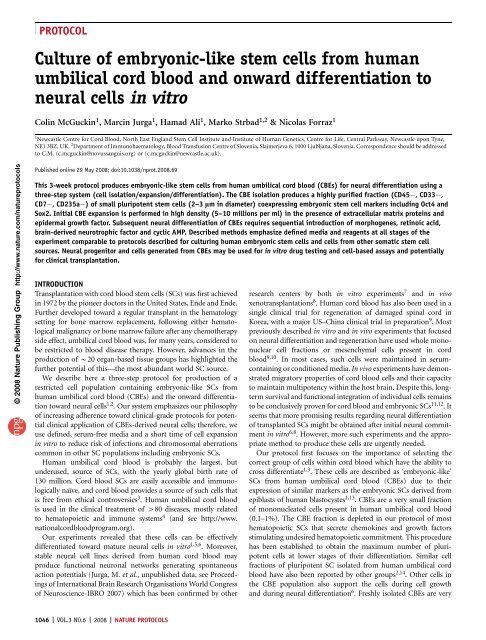
<strong>Culture</strong> <strong>of</strong> <strong>embryonic</strong>-<strong>like</strong> <strong>stem</strong> <strong>cells</strong> <strong>from</strong> <strong>human</strong><br />
umbilical cord blood and onward differentiation to<br />
neural <strong>cells</strong> in vitro<br />
Colin McGuckin 1 , Marcin Jurga 1 , Hamad Ali 1 , Marko Strbad 1,2 & Nicolas Forraz 1<br />
1 Newcastle Centre for Cord Blood, North East England Stem Cell Institute and Institute <strong>of</strong> Human Genetics, Centre for Life, Central Parkway, Newcastle upon Tyne,<br />
NE1 3BZ, UK. 2 Department <strong>of</strong> Immunohaematology, Blood Transfusion Centre <strong>of</strong> Slovenia, Slajmerjeva 6, 1000 Ljubljana, Slovenia. Correspondence should be addressed<br />
to C.M. (c.mcguckin@novussanguis.org) or (c.mcguckin@newcastle.ac.uk).<br />
Published online 29 May 2008; doi:10.1038/nprot.2008.69<br />
This 3-week protocol produces <strong>embryonic</strong>-<strong>like</strong> <strong>stem</strong> <strong>cells</strong> <strong>from</strong> <strong>human</strong> umbilical cord blood (CBEs) for neural differentiation using a<br />
three-step sy<strong>stem</strong> (cell isolation/expansion/differentiation). The CBE isolation produces a highly purified fraction (CD45 , CD33 ,<br />
CD7 , CD235a ) <strong>of</strong> small pluripotent <strong>stem</strong> <strong>cells</strong> (2–3 lm in diameter) coexpressing <strong>embryonic</strong> <strong>stem</strong> cell markers including Oct4 and<br />
Sox2. Initial CBE expansion is performed in high density (5–10 millions per ml) in the presence <strong>of</strong> extracellular matrix proteins and<br />
epidermal growth factor. Subsequent neural differentiation <strong>of</strong> CBEs requires sequential introduction <strong>of</strong> morphogenes, retinoic acid,<br />
brain-derived neurotrophic factor and cyclic AMP. Described methods emphasize defined media and reagents at all stages <strong>of</strong> the<br />
experiment comparable to protocols described for culturing <strong>human</strong> <strong>embryonic</strong> <strong>stem</strong> <strong>cells</strong> and <strong>cells</strong> <strong>from</strong> other somatic <strong>stem</strong> cell<br />
sources. Neural progenitor and <strong>cells</strong> generated <strong>from</strong> CBEs may be used for in vitro drug testing and cell-based assays and potentially<br />
for clinical transplantation.<br />
INTRODUCTION<br />
Transplantation with cord blood <strong>stem</strong> <strong>cells</strong> (SCs) was first achieved<br />
in 1972 by the pioneer doctors in the United States, Ende and Ende.<br />
Further developed toward a regular transplant in the hematology<br />
setting for bone marrow replacement, following either hematological<br />
malignancy or bone marrow failure after any chemotherapy<br />
side effect, umbilical cord blood was, for many years, considered to<br />
be restricted to blood disease therapy. However, advances in the<br />
production <strong>of</strong> B20 organ-based tissue groups has highlighted the<br />
further potential <strong>of</strong> this—the most abundant world SC source.<br />
We describe here a three-step protocol for production <strong>of</strong> a<br />
restricted cell population containing <strong>embryonic</strong>-<strong>like</strong> SCs <strong>from</strong><br />
<strong>human</strong> umbilical cord blood (CBEs) and the onward differentiation<br />
toward neural <strong>cells</strong> 1,2 . Our sy<strong>stem</strong> emphasizes our philosophy<br />
<strong>of</strong> increasing adherence toward clinical-grade protocols for potential<br />
clinical application <strong>of</strong> CBEs-derived neural <strong>cells</strong>; therefore, we<br />
use defined, serum-free media and a short time <strong>of</strong> cell expansion<br />
in vitro to reduce risk <strong>of</strong> infections and chromosomal aberrations<br />
common in other SC populations including <strong>embryonic</strong> SCs.<br />
Human umbilical cord blood is probably the largest, but<br />
underused, source <strong>of</strong> SCs, with the yearly global birth rate <strong>of</strong><br />
130 million. Cord blood SCs are easily accessible and immunologically<br />
naïve, and cord blood provides a source <strong>of</strong> such <strong>cells</strong> that<br />
is free <strong>from</strong> ethical controversies 3 . Human umbilical cord blood<br />
is used in the clinical treatment <strong>of</strong> 480 diseases, mostly related<br />
to hematopoietic and immune sy<strong>stem</strong>s 4 (and see http://www.<br />
nationalcordbloodprogram.org).<br />
Our experiments revealed that these <strong>cells</strong> can be effectively<br />
differentiated toward mature neural <strong>cells</strong> in vitro 1,5,6 . Moreover,<br />
stable neural cell lines derived <strong>from</strong> <strong>human</strong> cord blood may<br />
produce functional neuronal networks generating spontaneous<br />
action potentials (Jurga, M. et al., unpublished data, see Proceedings<br />
<strong>of</strong> International Brain Research Organisations World Congress<br />
<strong>of</strong> Neuroscience-IBRO 2007) which has been confirmed by other<br />
1046 | VOL.3 NO.6 | 2008 | NATURE PROTOCOLS<br />
research centers by both in vitro experiments 7 and in vivo<br />
xenotransplantations 8 . Human cord blood has also been used in a<br />
single clinical trial for regeneration <strong>of</strong> damaged spinal cord in<br />
Korea, with a major US–China clinical trial in preparation 9 . Most<br />
previously described in vitro and in vivo experiments that focused<br />
on neural differentiation and regeneration have used whole mononuclear<br />
cell fractions or mesenchymal <strong>cells</strong> present in cord<br />
blood 9,10 . In most cases, such <strong>cells</strong> were maintained in serumcontaining<br />
or conditioned media. In vivo experiments have demonstrated<br />
migratory properties <strong>of</strong> cord blood <strong>cells</strong> and their capacity<br />
to maintain multipotency within the host brain. Despite this, longterm<br />
survival and functional integration <strong>of</strong> individual <strong>cells</strong> remains<br />
to be conclusively proven for cord blood and <strong>embryonic</strong> SCs 11,12 .It<br />
seems that more promising results regarding neural differentiation<br />
<strong>of</strong> transplanted SCs might be obtained after initial neural commitment<br />
in vitro 6,8 . However, more such experiments and the appropriate<br />
method to produce these <strong>cells</strong> are urgently needed.<br />
Our protocol first focuses on the importance <strong>of</strong> selecting the<br />
correct group <strong>of</strong> <strong>cells</strong> within cord blood which have the ability to<br />
cross differentiate 1,3 . These <strong>cells</strong> are described as ‘<strong>embryonic</strong>-<strong>like</strong>’<br />
SCs <strong>from</strong> <strong>human</strong> umbilical cord blood (CBEs) due to their<br />
expression <strong>of</strong> similar markers as the <strong>embryonic</strong> SCs derived <strong>from</strong><br />
epiblasts <strong>of</strong> <strong>human</strong> blastocystes 1,13 . CBEs are a very small fraction<br />
<strong>of</strong> mononucleated <strong>cells</strong> present in <strong>human</strong> umbilical cord blood<br />
(0.1–1%). The CBE fraction is depleted in our protocol <strong>of</strong> most<br />
hematopoietic SCs that secrete chemokines and growth factors<br />
stimulating undesired hematopoietic commitment. This procedure<br />
has been established to obtain the maximum number <strong>of</strong> pluripotent<br />
<strong>cells</strong> at lower stages <strong>of</strong> their differentiation. Similar cell<br />
fractions <strong>of</strong> pluripotent SC isolated <strong>from</strong> <strong>human</strong> umbilical cord<br />
blood have also been reported by other groups 2,14 .Other<strong>cells</strong>in<br />
the CBE population also support the <strong>cells</strong> during cell growth<br />
and during neural differentiation 6 . Freshly isolated CBEs are very
© 2008<br />
Nature<br />
Publishing<br />
Group<br />
http:<br />
/ / www.<br />
nature.<br />
com/<br />
natureprotocols<br />
sensitive; therefore, we plate them initially in a high concentration<br />
(5–10 million <strong>cells</strong> per 1 ml) in TPOFLK (thrombopoietin, flt-3<br />
ligand and c-kit ligand) medium supplemented with extracellular<br />
matrix (ECM) proteins (collagen type IVand fibronectin) to reduce<br />
their apoptosis 14,15 . Initial high cell density and ECM proteins are<br />
crucial for CBEs survival and formation <strong>of</strong> aggregates similar to<br />
embryoid bodies. Appearance <strong>of</strong> embryoid bodies is the first<br />
phenotypic hallmark <strong>of</strong> proper in vitro growth <strong>of</strong> CBEs 1,14 .Such<br />
aggregation promotes cell–cell interactions and secretion <strong>of</strong> growth<br />
factors, which play a major role in cell survival. For this protocol,<br />
we highly recommend dynamic cell culture conditions based on<br />
observation <strong>of</strong> cell phenotypic changes and to adjust the timing<br />
regime for the CBE culture. The following steps are crucial for cell<br />
survival, expansion and differentiation: (i) formation <strong>of</strong> floating<br />
aggregates (16–24 h after isolation), (ii) size and number <strong>of</strong> floating<br />
<strong>cells</strong>/aggregates (1–7 d after isolation) and (iii) cell adhesion and<br />
differentiation (1–3 weeks after isolation).<br />
The expansion step where growth factors are introduced (epidermal<br />
growth factor (EGF), basic fibroblast growth factor (bFGF)<br />
and <strong>stem</strong> cell factor (SCF)) begins just after aggregates are formed<br />
(usually 16–24 h after CBE isolation). This step is important to<br />
increase number <strong>of</strong> CBEs as the initial number present in cord<br />
blood is B1 million (for an average cord blood volume <strong>of</strong> 80 ml).<br />
Presence <strong>of</strong> EGF is important for initial epithelial and neural<br />
commitment <strong>of</strong> CBEs. Through the expansion step, CBEs increase<br />
their size <strong>from</strong> 2–3 to 410 mm in diameter and begin to adhere<br />
spontaneously. However, it is worth emphasizing that prolonged<br />
in vitro expansion may trigger genetic instability (e.g., polyploidy)<br />
as reported previously 16 .<br />
Cell/aggregate adhesion and neural differentiation is further<br />
stimulated in our protocol by retinoic acid (RA—a morphogene<br />
with wide-range action) and brain-derived neurotrophic factor<br />
(BDNF—a major growth factor responsible for neural differentiation).<br />
After adhesion, most <strong>of</strong> the <strong>cells</strong> already coexpress Nestin and<br />
GFAP (markers <strong>of</strong> neural SCs). When their morphology changes to<br />
unipolar, we introduce dibutyryl cyclic AMP (dBcAMP) to stimulate<br />
their terminal differentiation 17 . We would <strong>like</strong> to stress that<br />
phenotype changes described in our protocol should be regarded as<br />
critical steps in neural cell development. These phenotypic changes<br />
MATERIALS<br />
REAGENTS<br />
.Umbilical cord blood unit ! CAUTION Parents’ full and informed consents,<br />
in adherence with the law, must be obtained before the delivery. All the<br />
hospital ethical requirements must be fulfilled.<br />
.Ficoll-Paque PREMIUM (GE Healthcare, cat. no. 17-5442-02)<br />
.DMEM/F-12 + GlutaMax (GIBCO, cat. no. 31331093)<br />
.B27 supplement (GIBCO, cat. no. 17504044)<br />
.N2 supplement (GIBCO, cat. no. 17502048)<br />
.Recombinant <strong>human</strong> SCF (rh SCF; ImmunoTools, cat. no. 11343325)<br />
m CRITICAL Growth factors and morphogens adhere to walls <strong>of</strong> containers<br />
and denature at low concentrations. Store them in appropriate aliquots<br />
according to the manufacturer’s recommendations at 20 1C.<br />
.Recombinant <strong>human</strong> thrombopoietin (rh TPO; ImmunoTools, cat. no.<br />
11343615)<br />
.Recombinant <strong>human</strong> Flt3-ligand (rh Flt3; ImmunoTools, cat. no. 11343305)<br />
.Recombinant <strong>human</strong> SCF (rh SCF; ImmunoTools, cat. no. 11343322)<br />
.Recombinant <strong>human</strong> FGF, basic (rh FGF-basic; ImmunoTools, cat. no.<br />
11343627)<br />
.Recombinant <strong>human</strong> EGF (rh EGF; ImmunoTools, cat. no. 11343407)<br />
PROTOCOL<br />
may slightly vary in time depending on age and quality <strong>of</strong> CBEs 18 .<br />
However, the above-mentioned phenotypic hallmarks should be<br />
easily distinguished in simple light microscope analysis enabling<br />
progression to the next step in the protocol. Another point <strong>of</strong> note,<br />
is that expression <strong>of</strong> mRNA, proteins (e.g., transcription factors)<br />
and their intracellular localization accompany phenotypic changes<br />
and have already been investigated elsewhere 1,14,19 .<br />
The limitations <strong>of</strong> this method are the very low number <strong>of</strong><br />
lineage-negative CBEs after their isolation (the mononuclear <strong>cells</strong><br />
depleted <strong>of</strong> CD45, CD33, CD7 and CD235a) and the simultaneous<br />
requirement for a high cell concentration for initial plating, which<br />
is crucial to support CBEs survival and formation <strong>of</strong> aggregates. For<br />
this reason, low volumes <strong>of</strong> cord blood or those cord blood units<br />
with limited numbers <strong>of</strong> CBEs might not allow the effective<br />
isolation <strong>of</strong> a decent amount <strong>of</strong> SCs. Therefore, further experiments<br />
need to be performed to develop a protocol that allows expansion<br />
<strong>of</strong> extremely small amounts (semi-clonal) <strong>of</strong> CBEs present in some<br />
cord blood units.<br />
For CBE isolation, expansion and differentiation, we describe<br />
defined serum-free culture media. Growth factors present in<br />
these culture media are introduced sequentially to promote CBE<br />
survival, expansion, differentiation and maturation toward neural<br />
phenotypes. The sequence <strong>of</strong> growth factors must be applied<br />
according to phenotypic changes in the cell morphology. Defined<br />
media, free <strong>from</strong> animal products, will allow the use <strong>of</strong> CBEs<br />
for drug testing and detailed analysis <strong>of</strong> factors regulating cell fates<br />
and for direct clinical transplantation, particularly for neurodegenerative<br />
diseases including stroke and also following trauma<br />
as, for instance, with spinal cord injury. But more basic and<br />
clinical research must be carried out by the scientific community<br />
worldwide to unravel this potential fully. It is important to note<br />
that beyond neural tissue production, <strong>cells</strong> <strong>of</strong> all three germ layers<br />
have been produced in our laboratory with this population.<br />
Because CBE populations can be directed to several different<br />
organ-based lineages, including hepatic, pancreatic and endothelial-<strong>like</strong><br />
tissues as an example, it is important to be careful<br />
with regard to the choice and source <strong>of</strong> growth factors and<br />
bioscaffolds to enable translation <strong>of</strong> those discoveries into biotechnology<br />
and clinical applications.<br />
.Fibronectin <strong>from</strong> <strong>human</strong> plasma (Sigma-Aldrich, cat. no. 86088-83-7)<br />
(see REAGENT SETUP)<br />
.Collagen type IV (Sigma-Aldrich, cat. no. C5533) (see REAGENT SETUP)<br />
.Heparin sodium salt (Sigma-Aldrich, cat. no. H4784-1G) (see REAGENT<br />
SETUP)<br />
.Penicillin–streptomycin (GIBCO, cat. no. 15070063)<br />
.Fungizone (GIBCO, cat. no. 15290026)<br />
.BDNF (ImmunoTools, cat. no. 11343375) (see REAGENT SETUP)<br />
.RA (Sigma-Aldrich, cat. no. R2625) (see REAGENT SETUP)<br />
.dBcAMP (Sigma-Aldrich, cat. no. D0627) m CRITICAL Only in the form<br />
<strong>of</strong> dBcAMP can the compound penetrate through cell membranes<br />
appropriately.<br />
.DMSO (Sigma-Aldrich, cat. no. D8418)<br />
.Deionized sterile water<br />
.10 PBS (Sigma-Aldrich, cat. no.P5493-1L)<br />
.1 Hanks’ balanced salt solution (HBSS) without CaCl 2 and MgCl 2<br />
(GIBCO–Invitrogen, cat. no. 14175-053)<br />
.ZENLAB 4.5—<strong>human</strong> albumin 4.5% solution Ph.Eur (Bio Products<br />
Laboratory, cat. no. ADAN7459)<br />
NATURE PROTOCOLS | VOL.3 NO.6 | 2008 | 1047
© 2008<br />
Nature<br />
Publishing<br />
Group<br />
http:<br />
/ / www.<br />
nature.<br />
com/<br />
natureprotocols<br />
PROTOCOL<br />
TABLE 1 | Abs used for flow cytometry (fluorescence-activated cell sorting) estimation <strong>of</strong> purity <strong>of</strong> isolated CBEs.<br />
Ab Type Volume used (ll) Cat. no. Distributor<br />
CD14 (pacific blue) Mouse IgG2A 5 558121 BD Pharmingen<br />
CD34 (PE/Cy7) Mouse IgG1 5 348811 BD<br />
CD45 (APC/Cy7) Mouse IgG1 5 557833 BD Pharmingen<br />
CD90 (FITC) Mouse IgG1 2 555595 BD Pharmingen<br />
CD133/1 (APC) Mouse IgG1 10 130-090-826 Miltenyi Biotech<br />
SSEA-4 (PE) Mouse IgG3 10 FAB1435P R & D Sy<strong>stem</strong>s<br />
.Gentran 40–Dextran 40 intravenous infusion BP 10% wt/vol in sodium<br />
chloride intravenous infusion BP 0.9% wt/vol (Baxter Healthcare Ltd,<br />
cat. no. B5043)<br />
.Mouse anti-<strong>human</strong> anti-GLyA/CD235a (DakoCytomation, cat. no. M0819)<br />
.Mouse IgG1 anti-<strong>human</strong> CD45 (Autogen Bioclear, cat. no. ABX252)<br />
.Mouse IgG1 anti-<strong>human</strong> CD33 (Autogen Bioclear, cat. no. AB163)<br />
.Mouse IgG3 anti-<strong>human</strong> CD7 (Autogen Bioclear, cat. no. AB164)<br />
.g-Globulin <strong>from</strong> <strong>human</strong> blood (Fluka Chemie GmbH, cat. no. 49000-5g)<br />
.Dynabeads PanMouse IgG (Dynal Biotech ASA, cat. no. 11042)<br />
.Abs for fluorescence-activated cell sorting (FACS) analysis: see Table 1<br />
.Primary Abs to provide markers for confocal analysis <strong>of</strong> stages <strong>of</strong> neural<br />
differentiation: see Table 2<br />
.Secondary Abs: Alexa 594 rabbit anti-goat (IgG H+L) (Invitrogen, cat. no.<br />
A11080), Alexa 594 goat anti-rabbit (IgG H+L) (Invitrogen, cat. no.<br />
A-11012), Alexa 488 rabbit anti-mouse (IgG H+L) (Invitrogen, cat. no.<br />
A-11059) and Alexa 488 goat anti-mouse (IgG1) (Invitrogen, cat. no. A-21121)<br />
.Washing buffer (see REAGENT SETUP)<br />
.Working buffer (see REAGENT SETUP)<br />
EQUIPMENT<br />
.15-ml Universal tubes (Greiner Bio-One, cat. no. 118271)<br />
.50-ml Universal tubes (Greiner Bio-One, cat. no. 210261)<br />
.10-ml Plastic pipettes (Greiner Bio-One, cat. no. 607180)<br />
.24-Well plate (Greiner Bio-One, cat. no. 662160)<br />
.96-Well plate, U-shape (Greiner Bio-One, cat. no. 650180)<br />
.Plastic pipettes (Delta Lab, cat. no. 200037)<br />
.Pipettes and tips (Jencons)<br />
.Shandon-thermo centrifuge (Thermo Scientific)<br />
.Syringes, 50 ml (Terumo Europe N.V.)<br />
.Syringe filters, 0.22 mm (PALL Life Sciences, cat. no. PN 4612)<br />
.Magnetic Particle Concentrator (MPC) (Dynal MPC-I; Dynal AS,<br />
cat. no. 120-01)<br />
.MPC (Dynal MPC-S; Dynal AS, cat. no. 120-00)<br />
.Cover glass (VWR, cat. no. 613-0149)<br />
.Microscope glass slides (VWR, cat. no. 631-0149)<br />
.Centrifuge (Rotanta 460R; Zentrifugen)<br />
.CO 2 incubator Hera Cell 240 (Heraeus)<br />
.Millipore Direct-Q (Millipore, cat. no. ZRQS5005Y)<br />
.Cell counter Coulter A C T diff2 (Becton Coulter)<br />
.Trypan Blue solution 0.4% (Sigma-Aldrich, cat. no. T8154)<br />
.Chamber for cell counting—Hemacytometer (Hauser Scientific,<br />
cat. no. 1492)<br />
REAGENTS SETUP<br />
See Table 3 for reagents used to prepare serum-free harvesting (SF-TPO) and<br />
expansion (SF-EFS) media.<br />
See Table 4 for the reagents used to prepare serum-free media for neural<br />
differentiation (SF-NEURO/D) and maturation (SF-NEURO/M). m CRITICAL<br />
Both neural differentiation media and neural maturation media tubes should<br />
be wrapped with aluminum foil because RA is light sensitive. All media<br />
should be stored at 4 1C for no 42 weeks.! CAUTION All media should<br />
be filtered before use using 0.22-mm filter to reduce contamination risks<br />
with pathogens.<br />
Fibronectin Reconstitute the powder by properly dissolving it in sterile water<br />
to give up a stock concentration <strong>of</strong> 1 mg ml 1 .<br />
Collagen type IV Reconstitute the powder by properly dissolving it in<br />
0.25% acetic acid to give a stock concentration <strong>of</strong> 1 mg ml 1 .<br />
Heparin Prepare a stock concentration <strong>of</strong> 1 mg ml 1 by dissolving heparin<br />
in DMEM/F12. m CRITICALThe solution must be filtered using 0.22-mmfilterto<br />
reduce contamination risks with pathogens.<br />
TABLE 2 | Abs used for confocal analysis <strong>of</strong> subsequent stages <strong>of</strong> neural differentiation.<br />
Primary Ab<br />
Undifferentiated <strong>cells</strong><br />
Host Isotype Suggested working dilution Manufacturer Cat. no.<br />
Oct4 Goat IgG 1:500 Abcam ab27985<br />
Sox2 Mouse IgG2a 1:50 R & D Sy<strong>stem</strong>s MAB2018<br />
Nanog Goat IgG 1:20 R & D Sy<strong>stem</strong>s AF1997<br />
Ki67 Mouse IgG1 1:100 Novocastra L111853<br />
Neural progenitors<br />
Nestin Mouse IgG1 1:100 R & D Sy<strong>stem</strong>s MAB1259<br />
GFAP* Rabbit IgG 1:1,000 Abcam Ab7779<br />
Neuroblasts and mature neurons<br />
NF-200 Mouse IgG1 1:800 Sigma-Aldrich N1042<br />
Doublecortin Rabbit IgG 1:500 Abcam Ab28941<br />
MAP2 Mouse IgG 1:1,000 Sigma-Aldrich M4403<br />
TUJ1 Rabbit IgG 1:2,000 Covance PRB435P<br />
NeuN Mouse IgG1 1:100 Chemicon MAB377<br />
PSD95 Rabbit IgG 1:500 Abcam Ab18258<br />
Astroglial <strong>cells</strong><br />
S100b Rabbit IgG 1:5,000 Swant 37A<br />
*Expression <strong>of</strong> GFAP is expressed by both astroglial <strong>cells</strong> and neural progenitors and should be considered a marker <strong>of</strong> undifferentiated neurons when coexpressed with Nestin.<br />
1048 | VOL.3 NO.6 | 2008 | NATURE PROTOCOLS
© 2008<br />
Nature<br />
Publishing<br />
Group<br />
http:<br />
/ / www.<br />
nature.<br />
com/<br />
natureprotocols<br />
BDNF Reconstitute the powder by dissolving it in sterile water to give a stock<br />
concentration <strong>of</strong> 10 mg ml 1 .<br />
RA Reconstitute the powder by dissolving it in DMSO to give a stock<br />
concentration <strong>of</strong> 0.05 M. ! CAUTION All vials containing RA should be wrapped<br />
with aluminum foil because RA is light sensitive. Degradation is a potential<br />
reason for failure <strong>of</strong> the protocol.<br />
Sterile water Water is deionized and filtered using Millipore Direct-Q with<br />
Millipak Express 20 filter—0.22 mm (Millipore, cat. no. 02001) and sterilized<br />
using dry heat autoclave. ! CAUTION After autoclaving, check the sterility<br />
indicator.<br />
Washing buffer: PBS/0.2% Albumin/Dextran Take 45.6 ml <strong>of</strong> PBS, mix it<br />
with the 4.4 ml <strong>of</strong> <strong>human</strong> albumin solution and add 50 ml <strong>of</strong> Dextran 40. Mix<br />
well and pass the buffer using a syringe (Terumo syringe 50 ml; Terumo Europe<br />
N.V.) equipped with sterile PALL Acrodisc syringe filter, 0.22 mm (PALLLife<br />
Sciences).<br />
Note: To prepare 1 PBS: dilute 10 ml <strong>of</strong> 10 PBS without calcium (CaCl2)<br />
and magnesium (MgCl2) in 90 ml sterile deionized water.<br />
Working buffer: HBSS/0.2% Albumin Add 2.2 ml <strong>of</strong> <strong>human</strong> albumin to<br />
47.8 ml HBSS. Mix well and pass the buffer using a syringe (Terumo syringe<br />
50 ml; Terumo Europe N.V.) equipped with sterile PALL Acrodisc syringe filter,<br />
0.22 mm (PALL Life Sciences). m CRITICAL pH <strong>of</strong> working and washing buffers<br />
should range between 7.2 and 7.4 to maintain cellular homeostasis.<br />
PROCEDURE<br />
Umbilical cord blood collection TIMING B5 min<br />
1| Collect umbilical cord blood units.<br />
m CRITICAL STEP Parents’ informed consents must be obtained<br />
before the delivery, and all the hospital ethical requirements<br />
must be fulfilled.<br />
m CRITICAL STEP During and after isolation, mix the collection bag gently to prevent coagulation <strong>of</strong> the cord blood. Processing <strong>of</strong><br />
the cord blood should be carried out as quickly as possible to prevent microreactions within the pack and/or hemolysis.<br />
2| Count the <strong>cells</strong> in the blood sample, using 80 ml <strong>of</strong> blood (see Box 1).<br />
TABLE 4 | Serum-free media for neural differentiation and maturation.<br />
Differentiation medium (SF-NEURO/D 50 ml)<br />
DMEM/F12 45 ml<br />
B27 (1:50) 1 ml<br />
N2 (1:25) 2 ml<br />
EGF (10 ng ml 1 ) 5 ml<br />
BDNF (10 ng ml 1 ) 50 ml<br />
RA (5 mM) 5 ml<br />
Collagen IV (1 mg ml 1 ) 50 ml<br />
Fibronectin (1 mg ml 1 ) 50 ml<br />
Pen/Strep (1:100) 500 ml<br />
Fungizone (1:100) 500 ml<br />
Maturation medium (SF-NEURO/M 50 ml)<br />
DMEM/F12 46 ml<br />
B27 (1:50) 1 ml<br />
N2 (1:25) 2 ml<br />
EGF (10 ng ml 1 ) 5 ml<br />
NGF (10 ng ml 1 ) 25 ml<br />
BDNF (10 ng ml 1 ) 50 ml<br />
RA (5 mM) 5 ml<br />
dBcAMP (100 mM) 50 ml<br />
Collagen IV (1 mg ml 1 ) 50 ml<br />
Fibronectin (1 mg ml 1 ) 50 ml<br />
Pen/Strep (1:100) 500 ml<br />
Fungizone (1:100) 500 ml<br />
BDNF, brain-derived neurotrophic factor; dBcAMP, dibutyryl cyclic AMP; EGF, epidermal growth factor;<br />
NGF, nerve growth factor; RA, retinoic acid.<br />
TABLE 3 | Serum-free media for CBEs harvesting and expansion.<br />
Harvesting medium (SF-TPO 50 ml)<br />
DMEM/F12 47 ml<br />
B27 (1:100) 0.5 ml<br />
N2 (1:50) 1 ml<br />
SCF (20 ng ml 1 ) 10 ml<br />
TPO (10 ng ml 1 ) 5 ml<br />
Flt3 (50 ng ml 1 ) 25 ml<br />
Fibronectin (5 mg ml 1 ) 250 ml<br />
Pen/Strep (1:100) 500 ml<br />
Fungizone (1:100) 500 ml<br />
Expansion medium (SF-EFS 50 ml)<br />
DMEM/F12 47 ml<br />
B27 (1:100) 0.5 ml<br />
N2 (1:50) 1 ml<br />
SCF (5 ng ml 1 ) 2.5 ml<br />
EGF (20 ng ml 1 ) 10 ml<br />
bFGF (20 ng ml 1 ) 10 ml<br />
Heparin (5 mg ml 1 ) 250 ml<br />
Fibronectin (5 mg ml 1 ) 250 ml<br />
Pen/Strep (1:100) 500 ml<br />
Fungizone (1:100) 500 ml<br />
bFGF, basic fibroblast growth factor; EGF, epidermal growth factor; SCF, <strong>stem</strong> cell factor;<br />
TPO, thrombopoietin.<br />
PROTOCOL<br />
3| According to the cell count, calculate appropriate<br />
volume to collect 100,000 <strong>of</strong> <strong>cells</strong> for flow cytometry (FACS)<br />
immunostaining and analysis (see Box 2, andTable 1).<br />
Isolation <strong>of</strong> umbilical cord blood mononuclear <strong>cells</strong> using<br />
Ficoll density gradient TIMING B2 h<br />
4| Dilute the blood <strong>from</strong> Step 1 (1:1) with sterile 1 PBS<br />
in a universal 50-ml tube. Recommended minimal volume<br />
is 20 ml.<br />
5| Layer the diluted blood slowly on 10 ml <strong>of</strong> Ficoll layer<br />
(ratio 4:1) in another 50-ml tube.<br />
m CRITICAL STEP Do not mix the tube; make sure the layers are<br />
not disturbed.<br />
6| Centrifuge the layered blood samples at 400g for 30 min at<br />
room temperature (20–22 1C). Acceleration and de-acceleration<br />
should be kept as low as possible to avoid any disturbance to<br />
<strong>cells</strong> in the white layer (Fig. 1).<br />
7| Collect the white layer using a plastic pipette in a new<br />
universal tube. Some <strong>cells</strong> might attach to the side <strong>of</strong> the<br />
tube in a form <strong>of</strong> a pellet, ‘rub’ them gently using the end <strong>of</strong><br />
the pipette and collect them (Fig. 1).<br />
! CAUTION Collecting too much <strong>of</strong> plasma might increase<br />
the chance <strong>of</strong> platelet contamination. Platelets can release<br />
significant levels <strong>of</strong> chemokines which (patient dependent)<br />
can change the stimulation pattern <strong>of</strong> the <strong>cells</strong>.<br />
NATURE PROTOCOLS | VOL.3 NO.6 | 2008 | 1049
© 2008<br />
Nature<br />
Publishing<br />
Group<br />
http:<br />
/ / www.<br />
nature.<br />
com/<br />
natureprotocols<br />
PROTOCOL<br />
BOX 1 | CELL COUNTING<br />
One part <strong>of</strong> the blood sample (60 ml) is used for counting the <strong>cells</strong> with automatic hematological cell counter Coulter A C T diff2 (Beckton<br />
Coulter), where all blood <strong>cells</strong> are counted including mononuclear <strong>cells</strong>, red blood <strong>cells</strong> and platelets.<br />
Second part <strong>of</strong> the sample (20 ml) is used for assessing the viability <strong>of</strong> the <strong>cells</strong> using Trypan Blue exclusion method and hemacyometer, as follows:<br />
1. Place a suitable volume <strong>of</strong> a cell suspension (10–20 ml) in an appropriate tube and add an equal volume <strong>of</strong> 0.4% Trypan Blue and gently mix.<br />
2. Allow to stand for 5 min at room temperature.<br />
3. Place 10 ml <strong>of</strong> stained <strong>cells</strong> in a hemacytometer and count the number <strong>of</strong> viable (unstained) and dead <strong>cells</strong> stained blue.<br />
4. Calculate the average number <strong>of</strong> unstained <strong>cells</strong> in each quadrant and multiply by 2 10 4 to find number <strong>of</strong> <strong>cells</strong> per ml. The percentage <strong>of</strong><br />
viable <strong>cells</strong> is the number <strong>of</strong> viable <strong>cells</strong> divided by the number <strong>of</strong> dead and viable <strong>cells</strong>.<br />
8| Centrifuge the cell suspension at 630g for 7 min at room temperature. Acceleration should be maximum with medium<br />
de-acceleration.<br />
9| Resuspend the pellet in B2 ml<strong>of</strong>1 PBS.<br />
10| Centrifuge the sample as described in Step 8.<br />
11| Take as much supernatant as possible without disturbing the pellet.<br />
12| Resuspend the pellet in 1 ml <strong>of</strong> 1 PBS.<br />
13| Count the <strong>cells</strong> using hematological cell counter and hemacytometer and asses the viability using Trypan Blue exclusion<br />
method (as described in Box 1).<br />
14| Check for abnormal levels <strong>of</strong> cell aggregation and possible platelet activation, which may disturb further isolation (Fig. 2).<br />
m CRITICAL STEP Platelets, when activated, form a fibrin net where mononuclear <strong>cells</strong> are trapped. That can be visible as fibrils in<br />
supernatant or as aggregates under the microscope.<br />
? TROUBLESHOOTING<br />
15| According to the cell count, calculate the appropriate volume to collect 10 5 <strong>cells</strong> for later comparison and troubleshooting<br />
by flow cytometry (FACS) immunostaining and analysis (as described in Box 2 and Table 1).<br />
Depletion <strong>of</strong> lineage positive <strong>cells</strong> TIMING B4 h<br />
16| To the remaining cell suspension volume (between 900 and 1,000 ml working volume), add 500 ml <strong>of</strong> 2% <strong>human</strong><br />
g-globulins diluted in PBS or working buffer.<br />
m CRITICAL STEP All operations must be performed in a laminar flow sterile hood unless stated otherwise. All reagents<br />
(Abs, working buffer, g-globulins, Dynabeads) and operations (e.g., centrifuging) should be used cooled at 4 1C oronice.<br />
17| Incubate <strong>cells</strong> for 20 min at 4 1C or on ice to block nonspecific Fc receptors (during the incubation, gently rock or rotate<br />
the tube to achieve homogenous binding).<br />
! CAUTION If automated rotator is not available, gently rotate tube manually every few minutes to avoid clumping and cellular<br />
adherence.<br />
18| Add mouse monoclonal anti-<strong>human</strong> anti-GlyA (CD235a); use 0.22 mg <strong>of</strong>Abper106<strong>cells</strong>, and mouse monoclonal<br />
anti-<strong>human</strong> anti-CD45; use 0.5 mg Abper106<strong>cells</strong>. Incubate <strong>cells</strong> for 30 min at 4 1C or on ice with gentle rocking or rotating.<br />
19| During the 30-min incubation, prepare the beads—monoclonal anti-mouse IgG Ab (PanMouse Dynabeads): vortex the<br />
beads before use and use 50 ml <strong>of</strong>beadsper10 7 <strong>cells</strong>.<br />
! CAUTION Be sure you are using mouse anti-<strong>human</strong> primary Abs and beads coupled to them (panmouse Abs conjugated<br />
with beads).<br />
BOX 2 | FLUORESCENCE-ACTIVATED CELL SORTING (FACS) ANALYSIS<br />
Flow cytometric analysis <strong>of</strong> <strong>cells</strong> taken at various stages <strong>of</strong> this procedure is an optional step, but it can be helpful in estimating the number <strong>of</strong><br />
undifferentiated <strong>stem</strong> <strong>cells</strong> and can provide further information about the purity <strong>of</strong> the isolated fraction.<br />
To prepare <strong>cells</strong> for fluorescence-activated cell sorting (FACS) analysis, add an appropriate volume <strong>of</strong> Abs (see Table 1) into cell suspension<br />
(10 5 <strong>cells</strong> in 100 ml), mix them and incubate at room temperature in dark for 20 min. Wash the <strong>cells</strong> according to the manufacturer’s<br />
recommendation or using BD wash/lyse assistant and perform the FACS analysis 18 . FACS analysis gives the indication <strong>of</strong> purity <strong>of</strong> CBE fraction<br />
(expected negative signal <strong>from</strong> CD45 and CD34 antibodies) and presence <strong>of</strong> <strong>embryonic</strong>-<strong>like</strong> <strong>cells</strong> (expected positive signal <strong>from</strong> SSEA-4 and<br />
CD133 antibodies) (see Table 1).<br />
1050 | VOL.3 NO.6 | 2008 | NATURE PROTOCOLS
© 2008<br />
Nature<br />
Publishing<br />
Group<br />
http:<br />
/ / www.<br />
nature.<br />
com/<br />
natureprotocols<br />
20| Washthebeadswiththesamevolumeoratleast1ml<br />
<strong>of</strong> washing buffer and mix.<br />
! CAUTION Avoid forming bubble/foam. Formation <strong>of</strong><br />
bubbles/foam can disturb migration/connection <strong>of</strong> beads to<br />
the well <strong>of</strong> the tube and decrease final cell yield at the end<br />
<strong>of</strong> the procedure.<br />
21| Apply the tube to a Dynal MPC for 2 min. Allow the<br />
beads to attach to the wall <strong>of</strong> the tube. Discard the<br />
supernatant.<br />
22| Take the tube <strong>from</strong> magnet and repeat the washing<br />
Steps 19 and 20 two times (total number <strong>of</strong> washes is three).<br />
23| After the 30-min incubation, wash excess <strong>of</strong> Ab <strong>from</strong><br />
sample (<strong>from</strong> Step 18) using 10 ml working buffer.<br />
24| Centrifuge the cell suspension at 630g for 7 min at 4 1C.<br />
The centrifuge should be chilled before use; acceleration<br />
should be maximum with medium de-acceleration.<br />
25| Discard supernatant and resuspend the <strong>cells</strong> using<br />
500 ml working buffer per 10 8 <strong>cells</strong>.<br />
! CAUTION Avoid forming bubble/foam. Formation <strong>of</strong> bubbles/foam can disturb migration/connection <strong>of</strong> beads to the well <strong>of</strong><br />
the tube and decrease final cell yield at the end <strong>of</strong> the procedure.<br />
26| Add washed beads (<strong>from</strong> Step 22) to the sample and incubate for 30 min at 4 1C or on ice with gentle rocking or rotating.<br />
27| Apply tube to MPC for 2 min.<br />
28| Collect the supernatant in a new sterile tube and mark as GlyA/CD45 negative <strong>cells</strong>.<br />
m CRITICAL STEP Do not discard supernatant.<br />
29| Rinse the positive fraction (beads remaining in the tube <strong>from</strong> Step 27) two times by adding 4 ml working buffer and reapplying<br />
the tube to MPC, and each time add the supernatant obtained to the tube containing GlyA/CD45 negative <strong>cells</strong> (<strong>from</strong> Step 28).<br />
m CRITICAL STEP Reapply the tube to MPC at least three times to remove all positively marked <strong>cells</strong> completely.<br />
30| Centrifuge the GlyA/CD45 negative <strong>cells</strong> as described in Step 24.<br />
31| Remove supernatant and resuspend the <strong>cells</strong> using 500 ml working buffer.<br />
32| Enumerate the <strong>cells</strong> and asses viability using Trypan Blue exclusion method (as described in Box 1).<br />
m CRITICAL STEP Check the aggregation <strong>of</strong> <strong>cells</strong> and possible platelet activation, which may disturb further isolation (as described<br />
in Step 14).<br />
? TROUBLESHOOTING<br />
33| Incubate sample for 30 min at 4 1C or on ice simultaneously with mouse monoclonal anti-<strong>human</strong> CD45/CD33/CD7 Ab.<br />
Use 1.0 mg <strong>of</strong>Abper106<strong>cells</strong>. *<br />
Figure 2 | Contamination <strong>of</strong> platelets in culture. The asterisk shows an<br />
aggregate <strong>of</strong> activated platelets, and arrow indicates clamped <strong>stem</strong> cell<br />
within. Scale bar, 10 mm.<br />
B<br />
F R<br />
Figure 1 | Separation <strong>of</strong> cord blood with Ficoll before (left tube) and after<br />
(right tube) centrifugation. Left tube shows distinct layer <strong>of</strong> diluted cord<br />
blood (B) over the Ficoll layer (F). After centrifugation, there should be<br />
distinct layers as shown in right tube (<strong>from</strong> bottom to the top: red blood<br />
<strong>cells</strong> and Ficoll (R), mononuclear <strong>cells</strong> with <strong>stem</strong> cell population (white layer)<br />
(W), plasma with platelets (P)).<br />
34| During the incubation, prepare beads as described in<br />
Steps 19–22.<br />
35| Wash excess <strong>of</strong> Ab <strong>from</strong> sample <strong>from</strong> Step 33 by adding<br />
10 ml <strong>of</strong> working buffer.<br />
36| Centrifuge the sample as described in Step 24.<br />
37| Remove supernatant and resuspend the <strong>cells</strong> using 500 ml<br />
working buffer.<br />
! CAUTION Avoid forming bubble/foam. Formation <strong>of</strong><br />
bubbles/foam can disturb migration/connection <strong>of</strong> beads to<br />
the well <strong>of</strong> the tube and decrease final cell yield at the end<br />
<strong>of</strong> the procedure.<br />
NATURE PROTOCOLS | VOL.3 NO.6 | 2008 | 1051<br />
P<br />
W<br />
PROTOCOL
© 2008<br />
Nature<br />
Publishing<br />
Group<br />
http:<br />
/ / www.<br />
nature.<br />
com/<br />
natureprotocols<br />
PROTOCOL<br />
38| Apply washed beads (<strong>from</strong> Step 35) to the sample and<br />
incubate for 30 min at 4 1C oronicewithgentlerockingor<br />
rotating.<br />
39| Apply tube to MPC for 2 min.<br />
40| Collect the supernatant in a new sterile tube and mark<br />
as lineage-negative <strong>cells</strong>.<br />
m CRITICAL STEP Do not discard supernatant.<br />
41| Rinse the positive fraction (beads remaining in the tube<br />
<strong>from</strong> Step 37) two times by adding 4 ml working buffer and<br />
reapplying the tube to MPC, and each time add the supernatant<br />
obtained to the tube containing lineage-negative <strong>cells</strong><br />
(<strong>from</strong> Step 40).<br />
m CRITICAL STEP Reapply the tube to MPC at least three times<br />
to remove all positively marked <strong>cells</strong> completely.<br />
42| Take a small sample <strong>of</strong> cell suspension (10–25 ml), and under the microscope check for the presence <strong>of</strong> magnetic beads<br />
using a glass slide or hemacytometer (Fig. 3a). Magnetic beads will affect cell viability (Fig. 3b). If necessary, repeat Step 37<br />
until no magnetic beads are observed under the microscope.<br />
? TROUBLESHOOTING<br />
43| Centrifuge the lineage-negative <strong>cells</strong> as described in Step 24.<br />
44| Remove supernatant and resuspend the <strong>cells</strong> in 500 ml chilled harvesting culture medium SF-TPO (see Table 3 and Fig. 4).<br />
m CRITICAL STEP Avoid rapid changes in temperature—adjust the temperature <strong>of</strong> culture medium and introduce it by dropping<br />
to cell pellet.<br />
45| Enumerate the <strong>cells</strong> and assess viability using Trypan Blue exclusion method, as described in Box 1.<br />
m CRITICAL STEP After this step, a restricted cell population containing CBEs (2–3 mm in diameter) should be obtained. This cell<br />
population will require expansion.<br />
46| Take 10 5 <strong>cells</strong> and perform the flow cytometry (FACS) immunostaining and analysis for characterization and potential<br />
troubleshooting (as described in Box 2 and Table 1).<br />
<strong>Culture</strong> and expansion <strong>of</strong> lineage-negative <strong>cells</strong> in serum-free media TIMING B3–9 d<br />
47| Take 60,000 <strong>cells</strong> and prepare cytospin slides. Cell number on each slide should be B10,000 <strong>cells</strong> per slide. Cytospin<br />
slides are prepared using Shandon-thermo centrifuge (90g for 3 min). Slides should put at room temperature for 15 min to<br />
dry, and then store them at 20 1C for later immunocytochemistry<br />
analysis. Proceed with the rest <strong>of</strong> <strong>cells</strong> (Fig. 4)<br />
to next step.<br />
m CRITICAL STEP Use no 410,000 <strong>cells</strong> per slide. Having too<br />
many <strong>cells</strong> on one slide would form a layer <strong>of</strong> <strong>cells</strong>, which would<br />
make the localization analysis <strong>of</strong> markers very difficult.<br />
Figure 4 | Embryonic-<strong>like</strong> <strong>stem</strong> <strong>cells</strong> freshly isolated <strong>from</strong> <strong>human</strong> umbilical<br />
cord blood. Scale bar, 10 mm. Note the relative homogeneity <strong>of</strong> the population<br />
with good sphericity and high nucleus to cytoplasmic ratio, typical <strong>of</strong><br />
immature <strong>stem</strong> cell populations.<br />
1052 | VOL.3 NO.6 | 2008 | NATURE PROTOCOLS<br />
a b<br />
Figure 3 | CBEs contaminated with magnetic beads. (a) Magneticbeads<br />
(arrows) attached on <strong>stem</strong> cell aggregate <strong>of</strong> lineage-negative <strong>cells</strong> after<br />
isolation. (b) If those <strong>cells</strong> are cultured, the beads could affect cell<br />
viability—<strong>cells</strong> undergo apoptosis and die. Scale bar, 10 mm.<br />
48| Plate the <strong>cells</strong> at a concentration ranging <strong>from</strong> 5 to<br />
10 million CBEs per ml <strong>of</strong> serum-free harvesting (SF-TPO)<br />
medium (see Table 3) and incubate at 37 1C at5%CO 2 for<br />
16–48 h (Fig. 5a).<br />
m CRITICAL STEP Go to the next step only when CBEs form<br />
aggregates (Fig. 5a) and the culture is not contaminated with<br />
platelets (Fig. 2; refer to Step 14).<br />
? TROUBLESHOOTING<br />
49| Change half the medium to serum-free expansion medium<br />
(SF-EFS), and incubate the <strong>cells</strong> at 37 1C in5%CO 2 for 24 h<br />
(see Table 3).<br />
50| Again, change half the medium to SF-EFS medium, and<br />
incubate <strong>cells</strong> at 37 1C in5%CO 2 for 1–7 d (Fig. 5b).
© 2008<br />
Nature<br />
Publishing<br />
Group<br />
http:<br />
/ / www.<br />
nature.<br />
com/<br />
natureprotocols<br />
a b<br />
Figure 5 | Harvesting and expansion <strong>of</strong> CBEs in serum-free media.<br />
(a) Aggregates <strong>of</strong> <strong>embryonic</strong>-<strong>like</strong> <strong>stem</strong> <strong>cells</strong> following the expansion phase<br />
(CBEs), formed in SF-TPO medium 16–48 h after isolation <strong>from</strong> <strong>human</strong><br />
umbilical cord blood. (b) Expected initial shape <strong>of</strong> the differentiated,<br />
collagen-adhered, neural progenitors (arrowheads) after 1–7 d in SF-EFS<br />
medium. Scale bars, 50 mm.<br />
! CAUTION During this incubation, change half culture medium every other day. If many floating <strong>cells</strong> are observed upon<br />
microscopical evaluation <strong>of</strong> the cell culture flask (inverted microscope), centrifuge the old medium, resuspend the pellet in a<br />
fresh medium and place it back in culture well with fresh medium.<br />
m CRITICAL STEP Within this step, at least some <strong>of</strong> free-floating single or aggregated CBEs should begin to adhere spontaneously<br />
and change their morphology (see Fig. 5b).<br />
Neural differentiation <strong>of</strong> lineage-negative <strong>cells</strong> TIMING B2–3 weeks<br />
51| Put sterile glass slide inserts in the wells <strong>of</strong> a 24-well plate, one slide per well.<br />
52| Coat the slides with collagen (1 mg ml 1 in 0.25% acetic acid) and incubate at 37 1C in5%CO2for 3 h to dry the<br />
collagen (see REAGENT SETUP).<br />
! CAUTION Washing the coated wells after the drying period with fresh medium might be required because collagen is<br />
initially too acidic and this can affect cell viability.<br />
53| Onthebasis<strong>of</strong>theconcentrationin<br />
the cell suspension, plate half a million<br />
<strong>cells</strong> in each well and then add 500 ml <strong>of</strong><br />
fresh neural differentiation medium<br />
(SF-NEURO/D) to each well (see Table 4).<br />
54| Incubate at 37 1C at5%CO2. Change half the SF-NEURO/D medium<br />
every other day for 1 week (Fig. 6a).<br />
m CRITICAL STEP Go to the next step<br />
only when CBEs are already attached<br />
and differentiated toward unipolar<br />
a b c<br />
neuroblasts (see Fig. 6a).<br />
? TROUBLESHOOTING<br />
55| After 1 week, change half the<br />
medium to neural maturation media<br />
(SF-NEURO/M) in each well (see Table 4).<br />
56| Change half the medium every<br />
third day for 1–2 weeks to obtain<br />
mature multiprocessed neural<br />
phenotypes (see Fig. 6b).<br />
d e f<br />
57| Do immunocytochemistry using<br />
Abs against adequate neural markers<br />
for differentiated <strong>cells</strong> and<br />
a b<br />
PROTOCOL<br />
Figure 6 | Differentiation and maturation <strong>of</strong> CBEs in serum-free media.<br />
(a) Early stages <strong>of</strong> neural progenitors developing toward more mature neural<br />
<strong>cells</strong> appear within the first week <strong>of</strong> differentiation in SF-NEURO/D medium.<br />
(b) Mature neurons present after 1–2 weeks <strong>of</strong> maturation in SF-NEURO/M<br />
medium (neurite-<strong>like</strong> process—arrowheads). The maturation process accompanies<br />
high apoptosis, particularly <strong>of</strong> <strong>cells</strong> that are not able to make the next stage <strong>of</strong><br />
differentiation (b, asterisk). However, the number <strong>of</strong> proliferating neuroblasts is<br />
still detectable though maturation period (b, arrow). Scale bars, 50 mm.<br />
20 µm 20 µm 20 µm<br />
20 µm 20 µm 20 µm<br />
Figure 7 | Intracellular localization <strong>of</strong> markers <strong>of</strong> pluripotency. Fluorescent microscope staining<br />
confirmation <strong>of</strong> cord blood <strong>embryonic</strong>-<strong>like</strong> <strong>stem</strong> <strong>cells</strong>, expressing, as example in this case, Oct4 (a) and<br />
Sox2 (b); (c) is a composite overlay <strong>of</strong> (a) and(b) and Ki67 (d), with additional Nanog (e); (f) isa<br />
composite overlay <strong>of</strong> (d) and(e).<br />
NATURE PROTOCOLS | VOL.3 NO.6 | 2008 | 1053<br />
*
© 2008<br />
Nature<br />
Publishing<br />
Group<br />
http:<br />
/ / www.<br />
nature.<br />
com/<br />
natureprotocols<br />
PROTOCOL<br />
<strong>embryonic</strong>/pluripotency markers for the undifferentiated <strong>cells</strong> on cytospin slides (see Table 2 for used and suggested markers)<br />
as reported previously 1,5 (Fig. 7).<br />
m CRITICAL STEP At this stage, you should have obtained the neural <strong>cells</strong>. Although some <strong>of</strong> the <strong>cells</strong> may still be <strong>of</strong> the<br />
neural progenitor variety rather than terminally differentiated <strong>cells</strong>, this provides more flexibility in the final use and analysis <strong>of</strong><br />
the <strong>cells</strong>.<br />
TIMING<br />
The collection, processing and negative depletion procedures can be performed in B6 h. The entire procedure <strong>from</strong> umbilical<br />
cord blood collection to obtaining <strong>cells</strong> with multiprocessed neural phenotypes can be performed in B3–4 weeks.<br />
? TROUBLESHOOTING<br />
Troubleshooting advice can be found in Table 5.<br />
TABLE 5 | Troubleshooting table.<br />
Steps Problem Reason/solution<br />
14, 32 Contamination <strong>of</strong> cell suspension with platelets Centrifuge the <strong>cells</strong> slowly (400g/10 min), discard the supernatant with<br />
platelets and collect the pellet. Repeat if necessary<br />
40 Existence <strong>of</strong> magnetic beads with negative fraction<br />
<strong>cells</strong><br />
ANTICIPATED RESULTS<br />
Our recent study indicated that umbilical cord blood contains various <strong>stem</strong> <strong>cells</strong> at different stages <strong>of</strong> their development 3 .Such<br />
cell fractions (e.g., CD133+; CBEs) might be isolated separately <strong>from</strong> one cord blood unit, expanded and committed toward<br />
different lineages: endothelial 1 and neural 14 , respectively.<br />
We found that CBEs could be differentiated very effectively toward neuronal phenotypes in vitro. Soon after isolation, CBEs<br />
are very small (diameter <strong>of</strong> 2–3 mm) and resemble <strong>embryonic</strong> 13 or very small <strong>embryonic</strong>-<strong>like</strong> 2 phenotypes reported previously by<br />
other research groups. CBEs express at this stage markers<br />
characteristic for pluripotent <strong>human</strong> <strong>embryonic</strong> <strong>stem</strong> <strong>cells</strong><br />
(e.g., surface markers: SSEA-3/4 and transcription factors:<br />
Oct4A and Sox2). During the stage <strong>of</strong> in vitro expansion<br />
(in the presence <strong>of</strong> EGF and bFGF), some <strong>of</strong> the <strong>cells</strong> start<br />
to differentiate spontaneously; they lose expression <strong>of</strong> <strong>embryonic</strong><br />
markers (mentioned earlier) and they increase in size<br />
(diameter up to 20 mm). These <strong>cells</strong> are in majority <strong>of</strong> neural<br />
progenitors expressing markers <strong>of</strong> proliferating neural <strong>stem</strong><br />
<strong>cells</strong> (Nestin, GFAP and Ki67, respectively). During the<br />
expansion stage, most <strong>of</strong> the undifferentiated <strong>cells</strong> remain<br />
in the aggregates formed within a few hours after isolation.<br />
Therefore, we consider such aggregates as an artificial <strong>stem</strong><br />
cell niche able to prevent the <strong>stem</strong> <strong>cells</strong> <strong>from</strong> spontaneous<br />
differentiation 14 .<br />
This protocol uses 2D culture conditions for the differentia-<br />
tion <strong>of</strong> CBEs into neural <strong>cells</strong>. <strong>Culture</strong> media, which promote<br />
neuronal differentiation, are supplemented with RA, BDNF and<br />
dBcAMP, and they are introduced sequentially. It is worth<br />
noting that proper sequential addition <strong>of</strong> neuromorphogenes<br />
is critical to the success <strong>of</strong> this protocol. We found, for example,<br />
that introducing cAMP and BDNF too early (when the <strong>cells</strong><br />
Reapply <strong>cells</strong> to magnetic particle concentrator to remove all magnetic<br />
beads along with positive <strong>cells</strong><br />
46 Total number <strong>of</strong> <strong>cells</strong> is less than one million Use ‘‘U’’ shape 96-well plate to enhance the <strong>cells</strong>’ survival and aggregate<br />
formation<br />
52 Low level <strong>of</strong> cell adhesion to plate surface Decrease the volume <strong>of</strong> media (minimum 300 ml) to enhance cell<br />
attachment to the plate surface<br />
1054 | VOL.3 NO.6 | 2008 | NATURE PROTOCOLS<br />
Figure 8 | Example <strong>of</strong> later stage neural phenotype <strong>from</strong> culture. The<br />
proportion <strong>of</strong> ovoid <strong>cells</strong> (highlighted by white arrows) reduces <strong>from</strong> previous<br />
cultures and epithelioid <strong>cells</strong> start to proliferate, acquiring a more defined<br />
neuroglial morphology with longer membrane projections and neurite-<strong>like</strong><br />
extensions. Adherent <strong>cells</strong> which are more spherical can also at this stage<br />
spontaneously cluster into compact neurospheres. Scale bar, 10 mm.
© 2008<br />
Nature<br />
Publishing<br />
Group<br />
http:<br />
/ / www.<br />
nature.<br />
com/<br />
natureprotocols<br />
do not express Nestin yet) may trigger apoptosis. Development <strong>of</strong> neurons in vitro <strong>from</strong> CBEs reflects the morphological changes<br />
<strong>of</strong> the neural <strong>stem</strong> <strong>cells</strong> observed in vivo. Round-shaped neural <strong>stem</strong> <strong>cells</strong> (Nestin+) (Fig. 5) give rise to bipolar proliferating<br />
neuroblasts (Fig. 6a), and subsequently they generate mature neuronal phenotypes (Figs. 6b and 8) withonemain,axon-<strong>like</strong><br />
protrusion and many dendritic-<strong>like</strong> small processes. These stages may be additionally characterized by expression <strong>of</strong> specific<br />
markers (see Table 2).<br />
We found that cord blood-derived lineage-negative <strong>stem</strong> <strong>cells</strong> differentiate significantly faster when they grow in 3D<br />
bioscaffolds. Such <strong>cells</strong> can be further co-cultured on biodegradable scaffolds to produce artificial neural tissue supported with<br />
microvessels 1 . We found that the optimal time for implantation <strong>of</strong> the <strong>cells</strong> on 3D scaffolds is the end <strong>of</strong> the expansion phase<br />
(stage 2). At this stage, CBEs are still maintained in culture as floating aggregates which are easy to transplant, and most <strong>of</strong><br />
the <strong>cells</strong> are already neural <strong>stem</strong> <strong>cells</strong>. CBEs accompanying other <strong>stem</strong> cell types present in cord blood are very useful in<br />
tissue engineering studies where more then one cell type is desired. Stem <strong>cells</strong> can potentially be obtained <strong>from</strong> only one cord<br />
blood unit for future autologous transplantation. Developed in our laboratory, this technique <strong>of</strong> separation <strong>of</strong> pluripotent and<br />
more committed <strong>stem</strong> <strong>cells</strong> <strong>from</strong> a single cord blood unit will allow in future the creation <strong>of</strong> artificial brain tissue constructs for<br />
regenerative medicine.<br />
ACKNOWLEDGMENTS Our work and the development <strong>of</strong> these protocols have been<br />
supported by: (i) the National Health Service Obstetrics and Gynaecology<br />
Directorate, Newcastle Hospitals Trust; (ii) Fondation Jerome LeJeune, Paris;<br />
(iii) Novus Sanguis adult <strong>stem</strong> <strong>cells</strong> charity, Paris; (iv) The Kuwaiti Government<br />
sponsorship <strong>of</strong> H.A.; and (v) The Blood Transfusion Service <strong>of</strong> Llubjana, Slovenia<br />
sponsorship <strong>of</strong> M.S.<br />
Published online at http://www.natureprotocols.com<br />
Reprints and permissions information is available online at http://npg.nature.com/<br />
reprintsandpermissions<br />
1. McGuckin, C.P. et al. Production <strong>of</strong> <strong>stem</strong> <strong>cells</strong> with <strong>embryonic</strong> characteristics <strong>from</strong><br />
<strong>human</strong> umbilical cord blood. Cell Prolif. 38, 245–255 (2005).<br />
2. Kucia, M. et al. Morphological and molecular characterization <strong>of</strong> novel population<br />
<strong>of</strong> CXCR4+ SSEA-4+ Oct-4+ very small <strong>embryonic</strong>-<strong>like</strong> <strong>cells</strong> purified <strong>from</strong> <strong>human</strong><br />
cord blood: preliminary report. Leukemia 21, 297–303 (2007).<br />
3. McGuckin, C.P. & Forraz, N. Potential for access to <strong>embryonic</strong>-<strong>like</strong> <strong>cells</strong> <strong>from</strong><br />
<strong>human</strong> umbilical cord blood. Cell Prolif. 41 (suppl. 1): 31–40 (2008).<br />
4. Ende, M. & Ende, N. Hematopoietic transplantation by means <strong>of</strong> fetal (cord)<br />
blood. A new method. Va. Med. Mon. 99, 276–280 (1972).<br />
5. Buzañska, L., Machaj, E.K., Zab"ocka, B., Pojda, Z. & Domañska-Janik, K. Human<br />
cord blood-derived <strong>cells</strong> attain neuronal and glial features in vitro. J. Cell Sci. 115,<br />
2131–2138 (2002).<br />
6. Jurga, M. et al. Neurogenic potential <strong>of</strong> <strong>human</strong> umbilical cord blood: neural-<strong>like</strong><br />
<strong>stem</strong> <strong>cells</strong> depend on previous long-term culture conditions. J. Neurosci. Res. 83,<br />
627–637 (2006).<br />
7. Chen, N. et al. Trophic factor induction <strong>of</strong> <strong>human</strong> umbilical cord blood <strong>cells</strong> in vitro<br />
and in vivo. J. Neural. Eng. 4, 130–145 (2007).<br />
8. Walczak, P. et al. Long-term cultured <strong>human</strong> umbilical cord neural-<strong>like</strong> <strong>cells</strong><br />
transplanted into the striatum <strong>of</strong> NOD SCID mice. Brain Res. Bull. 74, 155–163<br />
(2007).<br />
PROTOCOL<br />
9. Kang, K.S. et al. A 37-year-old spinal cord-injured female patient, transplanted <strong>of</strong><br />
multipotent <strong>stem</strong> <strong>cells</strong> <strong>from</strong> <strong>human</strong> UC blood, with improved sensory perception<br />
and mobility, both functionally and morphologically: a case study. Cytotherapy<br />
7, 368–373 (2005).<br />
10. Willing, A.E. et al. Intravenous versus intrastriatal cord blood administration in a<br />
rodent model <strong>of</strong> stroke. J. Neurosci. Res. 73, 296–307 (2003).<br />
11. McGuckin, C. et al. Embryonic-<strong>like</strong> <strong>stem</strong> <strong>cells</strong> <strong>from</strong> umbilical cord blood and<br />
potential for neural modeling. Acta Neurobiol. Exp. (Wars) 66, 321–329<br />
(2006).<br />
12. Sanberg, P.R. et al. Umbilical cord blood-derived <strong>stem</strong> <strong>cells</strong> and brain repair.<br />
Ann. NY Acad. Sci. 1049, 67–83 (2005).<br />
13. Pan, G. & Thomson, J.A. Nanog and transcriptional networks in <strong>embryonic</strong> <strong>stem</strong><br />
cell pluripotency. Cell Res. 17, 42–49 (2007).<br />
14. Habich, A. et al. Early appearance <strong>of</strong> <strong>stem</strong>/progenitor <strong>cells</strong> with neural-<strong>like</strong><br />
characteristics in <strong>human</strong> cord blood mononuclear fraction cultured in vitro.<br />
Exp. Hematol. 34, 914–925 (2006).<br />
15. McGuckin, C.P., Forraz, N., Pettengell, R. & Thompson, A. Thrombopoietin,<br />
flt3-ligand and c-kit-ligand modulate HOX gene expression in expanding cord<br />
blood CD133 <strong>cells</strong>. Cell Prolif. 37, 295–306 (2004).<br />
16. Imreh, M.P. et al. In vitro culture conditions favoring selection <strong>of</strong><br />
chromosomal abnormalities in <strong>human</strong> ES <strong>cells</strong>. J. Cell. Biochem. 99, 508–516<br />
(2006).<br />
17. Buzañska, L., Jurga, M., Stachowiak, E.K., Stachowiak, M.K. & Domañska-Janik, K.<br />
Neural <strong>stem</strong>-<strong>like</strong> cell line derived <strong>from</strong> a nonhematopoietic population <strong>of</strong><br />
<strong>human</strong> umbilical cord blood. Stem Cells Dev. 15, 391–406 (2006).<br />
18. McGuckin, C.P., Basford, C., Hanger, K., Habibollah, S. & Forraz, N. Cord blood<br />
revelations: the importance <strong>of</strong> being a first born girl, big, on time and to a<br />
young mother. Early Hum. Dev. 83, 733–741 (2007).<br />
19. Jurga, M., Buzanska, L., Ma"ecki, M., Habich, A. & Domañska-Janik, K. Function <strong>of</strong><br />
ID1 protein in <strong>human</strong> cord blood-derived neural <strong>stem</strong>-<strong>like</strong> <strong>cells</strong>. J. Neurosci. Res.<br />
84, 993–1002 (2006).<br />
NATURE PROTOCOLS | VOL.3 NO.6 | 2008 | 1055