Culture of embryonic-like stem cells from human
Culture of embryonic-like stem cells from human
Culture of embryonic-like stem cells from human

You also want an ePaper? Increase the reach of your titles
YUMPU automatically turns print PDFs into web optimized ePapers that Google loves.
© 2008<br />
Nature<br />
Publishing<br />
Group<br />
http:<br />
/ / www.<br />
nature.<br />
com/<br />
natureprotocols<br />
20| Washthebeadswiththesamevolumeoratleast1ml<br />
<strong>of</strong> washing buffer and mix.<br />
! CAUTION Avoid forming bubble/foam. Formation <strong>of</strong><br />
bubbles/foam can disturb migration/connection <strong>of</strong> beads to<br />
the well <strong>of</strong> the tube and decrease final cell yield at the end<br />
<strong>of</strong> the procedure.<br />
21| Apply the tube to a Dynal MPC for 2 min. Allow the<br />
beads to attach to the wall <strong>of</strong> the tube. Discard the<br />
supernatant.<br />
22| Take the tube <strong>from</strong> magnet and repeat the washing<br />
Steps 19 and 20 two times (total number <strong>of</strong> washes is three).<br />
23| After the 30-min incubation, wash excess <strong>of</strong> Ab <strong>from</strong><br />
sample (<strong>from</strong> Step 18) using 10 ml working buffer.<br />
24| Centrifuge the cell suspension at 630g for 7 min at 4 1C.<br />
The centrifuge should be chilled before use; acceleration<br />
should be maximum with medium de-acceleration.<br />
25| Discard supernatant and resuspend the <strong>cells</strong> using<br />
500 ml working buffer per 10 8 <strong>cells</strong>.<br />
! CAUTION Avoid forming bubble/foam. Formation <strong>of</strong> bubbles/foam can disturb migration/connection <strong>of</strong> beads to the well <strong>of</strong><br />
the tube and decrease final cell yield at the end <strong>of</strong> the procedure.<br />
26| Add washed beads (<strong>from</strong> Step 22) to the sample and incubate for 30 min at 4 1C or on ice with gentle rocking or rotating.<br />
27| Apply tube to MPC for 2 min.<br />
28| Collect the supernatant in a new sterile tube and mark as GlyA/CD45 negative <strong>cells</strong>.<br />
m CRITICAL STEP Do not discard supernatant.<br />
29| Rinse the positive fraction (beads remaining in the tube <strong>from</strong> Step 27) two times by adding 4 ml working buffer and reapplying<br />
the tube to MPC, and each time add the supernatant obtained to the tube containing GlyA/CD45 negative <strong>cells</strong> (<strong>from</strong> Step 28).<br />
m CRITICAL STEP Reapply the tube to MPC at least three times to remove all positively marked <strong>cells</strong> completely.<br />
30| Centrifuge the GlyA/CD45 negative <strong>cells</strong> as described in Step 24.<br />
31| Remove supernatant and resuspend the <strong>cells</strong> using 500 ml working buffer.<br />
32| Enumerate the <strong>cells</strong> and asses viability using Trypan Blue exclusion method (as described in Box 1).<br />
m CRITICAL STEP Check the aggregation <strong>of</strong> <strong>cells</strong> and possible platelet activation, which may disturb further isolation (as described<br />
in Step 14).<br />
? TROUBLESHOOTING<br />
33| Incubate sample for 30 min at 4 1C or on ice simultaneously with mouse monoclonal anti-<strong>human</strong> CD45/CD33/CD7 Ab.<br />
Use 1.0 mg <strong>of</strong>Abper106<strong>cells</strong>. *<br />
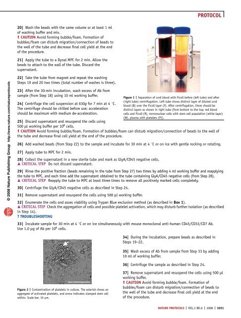
Figure 2 | Contamination <strong>of</strong> platelets in culture. The asterisk shows an<br />
aggregate <strong>of</strong> activated platelets, and arrow indicates clamped <strong>stem</strong> cell<br />
within. Scale bar, 10 mm.<br />
B<br />
F R<br />
Figure 1 | Separation <strong>of</strong> cord blood with Ficoll before (left tube) and after<br />
(right tube) centrifugation. Left tube shows distinct layer <strong>of</strong> diluted cord<br />
blood (B) over the Ficoll layer (F). After centrifugation, there should be<br />
distinct layers as shown in right tube (<strong>from</strong> bottom to the top: red blood<br />
<strong>cells</strong> and Ficoll (R), mononuclear <strong>cells</strong> with <strong>stem</strong> cell population (white layer)<br />
(W), plasma with platelets (P)).<br />
34| During the incubation, prepare beads as described in<br />
Steps 19–22.<br />
35| Wash excess <strong>of</strong> Ab <strong>from</strong> sample <strong>from</strong> Step 33 by adding<br />
10 ml <strong>of</strong> working buffer.<br />
36| Centrifuge the sample as described in Step 24.<br />
37| Remove supernatant and resuspend the <strong>cells</strong> using 500 ml<br />
working buffer.<br />
! CAUTION Avoid forming bubble/foam. Formation <strong>of</strong><br />
bubbles/foam can disturb migration/connection <strong>of</strong> beads to<br />
the well <strong>of</strong> the tube and decrease final cell yield at the end<br />
<strong>of</strong> the procedure.<br />
NATURE PROTOCOLS | VOL.3 NO.6 | 2008 | 1051<br />
P<br />
W<br />
PROTOCOL