Culture of embryonic-like stem cells from human
Culture of embryonic-like stem cells from human
Culture of embryonic-like stem cells from human

You also want an ePaper? Increase the reach of your titles
YUMPU automatically turns print PDFs into web optimized ePapers that Google loves.
© 2008<br />
Nature<br />
Publishing<br />
Group<br />
http:<br />
/ / www.<br />
nature.<br />
com/<br />
natureprotocols<br />
PROTOCOL<br />
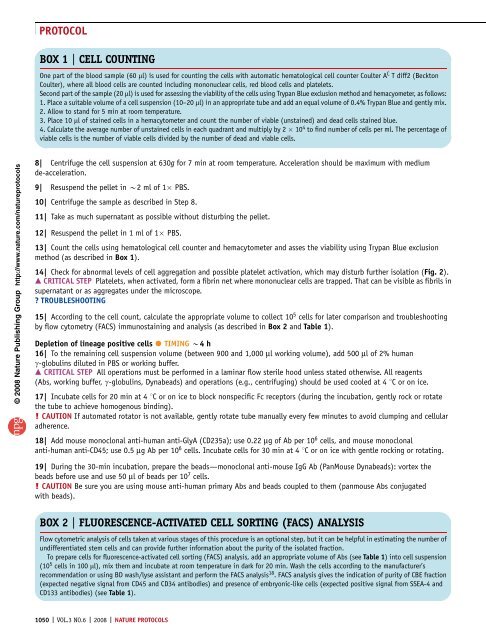
BOX 1 | CELL COUNTING<br />
One part <strong>of</strong> the blood sample (60 ml) is used for counting the <strong>cells</strong> with automatic hematological cell counter Coulter A C T diff2 (Beckton<br />
Coulter), where all blood <strong>cells</strong> are counted including mononuclear <strong>cells</strong>, red blood <strong>cells</strong> and platelets.<br />
Second part <strong>of</strong> the sample (20 ml) is used for assessing the viability <strong>of</strong> the <strong>cells</strong> using Trypan Blue exclusion method and hemacyometer, as follows:<br />
1. Place a suitable volume <strong>of</strong> a cell suspension (10–20 ml) in an appropriate tube and add an equal volume <strong>of</strong> 0.4% Trypan Blue and gently mix.<br />
2. Allow to stand for 5 min at room temperature.<br />
3. Place 10 ml <strong>of</strong> stained <strong>cells</strong> in a hemacytometer and count the number <strong>of</strong> viable (unstained) and dead <strong>cells</strong> stained blue.<br />
4. Calculate the average number <strong>of</strong> unstained <strong>cells</strong> in each quadrant and multiply by 2 10 4 to find number <strong>of</strong> <strong>cells</strong> per ml. The percentage <strong>of</strong><br />
viable <strong>cells</strong> is the number <strong>of</strong> viable <strong>cells</strong> divided by the number <strong>of</strong> dead and viable <strong>cells</strong>.<br />
8| Centrifuge the cell suspension at 630g for 7 min at room temperature. Acceleration should be maximum with medium<br />
de-acceleration.<br />
9| Resuspend the pellet in B2 ml<strong>of</strong>1 PBS.<br />
10| Centrifuge the sample as described in Step 8.<br />
11| Take as much supernatant as possible without disturbing the pellet.<br />
12| Resuspend the pellet in 1 ml <strong>of</strong> 1 PBS.<br />
13| Count the <strong>cells</strong> using hematological cell counter and hemacytometer and asses the viability using Trypan Blue exclusion<br />
method (as described in Box 1).<br />
14| Check for abnormal levels <strong>of</strong> cell aggregation and possible platelet activation, which may disturb further isolation (Fig. 2).<br />
m CRITICAL STEP Platelets, when activated, form a fibrin net where mononuclear <strong>cells</strong> are trapped. That can be visible as fibrils in<br />
supernatant or as aggregates under the microscope.<br />
? TROUBLESHOOTING<br />
15| According to the cell count, calculate the appropriate volume to collect 10 5 <strong>cells</strong> for later comparison and troubleshooting<br />
by flow cytometry (FACS) immunostaining and analysis (as described in Box 2 and Table 1).<br />
Depletion <strong>of</strong> lineage positive <strong>cells</strong> TIMING B4 h<br />
16| To the remaining cell suspension volume (between 900 and 1,000 ml working volume), add 500 ml <strong>of</strong> 2% <strong>human</strong><br />
g-globulins diluted in PBS or working buffer.<br />
m CRITICAL STEP All operations must be performed in a laminar flow sterile hood unless stated otherwise. All reagents<br />
(Abs, working buffer, g-globulins, Dynabeads) and operations (e.g., centrifuging) should be used cooled at 4 1C oronice.<br />
17| Incubate <strong>cells</strong> for 20 min at 4 1C or on ice to block nonspecific Fc receptors (during the incubation, gently rock or rotate<br />
the tube to achieve homogenous binding).<br />
! CAUTION If automated rotator is not available, gently rotate tube manually every few minutes to avoid clumping and cellular<br />
adherence.<br />
18| Add mouse monoclonal anti-<strong>human</strong> anti-GlyA (CD235a); use 0.22 mg <strong>of</strong>Abper106<strong>cells</strong>, and mouse monoclonal<br />
anti-<strong>human</strong> anti-CD45; use 0.5 mg Abper106<strong>cells</strong>. Incubate <strong>cells</strong> for 30 min at 4 1C or on ice with gentle rocking or rotating.<br />
19| During the 30-min incubation, prepare the beads—monoclonal anti-mouse IgG Ab (PanMouse Dynabeads): vortex the<br />
beads before use and use 50 ml <strong>of</strong>beadsper10 7 <strong>cells</strong>.<br />
! CAUTION Be sure you are using mouse anti-<strong>human</strong> primary Abs and beads coupled to them (panmouse Abs conjugated<br />
with beads).<br />
BOX 2 | FLUORESCENCE-ACTIVATED CELL SORTING (FACS) ANALYSIS<br />
Flow cytometric analysis <strong>of</strong> <strong>cells</strong> taken at various stages <strong>of</strong> this procedure is an optional step, but it can be helpful in estimating the number <strong>of</strong><br />
undifferentiated <strong>stem</strong> <strong>cells</strong> and can provide further information about the purity <strong>of</strong> the isolated fraction.<br />
To prepare <strong>cells</strong> for fluorescence-activated cell sorting (FACS) analysis, add an appropriate volume <strong>of</strong> Abs (see Table 1) into cell suspension<br />
(10 5 <strong>cells</strong> in 100 ml), mix them and incubate at room temperature in dark for 20 min. Wash the <strong>cells</strong> according to the manufacturer’s<br />
recommendation or using BD wash/lyse assistant and perform the FACS analysis 18 . FACS analysis gives the indication <strong>of</strong> purity <strong>of</strong> CBE fraction<br />
(expected negative signal <strong>from</strong> CD45 and CD34 antibodies) and presence <strong>of</strong> <strong>embryonic</strong>-<strong>like</strong> <strong>cells</strong> (expected positive signal <strong>from</strong> SSEA-4 and<br />
CD133 antibodies) (see Table 1).<br />
1050 | VOL.3 NO.6 | 2008 | NATURE PROTOCOLS