Culture of embryonic-like stem cells from human
Culture of embryonic-like stem cells from human
Culture of embryonic-like stem cells from human

You also want an ePaper? Increase the reach of your titles
YUMPU automatically turns print PDFs into web optimized ePapers that Google loves.
© 2008<br />
Nature<br />
Publishing<br />
Group<br />
http:<br />
/ / www.<br />
nature.<br />
com/<br />
natureprotocols<br />
PROTOCOL<br />
<strong>embryonic</strong>/pluripotency markers for the undifferentiated <strong>cells</strong> on cytospin slides (see Table 2 for used and suggested markers)<br />
as reported previously 1,5 (Fig. 7).<br />
m CRITICAL STEP At this stage, you should have obtained the neural <strong>cells</strong>. Although some <strong>of</strong> the <strong>cells</strong> may still be <strong>of</strong> the<br />
neural progenitor variety rather than terminally differentiated <strong>cells</strong>, this provides more flexibility in the final use and analysis <strong>of</strong><br />
the <strong>cells</strong>.<br />
TIMING<br />
The collection, processing and negative depletion procedures can be performed in B6 h. The entire procedure <strong>from</strong> umbilical<br />
cord blood collection to obtaining <strong>cells</strong> with multiprocessed neural phenotypes can be performed in B3–4 weeks.<br />
? TROUBLESHOOTING<br />
Troubleshooting advice can be found in Table 5.<br />
TABLE 5 | Troubleshooting table.<br />
Steps Problem Reason/solution<br />
14, 32 Contamination <strong>of</strong> cell suspension with platelets Centrifuge the <strong>cells</strong> slowly (400g/10 min), discard the supernatant with<br />
platelets and collect the pellet. Repeat if necessary<br />
40 Existence <strong>of</strong> magnetic beads with negative fraction<br />
<strong>cells</strong><br />
ANTICIPATED RESULTS<br />
Our recent study indicated that umbilical cord blood contains various <strong>stem</strong> <strong>cells</strong> at different stages <strong>of</strong> their development 3 .Such<br />
cell fractions (e.g., CD133+; CBEs) might be isolated separately <strong>from</strong> one cord blood unit, expanded and committed toward<br />
different lineages: endothelial 1 and neural 14 , respectively.<br />
We found that CBEs could be differentiated very effectively toward neuronal phenotypes in vitro. Soon after isolation, CBEs<br />
are very small (diameter <strong>of</strong> 2–3 mm) and resemble <strong>embryonic</strong> 13 or very small <strong>embryonic</strong>-<strong>like</strong> 2 phenotypes reported previously by<br />
other research groups. CBEs express at this stage markers<br />
characteristic for pluripotent <strong>human</strong> <strong>embryonic</strong> <strong>stem</strong> <strong>cells</strong><br />
(e.g., surface markers: SSEA-3/4 and transcription factors:<br />
Oct4A and Sox2). During the stage <strong>of</strong> in vitro expansion<br />
(in the presence <strong>of</strong> EGF and bFGF), some <strong>of</strong> the <strong>cells</strong> start<br />
to differentiate spontaneously; they lose expression <strong>of</strong> <strong>embryonic</strong><br />
markers (mentioned earlier) and they increase in size<br />
(diameter up to 20 mm). These <strong>cells</strong> are in majority <strong>of</strong> neural<br />
progenitors expressing markers <strong>of</strong> proliferating neural <strong>stem</strong><br />
<strong>cells</strong> (Nestin, GFAP and Ki67, respectively). During the<br />
expansion stage, most <strong>of</strong> the undifferentiated <strong>cells</strong> remain<br />
in the aggregates formed within a few hours after isolation.<br />
Therefore, we consider such aggregates as an artificial <strong>stem</strong><br />
cell niche able to prevent the <strong>stem</strong> <strong>cells</strong> <strong>from</strong> spontaneous<br />
differentiation 14 .<br />
This protocol uses 2D culture conditions for the differentia-<br />
tion <strong>of</strong> CBEs into neural <strong>cells</strong>. <strong>Culture</strong> media, which promote<br />
neuronal differentiation, are supplemented with RA, BDNF and<br />
dBcAMP, and they are introduced sequentially. It is worth<br />
noting that proper sequential addition <strong>of</strong> neuromorphogenes<br />
is critical to the success <strong>of</strong> this protocol. We found, for example,<br />
that introducing cAMP and BDNF too early (when the <strong>cells</strong><br />
Reapply <strong>cells</strong> to magnetic particle concentrator to remove all magnetic<br />
beads along with positive <strong>cells</strong><br />
46 Total number <strong>of</strong> <strong>cells</strong> is less than one million Use ‘‘U’’ shape 96-well plate to enhance the <strong>cells</strong>’ survival and aggregate<br />
formation<br />
52 Low level <strong>of</strong> cell adhesion to plate surface Decrease the volume <strong>of</strong> media (minimum 300 ml) to enhance cell<br />
attachment to the plate surface<br />
1054 | VOL.3 NO.6 | 2008 | NATURE PROTOCOLS<br />
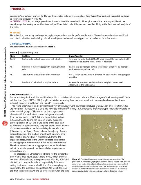
Figure 8 | Example <strong>of</strong> later stage neural phenotype <strong>from</strong> culture. The<br />
proportion <strong>of</strong> ovoid <strong>cells</strong> (highlighted by white arrows) reduces <strong>from</strong> previous<br />
cultures and epithelioid <strong>cells</strong> start to proliferate, acquiring a more defined<br />
neuroglial morphology with longer membrane projections and neurite-<strong>like</strong><br />
extensions. Adherent <strong>cells</strong> which are more spherical can also at this stage<br />
spontaneously cluster into compact neurospheres. Scale bar, 10 mm.