Correlation of histology and linear and nonlinear microscopy of the ...
Correlation of histology and linear and nonlinear microscopy of the ...
Correlation of histology and linear and nonlinear microscopy of the ...

Create successful ePaper yourself
Turn your PDF publications into a flip-book with our unique Google optimized e-Paper software.
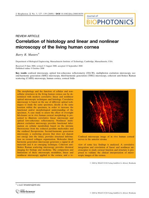
J. Biophoton. 2, No. 3, 127–139 (2009) / DOI 10.1002/jbio.200810039<br />
REVIEW ARTICLE<br />
<strong>Correlation</strong> <strong>of</strong> <strong>histology</strong> <strong>and</strong> <strong>linear</strong> <strong>and</strong> non<strong>linear</strong><br />
<strong>microscopy</strong> <strong>of</strong> <strong>the</strong> living human cornea<br />
Barry R. Masters*<br />
Department <strong>of</strong> Biological Engineering, Massachusetts Institute <strong>of</strong> Technology, Cambridge, Massachusetts, USA<br />
Received 25 June 2008, revised 15 August 2008, accepted 22 September 2008<br />
Published online 8 October 2008<br />
Key words: confocal <strong>microscopy</strong>, optical low-coherence reflectometry (OLCR), multiphoton excitation <strong>microscopy</strong>, second-harmonic<br />
generation (SHG) <strong>microscopy</strong>, third-harmonic generation (THG) <strong>microscopy</strong>, coherent anti-Stokes Raman<br />
scattering (CARS) <strong>microscopy</strong>, human cornea, corneal folds<br />
The morphology <strong>and</strong> <strong>the</strong> function <strong>of</strong> cellular <strong>and</strong> noncellular<br />
structures in <strong>the</strong> living human cornea can be determined<br />
with modern correlative <strong>linear</strong> <strong>and</strong> non<strong>linear</strong><br />
optical microscopic techniques <strong>and</strong> <strong>histology</strong>. Correlative<br />
<strong>microscopy</strong> is based on <strong>the</strong> use <strong>of</strong> different optical techniques<br />
to study <strong>the</strong> same specimen, ideally at <strong>the</strong> same<br />
location within <strong>the</strong> specimen, in order to increase <strong>the</strong><br />
functional <strong>and</strong>/or morphological underst<strong>and</strong>ing <strong>of</strong> <strong>the</strong><br />
specimen. A case study to assess <strong>the</strong> effect <strong>of</strong> overnight<br />
lid-closure on in vivo human corneal morphology is presented<br />
to illustrate correlative <strong>linear</strong> <strong>microscopy</strong> <strong>and</strong><br />
optical low-coherence reflectometry. Non<strong>linear</strong> multiphoton<br />
excitation <strong>microscopy</strong> provides functional information<br />
on cellular metabolism based on <strong>the</strong> intrinsic<br />
fluorescence from <strong>the</strong> reduced pyridine nucleotides <strong>and</strong><br />
<strong>the</strong> oxidized flavoproteins. Second-harmonic generation<br />
<strong>microscopy</strong>, a scattering process that does not deposit<br />
net energy into <strong>the</strong> tissue, provides structural information<br />
on corneal collagen organization. Molecular thirdharmonic<br />
generation <strong>microscopy</strong> generates a signal in all<br />
materials <strong>and</strong> it an emerging technique. Coherent anti-<br />
Stokes Raman scattering <strong>microscopy</strong> provides chemical<br />
imaging for biology <strong>and</strong> medicine. The comparison <strong>and</strong><br />
limitations <strong>of</strong> <strong>the</strong>se microscopic modalities, <strong>linear</strong> <strong>and</strong><br />
non<strong>linear</strong> <strong>microscopy</strong> applied to <strong>the</strong> cornea, <strong>and</strong> a re-<br />
* e-mail: bmasters@mit.edu<br />
Journal <strong>of</strong><br />
BIOPHOTONICS<br />
Confocal <strong>microscopy</strong> image <strong>of</strong> in vivo human corneal<br />
nerves in <strong>the</strong> anterior stroma<br />
view <strong>of</strong> some key findings is analyzed. A correlative<br />
integration <strong>and</strong> correlation <strong>of</strong> <strong>linear</strong> <strong>and</strong> non<strong>linear</strong> microscopies<br />
to study corneal function <strong>and</strong> structure is proposed<br />
to validate <strong>the</strong> clinical interpretation <strong>of</strong> microscopic<br />
images <strong>of</strong> <strong>the</strong> cornea.<br />
# 2009 by WILEY-VCH Verlag GmbH & Co. KGaA, Weinheim<br />
# 2009 by WILEY-VCH Verlag GmbH & Co. KGaA, Weinheim
Journal <strong>of</strong><br />
BIOPHOTONICS<br />
128<br />
1. Introduction<br />
B. R. Masters: <strong>Correlation</strong> <strong>of</strong> <strong>histology</strong> <strong>and</strong> <strong>linear</strong> <strong>and</strong> non<strong>linear</strong> <strong>microscopy</strong> <strong>of</strong> <strong>the</strong> living human cornea<br />
The history <strong>of</strong> <strong>the</strong> <strong>linear</strong> <strong>and</strong> non<strong>linear</strong> optical microscope<br />
is inextricably linked to <strong>the</strong> development <strong>of</strong><br />
new clinical instruments that play important roles in<br />
our underst<strong>and</strong>ing <strong>of</strong> cells <strong>and</strong> tissues <strong>and</strong> in <strong>the</strong><br />
early diagnosis <strong>and</strong> <strong>the</strong> prevention <strong>of</strong> disease. In <strong>the</strong><br />
domain <strong>of</strong> <strong>linear</strong> optics <strong>the</strong> response <strong>of</strong> <strong>the</strong> induced<br />
polarization to <strong>the</strong> incident electromagnetic field is<br />
<strong>linear</strong> with <strong>the</strong> amplitude <strong>of</strong> <strong>the</strong> field. Linear optical<br />
effects include one-photon absorption, scattering,<br />
<strong>and</strong> fluorescence. Exchange <strong>of</strong> energy <strong>and</strong> momentum<br />
can occur in <strong>linear</strong> interactions, e.g. absorption<br />
<strong>and</strong> scattering. Linear optical phenomena are characterized<br />
by a induced polarization PðtÞ in <strong>the</strong> material<br />
that depends in a <strong>linear</strong> manner on <strong>the</strong> electric<br />
field strength EðtÞ:<br />
PðtÞ ¼"0 c ð1Þ EðtÞ ; ð1Þ<br />
<strong>the</strong> constant <strong>of</strong> proportionality c ð1Þ is known as <strong>the</strong><br />
<strong>linear</strong> susceptibility <strong>and</strong> "0 is <strong>the</strong> permittivity <strong>of</strong> free<br />
space.<br />
Non<strong>linear</strong> optical effects are characterized by <strong>the</strong><br />
non<strong>linear</strong> response <strong>of</strong> <strong>the</strong> induced polarization to<br />
<strong>the</strong> incident electromagnetic field; <strong>the</strong> response is<br />
non<strong>linear</strong> in <strong>the</strong> amplitude <strong>of</strong> <strong>the</strong> field [1, 2]. For <strong>the</strong><br />
case <strong>of</strong> non<strong>linear</strong> optics, <strong>the</strong> optical response is described<br />
by expressing <strong>the</strong> induced polarization PðtÞ<br />
as a power series <strong>of</strong> <strong>the</strong> electric field strength:<br />
PðtÞ ¼"0½c ð1Þ EðtÞþc ð2Þ E 2 ðtÞþc ð3Þ E 3 ðtÞþ...Š : ð2Þ<br />
The quantities c ð2Þ <strong>and</strong> c ð3Þ are known as <strong>the</strong> second<strong>and</strong><br />
<strong>the</strong> third-order non<strong>linear</strong> optical susceptibilities.<br />
In writing <strong>the</strong>se two equations an important assumption<br />
is made: <strong>the</strong> polarization <strong>of</strong> <strong>the</strong> material at<br />
time t, only depends on <strong>the</strong> instantaneous value <strong>of</strong><br />
<strong>the</strong> electric field strength. It follows from this assumption,<br />
through <strong>the</strong> Kramers-Kronig relations,<br />
that <strong>the</strong> medium must be lossless <strong>and</strong> dispersion less.<br />
Typically non<strong>linear</strong> optical phenomena occur in <strong>the</strong><br />
presence <strong>of</strong> laser-matter interactions in <strong>the</strong> presence<br />
<strong>of</strong> very high electric field strengths. At high light<br />
intensities, <strong>the</strong> incident light modifies <strong>the</strong> induced<br />
polarization such that light waves can interact <strong>and</strong><br />
exchange energy <strong>and</strong> momentum. Examples <strong>of</strong><br />
non<strong>linear</strong> optical effects include <strong>the</strong> Pockels <strong>and</strong><br />
Kerr electro-optic effects, multiphoton excitation<br />
(MPE) [3], second-harmonic generation (SHG),<br />
third-harmonic generation (THG) [4], <strong>and</strong> coherent<br />
anti-Stokes Raman scattering (CARS) [5].<br />
Correlative <strong>microscopy</strong> is based on <strong>the</strong> use <strong>of</strong><br />
different optical techniques to study <strong>the</strong> same specimen,<br />
ideally at <strong>the</strong> same location within <strong>the</strong> specimen,<br />
in order to increase <strong>the</strong> functional <strong>and</strong>/or morphological<br />
underst<strong>and</strong>ing <strong>of</strong> <strong>the</strong> specimen. Typically,<br />
correlative <strong>microscopy</strong> could include <strong>linear</strong> (confocal<br />
<strong>microscopy</strong>, optical low-coherence reflectometry)<br />
toge<strong>the</strong>r with non<strong>linear</strong> <strong>microscopy</strong> (MPE, SHG,<br />
THG, <strong>and</strong> CARS).<br />
The purpose <strong>and</strong> <strong>the</strong> emphasis <strong>of</strong> this paper is to<br />
point out <strong>the</strong> limitations, <strong>and</strong> <strong>the</strong> multiple confounding<br />
factors that make <strong>the</strong> interpretation <strong>of</strong> corneal<br />
functional <strong>and</strong> morphological images difficult.<br />
1.1 Confocal <strong>microscopy</strong><br />
In <strong>the</strong> last decades <strong>the</strong>re have been improvements in<br />
both <strong>the</strong> resolution <strong>and</strong> <strong>the</strong> contrast <strong>of</strong> optical microscopes.<br />
The invention <strong>of</strong> various types <strong>of</strong> confocal<br />
microscopes, that generate contrast from <strong>the</strong> processes<br />
<strong>of</strong> <strong>linear</strong> absorption, scattering, <strong>and</strong> fluorescence,<br />
provides researchers in cell biology with<br />
extremely useful tools to investigate <strong>the</strong> biology <strong>of</strong><br />
cells in thick, highly scattering tissues. Confocal microscopes<br />
are based on <strong>the</strong> principle <strong>of</strong> spatial filtering<br />
<strong>and</strong> permit live cell imaging <strong>of</strong> cells <strong>and</strong> thick,<br />
highly scattering tissues with improved “optical sectioning”<br />
<strong>and</strong> improved contrast as compared to traditional<br />
epi-fluorescence microscopes [6].<br />
The first applications <strong>of</strong> confocal <strong>microscopy</strong><br />
were applied to cells <strong>and</strong> tissues that were fixed, <strong>and</strong><br />
stained with immunochemical stains <strong>of</strong> high specificity<br />
to delineate <strong>the</strong> fine structures in cells such as<br />
<strong>the</strong> cytoskeleton. Only later did researchers apply<br />
confocal <strong>microscopy</strong> to live cells <strong>and</strong> tissues both ex<br />
vivo <strong>and</strong> in vivo, i.e. three-dimensional imaging <strong>the</strong><br />
in vivo human cornea [7] <strong>and</strong> in vivo human skin<br />
[8].<br />
Two modes <strong>of</strong> contrast are implemented in confocal<br />
<strong>microscopy</strong>. The first mode generates contrast<br />
in <strong>the</strong> images with reflected <strong>and</strong> scattered light<br />
that occurs at <strong>the</strong> interfaces with different refractive<br />
indexes [6]. A registered stack <strong>of</strong> optical<br />
sections could <strong>the</strong>n be transformed in a digital<br />
computer into a three-dimensional volume that visualized<br />
<strong>the</strong> structure <strong>of</strong> cells <strong>and</strong> tissues, <strong>and</strong> <strong>the</strong>se<br />
computer generated structures could be visualized<br />
from any arbitrary orientation. The second mode<br />
generates <strong>the</strong> image contrast by exciting <strong>the</strong> fluorescence<br />
from ei<strong>the</strong>r intrinsic naturally occurring<br />
fluorescent molecules, for example, reduced pyridine<br />
nucleotides, NAD(P)H, <strong>and</strong> oxidized flavoproteins<br />
[6]. Both <strong>the</strong> reduced nicotinamide adenine<br />
dinucleotide, NADH, <strong>and</strong> <strong>the</strong> reduced nicotinamide<br />
adenine dinucleotide phosphate, NADPH, are denoted<br />
as NAD(P)H. The fluorescence <strong>of</strong> NAD(P)H<br />
is in <strong>the</strong> range <strong>of</strong> 400–500 nm. Alternatively, contrast<br />
in confocal <strong>microscopy</strong> could be based on<br />
fluorescent probes that are genetically over-expressed<br />
such as green fluorescent proteins (GFP),<br />
or molecular probes that were externally applies to<br />
<strong>the</strong> cells <strong>and</strong> tissues [6].<br />
# 2009 by WILEY-VCH Verlag GmbH & Co. KGaA, Weinheim www.biophotonics-journal.org
J. Biophoton. 2, No. 3 (2009) 129<br />
1.2 Confocal <strong>microscopy</strong> <strong>of</strong> <strong>the</strong> eye<br />
The application <strong>of</strong> confocal microscopes to <strong>the</strong> anterior<br />
segment <strong>of</strong> <strong>the</strong> eye, specifically <strong>the</strong> avascular<br />
cornea, a highly transparent <strong>and</strong> organized layered<br />
structure provided a new generation <strong>of</strong> diagnostic instruments<br />
[7, 9–12]. The human cornea consists <strong>of</strong><br />
superficial epi<strong>the</strong>lial cells, intermediate epi<strong>the</strong>lial<br />
cells, basal epi<strong>the</strong>lial cells, limbal stem cells, Bowman’s<br />
layer, stromal keratocytes, alternating layers<br />
<strong>of</strong> approximately orthogonal bundles <strong>of</strong> collagen fibers,<br />
nerve fibers, <strong>and</strong> on <strong>the</strong> posterior side, Descemet’s<br />
membrane <strong>and</strong> a single layer <strong>of</strong> endo<strong>the</strong>lial<br />
cells [11]. There is an interesting history <strong>of</strong> instrument<br />
development <strong>and</strong> innovation in <strong>the</strong> design <strong>of</strong><br />
instruments for corneal bio<strong>microscopy</strong> [13].<br />
The development <strong>of</strong> <strong>the</strong> scanning slit, clinical<br />
confocal microscope is not a new paradigm, but <strong>the</strong><br />
result <strong>of</strong> a continuous series <strong>of</strong> interlinked technical<br />
advances starting from <strong>the</strong> early work <strong>of</strong> Goldmann<br />
to Thaer’s development <strong>of</strong> <strong>the</strong> clinical scanning-slit<br />
confocal microscope [13]. There were many parallel<br />
developments in <strong>the</strong> advancement <strong>of</strong> confocal instruments<br />
for clinical use <strong>and</strong> <strong>the</strong> key figures include<br />
Goldmann, Maurice, Svishchev, Baer, Koester, Masters,<br />
<strong>and</strong> Thaer [7, 14]. The major problems that had<br />
to be solved in <strong>the</strong> development <strong>of</strong> a scanning slit<br />
confocal microscope for <strong>the</strong> clinical examination <strong>of</strong><br />
<strong>the</strong> living human eye include how to acquire images<br />
from <strong>the</strong> moving eye, <strong>and</strong> how to obtain a sufficient<br />
signal-to-noise ratio to form high contrast images<br />
that are acquired at video rates [15]. A recent book<br />
details <strong>the</strong> story <strong>of</strong> <strong>the</strong>se clinical developments that<br />
resulted in <strong>the</strong> modern scanning slit clinical confocal<br />
microscope [16]. All <strong>of</strong> <strong>the</strong>se instruments use an incoherent<br />
lamp as <strong>the</strong> light source.<br />
A recent design <strong>and</strong> development <strong>of</strong> a laser scanning<br />
confocal microscope that is applicable to <strong>the</strong> in<br />
vivo human cornea is based on <strong>the</strong> Rostock Cornea<br />
Module (RCM). This module attaches to a commercial<br />
Heidelberg Engineering scanning laser ophthalmoscope<br />
that is typically used for retinal imaging<br />
[17]. The attached module shifts <strong>the</strong> focus <strong>of</strong> <strong>the</strong> laser<br />
beam from <strong>the</strong> retinal surface to <strong>the</strong> cornea <strong>and</strong><br />
<strong>the</strong> focal position is operator controlled by <strong>the</strong> motion<br />
<strong>of</strong> an secondary movable lens. This scanning laser<br />
instrument has <strong>the</strong> capacity to produce high contrast<br />
confocal images over a 400 400 mm field <strong>of</strong><br />
view. It can also operate in a noncontact mode which<br />
may have several clinical advantages over <strong>the</strong> o<strong>the</strong>r<br />
types <strong>of</strong> clinical confocal microscopes that contact<br />
<strong>the</strong> cornea.<br />
Both <strong>the</strong> scanning slit confocal microscope, manufactured<br />
by Nidek, <strong>and</strong> <strong>the</strong> RCM unit manufactured<br />
by Heidelberg Engineering, have limitations<br />
<strong>and</strong> differences in <strong>the</strong>ir performance. Both instruments<br />
can operate in <strong>the</strong> noncontact mode which is<br />
www.biophotonics-journal.org<br />
REVIEW<br />
ARTICLE<br />
important to prevent transmission <strong>of</strong> microbes from<br />
patient to patient, <strong>and</strong> <strong>the</strong>y avoid <strong>the</strong> use <strong>of</strong> <strong>the</strong> index<br />
matching gel. Both instruments can image scars,<br />
opacities, <strong>and</strong> calcifications in <strong>the</strong> stroma; however,<br />
superficial scars <strong>and</strong> opacities will scatter <strong>the</strong> incident<br />
light <strong>and</strong> cause a shadow effect on <strong>the</strong> images<br />
from <strong>the</strong> deeper cellular layers.<br />
All types <strong>of</strong> clinical microscopes that are designed<br />
to image <strong>the</strong> full thickness <strong>of</strong> <strong>the</strong> human cornea<br />
have <strong>the</strong> capability to acquire images from <strong>the</strong><br />
anterior surface (superficial corneal epi<strong>the</strong>lial cells)<br />
to <strong>the</strong> posterior single layer <strong>of</strong> corneal endo<strong>the</strong>lial<br />
cells. Within <strong>the</strong> stroma on <strong>the</strong> anterior side <strong>the</strong> acellular<br />
Bowman’s layer provides a l<strong>and</strong>mark <strong>of</strong> depth<br />
within <strong>the</strong> stroma. Similarly on <strong>the</strong> posterior side <strong>of</strong><br />
<strong>the</strong> stroma <strong>the</strong> appearance <strong>of</strong> <strong>the</strong> acellular Descemet’s<br />
membrane <strong>and</strong> <strong>the</strong> easily recognized layer<br />
polygonal endo<strong>the</strong>lium provides a depth l<strong>and</strong>mark.<br />
What is confounding is <strong>the</strong> ability to accurately determine<br />
<strong>the</strong> depth <strong>of</strong> focus within <strong>the</strong> corneal stroma.<br />
Although some clinical instruments provide a<br />
movable intermediate lens that can be positioned<br />
<strong>and</strong> moved with high accuracy; that does not necessarily<br />
correspond to <strong>the</strong> accurate depth <strong>of</strong> <strong>the</strong> focal<br />
plane <strong>of</strong> <strong>the</strong> microscope objective within <strong>the</strong> cornea.<br />
Since <strong>the</strong> keratocyte density <strong>and</strong> <strong>the</strong> organization <strong>of</strong><br />
<strong>the</strong> corneal collagen fibers varies with depth within<br />
<strong>the</strong> corneal stroma it is important to solve this problem<br />
in <strong>the</strong> next generation <strong>of</strong> clinical confocal microscopes.<br />
2. Review <strong>of</strong> key clinical applications with<br />
<strong>linear</strong> confocal <strong>microscopy</strong><br />
2.1 Correlative light <strong>microscopy</strong> <strong>of</strong><br />
tangential sections <strong>and</strong> in vivo confocal<br />
<strong>microscopy</strong><br />
The foundations <strong>of</strong> confocal <strong>microscopy</strong>, <strong>the</strong> history<br />
<strong>of</strong> <strong>the</strong> bio<strong>microscopy</strong> <strong>of</strong> <strong>the</strong> eye, a tutorial on <strong>the</strong><br />
practical techniques <strong>of</strong> clinical confocal <strong>microscopy</strong><br />
<strong>of</strong> <strong>the</strong> eye, <strong>and</strong> a review <strong>of</strong> studies on <strong>the</strong> clinical examination<br />
<strong>of</strong> <strong>the</strong> cornea with <strong>the</strong> confocal microscope<br />
for <strong>the</strong> cases <strong>of</strong> aging, contact lens wear, corneas<br />
with known pathologies, <strong>and</strong> studies <strong>of</strong> <strong>the</strong><br />
morphological changes in <strong>the</strong> post surgical cornea<br />
were reviewed by Böhnke <strong>and</strong> Masters [11]. Many<br />
examples <strong>of</strong> correlative light <strong>microscopy</strong> <strong>of</strong> tangential<br />
sections from paraffin-embedded ex vivo human<br />
corneas that were stained with PAS reagent are<br />
shown adjacent to scanning-slit confocal images <strong>of</strong><br />
similar sections <strong>of</strong> <strong>the</strong> living in vivo human cornea.<br />
These multiple examples serve to illustrate <strong>the</strong> value<br />
<strong>of</strong> correlative light <strong>microscopy</strong> <strong>of</strong> ex vivo sections <strong>of</strong><br />
<strong>the</strong> cornea <strong>and</strong> <strong>the</strong> equivalent sections as observed<br />
# 2009 by WILEY-VCH Verlag GmbH & Co. KGaA, Weinheim
Journal <strong>of</strong><br />
BIOPHOTONICS<br />
130<br />
B. R. Masters: <strong>Correlation</strong> <strong>of</strong> <strong>histology</strong> <strong>and</strong> <strong>linear</strong> <strong>and</strong> non<strong>linear</strong> <strong>microscopy</strong> <strong>of</strong> <strong>the</strong> living human cornea<br />
with confocal <strong>microscopy</strong> <strong>of</strong> <strong>the</strong> in vivo human cornea.<br />
It is important to emphasize <strong>the</strong> comparison <strong>of</strong><br />
in vivo confocal images <strong>of</strong> <strong>the</strong> human cornea <strong>and</strong><br />
histological studies <strong>of</strong> ex vivo human corneas in order<br />
to minimize errors <strong>of</strong> interpretation <strong>of</strong> <strong>the</strong> clinical<br />
images. At <strong>the</strong> same time, investigators should be<br />
aware <strong>of</strong> <strong>the</strong> morphological differences between in<br />
vivo <strong>and</strong> ex vivo corneas, as well as <strong>the</strong> fixation <strong>and</strong><br />
<strong>the</strong> staining artifacts <strong>of</strong> classical <strong>histology</strong>.<br />
2.2 Long-term effects <strong>of</strong> contact lens wear<br />
on <strong>the</strong> human cornea: a new corneal<br />
degeneration<br />
This paper illustrates <strong>the</strong> confounding effect <strong>of</strong> acquiring<br />
clinical confocal images <strong>of</strong> <strong>the</strong> human cornea<br />
with inappropriate optical resolution [18]. We posed<br />
<strong>the</strong> following question: what are <strong>the</strong> long-term effects<br />
<strong>of</strong> contact lens wear on <strong>the</strong> human cornea? We<br />
answered this question by first acquiring confocal<br />
images through <strong>the</strong> full thickness <strong>of</strong> <strong>the</strong> in vivo human<br />
cornea, <strong>and</strong> <strong>the</strong>n by a frame by frame analysis<br />
to determine morphological changes within <strong>the</strong> cornea<br />
that are correlated with long-term contact lens<br />
wear, <strong>and</strong> do not occur in subjects that do not wear<br />
contact lenses.<br />
The scanning-slit confocal microscope with a<br />
50X/1.0 NA water immersion objective was used to<br />
investigate <strong>the</strong> corneal morphology in long-term<br />
contact lens wearers [18]. The authors investigated<br />
13 patients with a history <strong>of</strong> up to 26 years <strong>of</strong> s<strong>of</strong>t<br />
contact lens wear, 11 patients with a history <strong>of</strong> up to<br />
25 years <strong>of</strong> rigid gas permeable contact lens wear,<br />
<strong>and</strong> a control group <strong>of</strong> 29 normal subjects without a<br />
history <strong>of</strong> contact lens wear. For contact lens wearers<br />
epi<strong>the</strong>lial microcystic changes <strong>and</strong> alterations <strong>of</strong> endo<strong>the</strong>lial<br />
cell morphology were found as described<br />
previously. The significant new finding was <strong>the</strong>re<br />
were highly reflective panstromal microdot (submicron)<br />
deposits in <strong>the</strong> entire thickness <strong>of</strong> <strong>the</strong> stroma<br />
for <strong>the</strong> contact lens wearers. It was concluded that<br />
this newly observed stromal microdot degeneration<br />
scales with <strong>the</strong> years <strong>of</strong> contact lens wear <strong>and</strong> may<br />
be <strong>the</strong> early stage <strong>of</strong> a significant corneal disease.<br />
Fur<strong>the</strong>r correlative microscopic studies involved<br />
electron <strong>microscopy</strong> <strong>of</strong> ex vivo human corneas, <strong>and</strong><br />
spectroscopic studies <strong>of</strong> <strong>the</strong> microdots.<br />
Initially, o<strong>the</strong>r groups were not able to observe<br />
<strong>the</strong> stromal microdot deposits because <strong>the</strong>y used a<br />
Nipkow disk confocal microscope with a low magnification<br />
(20X) <strong>and</strong> a low NA microscope objective<br />
(0.5). The low magnification, low NA microscope objective<br />
did not provide <strong>the</strong> necessary resolution to<br />
image <strong>the</strong> stromal microdot deposits. Alternatively,<br />
<strong>the</strong> Nipkow disk t<strong>and</strong>em-scanning confocal micro-<br />
scope did not provide sufficient illumination to image<br />
<strong>the</strong> submicron stromal microdot deposits in <strong>the</strong><br />
cornea.<br />
This study illustrates <strong>the</strong> requirement <strong>of</strong> <strong>the</strong> appropriate<br />
optical resolution in studies with <strong>the</strong> clinical<br />
confocal microscope, as well as correlative electron<br />
<strong>microscopy</strong> to validate <strong>the</strong> clinical conclusions.<br />
2.3 Overnight lid closure <strong>and</strong> <strong>the</strong> reversible<br />
micro-folds in <strong>the</strong> stroma <strong>of</strong> <strong>the</strong> human<br />
cornea<br />
The next study shows <strong>the</strong> importance <strong>of</strong> correct experimental<br />
protocol to investigate transient phenomena<br />
in <strong>the</strong> in vivo human cornea. This case report<br />
illustrates <strong>the</strong> use <strong>of</strong> <strong>linear</strong> confocal <strong>microscopy</strong><br />
(back scattered <strong>and</strong> reflected incoherent light) toge<strong>the</strong>r<br />
with optical low-coherence reflectometry<br />
(OLCR) to study <strong>the</strong> physiological question: does<br />
overnight lid closure affect <strong>the</strong> structure <strong>of</strong> <strong>the</strong> cornea<br />
stroma?<br />
This is an example <strong>of</strong> correlative studies involves<br />
a case report on <strong>the</strong> investigation <strong>of</strong> overnight lid<br />
closure <strong>and</strong> <strong>the</strong> reversible micro-folds in <strong>the</strong> stroma<br />
<strong>of</strong> <strong>the</strong> human cornea. We used clinical confocal <strong>microscopy</strong><br />
<strong>and</strong> parallel reversible changes in corneal<br />
thickness as measured with optical low-coherence reflectometry<br />
(OLCR) to validate <strong>the</strong> interpretation <strong>of</strong><br />
<strong>the</strong> confocal images <strong>of</strong> reversible micro-folds in <strong>the</strong><br />
human cornea stroma.<br />
The cornea is a specialized tissue, which maintains<br />
its normal optical properties from <strong>the</strong> fluid control<br />
exerted by <strong>the</strong> endo<strong>the</strong>lial pumping function in<br />
<strong>the</strong> presence <strong>of</strong> an intact epi<strong>the</strong>lial barrier. Failure <strong>of</strong><br />
ei<strong>the</strong>r one <strong>of</strong> <strong>the</strong>se results in corneal fluid accumulation,<br />
which can be observed with <strong>the</strong> slit lamp as<br />
corneal haze <strong>and</strong> possibly epi<strong>the</strong>lial edema. A large<br />
increase <strong>of</strong> corneal thickness may lead to <strong>the</strong> appearance<br />
<strong>of</strong> corneal folds, which can be easily visualized<br />
with <strong>the</strong> slit lamp. These folds are reversible <strong>and</strong><br />
may disappear if corneal hydration control has been<br />
re-established [19].<br />
On a daily basis <strong>the</strong> normal human cornea is subjected<br />
to hypoxic conditions under <strong>the</strong> closed lid during<br />
sleep [20–21]. It is well known that overnight lid<br />
closure results in a reversible, transient, increase <strong>of</strong><br />
central corneal thickness. A diurnal variation in corneal<br />
sensitivity <strong>and</strong> thickness was studied; <strong>the</strong> overnight<br />
mean corneal swelling was 2.9 percent, <strong>and</strong><br />
after two hours <strong>of</strong> open eye conditions with normal<br />
blinking, <strong>the</strong> cornea had deswelled to <strong>the</strong> same<br />
thickness as <strong>the</strong> previous night [22]. Ano<strong>the</strong>r study<br />
<strong>of</strong> diurnal variations in human corneal thickness<br />
found a mean overnight increase <strong>of</strong> central corneal<br />
thickness to be 5.5 percent [23].<br />
# 2009 by WILEY-VCH Verlag GmbH & Co. KGaA, Weinheim www.biophotonics-journal.org
J. Biophoton. 2, No. 3 (2009) 131<br />
Micro-folds have previously been reported in <strong>the</strong><br />
human cornea under a variety <strong>of</strong> conditions <strong>and</strong><br />
pathology. When one observes human eye bank corneas<br />
with <strong>the</strong> confocal microscope <strong>the</strong>re are numerous<br />
micro-folds present in <strong>the</strong> highly swollen corneas.<br />
Also confocal <strong>microscopy</strong> has verified <strong>the</strong><br />
presence <strong>of</strong> micro-folds in keratoconus [24].<br />
The subject underwent a complete ocular examination<br />
prior to <strong>and</strong> after <strong>the</strong> completion <strong>of</strong> <strong>the</strong> study.<br />
The examination consisted <strong>of</strong> bio<strong>microscopy</strong> with a<br />
slit lamp, <strong>and</strong> with a clinical confocal microscope.<br />
Normal corneal morphology was observed in all corneal<br />
layers. The subject, aged 60, had no prior or<br />
present history <strong>of</strong> contact lens wear, no use <strong>of</strong> topical<br />
or systemic medication, nor any prior history <strong>of</strong><br />
eye or corneal disease.<br />
The method to induce corneal swelling was overnight<br />
lid closure. Prior to <strong>the</strong> onset <strong>of</strong> <strong>the</strong> overnight<br />
sleep period, one eyelid was b<strong>and</strong>aged to maintain<br />
<strong>the</strong> closed lid condition overnight. The surgeon carefully<br />
applied a no-pressure surgical b<strong>and</strong>age. Upon<br />
waking, <strong>the</strong> no-pressure surgical b<strong>and</strong>age was removed<br />
immediately before <strong>the</strong> slit lamp observation,<br />
<strong>and</strong> measurements <strong>of</strong> corneal thickness <strong>and</strong> confocal<br />
imaging <strong>of</strong> corneal morphology as described below<br />
were performed.<br />
The noninvasive, custom-built, optical low-coherence<br />
optical reflectometer (OLCR) was mounted on<br />
a clinical slit lamp from Haag Streit, Switzerl<strong>and</strong>.<br />
OLCR has high accuracy <strong>and</strong> precision to measure<br />
corneal thickness [25, 26]. Optical low-coherence<br />
reflectometry is a noninvasive interferometric optical<br />
technique that forms measures <strong>the</strong> optical pathlength<br />
<strong>of</strong> ocular tissue such as <strong>the</strong> cornea [27]. The<br />
contrast is formed from regions <strong>of</strong> tissue that show<br />
strong gradients <strong>of</strong> refractive index such as <strong>the</strong> aircornea<br />
interface <strong>and</strong> <strong>the</strong> posterior cornea-aqueous<br />
interface. The design <strong>of</strong> <strong>the</strong> optical reflectometer is<br />
based on a Michelson interferometer constructed<br />
with single-mode optical fibers. Details <strong>of</strong> <strong>the</strong> design<br />
<strong>of</strong> <strong>the</strong> rapid optical pachometer have previously<br />
been reported [28]. The OLCR measurements <strong>of</strong><br />
corneal thickness can be performed with high precision<br />
<strong>of</strong> about one micron <strong>and</strong> high intra- <strong>and</strong> intersession<br />
reproducibility [29–32].<br />
Immediately upon opening <strong>the</strong> patched eye <strong>and</strong><br />
observing <strong>the</strong> renewal <strong>of</strong> normal blinking <strong>the</strong> corneal<br />
thickness was determined at preset intervals<br />
with OLCR. Within five minutes <strong>of</strong> <strong>the</strong> first determination<br />
<strong>of</strong> corneal thickness <strong>the</strong> cornea was observed<br />
with <strong>the</strong> scanning slit confocal microscope. It is critical<br />
to follow this protocol as <strong>the</strong> micro-folds begin to<br />
disappear with <strong>the</strong> onset <strong>of</strong> normal blinking.<br />
Clinical confocal <strong>microscopy</strong> <strong>of</strong> <strong>the</strong> full thickness<br />
<strong>of</strong> <strong>the</strong> cornea was performed with a scanning slit<br />
confocal microscope (Confoscan 2) manufactured by<br />
Tomey, Germany. This microscope is based on <strong>the</strong><br />
scanning slit confocal microscope previously de-<br />
www.biophotonics-journal.org<br />
scribed [6, 7, 11]. The 50X, NA 1.0 water immersion<br />
microscope objective was used. The protocol for <strong>the</strong><br />
corneal examination with <strong>the</strong> clinical confocal microscope<br />
has been previously described [10, 11]. The<br />
key point is that in order to observe <strong>the</strong> transient<br />
micr<strong>of</strong>olds within <strong>the</strong> corneal stroma it is necessary<br />
to make <strong>the</strong> confocal microscopic observations within<br />
a few minutes <strong>of</strong> <strong>the</strong> initiation <strong>of</strong> normal blinking.<br />
This is critical since <strong>the</strong> folds rapidly disappear with<br />
time <strong>and</strong> are completely absent within one hour.<br />
The no-pressure surgical b<strong>and</strong>age which closed<br />
<strong>the</strong> eyelid was tolerated for <strong>the</strong> lid closure period<br />
from 10:00 PM to 8:00 AM. Upon removal <strong>of</strong> <strong>the</strong><br />
no-pressure surgical b<strong>and</strong>age, normal anterior segment<br />
morphology was found toge<strong>the</strong>r with an intact<br />
tear film <strong>and</strong> a normal blinking reflex was observed<br />
by slit-lamp examination. The corneal pachometry<br />
with OLCR yielded an overnight increase in corneal<br />
thickness <strong>of</strong> about 13 mm, <strong>and</strong> <strong>the</strong>n a consecutive<br />
deswelling in both experiments (see Table 1).<br />
The confocal <strong>microscopy</strong> examination performed<br />
prior to overnight lid closure showed a normal corneal<br />
structure in all layers from <strong>the</strong> corneal surface<br />
to <strong>the</strong> corneal endo<strong>the</strong>lium. At five minutes after<br />
opening <strong>the</strong> lid, following overnight lid closure, confocal<br />
<strong>microscopy</strong> <strong>of</strong> <strong>the</strong> central cornea was again<br />
performed, <strong>and</strong> micro-folds were observed in all corneal<br />
stromal layers (Figure 1 A, B, C)<br />
When <strong>the</strong>re are micro-folds present in <strong>the</strong> corneal<br />
stroma <strong>the</strong>y appear as a dark b<strong>and</strong> when observed<br />
with a clinical confocal microscope. The appearance<br />
<strong>of</strong> a dark b<strong>and</strong> may be explained by <strong>the</strong><br />
changed orientation <strong>of</strong> <strong>the</strong> keratocyte nuclei within<br />
<strong>the</strong> micro-folds. Localized tilting <strong>of</strong> <strong>the</strong> stromal lamellae<br />
may also change o<strong>the</strong>r optical properties. All<br />
<strong>of</strong> <strong>the</strong>se combined changes may result in a decrease<br />
in <strong>the</strong> amount <strong>of</strong> light that enters <strong>the</strong> collection cone<br />
<strong>of</strong> <strong>the</strong> microscope objective; thus we observe <strong>the</strong> appearance<br />
<strong>of</strong> dark b<strong>and</strong>s.<br />
After <strong>the</strong> closed lids are opened <strong>and</strong> normal<br />
blinking <strong>and</strong> tear film is re-established we found two<br />
concomitant changes: <strong>the</strong> corneal thickness returns<br />
to its normal values, <strong>and</strong> <strong>the</strong> micro-folds are no longer<br />
observed with confocal <strong>microscopy</strong>. The fact that<br />
<strong>the</strong> micro-folds were not present in <strong>the</strong> normal cornea<br />
prior to overnight lid closure, <strong>and</strong> were observed<br />
Table 1 Corneal thickness in overnight lid closure.<br />
Corneal thickness (mm) Experiment 1 Experiment 2*<br />
Before overnight<br />
lid closure<br />
519.6 mm 519.2 mm<br />
At lid opening 533.8 mm 531.0 mm<br />
After 1 hour 522.8 mm 520.8 mm<br />
After 4 hours 519.1 mm 519.0 mm<br />
*After a two day rest period<br />
REVIEW<br />
ARTICLE<br />
# 2009 by WILEY-VCH Verlag GmbH & Co. KGaA, Weinheim
Journal <strong>of</strong><br />
BIOPHOTONICS<br />
132<br />
B. R. Masters: <strong>Correlation</strong> <strong>of</strong> <strong>histology</strong> <strong>and</strong> <strong>linear</strong> <strong>and</strong> non<strong>linear</strong> <strong>microscopy</strong> <strong>of</strong> <strong>the</strong> living human cornea<br />
Figure 1 Confocal <strong>microscopy</strong> <strong>of</strong> <strong>the</strong> cornea immediately<br />
following overnight lid closure. Corneal micro-folds are<br />
observed in <strong>the</strong> full thickness <strong>of</strong> <strong>the</strong> stroma: (A) anterior<br />
stroma, (B) midstroma, <strong>and</strong> (C) posterior stroma. The microscope<br />
objective has a magnification <strong>of</strong> 50 . Scale<br />
Bar ¼ 50 mm.<br />
immediately following this condition suggests that<br />
<strong>the</strong>ir origin is related to this condition. Overnight lid<br />
closure results in <strong>the</strong> formation <strong>of</strong> micro-folds in <strong>the</strong><br />
stroma.<br />
It is postulated that cyclic process <strong>of</strong> corneal<br />
thickening over <strong>the</strong> period <strong>of</strong> sleep with <strong>the</strong> conco-<br />
mitant formation <strong>of</strong> micro-folds in <strong>the</strong> stroma, presumably<br />
due to <strong>the</strong> corneal thickening, <strong>and</strong> <strong>the</strong> reversal<br />
<strong>of</strong> <strong>the</strong> corneal thickening <strong>and</strong> <strong>the</strong> removal <strong>of</strong><br />
<strong>the</strong> folds in <strong>the</strong> stroma may present <strong>the</strong> cornea with<br />
<strong>the</strong> daily mechanical insult or stress.<br />
If <strong>the</strong> subject resumed normal blinking <strong>and</strong> <strong>the</strong>n<br />
after 30 minutes came to <strong>the</strong> eye clinic for <strong>the</strong> confocal<br />
eye examination <strong>and</strong> <strong>the</strong> measurements <strong>of</strong> corneal<br />
thickness with OLCR <strong>the</strong> transient morphological<br />
changes in <strong>the</strong> stroma would have been missed.<br />
Again, <strong>the</strong> correct experimental protocol is critical<br />
for human studies.<br />
This study demonstrates <strong>the</strong> correlation between<br />
<strong>the</strong> temporal course <strong>of</strong> <strong>the</strong> overnight corneal swelling<br />
<strong>and</strong> <strong>the</strong> appearance <strong>of</strong> folds in <strong>the</strong> stroma, <strong>and</strong><br />
<strong>the</strong> concomitant deswelling <strong>and</strong> loss <strong>of</strong> folds in <strong>the</strong><br />
stroma following lid opening <strong>and</strong> normal blinking, as<br />
observed with clinical confocal <strong>microscopy</strong> <strong>and</strong><br />
OLCR. Corneal thickness was measured to show <strong>the</strong><br />
temporal link (not causality) between <strong>the</strong> observations<br />
<strong>of</strong> <strong>the</strong> folds <strong>and</strong> <strong>the</strong> increased thickness <strong>of</strong> <strong>the</strong><br />
cornea. It is posited that <strong>the</strong> reduced oxygen concentration,<br />
changes lactate levels, results in corneal<br />
thickness increases, <strong>and</strong> <strong>the</strong>se thickness changes result<br />
in <strong>the</strong> observed micro-folds. It provides ano<strong>the</strong>r<br />
example <strong>of</strong> <strong>the</strong> limitations <strong>of</strong> confocal <strong>microscopy</strong><br />
applied to <strong>the</strong> human subject. If <strong>the</strong> confocal <strong>microscopy</strong><br />
was performed too late to observe <strong>the</strong> stromal<br />
folds, as explained in <strong>the</strong> previous paragraph, <strong>the</strong> results<br />
would be negative.<br />
2.4 O<strong>the</strong>r <strong>linear</strong> optical studies to study<br />
corneal function <strong>and</strong> morphology<br />
Ano<strong>the</strong>r optical technique to investigate tissue metabolism<br />
in <strong>the</strong> cornea is <strong>the</strong> measurement <strong>of</strong> <strong>the</strong> aut<strong>of</strong>luorescence<br />
<strong>of</strong> NAD(P)H [33, 34]. This field <strong>of</strong> cornea<br />
research was developed by Masters <strong>and</strong> Chance<br />
<strong>and</strong> has since emerged as a widespread technique to<br />
measure corneal cellular respiratory function [35].<br />
There are many confounding problems in both <strong>the</strong><br />
measurements <strong>and</strong> <strong>the</strong>ir interpretation <strong>and</strong> <strong>the</strong>y<br />
have been previously discussed [36]. In particular,<br />
<strong>the</strong>re is <strong>the</strong> problem <strong>of</strong> validation between <strong>the</strong> optical<br />
measurements <strong>of</strong> aut<strong>of</strong>luorescence <strong>and</strong> independent<br />
analytical studies <strong>of</strong> <strong>the</strong> cellular concentrations<br />
<strong>of</strong> <strong>the</strong> molecular concentrations that cause <strong>the</strong> aut<strong>of</strong>luorescence<br />
[37–39]. Ano<strong>the</strong>r confounding problem<br />
is that <strong>the</strong> cellular metabolism <strong>and</strong> <strong>the</strong> respiratory<br />
function <strong>of</strong> <strong>the</strong> various cell layers that comprise <strong>the</strong><br />
corneal epi<strong>the</strong>lium differ. Therefore, it is necessary<br />
to use an optical technique with <strong>the</strong> optical sectioning<br />
capability to image <strong>the</strong> aut<strong>of</strong>luorescence from<br />
each <strong>of</strong> <strong>the</strong> cell layers that form <strong>the</strong> epi<strong>the</strong>lium [40].<br />
Typically, this is not done in may investigations <strong>of</strong><br />
corneal aut<strong>of</strong>luorescence <strong>and</strong> that confounds <strong>the</strong> in-<br />
# 2009 by WILEY-VCH Verlag GmbH & Co. KGaA, Weinheim www.biophotonics-journal.org
J. Biophoton. 2, No. 3 (2009) 133<br />
terpretation <strong>of</strong> <strong>the</strong> experimental results since <strong>the</strong> results<br />
are averaged over <strong>the</strong> various layers <strong>of</strong> <strong>the</strong><br />
epi<strong>the</strong>lium. Typical applications <strong>of</strong> <strong>the</strong>se measurements<br />
are have been used to measure <strong>the</strong> corneal<br />
oxygen distribution with contact lens wear [40].<br />
Additionally, <strong>the</strong> measurement <strong>of</strong> aut<strong>of</strong>luorescence<br />
<strong>of</strong> diabetic <strong>and</strong> healthy human corneas in vivo<br />
at different excitation wavelengths may hold diagnostic<br />
potential [41]. The authors <strong>of</strong> this study found<br />
that aut<strong>of</strong>luorescence is increased in diabetes mellitus<br />
patients with retinopathy as compared to normal<br />
subjects. Fluorescence excitation was measured in<br />
<strong>the</strong> range 365 nm to 480 nm, <strong>and</strong> <strong>the</strong> fluorescence<br />
emission was measured in <strong>the</strong> range 532 nm to<br />
630 nm. The study is confounded by <strong>the</strong> lack <strong>of</strong> clear<br />
identification <strong>of</strong> <strong>the</strong> molecules involved in <strong>the</strong> aut<strong>of</strong>luorescence<br />
(<strong>the</strong> results suggest but do not confirm<br />
<strong>the</strong> involvement <strong>of</strong> flavins, NAD(P)H <strong>and</strong> ano<strong>the</strong>r<br />
unidentified fluorophore). Perhaps <strong>the</strong> study <strong>of</strong><br />
fluorescent lifetimes could help to identify <strong>the</strong><br />
sources <strong>of</strong> <strong>the</strong> aut<strong>of</strong>luorescence. Fluorescence lifetime<br />
measurement are less prone to <strong>the</strong> artifacts that<br />
plague measurements <strong>of</strong> fluorescence intensity.<br />
Corneal aut<strong>of</strong>luorescence, based on NAD(P)H<br />
fluorescence, can be used to measure <strong>the</strong> oxygen<br />
concentrations, oxygen flux, <strong>and</strong> <strong>the</strong> oxygen consumption<br />
under a variety <strong>of</strong> contact lenses made<br />
from different materials <strong>and</strong> with different oxygen<br />
diffusion coefficients <strong>and</strong> thicknesses [36]. As a correlative<br />
approach to <strong>the</strong> corneal cellular fluorecence<br />
studies cited above <strong>the</strong>re are <strong>the</strong>oretical models that<br />
may help to interrpt <strong>the</strong> fluorescence studies <strong>of</strong> corneal<br />
oxygen concentrations <strong>and</strong> cellular respiration.<br />
For example, in a recent investigation, <strong>the</strong> authors<br />
used a two-dimensional axi-symmetric finite element<br />
analysis (FEA) model for <strong>the</strong> contact lens on <strong>the</strong><br />
cornea to model <strong>the</strong> corneal oxygen distribution with<br />
contact lens wear. From <strong>the</strong>ir FEA approach, <strong>the</strong><br />
authors could model pr<strong>of</strong>iles <strong>of</strong> oxygen partial pressure,<br />
oxygen flux, <strong>and</strong> oxygen consumption form <strong>the</strong><br />
central corneal to <strong>the</strong> limbal junction [42]. The results<br />
<strong>of</strong> <strong>the</strong> modeling are highly dependent on <strong>the</strong><br />
details <strong>of</strong> <strong>the</strong> FEA model as well as <strong>the</strong> numerical<br />
values used in <strong>the</strong> simulation.<br />
3. Introduction to non<strong>linear</strong> optical<br />
<strong>microscopy</strong><br />
3.1 Multiphoton excitation fluorescence<br />
functional imaging <strong>of</strong> <strong>the</strong> cornea<br />
While <strong>the</strong> rate <strong>of</strong> one-photon absorption is proportional<br />
to <strong>the</strong> incident light intensity, <strong>the</strong> rate <strong>of</strong> twophoton<br />
absorption is proportional to <strong>the</strong> square <strong>of</strong><br />
<strong>the</strong> incident light intensity. The quantum mechanical<br />
www.biophotonics-journal.org<br />
REVIEW<br />
ARTICLE<br />
<strong>the</strong>oretical basis for multiphoton excitation processes<br />
in which <strong>the</strong> probability for <strong>the</strong> absorption <strong>of</strong> two<br />
photons from <strong>the</strong> ground state, to an excited state, via<br />
a virtual set <strong>of</strong> intermediate states, was first described<br />
in <strong>the</strong> 1931 dissertation <strong>of</strong> Maria Göppert-Mayer<br />
[30]. Masters has recently published an English translation<br />
<strong>of</strong> her 1931 dissertation paper in <strong>the</strong> H<strong>and</strong>book<br />
<strong>of</strong> Biomedical Non<strong>linear</strong> Optical Microscopy [31].<br />
The development <strong>of</strong> multiphoton excitation <strong>microscopy</strong><br />
solved critical problems for imaging live<br />
cells <strong>and</strong> tissues; <strong>the</strong> near-infrared light is less damaging<br />
<strong>the</strong>n ultraviolet light for <strong>the</strong> excitation <strong>of</strong> fluorescent<br />
molecules with absorption b<strong>and</strong>s in <strong>the</strong> ultraviolet,<br />
<strong>and</strong> <strong>the</strong> near-infrared light can penetrate<br />
deeper into tissues than ultraviolet light.<br />
The small focal volume <strong>of</strong> multiphoton excitation<br />
is a result <strong>of</strong> <strong>the</strong> physics <strong>of</strong> <strong>the</strong> excitation [32]. Only<br />
in <strong>the</strong> small focal volume <strong>of</strong> <strong>the</strong> high aperture microscope<br />
objective is <strong>the</strong> intensity sufficient to result in<br />
multiphoton excitation. There is no excitation outside<br />
<strong>of</strong> <strong>the</strong> focal volume, <strong>the</strong>refore, no spatial confocal<br />
filtering is necessary, <strong>and</strong> <strong>the</strong>re is no out-<strong>of</strong>-focus<br />
photobleaching <strong>and</strong> photodamage.<br />
Cellular metabolism can be monitored by imaging<br />
<strong>the</strong> intrinsic fluorescence <strong>of</strong> <strong>the</strong> reduced pyridine nucleotides,<br />
<strong>and</strong> <strong>the</strong> oxidized flavoproteins with twophoton<br />
excitation <strong>microscopy</strong> [43]. It is critical to validate<br />
<strong>the</strong> interpretation <strong>of</strong> NAD(P)H images acquired<br />
with multiphoton excitation <strong>microscopy</strong>. In a previous<br />
study <strong>of</strong> multiphoton imaging <strong>of</strong> <strong>the</strong> basal cells in <strong>the</strong><br />
ex vivo rabbit cornea, <strong>the</strong> source <strong>of</strong> <strong>the</strong> fluorescence<br />
was demonstrated to be NAD(P)H by <strong>the</strong> use <strong>of</strong> cyanide<br />
to block oxidative phosphorylation in <strong>the</strong> mitochondrial<br />
electron transport chain. In o<strong>the</strong>r investigations<br />
<strong>of</strong> NAD(P)H fluorescence both <strong>the</strong> emission<br />
spectra <strong>of</strong> <strong>the</strong> tissue independently <strong>the</strong> fluorescence<br />
lifetimes <strong>of</strong> <strong>the</strong> emission were measured [36]. The<br />
combination <strong>of</strong> emission spectra <strong>and</strong> fluorescence lifetime<br />
were used to validate <strong>the</strong> source <strong>of</strong> <strong>the</strong> fluorescence<br />
as NAD(P)H. It is strongly recommended that<br />
<strong>the</strong>se validation procedures be followed in order to<br />
reduce <strong>the</strong> ambiguity <strong>of</strong> <strong>the</strong> interpretation <strong>of</strong> <strong>the</strong> non<strong>linear</strong><br />
<strong>linear</strong> fluorescent images <strong>of</strong> cells <strong>and</strong> tissues.<br />
The subject <strong>of</strong> photodamage with femtosecond<br />
laser pulses is still an active area <strong>of</strong> research on multiphoton<br />
excitation <strong>microscopy</strong> <strong>of</strong> live cells [44]. A<br />
recent paper demonstrated <strong>the</strong> use <strong>of</strong> a pulse-picker<br />
to mitigate tissue damage <strong>and</strong> perhaps this technique<br />
could find use in clinical multiphoton excitation microscopes<br />
[45].<br />
3.2 Second-harmonic <strong>and</strong> third-harmonic<br />
generation <strong>microscopy</strong> <strong>of</strong> <strong>the</strong> cornea<br />
In 1961 Franken et al. experimentally demonstrated<br />
<strong>the</strong> generation <strong>of</strong> optical harmonics [46]. In second-<br />
# 2009 by WILEY-VCH Verlag GmbH & Co. KGaA, Weinheim
Journal <strong>of</strong><br />
BIOPHOTONICS<br />
134<br />
B. R. Masters: <strong>Correlation</strong> <strong>of</strong> <strong>histology</strong> <strong>and</strong> <strong>linear</strong> <strong>and</strong> non<strong>linear</strong> <strong>microscopy</strong> <strong>of</strong> <strong>the</strong> living human cornea<br />
harmonic generation (SHG) an incident wave <strong>of</strong> frequency<br />
w generates a new signal at <strong>the</strong> frequency<br />
2w. With <strong>the</strong> proper conditions <strong>the</strong> efficiency <strong>of</strong> this<br />
process can exceed 50 percent. SHG can only occur<br />
in media that does not contain inversion symmetry<br />
because all even-order non<strong>linear</strong> susceptibilities are<br />
zero in centrosymmetric media.<br />
SHG <strong>and</strong> third-harmonic generation (THG) can<br />
be explained by <strong>the</strong> <strong>the</strong>ory <strong>of</strong> <strong>the</strong> non<strong>linear</strong> susceptibility<br />
in which for <strong>the</strong> time domain <strong>the</strong> polarization<br />
is exp<strong>and</strong>ed in a power series <strong>of</strong> <strong>the</strong> sum <strong>of</strong> products<br />
<strong>of</strong> <strong>the</strong> <strong>linear</strong> susceptibility <strong>and</strong> <strong>the</strong> electric field, <strong>the</strong><br />
second-order susceptibility <strong>and</strong> <strong>the</strong> square <strong>of</strong> <strong>the</strong><br />
electric field, <strong>and</strong> <strong>the</strong> third-order susceptibility <strong>and</strong><br />
<strong>the</strong> cube <strong>of</strong> <strong>the</strong> electric field <strong>and</strong> higher-order terms.<br />
SHG is described by <strong>the</strong> second-order susceptibility,<br />
<strong>and</strong> THG is described by <strong>the</strong> third-order susceptibility<br />
[47]. The complete <strong>the</strong>oretical description <strong>of</strong><br />
SHG <strong>and</strong> THG requires that <strong>the</strong> polarization <strong>and</strong><br />
<strong>the</strong> vector properties <strong>of</strong> <strong>the</strong> electromagnetic field be<br />
accounted for in <strong>the</strong> analysis.<br />
THG has a broad potential for tissue imaging<br />
since it occurs in all materials, including dielectric<br />
materials with inversion symmetry [48]. In general<br />
THG is a weak process; however, it is dipole allowed.<br />
It occurs at all interfaces free from <strong>the</strong> constraint<br />
<strong>of</strong> phase-matching. Third-harmonic generation<br />
<strong>microscopy</strong> has a large potential for cell <strong>and</strong><br />
tissue imaging. As new techniques are developed for<br />
<strong>the</strong> surface-enhanced THG at interfaces <strong>the</strong> technique<br />
may become useful for imaging <strong>the</strong> cornea.<br />
SHG <strong>microscopy</strong> in tissues can be used to image<br />
microtubles, oriented protein structures <strong>and</strong> stacked<br />
membranes [49–51]. Ano<strong>the</strong>r emerging development<br />
is <strong>the</strong> use <strong>of</strong> THG <strong>microscopy</strong> with nano-gold<br />
particles to utilize <strong>the</strong> surface-plasmon-resonance effect<br />
[52].<br />
Both SHG <strong>and</strong> THG imaging do not exhibit <strong>the</strong><br />
saturation effects or <strong>the</strong> photobleaching effects that<br />
are associated with multiphoton excitation fluorescence<br />
<strong>microscopy</strong>. Therefore, SHG <strong>and</strong> THG <strong>microscopy</strong><br />
do not require ei<strong>the</strong>r intrinsic nor extrinsic<br />
fluorescent probes. These microscopic techniques<br />
can penetrate into millimeters <strong>of</strong> tissue <strong>and</strong> provide<br />
sub-micron three-dimensional optical sectioning.<br />
3.3 Review <strong>of</strong> some key applications with<br />
non<strong>linear</strong> optical <strong>microscopy</strong> <strong>of</strong> <strong>the</strong> cornea<br />
In ano<strong>the</strong>r investigation, <strong>the</strong> structure <strong>of</strong> <strong>the</strong> ex vivo<br />
rabbit cornea was studied with a multiphoton microscope<br />
that employed both SGH imaging <strong>and</strong> twophoton<br />
excited fluorescence (TPF) in a back scattering<br />
geometry [53]. Endogenous TPF <strong>and</strong> SHG signals<br />
from corneal cells <strong>and</strong> <strong>the</strong> extracellular matrix, re-<br />
spectively, were observed without exogenous dyes.<br />
The polarization dependence <strong>of</strong> <strong>the</strong> collagen SHG<br />
was used to study fiber orientation in <strong>the</strong> cornea. The<br />
authors demonstrated <strong>the</strong> spectra <strong>of</strong> <strong>the</strong> TPF fluorescence<br />
signal as well as <strong>the</strong> SHG signal in <strong>the</strong> collagen<br />
matrix <strong>of</strong> <strong>the</strong> cornea. The SHG signal had a quadratic<br />
dependence on <strong>the</strong> incident laser power. The<br />
authors compared <strong>the</strong> images from <strong>the</strong> TPF <strong>and</strong> <strong>the</strong><br />
SHG modes with Hematoxylin <strong>and</strong> Eosin (H & E<br />
stain) stained en face formalin-fixed sections <strong>of</strong> <strong>the</strong><br />
corneal stroma. Thus, <strong>the</strong>y demonstrated <strong>the</strong> utility<br />
<strong>of</strong> correlative <strong>microscopy</strong> to interpret <strong>the</strong> images.<br />
Histology is fixed, stained tissue specimens is a useful<br />
comparison for optical imaging; however, <strong>the</strong> user<br />
should be cautious <strong>and</strong> knowledgeable about <strong>the</strong> artifacts<br />
<strong>of</strong> <strong>histology</strong> such as tissue shrinkage.<br />
Ano<strong>the</strong>r study <strong>of</strong> corneal pathology in a transgenic<br />
mouse model shows <strong>the</strong> importance <strong>of</strong> measuring<br />
<strong>the</strong> emission spectra <strong>of</strong> <strong>the</strong> cornea to validate <strong>the</strong><br />
interpretation <strong>of</strong> <strong>the</strong> non<strong>linear</strong> <strong>microscopy</strong> [54]. The<br />
authors used non<strong>linear</strong> optical <strong>microscopy</strong> (two<br />
photon excited fluorescence, <strong>and</strong> second harmonic<br />
generation signals) to image <strong>the</strong> corneas <strong>of</strong> a transgenic<br />
mouse model. Images were presented <strong>of</strong> <strong>the</strong><br />
combined non<strong>linear</strong> signals from <strong>the</strong> cornea that<br />
consisted <strong>of</strong> TPF <strong>and</strong> SHG combined signals. To validate<br />
<strong>the</strong> images, H & E stained sections <strong>of</strong> <strong>the</strong> transgenic<br />
mouse cornea were presented. Metabolic activity<br />
in both <strong>the</strong> epi<strong>the</strong>lial (<strong>the</strong> authors did not specify<br />
which cell layer <strong>of</strong> <strong>the</strong> epi<strong>the</strong>lium was imaged) <strong>and</strong><br />
<strong>the</strong> endo<strong>the</strong>lial cellular layers were imaged by co-localizing<br />
<strong>the</strong> reduced pyridine nucleotide, NAD(P)H<br />
images <strong>and</strong> <strong>the</strong> flavin adenine dinucleotide (FAD)<br />
images in different colors. The authors also presented<br />
emission spectra for NAD(P)H <strong>and</strong> FAD in<br />
both <strong>the</strong> corneal epi<strong>the</strong>lial <strong>and</strong> endo<strong>the</strong>lial layers <strong>of</strong><br />
<strong>the</strong> transgenic mouse. Unfortunately, <strong>the</strong> emission<br />
spectra were not corrected for <strong>the</strong> instrument response.<br />
Second-harmonic generation <strong>microscopy</strong> is ano<strong>the</strong>r<br />
emerging area <strong>of</strong> non<strong>linear</strong> <strong>microscopy</strong> that is<br />
being applied to <strong>the</strong> cornea. SHG imaging <strong>of</strong> <strong>the</strong> excused<br />
porcine cornea after intrastromal femtosecond<br />
laser ablation was investigated to evaluate <strong>the</strong> next<br />
generation <strong>of</strong> laser surgical techniques [55]. This is<br />
an important approach since NIR fs laser surgery<br />
avoids <strong>the</strong> risks <strong>of</strong> mutagenicity <strong>and</strong> toxicity that accompanies<br />
<strong>the</strong> UV radiation <strong>of</strong> excimer lasers. Collagen<br />
has a noncentrosymmetric structure <strong>and</strong> can<br />
convert <strong>the</strong> incident ultrashort laser pulse to its second<br />
harmonic, thus providing a noninvasive imaging<br />
modality for <strong>the</strong> cornea. Since <strong>the</strong> SHG signal from<br />
<strong>the</strong> collagen in <strong>the</strong> cornea is emitted predominately<br />
in <strong>the</strong> forward direction (<strong>the</strong>re was only a very weak<br />
signal in <strong>the</strong> back scattered path), it was detected in<br />
<strong>the</strong> transmission mode. The authors demonstrated<br />
that <strong>the</strong> SHG signal is proportional to <strong>the</strong> second<br />
power <strong>of</strong> <strong>the</strong> normalized illumination intensity. High<br />
# 2009 by WILEY-VCH Verlag GmbH & Co. KGaA, Weinheim www.biophotonics-journal.org
J. Biophoton. 2, No. 3 (2009) 135<br />
contrast SHG images <strong>of</strong> corneal collagen fibers were<br />
obtained from <strong>the</strong> anterior corneal surface to about<br />
200 micron depth within <strong>the</strong> cornea.<br />
The problem <strong>of</strong> forward <strong>and</strong> back scattered higherharmonic<br />
signals is <strong>the</strong> subject <strong>of</strong> ano<strong>the</strong>r paper [56].<br />
Collagen is <strong>the</strong> most abundant protein in <strong>the</strong> human<br />
body <strong>and</strong> exists in connective tissues including tendons,<br />
<strong>the</strong> skin, <strong>the</strong> cornea <strong>and</strong> <strong>the</strong> sclera. SHG imaging<br />
was used to image <strong>the</strong> porcine corneal collagen<br />
fibrils <strong>and</strong> to demonstrate <strong>the</strong>ir regular arrangement<br />
that accounts for corneal transparency. The sclera<br />
collagen fibrils are in a more inhomogeneous arrangement<br />
<strong>and</strong> that results in a lack <strong>of</strong> transparency.<br />
The SHG microscope was set up to acquire both <strong>the</strong><br />
forward scattered SHG signal <strong>and</strong> also <strong>the</strong> backward<br />
scattered SHG signal.<br />
It should be stressed that only electron <strong>microscopy</strong><br />
can image <strong>the</strong> collagen fibrils, since <strong>the</strong> diameters<br />
<strong>and</strong> <strong>the</strong> spacing <strong>of</strong> <strong>the</strong> individual fibrils are<br />
far below <strong>the</strong> diffraction limit <strong>of</strong> <strong>the</strong> microscope. The<br />
SHG emission field is a function <strong>of</strong> <strong>the</strong> size <strong>and</strong> <strong>the</strong><br />
shape <strong>of</strong> <strong>the</strong> collagen fibrils, <strong>and</strong> is highly asymmetric<br />
due to <strong>the</strong> phase matching condition. It was<br />
previously demonstrated that objects with an axial<br />
size similar to <strong>the</strong> SHG wavelength shows forward<br />
directed SHG signals, while objects with an axial size<br />
less than l=10 produce equal SHG signals in both<br />
<strong>the</strong> forward <strong>and</strong> <strong>the</strong> backward directions [57].<br />
Since electron <strong>microscopy</strong> demonstrates that <strong>the</strong><br />
diameter <strong>of</strong> corneal collagen fibrils is approximately<br />
30 nm, <strong>the</strong>re should be significant backward SHG<br />
signal. Actual imaging showed <strong>the</strong> backward SHG<br />
signal from <strong>the</strong> cornea was extremely weak <strong>and</strong><br />
<strong>the</strong>re was no connection between <strong>the</strong> signals seen in<br />
<strong>the</strong> forward <strong>and</strong> <strong>the</strong> backward SHG imaging. In contrast<br />
to <strong>the</strong> cornea, <strong>the</strong> SHG images <strong>of</strong> <strong>the</strong> scleral<br />
collagen fibrils are similar in <strong>the</strong> forward <strong>and</strong> <strong>the</strong><br />
backward scattering directions. The authors suggest<br />
that backward SHG imaging may provide a sensitive<br />
clinical method to study corneal haze or cloudiness.<br />
The next study shows how SHG <strong>microscopy</strong> is<br />
used to investigate collagen structure in <strong>the</strong> ex vivo<br />
human cornea. Second-harmonic generation imaging<br />
was used to identify differences in corneal stromal<br />
collagen between normal human <strong>and</strong> keratoconus<br />
corneas [58]. The authors examined six normal<br />
corneas from eye bank donors <strong>and</strong> 13 corneas <strong>of</strong><br />
patients with keratoconus that were obtained after<br />
penetrating keratoplasty. A femtosecond titaniumsapphire<br />
laser with 800 nm output was used to generate<br />
second-harmonic signals at 400 nm from <strong>the</strong><br />
central <strong>and</strong> <strong>the</strong> paracentral corneas. The authors<br />
found in keratoconus corneas <strong>the</strong>re was less lamellar<br />
interweaving <strong>and</strong> a significant reduction or loss <strong>of</strong> lamellae<br />
inserting into Bowman’s layer. They conclude<br />
that compared with normal adult corneas, <strong>the</strong>re were<br />
marked abnormalities in <strong>the</strong> structure <strong>of</strong> <strong>the</strong> anterior<br />
corneal collagen lamellae <strong>of</strong> keratoconus corneas.<br />
www.biophotonics-journal.org<br />
Several groups have demonstrated <strong>the</strong> efficacy <strong>of</strong><br />
correlative <strong>microscopy</strong> in which multiphoton excitation<br />
<strong>microscopy</strong> <strong>and</strong> higher harmonic generation <strong>microscopy</strong><br />
are used to image <strong>the</strong> same cornea [59, 60].<br />
Multiphoton aut<strong>of</strong>luorescence <strong>and</strong> second-harmonic<br />
generation images were obtained from whole ex vivo<br />
porcine eyes. These images were obtained from <strong>the</strong><br />
cornea, <strong>the</strong> limbal region, <strong>the</strong> conjunctiva, <strong>and</strong> <strong>the</strong><br />
sclera. The 780 nm output <strong>of</strong> a femtosecond, titanium-sapphire<br />
laser was used to induce aut<strong>of</strong>luorescence<br />
in <strong>the</strong> region (435–700 nm) as well as secondgeneration<br />
signals at 390 nm. The SHG signals were<br />
used to image <strong>the</strong> collagen structures within <strong>the</strong> corneal<br />
stroma <strong>and</strong> <strong>the</strong> sclera. The authors obtained<br />
very weak SHG signals <strong>of</strong> <strong>the</strong> collagen, but <strong>the</strong>y were<br />
able to determine that <strong>the</strong> collagen in <strong>the</strong> cornea is<br />
organized in a depth-dependent fashion, whereas <strong>the</strong><br />
scleral collagen is more r<strong>and</strong>omly packed.<br />
O<strong>the</strong>r important applications <strong>of</strong> femtosecond lasers<br />
are in <strong>the</strong> area <strong>of</strong> using ultrashort laser pulses<br />
for refractive surgery. The use <strong>of</strong> ultrashort laser<br />
pulses (100–200 fs) can be used to generate microplasmas<br />
inside <strong>the</strong> cornea stromas [61, 62]. These<br />
plasmas can cut inside <strong>the</strong> tissue while leaving <strong>the</strong><br />
anterior corneal layers intact. The authors found that<br />
<strong>the</strong> extent <strong>of</strong> <strong>the</strong>rmal <strong>and</strong> mechanical damage to adjacent<br />
tissue is limited to 1 mm. Therefore, <strong>the</strong>se lasers<br />
have a potential for use in intrastromal refractive<br />
surgery.<br />
4. Discussion<br />
REVIEW<br />
ARTICLE<br />
The problems <strong>of</strong> interpretation <strong>of</strong> all types <strong>of</strong> microscopic<br />
images are formidable. There are optical aberrations<br />
in <strong>the</strong> imaging system <strong>and</strong> in <strong>the</strong> human eye,<br />
as well as artifacts due to specimen preparation <strong>of</strong><br />
ex vivo specimens. One solution to <strong>the</strong> problem <strong>of</strong><br />
aberrations is to incorporate an adaptive optics system<br />
(consisting <strong>of</strong> a wavefront sensor, a deformable<br />
mirror, <strong>and</strong> a feed-back control circuit) to minimize<br />
<strong>the</strong> spherical aberrations from <strong>the</strong> eye [61].<br />
A second problem is how to minimize <strong>the</strong> error<br />
<strong>of</strong> interpretation <strong>of</strong> corneal images. It is suggested<br />
that <strong>the</strong> use <strong>of</strong> correlative <strong>microscopy</strong>, in which similar<br />
specimens are imaged with multiple microscopic<br />
techniques, is helpful. An exemplar <strong>of</strong> this technique<br />
is a study <strong>of</strong> <strong>the</strong> structure <strong>of</strong> <strong>the</strong> lens fibers in ex vivo<br />
human lenses containing cataracts. A correlation <strong>of</strong><br />
both reflected light confocal <strong>microscopy</strong> on a freshly<br />
excised human lens <strong>and</strong> <strong>the</strong> subsequent fixing, embedding,<br />
<strong>and</strong> microtome sectioning for scanning<br />
electron <strong>microscopy</strong> <strong>of</strong> <strong>the</strong> identical regions <strong>of</strong> <strong>the</strong><br />
ocular lens, confirmed <strong>the</strong> interpretation <strong>of</strong> <strong>the</strong> confocal<br />
images [63]. Ano<strong>the</strong>r example is <strong>the</strong> comparison<br />
<strong>of</strong> confocal images from <strong>the</strong> in vivo images <strong>of</strong><br />
<strong>the</strong> cornea with ex vivo corneal whole mounts that<br />
# 2009 by WILEY-VCH Verlag GmbH & Co. KGaA, Weinheim
Journal <strong>of</strong><br />
BIOPHOTONICS<br />
136<br />
B. R. Masters: <strong>Correlation</strong> <strong>of</strong> <strong>histology</strong> <strong>and</strong> <strong>linear</strong> <strong>and</strong> non<strong>linear</strong> <strong>microscopy</strong> <strong>of</strong> <strong>the</strong> living human cornea<br />
are fixed, stained with specific fluorescent probes,<br />
embedded <strong>and</strong> sectioned.<br />
Finally, it is proposed that correlative <strong>microscopy</strong><br />
become <strong>the</strong> st<strong>and</strong>ard procedure to validate <strong>the</strong><br />
images that are obtained with <strong>linear</strong> <strong>and</strong> non<strong>linear</strong><br />
<strong>microscopy</strong>. It is proposed to develop a new clinical<br />
imaging system that may be based on several new<br />
types <strong>of</strong> <strong>linear</strong> <strong>and</strong> non<strong>linear</strong> microscopies; i.e. confocal<br />
reflected light <strong>microscopy</strong> with multispectral<br />
sources based on arrays <strong>of</strong> LEDs, multiphoton excitation<br />
<strong>microscopy</strong> based on cellular NAD(P)H fluorescence,<br />
<strong>and</strong> second-harmonic <strong>microscopy</strong> based on<br />
signals from collagen fibers. During <strong>the</strong> development<br />
<strong>and</strong> validation with both ex vivo specimens, in vivo<br />
animal corneas, <strong>and</strong> finally clinical studies that have<br />
prior approval from institutional review boards<br />
(IRB) it is necessary that both <strong>the</strong> safety <strong>and</strong> <strong>the</strong> validity<br />
<strong>of</strong> <strong>the</strong> imaging techniques be subjected to strict<br />
scrutiny.<br />
In <strong>the</strong> transition from <strong>the</strong> laboratory to <strong>the</strong> clinic<br />
<strong>the</strong>re are several cautions to be addressed. It is important<br />
to compare <strong>the</strong> images <strong>of</strong> <strong>the</strong> cornea that<br />
are obtained with confocal <strong>microscopy</strong>, <strong>and</strong> those<br />
obtained with multiphoton excitation <strong>microscopy</strong><br />
<strong>and</strong> second-harmonic generation <strong>microscopy</strong>. The<br />
clinical in vivo reflected light confocal <strong>microscopy</strong><br />
images show a resolution <strong>and</strong> a signal-to-noise ration<br />
that is similar to <strong>the</strong> those from ex vivo specimens<br />
<strong>and</strong> have been fairly well validated with both optical<br />
<strong>microscopy</strong> <strong>of</strong> sectioned whole mounts <strong>of</strong> human<br />
cornea as well as with electron <strong>microscopy</strong> studies <strong>of</strong><br />
ex vivo human cornea.<br />
Much more work is required to validate <strong>the</strong> ex vivo<br />
<strong>and</strong> <strong>the</strong> in vivo corneal images that are acquired with<br />
<strong>the</strong> non<strong>linear</strong> microscopic techniques such as multiphoton<br />
excitation <strong>microscopy</strong> <strong>and</strong> second-harmonic<br />
generation <strong>microscopy</strong>. It is proposed to use correlative<br />
<strong>microscopy</strong> that consists <strong>of</strong> imaging <strong>the</strong> same<br />
corneal specimen with reflected light confocal <strong>microscopy</strong>,<br />
multiphoton excitation <strong>microscopy</strong>, second-harmonic<br />
generation <strong>microscopy</strong>, <strong>and</strong> electron <strong>microscopy</strong>,<br />
in order to validate <strong>and</strong> to reduce <strong>the</strong> ambiguity<br />
<strong>of</strong> spurious claims <strong>of</strong> structural interpretation.<br />
The various <strong>linear</strong> <strong>and</strong> non<strong>linear</strong> optical microscopic<br />
techniques differ in both <strong>the</strong> physics <strong>of</strong> contrast<br />
generation, <strong>the</strong> deposition <strong>of</strong> energy in <strong>the</strong> specimen,<br />
<strong>and</strong> <strong>the</strong> potential for cell damage during<br />
image acquisition [44]. Confocal <strong>microscopy</strong> that operates<br />
in <strong>the</strong> reflected light mode generates contrast<br />
from spatial variation in <strong>the</strong> refractive index within<br />
<strong>the</strong> specimen. Boundaries with large differences <strong>of</strong><br />
refractive index, such as scar in <strong>the</strong> cornea are highly<br />
scattering <strong>and</strong> large signals are generated in <strong>the</strong><br />
back-scattering direction which is optimal for a noninvasive<br />
instrument to image <strong>the</strong> in vivo cornea.<br />
Before non<strong>linear</strong> optical techniques can be approved<br />
for routine clinical examination <strong>of</strong> <strong>the</strong> patient’s<br />
cornea <strong>the</strong>re are several concerns that must be<br />
addressed. The first concern is patient safety. While<br />
two photon excitation <strong>microscopy</strong> <strong>of</strong> <strong>the</strong> cornea can<br />
provide functional imaging <strong>the</strong> process does deposit<br />
energy into <strong>the</strong> tissue. Studies <strong>of</strong> animal corneas,<br />
<strong>and</strong> ex vivo human corneas are needed to address<br />
safety issues. Cell <strong>and</strong> tissue damage as a functions<br />
<strong>of</strong> pulse width, laser repetition rate, laser frequency,<br />
<strong>and</strong> peak pulse power are required to meet FDA approval<br />
for a new medical device. Non<strong>linear</strong> harmonic<br />
generation may prove to be <strong>the</strong> least damaging to<br />
cells <strong>and</strong> tissue since <strong>the</strong>re is no net deposition <strong>of</strong><br />
energy into <strong>the</strong> tissue.<br />
While <strong>the</strong> <strong>linear</strong> corneal imaging systems use<br />
ei<strong>the</strong>r incoherent lamps or coherent continuous<br />
lasers as <strong>the</strong> light source, non<strong>linear</strong> imaging systems<br />
typically require expensive femtosecond lasers.<br />
Therefore, non<strong>linear</strong> optical microscopes are much<br />
more expensive.<br />
Much <strong>of</strong> <strong>the</strong> work with non<strong>linear</strong> optical <strong>microscopy</strong><br />
is based on two techniques: (1) multiphoton<br />
excitation <strong>microscopy</strong> that is based on two-photon<br />
excitation <strong>of</strong> ei<strong>the</strong>r intrinsic fluorescent molecules<br />
such as NAD(P)H [43], or genetically over expressed<br />
green fluorescent, or extrinsic fluorescent<br />
molecules, or (2) second-harmonic generation <strong>microscopy</strong>.<br />
Multiphoton excitation <strong>microscopy</strong> is based<br />
on <strong>the</strong> non<strong>linear</strong> absorption <strong>of</strong> incident energy to<br />
form excited electronic states <strong>and</strong> <strong>the</strong> subsequent<br />
emission <strong>of</strong> fluorescence. One <strong>the</strong> o<strong>the</strong>r h<strong>and</strong>, second-harmonic<br />
generation <strong>microscopy</strong> is a non<strong>linear</strong><br />
scattering process (predominately in <strong>the</strong> forward<br />
direction) <strong>and</strong> <strong>the</strong>refore, <strong>the</strong>re is no energy deposition<br />
in <strong>the</strong> specimen. If second-harmonic generation<br />
<strong>microscopy</strong> is to become a clinical imaging modality<br />
for imaging <strong>the</strong> in vivo cornea, <strong>the</strong>n it is imperative<br />
that new techniques be developed to enhance <strong>the</strong><br />
back-scattered component <strong>of</strong> <strong>the</strong> SHG signal.<br />
Fur<strong>the</strong>rmore, while multiphoton excitation <strong>microscopy</strong><br />
is useful for corneal functional imaging based<br />
on <strong>the</strong> fluorescence <strong>of</strong> <strong>the</strong> naturally occurring<br />
NAD(P)H, SHG <strong>microscopy</strong> is more restrictive. In<br />
SGH <strong>microscopy</strong> an incipient wave <strong>of</strong> frequency w<br />
generates a signal at <strong>the</strong> frequency 2w. The physics<br />
<strong>of</strong> <strong>the</strong> process shows that all even-order non<strong>linear</strong><br />
susceptibilities vanish in centrosymmetric materials;<br />
<strong>the</strong>refore, SHG occurs in materials with no inversion<br />
symmetry. SHG <strong>microscopy</strong> is especially suitable for<br />
<strong>the</strong> imaging <strong>of</strong> collagen fibers.<br />
Because <strong>the</strong> back scattered SHG signal from <strong>the</strong><br />
cornea is <strong>of</strong>ten weak <strong>the</strong> interpretation <strong>of</strong> SHG corneal<br />
images is <strong>of</strong>ten ambiguous. Published papers<br />
contain SHG images <strong>of</strong> <strong>the</strong> cornea with putative interpretations<br />
<strong>of</strong> structures within <strong>the</strong> cornea. Correlative<br />
<strong>microscopy</strong> that compares SHG <strong>microscopy</strong><br />
<strong>and</strong> electron <strong>microscopy</strong> would serve to reduce <strong>the</strong><br />
ambiguity <strong>of</strong> image interpretation.<br />
The interpretation <strong>of</strong> <strong>the</strong> SHG <strong>microscopy</strong> <strong>of</strong> collagen<br />
1 fibrils was investigated by Williams, Zipfel<br />
# 2009 by WILEY-VCH Verlag GmbH & Co. KGaA, Weinheim www.biophotonics-journal.org
J. Biophoton. 2, No. 3 (2009) 137<br />
<strong>and</strong> Webb [64]. In <strong>the</strong>ir paper <strong>the</strong>y discussed in detail<br />
<strong>the</strong> principles governing SHG phase matching in<br />
<strong>the</strong> tightly focused microscopic optics. These principles<br />
will find useful application in <strong>the</strong> design <strong>of</strong> new<br />
clinical corneal non<strong>linear</strong> microscopes.<br />
A new development with great potential for live<br />
cell <strong>and</strong> tissue imaging as well as clinical non<strong>linear</strong><br />
imaging is third-harmonic generation (THG) <strong>microscopy</strong><br />
through <strong>the</strong> technique <strong>of</strong> resonance enhancement<br />
with an absorbing dye as developed in <strong>the</strong><br />
laboratory <strong>of</strong> C.-K. Sun [65]. This is <strong>the</strong> first report<br />
that demonstrates molecular THG <strong>microscopy</strong> with<br />
a nonfluorescent absorbing dye. This innovative contrast<br />
technique provides molecular labels that are<br />
specific <strong>and</strong> <strong>the</strong> signal is from <strong>the</strong> THG. The advantages<br />
<strong>of</strong> this high resolution technique include <strong>the</strong><br />
lack <strong>of</strong> electron transitions, no induced photobleaching,<br />
no induced photodamage, as compared to fluorescence<br />
or absorption based <strong>microscopy</strong>.<br />
Previously, <strong>the</strong> same group showed how metal<br />
nanoparticles can be used as an exogenous contrast<br />
agent for THG <strong>microscopy</strong> [65]. The technique is<br />
based on plasmon-resonance enhanced THG <strong>microscopy</strong><br />
[66].<br />
Ano<strong>the</strong>r non<strong>linear</strong> imaging technique with great<br />
biological <strong>and</strong> medical potential is coherent anti-<br />
Stokes Raman scattering (CARS) <strong>microscopy</strong> [67,<br />
68]. CARS may find useful applications in ocular tissue.<br />
CARS is a label-free imaging technique to image<br />
live cells <strong>and</strong> tissues based on molecular vibrational<br />
spectroscopy. Modern CARS instruments can<br />
acquire images at video-rates which permit imaging<br />
<strong>of</strong> cell migration <strong>and</strong> o<strong>the</strong>r dynamic processes. When<br />
<strong>the</strong> CARS microscope is tuned to specific molecular<br />
vibrational b<strong>and</strong>s <strong>the</strong> images can provide specific<br />
chemical information <strong>and</strong> that property differs from<br />
all <strong>of</strong> <strong>the</strong> imaging techniques that were discussed<br />
previously.<br />
5. Conclusion<br />
Fur<strong>the</strong>r progress in <strong>the</strong> development <strong>of</strong> new clinical<br />
optical instruments to investigate corneal function<br />
<strong>and</strong> structure is dependent on <strong>the</strong> need for improved<br />
signal detection in <strong>the</strong> back scattered direction <strong>of</strong><br />
SHG imaging. It is imperative that correlative <strong>microscopy</strong>,<br />
that compares collagen structure as seen with<br />
SHG <strong>microscopy</strong> <strong>and</strong> with electron <strong>microscopy</strong>, be<br />
used in order to validate <strong>the</strong> non<strong>linear</strong> optical <strong>microscopy</strong><br />
findings. New developments such as molecular<br />
THG <strong>microscopy</strong> <strong>and</strong> plasmon-resonance-enhanced<br />
THG <strong>microscopy</strong> are evidence <strong>of</strong> <strong>the</strong> rapid <strong>and</strong> innovative<br />
progress in non<strong>linear</strong> microscopic developments<br />
for live cell <strong>and</strong> tissue imaging, <strong>and</strong> eventually<br />
we shall see <strong>the</strong> development <strong>of</strong> clinical instruments<br />
that are based on <strong>the</strong>se noninvasive techniques [66].<br />
www.biophotonics-journal.org<br />
REVIEW<br />
ARTICLE<br />
Finally, <strong>the</strong>re is <strong>the</strong> necessity for a strong clinical<br />
collaboration in order that <strong>the</strong> questions posed in a<br />
particular study are ei<strong>the</strong>r important to gain new underst<strong>and</strong>ing<br />
<strong>of</strong> <strong>the</strong> cell biology <strong>of</strong> <strong>the</strong> cornea or from<br />
<strong>the</strong> diagnostic side <strong>of</strong> corneal imaging. Non<strong>linear</strong> optical<br />
<strong>microscopy</strong>, such as multiphoton excitation <strong>and</strong><br />
higher-harmonic generation, used in conjunction<br />
with <strong>linear</strong> confocal <strong>microscopy</strong>, when appropriately<br />
applied to <strong>the</strong> in vivo human cornea, with <strong>the</strong> prior<br />
safety studies documented, <strong>and</strong> with full validation<br />
in animal studies <strong>and</strong> on ex vivo human corneas,<br />
could provide clinicians <strong>and</strong> researchers with new diagnostic<br />
instruments.<br />
Acknowledgements The author is thankful for <strong>the</strong> support<br />
that he <strong>and</strong> Pr<strong>of</strong>essor M. Böhnke received for <strong>the</strong>ir<br />
shared <strong>the</strong> 1999 Alfred Vogt-Prize for Ophthalmology<br />
(<strong>the</strong> highest award in Switzerl<strong>and</strong> for scientific research in<br />
ophthalmology) from <strong>the</strong> Alfred Vogt-Stiftung zur Förderung<br />
der Augenheilkunde Zürich, for <strong>the</strong>ir work:<br />
“Confocal Microscopy <strong>of</strong> <strong>the</strong> Cornea” The author thanks<br />
Pr<strong>of</strong>essor M. Böhnke, formerly <strong>of</strong> <strong>the</strong> Department <strong>of</strong><br />
Ophthalmology, University <strong>of</strong> Bern, for help with <strong>the</strong> image<br />
acquisition <strong>and</strong> <strong>the</strong> corneal thickness measurements.<br />
Barry R. Masters is a Visiting<br />
Scientist, in <strong>the</strong> Department<br />
<strong>of</strong> Biological Engineering,<br />
at <strong>the</strong> Massachusetts<br />
Institute <strong>of</strong> Technology, <strong>and</strong><br />
was formerly a Pr<strong>of</strong>essor in<br />
Anatomy at <strong>the</strong> Uniformed<br />
Services University <strong>of</strong> <strong>the</strong><br />
Health Sciences. He is a Fellow<br />
<strong>of</strong> both <strong>the</strong> Optical<br />
Society <strong>of</strong> America (OSA)<br />
<strong>and</strong> The International Society<br />
for Optical Engineering<br />
(SPIE). Pr<strong>of</strong>essor Masters<br />
has published 80 refereed research papers <strong>and</strong> 110 book<br />
chapters. He is <strong>the</strong> editor or author <strong>of</strong>: Noninvasive Diagnostic<br />
Techniques in Ophthalmology; Medical Optical<br />
Tomography: Functional Imaging <strong>and</strong> Monitoring; Selected<br />
Papers on Confocal Microscopy; Selected Papers<br />
on Optical Low-Coherence Reflectometry <strong>and</strong> Tomography;<br />
Selected Papers on Multiphoton Excitation Microscopy;<br />
<strong>and</strong> Confocal Microscopy <strong>and</strong> Multiphoton Excitation<br />
Microscopy: <strong>the</strong> Genesis <strong>of</strong> Live Cell Imaging, <strong>and</strong><br />
(with Peter So) H<strong>and</strong>book <strong>of</strong> Biomedical Non<strong>linear</strong> Optical<br />
Microscopy. Pr<strong>of</strong>essor Masters is a Member <strong>of</strong> <strong>the</strong><br />
Editorial Board <strong>of</strong> three Journals: Computerized Medical<br />
Imaging <strong>and</strong> Graphics; Graefe’s Archive for Clinical<br />
<strong>and</strong> Experimental Ophthalmology, <strong>and</strong> Ophthalmic Research.<br />
His research interests include <strong>the</strong> development<br />
<strong>of</strong> in vivo <strong>microscopy</strong> <strong>of</strong> <strong>the</strong> human eye <strong>and</strong> skin <strong>and</strong> <strong>the</strong><br />
fractal analysis <strong>of</strong> branching vascular patterns.<br />
# 2009 by WILEY-VCH Verlag GmbH & Co. KGaA, Weinheim
Journal <strong>of</strong><br />
BIOPHOTONICS<br />
138<br />
References<br />
B. R. Masters: <strong>Correlation</strong> <strong>of</strong> <strong>histology</strong> <strong>and</strong> <strong>linear</strong> <strong>and</strong> non<strong>linear</strong> <strong>microscopy</strong> <strong>of</strong> <strong>the</strong> living human cornea<br />
[1] R. Menzel, Photonics, Linear <strong>and</strong> Non<strong>linear</strong> Interactions<br />
<strong>of</strong> Laser Light <strong>and</strong> Matter, Second Edition<br />
(Springer Verlag, Berlin, 2007).<br />
[2] R. W. Boyd, Non<strong>linear</strong> Optics, Third Edition (Academic<br />
Press, San Diego, 2008).<br />
[3] B. R. Masters, Selected Papers on Multiphoton Excitation<br />
Microscopy (Editor, B. R. Masters), Milestone<br />
Series, MS175 (SPIE Optical Engineering Press, Bellingham,<br />
WA, 2003).<br />
[4] C.-K. Sun, Adv. Biochem. Engin./Biotechnol. 95, 17<br />
(2005).<br />
[5] C. L. Evans <strong>and</strong> X. S. Xie, Annu. Rev. Anal. Chem. 1,<br />
883 (2008).<br />
[6] B. R. Masters, Selected Papers on Confocal Microscopy<br />
(Editor, B. R. Masters), Milestone Series MS 131 (SPIE<br />
Optical Engineering Press, Bellingham, WA, 1996).<br />
[7] B. R. Masters <strong>and</strong> A. A. Thaer, Appl. Opt. 33, 695<br />
(1994).<br />
[8] B. R. Masters, G. Gonnord, <strong>and</strong> P. Corcuff, J. Microsc.<br />
185, 329 (1996).<br />
[9] B. R. Masters, Noninvasive Diagnostic Techniques in<br />
Ophthalmology, Editor, B. R. Masters (Springer Verlag,<br />
New York, 1990).<br />
[10] B. R. Masters <strong>and</strong> M. Böhnke, Ophthal Res. 33, 125<br />
(2001).<br />
[11] M. Böhnke <strong>and</strong> B. R. Masters, Prog. Ret. Eye. Res.<br />
18, 553 (1999).<br />
[12] B. R. Masters <strong>and</strong> M. Böhnke, Annu. Rev. Biomed.<br />
Eng. 4, 69 (2002).<br />
[13] B. R. Masters, Exp. Eye Res. 78, 315 (2004).<br />
[14] B. R. Masters <strong>and</strong> P. T. C. So, editors, H<strong>and</strong>book <strong>of</strong><br />
Biomedical Non<strong>linear</strong> Optical Microscopy (Oxford<br />
University Press, New York, 2008).<br />
[15] B. R. Masters <strong>and</strong> A. A. Thaer, Bioimages 4, 129<br />
(1996).<br />
[16] B. R. Masters, Confocal Microscopy <strong>and</strong> Multiphoton<br />
Excitation Microscopy: The Genesis <strong>of</strong> Live Cell Imaging<br />
(SPIE Optical Engineering Press, Bellingham,<br />
WA, 2006).<br />
[17] J. Stave, G. Zinser, G. Grümmer, <strong>and</strong> R Guth<strong>of</strong>f,<br />
Ophthalmologe 99, 276 (2002).<br />
[18] M. Böhnke <strong>and</strong> B. R. Masters, Ophthalmology, 104,<br />
1887 (1997).<br />
[19] S. Dikstein <strong>and</strong> D. M. Maurice, J. Physiol. (London)<br />
221, 29 (1972).<br />
[20] N. Efron, L. G. Carney, Invest. Ophthalmol. Vis. Sci.<br />
18, 93 (1979).<br />
[21] B. A. Holden, G. W. Mertz, <strong>and</strong> J. J. McNally, Invest.<br />
Ophthalmol. Vis. Sci. 24, 218 (1983).<br />
[22] R. du Toit , J. A. Vega, D. Fonn, <strong>and</strong> T. Simpson, Cornea<br />
22, 205 (2003).<br />
[23] C. L. Harper, M. E. Boulton, D. Bennett, B. Marcyniuk,<br />
J. H. Jarvis-Evans, A. B. Tullo, <strong>and</strong> A. E. Ridgway, British<br />
Journal <strong>of</strong> Ophthalmology 80, 1068 (1996).<br />
[24] S. Somodi, C. Hahnel, C. Slowik, A. Richter, D. G. Weiss,<br />
<strong>and</strong> R. Guth<strong>of</strong>f, Ger. J. Ophthalmol. 5, 518 (1996).<br />
[25] R. Wälti, M. Böhnke, R. Gianotti, P. Bonvin, J. Ballif,<br />
<strong>and</strong> R. P. Salathé J. Biomed. Opt. 3, 253 (1998).<br />
[26] M. Böhnke, B. R. Masters, R. Wälti, J. J. Ballif, P. Chavanne,<br />
R. Gianotti, <strong>and</strong> R. P. Salathé, J. Biomed. Optics<br />
4, 152 (1999).<br />
[27] B. R. Masters, Selected Papers on Optical Low-Coherence<br />
Reflectometry <strong>and</strong> Tomography (Editor,<br />
B. R. Masters), Milestone Series MS 165 (SPIE Optical<br />
Engineering Press, Bellingham, WA, 2000).<br />
[28] J. Ballif, R. Gianotti, P. Chavanne, R. Wälti, <strong>and</strong> R. P.<br />
Salathé, Opt. Lett. 22, 757 (1998).<br />
[29] B. R. Masters, J. Biomedical Optics, 4, 236 (1999).<br />
[30] M. Göppert-Mayer, Ann. Phys. (Leipzig) 9, 273–295<br />
(1932).<br />
[31] B. R. Masters, in H<strong>and</strong>book <strong>of</strong> Biomedical Non<strong>linear</strong><br />
Optical Microscopy, edited by B. R. Masters <strong>and</strong><br />
P. T. C. So (Oxford University Press, New York,<br />
2008), chapter 4, p. 44-61.<br />
[32] B. R. Masters, P. T C. So, K. H. Kim, C. Buehler, <strong>and</strong><br />
E. Gratton, edited by P. M. Conn, Methods in Enzymology,<br />
Vol. 307 (Academic Press, San Diego, 1999),<br />
p. 513.<br />
[33] B. Chance, Circulation Research, Supp. I 38, I-31<br />
(1976).<br />
[34] B. Chance, Annu. Rev. Biophys. 20, 1 (1991).<br />
[35] B. R. Masters, S. Falk, <strong>and</strong> B. Chance, Curr. Eye Res.<br />
1, 623 (1982).<br />
[36] B. R. Masters, in: H<strong>and</strong>book <strong>of</strong> Biomedical Non<strong>linear</strong><br />
Optical Microscopy, eds. B R. Masters <strong>and</strong> P. T. C. So<br />
(Oxford University Press, New York, pp. 825–844, 2008).<br />
[37] B. R. Masters, A. K. Ghosh, J. Wilson, <strong>and</strong> F. M. Matschinsky,<br />
Invest. Ophthalmol. Vis. Sci. 30, 861 (1989).<br />
[38] B. R. Masters, Curr. Eye Res. 3, 23 (1984).<br />
[39] B. R. Masters, Curr. Eye Res. 4, 725 (1985).<br />
[40] B. R. Masters, in: The Cornea, Transactions <strong>of</strong> <strong>the</strong><br />
World Congress <strong>of</strong> <strong>the</strong> Cornea III, ed. H. D. Cavanagh<br />
(Raven Press, New York, 1988), pp. 281–286.<br />
[41] H. J. Van Schaik, C. Alkemade, W. Swart, <strong>and</strong> J. A.<br />
Van Best, Exp. Eye Res. 68, 1 (1999).<br />
[42] L. A. Alvord, W. J. Hall, L. D. Keyes, C. F. Morgan,<br />
<strong>and</strong> L. C. Winterton, Cornea 26, 654 (2007).<br />
[43] D. W. Piston, B. R. Masters, <strong>and</strong> W. W. Webb, J. Microsc.<br />
(Oxford) 178, 20 (1995).<br />
[44] K. König, J. Microsc. 200, 83 (2000).<br />
[45] B. R. Masters, P. T. C. So, C. Buehler, N. Barry,<br />
J. D. Sutin, W. W. Mantulin, <strong>and</strong> E. Gratton, Journal<br />
<strong>of</strong> Biomedical Optics 9, 1265 (2004).<br />
[46] P. A. Franken, A. E. Hill, C. W. Peters, <strong>and</strong> G. Weinrich,<br />
Phys. Rev. Lett. 7, 118 (1961).<br />
[47] R. W. Boyd, in H<strong>and</strong>book <strong>of</strong> Biomedical Non<strong>linear</strong><br />
Optical Microscopy, edited by B. R. Masters, P. T. C.<br />
So (Oxford University Press, New York, 2008), chapter<br />
6.<br />
[48] Y. Barad, H. Eisenberg, M. Horowitz, <strong>and</strong> Y. Silberberg,<br />
Appl. Phys. Lett. 70, 922 (1997).<br />
[49] J. A. Squier, M. Müller, G. J. Brakenh<strong>of</strong>f, <strong>and</strong> K. R<br />
Wilson, Optics Express 3, 315 (1998).<br />
[50] Y. Guo, P. P. Ho, H. Savage, D. Harris, P. Sacks,<br />
S. Schantz, F. Liu, N. Zhadin, <strong>and</strong> R. R. Alfano, Opt.<br />
Lett. 22, 1323 (1997).<br />
[51] P. J. Campagnola, A. C. Millard, M. Terasaki, P. E.<br />
Hoppe, C. J. Malone, <strong>and</strong> W. A. Mohler, Biophys. J.<br />
81, 493 (2002).<br />
# 2009 by WILEY-VCH Verlag GmbH & Co. KGaA, Weinheim www.biophotonics-journal.org
J. Biophoton. 2, No. 3 (2009) 139<br />
[52] D. Yelin, D. Oron, S. Thiberge, E. Moses, <strong>and</strong> Y. Silberberg,<br />
Opt. Exp. 11, 1385 (2003).<br />
[53] A. T. Yeh, N. Nassif, A. Zoumi, <strong>and</strong> B. J. Tromberg,<br />
Opt. Lett. 27, 2082 (2002).<br />
[54] J. G. Lyubovitsky, J. A. Spencer, T. B. Krasieva,<br />
B. Andersen, <strong>and</strong> B. J. Tromberg, J. Biomed. Opt. 11,<br />
014013 (2006).<br />
[55] M. Han, L. Zickler, G. Giese, M. Walter, F. H. Loesel,<br />
<strong>and</strong> J. F. Bille, J. Biomed Opt. 9, 760 (2004).<br />
[56] M. Han, G. Giese, <strong>and</strong> J. F. Bille, Optics Express 13,<br />
5791 (2005).<br />
[57] I. Moreaux, O. S<strong>and</strong>re, <strong>and</strong> J. Mertz, J. Opt. Soc. Am.<br />
B 17, 1685 (2000).<br />
[58] N. Morishige, A. J. Wahlert, M. C. Kenney, D. J. Brown,<br />
K. Kawamoto, T. Chikama, T. Nishida, <strong>and</strong> J. V. Jester,<br />
Invest. Ophthalmol. Vis. Sci. 48, 1087 (2007).<br />
[59] W.-L. Chen, Y. Sun, W. Lo, H.-Y. Tan, <strong>and</strong> C.-Y.<br />
Dong, Microsc. Res. Tech. 71, 83 (2008).<br />
[60] S.-W. Teng, H.-Y. Tan, J.-L. Peng, H.-H. Lin, K. H. Kim,<br />
W. Lo, Y. Sun, W.-C. Lin, S.-J. Lin, S.-H. Jee, P. T. C. So,<br />
<strong>and</strong> C.-Y. Dong, Invest. Ophthalmol. Vis. Sci. 47, 1216<br />
(2006).<br />
For subscription details please<br />
contact Wiley Customer Service:<br />
>> cs-journals@wiley.com (North <strong>and</strong> South America)<br />
>> service@wiley-vch.de (Germany/Austria/Switzerl<strong>and</strong>)<br />
>> cs-journals@wiley.co.uk (All o<strong>the</strong>r areas)<br />
www.biophotonics-journal.org<br />
[61] O. Albert, L. Sherman, G. Mourou, <strong>and</strong> T. B. Norris,<br />
Opt. Lett. 25, 52 (2000).<br />
[62] H. Lubatschowski, G. Maatz, A. Heisterkamp,<br />
U. Hetzel, W. Drommer, H. Welling, <strong>and</strong> W. Ertmer,<br />
Graefes Arch. Clin. Exp. Ophthalmol. 238, 33 (2000).<br />
[63] B. R. Masters, G. F. J. M. Vrensen, B. Willekens, <strong>and</strong><br />
J. Van Marle, Exp. Eye Res. 64, 371 (1997).<br />
[64] R. M. Williams, W. R. Zipfel, <strong>and</strong> W. W. Webb, Biophys.<br />
J. 88, 1377 (2005).<br />
[65] C.-H. Yu, S.-P. Tai, C.-T. Kung, W.-J. Lee, Y.-F. Chan,<br />
H.-Lin Liu, J.-Y. Lyu, <strong>and</strong> C.-K. Sun, Opt. Lett. 33,<br />
387 (2008).<br />
[66] S.-P. Tai, Y. Wu, D.-B. Shieh, L.-J. Chen, K.-J. Lin,<br />
C.-H. Yu, S.-W. Chu, C.-H. Chang, X.-Y. Shi, Y.-C.<br />
Wen, K.-H Lin, T.-M. Liu, <strong>and</strong> C.-K. Sun, Adv. Mater.<br />
(Weinheim, Ger.) 19, 4520 (2007).<br />
[67] C. L. Evans <strong>and</strong> X. S. Xie, Annu. Rev. Anal. Chem 1,<br />
883 (2008).<br />
[68] E. O. Potma <strong>and</strong> X. S. Xie, in H<strong>and</strong>book <strong>of</strong> Biomedical<br />
Non<strong>linear</strong> Optical Microscopy, edited by B. R.<br />
Masters <strong>and</strong> P. T. C. So (Oxford University Press,<br />
New York, 2008), chapter 7, 17.<br />
Increased frequency from 12 to 24 issues!<br />
2009. Volume 5, 24 issues.<br />
Print ISSN 1613-6810. Online ISSN 1613-6829<br />
For more information please visit<br />
46600901_bu<br />
or contact us at small@wiley-vch.de<br />
REVIEW<br />
ARTICLE<br />
provides <strong>the</strong> very best<br />
forum for experimental<br />
<strong>and</strong> <strong>the</strong>oretical studies<br />
<strong>of</strong> fundamental <strong>and</strong><br />
applied interdisciplinary<br />
research at <strong>the</strong> micro<strong>and</strong><br />
nanoscales<br />
# 2009 by WILEY-VCH Verlag GmbH & Co. KGaA, Weinheim